1
Flavonoids as positive allosteric modulators of α7 nicotinic receptors
Beatriz Elizabeth Nielsen1, Isabel Bermudez2 and Cecilia Bouzat1
1Instituto de Investigaciones Bioquímicas de Bahía Blanca, Departamento de
Biología, Bioquímica y Farmacia, Universidad Nacional del Sur-Consejo Nacional
de Investigaciones Científicas y Técnicas (CONICET), Bahía Blanca 8000,
Argentina.
2Department of Medical and Biological Sciences, Oxford Brookes University, Oxford
OX3 0BP, United Kingdom.
Correspondence to: Dr. Cecilia Bouzat. Instituto de Investigaciones Bioquímicas de
Bahía Blanca, UNS-CONICET, Bahía Blanca 8000, Argentina., E-mail:
ABBREVIATIONS
PAM: positive allosteric modulator
nAChR: nicotinic acetylcholine receptor
ACh: acetylcholine
pLGIC: pentameric ligand-gated ion channel
Neo: neoflavonoid 5,7-dihydroxy-4-phenylcoumarin
TMD: transmembrane domain
ECD: extracellular domain
Gen: genistein
Que: quercetin
5-HI: 5-hydroxyindole
PNU-120596: N-(5-Chloro-2,4-dimethoxyphenyl)-N'-(5-methyl-3-isoxazolyl)-urea
PNU-282987: N-[(3R)-1-Azabicyclo[2.2.2]oct-3-yl]-4-chlorobenzamide hydrochloride
NS-1738: N-(5-Chloro-2-hydroxyphenyl)-N'-[2-chloro-5-(trifluoromethyl)phenyl]urea
ROS: reactive oxygen species
DCFDA: 2′,7′-dichlorofluorescein diacetate
-Bgt: -bungarotoxin
2
ABSTRACT
The use of positive allosteric modulators (PAM) of α7 nicotinic receptors is a promising
therapy for neurodegenerative, inflammatory and cognitive disorders. Flavonoids are
polyphenolic compounds showing neuroprotective, anti-inflammatory and pro-cognitive
actions. Besides their well-known antioxidant activity, flavonoids trigger intracellular
pathways and interact with receptors, including α7. To reveal how the beneficial actions
of flavonoids are linked to α7 function, we evaluated the effects of three representative
flavonoids -genistein, quercetin and the neoflavonoid 5,7-dihydroxy-4-phenylcoumarin-
on whole-cell and single-channel currents. All flavonoids increase the maximal currents
elicited by acetylcholine with minimal effects on desensitization and do not reactivate
desensitized receptors, a behaviour consistent with type I PAMs. At the single-channel
level, they increase the duration of the open state and produce activation in long-
duration episodes with a rank order of efficacy of genistein > quercetin ≥ neoflavonoid.
By using mutant and chimeric α7 receptors, we demonstrated that flavonoids share
transmembrane structural determinants with other PAMs. The 7-PAM activity of
flavonoids results in decreased cell levels of reactive oxygen species. Thus, allosteric
potentiation of α7 may be an additional mechanism underlying neuroprotective actions
of flavonoids, which may be used as scaffolds for designing new therapeutic agents.
KEY WORDS
nicotinic receptor
patch-clamp
single-channel recordings
flavonoids
Cys-loop receptors
3
1. INTRODUCTION
The 7 nicotinic acetylcholine receptor (nAChR) is one of the most abundant nAChRs
in the brain and is a promising drug target for neurological, neurodegenerative,
inflammatory and cognitive disorders. It is a pentameric neurotransmitter-gated ion
channel (pLGIC) that contains an extracellular domain (ECD), which carries the agonist
binding sites at subunit interfaces; a transmembrane domain (TMD), which comprises
the pore and it is formed by four -helices of each subunit (TM1-TM4); and an
intracellular domain. Between ECD and TMD there is a structural transition zone that is
essential to couple agonist binding to channel opening (Bouzat et al., 2004). 7
responds to acetylcholine (ACh) by opening an intrinsic ion channel which triggers
rapid membrane depolarization and calcium influx (Dajas-Bailador and Wonnacott,
2004). It also triggers signal transduction pathways and the release of calcium from
intracellular stores (Bouzat et al., 2018; Corradi and Bouzat, 2016; Egea et al., 2015;
Guan et al., 2015; Kabbani and Nichols, 2018). 7 is highly expressed in hippocampus,
cortex and several subcortical limbic regions; and it is involved in cognition, sensory
processing information, attention, and working memory (Lendvai et al., 2013; Thomsen
et al., 2010; Uteshev, 2014). Decline of 7 activity has been associated with
Alzheimer’s disease and schizophrenia (Buckingham et al., 2009; Dineley et al., 2015;
Guan et al., 2000; Kadir et al., 2006; Kalmady et al., 2018; Ma and Qian, 2019;
Thomsen et al., 2010; Tregellas and Wylie, 2019). 7 is also present in non-neuronal
cells, where it plays a key role in immunity, inflammation and neuroprotection (Dineley
et al., 2015; Egea et al., 2015; Park et al., 2007; Shen and Yakel, 2012; Shytle et al.,
2004).
Enhancement of 7 activity is therefore emerging as a therapeutic strategy for
neurological and inflammatory disorders (Changeux and Taly, 2008; Corradi and
Bouzat, 2016; Dineley et al., 2015; Uteshev, 2014). Positive allosteric modulators
(PAMs) are promising therapeutic tools because they maintain the temporal and spatial
characteristics of the endogenous activation and are more selective than agonists
(Bouzat et al., 2018; Chatzidaki and Millar, 2015; Corradi and Bouzat, 2016; Yang et
al., 2017). Based on their effects on macroscopic currents, PAMs have been classified
as type I PAMs, that mainly enhance agonist-induced peak currents; and type II PAMs,
that also decrease desensitization and recover receptors from desensitized states
(Bertrand and Gopalakrishnan, 2007; Chatzidaki and Millar, 2015).
On the other hand, there is increasing epidemiological and experimental data
showing that dietary intake of flavonoids confers protection against multiple chronic
diseases, improves cognitive performance and reduces the incidence of age-related
4
neurological decline (Bakoyiannis et al., 2019; Ebrahimi and Schluesener, 2012;
Flanagan et al., 2018; Gildawie et al., 2018; Spencer et al., 2009; Vauzour et al., 2015).
Flavonoids are potent antioxidant and anti-inflammatory agents and have shown
neuroprotective effects in vitro and in vivo (Bakhtiari et al., 2017; Dajas et al., 2013;
Spencer et al., 2012; Vauzour et al., 2015). Consequently, they are considered as
potentially beneficial for age-related phenomena and neurodegenerative diseases,
including Alzheimer’s and Parkinson’s diseases (Bakhtiari et al., 2017; Bakoyiannis et
al., 2019; Ebrahimi and Schluesener, 2012; Kujawska and Jodynis-Liebert, 2018; Rossi
et al., 2008; Spencer et al., 2012).
Flavonoids are polyphenolic compounds ubiquitously present in plants. They
are derivatives of 2-phenyl-benzo-γ-pyrone, consisting in a 15-carbon skeleton that
comprises two phenyl rings (A and B), linked together by an oxygen-containing pyrone
ring (C) (Fig. 1A). According to the IUPAC, flavonoids are classified into flavonoids,
isoflavonoids and neoflavonoids depending on the position of the B-ring in the
benzopyrone moiety (Heim et al., 2002) (Fig. 1A). On the basis of the oxidation state of
the C-ring, the hydroxylation pattern, and the substitution of the 3-position, flavonoids
are also grouped into six subclasses named as flavanols, flavanones, flavones,
isoflavones, flavonols and anthocyanins (Corcoran et al., 2012; Panche et al., 2016).
Some benefits of flavonoids have been mainly atributted to their antioxidant
capacity that includes direct effects (free radical scavenging and metal chelating
activities) and indirect effects (modulating enzymes such as xanthine oxidase and nitric
oxide synthase) (Dajas et al., 2013). However, flavonoids also exert other effects that
reinforce their antioxidant and neuroprotective role, including modulation of signalling
pathways, transcription factors and gene expression (Bakhtiari et al., 2017;
Bakoyiannis et al., 2019; Dajas et al., 2013; Ebrahimi and Schluesener, 2012;
Flanagan et al., 2018; Spencer et al., 2012; Vauzour et al., 2015; Williams et al., 2004).
Moreover, in vivo beneficial effects of flavonoids take place at lower doses than those
required for their direct antioxidant activity, indicating that additional mechanisms
involving signalling processes may be implicated in their actions (Dajas et al., 2013;
Flanagan et al., 2018; Spencer et al., 2012; Williams et al., 2004). Among these
processes, flavonoids have been shown to directly modulate neurotransmitter
receptors and ion channels (Goutman et al., 2003; Hanrahan et al., 2011; Huang et al.,
1999; Huang and Dillon, 2000; Johnston, 2015; Lee et al., 2007, 2005; Shin et al.,
2010).
There are a few reports regarding their effects on 7, specifically showing that
genistein and quercetin enhance the macroscopic currents of 7 elicited by ACh
(Grønlien et al., 2007; Lee et al., 2010). Interestingly, the positive modulatory effects of
5
flavonoids seems to be specific for 7 among pLGICs since genistein inhibits GABAA
(Huang et al., 1999), glycine (Huang and Dillon, 2000), 5-HT3A, 42 and 34
receptors (Grønlien et al., 2007); and quercetin inhibits 5-HT3A (Lee et al., 2008, 2005),
glycine (Lee et al., 2007), GABAA (Goutman et al., 2003; Kim et al., 2015), muscle
nAChR (Lee et al., 2011c), 34 (Lee et al., 2011b), 42 (Goutman et al., 2003), and
910 receptors (Lee et al., 2011a).
Even though 7 cholinergic signalling and flavonoids are both clearly involved in
cognition, memory and neuroprotection as well as in the modulation of inflammatory
processes, only a few studies have focused on the molecular mechanisms underlying
the effects of these polyphenolic compounds on 7 activity.
Here by using whole-cell and single-channel current approaches combined with
mutagenesis, we have deciphered the molecular modulation of human 7 by flavonoids
with distinct position of the B-ring in the benzopyrone moiety: the isoflavone genistein
and the flavonol quercetin, previously reported as 7-PAMs (Grønlien et al., 2007; Lee
et al., 2010), and the neoflavonoid 5,7-dihydroxy-4-phenylcoumarin, whose activity on
7 is unknown. We have also demonstrated that the 7-PAM activity of flavonoids
constitutes a novel and additional molecular mechanism by which they exert their
antioxidant and neuroprotective role.
Overall, by providing novel information on 7 potentiation by flavonoids, our
study contributes to the understanding of the mechanisms of their biological activities
as well as their clinical relevance. Potentiation of 7 implies an additional process by
which these polyphenols may be beneficial for neurological and inflammatory
disorders.
6
2. MATERIALS AND METHODS
2.1. Drugs. Acetylcholine, 5-hydroxyindole (5-HI) and 5,7-dihydroxy-4-
phenylcoumarin were purchased from Sigma-Aldrich (St Louis, MO, USA). PNU-
120596 (N-(5-chloro-2,4-dimethoxyphenyl)-N'-(5-methyl-3-isoxazolyl)-urea) was
obtained from Tocris Biosciences (Bristol, UK). PNU-282987 [N-[(3R)-1-
azabicyclo[2.2.2]oct-3-yl]-4-chlorobenzamide hydrochloride], NS-1738 (N-(5-chloro-2-
hydroxyphenyl)-N'-[2-chloro-5-(trifluoromethyl)phenyl]urea), genistein (4',5,7-
trihydroxyisoflavone) and quercetin (2-(3,4-dihydroxyphenyl)-3,5,7-trihydroxychromen-
4-one) were purchased from Santa Cruz Biotechnology (Dallas, Texas, USA). -
bungarotoxin (-Bgt) was from ThermoFisher Scientific (MA, USA). Stock solutions
were prepared in water (ACh, 5-HI and -Bgt) or DMSO (PNU-120596, NS-1738 and
flavonoids).
2.2. Expression of 7 receptors in Xenopus laevis oocytes. Adult female
Xenopus laevis were purchased from Nasco (WI, USA). Xenopus care and
experimental procedures were in accordance with the UK Home Office regulations and
were approved by the Animal Use Committee of Oxford Brookes University. Stage V
and VI Xenopus oocytes were prepared as previously described (Carbone et al., 2009;
Mazzaferro et al., 2011), and then injected with 2-20 ng of human 7 subunit cDNA
(GenBank accession no X70297.1) into the nucleus in a volume of 23.0 nL, using a
Nanoject Automatic Oocyte Injector (Drummond, Broomall, USA). To favour the fast
expression of 7, its cDNA was co-injected with 1-2 ng of chaperone NACHO cDNA
(GenBank accession no BC050273.1) (Gu et al., 2016; Nielsen et al., 2018). Injected
oocytes were incubated until use at 18 ºC in Barth’s solution (88 mM NaCl, 1 mM KCl,
0.33 mM Ca(NO3)2, 0.41 mM CaCl2, 0.82 mM MgSO4, 2.4 mM NaHCO3, 10 mM
HEPES) supplemented with 0.1 mg/mL streptomycin, 1000 U/mL Penicillin and 100
μg/mL amikacin (pH 7.5 with 5 M NaOH). Oocytes were used for electrophysiological
recordings one to two days after injection (Carbone et al., 2009; Mazzaferro et al.,
2011; Nielsen et al., 2018).
2.3. Electrophysiological recordings in Xenopus laevis oocytes. Oocytes were
impaled by two microelectrodes filled with 3 M KCl (0.5–2.0 MΩ) and voltage-clamped
at –60 mV using HiClamp, an automated two-electrode voltage-clamp recording
system (Multi Channel Systems, Reutlingen, Germany) (Rego Campello et al., 2018).
Data were captured and analyzed with Data Mining software (Multi Channel Systems,
Reutlingen, Germany). All experiments were carried out at room temperature. The
7
oocytes were continuously perfused by Standard Oocyte Solution (SOS) containing
100 mM NaCl, 2mM KCl, 1.8 mM CaCl2, 1mM MgCl2 and 5 mM HEPES (pH 7.4 with 5
M NaOH). ACh was dissolved directly in SOS. The experimental flavonoid solutions
were prepared from the stock solution with a final DMSO concentration lower than 0.8
% (v/v). We have previously shown that DMSO concentrations below 1 % do not affect
7 activation properties (Andersen et al., 2016, 2013). The recordings were performed
in different oocytes (n indicates the number of independent experiments) and for each
condition, at least three different batches of oocytes from distinct Xenopus laevis
animals were used for experiments (N indicates the number of batches of oocytes).
To study the activity of different flavonoids, responses were evaluated following
co-application and preincubation protocols. Flavonoids were co-applied with ACh at a
concentration close to its EC20 (30 μM) for 7 receptor as described previously (Nielsen
et al., 2018). The peak current responses were normalized to the responses elicited by
ACh EC20 alone in the same oocyte (control current). A control current was elicited
before and after each current in presence of agonist and flavonoid. For all the
conditions, a 3-minute wash period allowed a total recovery of control currents.
Concentration–response curves for flavonoids were fitted by a non-linear least-squares
algorithm according to the equation:
𝐼 = 𝐼𝑚𝑎𝑥/[1 + (𝐸𝐶50/𝑥)𝑛]
in which Imax is the maximum obtainable peak current; EC50 is the concentration of the
agonist and flavonoid that elicits 50% of the maximum obtainable peak current; x is
flavonoid concentration and n is the slope factor.
2.4. Expression of 7 in BOSC23 cells. Receptors were transiently expressed in
BOSC23 cells, which are modified HEK 293T cells (kindly provided by Dr. Sine, Mayo
Clinic, USA). To discard mycoplasma contamination, cells were tested by 4,6-
Diamidino-2-phenylindole (DAPI) staining and fluorescence microscopy. The receptors
were human 7 wild-type (7 WT); human 7 quintuple mutant (7 TSLMF), which
carries five point mutations in the transmembrane domain (S223T, A226S, M254L,
I281M and V288F) (DaCosta et al., 2011) and is insensitive to PNU-120596; and the
high conductance form of the chimeric receptor containing the extracellular domain of
human 7 and the transmembrane domain of the mouse 5-HT3A receptor (7-5HT3A)
(Bouzat et al., 2004). Cells were transfected by calcium phosphate precipitation with
the subunit cDNAs alone or together with the chaperone Ric-3 cDNA (GenBank
accession no. NM_024557.5) for 7 WT and 7 TSLMF (Andersen et al., 2013; Bouzat
et al., 2008; Nielsen et al., 2018). GFP cDNA was incorporated during transfection (5 %
8
of total cDNA amount) to allow identification of transfected cells in green. All
transfections were carried out for about 8-12 hours in DMEM (Gibco) with 10 % FBS
(Internegocios) and were terminated by exchanging the medium. Cells were used for
whole-cell and single-channel recordings two to three days after transfection at which
time maximum functional expression levels are usually achieved (Andersen et al.,
2016; Bouzat et al., 2008, 1994; DaCosta et al., 2015, 2011; Nielsen et al., 2018).
2.5. Whole-cell recordings from BOSC23 cells. Macroscopic currents were
recorded in the whole-cell configuration at -50 mV as described previously (Andersen
et al., 2016; Bouzat et al., 2008; Corradi et al., 2009). The pipette was filled with
intracellular solution (ICS) containing 134 mM KCl, 5 mM EGTA,1 mM MgCl2, and 10
mM HEPES (pH 7.3). The extracellular solution (ECS) contained 150 mM NaCl, 1.8
mM CaCl2, 1 mM MgCl2, and 10 mM HEPES (pH 7.3). Agonist responses (control
currents) were obtained by a pulse of ECS containing the agonist. The PAMs and
flavonoids were dissolved in ECS from DMSO stock solutions. The final concentration
of DMSO used to solubilize PAMs and flavonoids was lower than 0.1 % (v/v).
Responses were evaluated following co-application protocols, where a 1.5-s
pulse of ECS containing ACh and flavonoid was applied. The duration of the recording
was 2.0 s. For all conditions, an 8-s wash period allowed total recovery of control
currents. The temporal parameters were selected considering the typical kinetics of α7
macroscopic currents in BOSC23 cells (Andersen et al., 2016; Corradi et al., 2009;
DaCosta et al., 2011).
To evaluate the recovery of desensitized receptors by flavonoids or other
PAMs, ACh was applied continuously for a 3.0-s period; 1.2 s after the beginning of the
ACh-pulse, the tested compound was simultaneously applied during the remaining 1.8
s in order to show the effects on desensitized receptors. In all cases, an 8-s wash
period allowed total recovery of control currents.
The solution exchange time was estimated by the open pipette protocol and
ranged from 0.1 to 1.0 ms (Andersen et al., 2016; Corradi et al., 2009). Currents were
filtered at 5 kHz and digitized at 20 kHz using an Axopatch 200B patch-clamp amplifier
(Molecular Devices, CA, USA) and acquired using WinWCP software (Strathclyde
Electrophysiology Software, University of Strathclyde, Glasgow, UK). The recordings
were analysed using the ClampFit software (Molecular Devices, CA, USA). Each
current represents the average from three to five individual traces obtained from the
same cell, which were aligned with each other at the maximum peak. Currents were
fitted by a double exponential function according to the equation:
9
𝐼(𝑡) = 𝐼𝑓𝑎𝑠𝑡 [𝑒𝑥𝑝(−𝑡/𝜏𝑓𝑎𝑠𝑡)] + 𝐼𝑠𝑙𝑜𝑤[𝑒𝑥𝑝(−𝑡/𝜏𝑠𝑙𝑜𝑤)] + 𝐼∞
in which t is time, Ifast and Islow are the amplitude for each component, I is the steady
state current value, and fast and slow are the fast and slow decay time constants,
respectively. The net charge was calculated by current integration (Papke and Papke,
2002). fast and slow were expressed as absolute values because these constants are
independent of the expression level of α7 in each cell. In contrast, the peak current and
the net charge responses were normalized to the responses elicited by ACh in the
same cell (control currents) because they vary with the expression level of α7.
2.6. Single-channel recordings in BOSC23 cells. Single channels were recorded
in the cell-attached patch configuration (Bouzat et al., 2008). Each patch corresponds
to a distinct cell (n indicates the number of independent experiments). For each
condition (different receptors or drugs), three or more cell transfections from distinct
days were performed for the recordings (N indicates the number of cell transfections).
For 7 WT and quintuple mutant, the bath and pipette solutions contained 142
mM KCl, 5.4 mM NaCl, 1.8 mM CaCl2, 1.7 mM MgCl2 and 10 mM HEPES (pH 7.4).
Only for the chimeric receptor 7-5HT3A, the bath and pipette solutions were free of
magnesium and with low-calcium concentration (0.2 mM CaCl2), in order to minimize
channel block by divalent cations as previously described (Andersen et al., 2016;
Rayes et al., 2005). Flavonoids (5-100 μM) were added to the pipette solution with
ACh. Thus, single channel activity was recorded in the continuous presence of the
drugs. The typical recording time was between 5 and 10 minutes. The final
concentration of DMSO used to solubilize flavonoids was lower than 0.1 % (v/v). This
DMSO concentration does not affect 7 activation properties (Andersen et al., 2016,
2013). ACh was solubilized directly in the pipette solution. Single-channel currents
were digitized at 5-10 μs intervals and low-pass filtered at a cut-off frequency of 10 kHz
using an Axopatch 200B patch-clamp amplifier (Molecular Devices, CA, USA). The
single-channel currents were recorded at -70 mV membrane potential that allows a
good signal-to-noise ratio. Analysis was performed with the program TAC (Bruxton
Corporation, Seattle, WA, USA) with the Gaussian digital filter at 9 kHz (Final cut-off
frequency 6.7 kHz). Events were detected by the half-amplitude threshold criterion
(Bouzat et al., 2004). To determine channel amplitude, events were tracked regardless
of current amplitude and amplitude histograms were then constructed. Open-time
histograms were fitted by the sum of exponential functions by maximum likelihood
using the program TACFit (Bruxton Corporation, Seattle, WA, USA). Bursts of channel
openings were identified as a series of closely separated openings preceded and
10
followed by closings longer than a critical duration, which was taken as the point of
intersection between closed components as previously described (Andersen et al.,
2016, 2013; Bouzat et al., 2008; Nielsen et al., 2018). Critical durations were defined
by the intersection between the first and second briefest components in the closed-time
histogram for bursts of 7 (200-400 µs) and second and third closed components for
bursts of 7 TSLMF (1-4 ms) and 7-5HT3A (2-5 ms), activated by ACh alone. In
presence of flavonoids, the critical time was defined between the second and the third
closed components for bursts of 7 (1-5 ms). For 7 TSLMF and 7-5HT3A, the
critical times did not show differences in absence or presence of flavonoids together
with ACh. The longest duration closed components from the closed-time histograms
were not analyzed because of their intrinsic variability that depends on the expression
level of 7 in each cell.
2.7. Measurement of Reactive Oxygen Species (ROS) production. BOSC23
cells transiently expressing human 7 WT were seeded at 3x10⁴ cells per well in clear-
bottom, black walled, 96-well plates (Nunc, 165305, Thermo Fisher Scientific)
previously coated with Poly-L-ornithine (Sigma Aldrich, St Louis, MO, USA) and
allowed to attach to the bottom of the wells for 24 h. Then, cells were exposed to
different treatments (agonist, -Bgt, flavonoids and distinct combinations) for additional
12 or 24 h in DMEM free of FBS. -Bgt was added 2 h before the other compounds to
allow complete blockade of 7. Subsequently, the cells were washed twice with PBS
and incubated with 10 μM 2′,7′-Dichlorofluorescein diacetate (DCFDA) (Santa Cruz
Biotechnology, Dallas, Texas, USA) for 20 minutes at 37 ºC. After incubation, loaded
cells were washed twice with PBS. The DCF fluorescence intensity is proportional to
the amount of ROS generated intracellularly (LeBel et al., 1992).
Fluorescence images were acquired with a Nikon Eclipse TE 2000 fluorescence
microscope (Nikon Instruments Inc., Melville, USA). The fluorescence intensity was
measured by a microplate reader (Fluroskan Ascent FL, Thermo Scientific) at an
excitation wavelength of 485 nm and an emission wavelength of 538 nm. The signals
were acquired at 1 min-intervals over a period of 30 min (Bian et al., 2015). Since DCF
can produce artifactual signal amplification upon light exposure by oxidation,
normalization dividing by fluorescence at time zero is not appropriate. Thus, the rate of
ROS increment as a function of time was calculated because this rate is solely
described by the level of cellular ROS (Koopman et al., 2006; Sepúlveda et al., 2013).
During the kinetic assay, dye controls (probe solution alone) and positive controls
(adding 1 mM H2O2 to control loaded cells) were assessed in order to verify the correct
11
performance of the probe (Bian et al., 2015; Sepúlveda et al., 2013). All assays were
performed in at least three individual experiments, each comprising three to six
replicates.
2.8. Statistical analysis. Data are presented as mean ± SEM or mean ± SD as
appropriate. Data sets that passed the Shapiro-Wilk test for normality and the Levene
Median test for equal variance were analysed using two-tailed Student’s t-test for
pairwise comparisons or OneWay ANOVA followed by Bonferroni’s post-hoc tests for
multiple comparisons. All the tests were performed with SigmaPlot 12.0 (Systat
Software, Inc.). Statistically significance differences were established at p-values<0.05
(p<0.05*, p<0.01**, p<0.001***). When post-hoc tests were applied, the p-values for
the comparison among groups were indicated in the corresponding Figures or Tables.
The number of independent experiments (n) and the number of batches of oocytes or
cell transfections (N) were indicated in the Tables or under Results.
Concentration-response curves were determined by nonlinear regression fits to
the Hill equation using Prism 5.0 (GraphPad, San Diego, CA). ROS production curves
were fitted by linear regression using Prism 5.0 (GraphPad, San Diego, CA).
12
3. RESULTS
3.1. Flavonoids enhance ACh-induced macroscopic currents recorded from
Xenopus laevis oocytes expressing 7.
The effects of representative flavonoids from distinct classes differing in the
position of the B-ring in the benzopyrone moiety were first assessed in oocytes
expressing human 7: quercetin as a flavonol (B-ring in position 2, flavonoid group),
genistein as an isoflavone (B-ring in position 3, isoflavonoid group), and 5,7-dihydroxy-
4-phenylcoumarin as a neoflavonoid (B-ring in position 4, neoflavonoid group). For
convenience, we referred to these compounds as Que, Gen and Neo, respectively (Fig.
1A).
In the presence of ACh, a large inward current was detected in oocytes
expressing human 7, whereas no currents were detected from oocytes injected with
1-2 ng of NACHO cDNA alone (n=17). Co-application of Gen, Que or Neo with 30 μM
ACh enhanced the macroscopic currents evoked by ACh in a concentration-dependent
manner (Table 1, Fig 1A). Full decay of 7 currents either in the absence or presence
of the flavonoids occurred during the duration of the pulse (20 s) (Fig. 1A).
Preincubation of the oocytes with the flavonoid solution for 10 s before the co-
application step also induced potentiation of the ACh-elicited currents by the three
flavonoids in a concentration-dependent manner. During the preincubation time with
flavonoids alone, currents were not evoked at any of the concentrations tested, thus
discarding an agonist role for these compounds.
We generated concentration-response curves for the flavonoids applied under
the two protocols, co-application with and without preincubation (Fig. 1B). The results
showed that the three types of flavonoids have α7-PAM activity, which was evidenced
by a statistically significant increase of the peak currents evoked by ACh (1.5 - 2-fold)
(Gen [t(20)=-39.799, p<0.001 for co-application and t(30)=-94.400, p<0.001 for
preincubation + co-application], Que [t(14)=-45.659, p<0.001 for co-application and
t(14)=-26.264, p<0.001 for preincubation + co-application] and Neo [t(18)=-48.488,
p<0.001 for co-application and t(16)=-67.500, p<0.001 for preincubation + co-
application]) (Fig. 1B and Table 1).
There were no statistically significant differences in the EC50 values [t(25)=-
0.617, p=0.543 for Gen, t(14)=1.330, p=0.205 for Que and t(17)=0.526, p=0.606 for
Neo] and in the degree of potentiation [t(25)=-1.095, p=0,284 for Gen, t(14)=-1.053,
p=0.310 for Que and t(17)=0.231, p=0.820 for Neo], for each flavonoid between the
two protocols thus indicating that co-application with the agonist is sufficient for
13
flavonoid activity. The rank order of potency for potentiation in the co-application
protocol was Gen ~ Neo > Que [F(2)=7.614, p=0.002<0.01]. With the preincubation
protocol, EC50 values did not show significant differences [F(2)=1.699, p=0.200].
Therefore, we conclude that the rank order of potency among flavonoids was Gen ~
Neo ≥ Que (Table 1).
The rank order of efficacy, measured by the increase in the peak currents, was
Gen > Que ~ Neo for both protocols [F(2)=28.276, p<0.001 for co-application and
F(2)=33.958, p<0.001 for preincubation + co-application] (Table 1).
Together, these findings indicate that the three types of flavonoids increase the
current amplitude elicited by the agonist, consistent with their actions as PAMs
(Bertrand and Gopalakrishnan, 2007; Bouzat et al., 2018; Chatzidaki and Millar, 2015;
Faghih et al., 2008; Williams et al., 2011).
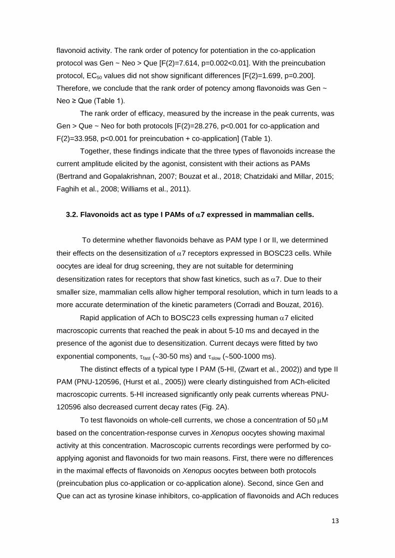
3.2. Flavonoids act as type I PAMs of 7 expressed in mammalian cells.
To determine whether flavonoids behave as PAM type I or II, we determined
their effects on the desensitization of 7 receptors expressed in BOSC23 cells. While
oocytes are ideal for drug screening, they are not suitable for determining
desensitization rates for receptors that show fast kinetics, such as 7. Due to their
smaller size, mammalian cells allow higher temporal resolution, which in turn leads to a
more accurate determination of the kinetic parameters (Corradi and Bouzat, 2016).
Rapid application of ACh to BOSC23 cells expressing human 7 elicited
macroscopic currents that reached the peak in about 5-10 ms and decayed in the
presence of the agonist due to desensitization. Current decays were fitted by two
exponential components, fast (30-50 ms) and slow (500-1000 ms).
The distinct effects of a typical type I PAM (5-HI, (Zwart et al., 2002)) and type II
PAM (PNU-120596, (Hurst et al., 2005)) were clearly distinguished from ACh-elicited
macroscopic currents. 5-HI increased significantly only peak currents whereas PNU-
120596 also decreased current decay rates (Fig. 2A).
To test flavonoids on whole-cell currents, we chose a concentration of 50 M
based on the concentration-response curves in Xenopus oocytes showing maximal
activity at this concentration. Macroscopic currents recordings were performed by co-
applying agonist and flavonoids for two main reasons. First, there were no differences
in the maximal effects of flavonoids on Xenopus oocytes between both protocols
(preincubation plus co-application or co-application alone). Second, since Gen and
Que can act as tyrosine kinase inhibitors, co-application of flavonoids and ACh reduces
14
the time needed for kinase inhibition and therefore the possibility of modifying the
phosphorylation state of the receptor (Grønlien et al., 2010, 2007; Huang et al., 1999;
Huang and Dillon, 2000).
In the presence flavonoids (50 µM), the maximal currents elicited by 100 μM
ACh increased but the decay time constants did not show any statistically significant
change (Fig. 2A, Table 1), which confirmed that they are type I PAMs, in agreement
with previous reports for Gen (Grønlien et al., 2007) and Que (Lee et al., 2010).
For Gen, the peak current increased 1.4 times [t(14)=-3.839, p=0.002<0.01]
and the net charge, 1.9 times [t(14)=-4.151, p<0.001] respect to the control (Table 1).
No significant differences in the decay time constants were observed in the presence of
Gen (Table 1) compared to the corresponding controls in its absence (fast=53 ± 21 ms
and slow=1062 ± 739 ms) [t(14)=-0.240, p=0.814 for fast and t(14)=1.225, p=0.241 for
slow].
For Que, the peak current increased 1.2 times [t(10)=-3.602, p=0.005<0.01]
but the net charge did not show a significant increase respect to the control
[t(10)=0.875, p=0.402] (Table 1). The decay time constants in the presence of Que
(Table 1) did not show significant differences with the corresponding control values
determined in the absence of Que (fast=47 ± 10 ms and slow=1235 ± 694 ms) [t(12)=-
1.096, p=0.294 for fast and t(12)=1.535, p=0.151 for slow].
For Neo, the peak current [t(12)=-4.725, p<0.001] and the net charge [t(12)=-
3.969, p=0.002<0.01] were statistically significantly increased respect to the control
(1.2 times and 1.9 times respectively, Table 1). In contrast, the decay time constants
in the presence of Neo (Table 1) did not show significant differences with respect to
those determined in its absence (fast=42 ± 7 ms and slow=1037 ± 653 ms) [t(12)=0.880,
p=0.396 for fast and t(12)=-0.135, p=0.895 for slow].
Although the increase in the peak currents elicited by 100 μM ACh was not
statistically different among flavonoids [F(2)=4.755, p=0.093], the trend in the rank
order for potentiation was the same as that established by concentration-response
curves obtained using Xenopus oocytes: Gen > Que ~ Neo.
Altogether, the results indicate that none of the flavonoids affect the decay
rates, but they increase the net charge. Also, in the presence of the flavonoids the ratio
of the changes in net charge/peak current is close to 1 (Table 1), as expected for type I
PAMs (Andersen et al., 2016; Papke and Papke, 2002).
We also evaluated the ability of flavonoids to reactivate desensitized receptors
since this is a property that differentiates type I from type II PAMs (Andersen et al.,
2016; Chatzidaki et al., 2015; Collins et al., 2011; Young et al., 2008). Figure 2B shows
15
that during continuous application of ACh (100 μM) 7 remained desensitized. A pulse
of extracellular solution (buffer) with no drugs applied to the desensitized receptors did
not elicit any significant response (0.11 ± 0.07-fold respect to the control current, n=5;
N=4, Fig. 2B). In contrast, 1 μM PNU-120596 reactivated desensitized receptors as
evidenced by a robust current, which is kinetically different from the original ACh-
induced current due to slower decay rate. Compared to the original responses,
reactivated currents by 1 μM PNU-120596 showed 10 ± 8-fold increase in the maximal
amplitude and a profound increase in the net charge (2088 ± 1767 %) (n=4, N=4). Type
I PAMs lacked the ability to recover desensitized currents or they elicited very small
currents. The recovered peak currents were 0.55 ± 0.16-fold for 2 mM 5-HI (n=7, N=5)
and 0.57 ± 0.13-fold for 10 μM NS-1738 (n=6, N=3) respect to the original currents
(Fig. 2B). For flavonoids, the reactivated currents were 0.40 ± 0.24-fold for Gen (n=5,
N=5), 0.34 ± 0.13-fold for Que (n=5, N=5) and 0.39 ± 0.09-fold for Neo (n=4, N=4) (Fig.
2B).
Thus, we conclude that the three flavonoids cannot produce significant
reactivation of desensitized 7 receptors, which is in line with their classification as
type I PAMs.
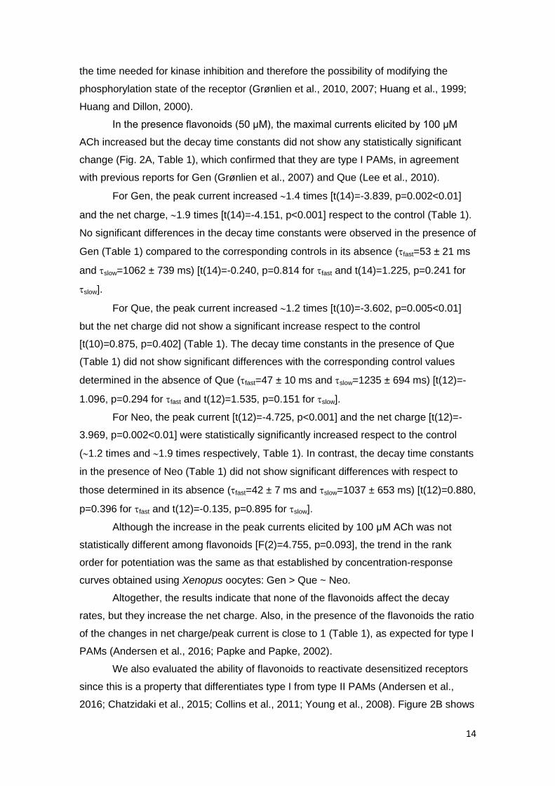
3.3. Deciphering 7 potentiation by flavonoids at the single-channel level.
To decipher the allosteric modulation of flavonoids at the molecular level, we
performed cell-attached patches in the presence of 100 µM ACh and flavonoids (5 µM-
100 µM) (Table 2 and Fig. 3). In the absence of flavonoids, single-channel currents
activated by 100 μM ACh appeared mainly as brief and isolated openings or as several
openings in quick succession, known as bursts (Table 2 and Fig.3). The open time and
burst duration histograms were described by the sum of two exponential components
(Fig. 3A).
In the presence of Gen, openings elicited by ACh were markedly prolonged and
coalesced into long-duration bursts (Fig. 3). Potentiation became evident in the
complete range of concentration evaluated, even at a concentration as low as 5 μM
(Table 2). The open duration histograms in the presence of Gen were described by
three exponential components whereas the burst duration histograms were fitted by
three exponential components for 5-10 μM Gen and by four exponential components
for 25-100 μM Gen (Fig. 3A). The maximal potentiation was reached at 25 µM. At 25-
100 M Gen, the mean open and burst durations were 6-fold and 35-fold longer than
in absence of the flavonoid, respectively (Table 2, Fig 3A). The differences in the
16
degree of potentiation among Gen concentrations were determined by the One-Way
ANOVA test [F(4)=18.882, p<0.001 for open and F(4)=42.421, p<0.001 for burst].
For Que, no changes were observed at 5 μM and potentiation became evident
at 10 μM, which is in line with the lower potency compared to Gen determined by
concentration-response curves. The open and burst duration histograms in presence of
Que at potentiating concentrations were fitted by three exponential components (Fig.
3A). At 10 μM, the mean open and burst durations increased slightly but the increase
was statistically significant. The maximal increase in mean open (3-fold) and burst
durations (10-fold) was reached at 25 μM (Table 2, Fig. 3A). The differences in the
degree of potentiation among Que concentrations were determined by the One-Way
ANOVA test [F(4)=25.842, p<0.001 for open and F(4)=55.755, p<0.001 for burst].
For Neo, potentiation became evident at 10 μM as an increase in the burst
duration but not in the open duration. At 25-50 M, potentiation reached the maximal
level with an increase of 2-fold and 8-fold in the mean open and burst durations,
respectively (Table 2, Fig. 3A). The open and burst duration histograms in presence of
Neo at potentiating concentrations were fitted by three exponential components (Fig.
3A). At 100 μM Neo, there was a decrease in potentiation that was evidenced as a
reduction of the mean burst and open durations. This observation may be explained by
additional channel blocking by high Neo concentrations. The differences in the degree
of potentiation among Neo concentrations were determined by the One-Way ANOVA
test [F(4)=7.848, p=0.001 for open and F(4)=14.686, p=0.005<0.01 for burst].
In summary, the comparison of the maximal effects among the different
flavonoids at 50 μM shows that Gen induces the highest increase in open and burst
durations and that the apparent efficacy rank order, measured as the increase in open
and burst durations, is Gen > Que ≥ Neo, similar to the rank order obtained from
concentration-response curves in oocytes [F(2)=71.330, p<0.001 for open and
F(2)=23.134, p<0.001 for burst] (Fig. 3B).
We also evaluated if flavonoids affect single-channel amplitude. For 7 in the
absence of PAMs, there is a wide range of channel amplitudes because the brief open
channel lifetime does not allow full resolution. However, in the presence of PAMs or if
only events longer than 0.3 ms are considered, it is possible to resolve the full
amplitude of the channel that is 10 pA (10.03 ± 0.32 pA, n=5, N=5) (Andersen et al.,
2013; Nielsen et al., 2018). In the presence of 50 μM flavonoid and 100 μM ACh, the
highest and major amplitudes were 10.19 ± 0.51 pA for Gen [t(8)=-0.612, p=0.557,
n=5, N=4], 9.78 ± 0.31 pA for Que [t(7)=1.175, p=0.278, n=4, N=3] and 10.26 ± 0.23 pA
for Neo [t(7)=-1.251, p=0.251, n=4, N=4]. Thus, flavonoids do not modify the single-
17
channel amplitude. Moreover, flavonoids allow complete resolution of the channel
amplitude due to their capability of increasing the mean open and burst durations.
3.4. Structural determinants of 7 potentiation by flavonoids
In order to provide further information about how flavonoids allosterically
modulate 7, we explored the action of Gen, Que and Neo on the 7-5HT3A chimera,
which carries human 7 sequence up to the beginning of the TM1 domain and mouse
5-HT3A sequence thereafter (Andersen et al., 2016; Bouzat et al., 2004; Rayes et al.,
2005). Whereas Gen and Que were shown to negatively modulate 5-HT3A receptors
(Goutman et al., 2003; Grønlien et al., 2010; Lee et al., 2008, 2005), both flavonoids
exert the opposite effect on 7 receptors. Thus, the use of the chimeric receptor
constitutes a good approach to identify the structural determinants of potentiation.
While Gen slightly inhibited the mean open and burst durations, Que and Neo
did not affect 7-5HT3A channel properties (Table 2, Fig. 4). Thus, none of the three
flavonoids (50 μM) act as PAMs of 7-5HT3A chimeric receptors. These results
suggest a prominent role of the 7 TMD or of the ECD-TMD interface to allow
potentiation by flavonoids.
Previous studies have shown that simultaneous mutations of five
transmembrane residues in 7 receptors inhibit potentiation by type II PAMs, proposing
this region as a PAM binding site (DaCosta et al., 2011; Young et al., 2008). However,
type I PAMs such as NS-1738 may bind to this transmembrane site as well (Collins et
al., 2011). We therefore sought to explore flavonoid actions at the quintuple mutant 7
(7 TSLMF).
Neither of the flavonoids (50 µM) potentiated 7 TSLMF, even more, Gen
decreased significantly the mean open duration (Table 2, Fig. 5). Thus, the five amino
acids in the transmembrane domain seem to be essential for flavonoid potentiation.
These results reveal that although the three flavonoids behave as type I PAMs,
they share the same structural determinants for potentiation located in the
transmembrane domain as the prototype type II PAM, PNU-120596.
3.5. 7 activation and potentiation by flavonoids decrease ROS intracellular
levels
Given the possibility that 7 potentiation is involved in the neuroprotective role
of flavonoids, we evaluated how their antioxidant effects are linked to 7 signalling.
18
We measured intracellular ROS levels using DCFDA and tested changes
mediated by exposing cells to flavonoids, to the specific 7 agonist PNU-282989
(Bodnar et al., 2005; Hajos et al., 2004) and to the agonist/flavonoid combination for 24
h. For these experiments, the concentration of flavonoids was 50 μM, at which the
maximal potentiation of 7 receptor was achieved, and the concentration of PNU-
282987 was 10 μM, as previously used in different in vitro and in vivo systems (Di
Cesare Mannelli et al., 2015; Hu et al., 2009; Navarro et al., 2016, 2015; Parada et al.,
2013, 2010; Tsoyi et al., 2011; Zanetti et al., 2016). The initial treatment duration was
24 h in order to ensure the activation of intracellular signalling pathways described for
7 and the tested flavonoids (Boadi et al., 2016; Dajas et al., 2013; Godoy et al., 2017;
Parada et al., 2013, 2010; Qian et al., 2015; Tsoyi et al., 2011; Williams et al., 2004).
Fluorescence images were taken after 24-h incubation under the different conditions.
The treatments with 50 μM flavonoid, 10 μM PNU-282987 or their combination
decreased the DCF fluorescence observed in cells in each visual field compared to the
control group pre-treated with DMSO (Fig. 6A).
In order to accurately quantify ROS levels, we determined the rate of intracellular
ROS generation by measuring the slope of DCF fluorescence as a function of time and
normalizing it to that of the control condition (Fig. 6B-C) (Koopman et al., 2006;
Sepúlveda et al., 2013). We also incorporated two additional controls: a dye control
(probe solution alone) and a positive control (1 mM H2O2) to verify the correct
performance and response capacity of the probe (Bian et al., 2015; Sepúlveda et al.,
2013). The linearity of the increase in fluorescence was maintained during the 30-min
measurement, and the set of points were fitted by linear regression (r2 0.96-0.99, Fig.
6B).
Consistent with the well-known antioxidant capacity of flavonoids, the intracellular
ROS production was significantly reduced in cells treated with flavonoids for 24 h
compared to non-treated cells [t(8)=7.840, p<0.001 for Gen, t(8)=12.010, p<0.001 for
Que and t(8)=7.016, p<0.001 for Neo, n=5, N=5 for each condition] (Fig. 6B-C). Under
our experimental conditions, there were no differences in ROS levels between Gen,
Que and Neo [F(2)=0.381, p=0.691]. Interestingly, when 7 was activated by 10 μM
PNU-282987, ROS levels also decreased respect to the control condition [t(20)=7.911,
p<0.001, n=11, N=11, Fig. 6B,C]. Although PNU-282987 is considered a selective 7
agonist, to further verify that it was mediating the effect through 7, we pre-treated the
cells with the specific antagonist -Bgt (500 nM) for 2 h before the addition of 10 µM
PNU-282987 (Fig. 6A-C). Under this condition, no statistically significant differences
were found respect to the control condition [t(16)=1.614, p=0.126, n=9, N=9, Fig. 6B-
19
C], as also observed by fluorescence microscopy (Fig. 6A). Thus, given that -Bgt
blocked the decrease in ROS levels exerted by PNU-282987, we confirmed that the
effect is mediated through 7. As an additional control, we also showed that -Bgt
alone did not affect ROS production [t(16)=-1.191, p=0.251, n=9, N=9, Fig. 6A-C].
In cells treated for 24 h with 10 μM PNU-282987 combined with 50 μM flavonoid,
the intracellular ROS levels decreased significantly respect to the control [t(8)=6.840,
p<0.001 for PNU + Gen, t(8)=8.602, p<0.001 for PNU + Que and t(8)=8.431, p<0.001
for PNU + Neo, n=5, N=5 for each condition, Fig 6]. However, no significant differences
were found between the conditions containing flavonoids in the absence or presence of
PNU-28298 [F(6)=1.932, p=0.110].
With the aim of dissecting the flavonoid 7-PAM effect from its potent antioxidant
effect independent of 7, which may govern the observed decrease in ROS production
at 24 h, we applied two different approaches. In one, we reduced the flavonoid
concentration (from 50 M to 10 M) and in the other we also reduced the time of
exposure to the flavonoid (from 24 h to 12 h). We performed this set of experiments
only for Gen since this flavonoid is the most effective as an 7 PAM.
Reducing Gen concentration from 50 M to 10 M in the 24 h-treatment showed
that Gen was still capable of decreasing ROS production compared to the control
condition [t(12)=18.120, p<0.001 n=3, N=3]. The decrease exerted by 10 M Gen
(20%) was slightly lower compared to that of 50 M (50%) [t(6)=2.859,
p=0.029<0.05, n=3, N=3]. Then, we combined 10 M Gen, whose antioxidant action
independent of 7 was submaximal at 24 h, with 10 M PNU-282987 and we observed
a more pronounced decrease in ROS levels than those exerted by either 10 M Gen
[t(4)=-5.604, p=0.005<0.01, n=3, N=3] or 10 M PNU-282987 [t(10)=2.883,
p=0.016<0.05, n=9, N=9). Nevertheless, in the combined treatment the maximal
achieved reduction in the ROS generation rate was similar to that obtained in presence
of 50 M Gen (alone or with PNU-282987) [F(2)=0.472, p=0.637]. Thus, it seems that
under these conditions similar minimal ROS levels can be reached either by the
antioxidant activity of Gen independent of 7 or by the combination of its antioxidant
activities mediated and not by 7.
To unmask even more the effect of Gen dependent of 7, we reduced the time of
exposure from 24 h to 12 h. With 12-h treatment, the antioxidant activity of Gen was
neither detected at 10 M nor at 50 M since ROS generation rate was not different
from that of the control condition [t(12)=-1.716, p=0.112, n=7, N=7 for 10 M Gen and
t(12)=-1.145, p=0.275, n=7, N=7 for 50 M Gen] (Fig. 6D-E). In contrast, incubation of
20
cells for 12 h with 10 M PNU-282987 reduced ROS production in a statistically
significant manner [t(12)=-1.145, p=0.0172<0.05, n=7, N=7], although the decrease
was lower than in the 24 h treatment [t(16)=2.429, p=0.0273<0.05, Fig. 6C,E).
Interestingly, 12-h co-incubation with PNU-282987 and Gen (10 M or 50 M)
produced a significant decrease in the ROS levels compared to the control condition
[t(12)=9.052, p<0.001, n=7, N=7 for PNU + Gen 10 M and t(12)=9.849, p<0.001, n=7,
N=7 for PNU + Gen 50 M]. The reduction was even more pronounced than that
induced by PNU-282987 alone [t(12)=3.154, p=0.00832<0.01, n=7, N=7 for PNU +
Gen 10 M and t(12)=3.523, p=0.00420<0.01, n=7, N=7 for PNU + Gen 50 M], thus
demonstrating that the 7-PAM activity of Gen enhanced cell protection. We confirmed
that the Gen effect was mediated through 7 since it was blocked by -Bgt (500 nM)
[t(4)=-0.804, p=0.466, n=3, N=3 for PNU + Gen 10 M + -Bgt and t(4)=-0.942,
p=0.399, n=3, N=3 for PNU + Gen 50 M + -Bgt, Fig. 6E). The effects determined by
the kinetic assays were in line with those determined by fluorescence microscopy (Fig.
6D).
Overall, this set of experiments revealed that the effects of Gen on ROS
production take place earlier through its activity as an 7 PAM than through its activity
mediated by 7-independent mechanisms.
21
4. DISCUSSION
Despite their common neurological and neuroprotective effects, the interactions
between flavonoids and 7 have been poorly explored. Here, by using a single-channel
approach we have elucidated, for the first time, the molecular mechanisms of the
flavonoid-induced potentiation of 7 receptors. Previous studies have shown that
flavonoids such as Gen (Grønlien et al., 2010, 2007) and Que (Lee et al., 2010)
potentiate 7 macroscopic currents through a type I PAM mechanism. We confirmed
these observations and identified a neoflavonoid as a novel 7 PAM. Our findings
clearly demonstrate that the type I PAM actions of all flavonoids tested increase the
open channel lifetime and induce activation in bursts, thus modulating 7 kinetics.
We studied three different classes of flavonoids differing in the position of the B-
ring in the benzopyrone moiety. Que is a flavonol that constitutes the major component
of flavonoids dietary intake and exhibits the most prominent antioxidant and anti-
inflammatory activity (Bakhtiari et al., 2017; Spencer et al., 2012). Gen is an isoflavone
abundant in soy, which improves short and long term memory, supresses inflammatory
pathways and oxidative stress, and may be involved in some estrogenic-receptor
signalling pathways (Bakhtiari et al., 2017; Ganai and Farooqi, 2015; Spencer et al.,
2012). The compound 5,7-dihydroxy-4-phenylcoumarin (Ulubelen et al., 1982) is a
neoflavonoid, which is the less explored class of flavonoids. The unique structure and
popularity of neoflavonoids in traditional medicine have made them attractive
pharmacological compounds. In this regard, the tested Neo is an inhibitor of cAMP
phosphodiesterase (Kusano et al., 1991), and an antioxidant (Veselinović et al., 2014),
anti-bacterial (Veselinović et al., 2015) and anti-melanogenic agent (Veselinović et al.,
2017).
4.1. Pharmacological characterization of flavonoids at the macroscopic and
single-channel level
Flavonoids enhance agonist-induced peak currents without significantly
affecting current decay rates and do not reactivate desensitized receptors. At the
single-channel level, they increase the open-channel lifetime and induce activation in
bursts, which are composed by several openings in quick succession, indicating that
they do affect 7 kinetics as described for other type I PAMs (Andersen et al., 2016).
The comparison of flavonoid potencies by the EC50 values determined from
macroscopic recordings shows that Gen and Neo are more potent than Que.
Regarding efficacy, Gen is the most efficacious 7 PAM, as based on the increase of
the maximal current and open and burst durations. Gen, Que and Neo increase 6, 3
22
and 2 times, respectively, the mean open duration and 35, 10 and 8 times,
respectively, the mean burst duration. However, the changes at the macroscopic level
are less pronounced. A similar lack of correlation has been reported for NS-1738
(Andersen et al., 2016). Thus, it appears that the burst duration is the most sensitive
parameter to quantify the potentiating effects, which, in turn, highlights the importance
of analysing single-channel kinetics for a better understanding of the mechanistic
changes (Nielsen et al., 2018). In this context, and based on the increase of the burst
duration, the rank order for 7 potentiation among several type I PAMs is NS-1378 ≥
Gen ≥ 5-HI > Que ≥ Neo.
In our study, the maximal level of potentiation was achieved by simultaneous
application with the agonist, in contrast to an earlier study which needed preincubation
with the flavonoid (Lee et al., 2010). This difference may be due to the differences in
the solution exchange rate of the perfusion system employed for recording in Xenopus
oocytes. Although Que and Gen are tyrosine kinase inhibitors (Akiyama et al., 1987;
Glossmann et al., 1981) and changes in 7 phosphorylation may affect its function
and/or cell expression (Charpantier et al., 2005; Cho et al., 2005), the rapid modulatory
effects by flavonoids on 7 and on other pLGICs have been shown to be independent
of tyrosine kinases (Grønlien et al., 2007; Huang et al., 1999; Huang and Dillon, 2000;
Lee et al., 2010). In our hands, the fact that co-application of flavonoids with ACh
rapidly affects 7 function in a reversible manner and that a receptor carrying
mutations at a PAM binding site, not involving phosphorylation sites, is insensitive to
flavonoid potentiation, supports a direct allosteric effect.
Our results also show that the position of the B-ring in the benzopyrone moiety
does not change the main mechanism by which flavonoids act in 7. However, it may
be involved, together with different hydroxylation patterns, in the slight differences in
efficacy and potency observed among flavonoids. Moreover, all tested flavonoids carry
hydroxyl substitutions in positions 5 and 7 in the A-ring that were identified as
necessary to afford neuronal protection (Dajas et al., 2013; Echeverry et al., 2010).
4.2. Structural determinants of flavonoid PAM activity
The binding site(s) for 7 PAMs have not been unequivocally identified from
crystal structures of 7 in complex with PAMs. Several PAMs, including type II PAMs
(PNU-120596 and TQS) and type I PAMs (ivermectin and LY-2087101) have been
proposed to bind to transmembrane site(s) (Collins and Millar, 2010; Young et al.,
2008), which may be shared by allosteric modulators displaying very distinct
pharmacological effects (Gill-Thind et al., 2015; Gill et al., 2013; Pałczynska et al.,
23
2012). However, other binding sites in the ECD and/or the ECD-TMD interface have
been proposed for type I PAMs (Bertrand et al., 2008; Grønlien et al., 2007;
Targowska-Duda et al., 2018). The 7-5HT3A receptor has been extensively used as a
model of 7 ECD as well as to dissect the main domains involved in drug modulation.
In accordance to previous observations, we found that this chimeric receptor is not
potentiated by flavonoids, although it is sensitive to other type I-PAMs, such as 5-HI
(Andersen et al., 2016; Grønlien et al., 2010; Williams et al., 2011).
The lack of flavonoid potentiation in 7-5HT3A indicates that other domains than
the ECD are required for potentiation. Further dissection of the amino acids involved
was obtained from the quintuple mutant 7 TSLMF, which is not potentiated by the
three classes of flavonoids, indicating the involvement of the transmembrane cavity in
flavonoid modulation. This receptor also shows significantly reduced potentiation by
type II PAMs (PNU-120596 and PAM-2) and by the type I PAM NS-1738, but full
potentiation by 5-HI (Andersen et al., 2016; DaCosta et al., 2011).
It is important to note that Gen not only does not potentiate the chimeric and
mutant receptors, but it also reduces the open and burst durations, suggesting that it
acts as an allosteric negative modulator (NAM) of both 7-5HT3A and the mutant 7
TSLMF. This result agrees with reports showing that PAMs can turn into negative
modulators in mutant receptors. For example, mutations in the transmembrane region
(S223M, M254L and S277V) convert ivermectin from PAM into a NAM (Collins and
Millar, 2010). In agreement, 7 TSLMF receptor contains two of these mutations
(S223M and M254L) and is inhibited by Gen.
The fact that Que and Neo neither potentiate nor inhibit the chimeric and mutant
receptors while Gen inhibits them may rely on different interactions due to their slight
differences in chemical structures. Our work opens doors for further work deciphering
the basis underlying the inhibitory effect of Gen.
We demonstrated that flavonoids exhibit a macroscopic type I PAM profile,
induce changes in single-channel properties similar to some type I PAMs (Andersen et
al., 2016), and require structural determinants of the transmembrane domain as type II
PAMs and some type I PAMs (NS-1738 and ivermectin). Thus, our work provides
additional information regarding 7 modulation and confirms that the prototypical type I
(5-HI) and type II (PNU-120596) PAMs may show the extreme behaviors of a wide
range of allosteric modulators. The classification of type I and type II appears to be an
oversimplification resulting mainly from macroscopic observations, which highlights the
importance of characterizing the molecular mechanisms at single-channel level.
24
4.3. Antioxidant activity of flavonoids through α7-PAM activity
We determined the interrelation between 7 modulation and flavonoids in ROS
levels, which are involved in aging and neurodegeneration (Schieber and Chandel,
2014). Activation of 7 by a selective agonist (PNU-282987) decreases the ROS
generation rate, in line with reports describing antioxidant and anti-inflamatory effects
of 7 on different cellular systems (Parada et al., 2013, 2010; Tsoyi et al., 2011). In 24
h-treatment, co-application of flavonoids with the agonist reduces the intracellular ROS
production but to an extent similar to that achieved by flavonoids alone at the maximal
concentrations.This observation may be explained by a common mechanism for
antioxidation mediated by flavonoids and 7-activation which has reached its maximal
level in our system and/or by the predominance of the flavonoid antioxidant capacity
independent of 7 at this prolonged time of treatment, thus obscuring the antioxidant
effect triggered by 7 activation.
The reduction of Gen exposure to 12 h allowed us to separate the antioxidant
effects of Gen independent of α7 from those dependent of α7. Although the antioxidant
activity of Gen alone is not detected at 12 h, the flavonoid potentiates the reduction in
ROS levels mediated by 7 activation, indicating that its action as type I PAM also
constitutes a mechanism of antioxidation. These results also revealed the importance
of 7 potentiation by flavonoids as an additional mechanism underlying their
neuroprotective role because the effects from their allosteric modulatory action occur at
an earlier stage than those related to their solely antioxidant action independent of 7.
It is important to note that the antioxidant effect mediated by 7 activation takes
place only in transfected cells, while the flavonoid antioxidant effects independent of
7, occur in all cells. Thus, the difference between both conditions may be
underestimated and therefore, the contribution of 7 signalling could be even higher
than here determined. Nevertheless, the significant reduction in ROS generation levels
in cells treated with the 7 selective agonist alone supports our model for measuring
the effects mediated by nAChR activation.
The dual ionotropic/metabotropic activity of 7 is responsible for its role in
neuroprotection. Mainly, the neuroprotective effects depend on NF-κB inhibition, which
has an antiinflammatory action, and on the pathway Jak2/PI3K/Akt leading to activation
of Nrf-2, a transcription factor primarily responsible for cellular defense against
oxidative stress (Parada et al., 2013; Tsoyi et al., 2011). Flavonoids also downregulate
NF-κB pathways and modulate several signalling cascades, including PI3K/Akt that
leads to upregulation of Nrf-2 (Dajas et al., 2013; Williams et al., 2004). It is possible
25
that 7 through both its ionotropic and metabotropic responses triggers more rapidly
these common pathways than flavonoids alone. In the presence of flavonoids, the more
sustained activation of 7 leading to higher calcium-influx potentiates the 7-triggered
intracellular pathways that mediate antioxidation. Thus, positive modulation on 7 may
represent a spare mechanism by which flavonoids exert their antioxidant activity and,
therefore, their neuroprotective role.
4.4. Therapeutic impact of flavonoids acting as α7 PAMs
Previous studies have explored the selectivity of flavonoids on different LGICs,
showing a positive effect only on 7 and an inhibitory effect on most neurotransmitter
receptors, including GABA-, serotonin-, ACh-, glycine- and glutamate-activated
receptors (Goutman et al., 2003; Grønlien et al., 2007; Huang et al., 1999; Huang and
Dillon, 2000; Lee et al., 2011a, 2011c, 2011b, 2008, 2007, 2005; Shin et al., 2010).
This strict selectivity for potentiation may result in a promising feature for therapy.
Furthermore, modifications of flavonoid structure have allowed different
pharmacological effects on GABA receptors (Hanrahan et al., 2011; Wasowski and
Marder, 2012). This demonstrates that not only the natural flavonoids, but also the
synthetic ones, exhibit a high potential for therapeutic development.
The advantages of PAMs over agonists for therapy are the maintainance of the
temporal and spatial pattern of the endogenous neurotransmitter, higher selectivity for
binding into allosteric sites, reduction of tolerance due to 7 desensitization and
neuroprotective action (Bertrand and Gopalakrishnan, 2007; Bouzat et al., 2018;
Chatzidaki and Millar, 2015; Faghih et al., 2008; Uteshev, 2014; Williams et al., 2011).
Nevertheless, there is a controversy about the possible cytotoxicity of PAMs due to the
high increase in intracellular calcium levels (Ng et al., 2007; Liu et al., 2009; Williams et
al., 2012; Guerra-Álvarez et al., 2015; Uteshev, 2016). Type II PAMs are the most
controversial due to their ability to potentiate 7 currents with high efficacy, decrease
desensitization and reactivate desensitized receptors (Guerra-Álvarez et al., 2015; Hu
et al., 2009; Ng et al., 2007; Uteshev, 2016; Williams et al., 2012). Type I PAMs may
be less cytotoxic than type II PAMs because potentiation is lower and receptor
desensitization still occurs, which acts as a filter against excessive stimulation (Guerra-
Álvarez et al., 2015; Hu et al., 2009; Ng et al., 2007; Williams et al., 2012). Therefore,
the use of flavonoids is a promising therapeutic strategy for enhancing 7 function,
because they do not change the desensitization rate and do not reactivate desensitized
receptors.
26
As natural compounds with a broad spectrum of beneficial pharmacological and
biological activities, including their role as 7 PAMs reported here, flavonoids are
suitable candidates for the treatment of multifactorial diseases. We here evaluated the
action of unmetabolized flavonoids but it has been shown for several compounds that
the conjugated and derivatives forms of polyphenols have similar or greater bioactivity
interacting with the same signalling pathways (Kawai, 2018; Unno et al., 2017).
The intake of flavonoids in the diet ranges from 60 to 350 mg/day (Johnston,
2015), reaching high nanomolar-low micromolar concentration levels in in vivo studies,
which are correlated with those showing effects in vitro (Krasieva et al., 2015; Schaffer
and Halliwell, 2012). Interestingly, some polyphenols are concentrated in neural tissue,
and therefore the achieved concentration may be higher (Kalt et al., 2008; Milbury and
Kalt, 2010). The effects of flavonoids as 7 PAMs occur at the low micromolar range
and, therefore, at clinically achievable concentrations. Furthermore, the beneficial
actions of these natural compounds may be due to a synergic effect of the multiplicity
of flavonoids ingested in the diet.
5. CONCLUSIONS
The α7 nicotinic receptor participates in cognition, neuroprotection and inflammation and
its potentiation is emerging as promising therapeutic strategy for neurological and
inflammatory disorders. On the other hand, flavonoids are plant polyphenolic compounds
showing neuroprotective, anti-inflammatory and pro-cognitive actions. Besides their well-
known antioxidant activity, flavonoids trigger intracellular pathways and interact with
receptors, including α7. We here identified a neoflavonoid as a novel α7 PAM and
deciphered the molecular mechanisms underlying the PAM actions of three classes of
flavonoids. They potentiate macroscopic responses without affecting receptor
desensitization, increase open-channel lifetime and induce channel activation in
episodes of successive openings, thus modulating α7 kinetics. Flavonoids share
transmembrane structural determinants with other non-structurally related PAMs.
Besides the well-described antioxidant actions of flavonoids, the enhancement of 7
activation has also a functional role reducing the ROS generation rate in human cells.
Thus, allosterically potentiation of α7 is proposed as an additional mechanism underlying
the neuroprotective actions of flavonoids, which, in turn, may be used as scaffolds for
designing new therapeutic agents. The identification of novel candidate PAMs as well as
the understanding of their actions at the molecular level are required for the still ongoing
development of these promising therapeutic compounds.

27
ACKNOWLEDGMENTS
This work was supported by grants from Universidad Nacional del Sur [PGI 24/B227]
and Agencia Nacional de Promoción Científica y Tecnológica [PICT-2015 0941, PICT-
2017 1170] to C.B.; a grant from Oxford Brookes University to I.B.; and a grant from
Company of Biologists [JCSTF-180207] to B.E.N.
28
REFERENCES
Akiyama, T., Ishida, J., Nakagawa, S., Ogawara, H., Watanabe, S., Itoh, N., Shibuya, M., Fukami, Y., 1987. Genistein, a specific inhibitor of tyrosine-specific protein kinases. J. Biol. Chem. 262, 5592–5595.
Andersen, N., Corradi, J., Sine, S.M., Bouzat, C., 2013. Stoichiometry for activation of neuronal α7 nicotinic receptors. Proc. Natl. Acad. Sci. U. S. A. 110, 20819–20824. https://doi.org/10.1073/pnas.1315775110
Andersen, N., Nielsen, B.E., Corradi, J., Tolosa, M.F., Feuerbach, D., Arias, H.R., Bouzat, C., 2016. Exploring the Positive Allosteric Modulation of Human α7 Nicotinic Receptors from a Single-Channel Perspective. Neuropharmacology 107, 189–200. https://doi.org/10.1016/j.neuropharm.2016.02.032
Bakhtiari, M., Panahi, Y., Ameli, J., Darvishi, B., 2017. Protective effects of flavonoids against Alzheimer’s disease-related neural dysfunctions. Biomed. Pharmacother. 93, 218–229. https://doi.org/10.1016/j.biopha.2017.06.010
Bakoyiannis, I., Daskalopoulou, A., Pergialiotis, V., Perrea, D., 2019. Phytochemicals and cognitive health: Are flavonoids doing the trick? Biomed. Pharmacother. 109, 1488–1497. https://doi.org/10.1016/j.biopha.2018.10.086
Bertrand, D., Bertrand, S., Cassar, S., Gubbins, E., Li, J., Gopalakrishnan, M., 2008. Positive Allosteric Modulation of the α7 Nicotinic Acetylcholine Receptor: Ligand Interactions with Distinct Binding Sites and Evidence for a Prominent Role of the M2-M3 Segment. Mol. Pharmacol. 74, 1407–1416. https://doi.org/10.1124/mol.107.042820
Bertrand, D., Gopalakrishnan, M., 2007. Allosteric modulation of nicotinic acetylcholine receptors. Biochem. Pharmacol. 74, 1155–1163. https://doi.org/10.1016/j.bcp.2007.07.011
Bian, Y.Y., Guo, J., Majeed, H., Zhu, K.X., Guo, X.N., Peng, W., Zhou, H.M., 2015. Ferulic acid renders protection to HEK293 cells against oxidative damage and apoptosis induced by hydrogen peroxide. Vitr. Cell. Dev. Biol. - Anim. 51, 722–729. https://doi.org/10.1007/s11626-015-9876-0
Boadi, W.Y., Amartey, P.K., Lo, A., 2016. Effect of quercetin, genistein and kaempferol on glutathione and glutathione-redox cycle enzymes in 3T3-L1 preadipocytes. Drug Chem. Toxicol. 39, 239–247. https://doi.org/10.3109/01480545.2015.1082135
Bodnar, A.L., Cortes-Burgos, L.A., Cook, K.K., Dinh, D.M., Groppi, V.E., Hajos, M., Higdon, N.R., Hoffmann, W.E., Hurst, R.S., Myers, J.K., Rogers, B.N., Wall, T.M., Wolfe, M.L., Wong, E., 2005. Discovery and structure-activity relationship of quinuclidine benzamides as agonists of α7 nicotinic acetylcholine receptors. J. Med. Chem. 48, 905–908. https://doi.org/10.1021/jm049363q
Bouzat, C., Bartos, M., Corradi, J., Sine, S.M., 2008. The interface between extracellular and transmembrane domains of homomeric Cys-loop receptors governs open-channel lifetime and rate of desensitization. J. Neurosci. 28, 7808–7819. https://doi.org/10.1523/JNEUROSCI.0448-08.2008
Bouzat, C., Bren, N., Sine, S.M., 1994. Structural basis of the different gating kinetics of fetal and adult acetylcholine receptors. Neuron 13, 1395–1402. https://doi.org/10.1016/0896-6273(94)90424-3
Bouzat, C., Gumilar, F., Spitzmaul, G., Wang, H., Rayes, D., Hansen, S.B., Taylor, P., Sine, S.M., 2004. Coupling of agonist binding to channel gating in an ACh-binding
29
protein linked to an ion channel. Nature 430, 896–900. https://doi.org/10.1038/nature02753
Bouzat, C., Lasala, M., Nielsen, B.E., Corradi, J., Esandi, M. del C., 2018. Molecular function of α7 nicotinic receptors as drug targets. J. Physiol. 596, 1847–1861. https://doi.org/10.1113/JP275101
Buckingham, S.D., Jones, A.K., Brown, L.A., Sattelle, D.B., 2009. Nicotinic Acetylcholine Receptor Signalling: Roles in Alzheimer’s Disease and Amyloid Neuroprotection. Pharmacol. Rev. 61, 39–61. https://doi.org/10.1124/pr.108.000562
Carbone, A.L., Moroni, M., Groot-Kormelink, P.J., Bermudez, I., 2009. Pentameric concatenated (α4)2 (β2)3 and (α4)3(β2)2 nicotinic acetylcholine receptors: subunit arrangement determines functional expression. Br. J. Pharmacol. 156, 970–981. https://doi.org/10.1111/j.1476-5381.2008.00104.x
Changeux, J.P., Taly, A., 2008. Nicotinic receptors, allosteric proteins and medicine. Trends Mol. Med. 14, 93–102. https://doi.org/10.1016/j.molmed.2008.01.001
Charpantier, E., Wiesner, A., Huh, K.-H., Ogier, R., Hoda, J.-C., Allaman, G., Raggenbass, M., Feuerbach, D., Bertrand, D., Fuhrer, C., 2005. Alpha7 neuronal nicotinic acetylcholine receptors are negatively regulated by tyrosine phosphorylation and Src-family kinases. J. Neurosci. 25, 9836–49. https://doi.org/10.1523/JNEUROSCI.3497-05.2005
Chatzidaki, A., Millar, N.S., 2015. Allosteric modulation of nicotinic acetylcholine receptors. Biochem. Pharmacol. 97, 408–417. https://doi.org/10.1016/j.bcp.2015.07.028
Chatzidaki, A., Oyley, J.M.D., Gill-thind, J.K., Sheppard, T.D., Millar, N.S., D’Oyley, J.M., Gill-thind, J.K., Sheppard, T.D., Millar, N.S., 2015. The influence of allosteric modulators and transmembrane mutations on desensitisation and activation of α7 nicotinic acetylcholine receptors. Neuropharmacology 97, 75–85. https://doi.org/10.1016/j.neuropharm.2015.05.006
Cho, C., Song, W., Leitzell, K., Teo, E., Meleth, A.D., Quick, M.W., Lester, R.A.J., 2005. Rapid upregulation of alpha7 nicotinic acetylcholine receptors by tyrosine dephosphorylation. J. Neurosci. 25, 3712–23. https://doi.org/10.1523/JNEUROSCI.5389-03.2005
Collins, T., Millar, N.S., 2010. Nicotinic Acetylcholine Receptor Transmembrane Mutations Convert Ivermectin from a Positive to a Negative Allosteric Modulator. Mol. Pharmacol. 78, 198–204. https://doi.org/10.1124/mol.110.064295
Collins, T., Young, G.T., Millar, N.S., 2011. Competitive binding at a nicotinic receptor transmembrane site of two α7-selective positive allosteric modulators with differing effects on agonist-evoked desensitization. Neuropharmacology 61, 1306–1313. https://doi.org/10.1016/j.neuropharm.2011.07.035
Corcoran, M.P., McKay, D.L., Blumberg, J.B., 2012. Flavonoid Basics: Chemistry, Sources, Mechanisms of Action, and Safety. J. Nutr. Gerontol. Geriatr. 31, 176–189. https://doi.org/10.1080/21551197.2012.698219
Corradi, J., Bouzat, C., 2016. Understanding the Bases of Function and Modulation of α7 Nicotinic Receptors: Implications for Drug Discovery. Mol. Pharmacol. 90, 288–299. https://doi.org/10.1124/mol.116.104240
Corradi, J., Gumilar, F., Bouzat, C., 2009. Single-channel kinetic analysis for activation and desensitization of homomeric 5-HT3A receptors. Biophys. J. 97, 1335–1345.
30
https://doi.org/10.1016/j.bpj.2009.06.018
DaCosta, C.J.B., Free, C.R., Corradi, J., Bouzat, C., Sine, S.M., 2011. Single-Channel and Structural Foundations of Neuronal α7 Acetylcholine Receptor Potentiation. J. Neurosci. 31, 13870–13879. https://doi.org/10.1523/JNEUROSCI.2652-11.2011
DaCosta, C.J.B., Free, C.R., Sine, S.M., 2015. Stoichiometry for α-bungarotoxin block of α7 acetylcholine receptors. Nat. Commun. 6, 8057–8067. https://doi.org/10.1038/ncomms9057
Dajas-Bailador, F., Wonnacott, S., 2004. Nicotinic acetylcholine receptors and the regulation of neuronal signalling. Trends Pharmacol. Sci. 25, 317–324. https://doi.org/10.1016/j.tips.2004.04.006
Dajas, F., Abin-Carriquiry, J.A., Arredondo, F., Echeverry, C., Felicia, R.-M., 2013. Neuroprotective Actions of Flavones and Flavonols: Mechanisms and Relationship to Flavonoid Structural Features. Cent. Nerv. Syst. Agents Med. Chem. 13, 30–35. https://doi.org/10.2174/1871524911313010005
Di Cesare Mannelli, L., Tenci, B., Zanardelli, M., Failli, P., Ghelardini, C., 2015. α7 Nicotinic Receptor Promotes the Neuroprotective Functions of Astrocytes against Oxaliplatin Neurotoxicity. Neural Plast. 2015, 396908. https://doi.org/10.1155/2015/396908
Dineley, K.T., Pandya, A.A., Yakel, J.L., 2015. Nicotinic ACh receptors as therapeutic targets in CNS disorders. Trends Pharmacol. Sci. 36, 96–108. https://doi.org/10.1016/j.tips.2014.12.002
Ebrahimi, A., Schluesener, H., 2012. Natural polyphenols against neurodegenerative disorders: Potentials and pitfalls. Ageing Res. Rev. 11, 329–345. https://doi.org/10.1016/j.arr.2012.01.006
Echeverry, C., Arredondo, F., Abin-Carriquiry, J.A., Midiwo, J.O., Ochieng, C., Kerubo, L., Dajas, F., 2010. Pretreatment with Natural Flavones and Neuronal Cell Survival after Oxidative Stress: A Structure−Activity Relationship Study. J. Agric. Food Chem. 58, 2111–2115. https://doi.org/10.1021/jf902951v
Egea, J., Buendia, I., Parada, E., Navarro, E., León, R., Lopez, M.G., 2015. Anti-inflammatory role of microglial alpha7 nAChRs and its role in neuroprotection. Biochem. Pharmacol. 97, 463–472. https://doi.org/10.1016/j.bcp.2015.07.032
Faghih, R., Gopalakrishnan, M., Briggs, C.A., 2008. Allosteric modulators of the α7 nicotinic acetylcholine receptor. J. Med. Chem. 51, 701–712. https://doi.org/10.1021/jm070256g
Flanagan, E., Müller, M., Hornberger, M., Vauzour, D., 2018. Impact of Flavonoids on Cellular and Molecular Mechanisms Underlying Age-Related Cognitive Decline and Neurodegeneration. Curr. Nutr. Rep. 7, 49–57. https://doi.org/10.1007/s13668-018-0226-1
Ganai, A.A., Farooqi, H., 2015. Bioactivity of genistein: A review of in vitro and in vivo studies. Biomed. Pharmacother. 76, 30–38. https://doi.org/10.1016/j.biopha.2015.10.026
Gildawie, K.R., Galli, R.L., Shukitt-Hale, B., Carey, A.N., 2018. Protective Effects of Foods Containing Flavonoids on Age-Related Cognitive Decline. Curr. Nutr. Rep. 7, 39–48. https://doi.org/10.1007/s13668-018-0227-0
Gill-Thind, J.K., Dhankher, P., D’Oyley, J.M., Sheppard, T.D., Millar, N.S., 2015. Structurally Similar Allosteric Modulators of α7 Nicotinic Acetylcholine Receptors
31
Exhibit Five Distinct Pharmacological Effects. J. Biol. Chem. 290, 3552–3562. https://doi.org/10.1074/jbc.M114.619221
Gill, J.K., Chatzidaki, A., Ursu, D., Sher, E., Millar, N.S., 2013. Contrasting Properties of α7-Selective Orthosteric and Allosteric Agonists Examined on Native Nicotinic Acetylcholine Receptors. PLoS One 8, e55047. https://doi.org/10.1371/journal.pone.0055047
Glossmann, H., Presek, P., Eigenbrodt, E., 1981. Quercetin inhibits tyrosine phosphorylation by the cyclic nucleotide-independent, transforming protein kinase, pp60src. Naunyn. Schmiedebergs. Arch. Pharmacol. 317, 100–102. https://doi.org/10.1007/BF00506266
Godoy, J.A., Lindsay, C.B., Quintanilla, R.A., Carvajal, F.J., Cerpa, W., Inestrosa, N.C., 2017. Quercetin Exerts Differential Neuroprotective Effects Against H2O2 and Aβ Aggregates in Hippocampal Neurons: the Role of Mitochondria. Mol. Neurobiol. 54, 7116–7128. https://doi.org/10.1007/s12035-016-0203-x
Goutman, J.D., Waxemberg, M.D., Doñate-Oliver, F., Pomata, P.E., Calvo, D.J., 2003. Flavonoid modulation of ionic currents mediated by GABA A and GABA C receptors. Eur. J. Pharmacol. 461, 79–87. https://doi.org/10.1016/S0014-2999(03)01309-8
Grønlien, J.H., Håkerud, M., Ween, H., Thorin-Hagene, K., Briggs, C. a, Gopalakrishnan, M., Malysz, J., 2007. Distinct profiles of alpha7 nAChR positive allosteric modulation revealed by structurally diverse chemotypes. Mol. Pharmacol. 72, 715–24. https://doi.org/10.1124/mol.107.035410
Grønlien, J.H., Ween, H., Thorin-Hagene, K., Cassar, S., Li, J., Briggs, C.A., Gopalakrishnan, M., Malysz, J., 2010. Importance of M2–M3 loop in governing properties of genistein at the α7 nicotinic acetylcholine receptor inferred from α7/5-HT3A chimera. Eur. J. Pharmacol. 647, 37–47. https://doi.org/10.1016/j.ejphar.2010.08.027
Gu, S., Matta, J.A., Lord, B., Harrington, A.W., Sutton, S.W., Davini, W.B., Bredt, D.S., 2016. Brain α7 Nicotinic Acetylcholine Receptor Assembly Requires NACHO. Neuron 89, 948–955. https://doi.org/10.1016/j.neuron.2016.01.018
Guan, Y.Z., Jin, X.D., Guan, L.X., Yan, H.C., Wang, P., Gong, Z., Li, S.J., Cao, X., Xing, Y.L., Gao, T.M., 2015. Nicotine Inhibits Microglial Proliferation and Is Neuroprotective in Global Ischemia Rats. Mol. Neurobiol. 51, 1480–1488. https://doi.org/10.1007/s12035-014-8825-3
Guan, Z.Z., Zhang, X., Ravid, R., Nordberg, A., 2000. Decreased protein levels of nicotinic receptor subunits in the hippocampus and temporal cortex of patients with Alzheimer’s disease. J. Neurochem. 74, 237–243. https://doi.org/10.1046/j.1471-4159.2000.0740237.x
Guerra-Álvarez, M., Moreno-Ortega, A.J., Navarro, E., Fernández-Morales, J.C., Egea, J., López, M.G., Cano-Abad, M.F., 2015. Positive allosteric modulation of alpha-7 nicotinic receptors promotes cell death by inducing Ca 2+ release from the endoplasmic reticulum. J. Neurochem. 133, 309–319. https://doi.org/10.1111/jnc.13049
Hajos, M., Hurst, R.S., Hoffmann, W., Krause, M., Wall, T.M., Higdon, N.R., Groppi, V.E., 2004. The Selective α7 Nicotinic Acetylcholine Receptor Agonist PNU-282987 [N-[(3R)-1-Azabicyclo[2.2.2]oct-3-yl]-4-chlorobenzamide Hydrochloride] Enhances GABAergic Synaptic Activity in Brain Slices and Restores Auditory Gating Deficits in Anesthetized Rats. J. Pharmacol. Exp. Ther. 312, 1213–1222.
32
https://doi.org/10.1124/jpet.104.076968
Hanrahan, J.R., Chebib, M., Johnston, G.A.R., 2011. Flavonoid modulation of GABA A receptors. Br. J. Pharmacol. 163, 234–245. https://doi.org/10.1111/j.1476-5381.2011.01228.x
Heim, K.E., Tagliaferro, A.R., Bobilya, D.J., 2002. Flavonoid antioxidants : chemistry , metabolism and structure-activity relationships. J. Nutr. Biochem. 13, 572–584. https://doi.org/10.1016/S0955-2863(02)00208-5
Hu, M., Gopalakrishnan, M., Li, J., 2009. Positive allosteric modulation of α7 neuronal nicotinic acetylcholine receptors: Lack of cytotoxicity in PC12 cells and rat primary cortical neurons. Br. J. Pharmacol. 158, 1857–1864. https://doi.org/10.1111/j.1476-5381.2009.00474.x
Huang, R.Q., Dillon, G.H., 2000. Direct inhibition of glycine receptors by genistein, a tyrosine kinase inhibitor. Neuropharmacology 39, 2195–2204. https://doi.org/10.1016/S0028-3908(00)00046-0
Huang, R.Q., Fang, M.J., Dillon, G.H., 1999. The tyrosine kinase inhibitor genistein directly inhibits GABA(A) receptors. Mol. Brain Res. 67, 177–183. https://doi.org/10.1016/S0169-328X(99)00061-3
Hurst, R.S., Hajos, M., Raggenbass, M., Wall, T.M., Higdon, N.R., Lawson, J.A., Rutherford-Root, K.L., Berkenpas, M.B., Hoffmann, W.E., Piotrowski, D.W., Groppi, V.E., Allaman, G., Ogier, R., Bertrand, S., Bertrand, D., Arneric, S.P., 2005. A Novel Positive Allosteric Modulator of the α7 Neuronal Nicotinic Acetylcholine Receptor: In Vitro and In Vivo Characterization. J. Neurosci. 25, 4396–4405. https://doi.org/10.1523/JNEUROSCI.5269-04.2005
Johnston, G.A.R., 2015. Flavonoid nutraceuticals and ionotropic receptors for the inhibitory neurotransmitter GABA. Neurochem. Int. 89, 120–125. https://doi.org/10.1016/j.neuint.2015.07.013
Kabbani, N., Nichols, R.A., 2018. Beyond the Channel: Metabotropic Signaling by Nicotinic Receptors. Trends Pharmacol. Sci. 39, 354–366. https://doi.org/10.1016/j.tips.2018.01.002
Kadir, A., Almkvist, O., Wall, A., Långström, B., Nordberg, A., 2006. PET imaging of cortical 11C-nicotine binding correlates with the cognitive function of attention in Alzheimer’s disease. Psychopharmacology (Berl). 188, 509–520. https://doi.org/10.1007/s00213-006-0447-7
Kalmady, S. V., Agrawal, R., Venugopal, D., Shivakumar, V., Amaresha, A.C., Agarwal, S.M., Subbanna, M., Rajasekaran, A., Narayanaswamy, J.C., Debnath, M., Venkatasubramanian, G., 2018. CHRFAM7A gene expression in schizophrenia: clinical correlates and the effect of antipsychotic treatment. J. Neural Transm. 125, 741–748. https://doi.org/10.1007/s00702-017-1833-5
Kalt, W., Blumberg, J.B., McDonald, J.E., Vinqvist-Tymchuk, M.R., Fillmore, S.A.E., Graf, B.A., O’Leary, J.M., Milbury, P.E., 2008. Identification of Anthocyanins in the Liver, Eye, and Brain of Blueberry-Fed Pigs. J. Agric. Food Chem. 56, 705–712. https://doi.org/10.1021/jf071998l
Kawai, Y., 2018. Understanding metabolic conversions and molecular actions of flavonoids in vivo:toward new strategies for effective utilization of natural polyphenols in human health. J. Med. Investig. 65, 162–165. https://doi.org/10.2152/jmi.65.162
Kim, H.-J., Lee, B.-H., Choi, S.-H., Jung, S.-W., Kim, H.-S., Hwang, S.-H., Pyo, M.-K.,
33
Kim, H.-C., Nah, S.-Y., 2015. Differential effects of quercetin glycosides on GABA C receptor channel activity. Arch. Pharm. Res. 38, 108–114. https://doi.org/10.1007/s12272-014-0409-2
Koopman, W.J.H., Verkaart, S., van Emst-de Vries, S.E., Grefte, S., Smeitink, J.A.M., Willems, P.H.G.M., 2006. Simultaneous quantification of oxidative stress and cell spreading using 5-(and-6)-chloromethyl-2′,7′-dichlorofluorescein. Cytom. Part A 69A, 1184–1192. https://doi.org/10.1002/cyto.a.20348
Krasieva, T.B., Ehren, J., O’Sullivan, T., Tromberg, B.J., Maher, P., 2015. Cell and brain tissue imaging of the flavonoid fisetin using label-free two-photon microscopy. Neurochem. Int. 89, 243–248. https://doi.org/10.1016/j.neuint.2015.08.003
Kujawska, M., Jodynis-Liebert, J., 2018. Polyphenols in Parkinson’s Disease: A Systematic Review of In Vivo Studies. Nutrients 10, 1–34. https://doi.org/10.3390/nu10050642
Kusano, A., Nikaido, T., Kuge, T., Ohmoto, T., Monache, G.D., Botta, B., Botta, M., Saitoh, T., 1991. Inhibition of adenosine 3’,5’-cyclic monophosphate phosphodiesterase by flavonoids from licorice roots and 4-arylcoumarins. Chem. Pharm. Bull. (Tokyo). 39, 930–933. https://doi.org/10.1248/cpb.39.930
LeBel, C.P., Ischiropoulos, H., Bondy, S.C., 1992. Evaluation of the Probe 2′,7′-Dichlorofluorescin as an Indicator of Reactive Oxygen Species Formation and Oxidative Stress. Chem. Res. Toxicol. 5, 227–231. https://doi.org/10.1021/tx00026a012
Lee, B.-H., Choi, S.-H., Shin, T.-J., Pyo, M.K., Hwang, S.-H., Kim, B.-R., Lee, S.-M., Lee, J.-H., Kim, H.-C., Park, H.-Y., Rhim, H., Nah, S.-Y., 2010. Quercetin enhances human α7 nicotinic acetylcholine receptor-mediated ion current through interactions with Ca(2+) binding sites. Mol. Cells 30, 245–53. https://doi.org/10.1007/s10059-010-0117-9
Lee, B.-H., Choi, S.H., Shin, T.J., Pyo, M.K., Hwang, S.H., Lee, S.M., Paik, H.D., Kim, H.C., Nah, S.Y., 2011a. Effects of quercetin on α9α10 nicotinic acetylcholine receptor-mediated ion currents. Eur. J. Pharmacol. 650, 79–85. https://doi.org/10.1016/j.ejphar.2010.09.079
Lee, B.-H., Hwang, S.H., Choi, S.H., Shin, T.J., Kang, J., Lee, S.M., Nah, S.Y., 2011b. Quercetin inhibits α3β4 nicotinic acetylcholine receptor-mediated ion currents expressed in Xenopus oocytes. Korean J. Physiol. Pharmacol. 15, 17–22. https://doi.org/10.4196/kjpp.2011.15.1.17
Lee, B.-H., Jeong, S.M., Lee, J.H., Kim, J.H., Yoon, I.S., Choi, S.H., Lee, S.M., Chang, C.G., Kim, H.C., Han, Y., Paik, H.D., Kim, Y., Nah, S.Y., 2005. Quercetin inhibits the 5-hydroxytryptamine type 3 receptor-mediated ion current by interacting with pre-transmembrane domain I. Mol Cells 20, 69–73.
Lee, B.-H., Lee, Jun Ho, Yoon, I.S., Lee, Joon Hee, Choi, S.H., Pyo, M.K., Jeong, S.M., Choi, W.S., Shin, T.J., Lee, S.M., Rhim, H., Park, Y.S., Han, Y.S., Paik, H.D., Cho, S.G., Kim, C.H., Lim, Y.H., Nah, S.Y., 2007. Human glycine α1 receptor inhibition by quercetin is abolished or inversed by α267 mutations in transmembrane domain 2. Brain Res. 1161, 1–10. https://doi.org/10.1016/j.brainres.2007.05.057
Lee, B.-H., Pyo, M.K., Lee, J.-H., Choi, S.-H., Shin, T.-J., Lee, S.-M., Lim, Y., Han, Y.S., Paik, H.-D., Cho, S.-G., Yun-Choi, H.S., Rhim, H., Nah, S.-Y., 2008. Differential Regulations of Quercetin and Its Glycosides on Ligand-Gated Ion Channels. Biol. Pharm. Bull. 31, 611–617. https://doi.org/10.1248/bpb.31.611
34
Lee, B.-H., Shin, T., Hwang, S., Choi, S., Kang, J., Kim, H., Park, C., Lee, S., Nah, S., 2011c. Inhibitory Effects of Quercetin on Muscle-type of Nicotinic Acetylcholine Receptor-Mediated Ion Currents Expressed in Xenopus Oocytes. Korean J. Physiol. Pharmacol. 15, 195–201. https://doi.org/10.4196/kjpp.2011.15.4.195
Lendvai, B., Kassai, F., Szájli, ágota, Némethy, Z., 2013. α7 Nicotinic acetylcholine receptors and their role in cognition. Brain Res. Bull. 93, 86–96. https://doi.org/10.1016/j.brainresbull.2012.11.003
Ma, K.G., Qian, Y.H., 2019. Alpha 7 nicotinic acetylcholine receptor and its effects on Alzheimer’s disease. Neuropeptides 73, 96–106. https://doi.org/10.1016/j.npep.2018.12.003
Mazzaferro, S., Benallegue, N., Carbone, A.L., Gasparri, F., Vijayan, R., Biggin, P.C., Moroni, M., Bermudez, I., 2011. Additional Acetylcholine (ACh) binding site at α4/α4 interface of (α4β2)2α4 nicotinic receptor influences agonist sensitivity. J. Biol. Chem. 286, 31043–31054. https://doi.org/10.1074/jbc.M111.262014
Milbury, P.E., Kalt, W., 2010. Xenobiotic Metabolism and Berry Flavonoid Transport across the Blood−Brain Barrier. J. Agric. Food Chem. 58, 3950–3956. https://doi.org/10.1021/jf903529m
Navarro, E., Buendia, I., Parada, E., León, R., Jansen-Duerr, P., Pircher, H., Egea, J., Lopez, M.G., 2015. Alpha7 nicotinic receptor activation protects against oxidative stress via heme-oxygenase I induction. Biochem. Pharmacol. 97, 473–481. https://doi.org/10.1016/j.bcp.2015.07.022
Navarro, E., Gonzalez-Lafuente, L., Pérez-Liébana, I., Buendia, I., López-Bernardo, E., Sánchez-Ramos, C., Prieto, I., Cuadrado, A., Satrustegui, J., Cadenas, S., Monsalve, M., López, M.G., 2016. Heme-Oxygenase I and PCG-1α Regulate Mitochondrial Biogenesis via Microglial Activation of Alpha7 Nicotinic Acetylcholine Receptors Using PNU282987 . Antioxid. Redox Signal. 27, 93–105. https://doi.org/10.1089/ars.2016.6698
Ng, H.J., Hogenkamp, D.J., Stevens, K.E., Gee, K.W., Zheng, L., Tran, M.B., Johnstone, T.B., Broide, R.S., Whittemore, E.R., 2007. Nootropic α7 nicotinic receptor allosteric modulator derived from GABAA receptor modulators. Proc. Natl. Acad. Sci. 104, 8059–8064. https://doi.org/10.1073/pnas.0701321104
Nielsen, B.E., Minguez, T., Bermudez, I., Bouzat, C., 2018. Molecular function of the novel α7β2 nicotinic receptor. Cell. Mol. Life Sci. 75, 2457–2471. https://doi.org/10.1007/s00018-017-2741-4
Pałczynska, M.M., Jindrichova, M., Gibb, A.J., Millar, N.S., 2012. Activation of α7 Nicotinic Receptors by Orthosteric and Allosteric Agonists : Influence on Single-Channel Kinetics and Conductance. Mol Pharmacol 82, 910–917. https://doi.org/10.1124/mol.112.080259
Panche, A.N., Diwan, A.D., Chandra, S.R., 2016. Flavonoids: an overview. J. Nutr. Sci. 5, 1–15. https://doi.org/10.1017/jns.2016.41
Papke, R.L., Papke, J.K.P., 2002. Comparative pharmacology of rat and human α7 nAChR conducted with net charge analysis. Br. J. Pharmacol. 137, 49–61. https://doi.org/10.1038/sj.bjp.0704833
Parada, E., Egea, J., Buendia, I., Negredo, P., Cunha, A.C., Cardoso, S., Soares, M.P., López, M.G., 2013. The Microglial α7-Acetylcholine Nicotinic Receptor Is a Key Element in Promoting Neuroprotection by Inducing Heme Oxygenase-1 via Nuclear Factor Erythroid-2-Related Factor 2. Antioxid. Redox Signal. 19, 1135–
35
1148. https://doi.org/10.1089/ars.2012.4671
Parada, E., Egea, J., Romero, A., García, A.G., López, M.G., 2010. Free Radical Biology & Medicine Poststress treatment with PNU282987 can rescue SH-SY5Y cells undergoing apoptosis via α 7 nicotinic receptors linked to a Jak2 / Akt / HO-1 signaling pathway. Free Radic. Biol. Med. 49, 1815–1821. https://doi.org/10.1016/j.freeradbiomed.2010.09.017
Park, H.J., Lee, P.H., Ahn, Y.W., Choi, Y.J., Lee, G., Lee, D.Y., Chung, E.S., Jin, B.K., 2007. Neuroprotective effect of nicotine on dopaminergic neurons by anti-inflammatory action. Eur. J. Neurosci. 26, 79–89. https://doi.org/10.1111/j.1460-9568.2007.05636.x
Qian, Y., Cao, L., Guan, T., Chen, L., Xin, H., Li, Y., Qian, Y., Cao, L., Guan, T., Chen, L., Xin, H., Li, Y., Zheng, R., Yu, D., 2015. Protection by genistein on cortical neurons against oxidative stress injury via inhibition of NF-kappaB , JNK and ERK signaling pathway Protection by genistein on cortical neurons against oxidative stress injury via inhibition of NF-kappaB , JNK and ERK s 0209. https://doi.org/10.3109/13880209.2014.962057
Rayes, D., Spitzmaul, G., Sine, S.M., Bouzat, C., 2005. Single-channel kinetic analysis of chimeric alpha7-5HT3A receptors. Mol. Pharmacol. 68, 1475–83. https://doi.org/10.1124/mol.105.015438
Rego Campello, H., Del Villar, S.G., Honraedt, A., Minguez, T., Oliveira, A.S.F., Ranaghan, K.E., Shoemark, D.K., Bermudez, I., Gotti, C., Sessions, R.B., Mulholland, A.J., Wonnacott, S., Gallagher, T., 2018. Unlocking Nicotinic Selectivity via Direct C‒H Functionalization of (−)-Cytisine. Chem 4, 1710–1725. https://doi.org/10.1016/j.chempr.2018.05.007
Rossi, L., Mazzitelli, S., Arciello, M., Capo, C.R., Rotilio, G., 2008. Benefits from dietary polyphenols for brain aging and Alzheimer’s disease. Neurochem. Res. 33, 2390–2400. https://doi.org/10.1007/s11064-008-9696-7
Schaffer, S., Halliwell, B., 2012. Do polyphenols enter the brain and does it matter? Some theoretical and practical considerations. Genes Nutr. 7, 99–109. https://doi.org/10.1007/s12263-011-0255-5
Schieber, M., Chandel, N.S., 2014. ROS function in redox signaling and oxidative stress. Curr. Biol. 24, 453–462. https://doi.org/10.1016/j.cub.2014.03.034
Sepúlveda, M., Gonano, L.A., Back, T.G., Chen, S.R.W., Vila Petroff, M., 2013. Role of CaMKII and ROS in rapid pacing-induced apoptosis. J. Mol. Cell. Cardiol. 63, 135–145. https://doi.org/10.1016/j.yjmcc.2013.07.013
Shen, J.X., Yakel, J.L., 2012. Functional α7 nicotinic ACh receptors on astrocytes in rat hippocampal CA1 slices. J. Mol. Neurosci. 48, 14–21. https://doi.org/10.1007/s12031-012-9719-3
Shin, T.-J., Choi, S.-H., Lee, B.-H., Pyo, M.K., Hwang, S.-H., Kim, B.-R., Lee, S.-M., Han, Y.S., Lee, J.-H., Park, J.-H., Kim, H.-C., Rhim, H., Nah, S.-Y., 2010. Effects of Quercetin on Human α-Amino-3-hydroxy-5-methyl-4- isoxazolepropionic Acid Receptor-Mediated Ion Currents. Biol. Pharm. Bull. 33, 1615–1619. https://doi.org/10.1248/bpb.33.1615
Shytle, R.D., Mori, T., Townsend, K., Vendrame, M., Sun, N., Zeng, J., Ehrhart, J., Silver, A.A., Sanberg, P.R., Tan, J., 2004. Cholinergic modulation of microglial activation by α7 nicotinic receptors. J. Neurochem. 89, 337–343. https://doi.org/10.1046/j.1471-4159.2004.02347.x
36
Spencer, J.P.E., Vafeiadou, K., Williams, R.J., Vauzour, D., 2012. Neuroinflammation: Modulation by flavonoids and mechanisms of action. Mol. Aspects Med. 33, 83–97. https://doi.org/10.1016/j.mam.2011.10.016
Spencer, J.P.E., Vauzour, D., Rendeiro, C., 2009. Flavonoids and cognition : The molecular mechanisms underlying their behavioural effects. Arch. Biochem. Biophys. 492, 1–9. https://doi.org/10.1016/j.abb.2009.10.003
Targowska-Duda, K.M., Kaczor, A.A., Jozwiak, K., Arias, H.R., 2018. Molecular interactions of type I and type II positive allosteric modulators with the human α7 nicotinic acetylcholine receptor: an in silico study. J. Biomol. Struct. Dyn. 1102, 1–29. https://doi.org/10.1080/07391102.2018.1427634
Thomsen, M.S., Hansen, H.H., Timmerman, D.B., Mikkelsen, J.D., 2010. Cognitive improvement by activation of α7 nicotinic acetylcholine receptors: from animal models to human pathophysiology. Curr. Pharm. Des. 16, 323–343. https://doi.org/10.2174/138161210790170094
Tregellas, J.R., Wylie, K.P., 2019. Alpha7 Nicotinic Receptors as Therapeutic Targets in Schizophrenia. Nicotine Tob. Res. 21, 349–356. https://doi.org/10.1093/ntr/nty034
Tsoyi, K., Chung, H.-T., Pae, H., Kim, H.J., Jang, H.J., Kim, J.W., Chang, H.K., Chang, K.C., Lee, J.H., Seo, H.G., Lee, Y.S., 2011. Stimulation of Alpha7 Nicotinic Acetylcholine Receptor by Nicotine Attenuates Inflammatory Response in Macrophages and Improves Survival in Experimental Model of Sepsis Through Heme Oxygenase-1 Induction. Antioxid. Redox Signal. 14, 2057–2070. https://doi.org/10.1089/ars.2010.3555
Ulubelen, A., Kerr, R.R., Mabry, T.J., 1982. Two new neoflavonoids and C-glycosylflavones from Passiflora serratodigitata. Phytochemistry 21, 1145–1147. https://doi.org/10.1016/S0031-9422(00)82434-6
Unno, K., Pervin, M., Nakagawa, A., Iguchi, K., Hara, A., Takagaki, A., Nanjo, F., Minami, A., Nakamura, Y., 2017. Blood–Brain Barrier Permeability of Green Tea Catechin Metabolites and their Neuritogenic Activity in Human Neuroblastoma SH-SY5Y Cells. Mol. Nutr. Food Res. 61, 1–18. https://doi.org/10.1002/mnfr.201700294
Uteshev, V., 2016. Are positive allosteric modulators of α7 nAChRs clinically safe? J. Neurochem. 136, 217–219. https://doi.org/10.1111/jnc.13236
Uteshev, V. V., 2014. The therapeutic promise of positive allosteric modulation of nicotinic receptors. Eur. J. Pharmacol. 727, 181–185. https://doi.org/10.1016/j.ejphar.2014.01.072
Vauzour, D., Martinsen, A., Layé, S., 2015. Neuroinflammatory processes in cognitive disorders: Is there a role for flavonoids and n-3 polyunsaturated fatty acids in counteracting their detrimental effects? Neurochem. Int. 89, 63–74. https://doi.org/10.1016/j.neuint.2015.08.004
Veselinović, J.B., Veselinović, A.M., Ilic-Tomic, T., Davis, R., O’Connor, K., Pavic, A., Nikodinovic-Runic, J., 2017. Potent anti-melanogenic activity and favorable toxicity profile of selected 4-phenyl hydroxycoumarins in the zebrafish model and the computational molecular modeling studies. Bioorganic Med. Chem. 25, 6286–6296. https://doi.org/10.1016/j.bmc.2017.09.021
Veselinović, J.B., Veselinović, A.M., Nikolić, G.M., Pešić, S.Z., Stojanović, D.B., Matejić, J.S., Mihajilov-Krstev, T.M., 2015. Antibacterial potential of selected 4-
37
phenyl hydroxycoumarins: Integrated in vitro and molecular docking studies. Med. Chem. Res. 24, 1626–1634. https://doi.org/10.1007/s00044-014-1245-0
Veselinović, J.B., Veselinović, A.M., Vitnik, Ž.J., Vitnik, V.D., Nikolić, G.M., 2014. Antioxidant properties of selected 4-phenyl hydroxycoumarins: Integrated in vitro and computational studies. Chem. Biol. Interact. 214, 49–56. https://doi.org/10.1016/j.cbi.2014.02.010
Wasowski, C., Marder, M., 2012. Flavonoids as GABAA receptor ligands: the whole story? J. Exp. Pharmacol. 4, 9–24. https://doi.org/10.2147/JEP.S23105
Williams, D.K., Peng, C., Kimbrell, M.R., Papke, R.L., 2012. Intrinsically Low Open Probability of α7 Nicotinic Acetylcholine Receptors Can Be Overcome by Positive Allosteric Modulation and Serum Factors Leading to the Generation of Excitotoxic Currents at Physiological Temperatures. Mol. Pharmacol. 82, 746–759. https://doi.org/10.1124/mol.112.080317
Williams, D.K., Wang, J., Papke, R.L., 2011. Positive allosteric modulators as an approach to nicotinic acetylcholine receptor-targeted therapeutics: Advantages and limitations. Biochem. Pharmacol. 82, 915–930. https://doi.org/10.1016/j.bcp.2011.05.001
Williams, R.J., Spencer, J.P.E., Rice-Evans, C., 2004. Flavonoids: Antioxidants or signalling molecules? Free Radic. Biol. Med. 36, 838–849. https://doi.org/10.1016/j.freeradbiomed.2004.01.001
Yang, T., Xiao, T., Sun, Q., Wang, K., 2017. The current agonists and positive allosteric modulators of α7 nAChR for CNS indications in clinical trials. Acta Pharm. Sin. B 7, 611–622. https://doi.org/10.1016/j.apsb.2017.09.001
Young, G.T., Zwart, R., Walker, A.S., Sher, E., Millar, N.S., 2008. Potentiation of α7 nicotinic acetylcholine receptors via an allosteric transmembrane site. Proc. Natl. Acad. Sci. U. S. A. 105, 14686–14691. https://doi.org/10.1073/pnas.0804372105
Zanetti, S.R., Ziblat, A., Torres, N.I., Zwirner, N.W., Bouzat, C., 2016. Expression and Functional Role of α7 Nicotinic Receptor in Human Cytokine-stimulated Natural Killer (NK) Cells. J. Biol. Chem. 291, 16541–16552. https://doi.org/10.1074/jbc.M115.710574
Zwart, R., De Filippi, G., Broad, L.M., McPhie, G.I., Pearson, K.H., Baldwinson, T., Sher, E., 2002. 5-Hydroxyindole potentiates human alpha 7 nicotinic receptor-mediated responses and enhances acetylcholine-induced glutamate release in cerebellar slices. Neuropharmacology 43, 374–84. https://doi.org/10.1016/S0028-3908(02)00094-1
38
FIGURE LEGENDS
Figure 1. Effects of flavonoids on macroscopic currents of human 7 evoked by
ACh in Xenopus oocytes.
(A) Left: Chemical structures of flavonoids: Genistein (Gen), Quercetin (Que) and 5,7-
dihydroxy-4-phenylcoumarin (Neo), showing the benzopyrone moiety (A and C-rings)
and the position in which B-ring is located for each class. Right: Representative traces
of macroscopic currents evoked by ACh at a concentration corresponding to EC20 (30
µM) alone (control currents, grey) and together with the flavonoid at different
concentrations (experimental currents, coloured traces, co-application protocol).
Currents were recorded from Xenopus oocytes at 60 mV holding potential using
HiClamp.
(B) Concentration-response curves obtained from macroscopic currents elicited by
ACh (30 µM) in the presence of different concentrations of Gen (red), Que (blue) and
Neo (green) were fitted by nonlinear regression to the Hill Equation, as described in
Materials and Methods. The two plots correspond to different protocols: co-application,
in which flavonoids were applied with the agonist (left) or preincubation plus co-
application, in which cells were preincubated for 10 s with the flavonoid and then the
flavonoid was applied together with the agonist (right). Data points are mean values ±
SEM. EC50 values, maximal potentiation, Hill slope and the number of independent
experiments (n) and batches of oocytes (N) are shown in Table 1.
Figure 2. Potentiation and recovery from desensitization of human 7 by
flavonoids and typical PAMs.
(A) Macroscopic currents were recorded in the whole-cell configuration from BOSC23
cells expressing human 7. Top: Representative whole-cell 7-currents evoked by
ACh (black traces) and ACh co-applied with the prototype type I PAM (5-HI) and type II
PAM (PNU-120596) (grey traces). Bottom: Representative whole-cell 7-currents
elicited by ACh (black traces) and by ACh co-applied with a given flavonoid (grey
traces). The horizontal bars over the currents indicate the application of agonist alone
(black) or together with PAMs or flavonoids (grey). Holding potential: 50 mV.
(B) Recovery of 7 from desensitization by PAMs and flavonoids was assessed.
Continuous exposure of 7 to ACh (100 µM) results in 7 desensitization. After
reaching the steady state current in the continuous presence of ACh, cells were
exposed to a pulse of buffer alone or containing different drugs: type II PAM (1 μM
PNU-120596) and type I PAMs (2 mM 5-HI and 10 μM NS-1738) (left) and flavonoids
Gen, Que and Neo (50 μM) (right). The traces shown are representative for each
39
condition. The control currents evoked by ACh (black traces) were normalized among
the different conditions for comparison of the reactivated currents (grey traces). The
horizontal bars over the currents indicate the application of agonist alone (black) or
together with buffer or drugs (grey). Holding potential: 50 mV.
Figure 3. Single-channel current profile of 7 in the presence of flavonoids.
Single-channel currents were recorded in the cell-attached configuration from BOSC23
cells expressing human 7.
(A) Left: Typical traces of 7 channel currents in the presence of 100 M ACh alone or
combined with 50 M Gen, 50 M Que and 50 M Neo. Channel openings are shown
as upward deflections. Right: Representative open and burst duration histograms for
each condition. Membrane potential: -70 mV. Filter: 9 kHz.
(B) Mean open (open) and burst (burst) durations in presence of 50 M Gen, 50 M Que
and 50 M Neo. The data correspond to the duration of the slowest component of each
corresponding histogram. Data are plotted as mean ± SD. The n (number of
independent experiments) for each condition was: (-) n=5, Gen n=5, Que n=4 and Neo
n=4. The number of cell transfections (N) for each condition is shown in Table 2.
Statistical significance among groups was determined by OneWay ANOVA followed by
Bonferroni’s post-hoc tests for multiple comparisons. The symbol * indicates
differences with control without flavonoid, # indicates differences with Gen condition
and & indicates differences with Que condition. ns indicates no significant differences
among the conditions. For all the cases, the number of symbols (one, two or three)
indicates different significant p-values independently of the type of symbol (For
instance, p<0.05*, p<0.01**, p<0.001***).
Figure 4. Effects of flavonoids on chimeric 7-5HT3A receptors at the single-
channel level.
(A) Left: Typical traces from single-channel recordings of chimeric 7-5HT3A in the
presence of 500 μM ACh alone or combined with 50 μM Gen, 50 μM Que or 50 μM
Neo. Channel openings are shown as upward deflections. Right: Representative open
and burst duration histograms for each condition. Membrane potential: -70 mV. Filter: 9
kHz.
(B) Mean open (open) and burst (burst) durations in presence of 50 μM Gen, 50 μM Que
and 50 μM Neo. The data correspond to the duration of the slowest component of each
corresponding histogram. Data are plotted as mean ± SD. The n (number of
independent experiments) for each condition was: (-) n=5, Gen n=4, Que n=5 and Neo
40
n=4. The number of cell transfections (N) for each condition is shown in Table 2.
Statistical significance was determined by two-tailed Student’s t-test. The symbol *
indicates statistically significant differences with control without flavonoid.
Figure 5. Effects of flavonoids on the quintuple mutant 7 TSLMF at the single-
channel level.
(A) View of the -helices TM1, TM2 and TM3 with the five mutations that make 7
insensitive to PNU-120596 (DaCosta et al., 2011).
(B) Left: Typical traces from single-channel recordings of 7 TSLMF in the presence of
100 μM ACh alone or combined with 50 μM Gen, 50 μM Que and 50 μM Neo. Channel
openings are shown as upward deflections. Right: Representative open and burst
duration histograms for each condition. Membrane potential: -70 mV. Filter: 9 kHz.
(C) Mean open (open) and burst (burst) durations in presence of 50 μM Gen, 50 μM Que
and 50 μM Neo. Data are plotted as mean ± SD. The number of independent
experiments (n) for each condition was: (-) n=5, Gen n=4, Que n=5 and Neo n=4. The
number of cell transfections (N) for each condition is shown in Table 2. Statistical
significance was determined by two-tailed Student’s t-test. The symbol * indicates
comparison with control without flavonoid.
Figure 6. Effects of flavonoids on intracellular ROS production.
24-h treatment (A-C) (A) Representative fluorescence images of the effects on
intracellular ROS levels after 24 h treatment with flavonoids alone (50 M), 10 M
PNU-282987 (selective agonist), 500 nM -Bgt (selective antagonist), 10 M PNU-
282987 plus 500 nM -Bgt, and flavonoids (50 M) plus 10 M PNU-282987. The DCF
fluorescence was observed under an inverted fluorescence microscope. Scale bar: 50
μm.
(B) The plot shows the mean curves describing time course of ROS production for
each condition after 24 h-treatment fitted by linear regression: control, 50 μM Gen, 50
μM Que, 50 μM Neo, 10 μM PNU-282987, 500 nM -Bgt, 10 μM PNU-282987 + 500
nM -Bgt, 10 μM PNU-282987 + 50 μM Gen, 10 μM PNU-282987 + 50 μM Que and 10
μM PNU-282987 + 50 μM Neo. The curves corresponding to the positive control with
H2O2 (added at time zero of the assay) and to the dye control (solution of the probe
alone) are also shown. Data are plotted as mean ± SEM, normalizing the slope of each
condition to the slope of the control condition. For clarity reasons, we refer to PNU-
282987 (7 selective agonist) as PNU in the figure.
41
(C) The bar chart shows the normalized DCF slope (ROS generation rate) obtained
from the previous plot in the different conditions after 24 h-exposure. Data are plotted
as mean ± SD. The number of independent experiments (n) were: DMSO control n=11,
Gen n=5, Que n=5, Neo n=5, PNU-282987 n=11, -Bgt n=9, PNU-282987 + -Bgt
n=9, PNU-282987 + Gen n=5, PNU-282987 + Que n=5 and PNU-282987 + Neo n=5.
The number of cell transfections (N) coincides with the number of independent
experiments (n) for each condition. The symbol * indicates comparison to the DMSO
control by two-tailed Student’s t-test. For clarity reasons, we refer to PNU-282987 (7
selective agonist) as PNU in the figure. The slope corresponding to cells treated with
H2O2 is shown for comparative purposes.
12-h treatment (D-E) (D) Representative fluorescence images of the effects of 50 M
Gen, 10 M PNU-282987, 10 M PNU-282987 + 50 M Gen and 10 M PNU-282987
+ 50 M Gen + 500 nM -Bgt on intracellular ROS levels after 12 h treatment. The
DCF fluorescence was observed under an inverted microscope. Scale bar: 50 μm.
(E) The bar chart shows the normalized DCF slope (ROS generation rate) in the
different conditions after 12 h-exposure. Data are plotted as mean ± SD. The number
of independent experiments (n) were: DMSO control n=7, 10 M Gen n=7, 50 M Gen
n=7, PNU-282987 n=7, PNU-282987 + 10 M Gen n=7, PNU-282987 + 50 M Gen
n=7, PNU-282987 + 10 M Gen + -Bgt n=3 and PNU-282987 + 50 M Gen + -Bgt
n=3. The number of cell transfections (N) coincides with the number of independent
experiments (n) for each condition. The symbol * indicates comparison to the DMSO
control, by two-tailed Student’s t-test. For clarity reasons, we refer to PNU-282987 (7
selective agonist) as PNU in the figure.
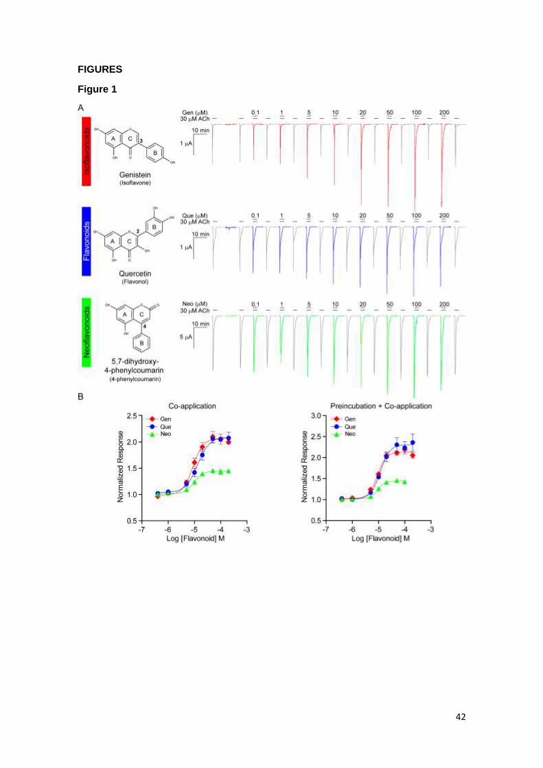
42
FIGURES
Figure 1
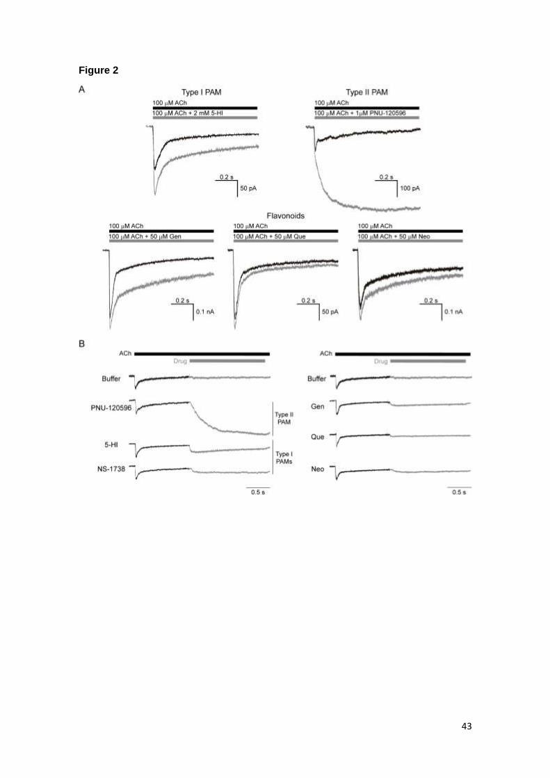
43
Figure 2
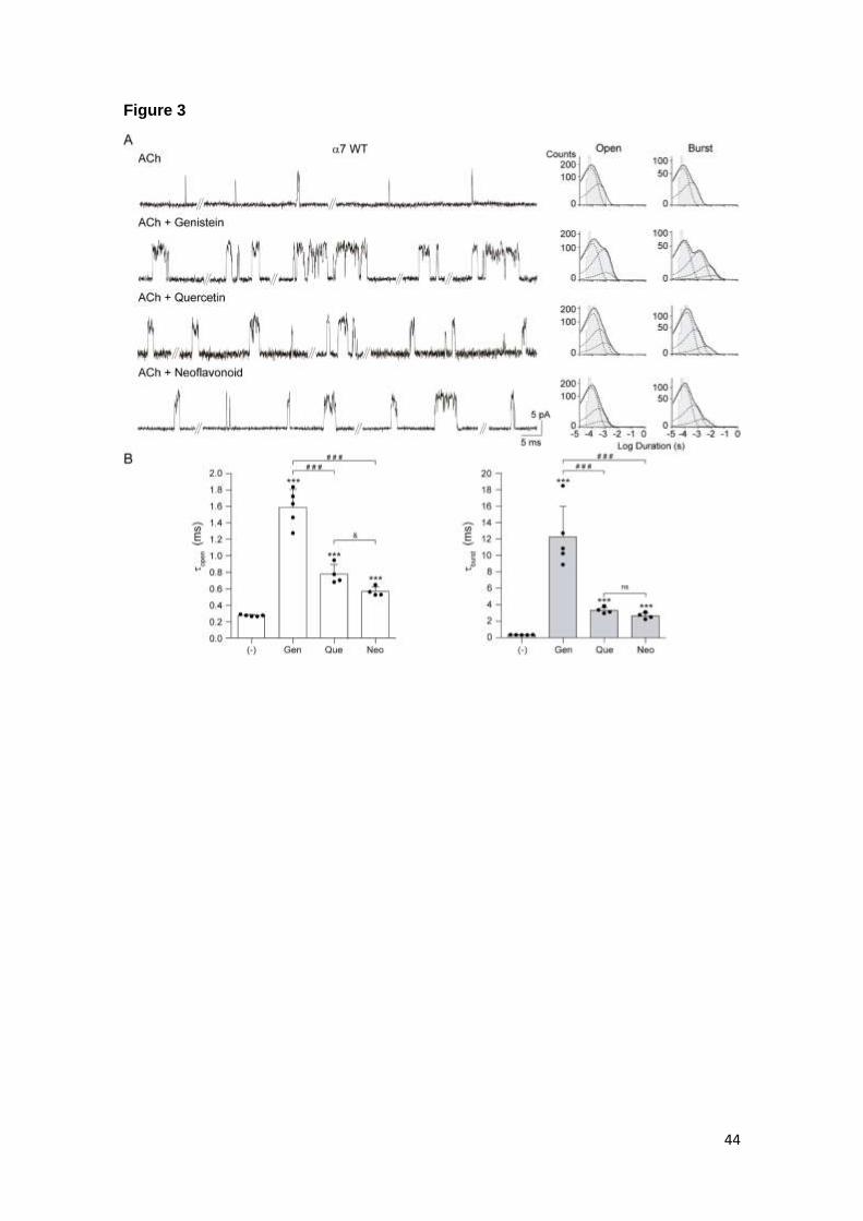
44
Figure 3
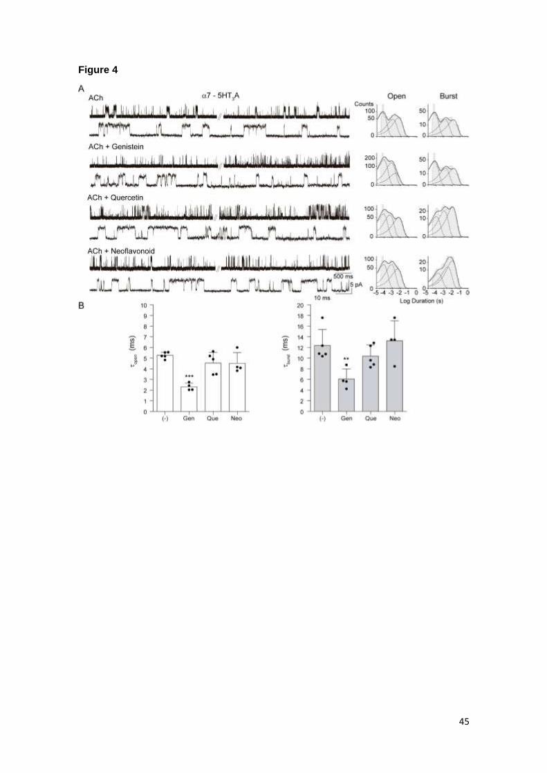
45
Figure 4
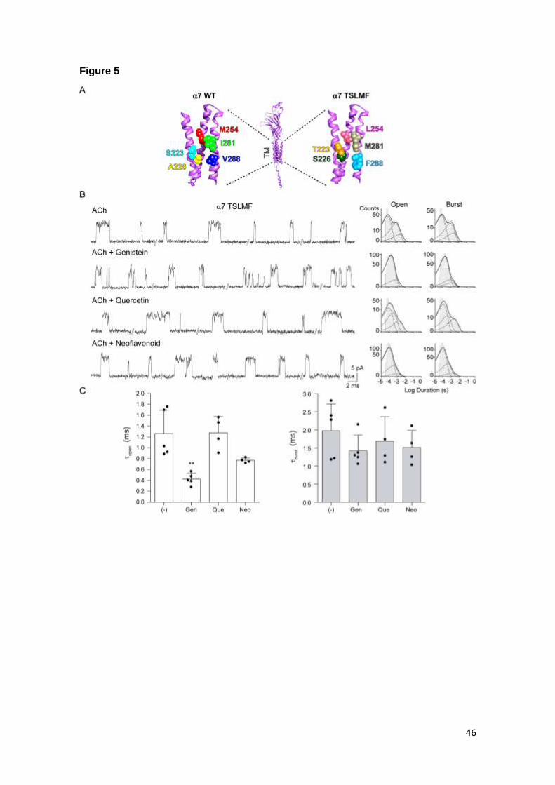
46
Figure 5
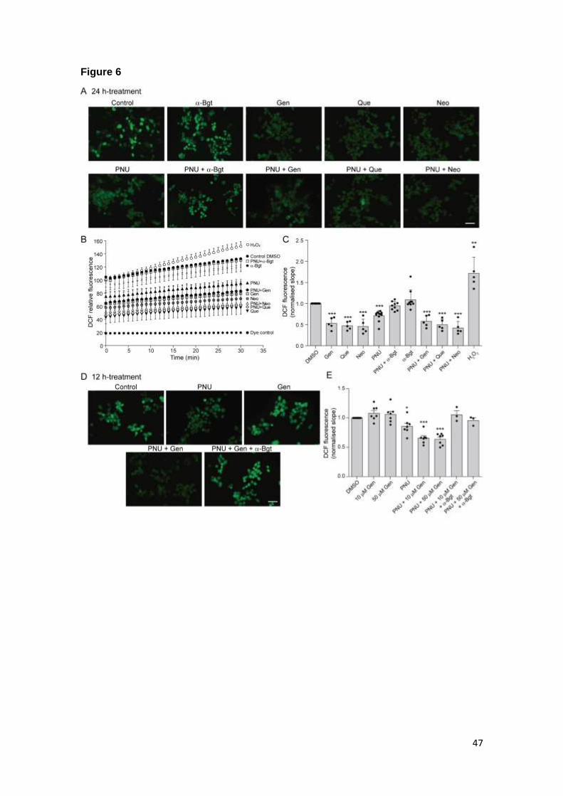
47
Figure 6
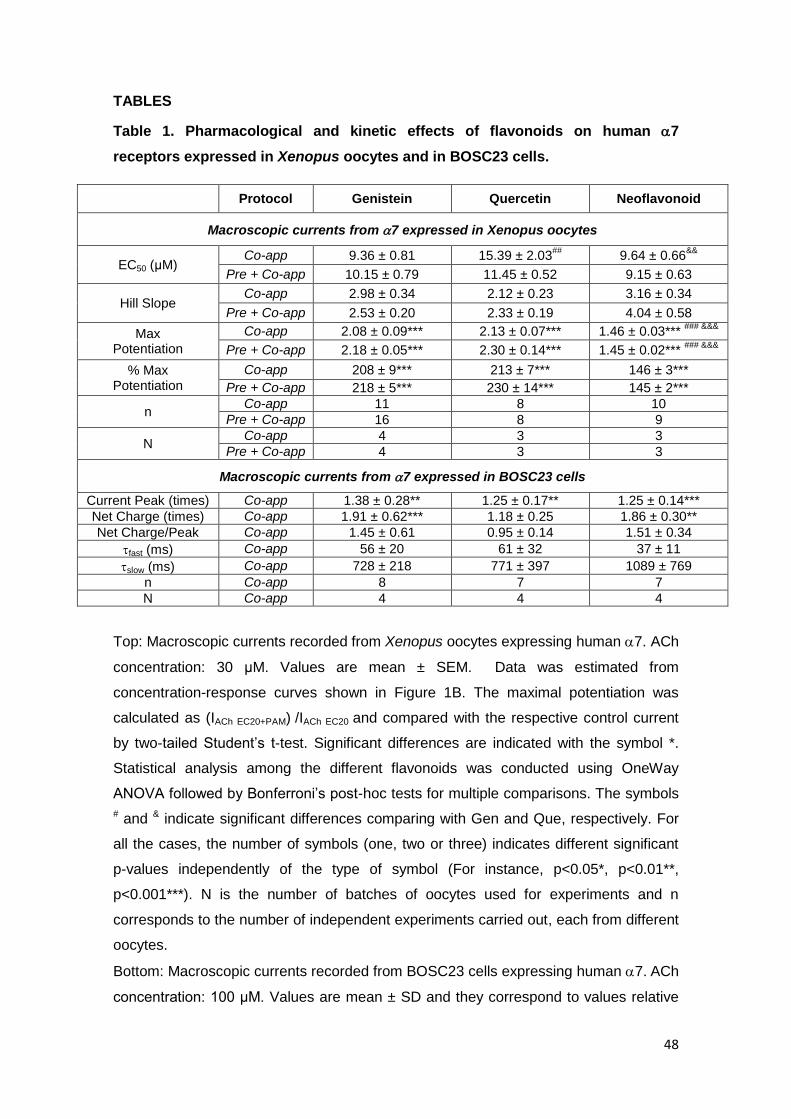
48
TABLES
Table 1. Pharmacological and kinetic effects of flavonoids on human 7
receptors expressed in Xenopus oocytes and in BOSC23 cells.
Protocol Genistein Quercetin Neoflavonoid
Macroscopic currents from 7 expressed in Xenopus oocytes
EC50 (μM) Co-app 9.36 ± 0.81 15.39 ± 2.03
## 9.64 ± 0.66
&&
Pre + Co-app 10.15 ± 0.79 11.45 ± 0.52 9.15 ± 0.63
Hill Slope Co-app 2.98 ± 0.34 2.12 ± 0.23 3.16 ± 0.34
Pre + Co-app 2.53 ± 0.20 2.33 ± 0.19 4.04 ± 0.58
Max Potentiation
Co-app 2.08 ± 0.09*** 2.13 ± 0.07*** 1.46 ± 0.03*** ### &&&
Pre + Co-app 2.18 ± 0.05*** 2.30 ± 0.14*** 1.45 ± 0.02*** ### &&&
% Max Potentiation
Co-app 208 ± 9*** 213 ± 7*** 146 ± 3***
Pre + Co-app 218 ± 5*** 230 ± 14*** 145 ± 2***
n Co-app 11 8 10
Pre + Co-app 16 8 9
N Co-app 4 3 3
Pre + Co-app 4 3 3
Macroscopic currents from 7 expressed in BOSC23 cells
Current Peak (times) Co-app 1.38 ± 0.28** 1.25 ± 0.17** 1.25 ± 0.14***
Net Charge (times) Co-app 1.91 ± 0.62*** 1.18 ± 0.25 1.86 ± 0.30**
Net Charge/Peak Co-app 1.45 ± 0.61 0.95 ± 0.14 1.51 ± 0.34
fast (ms) Co-app 56 ± 20 61 ± 32 37 ± 11
slow (ms) Co-app 728 ± 218 771 ± 397 1089 ± 769
n Co-app 8 7 7
N Co-app 4 4 4
Top: Macroscopic currents recorded from Xenopus oocytes expressing human 7. ACh
concentration: 30 μM. Values are mean ± SEM. Data was estimated from
concentration-response curves shown in Figure 1B. The maximal potentiation was
calculated as (IACh EC20+PAM) /IACh EC20 and compared with the respective control current
by two-tailed Student’s t-test. Significant differences are indicated with the symbol *.
Statistical analysis among the different flavonoids was conducted using OneWay
ANOVA followed by Bonferroni’s post-hoc tests for multiple comparisons. The symbols
# and & indicate significant differences comparing with Gen and Que, respectively. For
all the cases, the number of symbols (one, two or three) indicates different significant
p-values independently of the type of symbol (For instance, p<0.05*, p<0.01**,
p<0.001***). N is the number of batches of oocytes used for experiments and n
corresponds to the number of independent experiments carried out, each from different
oocytes.
Bottom: Macroscopic currents recorded from BOSC23 cells expressing human 7. ACh
concentration: 100 μM. Values are mean ± SD and they correspond to values relative
49
to the control in the same cell (peak current and net charge) or absolute values (decay
time constants, ) as described in Materials and Methods. Statistical analysis was
conducted by two-tailed Student’s t-test comparing with the respective control
macroscopic current in absence of flavonoid (p<0.05*, p<0.01**, p<0.001***). n
corresponds to the number of independent experiments, each from different cell
patches, and N indicates the number of cell transfections.
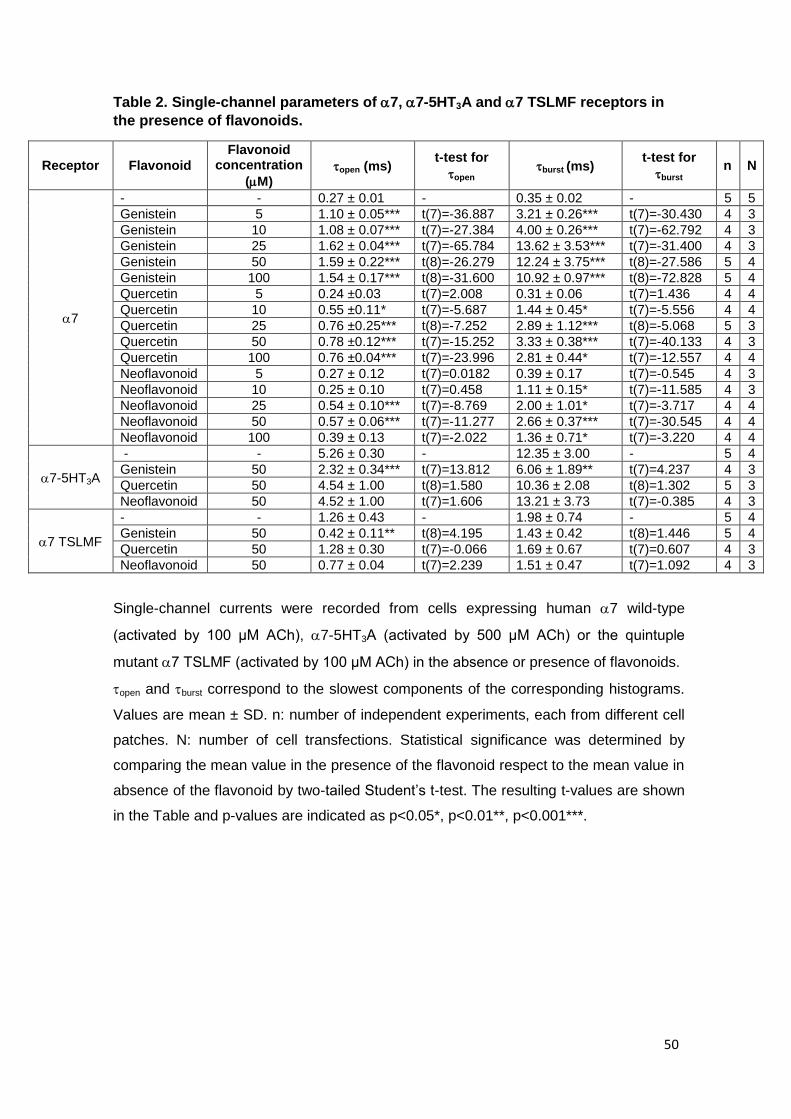
50
Table 2. Single-channel parameters of 7, 7-5HT3A and 7 TSLMF receptors in
the presence of flavonoids.
Receptor Flavonoid
Flavonoid concentration
(M) open (ms)
t-test for
open burst (ms)
t-test for
burst n N
7
- - 0.27 ± 0.01 - 0.35 ± 0.02 - 5 5
Genistein 5 1.10 ± 0.05*** t(7)=-36.887 3.21 ± 0.26*** t(7)=-30.430 4 3
Genistein 10 1.08 ± 0.07*** t(7)=-27.384 4.00 ± 0.26*** t(7)=-62.792 4 3
Genistein 25 1.62 ± 0.04*** t(7)=-65.784 13.62 ± 3.53*** t(7)=-31.400 4 3
Genistein 50 1.59 ± 0.22*** t(8)=-26.279 12.24 ± 3.75*** t(8)=-27.586 5 4
Genistein 100 1.54 ± 0.17*** t(8)=-31.600 10.92 ± 0.97*** t(8)=-72.828 5 4
Quercetin 5 0.24 ±0.03 t(7)=2.008 0.31 ± 0.06 t(7)=1.436 4 4
Quercetin 10 0.55 ±0.11* t(7)=-5.687 1.44 ± 0.45* t(7)=-5.556 4 4
Quercetin 25 0.76 ±0.25*** t(8)=-7.252 2.89 ± 1.12*** t(8)=-5.068 5 3
Quercetin 50 0.78 ±0.12*** t(7)=-15.252 3.33 ± 0.38*** t(7)=-40.133 4 3
Quercetin 100 0.76 ±0.04*** t(7)=-23.996 2.81 ± 0.44* t(7)=-12.557 4 4
Neoflavonoid 5 0.27 ± 0.12 t(7)=0.0182 0.39 ± 0.17 t(7)=-0.545 4 3
Neoflavonoid 10 0.25 ± 0.10 t(7)=0.458 1.11 ± 0.15* t(7)=-11.585 4 3
Neoflavonoid 25 0.54 ± 0.10*** t(7)=-8.769 2.00 ± 1.01* t(7)=-3.717 4 4
Neoflavonoid 50 0.57 ± 0.06*** t(7)=-11.277 2.66 ± 0.37*** t(7)=-30.545 4 4
Neoflavonoid 100 0.39 ± 0.13 t(7)=-2.022 1.36 ± 0.71* t(7)=-3.220 4 4
7-5HT3A
- - 5.26 ± 0.30 - 12.35 ± 3.00 - 5 4
Genistein 50 2.32 ± 0.34*** t(7)=13.812 6.06 ± 1.89** t(7)=4.237 4 3
Quercetin 50 4.54 ± 1.00 t(8)=1.580 10.36 ± 2.08 t(8)=1.302 5 3
Neoflavonoid 50 4.52 ± 1.00 t(7)=1.606 13.21 ± 3.73 t(7)=-0.385 4 3
7 TSLMF
- - 1.26 ± 0.43 - 1.98 ± 0.74 - 5 4
Genistein 50 0.42 ± 0.11** t(8)=4.195 1.43 ± 0.42 t(8)=1.446 5 4
Quercetin 50 1.28 ± 0.30 t(7)=-0.066 1.69 ± 0.67 t(7)=0.607 4 3
Neoflavonoid 50 0.77 ± 0.04 t(7)=2.239 1.51 ± 0.47 t(7)=1.092 4 3
Single-channel currents were recorded from cells expressing human 7 wild-type
(activated by 100 μM ACh), 7-5HT3A (activated by 500 μM ACh) or the quintuple
mutant 7 TSLMF (activated by 100 μM ACh) in the absence or presence of flavonoids.
open and burst correspond to the slowest components of the corresponding histograms.
Values are mean ± SD. n: number of independent experiments, each from different cell
patches. N: number of cell transfections. Statistical significance was determined by
comparing the mean value in the presence of the flavonoid respect to the mean value in
absence of the flavonoid by two-tailed Student’s t-test. The resulting t-values are shown
in the Table and p-values are indicated as p<0.05*, p<0.01**, p<0.001***.
Recommended