Immunological Identity of the Small Subunit of HL-A Antigens and β2-microglobulin and ItsTurnover on the Cell MembraneAuthor(s): Peter Cresswell, Timothy Springer, Jack L. Strominger, Mervyn J. Turner,Howard M. Grey and Ralph T. KuboSource: Proceedings of the National Academy of Sciences of the United States of America,Vol. 71, No. 5 (May, 1974), pp. 2123-2127Published by: National Academy of SciencesStable URL: http://www.jstor.org/stable/63598 .
Accessed: 03/05/2014 20:39
Your use of the JSTOR archive indicates your acceptance of the Terms & Conditions of Use, available at .http://www.jstor.org/page/info/about/policies/terms.jsp
.JSTOR is a not-for-profit service that helps scholars, researchers, and students discover, use, and build upon a wide range ofcontent in a trusted digital archive. We use information technology and tools to increase productivity and facilitate new formsof scholarship. For more information about JSTOR, please contact [email protected].
.
National Academy of Sciences is collaborating with JSTOR to digitize, preserve and extend access toProceedings of the National Academy of Sciences of the United States of America.
http://www.jstor.org
This content downloaded from 130.132.123.28 on Sat, 3 May 2014 20:39:02 PMAll use subject to JSTOR Terms and Conditions
Proc. Nat. Acad. Sci. USA Vol. 71, No. 5, pp. 2118-2122, May 1974
Cyclic-AMP-Controlled Oscillations in Suspended Dictyostelium Cells: Their Relation to Morphogenetic Cell Interactions
(chemotaxis/sline molds/cell aggregation/membrane receptors/dissipative structures)
GUNTHER GERISCH* AND BENNO HESSt * Friedrich-Miescher-Laboratorium der Max-Planck-Gesellschaft, 74 Tabingen; and tMax-Planck-Institut fAr Erniihrungsphysiologie, 46 Dortmund, Germany
Communicated by Britton Chance, November 28, 1973
ABSTRACT Periodic spikes of decreased optical density were recorded in cell suspensions of Dictyostelium discoideum. Spike formation as well as clhanges in the redox state of cytochrome b are facultatively and inde- pendently coupled to an oscillating system which is under developmental control and presumably underlies signal transmission in aggregating cells.
Cyclic AMP triggers a double response, the slow com- ponent resembling the spikes formed during spontaneous oscillations. The fast component shows characteristics of the chemotactic response to cyclic AMP. The receptor system is suiggested to sense changes of cyclic AMP con- centration in time. Cyclic AMP pulses interact with the oscillating system, resulting in phase shift or suppression of spike formation, and in the induction of oscillations in an early stage of development before the onset of spon- taneous oscillations. Continuous flow application of cyclic AMP does not change frequency up to flow rates which extinguish oscillations.
After the end of growth, cells of the slime mold Dictyostelium discoideum aggregate in response to chernotactic stimuli. This process is an example of self-organization of spatial patterns by chemical cell communication, starting with a layer of randomly distributed identical cells (1-3). Aggregation terri- tories are controlled by centers which typically release chemo- tactic signals in pulses with a frequency of 0.2-0.3 min-' (4, 5). The cells around a center respond by orientated cell movement, and also by producing a pulse to which the outer neighboring cells respond after a signal input/output delay of ) 15 sec. So waves of chemotactic pulses can be propagated over a distance much larger than the chemotactic action radius of an aggregation center (4, 5).
cAMP elicits a chemotactic response (6, 7), and when ap- plied in pulses induces propagated waves, thus simulating transmitter action (8). Extracellular cAl\IP is rapidly de- stroyed by extracellular as well as cell-bound phosphodi- esterases (9, 10). Periodic activities and cAMP effects can be recorded optically in stirred cell suspensions. This makes it possible to investigate the molecular basis of morphogenetic cell communication under conditions similar to those used in studying oscillations of the glycolytic pathway in yeast cell suspensions (11, 12). In the present paper we report that the ability to oscillate in suspensions is related to the morpho- genetic capacity of the cells, and describe interactions of cAMP with the oscillating system.
Abbreviations: cAMP, cyclic adenosine 3',5'-monophosphate; cGMP, cyclic guanosine 3',5'-monophosphate; cTMP, cyclic inosine 3', '-monophosphate; t,, developmental stage of cells timed as n hours after their removal from growth medium.
METHODS
Dictyostelium discoideum strain Ax-2 (13) was cultivated at 22-25? axenically on growth medium containing 1.8% mal- tose (13) up to cell densities of 0.3 to 1.4. 107/ml. The cells were washed three times in the cold with 0.017 M Soerensen phosphate buffer, pH 6.0, resuspended in the buffer, adjusted to 1- 107/ml, and shaken. The time of resuspension was taken as the beginning of cell differentiation to aggregation com- petence. After various times, cells were centrifuged and ad- justed in cold buffer to 2- 108/ml. From an ice bath, 2 ml of the suspension were transferred into a cuvette with an optical pathway of 1 cm, and agitated by bubbling oxygen with a constant flow rate of 24 4+ 1 nIl/mii through two syringes. For all measurements taken at 405 and 430 nm, the cuvette was kept at 230. The spectrophotometer and the continuous flow equipment used are described in (14) and (12), respec- tively.
Recording of cytochrome b was based on oxygen-dithionite difference spectra determined at liquid air temperature in suspensions of 108 cells per ml using a Johnson Foundation split beam spectrophotometer. Peaks were found for cyto- chromes a, a3 at 598, cl at 553, c at 548, and b at 562 and 560, as well as a Soret region with a peak at 425 and shoulders at 430 and 445 nm. The redox state of cytochrome b in living cell suspensions was recorded at 430 nm using 405 nm as the ref- erence wavelength. The latter was simultaneously used for recording optical density.
RESULTS
Spikes and Sinusoidal Oscillations. After separation from the growth medium, suspended cells of D. discoideum Ax-2 pass through a pre-aggregation phase of about 9 hr before they acquire full aggregation competence (9, 15). Within the first 5 hr of this phase, no spontaneous oscillations were ob- served. When, however, cells were harvested 6-14 hr after separation from the growth medium, oscillations began im- mediately after transfer from an ice bath into the optical cuvette. Regularly, an initial series of spikes was recorded, followed by sinusoidal oscillations (Fig. 1). Sometimes several cycles of only sinusoidal oscillations were intercalated be tween spike-generating cveles. The mean spike frequency was 0.14 min-', and the frequency increased upon cessation of spike forrimation, with a mean acceleration factor of 1.20. These results indicate that the cellular activity underlying spike formation, although being coupled to an oscillating system, is not an indispensable part of it.
2118
This content downloaded from 130.132.123.28 on Sat, 3 May 2014 20:39:02 PMAll use subject to JSTOR Terms and Conditions
2124 Immunology: Cresswell et al. Proc. Nat. Acad. Sci. USA 71 (1974)
100_
80 - A-Microglobulin
~60- S
/ L-~~~~~~~~A7,12 40-
20-
I I 1 1 Is { ,,1 0.001 0.01 0.1
pg Protein
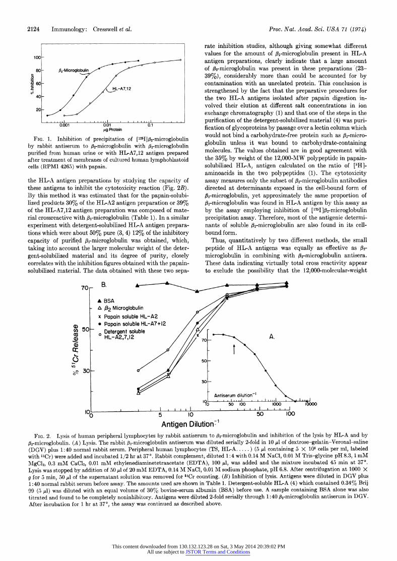
FIG. 1. Inhibition of precipitation of ['2511f.32-microglobulin by rabbit antiserum to #g-microglobulin with ,B2-microglobulin purified from human urine or with HLA7,12 antigen prepared after treatment of membranes of cultured human lymphoblastoid cells (RPM1 4265) with papain.
the HL-A antigen preparations by studying the capacity of these antigens to inhibit the cytotoxicity reaction (Fig. 2B). By this method it was estimated that for the papain-solubi- lized produicts 30% of the HL-A2 antigen preparation or 39% of the HL-A7,12 antigen preparation was composed of mate- rial crossreactive with fl2-microglobulin (Table 1). In a similar experiment with detergent-solubilized HL-A antigen prepara- tions which were about 50% pure (3, 4) 12% of the inhibitorv capacity of purified ,32-microglobulin was obtained, which, taking into account the larger molecular weight of the deter- gent-solubilized material and its degree of purity, closely correlates with the inhibition figures obtained with the papain- solubilized material. The data obtained with these two sepa-
rate inhibition studies, although giving somewhat different values for the amount of 32-mnicroglobulin present in HL-A antigen preparations, clearly indicate that a large amount of 32-microglobulin was present in these preparations (23- 39%), considerably more than could be accounted for by contamination with an unrelated protein. This conclusion is strengthened by the fact that the preparative procedures for the two HL-A antigens isolated after papain digestion in- volved their elution at different salt concentrations in ion exchange chromatography (1) and that one of the steps in the purification of the detergent-solubilized material (4) was puri- fication of glycoproteins by passage over a lectin column which would not bind a carbohydrate-free protein such as A32-micro- globulin unless it was bound to carbohydrate-containing molecules. The values obtained are in good agreement with the 35% by weight of the 12,000-MW polypeptide in papain- solubilized HL-A, antigen calculated on the ratio of [5H]- aminoacids in the two polypeptides (1). The cytotoxicity assay measures only the subset of #32-microglobulin antibodies directed at determinants exposed in the cell-bound form of 02-microglobulin, yet approximately the same proportion of 32-Microglobulin was found in HL-A antigen by this assay as
by the assay employing inhibition of [1251 ]32-microglobulin precipitation assay. Therefore, most of the antigenic determi- nants of soluble 02-microglobulin are also found in its cell- bound form.
Thus, quantitatively by two different methods, the small peptide of HL-A antigens was equally as effective as 32-
microglobulin in combining with 02-microglobulin antisera. These data indicating virtually total cross reactivity appear to exclude the possibility that the 12,000-molecular-weight
70 B
A BSA - a 2 Microglobulin
x Papoih soluble HL-A2 * Papain soluble HL-A7+12
dJ) D0 Detergent soluble cu 0HL-A2,7,12
0~~~~~~~~~~~~~7
~~~~~30 ~ ~ ~ ~ ~ 0
Antiserum dilution-1
10C, I , I, .,,, I I -I I,,,, I
0 ) 5 10 50 100
Antigen Dilution' FIG. 2. Lysis of human peripheral lymphocytes by rabbit antiserum to 32-microglobulin and inhibition of the lysis by HL-A and by
32-microglobulin. (A) Lysis. The rabbit 32-microglobulin antiserum was diluted serially 2-fold in 10 IAd of dextrose-gelatin-Veronal-saline (DGV) plus 1:40 normal rabbit serum. Peripheral human lymphocytes (TS, HL-A ..... ) (5 JAl containing 3 X 10⁶ cells per ml, labeled with 51Cr) were added and incubated 1/2 hr at 37?. Rabbit complement, diluted 1:4 with 0.14 M NaCl, 0.01 M Tris-glycine pH 8.3, 1 mM MgCl2, 0.3 mM CaC12, 0.01 mM ethylenediaminetetraacetate (EDTA), 100 Il, was added and the mixture incubated 45 min at 37?. Lysis was stopped by addition of 50 ,ul of 20 mM EDTA, 0.14 M NaCl, 0.01 M sodium phosphate, pH 6.8. After centrifugation at 1000 X g for 5 min, 50 .dA of the supernatant solution was removed for 5'Cr counting. (B) Inhibition of lysis. Antigens were diluted in DGV plus 1:40 normal rabbit serum before assay. The amounts used are shown in Table 1. Detergent-soluble HL-A (4) which contained 0.34% Brij 99 (5 ,l) was diluted with an equal volume of 30% bovine-serum albumin (BSA) before use. A sample containing BSA alone was also titrated and found to be completely noninhibitory. Antigens were diluted 2-fold serially through 1:40 32-microglobulin antiserum in D)GV. After incubation for 1 hr at 370, the assay was continued as described above.
This content downloaded from 130.132.123.28 on Sat, 3 May 2014 20:39:02 PMAll use subject to JSTOR Terms and Conditions
Proc. Nat. Acad. Sci. USA 71 (1974) #2-Microglobulin and HL-A Antigens 2125
4 6 43j
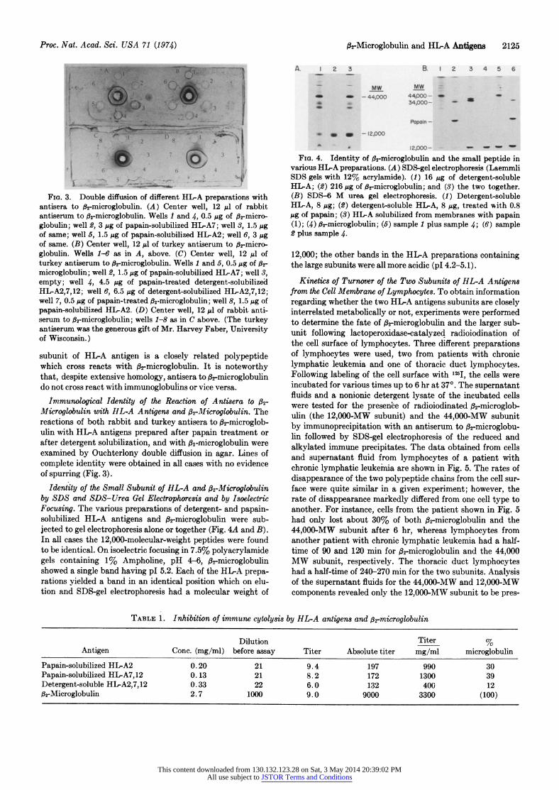
FIG. 3. Double diffusion of different HLA preparations with antisera to 8j-microglobulin. (A) Center well, 12 ul of rabbit antiserum to 62rmicroglobulin. Wells 1 and 4, 0.5 ,ug of 62-micro- globulin; well 2, 3 MAg of papain-solubilized HIA7; well 3, 1.5 Mg of same; well 5, 1.5 MAg of papain-solubilized HLA2; well 6, 3 Mug of same. (B) Center well, 12,ul of turkey antiserum to #2-micro- globulin. Wells 1-6 as in A, above. (C) Center well, 12 ul of turkey antiserum to r-microglobulin. Wells 1 and 5, 0.5 Mg of l2-
microglobulin; well 2, 1.5 MAg of papain-solubilized HL-A7; well 3, empty; well 4, 4.5 Mg of papain-treated detergent-solubilized HL-A2,7,12; well 6, 6.5 MAg of detergent-solubilized HLA2,7,12; well 7, 0.5 Mg of papain-treated ,2-microglobulin; well 8, 1.5 ug of papain-solubilized HI-A2. (D) Center well, 12 Ml of rabbit anti- serum to ,B2-microglobulin; wells 1-8 as in C above. (The turkey antiserum was the generous gift of Mr. Harvey Faber, University of Wisconsin.)
subunit of HL-A antigen is a closely related polypeptide which cross reacts with #2-microglobulin. It is noteworthy that, despite extensive homology, antisera to #2rmicroglobulin do not cross react with immunoglobulins or vice versa.
Immunological Identity of the Reaction of Antisera to 02-
Microglobulin with HL-A Antigens and #2-MIicroglobulin. The reactions of both rabbit and turkey antisera to #2-microglob- ulin with HL-A antigens prepared after papain treatment or after detergent solubilization, and with j32-microglobulin were examined by Ouchterlony double diffusion in agar. Lines of complete identity were obtained in all cases with no evidence of spurring (Fig. 3).
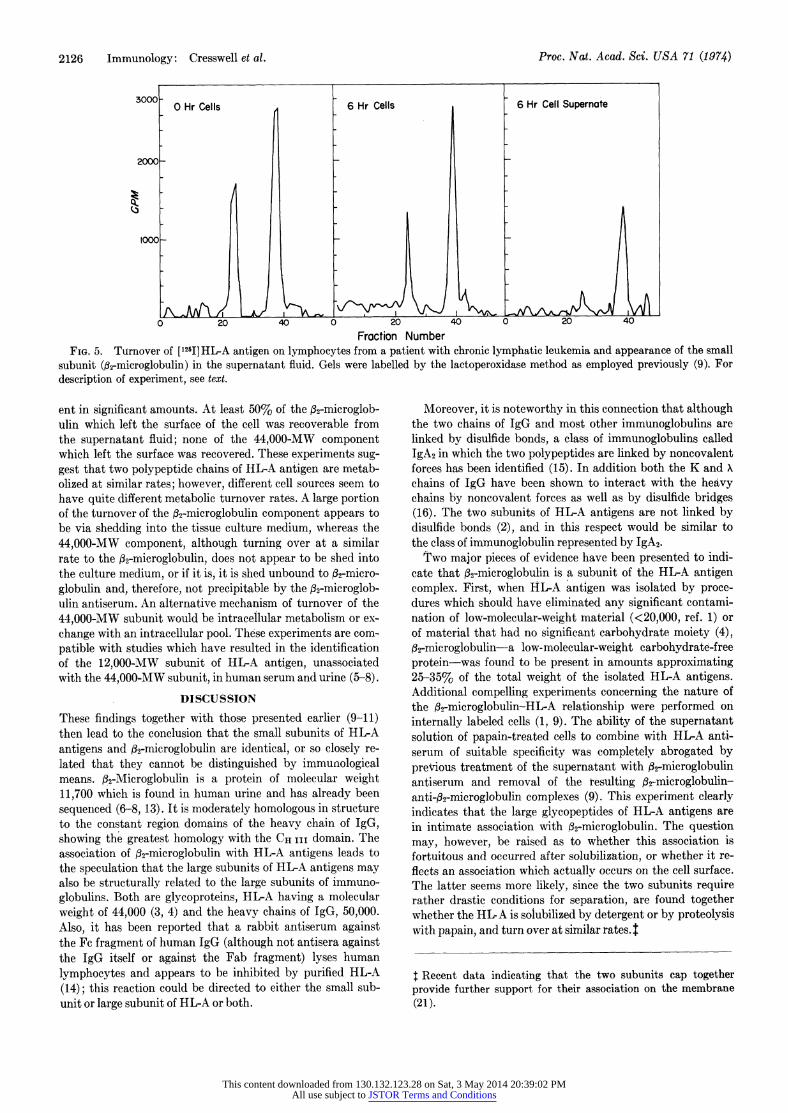
Identity of the Small Subunit of HL-A and 132-Microglobulin by SDS and SDS-Urea Gel Electrophoresis and by Isoelectric Focusing. The various preparations of detergent- and papain- solubilized HL-A antigens and #2-microglobulin were sub- jected to gel electrophoresis alone or together (Fig. 4A and B). In all cases the 12,000-molecular-weight peptides were found to be identical. On isoelectric focusing in 7.5% polyacrylamide gels containing 1% Ampholine, pH 4-6, 2-microglobulin showed a single band having pl 5.2. Each of the HL-A prepa- rations yielded a band in an identical position which on elu- tion and SDS-gel electrophoresis had a molecular weight of
A. 1 2 3 B. 1 2 3 4 5 6
MW-
12,000- FIG. 4. Identity of 6r-microglobulin and the small peptide in
various HI-A preparations. (A) SDS-gel electrophoresis (Laemmli SDS gels with 12% acrylamide). (1) 16 ,ug of detergent-soluble HL-A; (2) 216 ,g of i2rmicroglobulin; and (3) the two together. (B) SDS-6 M urea gel electrophoresis. (1) Detergent-soluble HI-A, 8 ,g; (2) detergent-soluble HL-A, 8 ,ug, treated with 0.8 ,ug of papain; (3) HIA solubilized from membranes with papain (1); (4) 02-microglobulin; (5) sample 1 plus sample 4; (6) sample 2 plus sample 4.
12,000; the other bands in the HL-A preparations containing the large subunits were all more acidic (pl 4.2-5.1).
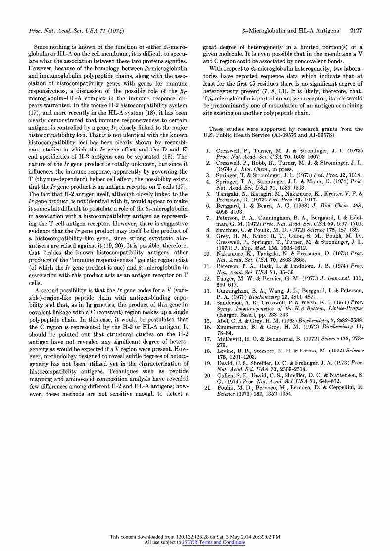
Kinetics of Turnover of the Two Subunits of HL-A Antigens from the Cell Membrane of Lymphocytes. To obtain information regarding whether the two HL-A antigens subunits are closely interrelated metabolically or not, experiments were performed to determine the fate of 2-microglobulin and the larger sub- unit following lactoperoxidase-catalyzed radioiodination of the cell surface of lymphocytes. Three different preparations of lymphocytes were used, two from patients with chronic lymphatic leukemia and one of thoracic duct lymphocytes. Following labeling of the cell surface with 125I, the cells were incubated for various times up to 6 hr at 37?. The supernatant fluids and a nonionic detergent lysate of the incubated cells were tested for the presence of radioiodinated fl2-microglob- ulin (the 12,000-MW subunit) and the 44,000-MW subunit by immunoprecipitation with an antiserum to f32-microglobu- lin followed by SDS-gel electrophoresis of the reduced and alkylated immune precipitates. The data obtained from cells and supernatant fluid from lymphocytes of a patient with chronic lymphatic leukemia are shown in Fig. 5. The rates of disappearance of the two polypeptide chains from the cell sur- face were quite similar in a given experiment; however, the rate of disappearance markedly differed from one cell type to another. For instance, cells from the patient shown in Fig. 5 had only lost about 30% of both 62-microglobulin and the 44,000-MW subunit after 6 hr, whereas lymphocytes from another patient with chronic lymphatic leukemia had a half- time of 90 and 120 min for 2-microglobulin and the 44,000 MW subunit, respectively. The thoracic duct lymphocytes had a half-time of 240-270 min for the two subunits. Analysis of the supernatant fluids for the 44,000-MW and 12,000-MW components revealed only the 12,000-MW subunit to be pres-
TABLE 1. Inhibition of immune cytolysis by HL-A antigens and ,2-microglobulin
Dilution Titer % Antigen Conc. (mg/ml) before assay Titer Absolute titer mg/ml microglobulin
Papain-solubilized HL-A2 0.20 21 9.4 197 990 30 Papain-solubilized HLA7,12 0.13 21 8.2 172 1300 39 Detergent-soluble HIA2,7,12 0.33 22 6.0 132 400 12 6rMicroglobulin 2.7 1000 9.0 9000 3300 (100)
This content downloaded from 130.132.123.28 on Sat, 3 May 2014 20:39:02 PMAll use subject to JSTOR Terms and Conditions
2126 Immunology: Cresswell et al. Proc. Nat. Acad. Sci. USA 71 (1974)
3000 0 Hr Cells 6 Hr Cells 6 Hr Cell Supernote
2000_
0 20 40 0 20 40 0 20 40
Fraction Number FIG. 5. Turnover of [12I]HL-A antigen on lymphocytes from a patient with chronic lymphatic leukemia and appearance of the small
subunit (02-microglobulin) in the supernatant fluid. Gels were labelled by the lactoperoxidase method as employed previously (9). For description of experiment, see text.
ent in significant amounts. At least 50% of the f32-microglob- ulin which left the surface of the cell was recoverable from the supernatant fluid; none of the 44,000-MW component which left the surface was recovered. These experiments sug- gest that two polypeptide chains of HL-A antigen are metab- olized at similar rates; however, different cell sources seem to have quite different metabolic turnover rates. A large portion of the turnover of the 32-microglobulin component appears to be via shedding into the tissue culture medium, whereas the 44,000-MW component, although turning over at a similar rate to the 32-microglobulin, does not appear to be shed into the culture medium, or if it is, it is shed unbound to f2-micro- globulin and, therefore, not precipitable by the,f2-microglob- ulin antiserum. An alternative mechanism of turnover of the 44,000-MW subunit would be intracellular metabolism or ex- change with an intracellular pool. These experiments are com- patible with studies which have resulted in the identification of the 12,000-MW subunit of HL-A antigen, unassociated with the 44,000-MW subunit, in human serum and urine (5-8).
DISCUSSION
These findings together with those presented earlier (9-11) then lead to the conclusion that the small subunits of HL-A antigens and 32-microglobulin are identical, or so closely re- lated that they cannot be distinguished by immunological means. ,32-Microglobulin is a protein of molecular weight 11,700 which is found in human urine and has already been sequenced (6-8, 13). It is moderately homologous in structure to the constant region domains of the heavy chain of IgG, showing the greatest homology with the CH III domain. The association of 032-microglobulin with HL-A antigens leads to the speculation that the large subunits of HL-A antigens may also be structurally related to the large subunits of immuno- globulins. Both are glycoproteins, HL-A having a molecular weight of 44,000 (3, 4) and the heavy chains of IgG, 50,000. Also, it has been reported that a rabbit antiserum against the Fc fragment of human IgG (although not antisera against the IgG itself or against the Fab fragment) lyses human lymphocytes and appears to be inhibited by purified HL-A (14); this reaction could be directed to either the small sub- unit or large subunit of HL-A or both.
Moreover, it is noteworthy in this connection that although the two chains of IgG and most other immunoglobulins are linked by disulfide bonds, a class of immunoglobulins called IgA2 in which the two polypeptides are linked by noncovalent forces has been identified (15). In addition both the K and X chains of IgG have been shown to interact with the heavy chains by noncovalent forces as well as by disulfide bridges (16). The two subunits of HL-A antigens are not linked by disulfide bonds (2), and in this respect would be similar to the class of immunoglobulin represented by IgA2.
Two major pieces of evidence have been presented to indi- cate that 32-microglobulin is a subunit of the HL-A antigen complex. First, when HL-A antigen was isolated by proce- dures which should have eliminated any significant contami- nation of low-molecular-weight material (<20,000, ref. 1) or of material that had no significant carbohydrate moiety (4),
02-microglobulin-a low-molecular-weight carbohydrate-free protein-was found to be present in amounts approximating 25-35% of the total weight of the isolated HL-A antigens. Additional compelling experiments concerning the nature of the 032-microglobulin-HL-A relationship were performed on internally labeled cells (1, 9). The ability of the supernatant solution of papain-treated cells to combine with HL-A anti- serum of suitable specificity was completely abrogated by previous treatment of the supernatant with O32-microglobulin antiserum and removal of the resulting 032-microglobulin- anti-fl2-microglobulin complexes (9). This experiment clearly indicates that the large glycopeptides of HL-A antigens are in intimate association with 182-microglobulin. The question may, however, be raised as to whether this association is fortuitous and occurred after solubilization, or whether it re- flects an association which actuallv occurs on the cell surface. The latter seems more likely, since the two subunits require rather drastic conditions for separation, are found together whether the HL A is solubilized by detergent or by proteolysis with papain, and turn over at similar rates. 1
I Recent data indicating that the two subunits cap together provide further support for their association on the membrane (21).
This content downloaded from 130.132.123.28 on Sat, 3 May 2014 20:39:02 PMAll use subject to JSTOR Terms and Conditions
Proc. Nat. Acad. Sci. USA 71 (1974) ,2-Microglobulin and HILA Antigens 2127
Since nothing is known of the function of either ,s2-micro- globulin or HL-A on the cell membrane, it is difficult to specu- late what the association between these two proteins signifies. However, because of the homology between#2-microglobulin and immunoglobulin polypeptide chains, along with the asso- ciation of histocompatibility genes with genes for immune responsiveness, a discussion of the possible role of the j2-
microglobulin-HL-A complex in the immune response ap- pears warranted. In the mouse H-2 histocompatibility system (17), and more recently in the HL-A system (18), it has been clearly demonstrated that immune responsiveness to certain antigens is controlled by a gene, ir, closely linked to the major histocompatibility loci. That it is not identical with the known histocompatibility loci has been clearly shown by recombi- nant studies in which the Ir gene effect and the D and K end specificities of H-2 antigens can be separated (19). The nature of the Ir gene product is totally unknown, but since it influences the immune response, apparently by governing the T (thymus-dependent) helper cell effect, the possibility exists that the Ir gene product is an antigen receptor on T cells (17). The fact that H-2 antigen itself, although closely linked to the Ir gene product, is not identical with it, would appear to make it somewhat difficult to postulate a role of the f2-microglobulin in association with a histocompatibility antigen as represent- ing the T cell antigen receptor. However, there is suggestive evidence that the Ir gene product may itself be the product of a histocompatibility-like gene, since strong cytotoxic allo- antisera are raised against it (19, 20). It is possible, therefore, that besides the known histocompatibility antigens, other products of the "immune responsiveness" genetic region exist (of which the Ir gene product is one) and fl2-microglobulin in association with this product acts as an antigen receptor on T cells.
A second possibility is that the Ir gene codes for a V (vari- able)-region-like peptide chain with antigen-binding capa- bility and that, as in Ig genetics, the product of this gene in covalent linkage with a C (constant) region makes up a single polypeptide chain. In this case, it would be postulated that the C region is represented by the H-2 or HL-A antigen. It should be pointed out that structural studies on the H-2 antigen have not revealed any significant degree of hetero- geneity as would be expected if a V region were present. How- ever, methodolbgy designed to reveal subtle degrees of hetero- geneity has not been utilized yet in the characterization of histocompatibility antigens. Techniques such as peptide mapping and amino-acid composition analysis have revealed few differences among different H-2 and HL-A antigens; how- ever, these methods are not sensitive enough to detect a
great degree of heterogeneity in a limited portion(s) of a given molecule. It is even possible that in the membrane a V and C region could be associated by noncovalent bonds.
With respect to fl2-microglobulin heterogeneity, two labora- tories have reported sequence data which indicate that at least for the first 45 residues there is no significant degree of heterogeneity present (7, 8, 13). It is likely, therefore, that, if 02-microglobulin is part of an antigen receptor, its role would be predominantly one of modulation of an antigen combining site existing on another polypeptide chain.
These studies were supported by research grants from the U.S. Public Health Service (AI-09576 and AI-09578)
1. Cresswell, P., Turner, M. J. & Strominger, J. L. (1973) Proc. Nat. Acad. Sci. USA 70, 1603-1607.
2. Cresswell, P., Robb, R., Turner, M. J. & Strominger, J. L. (1974) J. Biol. Chem., in press.
3. Springer, T. & Strominger, J. L. (1973) Fed. Proc. 32, 1018. 4. Springer, T. A., Strominger, J. L. & Mann, D. (1974) Proc.
Nat. Acad. Sci. USA 71, 1539-1543. 5. Tanigaki, N., Katagiri, M., Nakamuro, K., Kreiter, V. P. &
Pressman, D. (1973) Fed. Proc. 43, 1017. 6. Berggard, I. & Bearn, A. G. (1968) J. Biol. Chem. 243,
4095-4103. 7. Peterson, P. A., Cunningham, B. A., Bergaard, I. & Edel-
man, G. M. (1972) Proc. Nat. Acad. Sci. USA 69, 1697-1701. 8. Smithies, 0. & Poulik, M. D. (1972) Science 175, 187-189. 9. Grey, H. M., Kubo, R. T., Colon, S. M., Poulik, M. D.,
Cresswell, P., Springer, T., Turner, M. & Strominger, J. L. (1973) J. Exp. Med. 138, 1608-1612.
10. Nakamuro, K., Tanigaki, N. & Pressman, D. (1973) Proc. Nat. Acad. Sci. USA 70, 2863-2865.
11. Peterson, P. A., Rask, L. & Lindblom, J. B. (1974) Proc. Nat. Acad. Sci. USA 71, 35-39.
12. Fanger, M. W. & Bernier, G. M. (1973) J. Immunol. 111, 609-617.
13. Cunningham, B. A., Wang, J. L., Berggard, I. & Peterson, P. A. (1973) Biochemistry 12, 4811-4821.
14. Sanderson, A. R., Cresswell, P. & Welsh, K. I. (1971) Proc. Symp. Immunogenetics of the H-2 System, Liblice-Prague (Karger, Basel), pp. 238-243.
15. Abel, C. A. & Grey, H. M. (1968) Biochemistry 7, 2682-2688. 16. Zimmerman, B. & Grey, H. M. (1972) Biochemistry 11,
78-84. 17. McDevitt, H. 0. & Benacerraf, B. (1972) Science 175, 273-
279. 18. Levine, B. B., Stember, R. H. & Fotino, M. (1972) Science
178, 1201-1203. 19. David, C. S., Shreffler, D. C. & Frelinger, J. A. (1973) Proc.
Nat. Acad. Sci. USA 70, 2509-2514. 20. Cullen, S. E., David, C. S., Shreffler, D. C. & Nathenson, S.
G. (1974) Proc. Nat. Acad. Sci. USA 71, 648-652. 21. Poulik, M. D., Bernoco, M., Bernoco, D. & Ceppellini, R.
Science (1973) 182, 1352-1354.
This content downloaded from 130.132.123.28 on Sat, 3 May 2014 20:39:02 PMAll use subject to JSTOR Terms and Conditions
Recommended