Linfoma di Hodgkin
Roberto Pacelli
Dipartimento di Diagnostica per
Immagini e Radioterapia Università “Federico II” di Napoli
& Istituto di Biostrutture e Bioimmagini
C.N.R
Oncoematologia e nuove tecniche Radioterapiche
Pusey W. Cases of sarcoma and of Hodgkin’s disease treated by exposure to X-rays: a preliminary report. JAMA 1902; 38:166-169
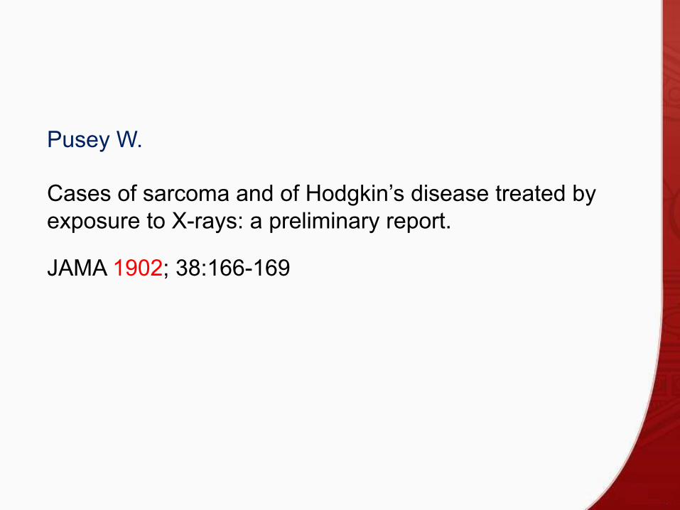
Radiotherapy “extended field”
R. Gilbert “Radiotherapy in Hodgkin’s disease. (Malignant Granulomatosis); anatomic and
clinical foundations; governing principles; results” American Journal of Roentgenology 41:198, 1939. M.V. Peters “A study of survivals in Hodgkin’s disease treated radiologically” American Journal of Roentgenology 63:299, 1950. H.S. Kaplan “The radical radiotherapy of regionally localized Hodgkin’s disease,” Radiology 78:553–561, 1962.
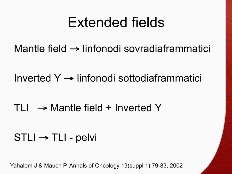
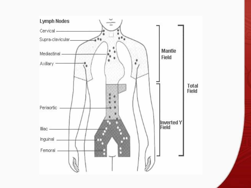
Extended fields
Mantle field → linfonodi sovradiaframmatici Inverted Y → linfonodi sottodiaframmatici TLI → Mantle field + Inverted Y STLI → TLI - pelvi
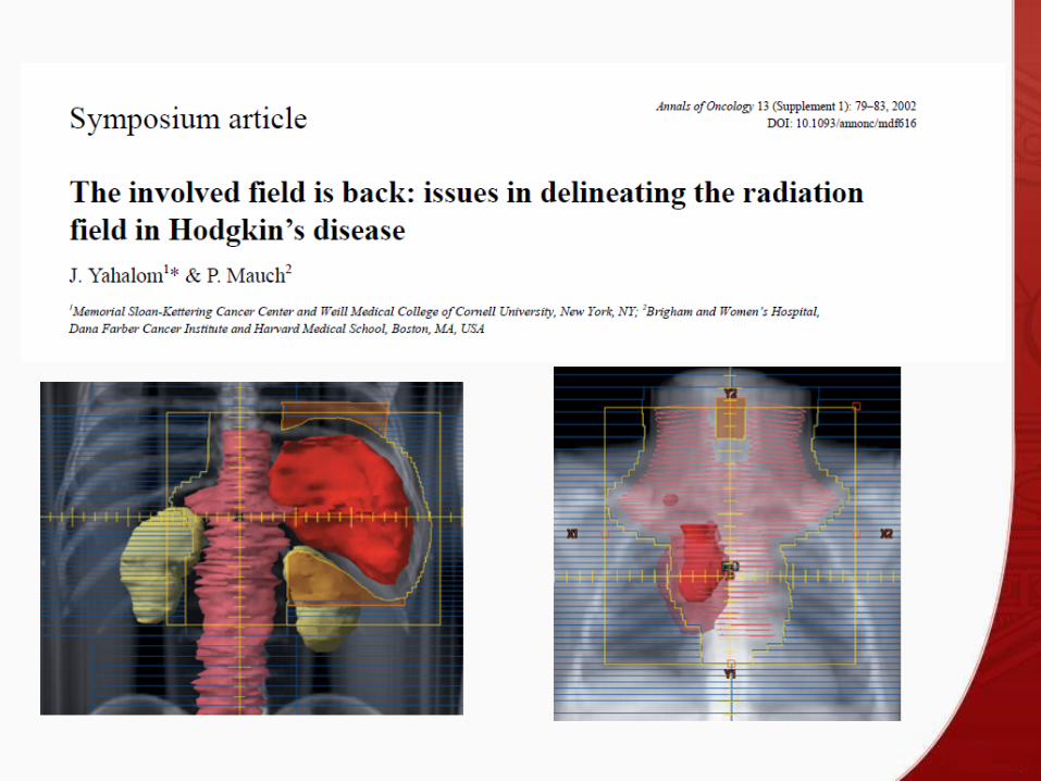
Yahalom J & Mauch P. Annals of Oncology 13(suppl 1):79-83, 2002
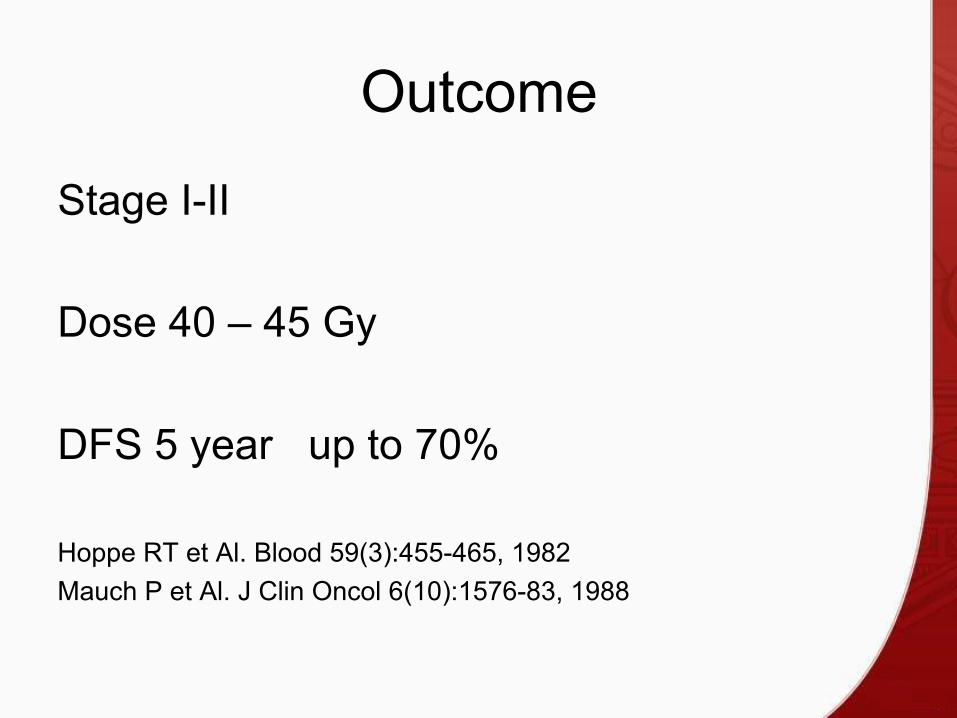
Outcome
Stage I-II Dose 40 – 45 Gy DFS 5 year up to 70% Hoppe RT et Al. Blood 59(3):455-465, 1982 Mauch P et Al. J Clin Oncol 6(10):1576-83, 1988
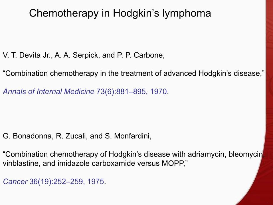
V. T. Devita Jr., A. A. Serpick, and P. P. Carbone, “Combination chemotherapy in the treatment of advanced Hodgkin’s disease,” Annals of Internal Medicine 73(6):881–895, 1970. G. Bonadonna, R. Zucali, and S. Monfardini, “Combination chemotherapy of Hodgkin’s disease with adriamycin, bleomycin, vinblastine, and imidazole carboxamide versus MOPP,” Cancer 36(19):252–259, 1975.
Chemotherapy in Hodgkin’s lymphoma
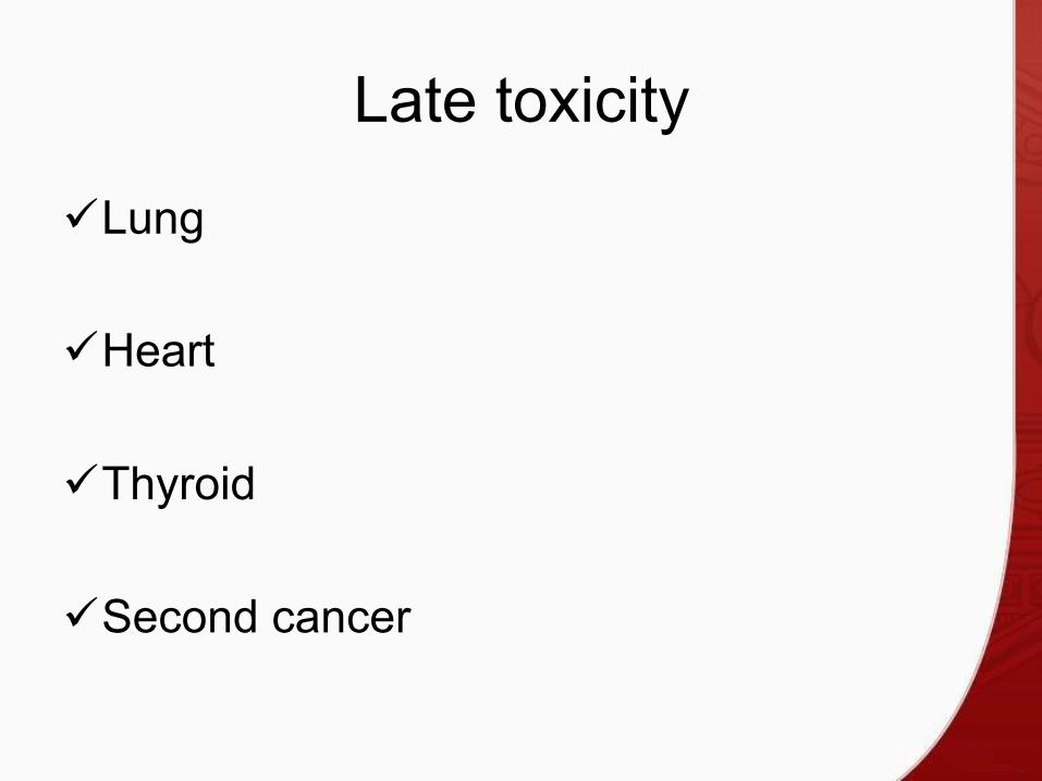
Late toxicity
ü Lung
ü Heart
ü Thyroid
ü Second cancer
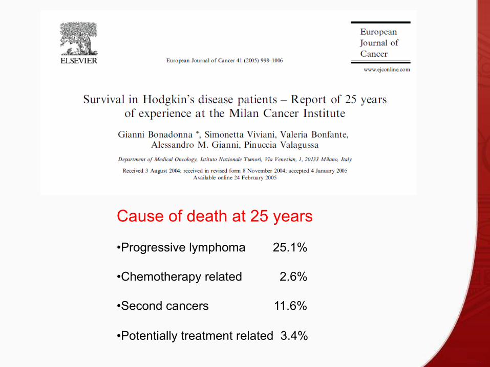
Cause of death at 25 years • Progressive lymphoma 25.1%
• Chemotherapy related 2.6% • Second cancers 11.6%
• Potentially treatment related 3.4%
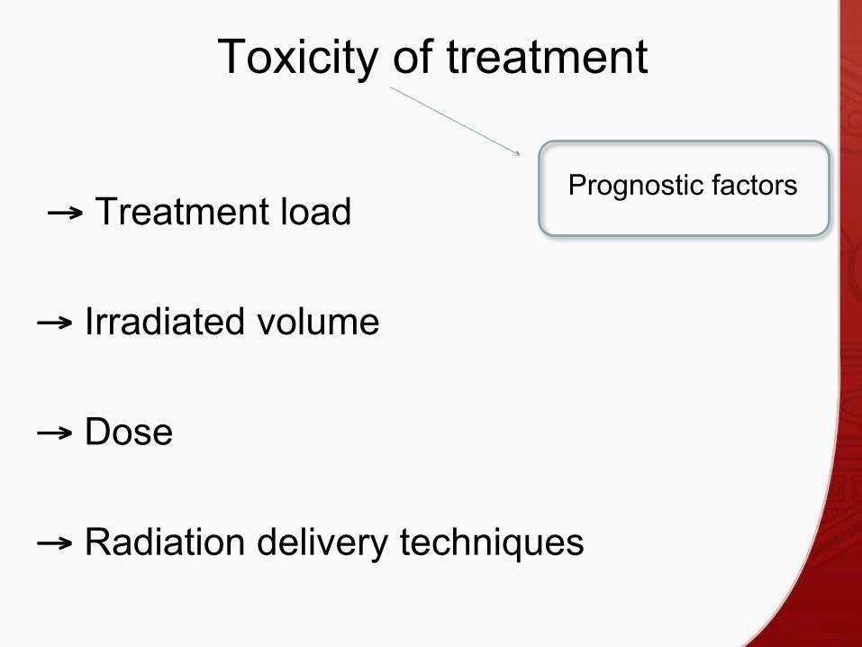
Toxicity of treatment
→ Treatment load → Irradiated volume → Dose → Radiation delivery techniques
Prognostic factors
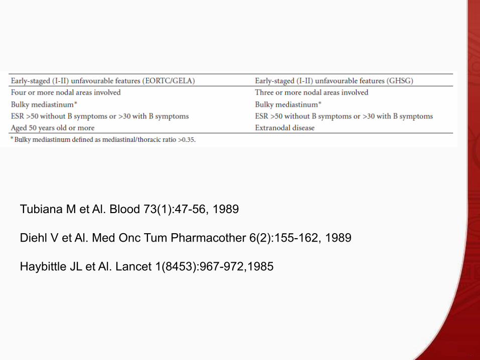
Tubiana M et Al. Blood 73(1):47-56, 1989 Diehl V et Al. Med Onc Tum Pharmacother 6(2):155-162, 1989 Haybittle JL et Al. Lancet 1(8453):967-972,1985
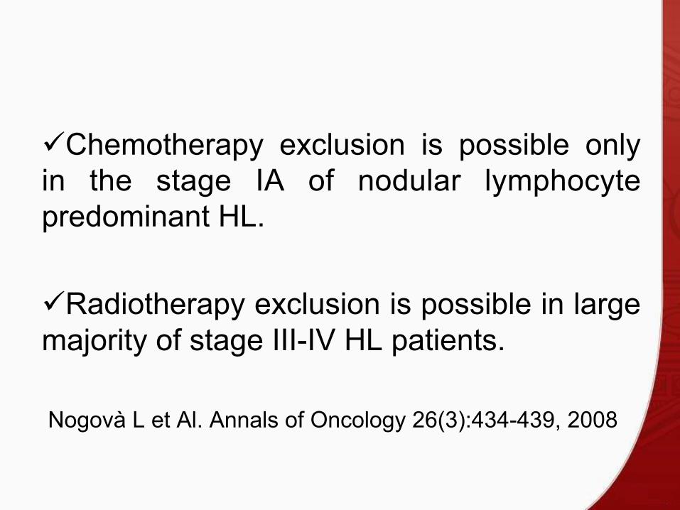
ü Chemotherapy exclusion is possible only in the stage IA of nodular lymphocyte predominant HL.
ü Radiotherapy exclusion is possible in large majority of stage III-IV HL patients. Nogovà L et Al. Annals of Oncology 26(3):434-439, 2008
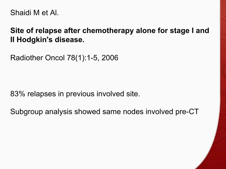
Shaidi M et Al. Site of relapse after chemotherapy alone for stage I and II Hodgkin's disease. Radiother Oncol 78(1):1-5, 2006 83% relapses in previous involved site. Subgroup analysis showed same nodes involved pre-CT
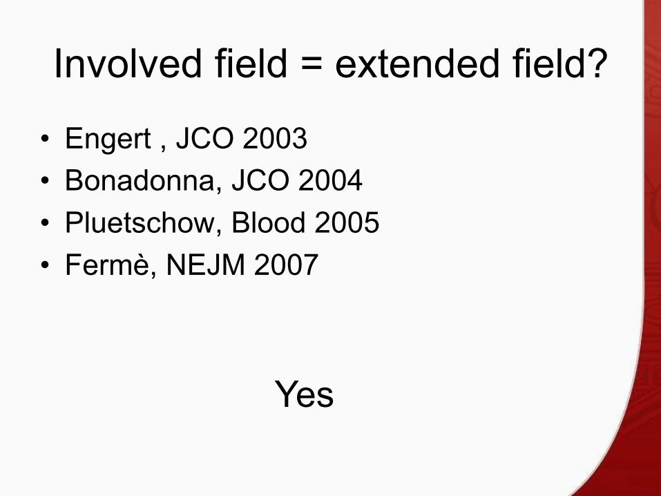
Involved field = extended field?
• Engert , JCO 2003 • Bonadonna, JCO 2004 • Pluetschow, Blood 2005 • Fermè, NEJM 2007
Yes
Involved field=involved node?
Campbell, JCO 2008 May be yes
< Dose
Schewe et Al. IJROBP, 1988
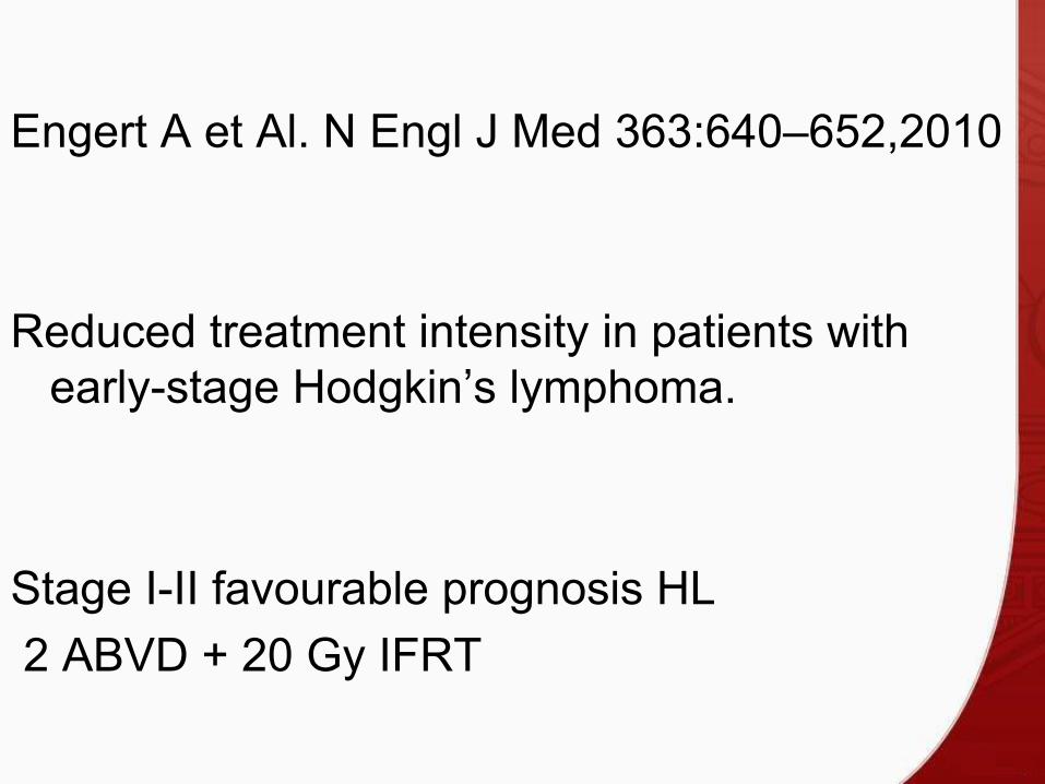
Engert A et Al. N Engl J Med 363:640–652,2010 Reduced treatment intensity in patients with
early-stage Hodgkin’s lymphoma. Stage I-II favourable prognosis HL 2 ABVD + 20 Gy IFRT
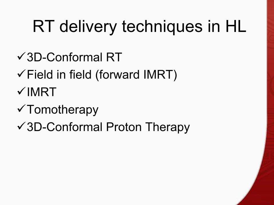
RT delivery techniques in HL
ü 3D-Conformal RT ü Field in field (forward IMRT) ü IMRT ü Tomotherapy ü 3D-Conformal Proton Therapy
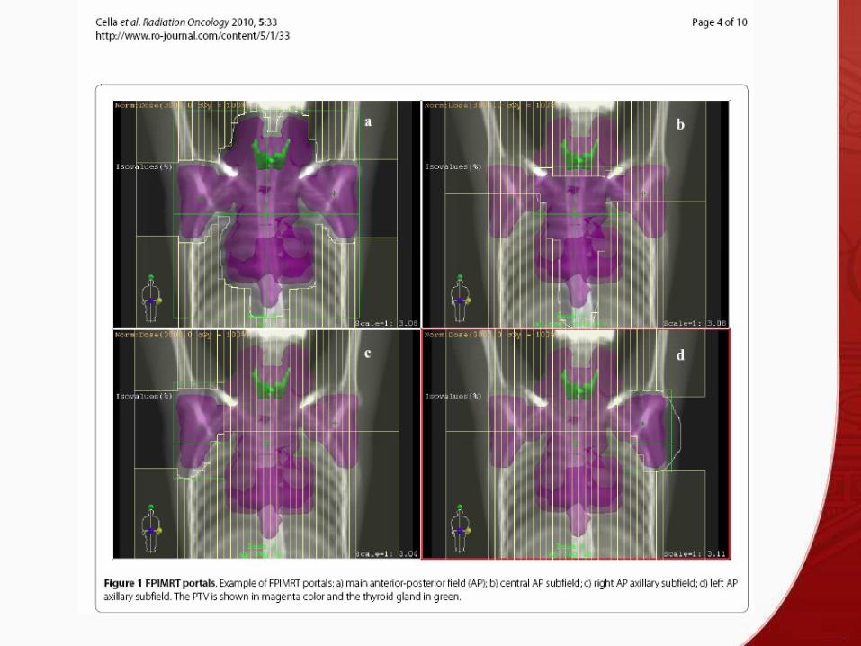
Chera BS et Al. Int. J. Radiation Oncology Biol. Phys. 75(4):1173–1180, 2009
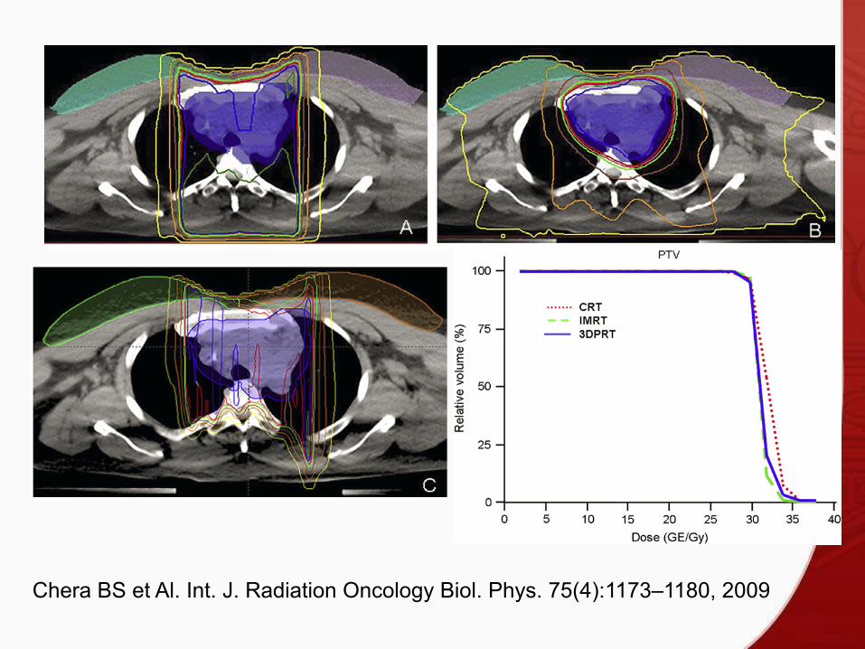
Chera BS et Al. Int. J. Radiation Oncology Biol. Phys. 75(4):1173–1180, 2009
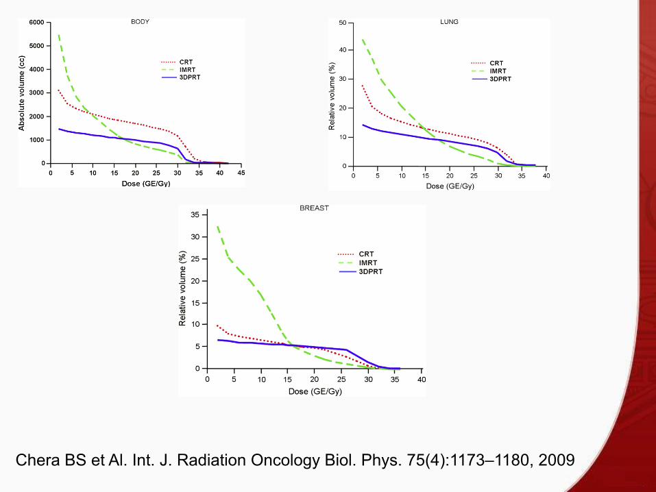
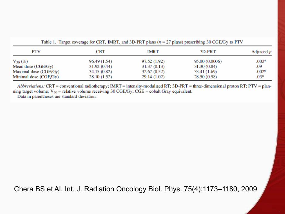
Chera BS et Al. Int. J. Radiation Oncology Biol. Phys. 75(4):1173–1180, 2009
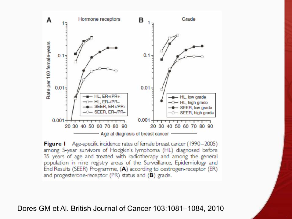
Dores GM et Al. British Journal of Cancer 103:1081–1084, 2010
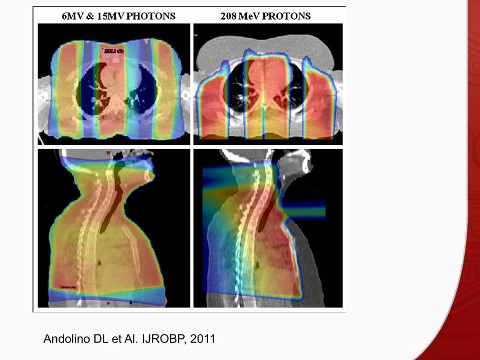
Andolino DL et Al. IJROBP, 2011
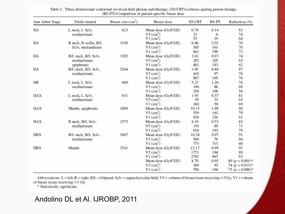
Andolino DL et Al. IJROBP, 2011
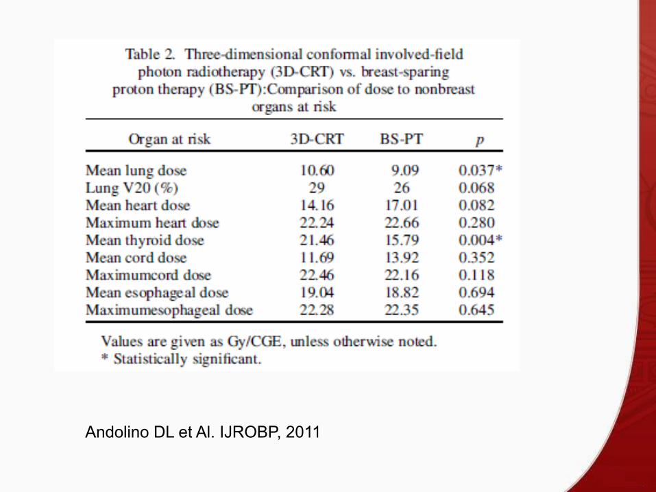
Andolino DL et Al. IJROBP, 2011
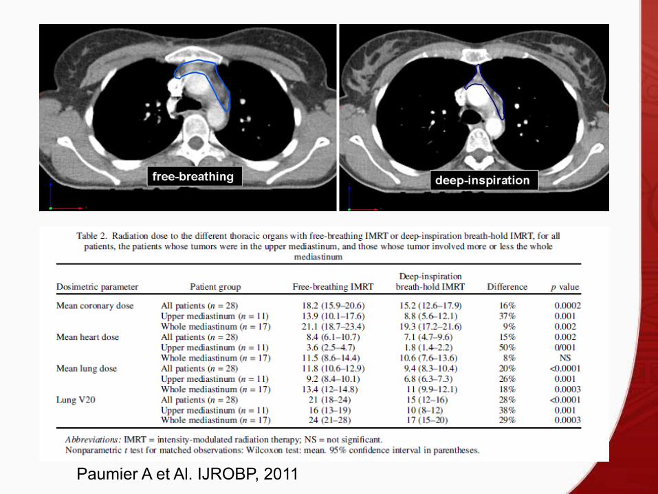
Paumier A et Al. IJROBP, 2011
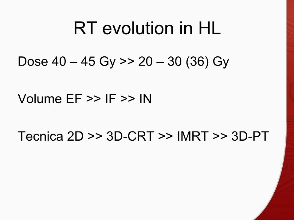
RT evolution in HL
Dose 40 – 45 Gy >> 20 – 30 (36) Gy Volume EF >> IF >> IN Tecnica 2D >> 3D-CRT >> IMRT >> 3D-PT
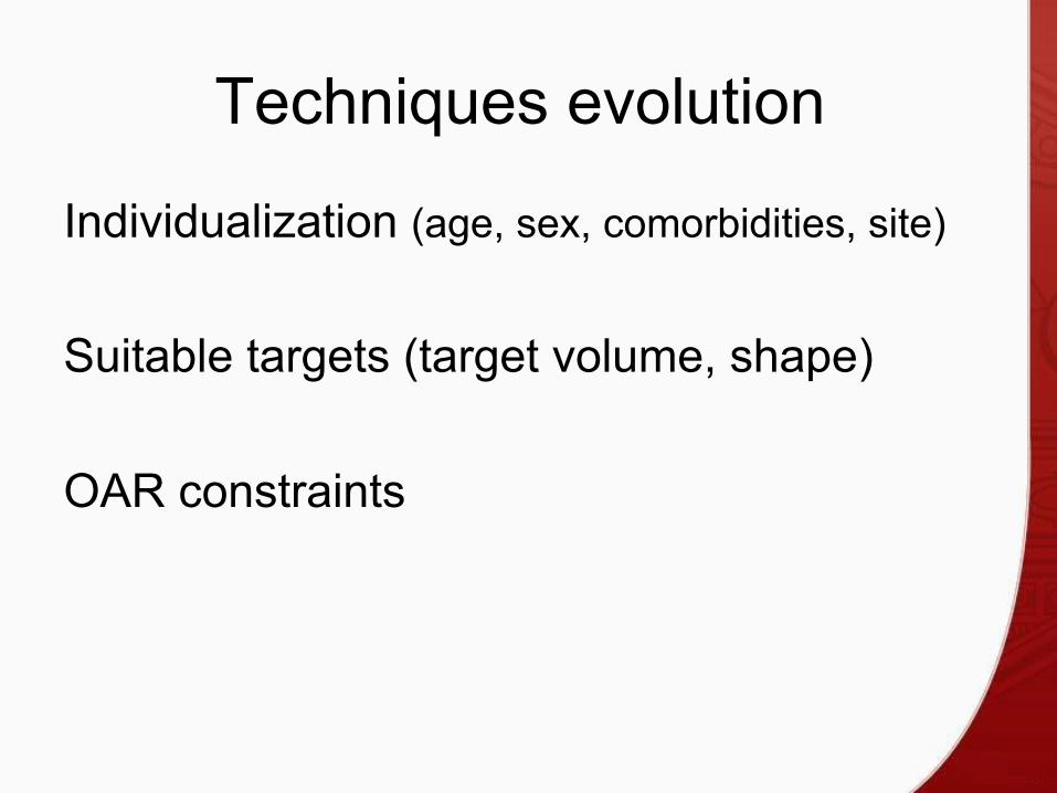
Techniques evolution
Individualization (age, sex, comorbidities, site) Suitable targets (target volume, shape) OAR constraints
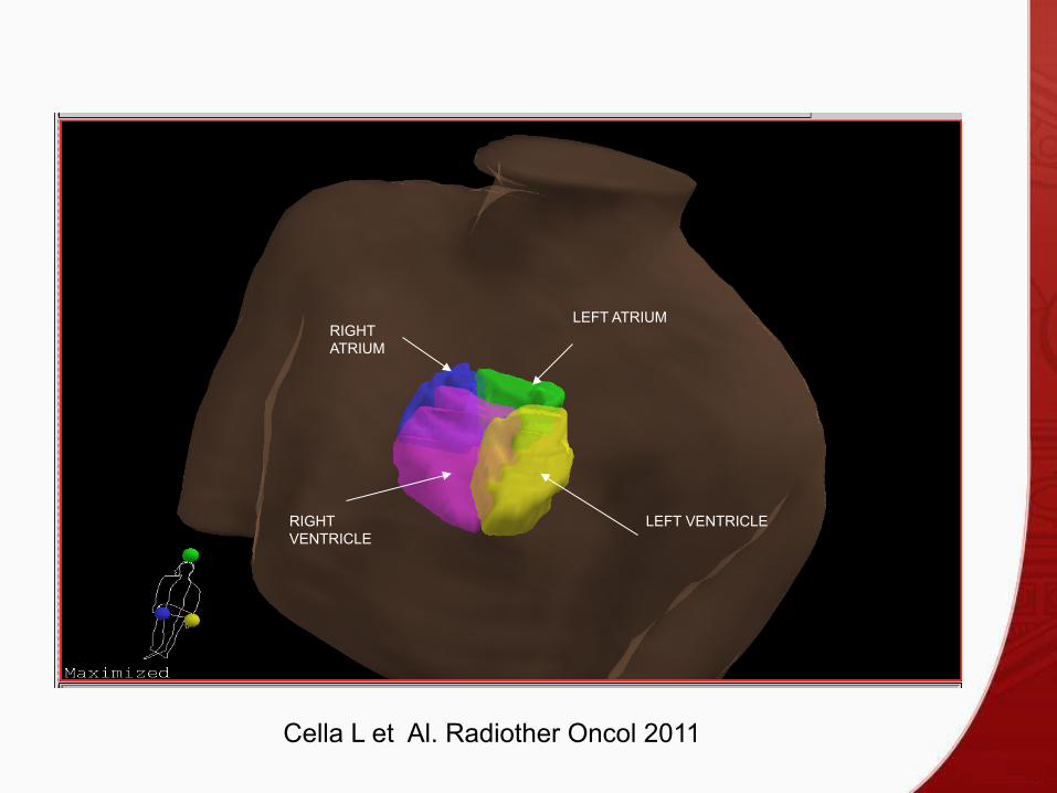
RIGHT ATRIUM
LEFT ATRIUM
RIGHT VENTRICLE
LEFT VENTRICLE
Cella L et Al. Radiother Oncol 2011
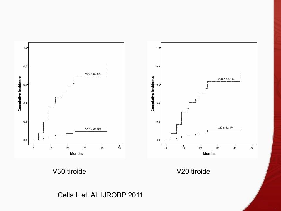
V30 tiroide V20 tiroide
Cella L et Al. IJROBP 2011
Conclusions ü Evolution in the indication, volume, dose and technical delivery of the radiation treatment in HL paves the way to a significant improvement of the late toxicity associated with older treament modalities.
ü Better knowledge of toxicity constraints coupled to an individualization of patients therapy technique will be necessary to fully utilize the available technology .
Recommended