The Muscular System
Miss Ulrich
Muscle cells cannot partially contract. They
act on the ‘all or none’ principle. They either contract 100% or do not contract at all.
You cannot turn fat into muscle by exercising.
You cannot ‘spot reduce’ i.e. you cannot get rid of your spare tire by doing sit-ups.
Interesting Facts
When you are cold, your muscles produce
rapid contractions to generate body heat (shivering).
A cramp is a painful muscle contraction.
Tetanus is a very severe type of
contraction. It is a persistent contraction that can be caused by a bacterial infection. Sometimes you get a ‘tetanus shot’ to prevent this. Tetanus can cause lockjaw.
A spasm is rapid involuntary contraction of a muscle. You may have had one in your eye before - tick.
You are always moving. Even when you are
sleeping, your muscles are working. Movement only stops when life stops.
Movement within cells is caused by chemical reactions. All other body movements are caused by muscles.
Movement
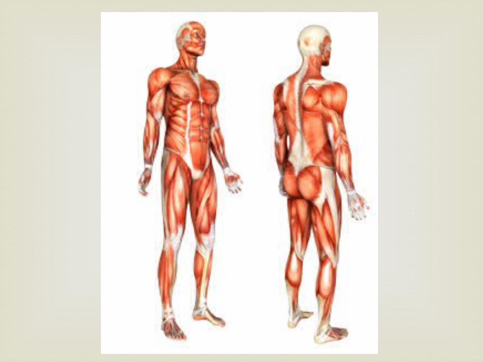
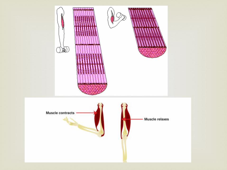
You have more than 650 muscles.
Muscles make up 40% of your body mass.
Muscles work by contracting. When a muscle contracts it shortens. Without your muscles, your bones could not move. When a muscle contracts it pulls on a bone, producing movement. Muscles can only pull bone; they cannot push bones.
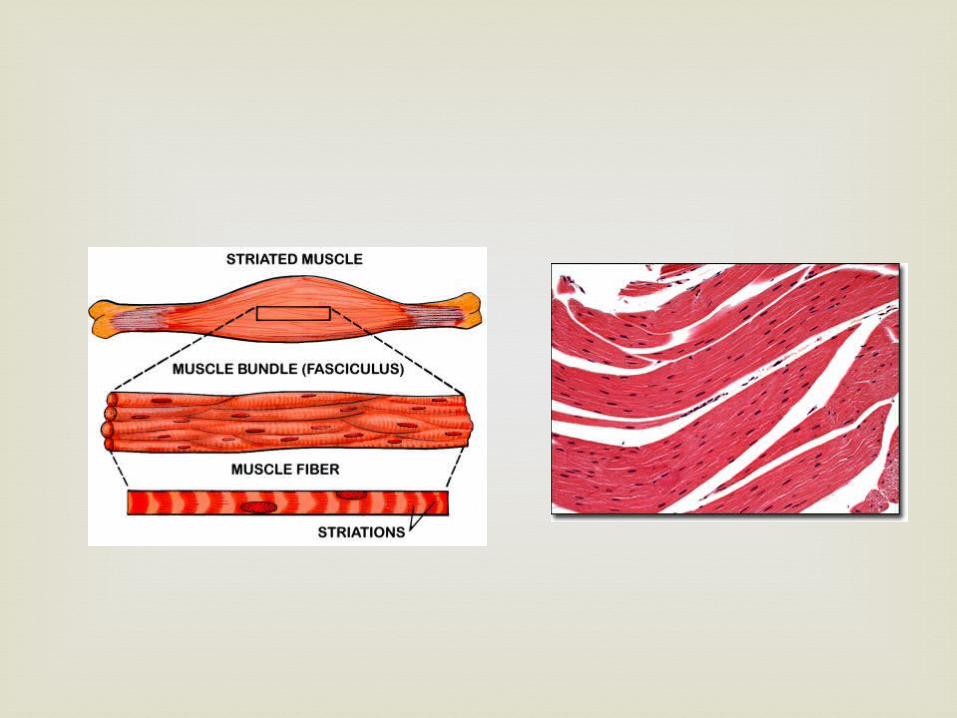
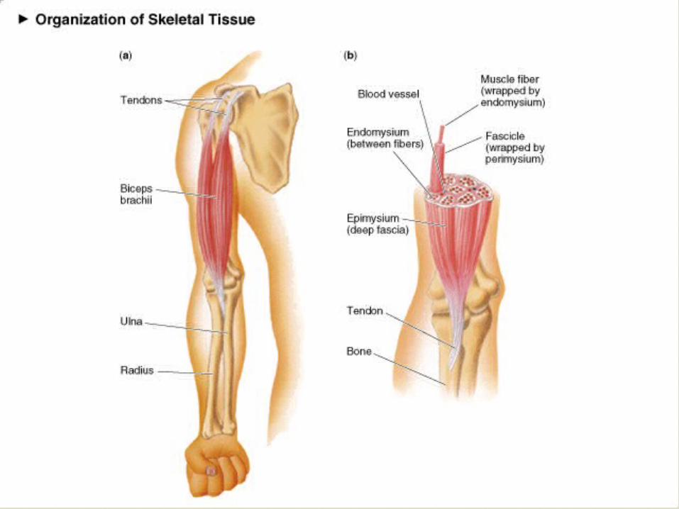
Structure of Skeletal Muscle:Connective Tissue Covering
Epimysium Surrounds entire muscle
Perimysium Surrounds bundles of muscle fibers
Fascicles Endomysium
Surrounds individual muscle fibers
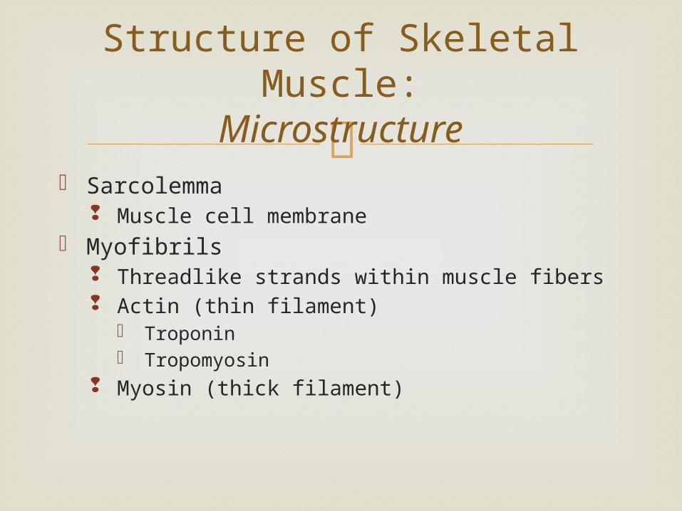
Structure of Skeletal Muscle:Microstructure
Sarcolemma Muscle cell membrane
Myofibrils Threadlike strands within muscle fibers Actin (thin filament)
Troponin Tropomyosin
Myosin (thick filament)
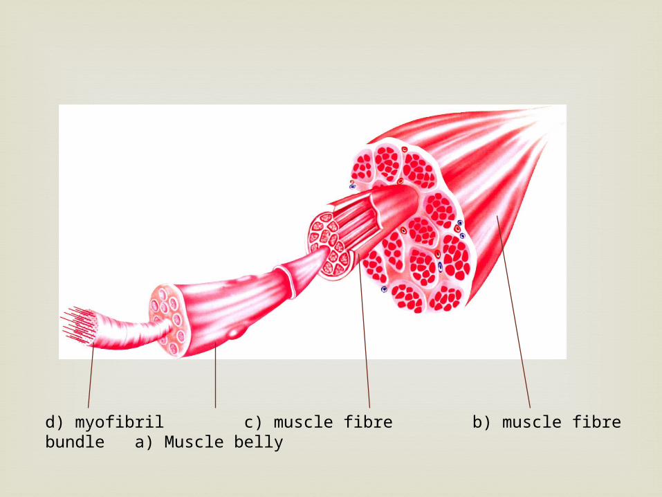
d) myofibril c) muscle fibre b) muscle fibre bundle a) Muscle belly
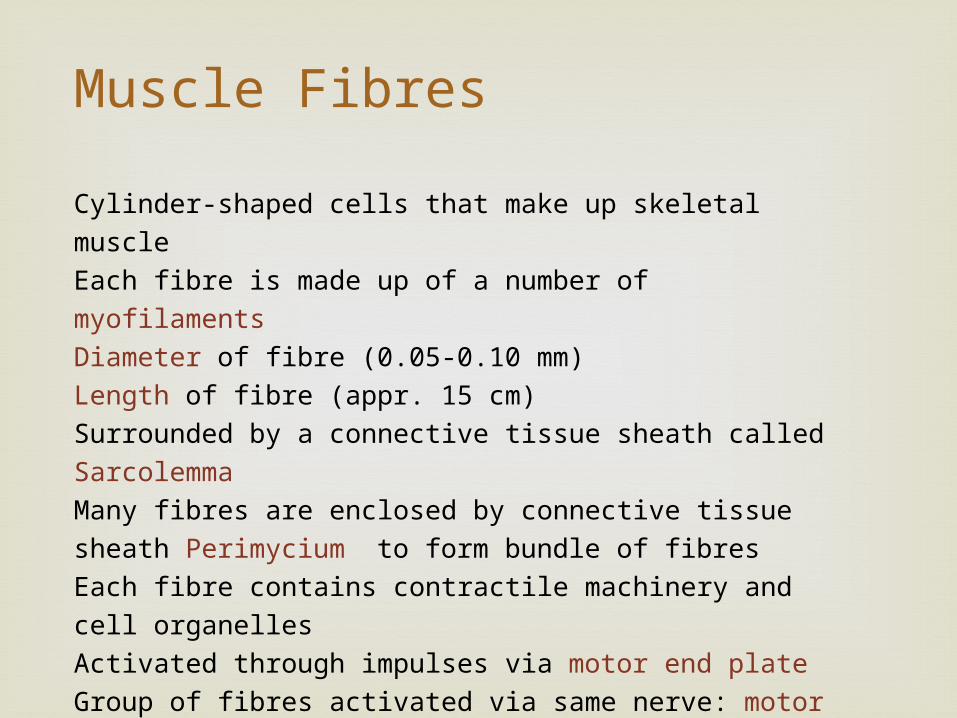
Cylinder-shaped cells that make up skeletal muscle
Each fibre is made up of a number of myofilaments
Diameter of fibre (0.05-0.10 mm)
Length of fibre (appr. 15 cm)
Surrounded by a connective tissue sheath called Sarcolemma
Many fibres are enclosed by connective tissue sheath Perimycium to
form bundle of fibres
Each fibre contains contractile machinery and cell organelles
Activated through impulses via motor end plate
Group of fibres activated via same nerve: motor unit
Each fibre has capillaries that supply nutrients and eliminate waste
Muscle Fibres
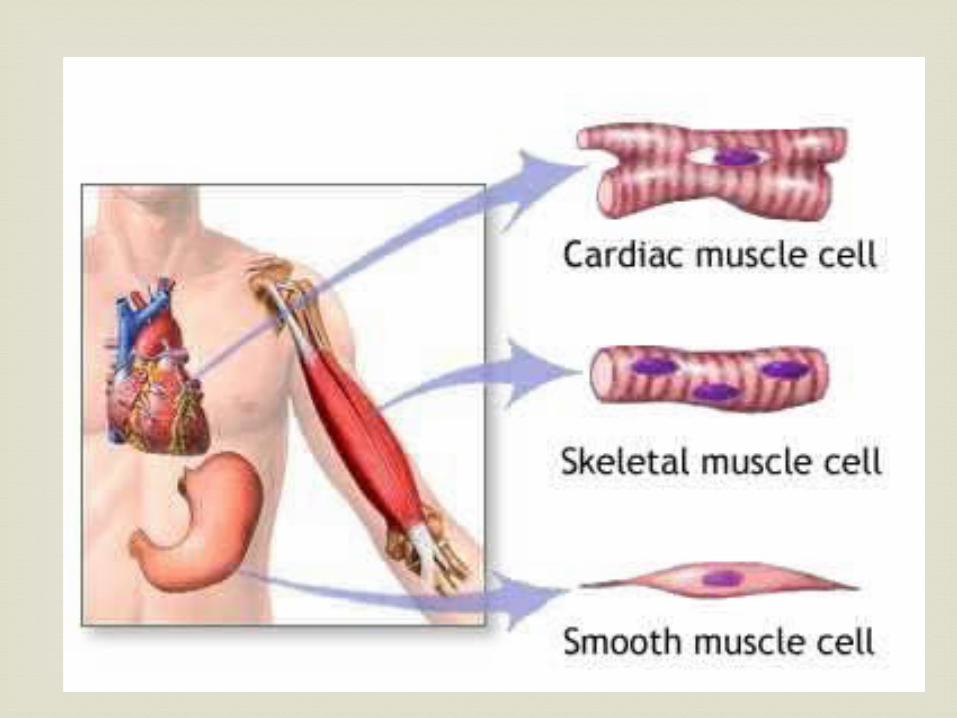
Not all our muscles are used for locomotion.
Some allow us to wink, swallow etc. There are three main types of muscles. At the cellular level they all have the same function – to contract. When we move beyond the cellular level we see differences in their functions:
Types of Muscles
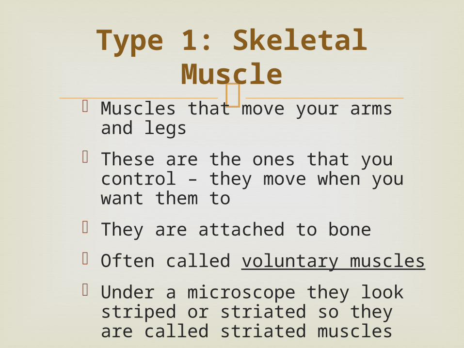
Muscles that move your arms and legs
These are the ones that you control – they move when you want them to
They are attached to bone
Often called voluntary muscles
Under a microscope they look striped or striated so they are called striated muscles
Type 1: Skeletal Muscle
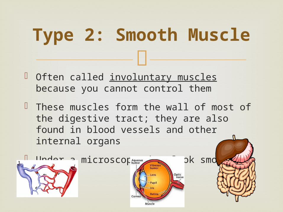
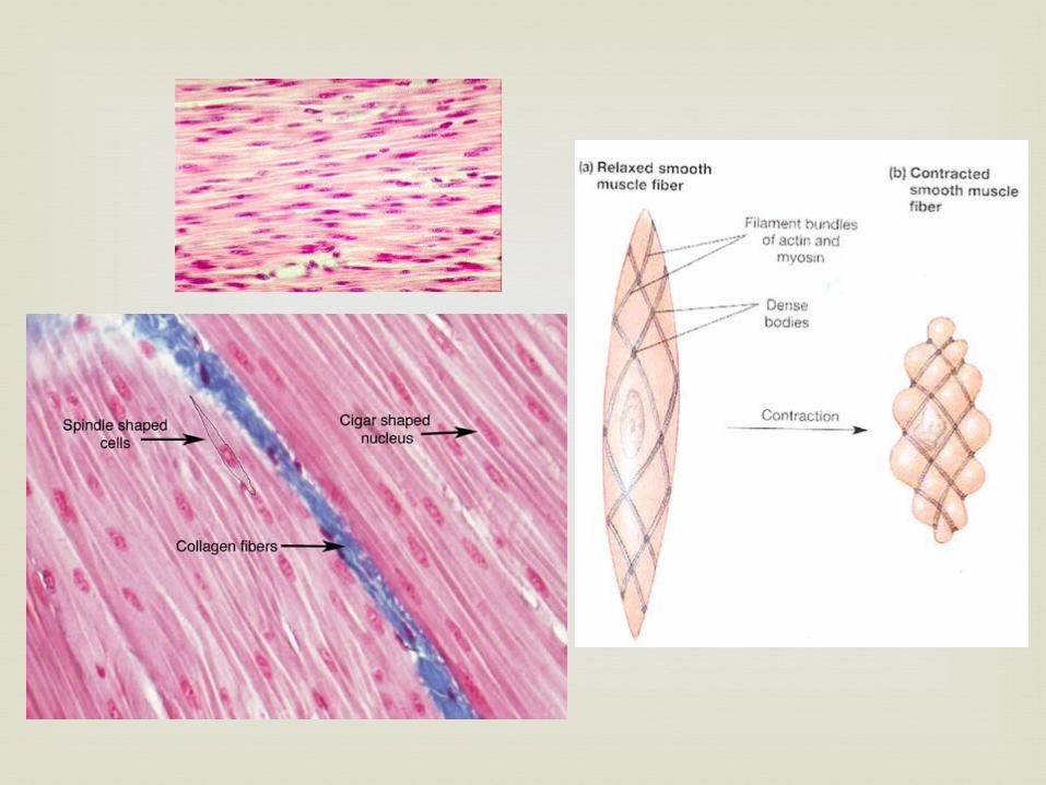
Often called involuntary muscles because you
cannot control them
These muscles form the wall of most of the digestive tract; they are also found in blood vessels and other internal organs
Under a microscope they look smooth
Type 2: Smooth Muscle
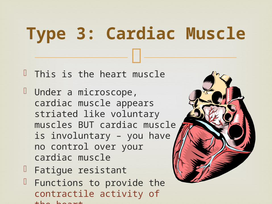
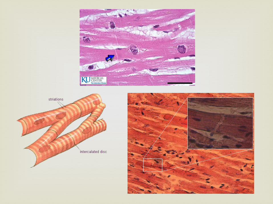
This is the heart muscle
Under a microscope, cardiac muscle appears striated like voluntary muscles BUT cardiac muscle is involuntary – you have no control over your cardiac muscle
Fatigue resistant Functions to provide the contractile
activity of the heart
Type 3: Cardiac Muscle
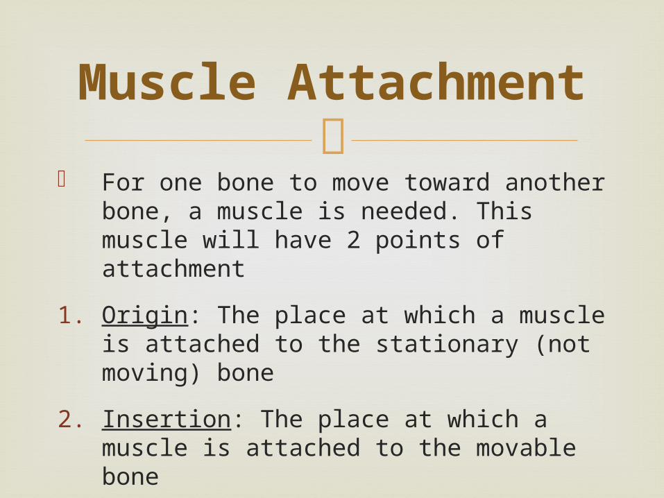
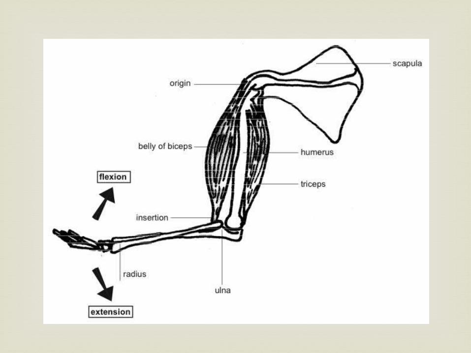
For one bone to move toward another
bone, a muscle is needed. This muscle will have 2 points of attachment
1. Origin: The place at which a muscle is attached to the stationary (not moving) bone
2. Insertion: The place at which a muscle is attached to the movable bone
Muscle Attachment
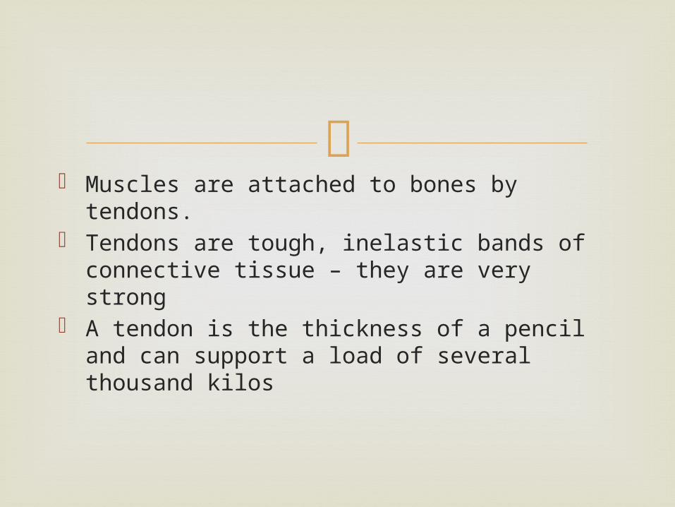
Muscles are attached to bones by tendons. Tendons are tough, inelastic bands of
connective tissue – they are very strong A tendon is the thickness of a pencil and can
support a load of several thousand kilos

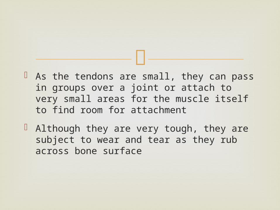
As the tendons are small, they can pass in
groups over a joint or attach to very small areas for the muscle itself to find room for attachment
Although they are very tough, they are subject to wear and tear as they rub across bone surface
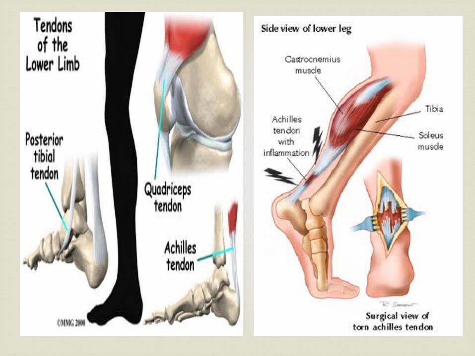
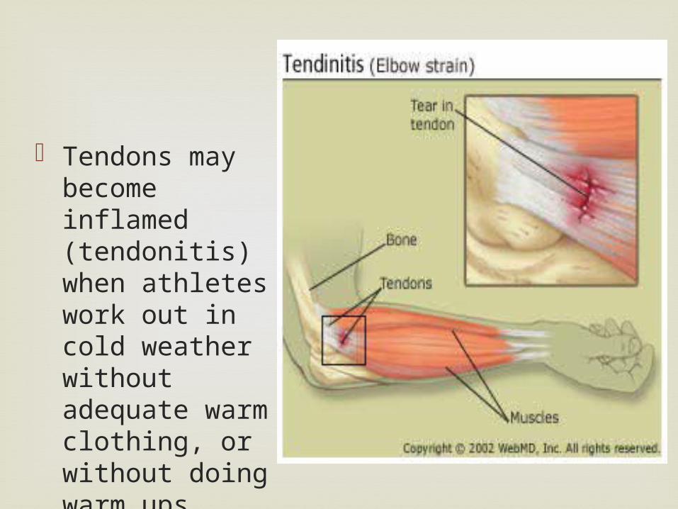
Tendons may become inflamed (tendonitis) when athletes work out in cold weather without adequate warm clothing, or without doing warm ups
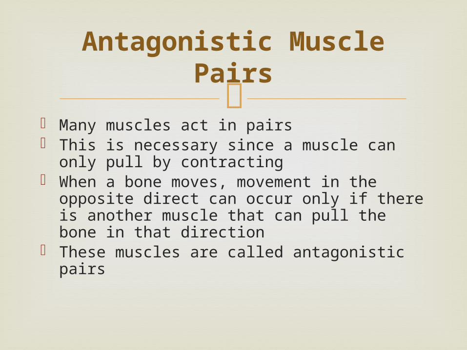
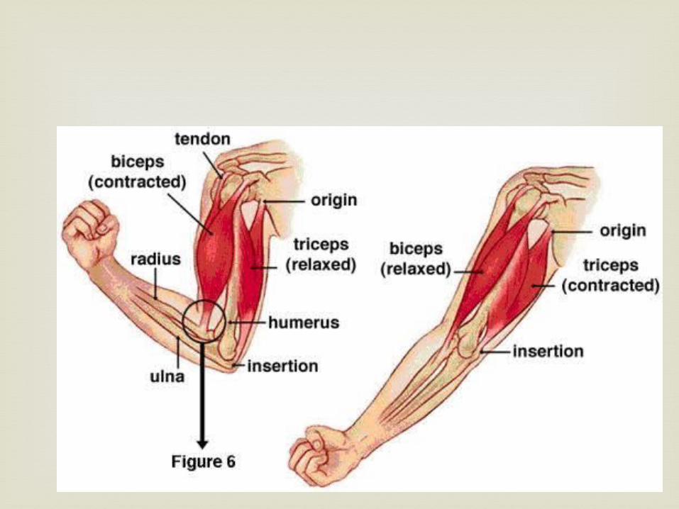
Many muscles act in pairs This is necessary since a muscle can only pull
by contracting When a bone moves, movement in the
opposite direct can occur only if there is another muscle that can pull the bone in that direction
These muscles are called antagonistic pairs
Antagonistic Muscle Pairs
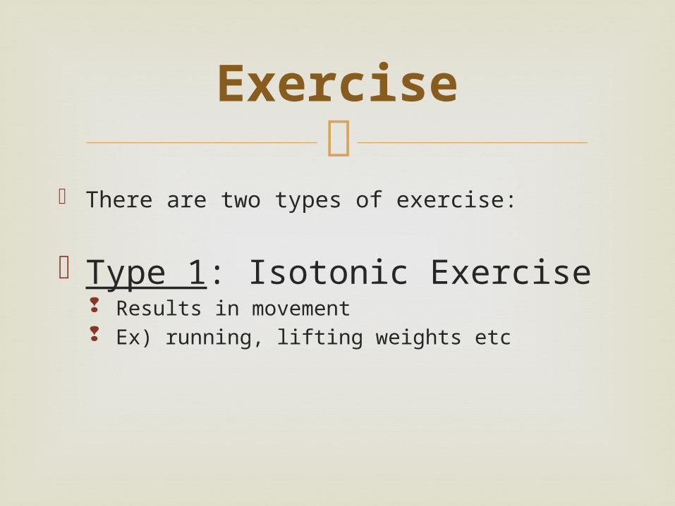
There are two types of exercise:
Type 1: Isotonic Exercise Results in movement Ex) running, lifting weights etc
Exercise
Muscles are pitted against each other This is exercise that does NOT result in
movement Ex) Pushing a wall; hooking fingers together
and trying to pull hands apart Such exercises have been shown to increase
strength and muscle size rapidly
Type 2: Isometric Exercise
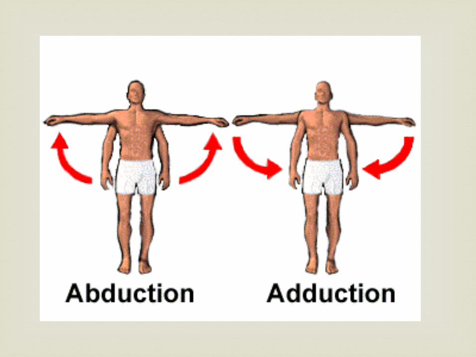
1. Abduction: movement away from the side of
the trunk or midline of the body Ex) raising arms to the side; swinging leg to the side
2. Adduction: movement toward the trunk or midline (opposite of abduction)
Movement in Joints
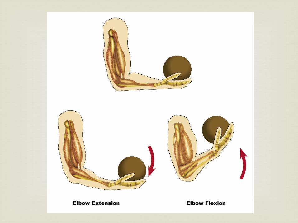
3. Flexion: bending or bringing
bones together Ex) bending elbow or knee
4. Extension: straightening Ex) straightening elbow or knee

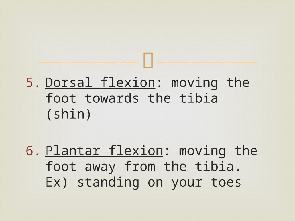
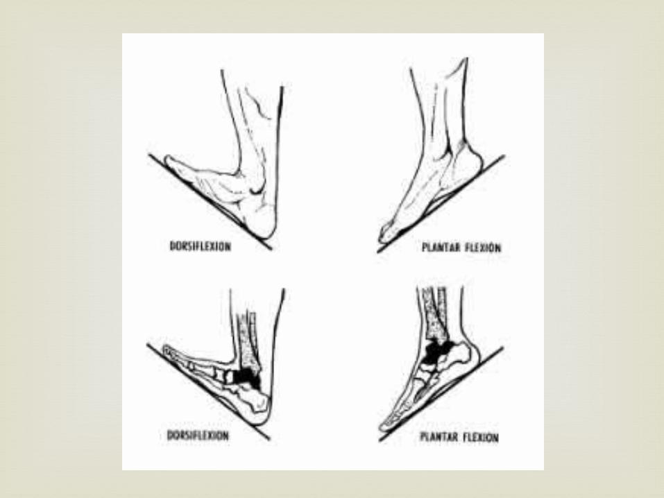
5. Dorsal flexion: moving the foot
towards the tibia (shin)
6. Plantar flexion: moving the foot away from the tibia. Ex) standing on your toes
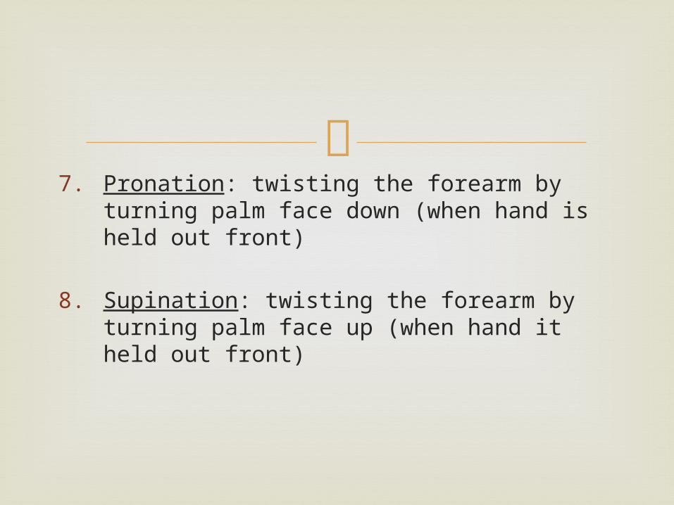
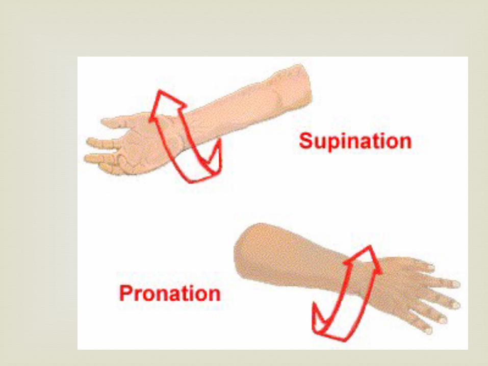
7. Pronation: twisting the forearm by turning
palm face down (when hand is held out front)
8. Supination: twisting the forearm by turning palm face up (when hand it held out front)
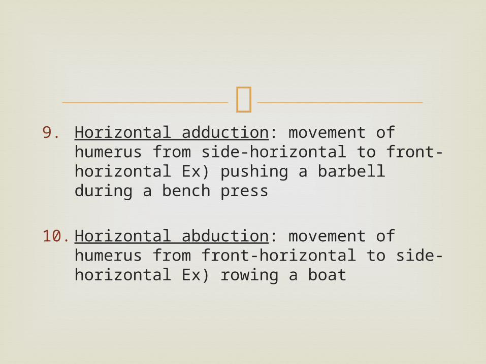
9. Horizontal adduction: movement of humerus
from side-horizontal to front-horizontal Ex) pushing a barbell during a bench press
10. Horizontal abduction: movement of humerus from front-horizontal to side-horizontal Ex) rowing a boat
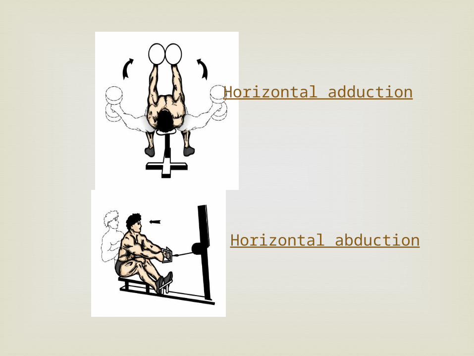
Horizontal adduction
Horizontal abduction
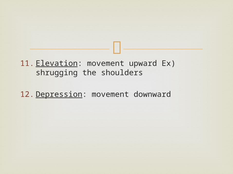
11. Elevation: movement upward Ex) shrugging
the shoulders
12. Depression: movement downward
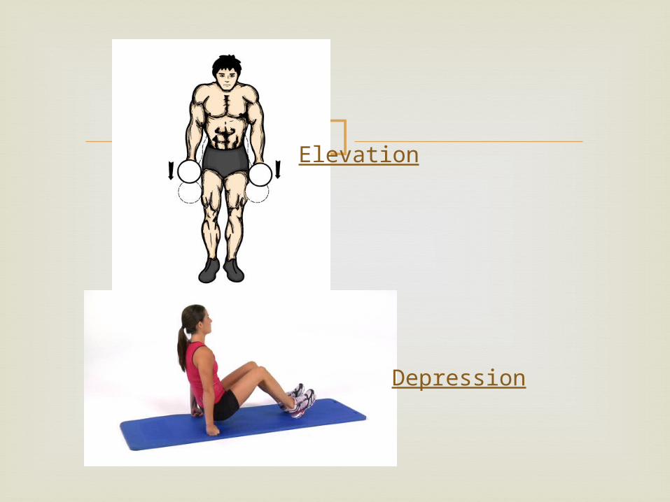
Elevation
Depression
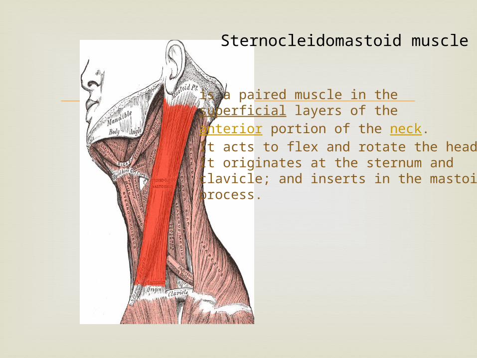
Sternocleidomastoid muscle
is a paired muscle in the superficial layers of the anterior portion of the neck. It acts to flex and rotate the head.It originates at the sternum and clavicle; and inserts in the mastoid process.
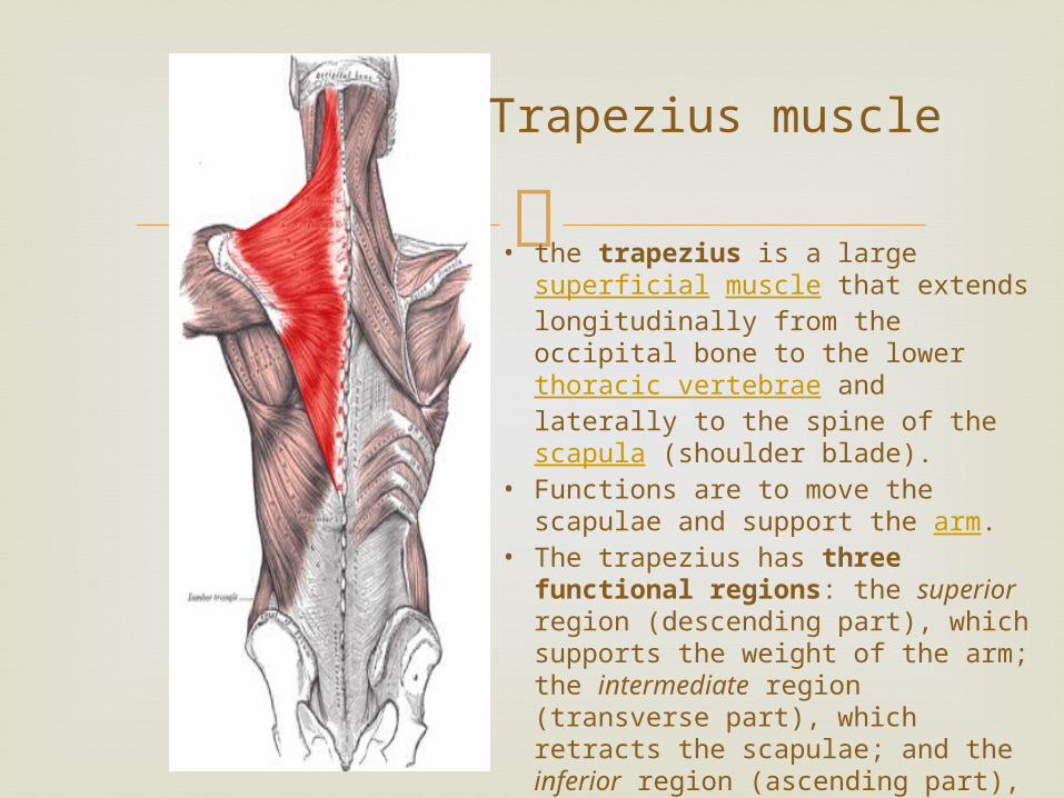
Trapezius muscle
• the trapezius is a large superficial muscle that extends longitudinally from the occipital bone to the lower thoracic vertebrae and laterally to the spine of the scapula (shoulder blade).
• Functions are to move the scapulae and support the arm.
• The trapezius has three functional regions: the superior region (descending part), which supports the weight of the arm; the intermediate region (transverse part), which retracts the scapulae; and the inferior region (ascending part), which medially rotates and depresses the scapulae.
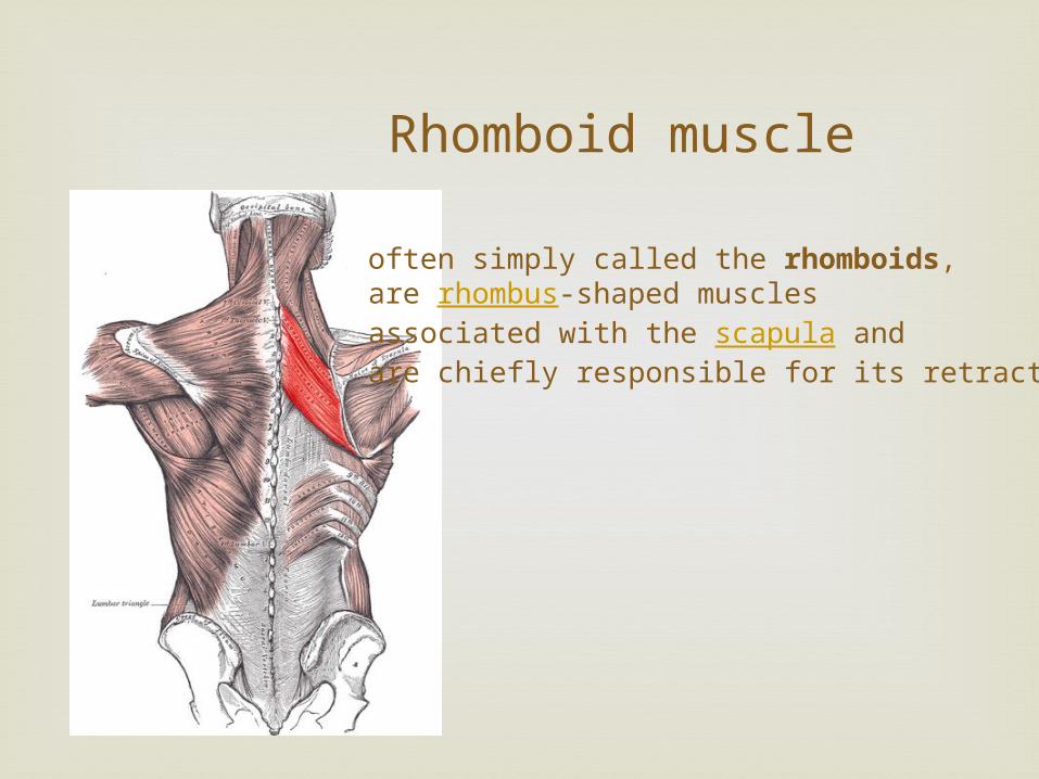
Rhomboid muscle
often simply called the rhomboids, are rhombus-shaped muscles associated with the scapula and are chiefly responsible for its retraction.
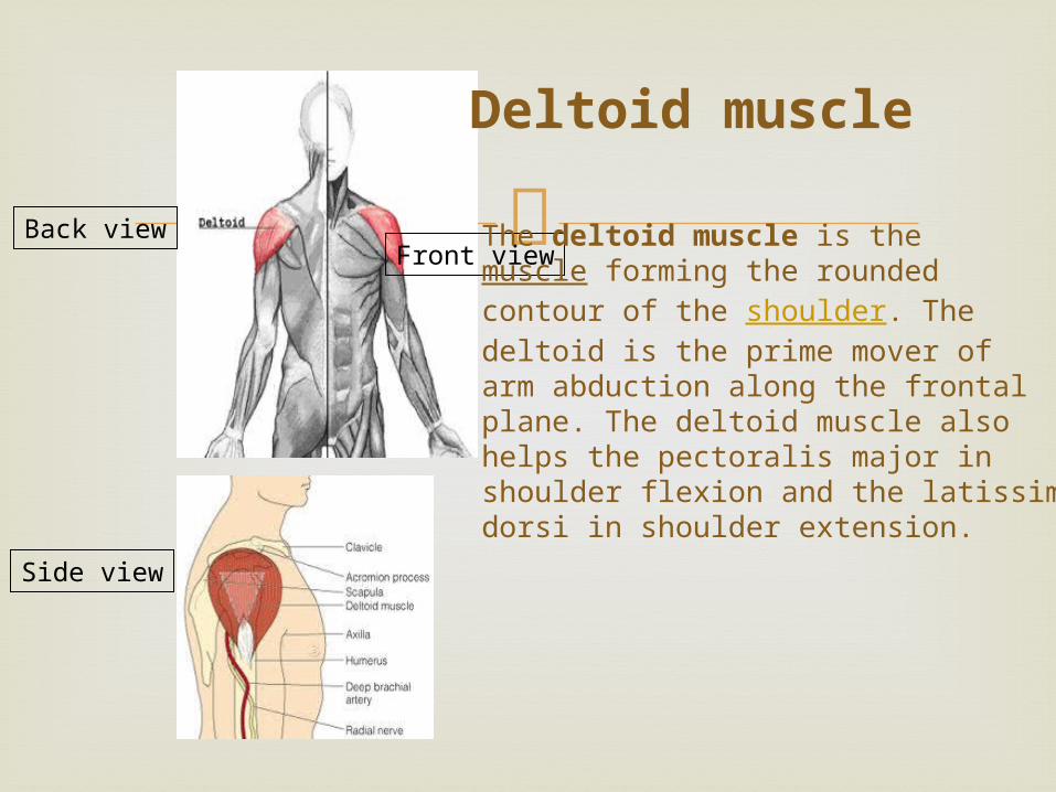
Back view
Side view
Front viewThe deltoid muscle is the muscle forming the rounded contour of the shoulder. The deltoid is the prime mover of arm abduction along the frontal plane. The deltoid muscle also helps the pectoralis major in shoulder flexion and the latissimusdorsi in shoulder extension.
Deltoid muscle
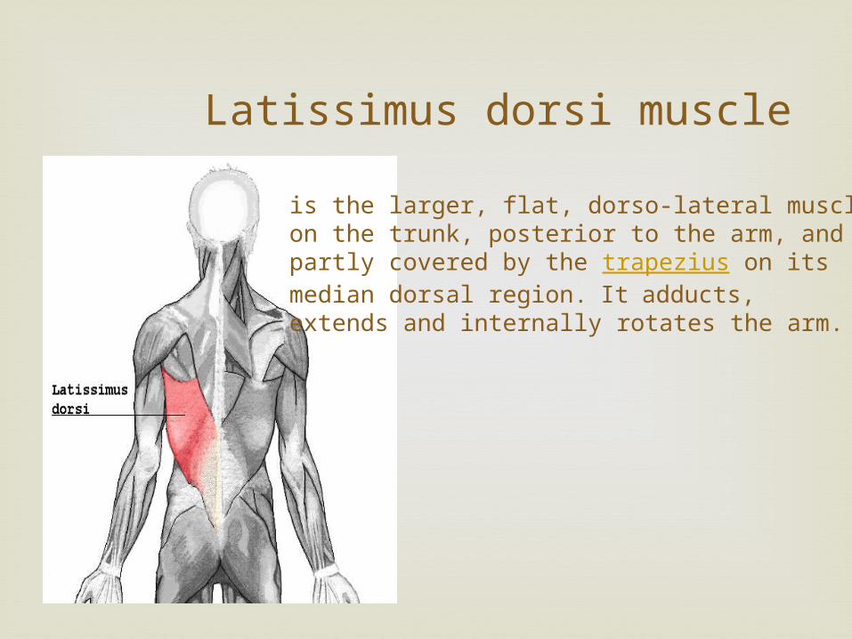
Latissimus dorsi muscle
is the larger, flat, dorso-lateral muscleon the trunk, posterior to the arm, and partly covered by the trapezius on its median dorsal region. It adducts, extends and internally rotates the arm.
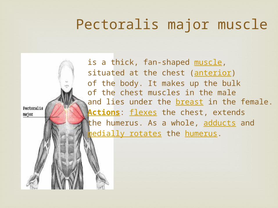
Pectoralis major muscle
is a thick, fan-shaped muscle, situated at the chest (anterior) of the body. It makes up the bulk of the chest muscles in the male and lies under the breast in the female.Actions: flexes the chest, extendsthe humerus. As a whole, adducts and medially rotates the humerus.
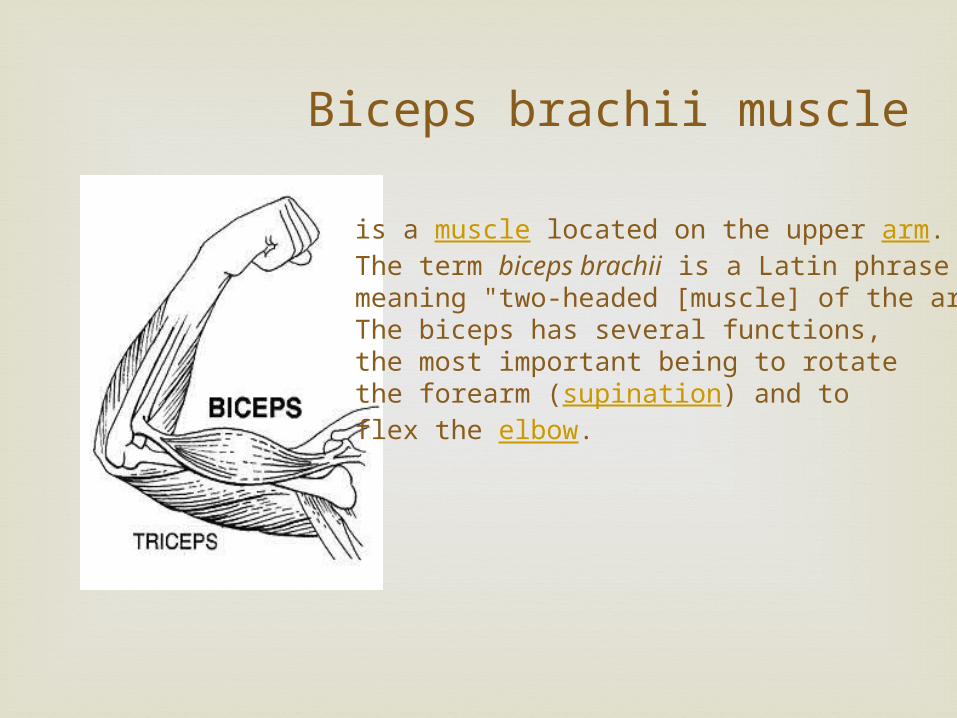
Biceps brachii muscle
is a muscle located on the upper arm. The term biceps brachii is a Latin phrase meaning "two-headed [muscle] of the arm", The biceps has several functions, the most important being to rotate the forearm (supination) and to flex the elbow.
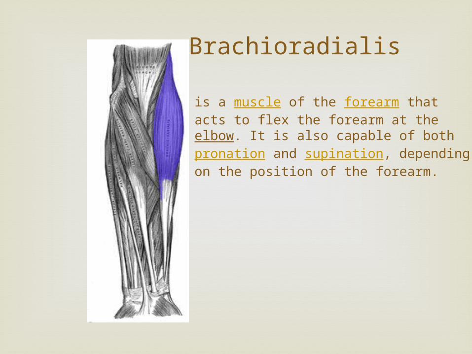
Brachioradialis
is a muscle of the forearm that acts to flex the forearm at the elbow. It is also capable of both pronation and supination, depending on the position of the forearm.
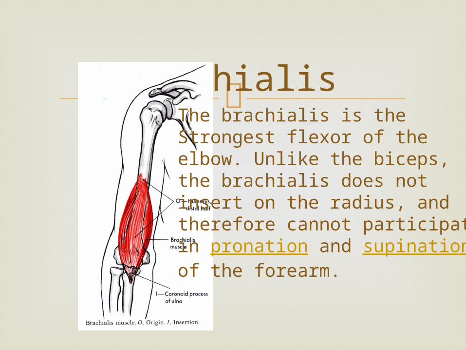
Brachialis
The brachialis is the Strongest flexor of the elbow. Unlike the biceps, the brachialis does not insert on the radius, and therefore cannot participate in pronation and supination of the forearm.
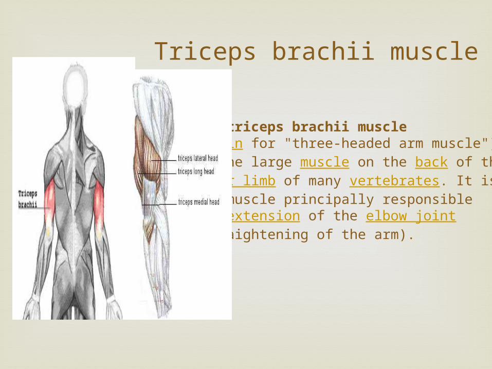
Triceps brachii muscle
The triceps brachii muscle (Latin for "three-headed arm muscle") is the large muscle on the back of the upper limb of many vertebrates. It is the muscle principally responsible for extension of the elbow joint (straightening of the arm).
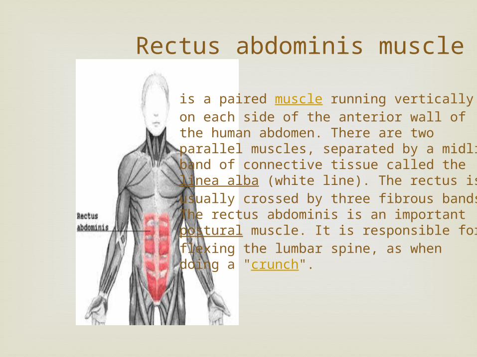
Rectus abdominis muscle
is a paired muscle running vertically on each side of the anterior wall of the human abdomen. There are two parallel muscles, separated by a midline band of connective tissue called the linea alba (white line). The rectus is usually crossed by three fibrous bands.The rectus abdominis is an important postural muscle. It is responsible for flexing the lumbar spine, as when doing a "crunch".
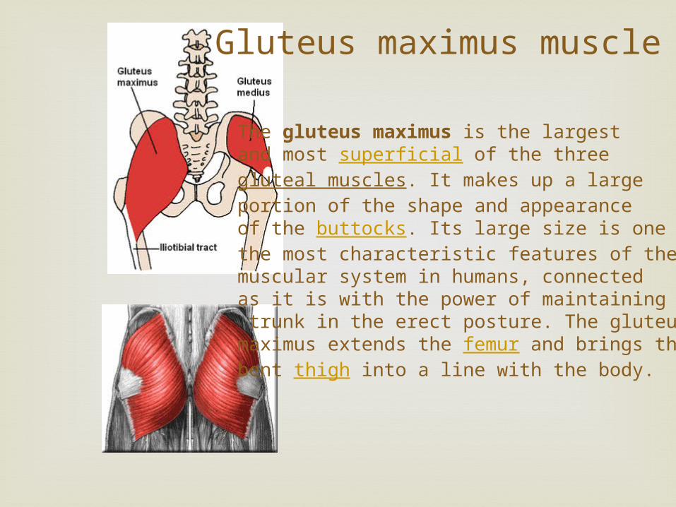
Gluteus maximus muscle
The gluteus maximus is the largest and most superficial of the three gluteal muscles. It makes up a large portion of the shape and appearance of the buttocks. Its large size is one of the most characteristic features of the muscular system in humans, connected as it is with the power of maintaining the trunk in the erect posture. The gluteus maximus extends the femur and brings the bent thigh into a line with the body.
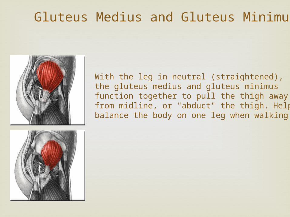
With the leg in neutral (straightened), the gluteus medius and gluteus minimus function together to pull the thigh away from midline, or "abduct" the thigh. Helps balance the body on one leg when walking.
Gluteus Medius and Gluteus Minimus
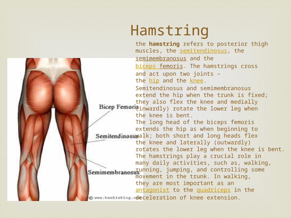
Hamstring the hamstring refers to posterior thigh muscles, the semitendinosus, the semimembranosus and the biceps femoris. The hamstrings cross and act upon two joints – the hip and the knee.Semitendinosus and semimembranosus extend the hip when the trunk is fixed; they also flex the knee and medially (inwardly) rotate the lower leg when the knee is bent.The long head of the biceps femoris extends the hip as when beginning to walk; both short and long heads flex the knee and laterally (outwardly) rotates the lower leg when the knee is bent.The hamstrings play a crucial role in many daily activities, such as, walking, running, jumping, and controlling some movement in the trunk. In walking, they are most important as an antagonist to the quadriceps in the deceleration of knee extension.
Quadriceps
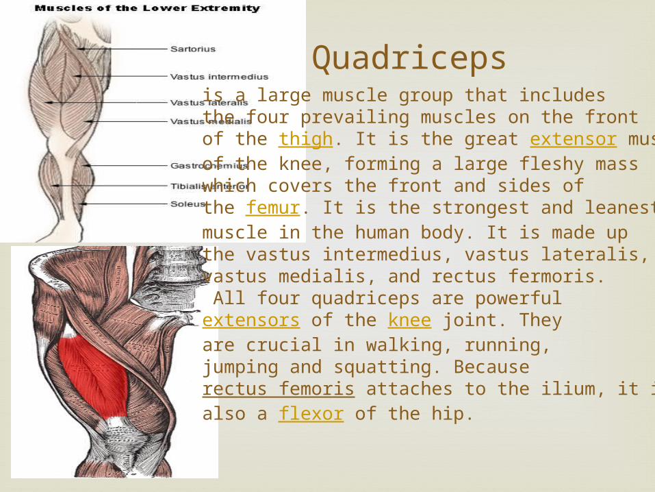
is a large muscle group that includes the four prevailing muscles on the front of the thigh. It is the great extensor muscle of the knee, forming a large fleshy mass which covers the front and sides of the femur. It is the strongest and leanest muscle in the human body. It is made upthe vastus intermedius, vastus lateralis,vastus medialis, and rectus fermoris. All four quadriceps are powerful extensors of the knee joint. They are crucial in walking, running, jumping and squatting. Because rectus femoris attaches to the ilium, it is also a flexor of the hip.
Sartorius muscle
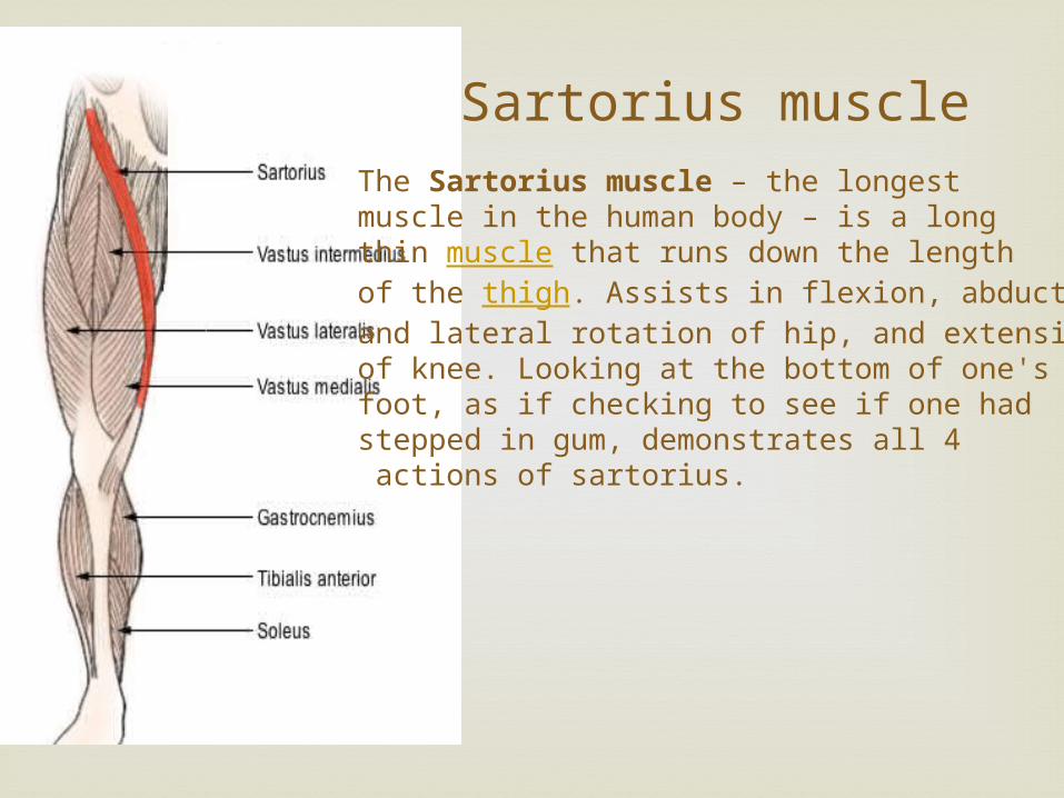
The Sartorius muscle – the longest muscle in the human body – is a long thin muscle that runs down the length of the thigh. Assists in flexion, abduction and lateral rotation of hip, and extension of knee. Looking at the bottom of one's foot, as if checking to see if one had stepped in gum, demonstrates all 4 actions of sartorius.
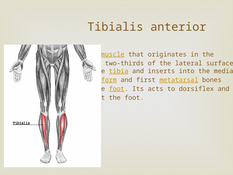
Tibialis anterior
is a muscle that originates in the upper two-thirds of the lateral surface of the tibia and inserts into the medial cuneiform and first metatarsal bones of the foot. Its acts to dorsiflex and invert the foot.

Gastrocnemius
is a very powerful superficial pennate muscle that is in the back part of the lower leg. It runs from its two heads just above the knee to the heel, and is involved in standing, walking, running and jumping. Along with the soleus muscle it forms the calf muscle. Its function is plantar flexing the foot atthe ankle joint and flexing the leg at the knee joint.
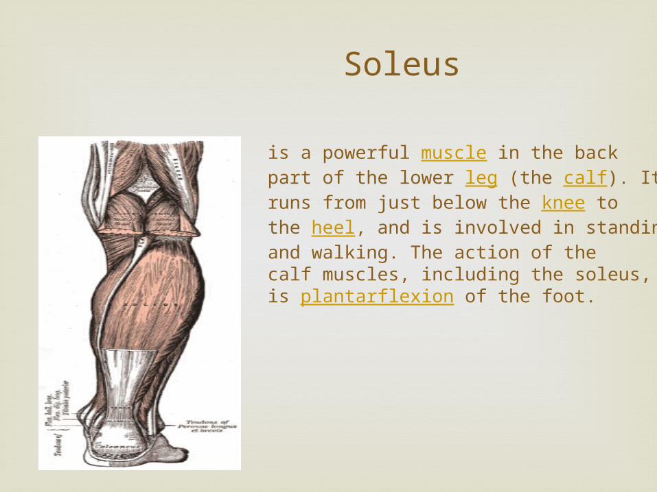
Soleus
is a powerful muscle in the back part of the lower leg (the calf). It runs from just below the knee to the heel, and is involved in standing and walking. The action of the calf muscles, including the soleus, is plantarflexion of the foot.
Muscular Disorders
Muscular Dystrophy
Muscular dystrophy (MD) is a group of rare inherited muscle diseases in which muscle fibers are unusually susceptible to damage.
Muscles, primarily voluntary muscles, become progressively weaker. In the late stages of muscular dystrophy, fat and connective tissue often replace muscle fibers. In some types of muscular dystrophy, heart muscles, other involuntary muscles and other organs are affected.
The most common types of muscular dystrophy appear to be due to a genetic deficiency of the muscle protein dystrophin.
There's no cure for muscular dystrophy, but medications and therapy can slow the course of the disease.
M.D. Types
There are nine major types of MD affecting people of all ages, from infancy to middle age or later. The two most common types of MD affect children:
Duchenne muscular dystrophy (DMD) - most common in children. Usually first seen in boys 2-5 years of age. Most die in their late teens
Becker muscular dystrophy (BMD) - Generally affects older boys and young men, and progresses more slowly. Usually can walk well into adultood
Myotonic dystrophyproduces stiffness of muscles and an inability to relax muscles at will (myotonia), as well as the muscle weakness of the other forms of muscular dystrophy.
Although this form of MD can affect children, it often doesn't affect people until adulthood. It can vary greatly in its severity. Muscles may feel stiff after using them. Progression of this form of MD is slow.
1 in 3000 Boys. Females are rarely affected, but often carriers
Signs and symptoms
They vary according to the type of muscular dystrophy. In general, they may include:
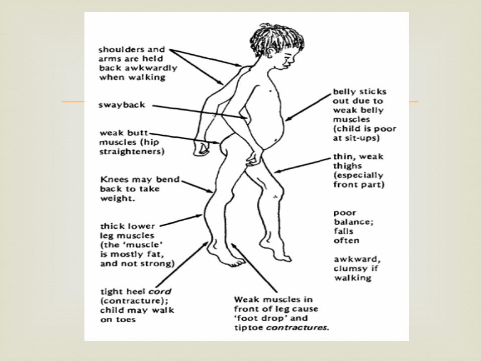
Muscle weakness Apparent lack of coordination Progressive crippling, resulting in contractures of
the muscles around your joints and loss of mobility
Many specific signs and symptoms vary from among the different forms of MD. Each type is different in the age of onset, what parts of the body the symptoms primarily affect and how rapidly the disease progresses.
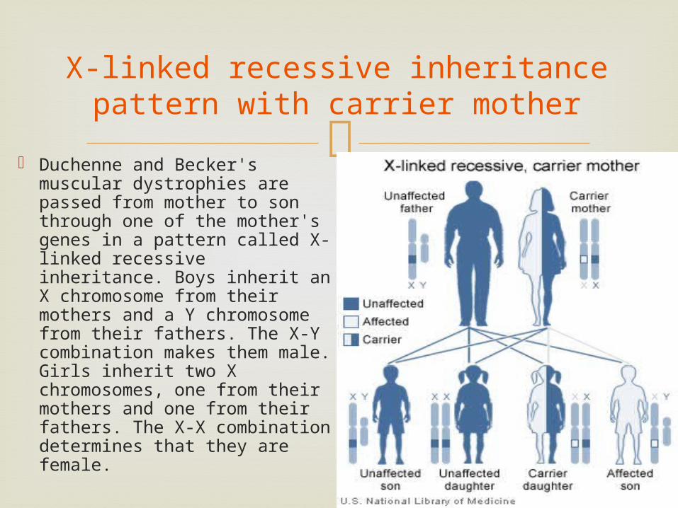
X-linked recessive inheritance pattern with carrier mother
Duchenne and Becker's muscular dystrophies are passed from mother to son through one of the mother's genes in a pattern called X-linked recessive inheritance. Boys inherit an X chromosome from their mothers and a Y chromosome from their fathers. The X-Y combination makes them male. Girls inherit two X chromosomes, one from their mothers and one from their fathers. The X-X combination determines that they are female.
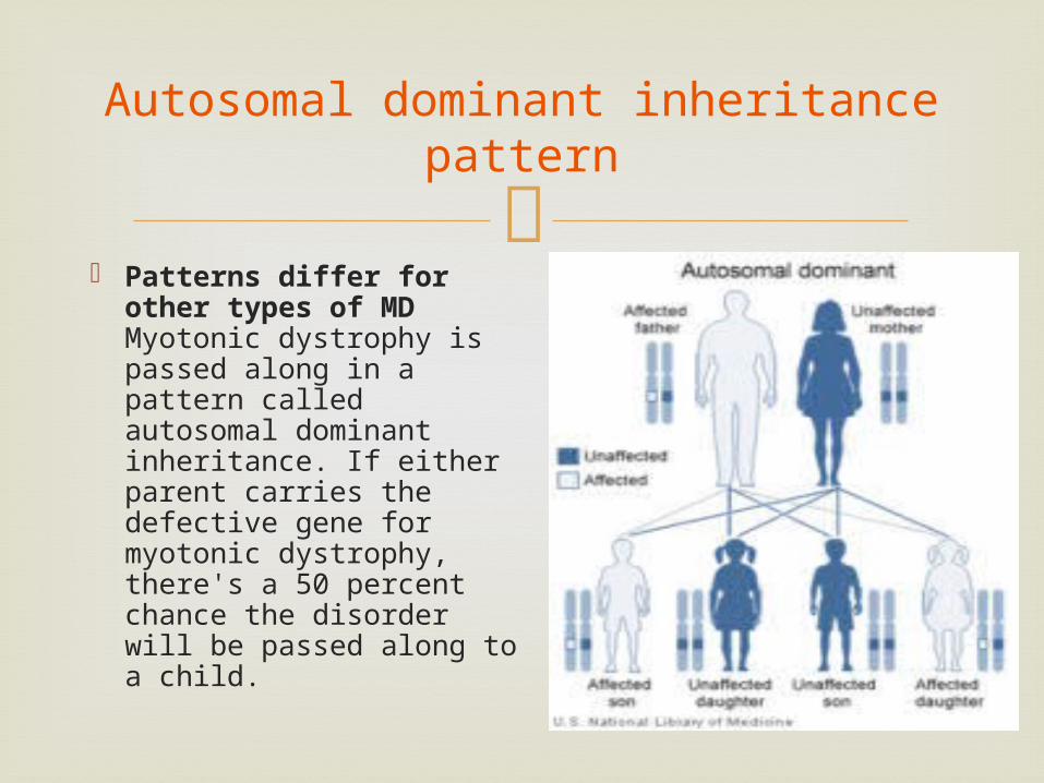
Autosomal dominant inheritance pattern
Patterns differ for other types of MDMyotonic dystrophy is passed along in a pattern called autosomal dominant inheritance. If either parent carries the defective gene for myotonic dystrophy, there's a 50 percent chance the disorder will be passed along to a child.
Treatment
There's currently no cure for any form of muscular dystrophy. Research into gene therapy may eventually provide treatment to stop the progression of some types of muscular dystrophy.
Current treatment is designed to help prevent or reduce deformities in the joints and the spine and to allow people with MD to remain mobile as long as possible.
Treatments may include various types of physical therapy, medications, assistive devices (braces) and surgery.
Fibrodysplasia
ossificans progressiva

Stone Man Syndrome Extremely rare disease of connective tissue A mutation of the body’s repair mechanism causes
fibrous tissue (muscle, tendon, ligament) to be ossified when damaged.
In some cases, injuries can cause joints to become permanently frozen in place.
The gene that causes ossification is normally deactivated after a fetus' bones are formed in the womb, but in patients with FOP, the gene keeps working.
Fibrodysplasia ossificans
progressiva
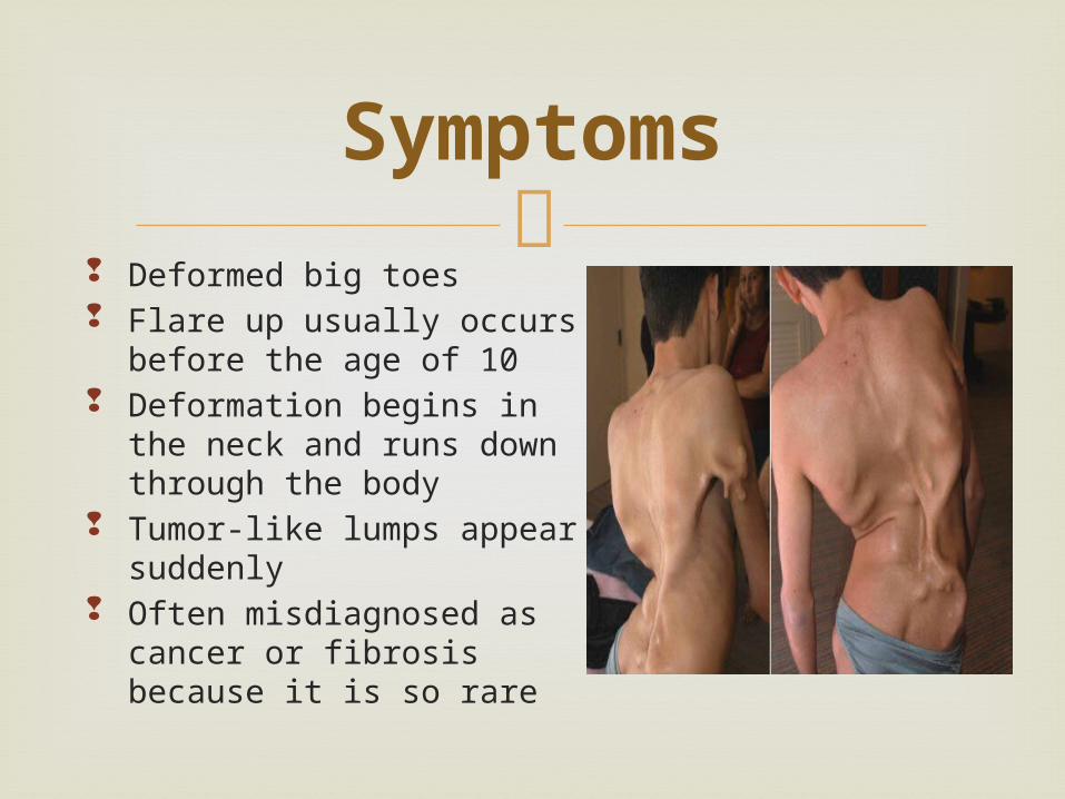
Deformed big toes Flare up usually occurs
before the age of 10 Deformation begins in the
neck and runs down through the body
Tumor-like lumps appear suddenly
Often misdiagnosed as cancer or fibrosis because it is so rare
Symptoms
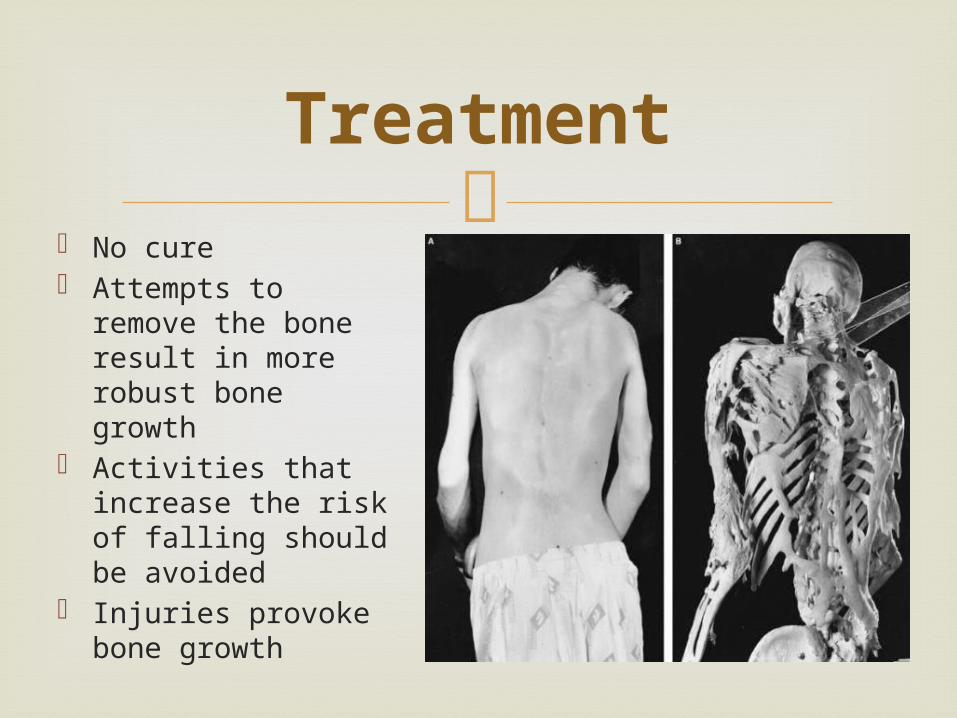
No cure Attempts to
remove the bone result in more robust bone growth
Activities that increase the risk of falling should be avoided
Injuries provoke bone growth
Treatment
Recommended