Radioembolización Y90
Downstaging y tratamiemto en lista del HCC
Fernando Pardo
Clínica Universidad de Navarra

Embolizing Particles for Intraarterial Therapy
Sangro B, et al. J Nucl Med Radiat Ther 2011
Resin Glass
Mean Diameter 35 µm 25 µm
Activity/sphere 50 Bq 2,500 Bq
300
100
35

Sangro B et al. J Hepatol 2012

Tratamiento transarterial del HCC
RE
TACE
RERE RE
Sangro B et al. J Hepatol 2012
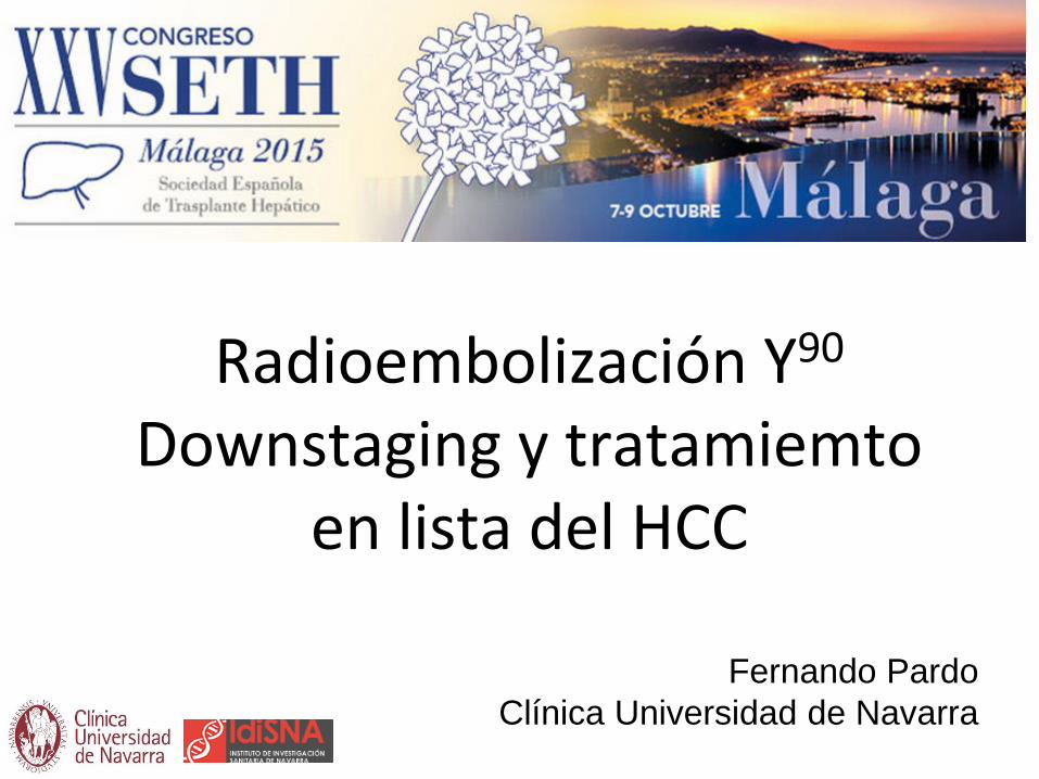
Tto transarterial del HCC
Características
TACE• Isquemia ± fármaco
• Partículas medias-grandes
• Selectiva o superselectiva
• Varias sesiones
• Grandes diferencias entre Centros en dispositivos y procedimientos
RE• Irradiación
• Partículas muy pequeñas
• Global a superselectiva
• Sesión única
• Procedimientos altamente reproducibles
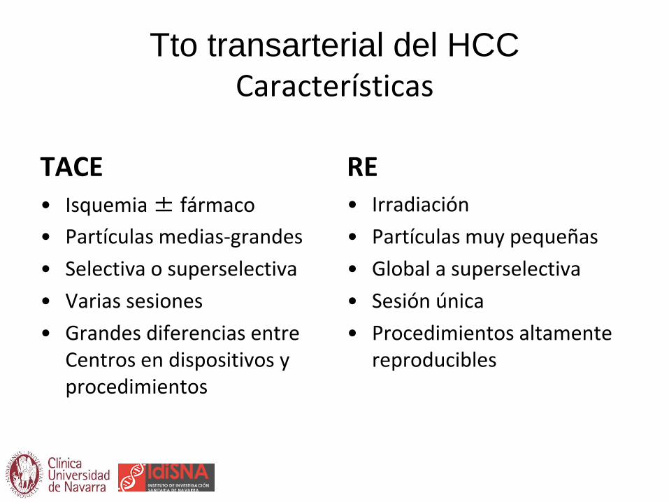
Tratamiento intra-arterial del HCC
Niveles de evidencia
TACE• Ensayos randomizados
controlados (2 positivos)
• Metaanalysis (3 positivos)
• Nivel 1
RE• Grandes series prospectivas
de cohortes
• Nivel 2
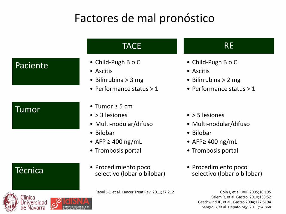
Factores de mal pronóstico
• > 5 lesiones
• Multi-nodular/difuso
• Bilobar
• AFP≥ 400 ng/mL
• Trombosis portal
Tumor
• Child-Pugh B o C
• Ascitis
• Bilirrubina > 2 mg
• Performance status > 1
Paciente
• Procedimiento poco selectivo (lobar o bilobar)Técnica
Raoul J-L, et al. Cancer Treat Rev. 2011;37:212
• Tumor ≥ 5 cm
• > 3 lesiones
• Multi-nodular/difuso
• Bilobar
• AFP ≥ 400 ng/mL
• Trombosis portal
• Child-Pugh B o C
• Ascitis
• Bilirrubina > 3 mg
• Performance status > 1
• Procedimiento poco selectivo (lobar o bilobar)
RETACE
Goin J, et al. JVIR 2005;16:195Salem R, et al. Gastro. 2010;138:52
Geschwind JF, et al. Gastro 2004;127:S194Sangro B, et al. Hepatology. 2011;54:868

Candidatos a tratamiento intra-arterial
• Estadio precoz a intermedio
– Lista de espera para trasplante
– Irresecable por localización (downstaging)
– Tumor único no accesible a RFA
– Tumores pequeños tratables superselectivamente
• Estadio intermedio o avanzado
– Tumor grande o múltiple no tratable superselectivamente
– Invasión portal sin enfermedad extrahepática
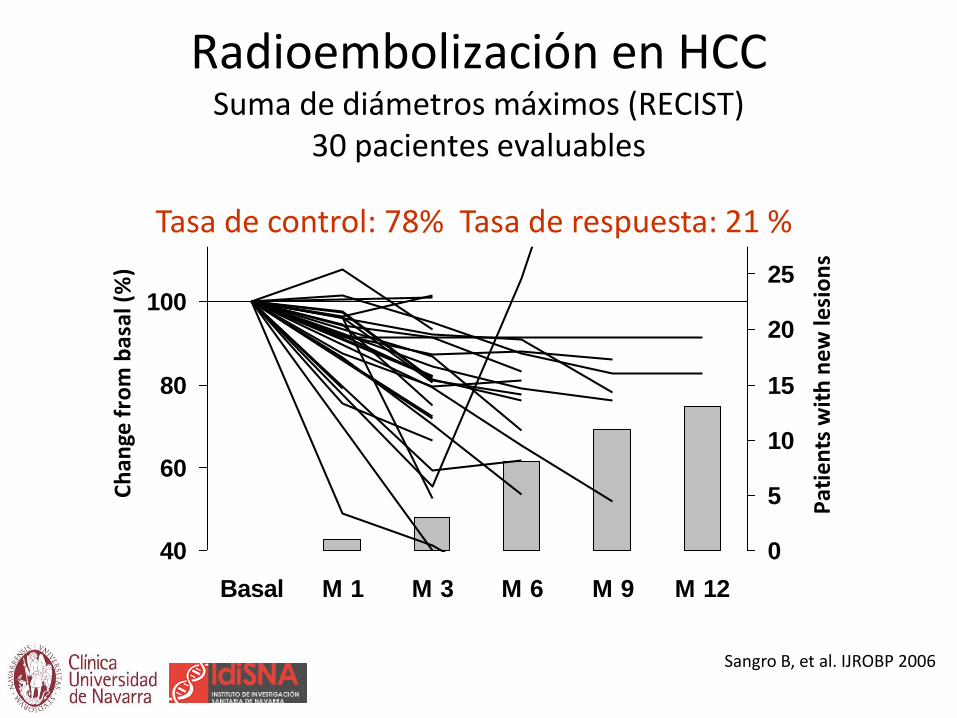
Radioembolización en HCC Suma de diámetros máximos (RECIST)
30 pacientes evaluables
40
60
80
100
120
Basal M 1 M 3 M 6 M 9 M 12
Ch
ange
fro
m b
asal
(%
)
0
5
10
15
20
25
30
Pat
ien
ts w
ith
new
lesi
on
s
Sangro B, et al. IJROBP 2006
Tasa de control: 78% Tasa de respuesta: 21 %
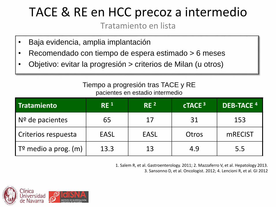
TACE & RE en HCC precoz a intermedioTratamiento en lista
• Baja evidencia, amplia implantación
• Recomendado con tiempo de espera estimado > 6 meses
• Objetivo: evitar la progresión > criterios de Milan (u otros)
Tratamiento RE 1 RE 2 cTACE 3 DEB-TACE 4
Nº de pacientes 65 17 31 153
Criterios respuesta EASL EASL Otros mRECIST
Tº medio a prog. (m) 13.3 13 4.9 5.5
1. Salem R, et al. Gastroenterology. 2011; 2. Mazzaferro V, et al. Hepatology 2013. 3. Sansonno D, et al. Oncologist. 2012; 4. Lencioni R, et al. GI 2012
Tiempo a progresión tras TACE y REpacientes en estadio intermedio
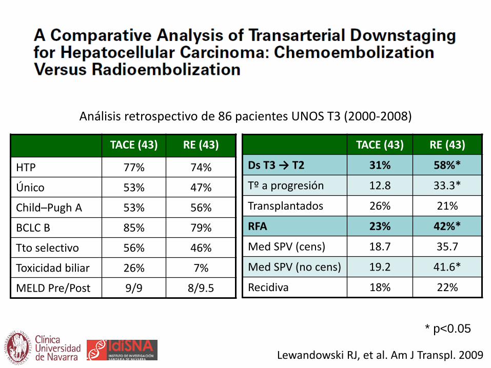
Lewandowski RJ, et al. Am J Transpl. 2009
Análisis retrospectivo de 86 pacientes UNOS T3 (2000-2008)
TACE (43) RE (43)
HTP 77% 74%
Único 53% 47%
Child–Pugh A 53% 56%
BCLC B 85% 79%
Tto selectivo 56% 46%
Toxicidad biliar 26% 7%
MELD Pre/Post 9/9 8/9.5
TACE (43) RE (43)
Ds T3 → T2 31% 58%*
Tº a progresión 12.8 33.3*
Transplantados 26% 21%
RFA 23% 42%*
Med SPV (cens) 18.7 35.7
Med SPV (no cens) 19.2 41.6*
Recidiva 18% 22%
* p<0.05
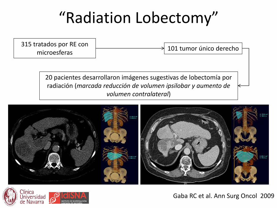
“Radiation Lobectomy”
315 tratados por RE con microesferas
101 tumor único derecho
20 pacientes desarrollaron imágenes sugestivas de lobectomía por radiación (marcada reducción de volumen ipsilobar y aumento de
volumen contralateral)
Gaba RC et al. Ann Surg Oncol 2009
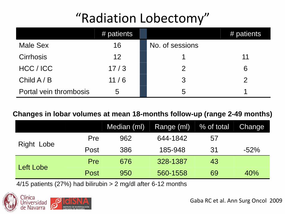
“Radiation Lobectomy”# patients # patients
Male Sex 16 No. of sessions
Cirrhosis 12 1 11
HCC / ICC 17 / 3 2 6
Child A / B 11 / 6 3 2
Portal vein thrombosis 5 5 1
Median (ml) Range (ml) % of total Change
Right LobePre 962 644-1842 57
Post 386 185-948 31 -52%
Left LobePre 676 328-1387 43
Post 950 560-1558 69 40%
Gaba RC et al. Ann Surg Oncol 2009
Changes in lobar volumes at mean 18-months follow-up (range 2-49 months)
4/15 patients (27%) had bilirubin > 2 mg/dl after 6-12 months
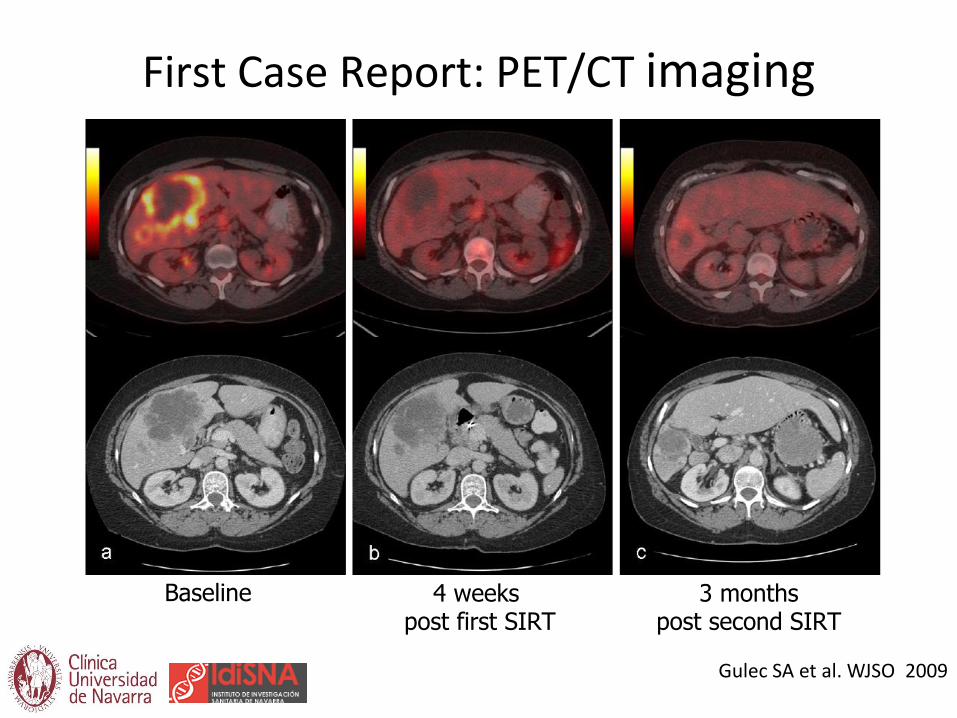
First Case Report: PET/CT imaging
Baseline 4 weeks post first SIRT
3 monthspost second SIRT
Gulec SA et al. WJSO 2009
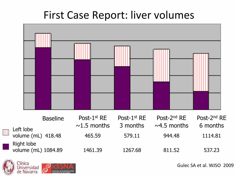
First Case Report: liver volumes
2500
2000
1000
500
0
Liv
er
Volu
me (
mL)
1500
Left lobevolume (mL)
Right lobevolume (mL)
418.48 465.59 579.11 944.48 1114.81
1084.89 1461.39 1267.68 811.52 537.23
Baseline Post-1st RE
~1.5 months
Post-1st RE
3 months
Post-2nd RE
~4.5 months
Post-2nd RE
6 months
Gulec SA et al. WJSO 2009
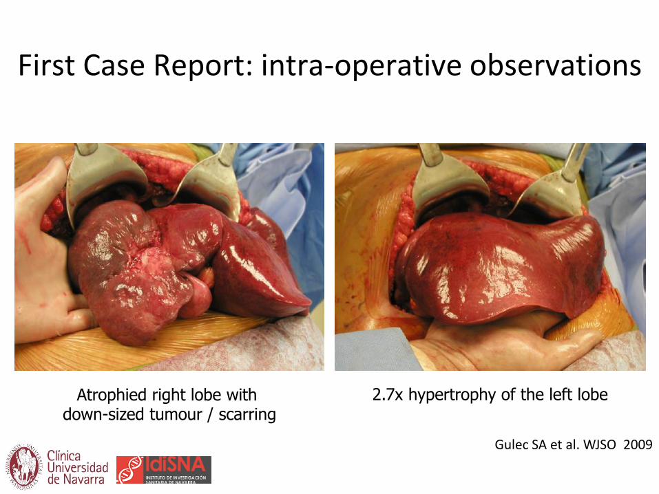
First Case Report: intra-operative observations
Atrophied right lobe with down-sized tumour / scarring
2.7x hypertrophy of the left lobe
Gulec SA et al. WJSO 2009
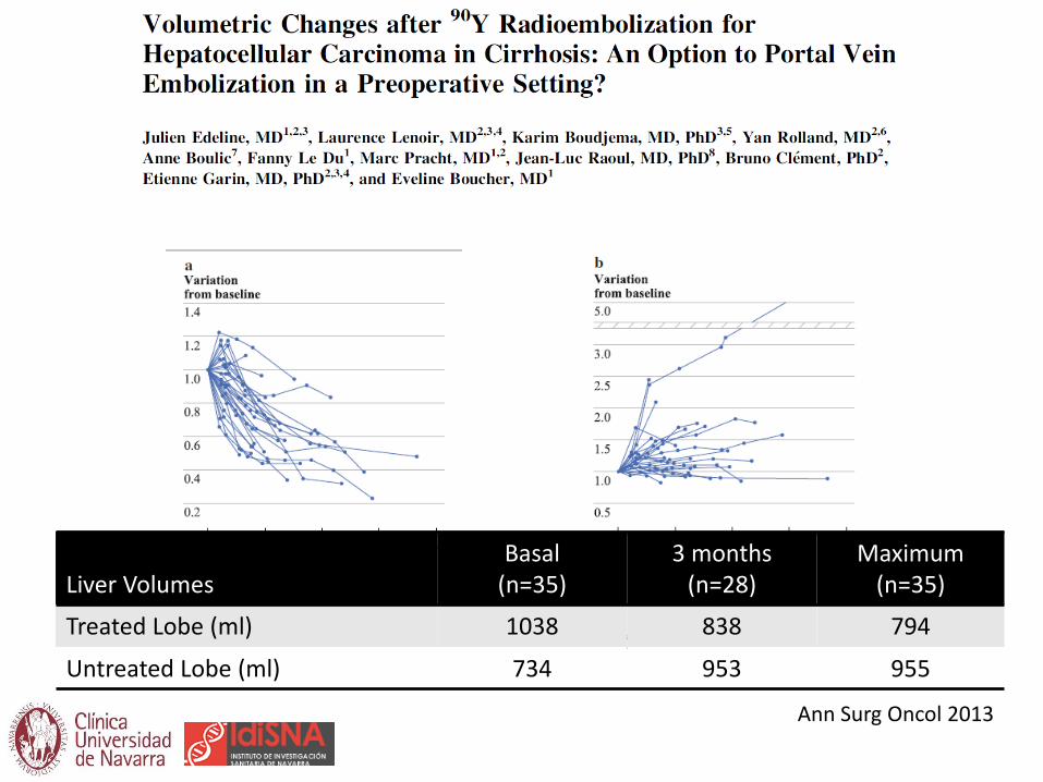
Ann Surg Oncol 2013
Lóbulo tratado Lóbulo contralateral
Liver VolumesBasal
(n=35)3 months
(n=28)Maximum
(n=35)
Treated Lobe (ml) 1038 838 794
Untreated Lobe (ml) 734 953 955
HPB (Oxford) 2013
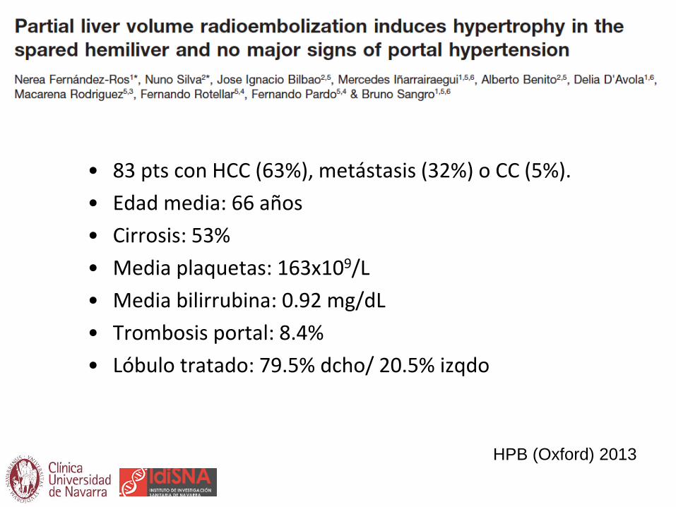
• 83 pts con HCC (63%), metástasis (32%) o CC (5%).
• Edad media: 66 años
• Cirrosis: 53%
• Media plaquetas: 163x109/L
• Media bilirrubina: 0.92 mg/dL
• Trombosis portal: 8.4%
• Lóbulo tratado: 79.5% dcho/ 20.5% izqdo
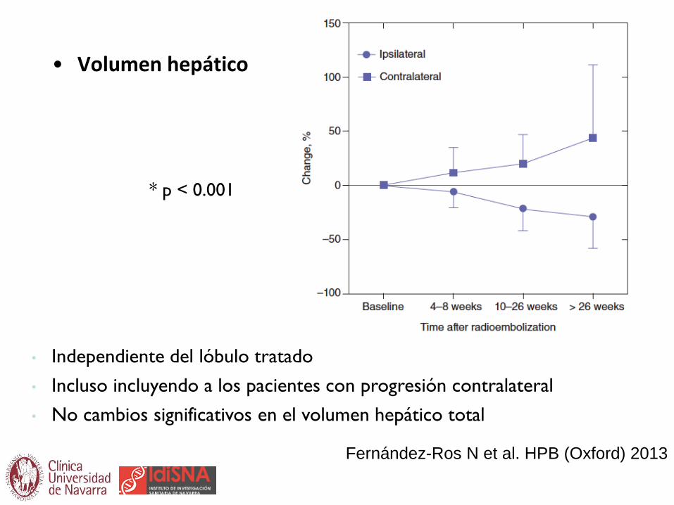
• Volumen hepático
• Independiente del lóbulo tratado
• Incluso incluyendo a los pacientes con progresión contralateral
• No cambios significativos en el volumen hepático total
* p < 0.001
Fernández-Ros N et al. HPB (Oxford) 2013
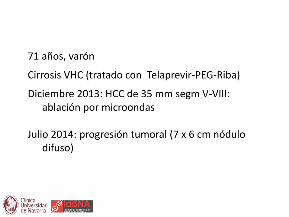
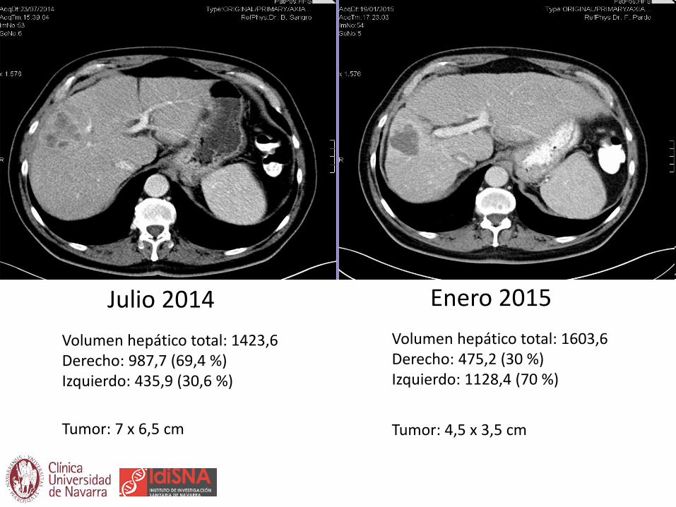
71 años, varón
Cirrosis VHC (tratado con Telaprevir-PEG-Riba)
Diciembre 2013: HCC de 35 mm segm V-VIII: ablación por microondas
Julio 2014: progresión tumoral (7 x 6 cm nódulo difuso)

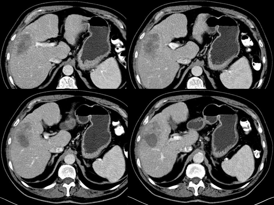
Lesión 7 x 4,7 x 6,5 cm.Áreas sólido quísticas en relación con tratamiento previo.Periferia de la lesión áreas nodulares con captación de contraste y lavado en fase portal.HCC tratado, con áreas activas
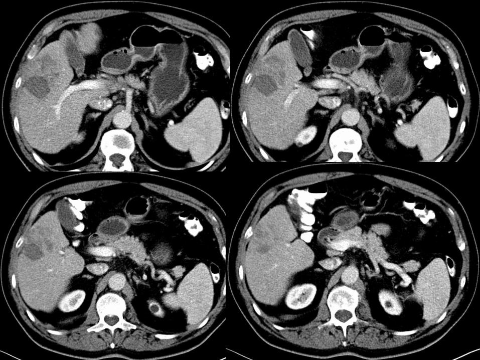

90Y-microspheres PET/CT distribution
single dose trans-arterial 90Y microspheres administered activity 1,8GBq.
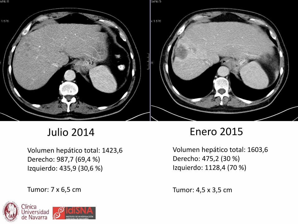
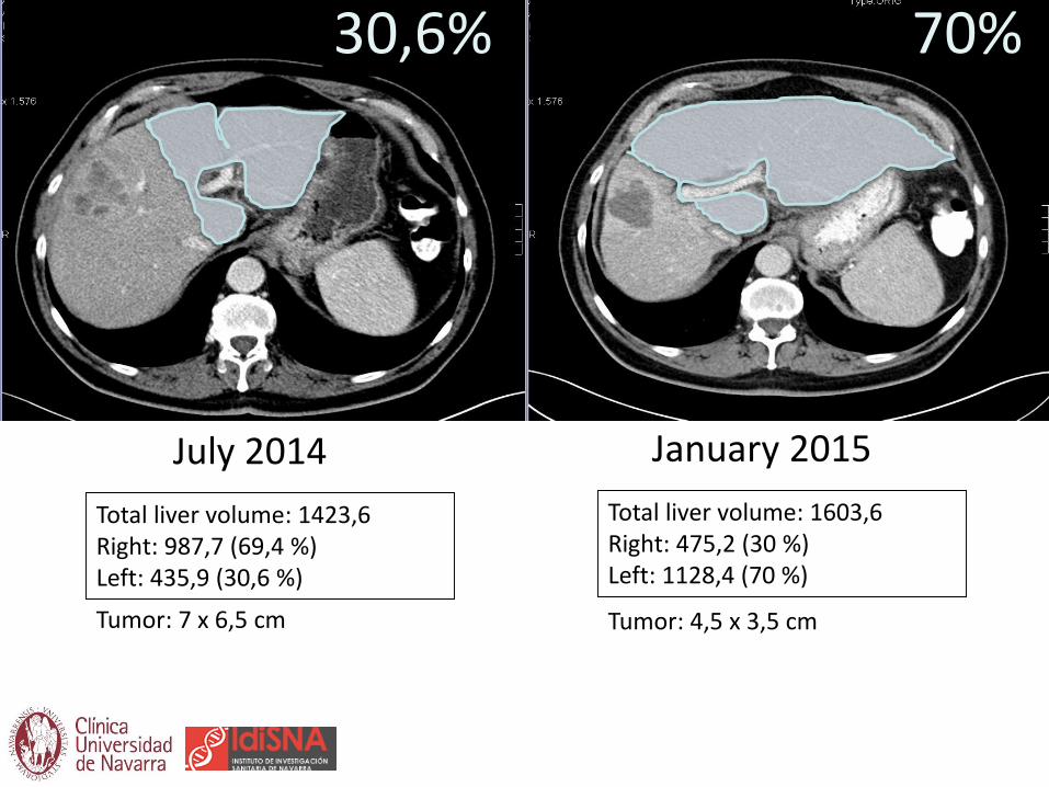
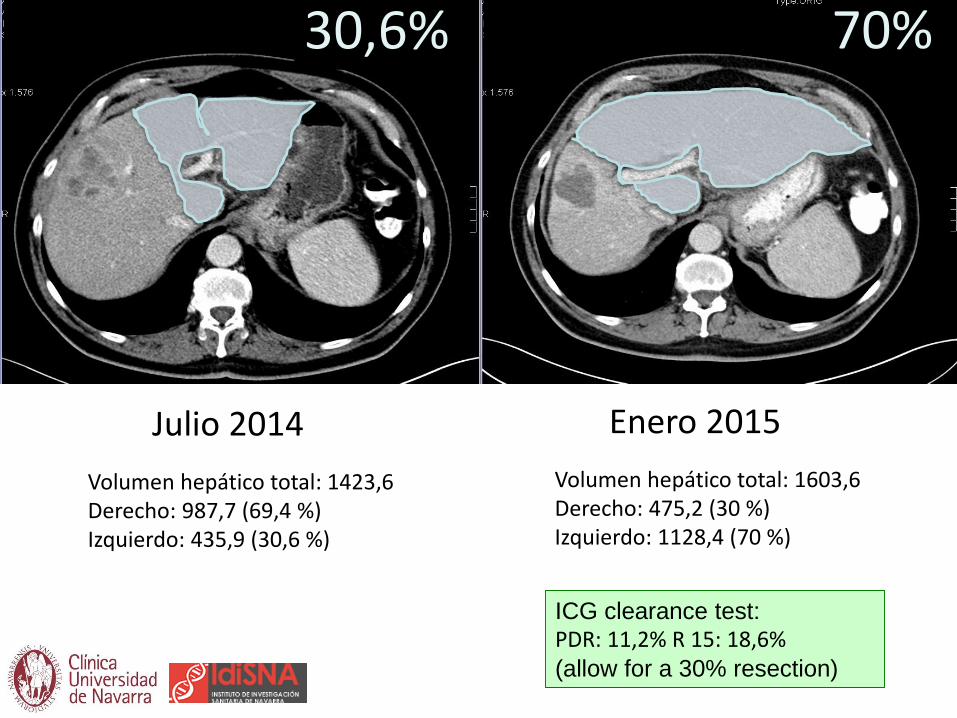
Julio 2014
Volumen hepático total: 1423,6Derecho: 987,7 (69,4 %)Izquierdo: 435,9 (30,6 %)
Enero 2015
Volumen hepático total: 1603,6Derecho: 475,2 (30 %)Izquierdo: 1128,4 (70 %)
Tumor: 4,5 x 3,5 cmTumor: 7 x 6,5 cm
Julio 2014
Volumen hepático total: 1423,6Derecho: 987,7 (69,4 %)Izquierdo: 435,9 (30,6 %)
Enero 2015
Volumen hepático total: 1603,6Derecho: 475,2 (30 %)Izquierdo: 1128,4 (70 %)
Tumor: 4,5 x 3,5 cmTumor: 7 x 6,5 cm
July 2014
Total liver volume: 1423,6Right: 987,7 (69,4 %)Left: 435,9 (30,6 %)
January 2015
Total liver volume: 1603,6Right: 475,2 (30 %)Left: 1128,4 (70 %)
Tumor: 4,5 x 3,5 cmTumor: 7 x 6,5 cm
30,6% 70%
30,6% 70%
ICG clearance test:PDR: 11,2% R 15: 18,6%(allow for a 30% resection)
Julio 2014
Volumen hepático total: 1423,6Derecho: 987,7 (69,4 %)Izquierdo: 435,9 (30,6 %)
Enero 2015
Volumen hepático total: 1603,6Derecho: 475,2 (30 %)Izquierdo: 1128,4 (70 %)

Local therapies
TACE
DCBeads
Disease control
PVE Hypertrophy FLR
SIRT
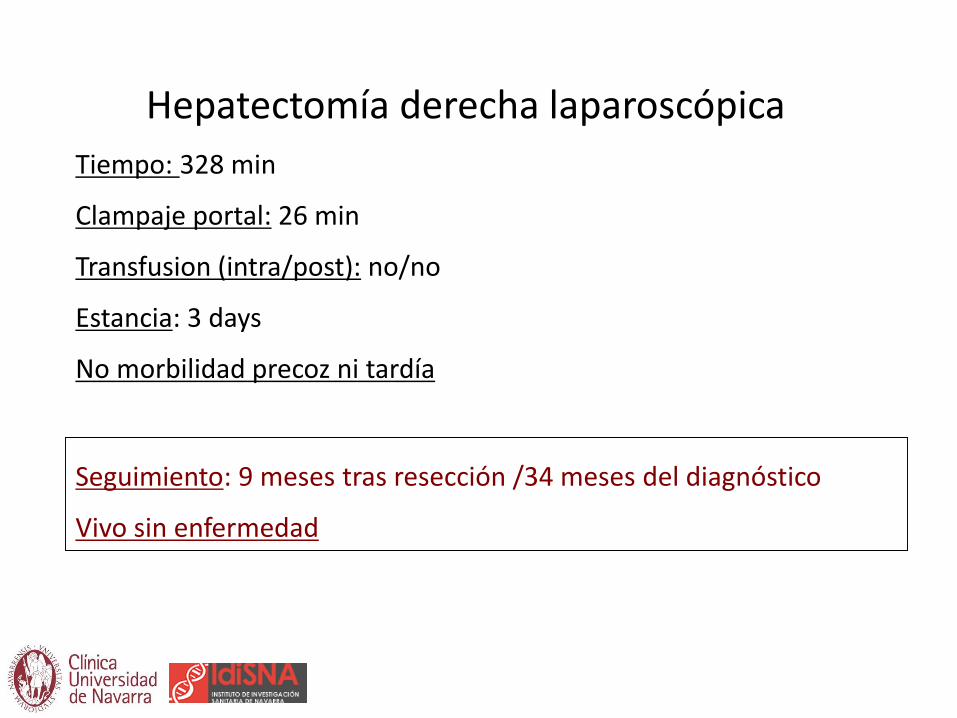
Tiempo: 328 min
Clampaje portal: 26 min
Transfusion (intra/post): no/no
Estancia: 3 days
No morbilidad precoz ni tardía
Hepatectomía derecha laparoscópica
Seguimiento: 9 meses tras resección /34 meses del diagnóstico
Vivo sin enfermedad
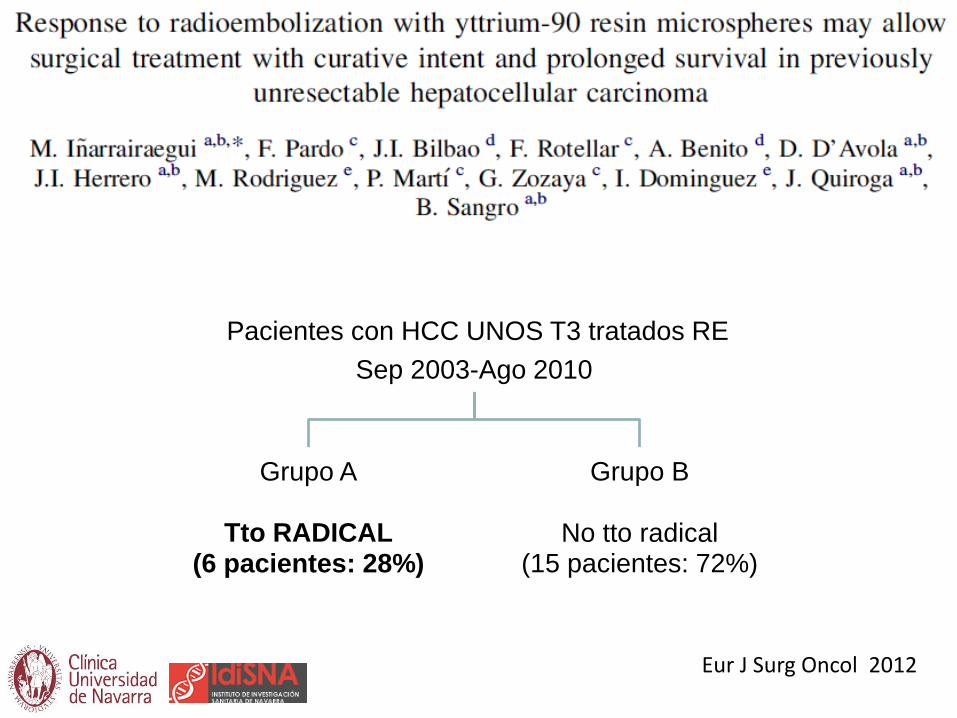
Pacientes con HCC UNOS T3 tratados RE
Sep 2003-Ago 2010
Grupo A
Tto RADICAL(6 pacientes: 28%)
Grupo B
No tto radical(15 pacientes: 72%)
Eur J Surg Oncol 2012

Down-Staging HCC con Y90-Radioembolización
Grupo A
6 pacientes
Grupo B
15 pacientes
p
Edad (años) 62 73 0.006
Sexo (hombres) 100% 73% 0.2
Cirrosis 67% 73% 1
Bilirrubina (mg/dL) 1.0 0.9 0.3
Tratamientos previos 50% 27% 0.1
Volumen Tumoral (mL) 583 137 0.01
Sesión única 83 % 73 % 0.7
Enfermedad unilobar 100% 73% 0.2
Actividad/vol tumor (GBq/mL) 0.37 0.89 0.34
Iñarrairaegui M, et al. Eur J Surg Oncol 2012

Pt Edad CirrosisBil
(mg/dl)
#
nódulos
Tamaño
(cms)
AFP
(UI/ml)
1 72 Sí 0.94 1 8.4 11
2 63 Sí 1.20 1 14.2 17
3 56 Sí 1.28 2 5.5 2
4 66 No 0.88 1 11.5 2
5 62 Sí 1.03 1 13.0 2
6 60 No 0.94 1 11.0 51463
Grupo A: 6 pacientes (28.5%)
Iñarrairaegui M, et al. Eur J Surg Oncol 2012
Down-Staging HCC con Y90-Radioembolización

Down-Staging HCC by Y90-Radioembolization
Dose
(GBq)
Treated
area
Radical
therapy
Interval
(months)Status
Time from radical
therapy
1 2.80 VII-VIIIRFA+
Resection22 Alive FOD 20 mo
2 3.25 RHL LDLT 10 Alive FOD 49 mo
3 2.05 RHL DDLT 35 Alive FOD 52 mo
4 3.02 RHL Resection 2 Alive FOD 49 mo
53.00;
2.00RHL Resection 14 Alive FOD 24 mo
6 1.42 RHL Resection 11 DWD 34 mo
Iñarrairaegui M, et al. Eur J Surg Oncol 2012
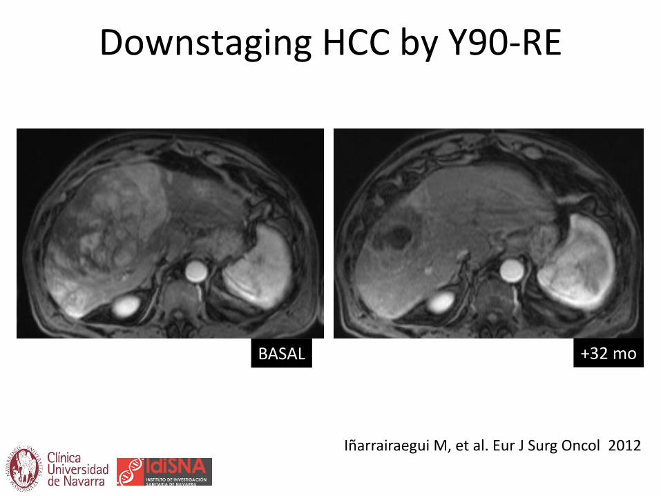
Downstaging HCC by Y90-RE
BASAL +32 mo
Iñarrairaegui M, et al. Eur J Surg Oncol 2012

Downstaging HCC by Y90-RE
BASAL +10 mo
Iñarrairaegui M, et al. Eur J Surg Oncol 2012
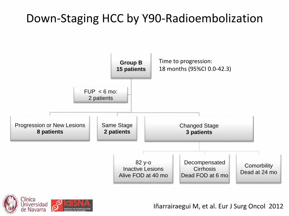
Group B 15 patients
Progression or New Lesions8 patients
Same Stage2 patients
Changed Stage3 patients
82 y-oInactive Lesions
Alive FOD at 40 mo
DecompensatedCirrhosis
Dead FOD at 6 mo
ComorbilityDead at 24 mo
FUP < 6 mo: 2 patients
Time to progression: 18 months (95%CI 0.0-42.3)
Down-Staging HCC by Y90-Radioembolization
Iñarrairaegui M, et al. Eur J Surg Oncol 2012
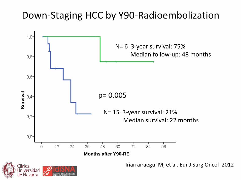
p= 0.005
N= 6 3-year survival: 75%Median follow-up: 48 months
N= 15 3-year survival: 21%Median survival: 22 months
Down-Staging HCC by Y90-Radioembolization
Months after Y90-RE
Su
rviv
al
Iñarrairaegui M, et al. Eur J Surg Oncol 2012
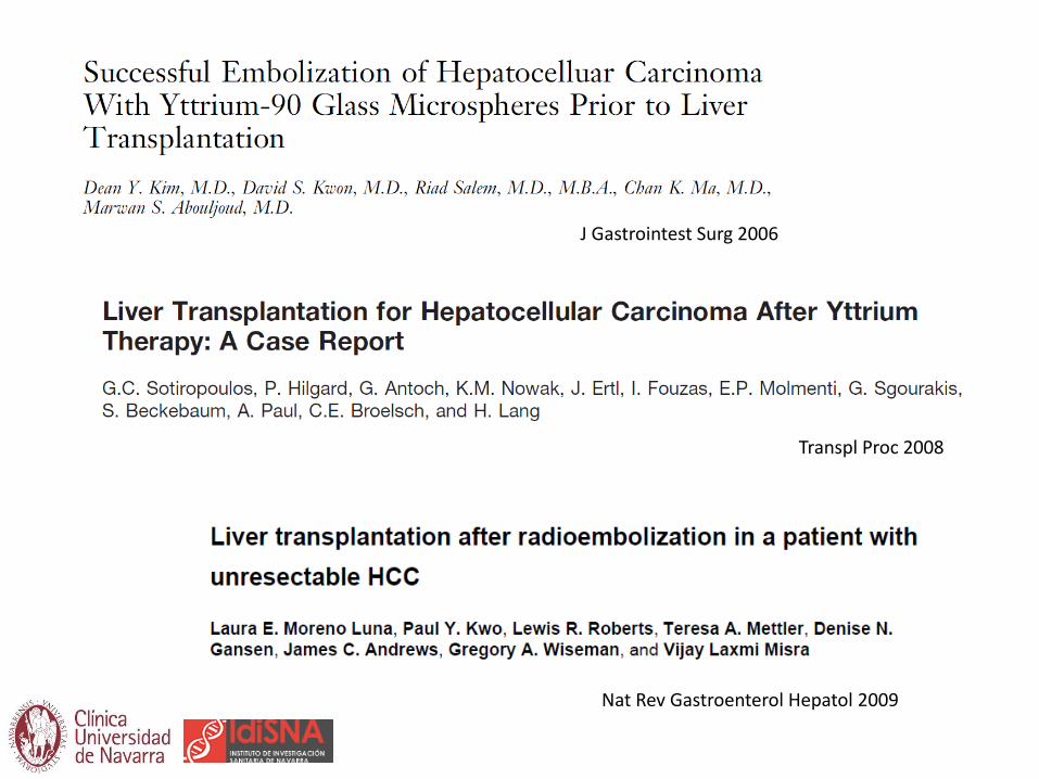
Nat Rev Gastroenterol Hepatol 2009
Transpl Proc 2008
J Gastrointest Surg 2006
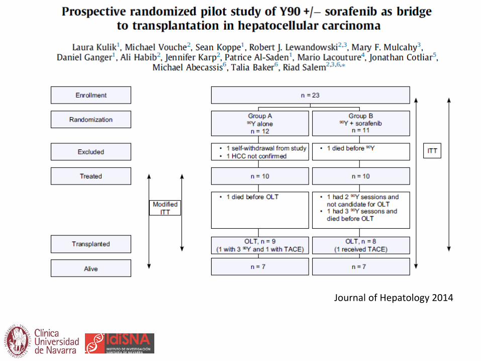
Journal of Hepatology 2014
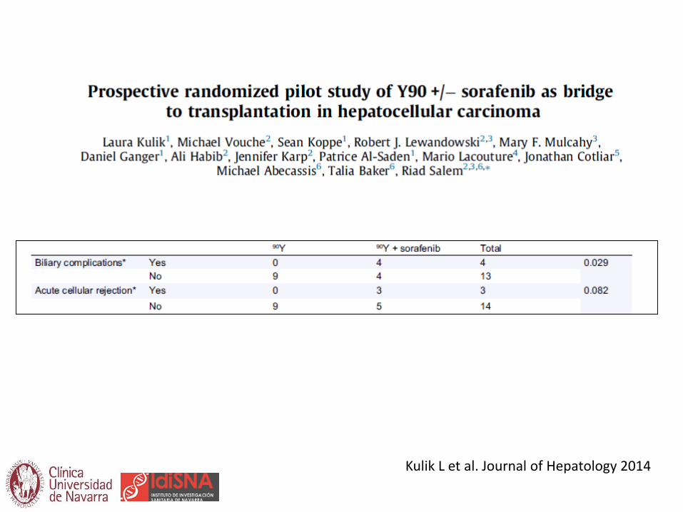
Kulik L et al. Journal of Hepatology 2014
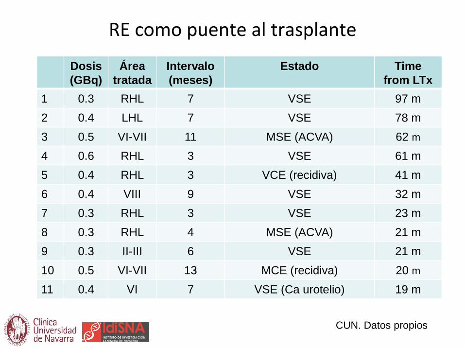
RE como puente al trasplante
Dosis
(GBq)
Área
tratada
Intervalo
(meses)
Estado Time
from LTx
1 0.3 RHL 7 VSE 97 m
2 0.4 LHL 7 VSE 78 m
3 0.5 VI-VII 11 MSE (ACVA) 62 m
4 0.6 RHL 3 VSE 61 m
5 0.4 RHL 3 VCE (recidiva) 41 m
6 0.4 VIII 9 VSE 32 m
7 0.3 RHL 3 VSE 23 m
8 0.3 RHL 4 MSE (ACVA) 21 m
9 0.3 II-III 6 VSE 21 m
10 0.5 VI-VII 13 MCE (recidiva) 20 m
11 0.4 VI 7 VSE (Ca urotelio) 19 m
CUN. Datos propios
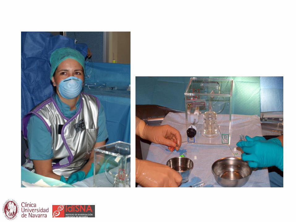
- Seguro para el cirujano?
- Seguro para el paciente?
Seguridad de la cirugía post-SIRT
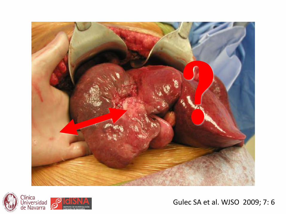
Gulec SA et al. WJSO 2009; 7: 6
?
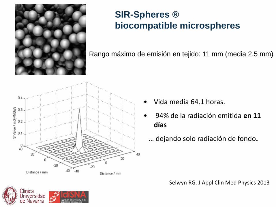
SIR-Spheres ®
biocompatible microspheres
Selwyn RG. J Appl Clin Med Physics 2013
Rango máximo de emisión en tejido: 11 mm (media 2.5 mm)
• Vida media 64.1 horas.
• 94% de la radiación emitida en 11 días
… dejando solo radiación de fondo.
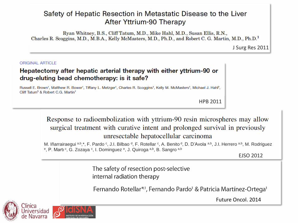
J Surg Res 2011
HPB 2011
M. Iñarrairaegui a,b,*, F. Pardo c, J.I. Bilbao d, F. Rotellar c, A. Benito d, D. D’Avola a,b, J.I. Herrero a,b, M. Rodrigueze, P. Martı c, G. Zozaya c, I. Dominguez e, J. Quiroga a,b, B. Sangro a,b
EJSO 2012
Future Oncol. 2014
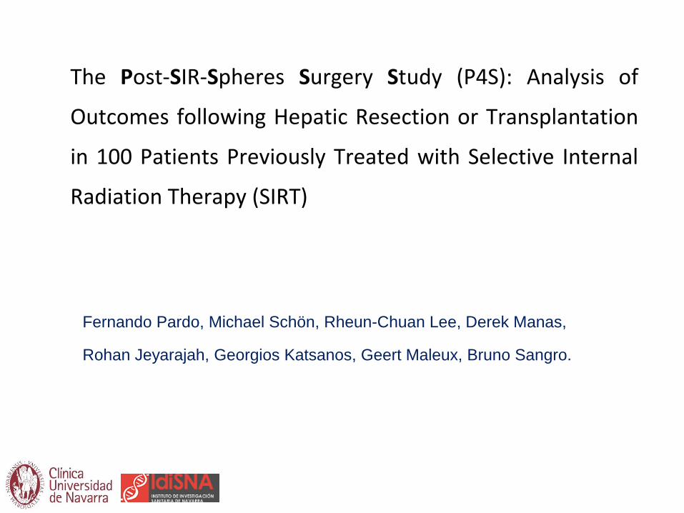
The Post-SIR-Spheres Surgery Study (P4S): Analysis of
Outcomes following Hepatic Resection or Transplantation
in 100 Patients Previously Treated with Selective Internal
Radiation Therapy (SIRT)
Fernando Pardo, Michael Schön, Rheun-Chuan Lee, Derek Manas,
Rohan Jeyarajah, Georgios Katsanos, Geert Maleux, Bruno Sangro.
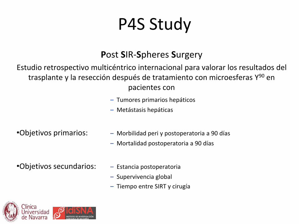
Post SIR-Spheres SurgeryEstudio retrospectivo multicéntrico internacional para valorar los resultados del
trasplante y la resección después de tratamiento con microesferas Y90 en pacientes con
– Tumores primarios hepáticos
– Metástasis hepáticas
•Objetivos primarios: – Morbilidad peri y postoperatoria a 90 días
– Mortalidad postoperatoria a 90 días
•Objetivos secundarios: – Estancia postoperatoria
– Supervivencia global
– Tiempo entre SIRT y cirugía
P4S Study

Center City Country PIEntered Patients Clean patients
Clinica Universidad de Navarra Pamplona Spain Fernando Pardo 30 30
Klinikum Karlsruhe Karlsruhe Germany Michael Schöen 11 10
UZ Gasthuisberg Leuven Belgium Geert Maleux 5 5Institut Jules Bordet Brussels Belgium Vincent Donckier 7 7
Newcastel Hospital Newcastle UK Derek Manas 7 7S Orsola Malpighi Bologna Italy Daniele Pinna 5 5EUROPE 65 64
St. Francis Hospital Tulsa USA Kevin Fisher 2 2
Carolinas MC Charlotte USA Samuel Baker 5 5Methodist Dallas MC Dallas USA Rohan Jeyarajah 7 7USA 14 14
Chinese Univ Hong Kong Hong Kong China Joseph Lau 9
Singapore General Hospital Singapore Singapore Pierce Chow 4 4
Taipei Veterans General Hospital Taipei Taiwan Lee Rheun-Chuan 8 8
Wakefield Gastro Clinic Wakefield New Zealand Richard Stubbs 4 4
Austin Hospital Austin Australia Paul Gow 2 2
St Vincent Hospital Sydney Australia Francis Chu 5 5ASIA-OCEANIA 32 23
111 101
P4S Study
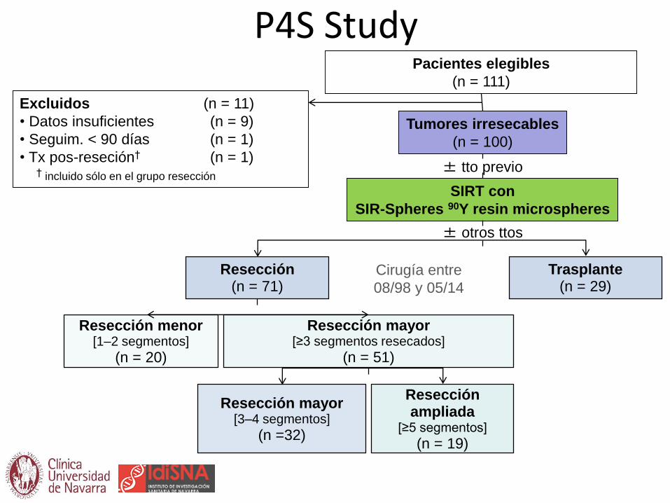
Resección mayor[3–4 segmentos]
(n =32)
Resección ampliada
[≥5 segmentos]
(n = 19)
Resección menor[1–2 segmentos]
(n = 20)
Resección mayor[≥3 segmentos resecados]
(n = 51)
SIRT con
SIR-Spheres 90Y resin microspheres
± otros ttos
± tto previo
Pacientes elegibles
(n = 111)
Tumores irresecables
(n = 100)
Excluidos (n = 11)
• Datos insuficientes (n = 9)
• Seguim. < 90 días (n = 1)
• Tx pos-reseción† (n = 1)† incluido sólo en el grupo resección
P4S Study
Trasplante(n = 29)
Resección(n = 71)
Cirugía entre
08/98 y 05/14
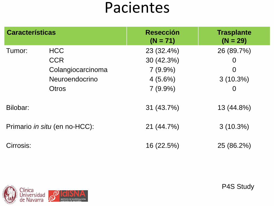
Pacientes
Características Resección
(N = 71)
Trasplante
(N = 29)
Tumor: HCC
CCR
Colangiocarcinoma
Neuroendocrino
Otros
23 (32.4%)
30 (42.3%)
7 (9.9%)
4 (5.6%)
7 (9.9%)
26 (89.7%)
0
0
3 (10.3%)
0
Bilobar: 31 (43.7%) 13 (44.8%)
Primario in situ (en no-HCC): 21 (44.7%) 3 (10.3%)
Cirrosis: 16 (22.5%) 25 (86.2%)
P4S Study
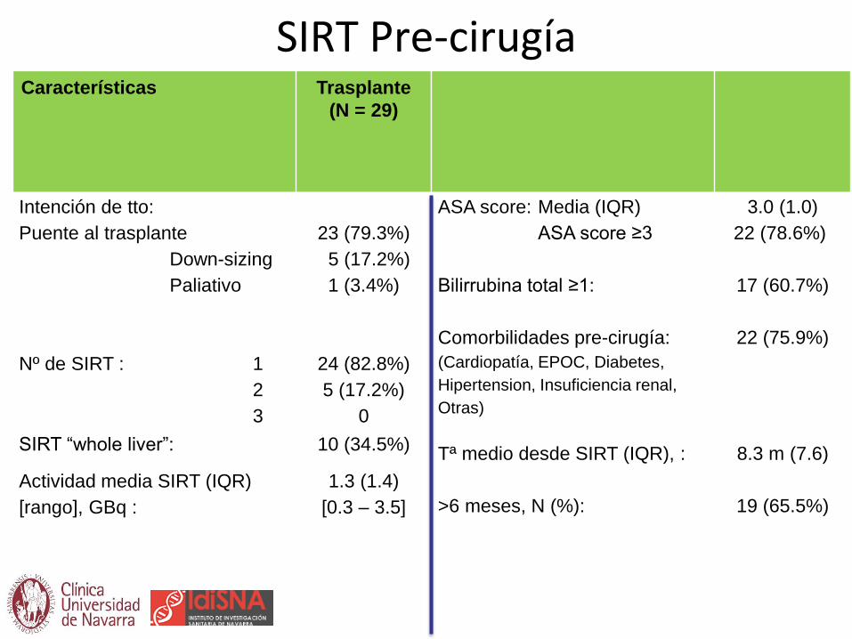
SIRT Pre-cirugíaCaracterísticas Trasplante
(N = 29)
Intención de tto:
Puente al trasplante
Down-sizing
Paliativo
23 (79.3%)
5 (17.2%)
1 (3.4%)
Nº de SIRT : 1
2
3
24 (82.8%)
5 (17.2%)
0
SIRT “whole liver”: 10 (34.5%)
Actividad media SIRT (IQR)
[rango], GBq :
1.3 (1.4)
[0.3 – 3.5]
ASA score: Media (IQR)
ASA score ≥3
3.0 (1.0)
22 (78.6%)
Bilirrubina total ≥1: 17 (60.7%)
Comorbilidades pre-cirugía:
(Cardiopatía, EPOC, Diabetes,
Hipertension, Insuficiencia renal,
Otras)
22 (75.9%)
Tª medio desde SIRT (IQR), :
>6 meses, N (%):
8.3 m (7.6)
19 (65.5%)

Complicaciones peri-postoperatorias y resultadosComplicaciones Trasplante
(N = 29)
Total: CD ≥1
CD ≥3
15 (51.7%)
4 (13.8%)
Fallo hepático: CD ≥1
CD ≥3
1 (3.4%)
0
Herida: CD ≥1
CD ≥3
1 (3.4%)
0
Cardiovascular CD ≥1
CD ≥3
1 (3.4%)
0
Pulmonar CD ≥1
CD ≥3
1 (3.4%)
0
Renal CD ≥1
CD ≥3
2 (6.9%)
0
Evolución
Estancia media
días (IQR): 11.0 (10.0)
Reingreso a 90 días: 9 (31.0%)
Mortalidad 30 días
90 días
0
0
Seguimiento medio:
SIRT
Cirugía
48.3 meses
40.2 meses
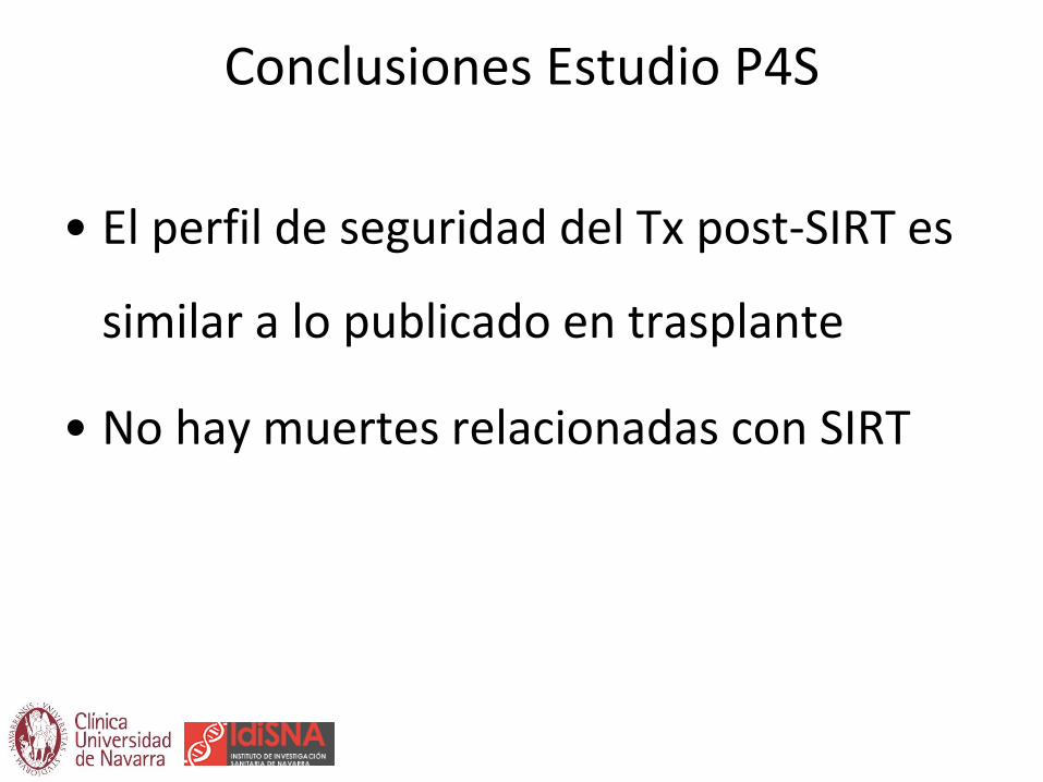
Conclusiones Estudio P4S
• El perfil de seguridad del Tx post-SIRT es
similar a lo publicado en trasplante
• No hay muertes relacionadas con SIRT

Integration of SIRT in the HCC BCLCstaging classification and treatment schedule
HCC
PS 0
Child A
PS 0–2
Child A–B
Resection TACE
Stage 0
Very Early Stage
Stage A
Early Stage
Stage B
Intermediate Stage
Stage C
Advanced Stage
Stage D
End Stage
PS >2
Child C
single <2 cm or
carcinoma in situ
single nodule or
3 nodules <3 cm
PS 0
multinodular; PS 0portal vein invasion,
N1 M1 or PS 1–2
PS >2 or Child C (unless within
transplant criteria)
portal pressure;
bilirubin
normal
increased associated diseases
Liver Transplant Ablation
no yes
single 3 nodules <3 cm
symptomatic
OS <3 mo
10% of patients
Curative Treatments – 5-yr survival 40–70%
30% of patients
OS 20 months (14-45)
20% of patients
failed
TACE
unilobar
fewer nodules
smaller burden
bilobar
multinodular
larger burden
fit/suitable
for SIRT i.e.
liver-dominant;
bilirubin
<2 mg/dL;
Child A or <B7
fit/suitable
for sorafenib
i.e.
main PVT
EHD
SIRTSIRT/
sorafenib
OS 11 months (6-14)
40% of patients
sorafenib
SIRT?
¡Muchas gracias!
Recommended