The Immune System
Chapter 21
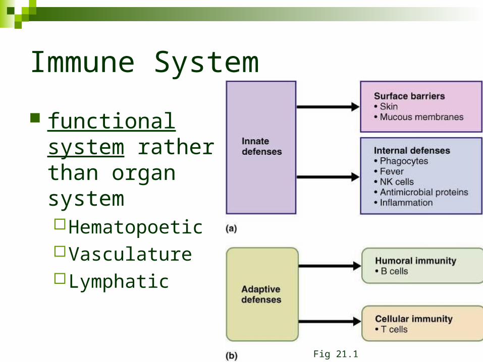
Immune System
functional system rather than organ systemHematopoetic VasculatureLymphatic
Fig 21.1
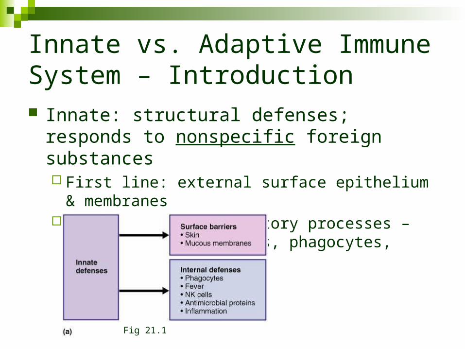
Innate vs. Adaptive Immune System – Introduction Innate: structural defenses; responds to
nonspecific foreign substances First line: external surface epithelium & membranes Second line: inflammatory processes – antimicrobial
proteins, phagocytes, etc.
Fig 21.1
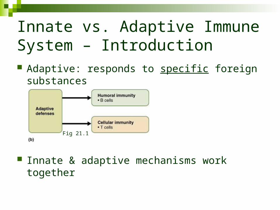
Innate vs. Adaptive Immune System – Introduction Adaptive: responds to specific foreign substances
Innate & adaptive mechanisms work together
Fig 21.1
Innate, Surface Defenses
Skin physical barrier to microbes Keratin resistant to most bacterial enzymes & toxins secretions are acidic pH 3-5
Mucosa physical barrier & produces a variety of protective chemicals
Gastric mucosa very acidic & produces proteolytic enzymes
Saliva & lacrimal fluid contain lysozyme Mucous
traps bacteria & moves them away from epithelial surface
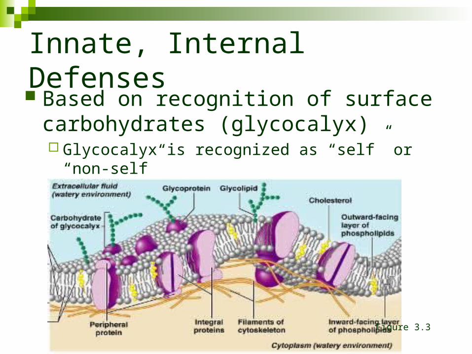
Innate, Internal Defenses Based on recognition of surface
carbohydrates (glycocalyx) Glycocalyx is recognized as “self” or “non-self”
Figure 3.3
Innate, Internal Defenses
Phagocytes Macrophages: derived from monocytes
Free Macrophages: roam through tissues Fixed Macrophages: Kupffer cells (liver) & microglia (brain) Ingest cellular debris, foreign material, bacteria, fungi
Neutrophils: ingest pathogens Eosinophils: weakly phagocytic of pathogens. Attack
parasites (degranulation) Mast Cells: phagocytic of various bacteria
Innate, Internal Defenses
Phagocytic mechanisms: Adherence: cell binds to invader
Aided by opsonization (a chemical process that enhances binding via complement & antibodies)
Ingestion: formation of phagolysosomes Respiratory Bursts: merge phagosome with lysosome & flood
phagolysosome with free radicals (macrophage) Defensins: proteins that crystallize out of solution & pierce
pathogen membranes (neutrophils)
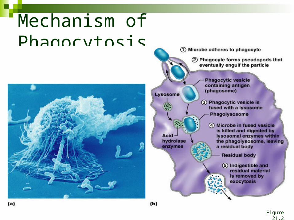
Mechanism of Phagocytosis
Figure 21.2
Innate, Internal Defenses
Natural Killer Cells: Small population of large granular lymphocytes Non specific for “non-self” Not phagocytic: attack is by release of perforins that
perforate the target cell plasma membrane. Shortly after perforation the target nucleus disintegrates.
Release chemicals that enhance the inflammatory response
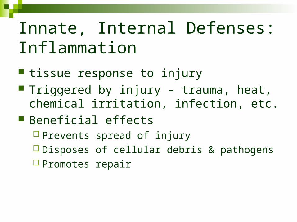
Innate, Internal Defenses: Inflammation tissue response to injury Triggered by injury – trauma, heat, chemical
irritation, infection, etc. Beneficial effects
Prevents spread of injury Disposes of cellular debris & pathogens Promotes repair
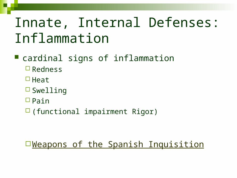
Innate, Internal Defenses: Inflammation cardinal signs of inflammation
Redness Heat Swelling Pain (functional impairment Rigor)
Weapons of the Spanish Inquisition
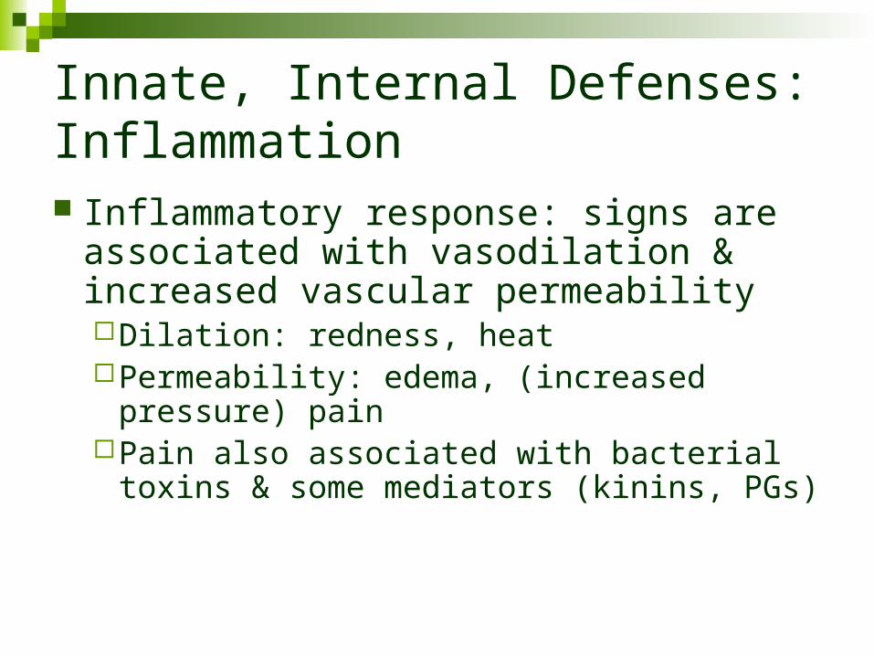
Innate, Internal Defenses: Inflammation Inflammatory response: signs are
associated with vasodilation & increased vascular permeabilityDilation: redness, heatPermeability: edema, (increased pressure)
painPain also associated with bacterial toxins &
some mediators (kinins, PGs)
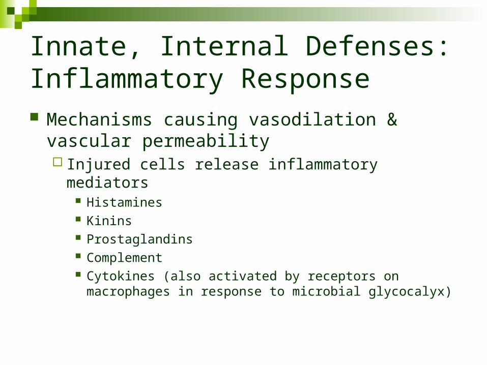
Innate, Internal Defenses: Inflammatory Response Mechanisms causing vasodilation & vascular
permeability Injured cells release inflammatory mediators
Histamines Kinins Prostaglandins Complement Cytokines (also activated by receptors on macrophages in
response to microbial glycocalyx)
Innate, Internal Defenses: Inflammatory Response Edema
Dilutes harmful substancesProvides nutrients (& O2) for repairEnhances entry of clotting protein
Epithelial breaches also stimulate -defensin release from epithelial cells
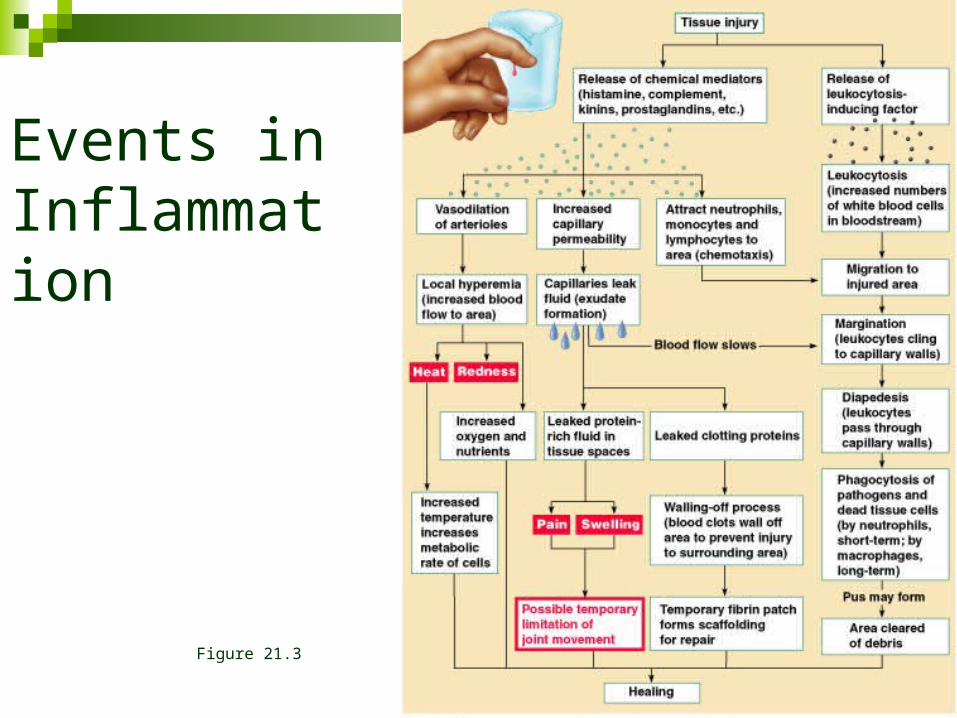
Events in Inflammation
Figure 21.3
Innate, Internal Defenses: Inflammatory Response Phagocyte mobilization: infiltration of damaged
area by neutrophils & macrophages
Innate, Internal Defenses: Inflammatory Response Leukocytosis: leukocytosis inducing factors
released by injured cells promote rapid release of WBCs from marrow
Margination: increased vascular permeability causes decreased fluid in vessels; blood flow slows & neutrophils are able to move to vessel margins. Here endothelial markers (CAMs) allow neutrophils to cling to vessel walls (pavementing).
Innate, Internal Defenses: Inflammatory Response Diapedesis: neutrophils migrate through
capillary walls Chemotaxis – inflammatory chemicals attract
neutrophils to move up the chemical concentration gradient (neutrophils respond first)
As the process continues, monocytes diapedes into the area & become macrophages. With chronic inflammation, macrophages predominate
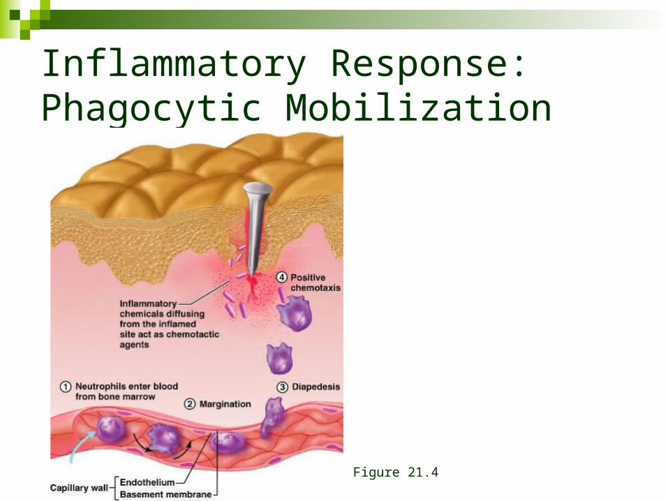
Inflammatory Response:Phagocytic Mobilization
Figure 21.4
Innate, Internal Defenses: Inflammatory Response Macrophages clean up cellular debris &
pathogens If pathogens were associated with the injury,
activation of the complement cascade occurs & elements of adaptive immunity join the process
Innate, Internal Defenses
Viral replication – (viruses lack metabolic processes) Viruses release nucleic acid (RNA or DNA) into cytoplasm. The information on the nucleic acid is incorporated into the cell’s DNA. Normal cellular mechanisms then produce viral structural components. Multiple new viral particles are produced & released from the cell (sometimes killing the cell)
Innate, Internal Defenses
Antiviral proteins: interferon & complement Interferon: some cells produce & release
interferons (IFNs) when invaded by virus Released interferons stimulate nearby cells to
produce proteins (PKR) that interfere with viral replication by disrupting protein synthesis & the ribosome
Not virus specific.
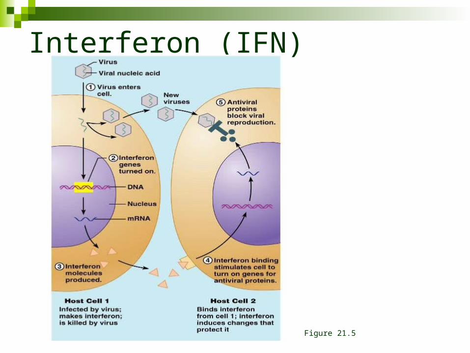
Interferon (IFN)
Figure 21.5
Innate, Internal Defenses
Complement – a group of plasma proteins (20) that are activated in the presence of foreign substances
Complement activation enhances & amplifies inflammation
Bacteria & some other cell types are lysed by complement activation
Complement activation enhances both innate & adaptive defenses
Innate, Internal Defenses
Complement activation pathways Classical pathway: requires antibodies
Antibodies bind to target (antigen) Complement protein C1 binds to the antibody-
antigen complex (complement fixation) Alternative pathway: complement factors interact with
microorganism glycocalyx Both pathways lead to a cascade of protein activation,
leading to activation of C3
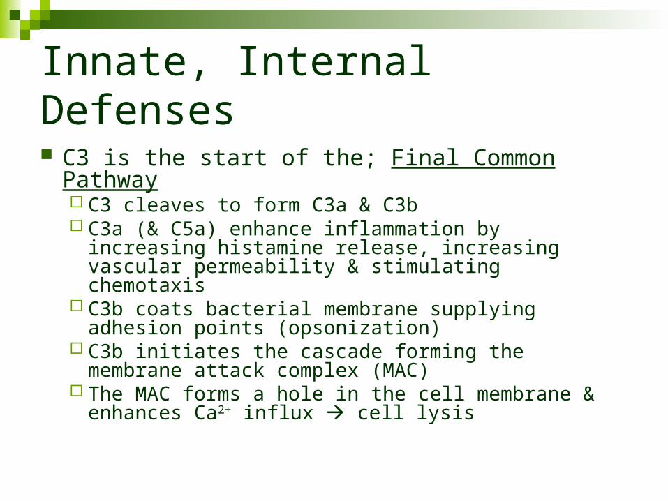
Innate, Internal Defenses
C3 is the start of the; Final Common Pathway C3 cleaves to form C3a & C3b C3a (& C5a) enhance inflammation by increasing
histamine release, increasing vascular permeability & stimulating chemotaxis
C3b coats bacterial membrane supplying adhesion points (opsonization)
C3b initiates the cascade forming the membrane attack complex (MAC)
The MAC forms a hole in the cell membrane & enhances Ca2+ influx cell lysis
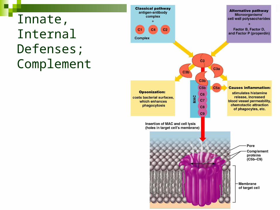
Innate, Internal Defenses; Complement
Figure 21.6
Innate, Internal Defenses
C-reactive proteins (CRP) produced by the liver in response to inflammatory molecules can activate the classical pathway by binding to membrane & activating C1. Also participates in opsonization.
Fever – a systemic response to infection. Leukocytes & macrophages release pyrogens that raise the hypothalamic “set point” for temperature
ADAPTIVE DEFENSES
ADAPTIVE DEFENSES
Innate & adaptive mechanisms work together in a cohesive fashion
Adaptive Defenses: Characteristics
Specificity: directed at specific targets
Systemic: not restricted to initial site of infection / invasion
Memory: after initial exposure & activation, a more rapid & more vigorous response is made to subsequent exposures to pathogens
(secondary response)
Adaptive Defenses: Components
Humoral Immunity: (antibody mediated immunity) provided by antibodies floating free in body fluids
Cell mediated immunity: lymphocytes directly attack specific invaders by
lysis or indirect attack by initiating inflammation and/or activating other lymphocytes & macrophages
Adaptive, Humoral Immunity
Antigen = any substance that can mobilize the immune system & provoke an immune response*
*Humoral and/or cell mediated
Adaptive, Humoral Immunity
Complete antigens (proteins, nucleic acids, lipids, polysaccharides): Immunogenicity: the ability to stimulate specific
lymphocytes & specific antibodies Reactivity: the ability to react with activated
lymphocytes & antibodies Hapten (an incomplete antigen): a smaller
molecule that is not immunogenic until attached to proteins
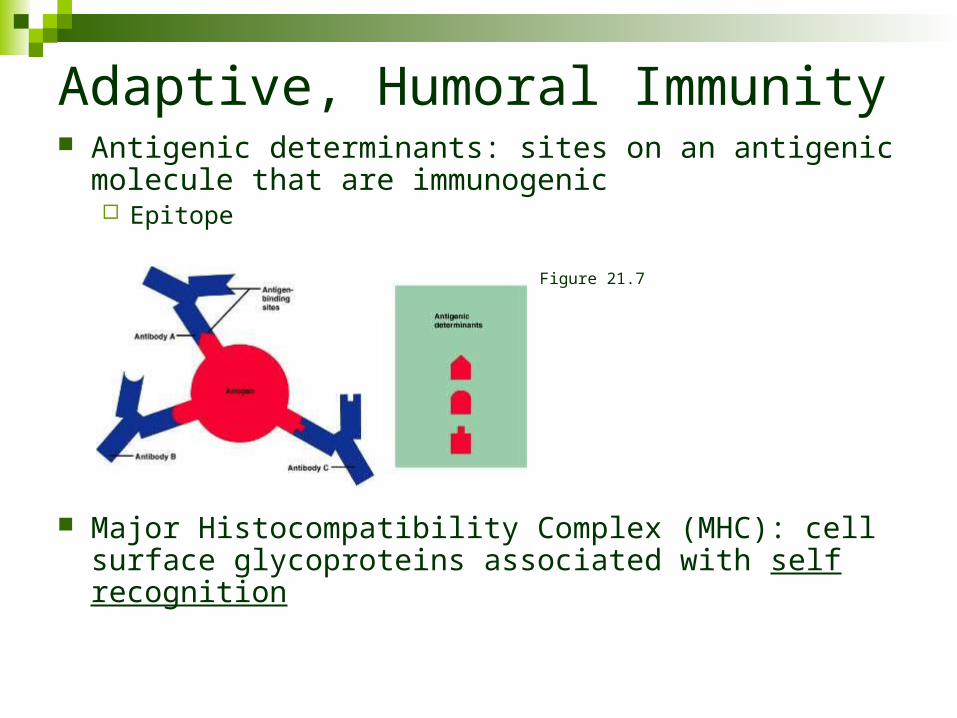
Adaptive, Humoral Immunity Antigenic determinants: sites on an antigenic molecule
that are immunogenic Epitope
Major Histocompatibility Complex (MHC): cell surface glycoproteins associated with self recognition
Figure 21.7
Adaptive Immune System: Cells
LymphocytesT-cellsB-cells
Antigen Presenting Cells (APCs)
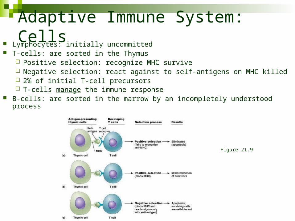
Adaptive Immune System: Cells Lymphocytes: initially uncommitted T-cells: are sorted in the Thymus
Positive selection: recognize MHC survive Negative selection: react against to self-antigens on MHC killed 2% of initial T-cell precursors T-cells manage the immune response
B-cells: are sorted in the marrow by an incompletely understood process
Figure 21.9
Adaptive Immune System: Cells
Immunocompetence: as T- or B-cells mature they become immunocompetent, they display receptors on their cell membrane for a specific antigen.
All of the receptors on one cell are identical; immunity depends upon genetic coding for appropriate receptors.
Adaptive Immune System: Cells
Antigen Presenting Cells (APCs) APCs ingest foreign material, then present
antigenic fragments on their cell surface where they are recognized by T-cells T-cells: respond to antigen only if it is displayed on plasma membrane.
APCs: Macrophages & B lymphocytes Interactions between APCs & lymphocytes &
lymphocyte-lymphocyte interactions are critical to immune response
Adaptive, Humoral response
Humoral response (clonal selection) B-cells: Antigen challenge to naïve
immunocompetent B-cell Antigen binds to B-cell receptors & form cross-
links between receptors Cross linked antigen-receptor complex
undergoes endocytosis; B-cell presents to T-cell
Humoral Immunity
Active humoral immunity: B-cells encounter & respond to antigen to produce an
antibody
Passive humoral immunity: Introduced “non-native” antibody
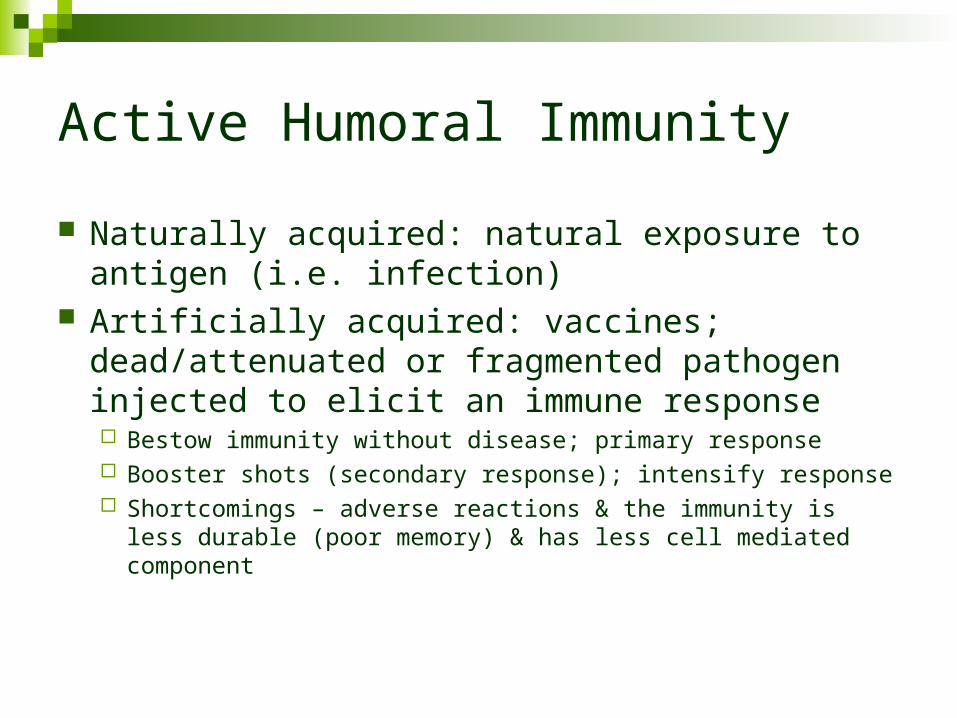
Active Humoral Immunity
Naturally acquired: natural exposure to antigen (i.e. infection)
Artificially acquired: vaccines; dead/attenuated or fragmented pathogen injected to elicit an immune response Bestow immunity without disease; primary response Booster shots (secondary response); intensify response Shortcomings – adverse reactions & the immunity is less durable
(poor memory) & has less cell mediated component
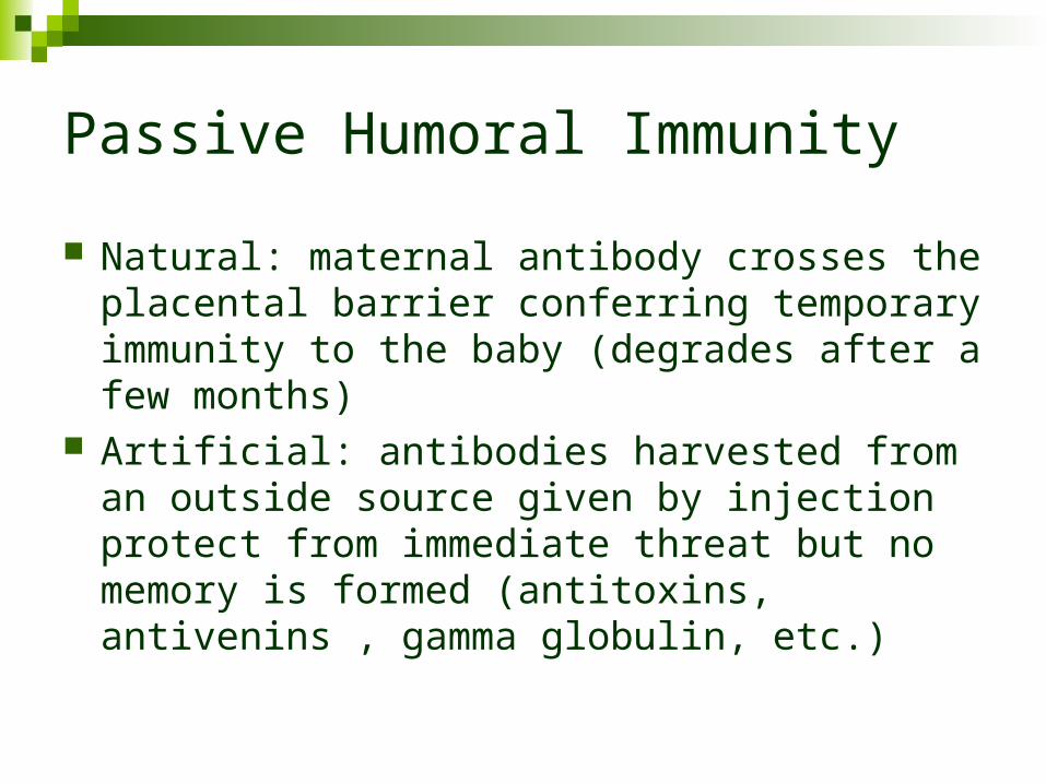
Passive Humoral Immunity
Natural: maternal antibody crosses the placental barrier conferring temporary immunity to the baby (degrades after a few months)
Artificial: antibodies harvested from an outside source given by injection protect from immediate threat but no memory is formed (antitoxins, antivenins , gamma globulin, etc.)
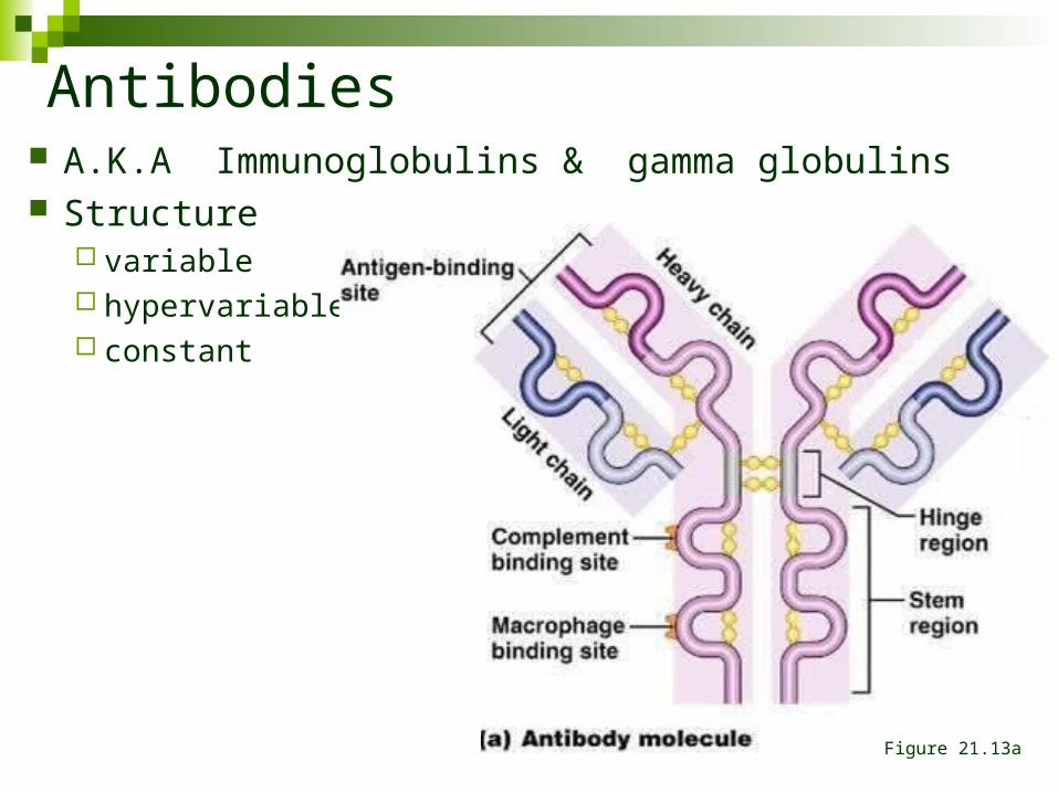
Antibodies A.K.A Immunoglobulins & gamma globulins Structure
variable hypervariable constant
Figure 21.13a
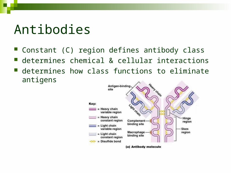
Antibodies Constant (C) region defines antibody class determines chemical & cellular interactions determines how class functions to eliminate antigens
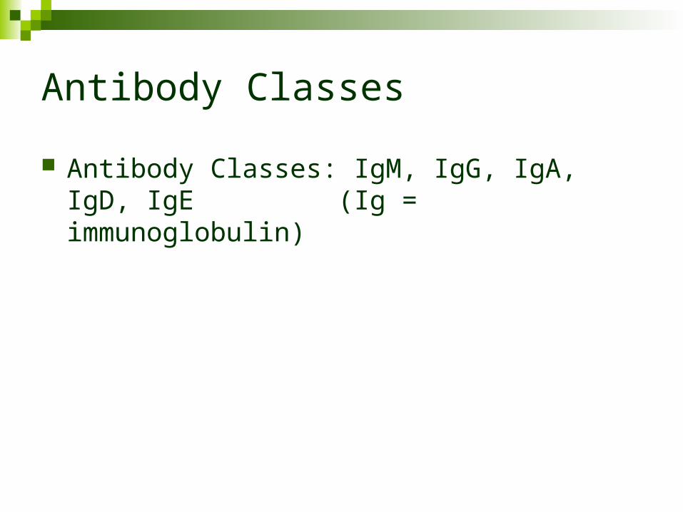
Antibody Classes
Antibody Classes: IgM, IgG, IgA, IgD, IgE (Ig = immunoglobulin)
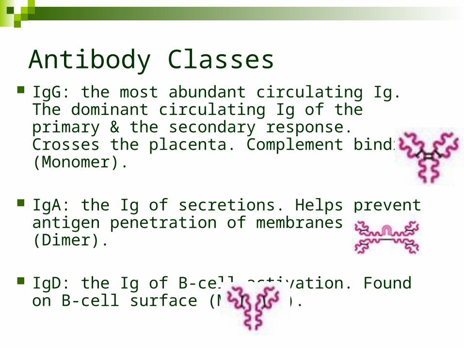
Antibody Classes IgG: the most abundant circulating Ig. The
dominant circulating Ig of the primary & the secondary response. Crosses the placenta. Complement binding (Monomer).
IgA: the Ig of secretions. Helps prevent antigen penetration of membranes (Dimer).
IgD: the Ig of B-cell activation. Found on B-cell
surface (Monomer).
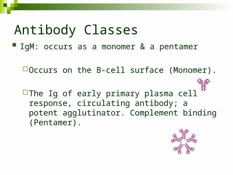
Antibody Classes IgM: occurs as a monomer & a pentamer
Occurs on the B-cell surface (Monomer).
The Ig of early primary plasma cell response, circulating antibody; a potent agglutinator. Complement binding (Pentamer).
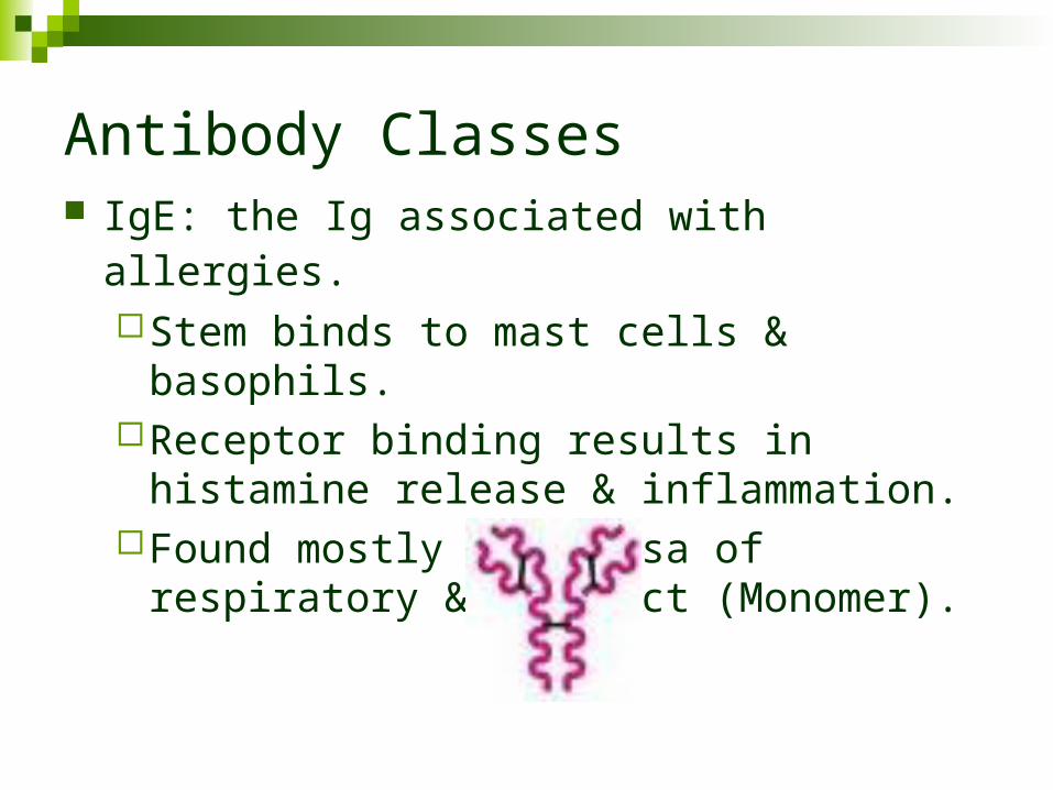
Antibody Classes IgE: the Ig associated with allergies.
Stem binds to mast cells & basophils.Receptor binding results in histamine release
& inflammation.Found mostly in mucosa of respiratory & GI
tract (Monomer).
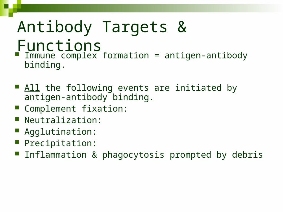
Antibody Targets & Functions Immune complex formation = antigen-antibody binding.
All the following events are initiated by antigen-antibody binding.
Complement fixation: Neutralization: Agglutination: Precipitation: Inflammation & phagocytosis prompted by debris
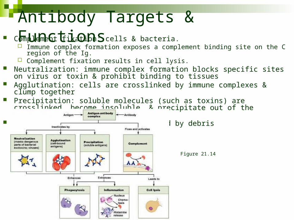
Antibody Targets & Functions Complement fixation: cells & bacteria.
Immune complex formation exposes a complement binding site on the C region of the Ig. Complement fixation results in cell lysis.
Neutralization: immune complex formation blocks specific sites on virus or toxin & prohibit binding to tissues
Agglutination: cells are crosslinked by immune complexes & clump together Precipitation: soluble molecules (such as toxins) are crosslinked, become insoluble,
& precipitate out of the solution Inflammation & phagocytosis prompted by debris
Figure 21.14
Antibody Targets & Functions
Monoclonal antibodies: antibodies produced by descendants of a single cell Pure antibody preparations that are specific for a
single antigenic determinant Research / diagnostic / therapeutic use
Cell Mediated Immune Response
T-cell activation: involves recognition of PM surface antigens only Antigen is combined with MHC & displayed on PM T-cell receptors: bind to the MHC & are stimulated by
the associated antigen The addition of a co-stimulator (cytokines,
interleukins, etc) prompts the T-cell to form a clone In the absence of a co-stimulator the T-cell becomes
tolerant to antigen (anergy)
Cell Mediated: MHC
MHC occurs as two classes MHC I on virtually all tissue cells MHC II only on PM some immune system cells
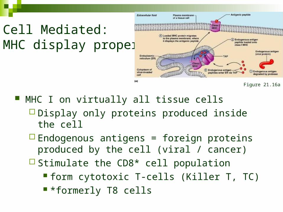
Cell Mediated: MHC display properties
MHC I on virtually all tissue cells Display only proteins produced inside the cell Endogenous antigens = foreign proteins produced by
the cell (viral / cancer) Stimulate the CD8* cell population
form cytotoxic T-cells (Killer T, TC) *formerly T8 cells
Figure 21.16a
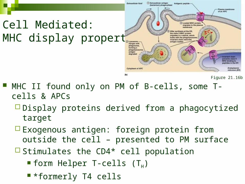
Cell Mediated: MHC display properties
MHC II found only on PM of B-cells, some T-cells & APCs Display proteins derived from a phagocytized target Exogenous antigen: foreign protein from outside the cell –
presented to PM surface Stimulates the CD4* cell population
form Helper T-cells (TH) *formerly T4 cells
Figure 21.16b
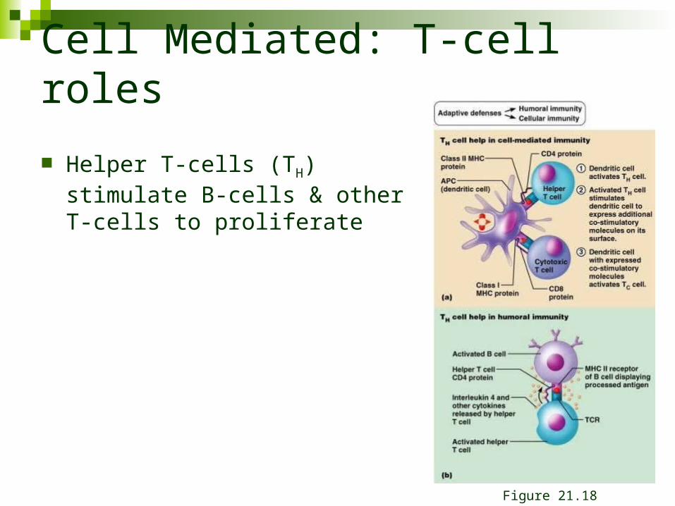
Cell Mediated: T-cell roles
Helper T-cells (TH) stimulate B-cells & other T-cells to proliferate
Figure 21.18
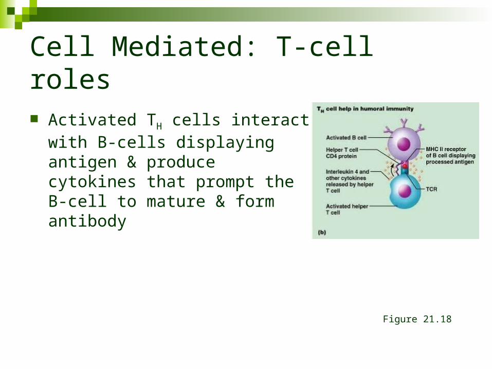
Cell Mediated: T-cell roles
Activated TH cells interact with B-cells displaying antigen & produce cytokines that prompt the B-cell to mature & form antibody
Figure 21.18
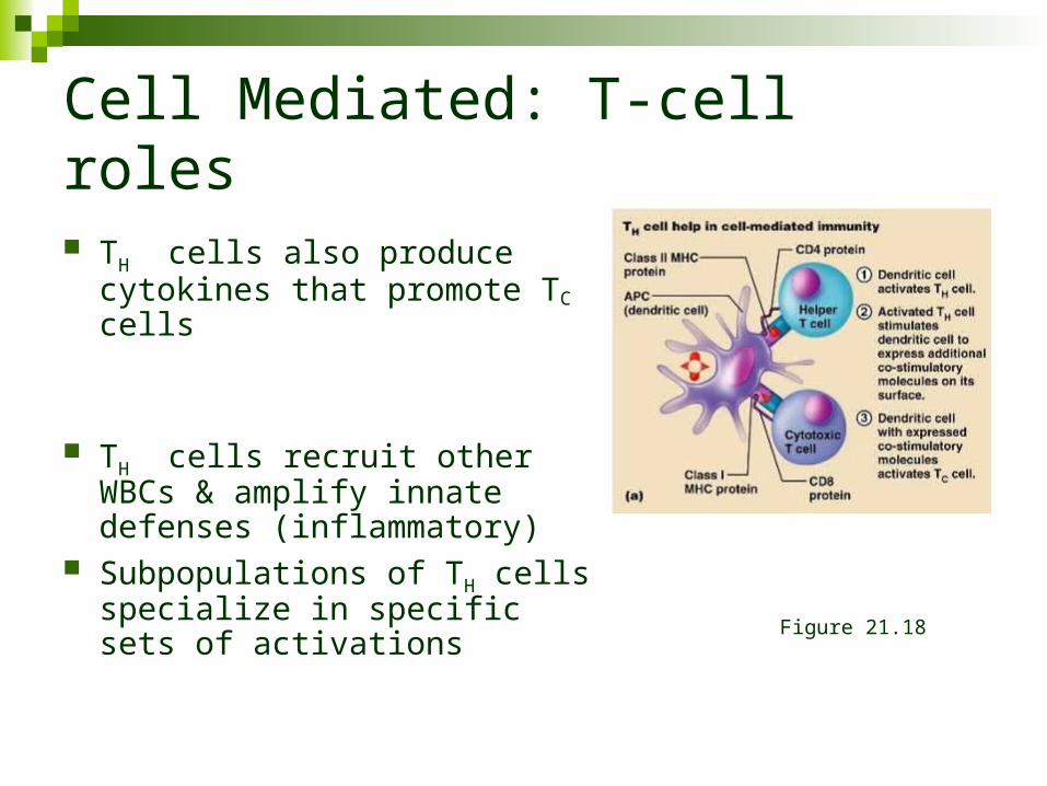
Cell Mediated: T-cell roles
TH cells also produce cytokines that promote TC cells
TH cells recruit other WBCs & amplify innate defenses (inflammatory)
Subpopulations of TH cells specialize in specific sets of activations
Figure 21.18
Cell Mediated: T-cell roles
Cytotoxic T-cells (TC, Killer T): directly attack & kill cells with specific antigen
Activated TC cells are co-stimulated by TH cells
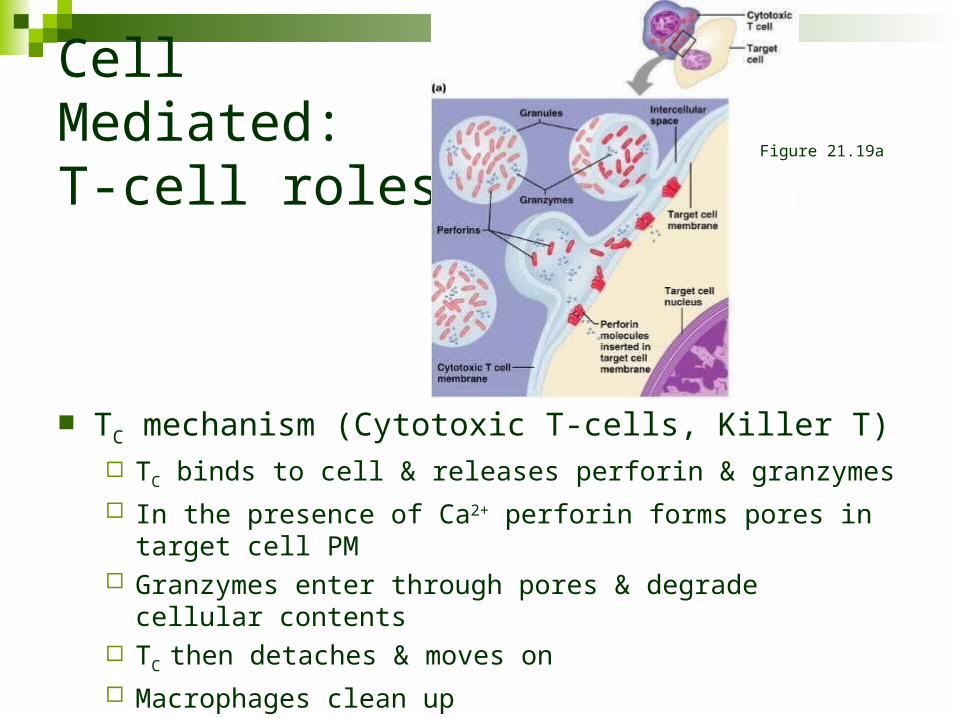
Cell Mediated: T-cell roles
TC mechanism (Cytotoxic T-cells, Killer T) TC binds to cell & releases perforin & granzymes
In the presence of Ca2+ perforin forms pores in target cell PM Granzymes enter through pores & degrade cellular contents TC then detaches & moves on
Macrophages clean up
Figure 21.19a
Cell Mediated: T-cell roles
Other T-cells *Regulatory T-cells (TReg): release inhibitory cytokines
that suppress B-cell & T-cell activity Help to prevent autoimmune events *formerly Suppressor T (TS)
Gamma Delta T-cells (Tgd): live in the intestine. Function in surveillance & are triggered much like NK cells
Organ Transplants/Rejections
Types of Organ Transplants Autograft: tissue graft from one body site to another
(same person) Isograft: graft received from a genetically identical
donor (identical twin) Allograft: graft received from genetically non-identical
donor (same species) Xenograft: graft received from another species of
animal
Organ Transplants/Rejections
Transplant rejection: mediated by the immune system (especially TC, NK, antibodies) Auto/Isograft: MHC compatible Xenograft: most MHC incompatible Allograft: attempt to obtain the best MHC match
Organ Transplants/Rejections
Immunosuppressive therapy: used to delay/prevent rejection Corticosteroids: suppress inflammation Antiproliferative: prevent/kill rapidly dividing cells Immunosuppressant: prevent/kill rapidly dividing cells Side effects tend to be harsh Increased risk of infection
Immunologic Dysfunction
Immunodeficiency Congenital/Genetic: varied inborn errors Acquired:
Drugs: immunosuppressive / cancer drugsRadiation therapy – marrow
Cancer: can be viewed as a failure of immune surveillance
Hodgkin’s disease: lymph node cancer AIDS/HIV: kills TH cells
Immunologic Dysfunction
Autoimmune disease: production of antibody & TH against self tissues Examples & tissue effected
Multiple sclerosis: white matter of nervous system Graves disease: thyroid Type I diabetes mellitus: beta cells of pancreas Systemic Lupus Erythrematosis: (anti DNA) kidneys, heart,
lungs & skin Rheumatoid Arthritis: destroys joints (cartilage) Glomerulonephritis: impaired renal function (may be
secondary to other autoimmune disease)
Immunologic Dysfunction
Mechanisms of immunologic dysfunction Failure of lymphocyte programming New self antigens
Gene mutation Structural change – haptens, infection
Foreign antigens that closely resemble self antigen resulting in cross reactivity.
Immunologic Dysfunction
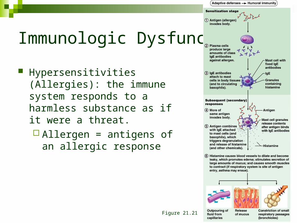
Hypersensitivities (Allergies): the immune system responds to a harmless substance as if it were a threat. Allergen = antigens of an
allergic response
Figure 21.21
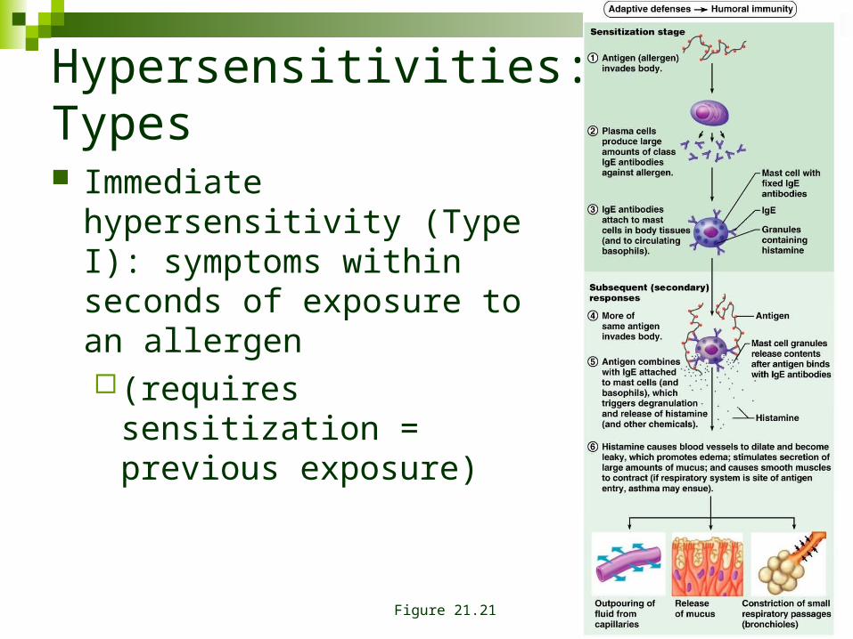
Hypersensitivities: Types
Immediate hypersensitivity (Type I): symptoms within seconds of exposure to an allergen(requires sensitization =
previous exposure)
Figure 21.21
Hypersensitivities: Type I
Anaphylaxis (IgE mediated; mast / basophils) Local: histamine induced vasodilation & increased
permeability. Watery eyes, runny nose, itching & redness. Respiratory allergy induced asthma
Systemic: anaphylactic shock: associated with allergens that have systemic distribution. Widespread vasodilation, airway swelling
Atopy: the tendency to display Type I symptoms to certain environmental antigens without prior sensitization
Hypersensitivities: Types II & III
Subacute hypersensitivity (IgG & IgM mediated) Cytotoxic reactions (Type II): antibodies bind to cellular
antigens promoting complement fixation / inflammation / phagocytosis (transfusion reaction)
Immune complex h. (Type III): widely distributed antigen reacts with antibody.
Antigen-antibody complexes cannot be cleared; persistent inflammation / tissue damage (farmer’s lung; associated with autoimmune disorders)
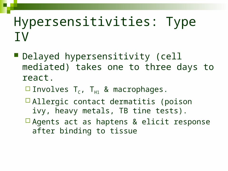
Hypersensitivities: Type IV
Delayed hypersensitivity (cell mediated) takes one to three days to react. Involves TC, TH1 & macrophages.
Allergic contact dermatitis (poison ivy, heavy metals, TB tine tests).
Agents act as haptens & elicit response after binding to tissue
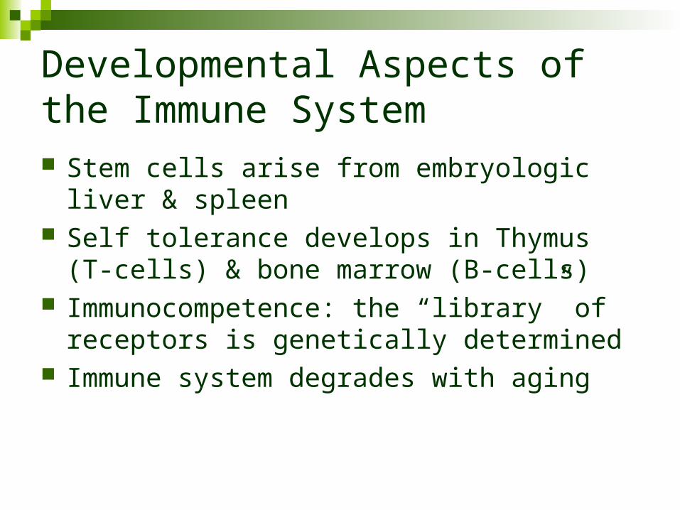
Developmental Aspects of the Immune System Stem cells arise from embryologic liver & spleen Self tolerance develops in Thymus (T-cells) &
bone marrow (B-cells) Immunocompetence: the “library” of receptors is
genetically determined Immune system degrades with aging

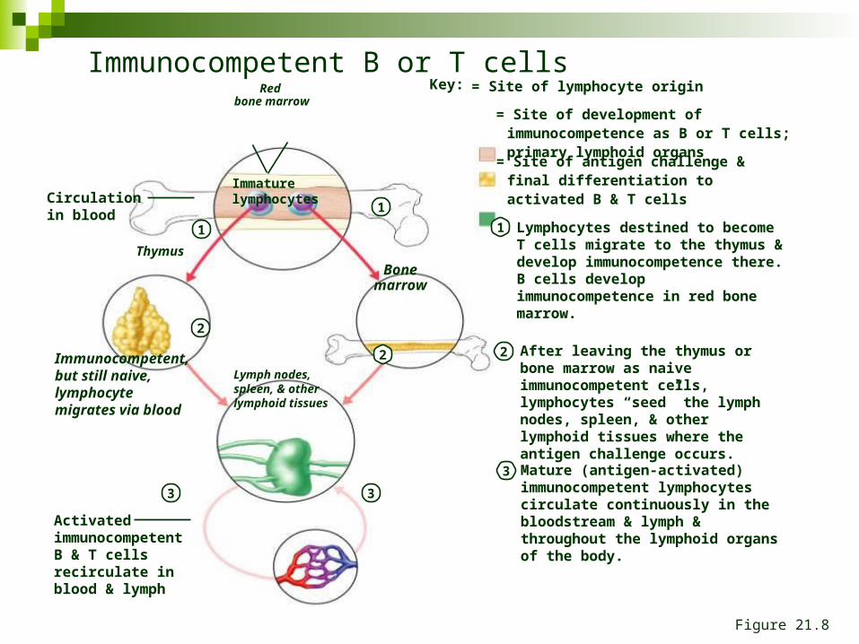
Red bone marrow
1
2
3
Immunocompetent, but still naive, lymphocyte migrates via blood
Mature (antigen-activated) immunocompetent lymphocytes circulate continuously in the bloodstream & lymph & throughout the lymphoid organs of the body.
Key: = Site of lymphocyte origin
= Site of development of immunocompetence as B or T cells; primary lymphoid organs
= Site of antigen challenge & final differentiation to activated B & T cells Immature
lymphocytesCirculation in blood
1
1 Lymphocytes destined to become T cells migrate to the thymus & develop immunocompetence there. B cells develop immunocompetence in red bone marrow.
Thymus
Bonemarrow
Lymph nodes, spleen, & other lymphoid tissues
2 2 After leaving the thymus or bone marrow as naive immunocompetent cells, lymphocytes “seed” the lymph nodes, spleen, & other lymphoid tissues where the antigen challenge occurs.
3 3
Activated immunocompetent B & T cells recirculate in blood & lymph
Immunocompetent B or T cells
Figure 21.8
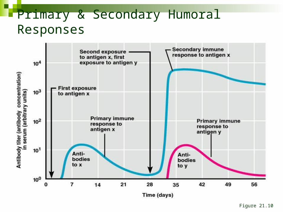
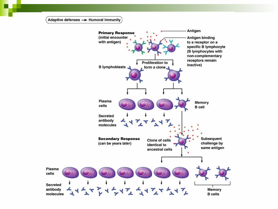
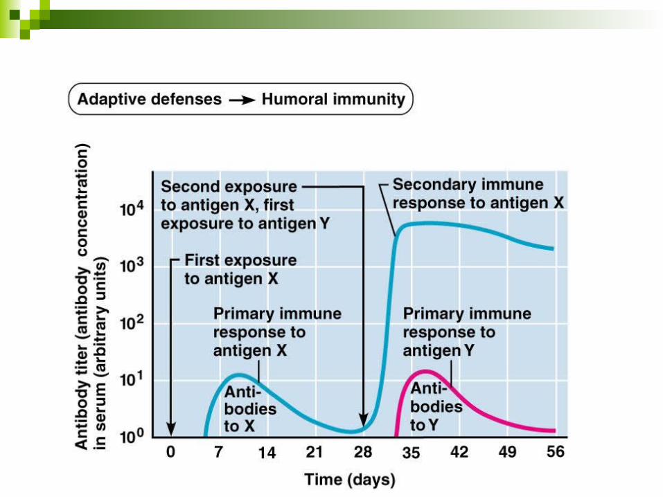
Primary & Secondary Humoral Responses
Figure 21.10
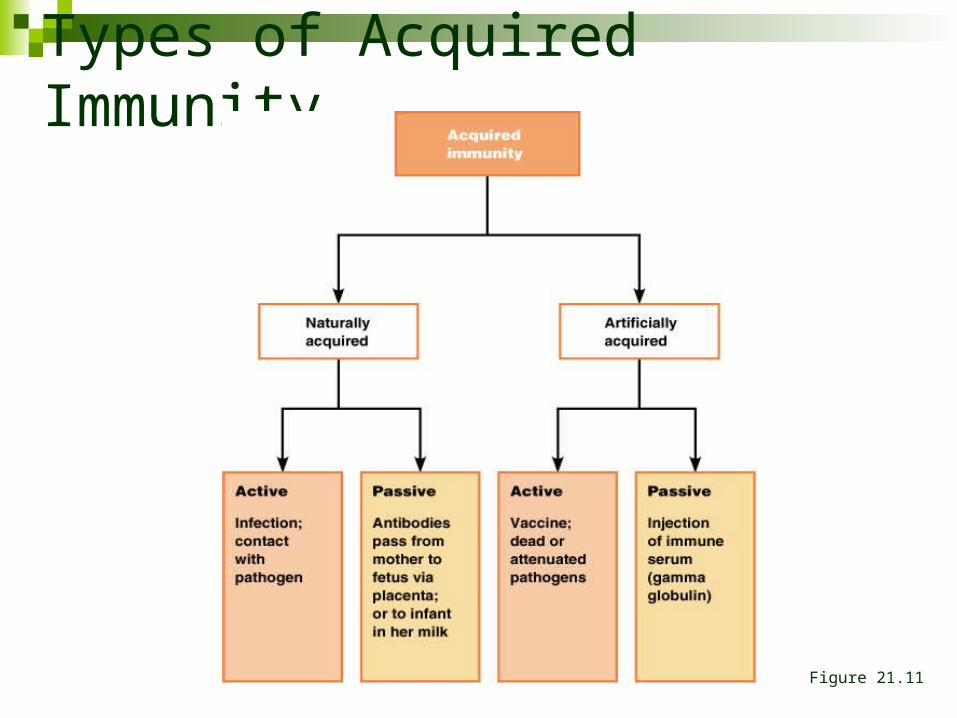
Types of Acquired Immunity
Figure 21.11
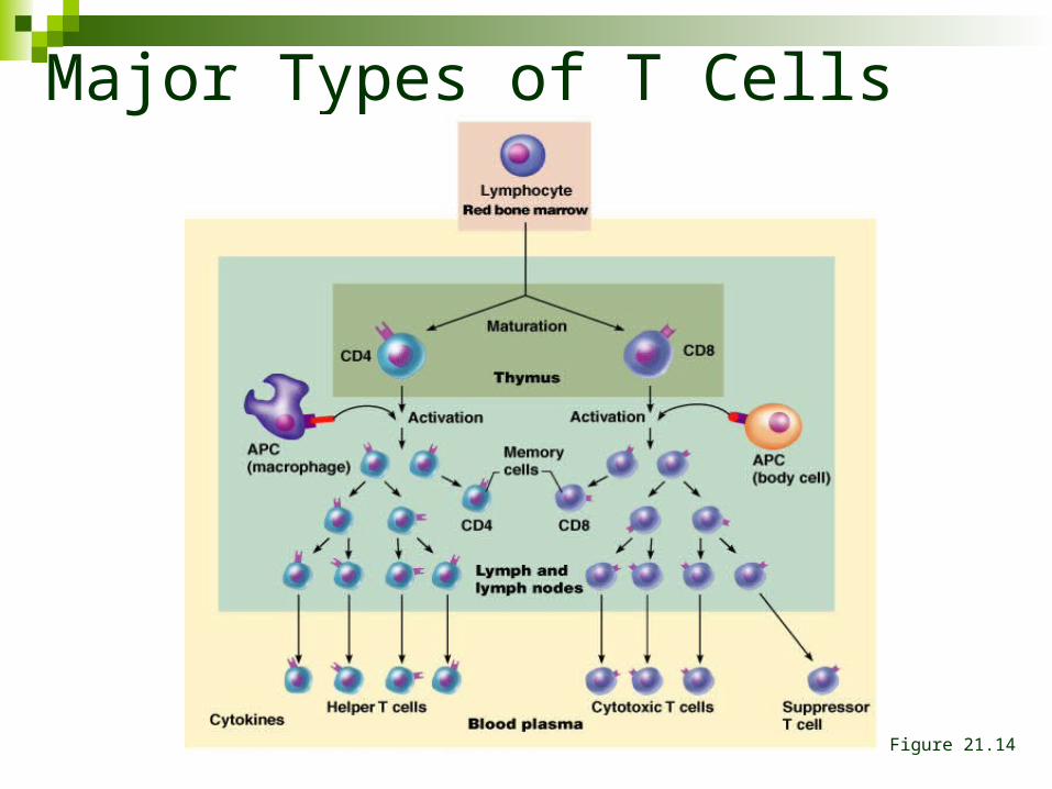
Major Types of T Cells
Figure 21.14
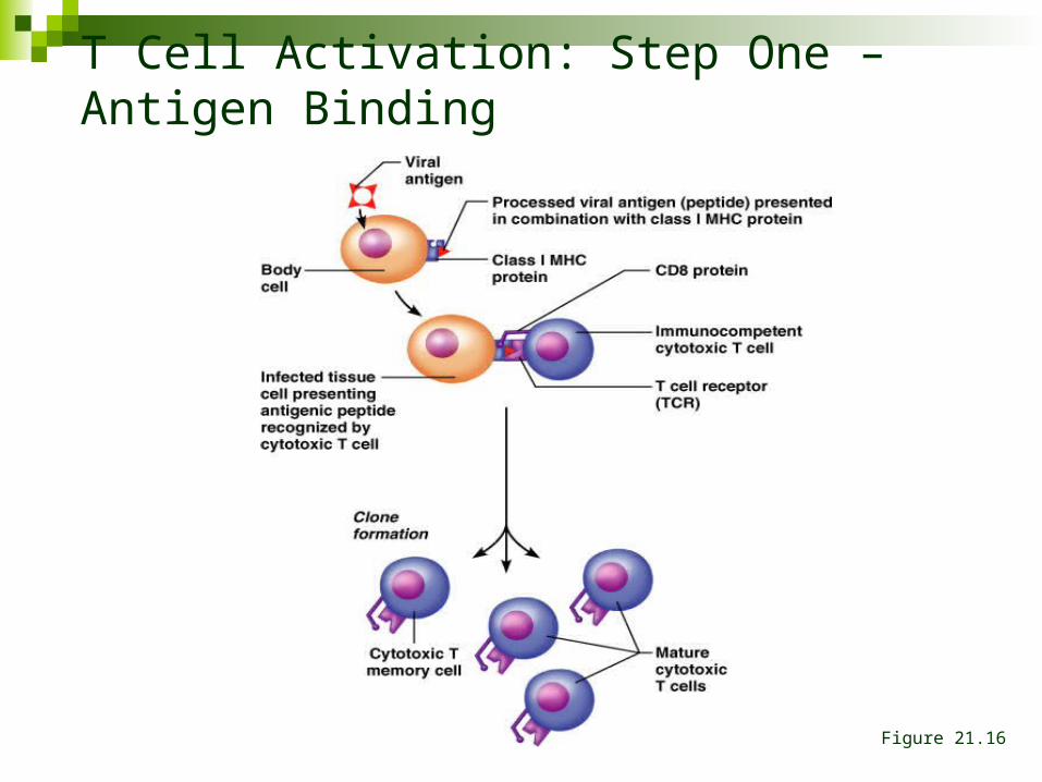
T Cell Activation: Step One – Antigen Binding
Figure 21.16
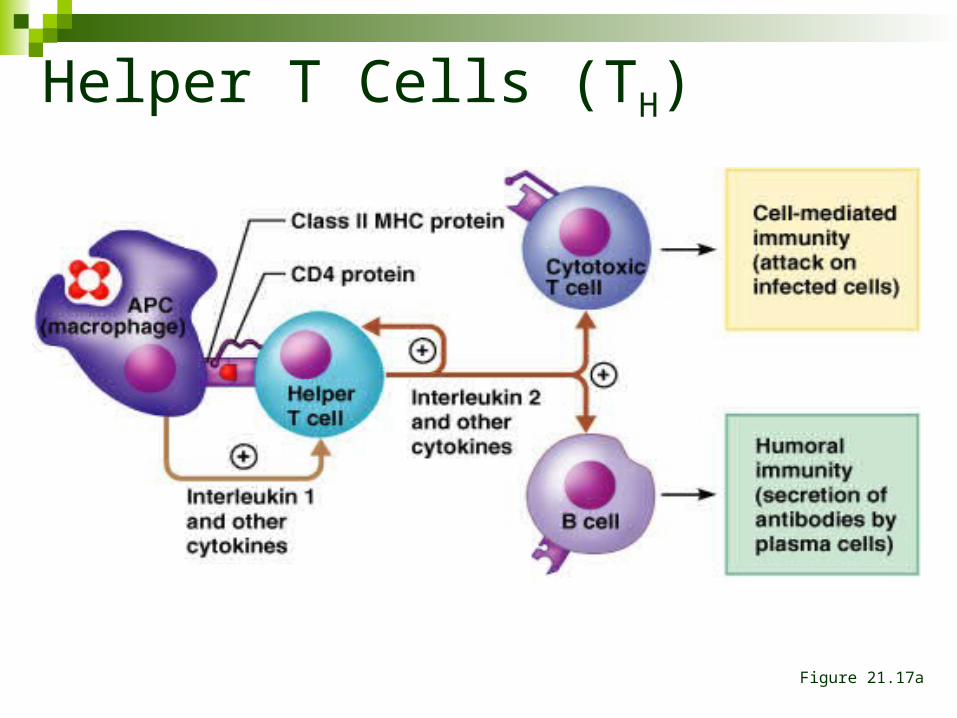
Helper T Cells (TH)
Figure 21.17a
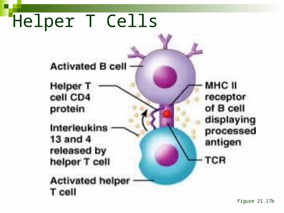
Helper T Cells
Figure 21.17b
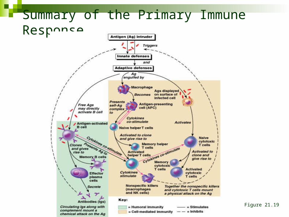
Summary of the Primary Immune Response
Figure 21.19
Recommended