UNIVERSIDADE FEDERAL DE PELOTAS
Programa de Pós-Graduação em Biotecnologia
Tese
Expressão heteróloga e utilização da proteína recombinante EMA-1 de Theileria equi como
imunobiológico
Leandro Quintana Nizoli
Pelotas, 2009

LEANDRO QUINTANA NIZOLI
Expressão heteróloga e utilização da proteína recombinante EMA-1 de Theileria equi como
imunobiológico
Orientador: Fábio Pereira Leivas Leite
Co-orientador: Fabricio Rochedo Conceição
Pelotas, 2009
Tese apresentada ao Programa de Pós-Graduação em Biotecnologia da Universidade Federal de Pelotas, como requisito parcial à obtenção do títulode Doutor em Ciências (área de conhecimento: Doenças Parasitárias).

Dados de catalogação na fonte: Ubirajara Buddin Cruz – CRB-10/901 Biblioteca de Ciência & Tecnologia - UFPel
N737e Nizoli, Leandro Quintana
Expressão heteróloga e utilização da proteína recombinante EMA-1 de Theileria equi como imunobiológico / Leandro Quintana Nizoli; orientador Fábio Pereira Leivas Leite ; co-orientador Fabricio Rochedo Conceição. – Pelotas, 2009. – 67f. : il. – Tese (Doutorado). Programa de Pós-Graduação em Biotecnologia. Centro de Biotecnologia. Universidade Federal de Pelotas. Pelotas, 2009.
1.Biotecnologia. 2.Theileria equi. 3.Imunobiológicos.
4.EMA-1. 5.Equinos. 6.Proteína recombinante. 7.Pichia pastoris. I.Leivas Leite, Fábio Pereira. II.Conceição, Fabrício Rochedo. III.Título
CDD: 636.1

Banca examinadora:
Profa. Dra. Tânia Regina Bettin dos Santos, Universidade Federal de Pelotas
Prof. Dr. Carlos Eduardo Wayne Nogueira, Universidade Federal de Pelotas
Prof. Dr. Renato Andreotti e Silva, EMBRAPA - CNPGC
Prof. Dr. Fábio Pereira Leivas Leite, Universidade Federal de Pelotas

AGRADECIMENTOS
A Deus pela luz que a cada dia me fortalece nos momentos difíceis e alegres
da jornada.
Aos professores e funcionários do Programa de Pós-graduação em
Biotecnologia que contribuíram em definitivo para meu crescimento pessoal e
acadêmico.
Ao Prof. Fábio Leite, pela orientação, compreensão, confiança e amizade
durante o decorrer do trabalho.
Ao professor Fabricio Conceição pelo apoio à pesquisa e orientação para a
realização desta tese.
Ao professor Sergio Silva pela grande amizade, conhecimentos transmitidos e
pelo estímulo à vida acadêmica.
A todos colegas e estagiários que foram parte da história que escrevemos
neste período,
Aos meus familiares e amigos por terem me apoiado e incentivado a seguir
com a realização deste projeto.
A CAPES pelo auxilio financeiro durante o doutorado.
A todos de que de uma forma ou outra tenha colaborado e apoiado a
realização deste trabalho.
Muito Obrigado!

RESUMO
NIZOLI, Leandro Quintana. Expressão heteróloga e utilização da proteína recombinante EMA-1 de Theileria equi como imunobiológico . 2009. 67f. Tese (Doutorado) – Programa de Pós-Graduação em Biotecnologia. Universidade Federal de Pelotas, Pelotas. A Theileriose eqüina é considerada uma das principais doenças parasitárias que acometem os eqüinos, acarretando grande impacto econômico na equinocultura. A doença é causada pelo hematozoário Theileria equi. As perdas econômicas associadas à theileriose eqüina estão relacionadas tanto aos fatores clínicos, quanto à restrição ao trânsito internacional de animais soropositivos, já que animais portadores crônicos são passíveis de reagudizações, gerando perda de performance física e reprodutiva, e são potencialmente disseminadores da enfermidade. Nos últimos anos, os estudos sobre o diagnóstico imunológico e vacinação contra T. equi concentram-se na obtenção de frações antigênicas. Na membrana externa deste protozoário foram caracterizadas proteínas principais de superfície denominadas de EMAs (equi merozoite antigen). Dentre estas, a EMA-1 destaca-se como antígeno para diagnóstico em função de sua conservação entre diversos isolados. Seu papel também tem sido caracterizado como imunógeno por estimular forte resposta humoral com produção de anticorpos em animais infectados, podendo ser usado como ferramenta para imunodiagnóstico dessa doença. EMA-1 é também um potencial candidato como antígeno vacinal no controle da theileriose equina. Neste estudo utilizou-se o sistema eucariótico de expressão baseado na levedura metilotrófica Pichia pastoris, para a produção da proteína EMA-1 de T. equi e a avaliação quanto a sua antigenicidade e imunogenicidade. Quanto a sua antigenicidade, a proteína recombinante foi reconhecida por anticorpos de animais portadores crônicos de T. equi, sugerindo que epítopos nativos foram conservados na proteína recombinante. Também foi observado que a proteína recombinante foi capaz de gerar resposta imune em camundongos vacinados com esta proteína. Os dados obtidos neste estudo demonstram que a levedura P. pastoris é um sistema de expressão heterólogo adequado para a produção da proteína EMA-1 de T. equi, podendo ser utilizada como imunobiológico no desenvolvimento de testes diagnósticos e vacina recombinante. Palavras-chave: Theileria equi, equinos, Pichia pastoris, EMA-1, proteína
recombinante.

ABSTRACT
NIZOLI, Leandro Quintana. Expressão heteróloga e utilização da proteína recombinante EMA-1 de Theileria equi como imunobiológico . 2009. 67f. Tese (Doutorado) – Programa de Pós-Graduação em Biotecnologia. Universidade Federal de Pelotas, Pelotas.
Equine theileriosis is considered to be one of the most important parasitic diseases that affect horses, and has great economic impact on the equine industry. The disease is caused by the etiologic agent Theileria equi, which is classified as a hematozoan. The losses associated with equine theileriosis are related to clinical manifestation as well as restriction to international travel to positive horses. Chronic infected equines suffer the risk of the disease relapse which leads to losses in reproduction performance and are potentially disseminators of the disease. In the last years, studies on the immunologic diagnosis and vaccination against T. equi have focused to obtain distinct antigenic proteins. On the outer membrane of this protozoan, major surface proteins has been characterized and named as EMAs (equi merozoite antigen). Of these, EMA-1 has been used as antigen for diagnosis due to its conservation in diverse isolates. Its role as a potential immunogen has been well documented due its ability to stimulate a humoral response with production of specific antibodies in infected animals. Through this antibodies one can used as tool for immune diagnostic of this disease. EMA-1 is also a strong candidate to be use as a vaccine in the control of equine theileriosis. In this study we used the Pichia pastoris yeast as expression system for the production of the EMA-1 protein of T. equi and evaluated its antigenicity and immunogenicity. When tested for antigenicity, the recombinant protein was recognized by antibodies form chronic T. equi infected horses, suggesting that epitopes of the native were conserved in the recombinant protein. Also we were able to observe that this protein was immunogenic in mice. The data obtained in this study demonstrated that the yeast P. pastoris is an expression system of heterologous protein suitable for the production of EMA-1 from T. equi.
Keywords: Theileria equi, equines, Pichia pastoris, EMA-1, recombinant protein.

SUMÁRIO
1 INTRODUÇÃO GERAL .................................. ................................................................................ 6
2 OBJETIVOS ......................................... ......................................................................................... 10
OBJETIVO GERAL ................................................................................................................................. 10 OBJETIVOS ESPECÍFICOS ..................................................................................................................... 10
3 ARTIGO 1
EQUINE THEILERIOSIS: EPIDEMIOLOGICAL ASPECTS, DIAGN OSTIC AND TREATMENT ....... 12
ABSTRACT ....................................................................................................................................... 13 INTRODUCTION ............................................................................................................................... 14 EPIDEMIOLOGICAL ASPECTS........................................................................................................ 15 DIAGNOSTIC TESTS ........................................................................................................................ 18 TREATMENT ..................................................................................................................................... 19 CONCLUSIONS ................................................................................................................................ 20 REFERENCES .................................................................................................................................. 21
4 ARTIGO 2
CLONING AND EXPRESSION OF MEROZOITE ANTIGEN 1 OF THEILERIA EQUI GENE IN THE METHYLOTROPHIC YEAST PICHIA PASTORIS ............................................................................... 30
ABSTRACT ....................................................................................................................................... 31 INTRODUCTION ............................................................................................................................... 32 MATERIALS AND METHODS ........................................................................................................... 33 RESULTS AND DISCUSSION .......................................................................................................... 38 CONCLUSION................................................................................................................................... 41 ACKNOWLEDGES ............................................................................................................................ 41 REFERENCES .................................................................................................................................. 41 FIGURES ........................................................................................................................................... 44
5 ARTIGO 3
IMUNOGENICIDADE E ANTIGENICIDADE DA PROTEÍNA RECOMB INANTE EMA-1 DE THEILERIA EQUI EXPRESSA EM PICHIA PASTORIS ...................................................................... 49
ABSTRACT ....................................................................................................................................... 49 RESUMO ........................................................................................................................................... 50 INTRODUCTION ............................................................................................................................... 50 MATERIALS AND METHODS ........................................................................................................... 51 RESULTS AND DISCUSSION .......................................................................................................... 53 ACKNOWLEDGEMENTS ................................................................................................................. 55 REFERENCES .................................................................................................................................. 55 FIGURES ........................................................................................................................................... 57
6 CONSIDERAÇÕES FINAIS .............................. ............................................................................ 58
7 CONCLUSÕES GERAIS ................................. ............................................................................. 60
8 REFERÊNCIAS ............................................................................................................................. 61

1 INTRODUÇÃO GERAL
A primeira descrição do parasito foi feita por Guglielmi em 1899 na África do
Sul, sendo posteriormente classificado e descrito como Babesia equi por Laveran
em 1901. No entanto, descobertas a respeito do ciclo de vida do parasito, como
multiplicação em linfócitos e ausência de transmissão transovariana nos carrapatos
vetores (SCHEIN et al., 1981; SCHEIN, 1988; UETI et al., 2008), indicam que T. equi
não é uma babésia clássica e, desde então, sua taxonomia tem sido questionada.
Considerando similaridades do parasito com organismos da família Theileriidae, a
reclassificação de Babesia equi como Theileria equi passou a ser aceita
(MEHLHORN & SCHEIN, 1998). Entretanto, estudos filogenéticos, utilizando RNA
ribossomal e proteínas de superfície de parasitos de ambas as famílias, indicam que
T. equi pode representar um terceiro grupo, diferente de Babesia ou Theileria
(ALLSOPP et al., 1994; KATZER et al., 1998; ZAHLER et al., 2000a; ZAHLER et al.,
2000b; CRIADO-FORNELIO et al., 2003a,b)
A theileriose equina é uma importante doença parasitária que acomete os
equinos de forma endêmica no território brasileiro, assim como, em diversos outros
países (KERBER at al., 1999; PFEIFER et al., 1995). Esta doença vem sendo
estudada à dezena de anos, principalmente em função do elevado número de
distúrbios que pode acarretar aos animais acometidos, bem como as enormes
perdas econômicas na equideocultura mundial (FRIEDHOFF et al., 1990;
KNOWLES, 1996). Nos Estados Unidos, Canadá, Austrália e Japão, assim como,
em alguns países da Europa e América Latina, onde o parasito não ocorre de forma
endêmica, são mantidas rigorosas medidas de controle que impedem a entrada de
animais soropositivos (OIE, 2008). Nestes países, apesar da doença ser
considerada exótica, o risco de tornar-se endêmica é constante devido à existência
dos carrapatos vetores, pertencentes aos gêneros Amblyomma, Dermacentor e
Rhipicephalus (KERBER et al., 1999; GUIMARÃES et al., 1998a,b; BATTSETSEG et
al., 2002; STILLER et al., 2002; UETI et al., 2008). Portanto, medidas de controle,
além dos testes sorológicos, muitas vezes incluem quarentena e controle de

7
carrapatos (MARTIN, 1999). Estes procedimentos, além de extremamente
dispendiosos, afetam negativamente o mercado internacional de eqüinos e a
participação em competições eqüestres internacionais.
O parasito encontra-se mundialmente distribuído em regiões tropicais e
subtropicais, sendo a prevalência da infecção diretamente relacionada com a
ocorrência dos carrapatos vetores. É estimado que 90% da população mundial de
eqüinos esteja exposta à T. equi, ainda que em alguns países a infecção não ocorra
de forma endêmica (FRIEDHOFF et al., 1990). No Brasil, estudos epidemiológicos
utilizando imunofluorescência indireta para detecção de anticorpos anti T. equi têm
registrado prevalências de 49,2% em São Paulo (HEUCHERT et al., 1999), 57,9%
no Rio Grande do Sul (CUNHA et al., 1996), 60,45% em Minas Gerais (RIBEIRO et
al., 1999) e 72% a 100% no Rio de Janeiro (TENTER & FRIEDHOFF, 1986;
PFEIFER et al., 1995).
Infecções por T. equi caracterizam-se pelo desenvolvimento de anemia
hemolítica progressiva nos animais infectados, sendo a patogenia da enfermidade
determinada principalmente pela lise de eritrócitos durante a invasão e multiplicação
do parasito (KNOWLES, Jr. et al., 1994; LORDING, 2008). Quando eqüinos
suscetíveis são infectados desenvolvem a fase aguda da doença, a qual cursa com
febre, anemia, hemorragias petequiais de mucosas, hemoglobinúria e icterícia
(ZOBBA et al., 2008). A gravidade da doença neste estágio depende da virulência
da cepa, dose do inóculo e condição imunológica do animal (CUNHA et al., 1998;
AMBAWAT et al., 1999). A mortalidade em infecções por T. equi é baixa, em geral
os animais recuperam-se da fase aguda da doença, porém permanecem como
portadores do parasito (UETI et al., 2005). Durante a fase crônica da infecção, sinais
clínicos inespecíficos como inapetência, perda de peso e queda no desempenho
físico e reprodutivo são comuns (SCHEIN, 1988). Em casos de imunossupressão a
reagudização da doença é favorecida e os animais podem apresentar diferentes
graus de anemia, com agravamento dos sinais clínicos (OLADOSU, 1988;
OLADOSU & OLUFEMI, 1992; CUNHA et al., 1997; NOGUEIRA et al., 2005).
Animais infectados por T. equi desenvolvem uma sólida imunidade que
protege contra a doença clínica no caso de re-exposições ao parasito (CUNHA et al.,
2006). Esta proteção tem sido atribuída à contínua estimulação do sistema imune
por parasitos que persistem no organismo durante a fase crônica da enfermidade
(SCHEIN, 1988). Experimentos conduzidos em diferentes modelos de infecções com
8
patógenos intraeritrocitários, que possuem uma patogenia semelhante à de T. equi,
propõe que através da produção de interferon gama (IFN-γ), linfócitos T CD4+ podem
ativar macrófagos e estimular a produção de anticorpos por linfócitos B, mecanismos
estes que atuariam na eliminação dos parasitos (IGARASHI et al., 1999; PALMER et
al., 1999; HELMBY & TROYE-BLOMBERG, 2000). Macrófagos ativados não
somente apresentam uma intensa capacidade fagocitária como produzem uma série
de intermediários reativos de oxigênio e nitrogênio que são tóxicos para parasitos
(SU & STEVENSON, 2000; SHODA et al., 2000). Anticorpos por sua vez também
atuam em mecanismos citotóxicos, como opsonização e fixação de complemento,
importantes no controle da multiplicação dos parasitos (AUCAN et al., 2000; CHEN
et al., 2000a; CHEN et al., 2000b; TAYLOR et al., 2001). A participação de anticorpos
na neutralização de merozoítos, impedindo diretamente a invasão de eritrócitos
também tem sido demonstrada (CAVINATO et al., 2001).
Eqüinos infectados com T. equi desenvolvem altos títulos de anticorpos contra
proteínas de superfície de merozoítos, o que sugere que os mesmos estão
envolvidos no controle da multiplicação e eliminação do parasito. Assim sendo, tanto
mecanismos celulares como aqueles dependentes de anticorpos parecem
desempenhar papéis fundamentais no controle de hematozoários (KNOWLES et al.,
1994; CUNHA et al. 2006). Em T. equi, a proteína equi merozoite antigen–1 (EMA-1)
é um antígeno de 34 kDa expresso na superfície do merozoíto, predominantemente
reconhecido por anticorpos de eqüinos infectados por T. equi (KNOWLES et al.,
1997). Comparações entre seqüências de aminoácidos da EMA-1, obtidas de
diferentes isolados de T. equi, têm revelado identidades superiores a 80%
(NICOLAIEWSKY et al., 2001; XUAN et al., 2001).
O desenvolvimento de vacinas representa um campo de grande interesse na
pesquisa envolvendo hemoparasitos de importância veterinária. Os principais
obstáculos nesta área são o desconhecimento a respeito da imunidade protetora
desenvolvida pelo hospedeiro e a grande variedade de mecanismos de evasão do
sistema imune utilizados pelos parasitos (JENKINS, 2001). Vários estudos têm sido
realizados buscando elucidar estes aspectos e um modelo de como o hospedeiro
mantém hemoparasitos sob controle e evita surtos clínicos em subseqüentes
exposições ao agente tem sido proposto (BROWN & PALMER, 1999; BROWN,
2001).

9
Estudos desenvolvimentos no campo da biologia molecular tornaram possível
expressar genes de antígenos de diferentes patógenos em sistemas heterólogos.
Pichia pastoris é uma levedura metilotrófica, capaz de utilizar o metanol como fonte
de carbono e energia que pode ser geneticamente modificada para expressar
proteínas heterólogas (CEREGHINO L.G.P. & CREGG, 1999; CEREGHINO L.J. &
CREGG, 2000). Durante os últimos anos, ela tem se tornado um importante sistema
de produção de uma variedade de proteínas heterólogas (NIZOLI et al., 2007;
DUMMER et al., 2007). A produção destas proteínas recombinantes possibilita
determinar os antígenos que representam os principais alvos da resposta imune dos
hospedeiros vertebrados. A partir da identificação destes antígenos, é possível
testar, por imunização ativa, o potencial imunoprotetor de cada uma destas
proteínas. Estes estudos tornam os antígenos protetores, ou seus epítopos mais
importantes, fortes candidatos para serem utilizados como constituintes de vacinas
recombinantes contra o respectivo patógeno.
Assim, os grandes desafios nas pesquisas envolvendo T. equi concentram-se
principalmente no desenvolvimento de vacinas e no diagnóstico do parasito,
especialmente na detecção de portadores assintomáticos.

2 OBJETIVOS
Objetivo Geral
Produzir a proteína EMA-1 de Theileria equi recombinante em Pichia pastoris.
Objetivos Específicos
- Clonar o gene EMA-1 em sistema Pichia pastoris;
- Expressar a proteína EMA-1 de Theileria equi em levedura Pichia pastoris;
- Avaliar a potencial antigênico e imunogênico da proteína EMA-1.

3 ARTIGO 1
EQUINE THEILERIOSIS: EPIDEMIOLOGICAL ASPECTS, DIAGN OSTIC AND
TREATMENT
Artigo científico formatado segundo as normas da re vista Journal of Equine
Veterinary Science

EQUINE THEILERIOSIS: EPIDEMIOLOGICAL ASPECTS, DIAGN OSTIC 1
AND TREATMENT 2
3
EQUINE THEILERIOSIS 4
5
Leandro Quintana Nizolia, Marcelo Mendes Götzea, Fabricio Rochedo Conceiçãoa, 6
Fábio Pereira Leivas Leiteab* 7
8
9
10
a Centro de Biotecnologia, b Instituto de Biologia Departamento de Microbiologia e 11
Parasitologia, Universidade Federal de Pelotas, Pelotas, RS, Brazil. 12
13
14
* Corresponding author: Phone: +55 53 3227-2770; fax: +55 53 3275-7555; E-mail address: 15
[email protected] (F. P. L. Leite). Centro de Biotecnologia, Universidade Federal de 16
Pelotas, CP 354, CEP: 96010-900, Pelotas, RS, Brazil. 17
18
19
20
21
22
23
24

13
ABSTRACT 25
26
Equine theileriosis is a tick-borne protozoal disease of horses, mules, donkeys and zebras. The 27
etiological agent is blood parasite named Theileria equi. Infected animals may remain carriers 28
of these parasites for long periods and act as sources of infection for ticks, which act as 29
vectors. The parasites are found inside the erythrocytes of the infected animals. Animals 30
which are low-level carriers of T. equi parasite or ticks which may act as reservoirs pose a risk 31
of introduction of these parasites to diseases-free areas as a result of the increased movement 32
of horses worldwide. In this review, the biology, distribution, pathogenicity as well diagnosis 33
and treatment of equine theileriosis are discussed. 34
35
Key words: equine theileriosis, Theileria equi, diagnostic, treatment 36
37
38
39
40
41
42
43
44
45

14
INTRODUCTION 46
47
Equine theileriosis, caused by Theileria equi (formerly Babesia equi) (1), is considered 48
to be the most important tick-borne disease of horse in tropical and subtropical areas 49
including Central and South America, Africa, Asia and Southern Europe (2). At present, only 50
Australia, Canada, New Zealand, Japan, UK, and USA are considered equine piroplasmosis-51
free and have in place stringent regulatory important restrictions to prevent introduction of 52
these parasites (3). 53
T. equi are intraerythrocytic protozoa of equids that are transmitted by ixodid ticks (4). 54
Ticks belonging to the genera Hyalomma, Dermacentor, and Rhipicephalus transmit these 55
parasites (4-7). Recent work has shown the potential for transmission of T. equi by 56
Rhipicephalus (Boophilus) microplus (8-11). 57
T. equi is one of the small species of protozoa and has piroplasms in the erythrocytes 58
that appear oval, circle, ameboid, or as double pears, and measure 2 to 3 µm in length. 59
Theileria spp. has a schizogony period in the lymphoblastoid cells different from the genera 60
Babesia. (12-14). 61
Of the estimated world population of 58 million horses, 90% are bred in endemic areas 62
(2). In endemic countries, the control of equine piroplasmosis is important to keep 63
international markets open to the horse industry (15). For this reason many countries prohibit 64
the importation of horses because of the high prevalence of asymptomatic carrier animals in 65
the region. 66
Sub-clinical infections have particular relevance to the horse-racing industry where the 67
geographical movement of presumably healthy horses may aid in the spread of T. equi or 68
where sub-clinical infection may negatively affect the animal’s performance. It has also been 69
shown that strenuous exercise, such as that experienced with horse-racing, can cause sub-70

15
clinical infections to become acute (16,17). Thus there is a real need for the diagnosis of both 71
clinical and sub-clinical infections. In this review, the biology, distribution, pathogenicity as 72
well diagnostic and treatment of equine theileriosis are discussed. 73
74
EPIDEMIOLOGICAL ASPECTS 75
76
Thoroughbred racing industry is particularly strong affected by equine theileriosis, 77
acute infections resulting in missed training sessions and races and hence serious loss of 78
income to owners and trainers. Income is also lost to owners through abortion in stud mares 79
which are T. equi carriers (2,18). 80
Authors have demonstrated occurrences varying from 49.2% to 100% in the southern 81
and south-eastern states of Brazil. They cited different epidemiologic conditions but with high 82
tick infections (19-24). 83
The effects of the variation of prevalence has been observed with different categories 84
and breeding systems in different regions of Brazil (21,25). Previous studies have shown that 85
the prevalence of equine theileriosis in Brazil is serious (26-28). Transmission of theileriosis 86
is usually influenced by the dynamics of vector populations, and these are directly influenced 87
by climatic conditions (29). 88
Animals sensitive to the disease die within 24 to 48 hours after the development of 89
first clinical signs (30). In chronic cases, the disease continues for months and these animals 90
deteriorate to a worse condition within 3 to 4 years. The protozoa may not be seen 91
microscopically during the subclinical phase of the disease; therefore, serologic tests need to 92
be done to diagnose the disease (31-33). 93
Clinical signs of infection vary from asymptomatic to acute, fever, jaundice, anaemia, 94
icterus, and even death may occur (30). Intra-uterine infections with T. equi may result in 95
16
abortion and neonatal death (30,34). Horses that recover from acute infection may act as 96
reservoirs for transmission to ticks which subsequently infect other equids. Various authors 97
postulate that T. equi persists for life-long infection in its equine host (2,18,35,36). 98
In T. equi infection, clinical parasitaemia may exceed 20% but 1-5% parasitaemia is 99
more commonly observed in field conditions (37). In latent carrier equids, it is very difficult 100
to demonstrate the parasite in stained blood smears because the parasitaemia is extremely low. 101
Equids reared in endemic areas usually get infected at early age and become immune tolerant 102
throughout their life span. Nevertheless, outbreak of T. equi infection may occur in these 103
equids consequent to unfavorable health conditions (16). 104
Foals born of pre immune mares are naive at birth and acquire passive immunity 105
through colostrum (38). Maternal antibodies, against T. equi, decline steadily to extinction by 106
about four months from birth (38); however in endemic areas it is expected that primary 107
infection takes place before they decrease. It has been demonstrated that under these 108
conditions foals acquire the infection shortly after birth with the majority showing patent 109
parasitemia before 42 days of age (39). Regarding T. equi, transplacental transmission can 110
occurs and takes place during the first four months of gestation and appears to be a rule rather 111
than an exception, as recently demonstrated by a DNA probe (40). Recently was demonstrated 112
the presence of DNA of T. equi parasite in neonate foal’s blood born from a latently infected 113
mare, using specific DNA probe (41). They opined that parasite transmission occurred during 114
pregnancy despite the fact that latently infected mare was having normal placenta as the 115
foaling and foals were normal. Passage of infected erythrocytes or extra cellular parasitic 116
form across the placental barrier is the probable mode of transmission of T. equi and this may 117
occur as a result of damage to placental vessel in the event of abortion which could have lead 118
to mixing of maternal and foetal blood (42). 119

17
Kumar et al. (43) indicated that new-born foals were born naive and passively 120
transferred immunity is transitory which wanes after a period of time rendering the foals 121
susceptible to natural T. equi infection, after 63-77 days after foaling. This will help in 122
reducing the losses due to disease condition in new-born foals by following suitable 123
management practices at farm after waning of antibody titers. 124
Positive titers for T. equi from the first day of life on can be explained by maternal 125
antibodies, especially in those cases where the foals became negative thereafter. This occurred 126
between the first and fourth month. Maternal antibodies for T. equi have been demonstrated 127
before until about 4 months of age (in the CF test) in foals of field-infected mares (38). 128
Immunocompetent young horses infected with T. equi preferentially produce 129
antibodies to erythrocyte-stage antigens of 30 and 34 kDa during resolution of acute infection 130
(15,44). These antigens (equi merozoite antigens-1 and -2) are erythrocyte-stage which 131
possess a surface epitope that is conserved worldwide and induce specific high antibody 132
levels (15, 45, 46). Knowles Jr.(1991) had shown that a protein epitope shared by EMA-1 and 133
EMA-2 was immunodominant for antibody induction and shared by isolates worldwide (46). 134
To survive and replicate in the erythrocyte, members of the Apicomplexa phylum and 135
the intra-erythrocytic parasite Plasmodium falciparum export proteins that interacts with and 136
dramatically modify the properties of the host erythrocyte. As part of this process, P. 137
falciparum appears to establish a system within the erythrocyte cytosol that allows the correct 138
trafficking of parasite proteins to their final cellular destinations (47). 139
The pathways and components of these complex trafficking processes are fundamental 140
to the survival of P. falciparum in vivo, and are major determinants of this parasite’s unique 141
pathogenicity (47). Recently has been showed that the parasite T. equi EMA-2 in the infected 142
erythrocytic cytoplasm can be exported to the membrane and this can affect the parasite’s 143
erythrocytic binding behavior (48). 144

18
DIAGNOSTIC TESTS 145
146
Several direct and indirect detection methods, including blood smears (49), in-vitro 147
cultures (50,51), DNA probes (13) and serology (52), have been used to diagnose T. equi 148
infection. 149
Microscopic detection from blood smears has been used for the most standard 150
diagnosis of equine theileriosis (53); however, it has many limitations. The technique is 151
relatively laborious when large numbers of blood smear samples need to be simultaneously 152
quantified. While the identification of parasites in blood smears constitutes the definitive 153
diagnosis of equine infection, it bears certain limitations, particularly during apparent or 154
chronic infection due to low parasitemias (54). 155
Among the molecular tools, PCR is the most commonly used, including other PCR-156
derived tools, such as the multiplex and nested PCR (13,55). However, these molecular tools 157
require thermo cyclers and several other expensive operations, which make them unsuitable 158
for routine diagnosis, especially in resource-poor countries (56). 159
A primary PCR has been shown to detect T. equi infection in horses however, in all 160
cases the parasitemia level was sufficiently high for the organism to be detected by 161
microscopic examination (30). More recently, a nested PCR for T. equi based on the 162
merozoite antigen ema-1 gene sequence has been reported to give increased sensitivity (13). 163
A variety of serological tests such as indirect fluorescent antibody test (IFAT), 164
enzyme-linked immunosorbent assay (ELISA) and complement fixation test (CFT) have been 165
used to detect specific antibodies. Both the complement fixation test and the 166
immunofluorescent antibody test have been used for many years for diagnosis (57). 167

19
These methods have a low sensitivity for detecting latent infections. However, it has 168
been reported that because of its low sensitivity and specificity, the complement fixation test 169
fails to discriminate accurately between negative and carrier animals (19). 170
Compared with ELISA, however, IFAT is time consuming and requires large amounts 171
of parasites. Moreover, the estimation of the intensity of fluorescence is subjective and 172
requires the participation of experts, which has hindered the standardization and 173
comparability of the results (58). 174
Recently, the competitive ELISA (cELISA) using recombinant antigens was 175
developed as a more specific method than CFT, IFAT and other iELISA for the serodiagnosis 176
of piroplasmosis (32,47,59-64). Thus, several serological assays such as ELISAs are often 177
more sensitive and specific have been developed to advance diagnosis of equine 178
piroplasmosis. 179
A competitive inhibition ELISA, employing a monoclonal antibody and a recombinant 180
T. equi merozoite protein (EMA-1), was shown to be specie-specific for anti-T. equi 181
antibodies, as horses infected with B. caballi tested negative (46). It has been shown that an 182
ELISA using recombinant EMA-1 expressed in insect cells by baculovirus (65) and Pichia 183
pastoris (66) can be a useful diagnostic reagent for detection of T. equi infection in horses, 184
being more sensitive than CFT and IFAT. 185
186
TREATMENT 187
188
Treatment of piroplasmosis varies depending on the location of the horse and the goal 189
of treatment. In endemic regions, suppressing clinical signs without eliminating the organism 190
from the body is desirable because premonition depends on the continued presence of the 191
parasite at low levels (67). 192

20
The use of the dipropionate of imidocarb is recommended in solution 10%, 2.4.mg.kg1 193
four times, with break of 72 hours. However, the protocol of one dose of 1.2 mg.kg1 is 194
efficient to T. equi, not presenting difference of the treated animals with 2.4 mg.kg1, with the 195
advantages of reduce costs and diminish risks of effects to the horses (17). 196
Babesiacidal/theilericidal drugs used in the treatment of equine theileriosis are limited 197
and are either ineffective in completely eliminating the parasites and/or cause severe side 198
effects (68,69). Thus, a continuous search for alternative and effective chemotherapeutic 199
drugs is necessary. 200
The most reliable method to control equine piroplamosis remains preventing entry of 201
infected equine and ensuring that animals entering from endemic countries are thoroughly 202
checked and found to be free of ticks. 203
204
CONCLUSIONS 205
206
As a result, equine theileriosis is a very important equine disease. Serologic 207
examination is better than microscopic examination to determine the prevalence of the 208
disease. To combat the disease, the animal owners should be informed about the importance 209
of the disease and the danger of tick infestation. Considering the high prevalence of T. equi 210
infection in Brazil further studies are required to define the population of the infection. 211
Control strategies for horses used for recreational riding should based on reducing their 212
exposure to ticks by preventing them grazing areas with high tick infestation, preventing 213
contact with high tick infestation, preventing contact with wild horses, spraying horses with 214
acaricides and treating positive horses. In conclusion, the hardest challenges in the researches 215
involving T. equi concentrate mainly in the development of treatment protocols and in the 216
diagnosis of the parasite, especially in the detection of chronic cases of equine theileriosis. 217

21
REFERENCES 218
219
1. Mehlhorn H, Schein E. Redescription of Babesia equi Laveran, 1901 as Theileria equi. 220
Parasitol Res 1998;84:467-475. 221
2. Schein E. Equine babesiosis. In: Babesiosis of Domestic Animals and Man, Ristic M (Ed.), 222
CRC Press, Inc., Boca Raton, FL, 1988, pp. 197-208. 223
3. Friedhoff KT, Tenter AM, Muller I. Haemoparasites of equines: impact on international 224
trade of horses. Rev Sci Tech Off Int Epiz 1990;9:1187-1194. 225
4. Friedhoff KT. Transmission of babesia. In: Babesiosis of Domestic Animals and Man, 226
Ristic M. (Ed.), CRC Press, Boca Raton, FL, 1988. pp. 23-52. 227
5. Kumar S, Malhotra DV, Sangwan AK, Goel P, Kumar A , Kumar S. Infectivity rate and 228
transmission potential of Hyalomma anatolicum anatolicum ticks for Babesia equi 229
infection. Vet Parasitol 2007;144:338-343. 230
6. Battsetseg B, Lucero S, Xuan X, Claveria F, Byambaa B, Battur B, Boldbaatar D, Batsukh 231
Z, Khaliunaa T, Battsetseg G, Igarashi I, Nagasawa H, Fujisaki K. Detection of equine 232
Babesia spp. gene fragments in Dermacentor nuttalli Olenev 1929 infesting mongolian 233
horses, and their amplification in egg and larval progenies. J Vet Med Sci 2002;64:727-234
730. 235
7. Stiller D, Goff WL, Johnson LW, Knowles DP. Dermacentor variabilis and Boophilus 236
microplus (Acari: Ixodidae): experimental vectors of Babesia equi to equids. J Med 237
Entomol 2002;39:667-670. 238
8. Ueti MW, Palmer GH, Kappmeyer LS, Statdfield M, Scoles GA, Knowles DP. Ability of 239
the vector tick Boophilus microplus to acquire and transmit Babesia equi following feeding 240
on chronically infected horses with low-level parasitemia. J Clin Microbiol 2005;43:3755-241
3759. 242

22
9. Ueti MW, Palmer GH, Kappmeyer LS, Scoles GA, Knowles DP. Expression of equi 243
merozoite antigen 2 during development of Babesia equi in the midgut and salivary gland 244
of the vector tick Boophilus microplus. J Clin Microbiol 2003;41:5803-5809. 245
10. Battsetseg B, Lucero S, Xuan X, Claveria FG, Inoue N, Alhassan A, Kanno T, Igarashi I, 246
Nagasawa H, Mikami T, Fujisaki K. Detection of natural infection of Boophilus microplus 247
with Babesia equi and Babesia caballi in Brazilian horses using nested polymerase chain 248
reaction. Vet Parasitol 2002;107:351-357. 249
11. Guimarães AM, Lima JD, Ribeiro MFB. Sporogony and experimental transmission of 250
Babesia equi by Boophilus microplus. Parasitol Res 1998;84:323-327. 251
12. Inci A. Detection of Babesia caballi (Nuttall, 1910) and Babesia equi (Laveran, 1901) in 252
horses by microscopic examination in a military farm in Gemlik. Tr J Vet Anim Sci 253
1997;21:43-46. 254
13. Nicolaiewsky TB, Richte MF, Lunge VR, Cunha CW, Delagostin O, Ikuta N, Fonseca 255
AS, Silva SS, Ozaki LS. Detection of Babesia equi (Laveran, 1901) by nested polymerase 256
chain reaction. Vet. Parasitol 2001;101:9-21. 257
14. Kettle DS. Medical and veterinary entomology. 2nd ed. UK: CAB International, 1995. 258
15. Knowles DP. Control of Babesia equi parasitemia. Parasitol Today 1996;12:195-198. 259
16. Hailat NQ, Lafi SQ, Al-Darraji AM, Al-Ani FK. Equine babesiosis associated with 260
strenuous exercise: clinical and pathological studies in Jordan. Vet Parasitol 1997;69:1-8. 261
17. Ribas LM, Silva SS, Nogueira CEW, Nizoli LQ, Krause LE. Avaliação da eficácia 262
terapêutica do dipropionato de imidocarb no tratamento de eqüinos durante infecção aguda 263
por Babesia equi. A Hora Veterinária 2003;139:21-24. 264
18. Ueti MW, Palmer GH, Scoles GA, Kappmeyer LS, Knowles DP. Persistently infected 265
horses are reservoirs for intrastadial tick-borne transmission of the apicomplexan parasite 266
Babesia equi. Infect Immun 2008;76:3525-3529. 267

23
19. Tenter AM, Friedhoff KT. Serodiagnosis of experimental and natural Babesia equi and B. 268
caballi infections. Vet Parasitol 1986;20:49-61. 269
20. Cunha CW. Babesiose Eqüina: Padronização da Reação de Imunofluorescência Indireta 270
para sorodiagnóstico em eqüinos Puro Sangue Inglês. Pelotas-RS. 57p. Dissertação 271
(Mestrado) - Curso de Pós-Graduação em Veterinária, Universidade Federal de Pelotas, 272
1993. 273
21. Cunha CW, Silva SS, Pimentel CA, Dapper E. Avaliação da freqüência de eqüinos 274
soropositivos a Babesia equi no Jóquei Clube de Pelotas e em dois Haras da zona sul do 275
Rio Grande do Sul, RS. Rev Bras Paras Vet 1996;5:119-122. 276
22. Souza AP, Bellato V, Sartor AA, Silva AB. Prevalência de anticorpos anti-Babesia equi 277
em eqüinos no planalto catarinense. Ciência Rural 2000;30:119-121. 278
23. Xuan X, Nagai A, Battsetseg B, Fukumoto S, Makala LH, Inoue N, Igarashi I, Mikami T, 279
Fujisaki K. Diagnosis of equine piroplasmosis in Brazil by serodiagnostic methods with 280
recombinant antigens. J Vet Med S 2001;63:1159-1160. 281
24. Heim A, Passos LM, Ribeiro MF, Costa-Júnior LM, Bastos CV, Cabral DD, Hirzmann J, 282
Pfister K. Detection and molecular characterization of Babesia caballi and Theileria equi 283
isolates from endemic areas of Brazil. Parasitol Res 2007;102:63-68. 284
25. Botteon PT, Botteon RCCM, Linhares GFC, Massard CL, Loss ZG. Seroprevalencia de 285
Babesia equi en tres diferentes sistemas de crianza de equinos de Brasil. Parasitol 286
Latinoam 2002; 57:141-145. 287
26. Kerber CE, Ferreira F, Pereira MC. Control of equine piroplasmosis in Brazil. Onder J 288
Vet Res 1999;66:123-127. 289
27. Heuchert CMS, Giulli V, Athaide DF, Böse R, Friedhoff KT. Seroepidemiologic studies 290
on Babesia equi and Babesia caballi infections in Brazil. Vet Parasitol 1999;85:1-11. 291

24
28. Ribeiro MF, Costa JO, Guimarães AM. Epidemiological aspects of Babesia equi in horses 292
in Minas Gerais, Brazil. Vet Res Commun 1999;23:385-390. 293
29. Pfeifer BI, Bose R, Peymann B, Friedhoff KT. Epidemiological aspects of equine 294
babesioses in a herd of horses in Brazil. Vet Parasitol 1995;58:1-8. 295
30. Zobba R, Ardu M, Niccolini S, Chessa B, Manna L, Cocco R, Parpagliae MLP. Clinical 296
and laboratory findings in Equine Piroplasmosis. J Equine Vet Science 2008;28:301-308. 297
31. Bashiruddin JB, Camma C, Rebelo E. Molecular detection of Babesia equi and Babesia 298
caballi in horse blood by PCR amplification of parts of the 16S rRNA gene. Vet Parasitol 299
1999;84:75-83. 300
32. Ikadai H, Osorio CR, Xuan X, Igarashi I, Nagasawa H, Fujisaki K, Suzuki N, Mikami T. 301
Detection of Babesia caballi infection by enzyme-linked immunosorbent assay using 302
recombinant 48-kDa merozoite rhoptry protein. Int J Parasitol 2000;30: 633-635. 303
33. Shkap V, Cohen I, Leibovitz B, Savitsky I, Pipano E, Avni G, Shofer S, Giger U, 304
Kappmeyer L, Knowles D. Seroprevalence of Babesia equi among horses in Israel using 305
competitive inhibition ELISA and IFA assays. Vet Parasitol 1998;76:251-259. 306
34. Phipps LP, Otter A. Transplacental transmission of Theileria equi in two foals born and 307
reared in the United Kingdom. Vet Rec 2004;154:406-408. 308
35. Hourrigan JL, Knowles RC. Equine piroplamsosis (EP). Am Assoc Equine Pract 309
1979;1:119-128. 310
36. De Waal DT, Van Heerden J. Equine babesiosis. In: Infectious Diseases of Livestock with 311
Special Reference to South Africa, Coetzer J, Thomson GR, Tustin RC (Eds), Oxford 312
University Press, Cape Town, 1994, pp. 295-304. 313
37. Kumar S, Kumar R. Diagnosis of Babesia equi infection: an update on the methods 314
available. CAB Reviews: Perspectives in Agriculture, Veterinary Science, Nutrition and 315
Natural Resources 2007; 2:1-14. 316

25
38. Donnelly J, Phipps LP, Watkins KL. Evidence of maternal antibodies to Babesia equi and 317
Babesia caballi in foals of seropositive mares. Equine Vet J 1982;14:126-128. 318
39. Ribeiro MFB, Saito JF, Pimentel PV. Babesiose equina. Primo-infecção de potros em área 319
endêmica. Arq Bras Med Vet Zootec 1995;47:641-647. 320
40. Lewis B, Penzhorn B, Allsopp P. Transplacental transmission of Babesia (Theileria) equi 321
in horses: the rule rather than exception? Fifth Biennial Conference STVM, Proceedings, 322
Key West, p.113, 1999. 323
41. Allsopp MT, Lewis BD, Penzhorn BL. Molecular evidence for transplacental transmission 324
of Theileria equi from carrier mares to their apparently healthy foals. Vet Parasitol 325
2007;148:130-136. 326
42. Erbsloh JK. Babesiosis in the newborn foal. J Reprod Fertil Suppl. 1975;23:725-726. 327
43. Kumar S, Kumar R, Gupta AK, Dwivedi SK. Passive transfer of Theileria equi antibodies 328
to neonate foals of immune tolerant mares. Vet Parasitol 2008;151:80-85. 329
44. Knowles DP, Kappmeyer LS, Perryman LE. Specific immune responses are required to 330
control parasitemia in Babesia equi infection. Infect Immun 1994;62:1909-1913. 331
45. Kappmeyer LS, Perryman LE, Knowles DP. A Babesia equi gene encodes a surface 332
protein with homology to Theileria species. Mol Biochem Parasitol 1993;62:121-124. 333
46. Knowles DP, Perryman LE, Kappmeyer LS, Hennager SG. Detection of equine antibody 334
to Babesia equi merozoite protein by a monoclonal antibody based competitive inhibition 335
enzyme linked immunosorbent assay. J Clin Microbiol 1991;29:2056- 2058. 336
47. Cooke BM, Lingelbach K, Bannister LH, Tilley L. Protein trafficking in Plasmodium 337
falciparum-infected red blood cells. Trends Parasitol 2004;20:581-589. 338
339
340

26
48. Kumar S, Yokoyama N, Kim JY, Huang X, Inoue N, Xuan X, Igarashi I, Sugimoto C. 341
Expression of Babesia equi EMA-1 and EMA-2 during merozoite developmental stages in 342
erythrocyte and their interaction with erythrocytic membrane skeleton. Mol Biochem 343
Parasitol 2004;133:221-227. 344
49. Retief GB. A comparison of equine piroplasmosis in South Africa and the United States. J 345
Am Vet Med Assoc 1964;145:912-916. 346
50. Holman PJ, Becu T, Bakos E, Polledo G, Cruz D, Wagner GG. Babesia equi field isolates 347
cultured from horse blood using a microcentrifuge method. J Parasitol 1998;84:696-699 348
51. Alhassan A, Govind Y, Tam NT, Thekisoe OM, Yokoyama N, Inoue N, Igarashi I. 349
Comparative evaluation of the sensitivity of LAMP, PCR and in vitro culture methods for 350
the diagnosis of equine piroplasmosis. Parasitol Res 2007;100:1165-1168. 351
52. Huang X, Xuan X, Yokoyama N, Katayama Y, Anzai T, Igarashi I. Evaluation of 352
enzyme-linked immunosorbent assays with recombinant antigens for the serodiagnosis of 353
equine Babesia infections. Vet Parasitol 2006;140:158-161 354
53. Bose R, Jorgensen WK, Dalgliesh RJ, Friedhoff KT, De vos AJ. Current state and future 355
trends in the diagnosis of babesiosis. Vet Parasitol 1995;57:61-74. 356
54. Krause PJ, Speilman TA, Ryan R, Magera J, Rajan TV, Christianson D, Alberghini TV, 357
Bow L, Persing D. Comparison of PCR with blood smear and inoculation of small animals 358
for diagnosis of Babesia microti parasitemia. J Clin Microbiol 1996;34:2791-2794. 359
55. Alhassan A, Pumidonming W, Okamura M, Hirata H, Battsetseg B, Fujisaki K, 360
Yokoyama N, Igarashi I. Development of a single-round and multiplex PCR method for 361
the simultaneous detection of Babesia caballi and Babesia equi in horse blood. Vet 362
Parasitol 2005;129:43-49. 363
364

27
56. Alhassan A, Thekisoe OM, Yokoyama N, Inoue N, Motloang MY, Mbati PA, Yin H, 365
Katayama Y, Anzai T, Sugimoto C, Igarashi I. Development of loop-mediated isothermal 366
amplification (LAMP) method for diagnosis of equine piroplasmosis. Vet Parasitol 367
2007;143:155-160. 368
57. Bruning A. Equine piroplasmosis an update on diagnosis, treatment and prevention. Br 369
Vet J 1996;152:139-151. 370
58. Huang X, Xuan X, Yokoyama N, Katayama Y, Anzai T, Igarashi I. Evaluation of 371
enzyme-linked immunosorbent assays with recombinant antigens for the serodiagnosis of 372
equine Babesia infections. Vet Parasitol 2006;140:158-161. 373
59. Knowles DP, Kappmeyer LS, Stiller D, Hennager SG, Perryman LE. Antibody to a 374
recombinant merozoite protein epitope identifies horses infected with Babesia equi. J Clin 375
Microbiol 1992;30:3122-3126. 376
60. Knowles DP, Kappmeyer LS, Perryman LE. Genetic and biochemical analysis of 377
erythrocyte-stage surface antigens belonging to a family of highly conserved proteins of 378
Babesia equi and Theileria species. Mol Biochem Parasitol 1997;90:69-79. 379
61. Katz J, Dewald R, Nicholson J. Procedurally similar competitive immunoassay systems 380
for the serodiagnosis of Babesia equi, Babesia caballi, Trypanosoma equiperdum, and 381
Burkholderia mallei infection in horses. J Vet Diagn Invest 2000;12:46-50. 382
62. Rhalem A, Sahibi H, Lasri S, Johnson WC, Kappmeyer LS, Hamidouch A, Knowles DP, 383
Goff WL. Validation of a competitive enzyme-linked immunosorbent assay for diagnosing 384
Babesia equi infections of Moroccan origin and its use in determining the seroprevalence 385
of B. equi in Morocco. J Vet Diagn Invest 2001;13: 249-251. 386
387
388

28
63. Xuan X, Chahan B, Huang X, Yokoyama N, Makala LH, Igarashi I, Fujisaki K, 389
Maruyama S, Sakai T, Mikami T. Diagnosis of equine piroplasmosis in Xinjiang province 390
of China by the enzyme-linked immunosorbent assays using recombinant antigens. Vet 391
Parasitol 2002;108:179-182. 392
64. OIE, 2007. Manual of Diagnostic Tests and Vaccines for Terrestrial Animals, equine 393
piroplasmosis. Available at: http://www.oie.int/eng/normes/mmanual/A_00084.htm 394
(accessed January 2009). 395
65. Xuan X, Larsen A, Ikadai H, Tanaka T, Igarashi I, Nagasawa H, Fujisaki K, Toyoda Y, 396
Suzuki N, Mikami T. Expression of Babesia equi merozoite antigen 1 in insect cells by 397
recombinant baculovirus and evaluation of its diagnostic potential in an enzyme-linked 398
immunosorbent assay. J Clin Microbiol 2001;39:705-709. 399
66. Nizoli LQ, Conceição FR, Silva SS, Dummer LA, Santos Jr AG, Leite FPL. 400
Immunogenicity and antigenicity of the recombinant EMA-1 protein of Theileria equi 401
expressed in the yeast Pichia pastoris. Brazil J Vet Parasitol 2009;18:in press. 402
67. Sellon DC. Disorders of the hematopoietic system. In: Equine internal medicine, Reed M, 403
Bayly WM, Sellon DC (eds), Philadelphia, Elsevier, 2004, 735p. 404
68. Kuttler KL, Zaugg JL, Gipson CA. Imidocarb and parvaquone in the treatment of 405
piroplasmosis (Babesia equi) in equids. Am J Vet Res 1987;48:1613-1616. 406
69. Zaugg JL, Lane VM. Efficacy of buparvaquone as a therapeutic and clearing agent of 407
Babesia equi of European origin in horses. Am J Vet Res 1992;53:1396-1399. 408
409

4 ARTIGO 2
CLONING AND EXPRESSION OF MEROZOITE ANTIGEN 1 OF Theileria equi
GENE IN THE METHYLOTROPHIC YEAST Pichia pastoris
Artigo científico formatado de acordo com as normas da revista Protein
Expression and Purification
CLONING AND EXPRESSION OF MEROZOITE ANTIGEN 1 OF Theileria equi
GENE IN THE METHYLOTROPHIC YEAST Pichia pastoris
Leandro Quintana Nizolia, Luana Alves Dummera, Fabricio Rochedo Conceiçãoa, Carlos Gil
Turnesa, Fábio Pereira Leivas Leitea,b
a Centro de Biotecnologia, b Departamento de Microbiologia e Parasitologia, Instituto de
Biologia, c Laboratório de Doenças Parasitárias, Faculdade de Veterinária, Universidade
Federal de Pelotas (UFPel), Pelotas, RS, Brazil;
* Corresponding author: Phone: +55 53 3227-2770; fax: +55 53 3275-7353. E-mail address:
[email protected] (F. P. L. Leite). Universidade Federal de Pelotas, CP 354, CEP,
96010-900, Pelotas, RS, Brazil.

31
ABSTRACT
The equine piroplasmosis caused by Theileria equi is a tick-borne protozoal infection of
horses, causing damage to animal health and economic losses. In T. equi, two merozoite
surface proteins, equi merozoite antigen EMA-1 and EMA-2, have been identified as the most
immunodominant antigens, suggesting that they might be used as immunobiological tools.
This study focused on expression and purification of truncated recombinant EMA-1 of T. equi
in P. pastoris. The DNA encoding EMA-1 was cloned by PCR from strain Pelotas of T. equi.
After PCR and construction of expression vector, EMA-1 was expressed in P. pastoris yeast
X-33 and secreted into the culture medium. The secreted EMA-1 was purified using
ammonium sulfate at 80%. The recombinant protein was imunogenic and antigenic. This
study provides a new alternative for expression and utilization of recombinant protein EMA-
1.
Key Words: Theileria equi; Recombinant vaccine; Pichia pastoris; EMA-1
Running headline: Truncated EMA-1 expressed in yeast Pichia pastoris

32
INTRODUCTION
The equine piroplasmosis caused by Theileria equi is a tick-borne protozoal infection
of horses, it causes great damage to animal health and important economical losses. The
disease is a haemoparasitosis caused by Theileria equi and Babesia caballi, is a tick-borne
hemoprotozoan disease affecting horses worldwide [1]. Through infecting and destroying
erythrocytes, it can compromise the equine function, leading to loss of vitality and decrease in
the performance of infected animals. Equine piroplasmosis caused by T. equi is more
pathogenic and widespread in horses than that by B. caballi [2], it causes a persistent infection
for which drug therapy or vaccination are not available [1,3]. The T. equi infection is
characterized by fever, anemia, icterus, and hepato and splenomegaly [4].
Merozoite surface proteins are important in the pathogenesis of hemoprotozoan
diseases because of their role in parasite recognition, attachment to and penetration of host
erythrocytes [5,6]. In T. equi, two kinds of merozoite surface proteins, equi merozoite antigen
EMA-1 and EMA-2, have been identified as the most immunodominant antigens [7,8,9].
EMA-1 is geographically conserved among all T. equi isolates [8,10] and shares significantly
high identity in amino acid sequence with the counterpart proteins of many Theileria parasites
[7,11]. Additionally, EMA-1 has glycosyl-phosphatidylinositol (GPI) anchor-specific motifs
in their sequence, suggesting that these proteins might be expressed on the outer surface of
merozoite with a GPI anchor [11]. The T. equi merozoite express surface protein of molecular
mass 34 kDa, which are strongly recognized by antibodies produced in infected animals
[11,12]. Further, EMA-1/2 are members of the major piroplasm surface protein (MPSP)
family that is conserved among the genus Theileria, but the biological role of MPSP has not
yet been clarified. Therefore, functional studies on EMA family proteins may also provide

33
insight into the biological significance of MPSP expression in the intra-erythrocytic
development of Theileria parasites.
The methylotrophic yeast Pichia pastoris is one of the dominant expression systems in
molecular biology due to its stable and high-level expression of heterologous proteins [13,14].
Over 400 proteins from prokaryotes, eukaryotes, and viruses have now been successfully
expressed in this yeast [15]. The P. pastoris expression system offers many advantages,
including its ease of usage relative to other eukaryotic expression systems, the possibility of
high-level expression of foreign proteins. The yeast is also able to introduce eukaryotic post-
translational modifications such as glycosylation and proteolytic processing [16].
In this study, we report the successful cloning of the truncated DNA sequence of T.
equi gene. Its expression in P. pastoris and characterization of the recombinant protein were
also investigated. This is the first report of the cloning and expression of EMA-1 of T. equi in
P. pastoris system.
MATERIALS AND METHODS
Strains, plasmids and yeast culture media
Escherichia coli TOP10 cells were used as a host for plasmid propagation. For yeast
transformation, the P. pastoris transfer plasmid pPICZαB containing the 5’ alcohol oxidase 1
(AOX1) promoter and the 3’ AOX1 transcription termination sequences was used. pPICZαB
also contains the dominant selectable marker antibiotic zeocin, which is bifunctional in both
Pichia and E. coli. Pichia pastoris host strain X-33 was used for protein expression
experiments. These products were purchased from Invitrogen (Carlsbad, CA).
Pichia pastoris cells were cultured in YPD medium (1% yeast extract, 2% peptone,
and 2% D-glucose) or BMGY medium (1% yeast extract, 2% peptone, 1.34% yeast nitrogen

34
base, 4x10⁻⁵% biotin, 1% glycerol and 100 mM potassium phosphate [pH 6.0]). YPDS plates
(YPD medium plus 1 M sorbitol and 2% agar (w/v)) containing 100 µg/ml zeocin (Invitrogen,
San Diego, CA) were used for selection of Pichia transformants. BMMY medium (1% yeast
extract, 2% peptone, 1.34% yeast nitrogen base, 4x10⁻⁵% biotin, 0.5% methanol, and 100 mM
potassium phosphate [pH 6.0]) was used for protein induction.
DNA extraction and PCR amplification
The EMA-1 gene (GenBank accession no AF261824) without the native signal
peptide sequence was amplified by PCR using DNA obtained as previously described [17].
Amplification reaction was performed in a thermocycler (Mastercycle Eppendorf). The PCR
was subjected to amplification in a 25 µl mixture containing ~20 ng of genomic DNA, 0.2 µl
Taq DNA polymerase (5U/µL), 2.5 µl 10x PCR buffer, 1.0 µL 50 mM MgCl2, 0.5 µl 10 mM
dNTP, 1.0 µl 10 pmol/µL primer 1 and primer 2 (Table 1) and ddH2O, under the following
conditions: 94 °C for 5 min (1 cycle); 94 °C for 60 sec, 54 °C for 60 sec, 72 °C for 1 min (40
cycles); and 72 °C for 7 min (1 cycle). All primers used in this study were synthesized in
MWG-Biotech AG (USA).
Construction of expression plasmid pPICZalpbaB-EMA1
The amplified PCR products after purification and cleavage with EcoRI and KpnI were
cloned into expression vector pPICZαB digested by the same restriction enzymes and the
resultant plasmid was transformed to TOP10F competent cells. The transformants were
cultured in LB plate (1% tryptone, 0.5% yeast extract, 0.5% NaCl, and 2% agar) with zeocin
(25 µg/ml), for screening the positive clones. The recombinant plasmids with EMA-1
truncated gene were identified with ultra-rapid microprep plasmid extraction [18], proper
insert orientation was tested by restriction endonuclease cleavage, and confirmed by
35
sequencing with the DYEnamic ET terminators sequencing kit (GE Healthcare, Giles, United
Kingdom) following the manufacturer’s protocol. Sequence determination was performed in a
MegaBACE 500 automatic sequencer (GE Healthcare). Sequencing reactions were performed
using primers 5’AOX1 and 3’AOX1 vector-specific and primers used for PCR amplification
(Table 1). Sequence analyses were assembled using Vector NTI 10, AlignX and
ContigExpress (Invitrogen). Homologies analyses were performed with the NCBI database
and BLAST [19].
Transformation and screening of P. pastoris expression strains
The plasmid pPICZαB-EMA1 was linearized by digestion with restriction enzyme
PmeI for integration into the chromosomal DNA of Pichia pastoris X-33. Transformation of
the linearized plasmid was carried out as described in the Invitrogen Pichia Expression kit
manual. Approximately 250 colonies were obtained on the YPDS plates containing 100 µg/ml
Zeocin after growing for 3 days.
For confirm integration of the EMA-1 gene into the yeast genome, the colonies were
subsequently screened by colony blotting. The colony blotting assay was performed as
previously described [20], with some modifications. Briefly, a hundred Zeocin recombinant
colonies were plated onto BMMY agar medium (1% yeast extract, 2 % peptone, 1.34% yeast
nitrogen base, 4x10⁻⁵% biotin, 0,5% methanol, 100 mM potassium phosphate [pH 6.0] and
1% agar) and incubated at 28°C for 3 days. Every 24 h, 1% of total medium volume of
absolute methanol was added on the top of plates. Pre-cut nitrocellulose membrane (Hybond
C, Amersham Biosciences) was then left standing for 3 h at 28°C on the surface colonies.
Membrane was then washed with PBS (pH 7.4) plus 0.05% Tween-20 (PBS-T) prior to
remove adhering colonies fragments. Membrane was blocked for 1 hour at room temperature
with gentle agitation in PBS-T buffer containing 5% of non-fat milk. After, the membrane
36
was incubated for 1 hour with monoclonal antibody (MAb) anti-His peroxidase conjugate.
The reaction was developed with diaminobenzidine (Sigma).
Expression of rEMA-1 on shaker and bioreactor cultures
The selected positive clones were used to inoculate 5 ml BMGY medium and
incubated overnight at 30° C with shaking at 250 rpm. A 0.3 ml aliquot of the culture was
used to determine cell density and viability. The remainder was harvested by centrifugation at
5,000 x g for 5 min. To induce protein expression, the cells were resuspended in BMMY. A
total of 10 ml cell suspension in BMMY was incubated at 30 °C with shaking at 250 rpm for
7 days. The cultures were supplemented with 100% methanol to a final concentration of 1%,
and this step was repeated every 24h. After methanol induction for various periods of time,
aliquots were withdrawn for cell viability assay and expression analysis. Recombinant EMA-
1 was detected by Dot-Blotting procedure using MAb Anti-6xHIS HRP conjugated. One
transformant with high expression level in the culture supernatant was used for large-scale
expression. The transformed strain with the highest activity screened in the shake flask
expression was cultivated in bioreactor (Biostat B, B. Braun Biotech). For large-scale
expression, recombinant X-33 was pre-inoculated in YPD medium for 24h at 28°C in
agitation of 250 rpm and then cells were placed in reactor containing BMGY medium.
Temperature was controlled at 30ºC and agitation was set at 300 rpm and aeration rate of 1
vvm. After complete consumption of glycerol in the medium, 1.0% of methanol was added
every 24 h to induce expression during 4 days. Recombinant EMA-1 expression was detected
by Dot-Blotting as mentioned before. After that, cells were harvested in 5,000 x g at 4ºC for
10 min and supernatant stored at 4°C until purification.

37
Precipitation of protein
For precipitation, the culture (1 L) was first centrifuged at 5,000 x g for 30 min at 4°C,
and the resultant supernatant containing the secreted EMA-1 was pooled and mixed with
ammonium sulfate to a saturation of 80% and incubation at 4°C for 16 hours. The mixture
was centrifuged at 12,000 x g for 30 min, and the resultant pellet was resuspended in PBS (pH
7.0), followed by overnight dialysis against the same buffer at 4° C using a membrane of 30
kDa cut-off. After dialysis, the samples were centrifuged to remove any insoluble materials;
the resultant supernatant was pooled and stored at -20°C. The protein quantitation was
determined using the BCATM Protein Assay Kit (Pierce Chemical Company) with bovine
serum albumin (BSA) as a standard.
SDS-PAGE and Dot-Blotting
Purified proteins were boiled for 10 min in SDS-PAGE loading buffer and separated
on 10% separating gel and then submitted to electrophoresis in Bio-Rad Mini-PROTEAN
Tetra Electrophoresis System. Gel was stained with Coomassie Brilliant Blue R250. For Dot-
Blotting analysis was performed as described elsewhere [21], with some modifications.
Supernatant of yeast X-33/pPICZαB was used as negative control. Proteins adsorption was
carried out by spotting 5 µl in nitrocellulose membrane pieces (2.0 x 1.0 cm). After blocking
and washed, membranes were probed also as described and detected with DAB solution.
Reactions were stopped with distilled water washes. Membrane was blocked with 5% non-fat
dry milk in PBS-T pH 7.4 for 1h and after several washes with PBS-T, antigenic proteins
spots were detected by incubating membrane with the following sera: MAb Anti-6xHIS HRP
conjugated (SIGMA); anti-EMA-1 monoclonal antibody (Babesia equi Antibody. Test Kit,
cELISA, VMRD. Inc, Pullman); sera of horse naturally infected with T. equi or sera of
negative horse, also for 1 h and then washed with PBS-T. Membranes were incubated for 1 h

38
with anti-mouse (1:2.000) and anti-horse (1:10.000) immunoglobulins HRP conjugated. After
that, membranes were washed again and then placed in DAB solution (0,6 mg
Diaminobenzidine, 0,03% nickel sulfate, 50 mM Tris-HCl [pH 8.0], and hydrogen peroxide
[30 vol]) until colored reaction began to appear, and then stopped with distilled water washes.
RESULTS AND DISCUSSION
Construction of expression vector and P. pastoris transformation
The P. pastoris gene expression system is an attractive method with which to produce
a variety of intercellular and extracellular proteins [13]. Since, we previously isolated a DNA
encoding this protein, we decided to clone the DNA sequence encoding EMA-1 without its
native signal peptide in the pPICZαB plasmid, in frame with the yeast a-factor signal
sequence, for secretion of protein containing a 6His-Tag at its C-terminus. PCR amplification
of the EMA-1 gene yielded a 755 bp DNA (Fig. 2A) fragment with the expected sequence.
The amplified EMA-1 gene was inserted into the vector pPICZαB. The positive insert in the
recombinant plasmids was screened by restriction endonuclease cleavage with XhoI and three
expected fragments were showed (Fig. 2B). Then, constructed expression vector
pPICZαB/EMA-1 (Fig. 1) was used for E. coli strain TOP10F transformation, which result in
several colonies Zeocin resistant. About 50 colonies were picked for an ultra-rapid procedure
screening [18] which shown 11 recombinants. One of these was replicated and then
sequenced. Sequencing analysis showed that the EMA-1 gene from T. equi was sequenced.
The lengths of the sequenced regions varied from nucleotide 121 to 735. A consistent
alignment provided by 615 nucleotides in EMA-1 gene revealed high degrees of identity with
others sequences EMA-1. The sequence was submitted at GenBank (accession no FJ628171)
(data not show).
39
After the recombinant vector was constructed and verified experimentally to be
correct, it was transformed into P. pastoris strain X-33 competent cells by electroporation,
and approximately 250 colonies were obtained on the YPDS plates after growing for 3 days.
These colonies were picked up and spotted on 100 µg/ml Zeocin YPD plates.
Expression of EMA-1 on shaker and bioreactor cultures
The screening was performed by colony blotting assay which cells growing on
BMMY plates are induced with methanol and the secreted proteins can be detected with MAb
Anti-6xHIS HRP conjugated (Fig. 3). No transformed X-33 was used with a negative control
and recombinant B subunit of heat labile enterotoxin from E. coli (LTB) with HIS-tag was
used as positive control added on nitrocellulose membrane after induction. From one hundred
colonies chosen for Colony blotting assay, three shown positive recombinant protein
expression in different levels and one of these positive colony with apparently higher
expression level was chosen for shaker flasks and bioreactor growth.
Recombinant P. pastoris that expressed EMA-1 of T. equi on higher level chose by
Colony blotting assay was used for small scale expression on shaker in order to verify the best
expression time after induction. Secreted protein was detected by Dot-Blotting assay and best
results were obtained with 96 h of induction (Fig. 4). As negative control, no transformed P.
pastoris X-33 was also growth in BMGY and induced with 1% of methanol on BMMY
medium. In bioreactor, fed-batch process continued for a period of 4 days was performed.
After 24 h of glycerol growth, when this was exhausted, cells were induced with 1%
methanol. Protein secretion was detected with Dot-blotting, as described above.

40
Precipitation and characterization of protein rEMA-1
In the precipitation of recombinant EMA-1 expressed in P. pastoris X-33 cells,
ammonium sulfate precipitation was carried out to concentrate the protein, while the best
EMA-1 precipitation yield was reached at 80% ammonium sulfate saturation level. SDS-
PAGE analysis the culture supernatant of recombinant strain revealed that the EMA-1
secreted into supernatant compared to no-transformant and indicated a major protein band at a
molecular mass of ~45 kD, which is consistent with the molecular mass of EMA-1 (Fig. 5).
The final yield of the purified protein was quantified by BCA Proteins Assay and resulted in a
yield of ~389 mg of rEMA-1 per liter of cell culture supernatant.
Dot blot is a technique for detecting and identifying proteins, similar to the Western
blot technique but differing in that protein samples are not separated electrophoretically but
are spotted through circular templates directly onto the membrane [21]. Antigens may be
applied directly to nitrocellulose membrane as a discrete spot (dot) to give a simple and
reliable assay [22]. Purified EMA-1 was utilized to evaluate antigenic responses. Dot blot
analysis showed a positive reaction of the supernatant of recombinant strain using anti-
histidine monoclonal antibody, monoclonal antibody anti-EMA-1, and serum of equine
positive carrier of T. equi, and did not show reactivity with serum of negative animal. No
reactivity was observed with control negative protein. The immunogenicity of rEMA-1
protein was demonstrated by IFAT using sera from recombinant protein immunized mice
using aluminum hydroxide as adjuvant. All animals vaccinated with rEMA-1 developed a
high specific antibody response (data no showed) [23].
The P. pastoris expression system has been used for the production of a wide variety
of proteins [16]. However, to the best of our knowledge this is the first report on the cloning
and expression of EMA-1 protein in P. pastoris.

41
CONCLUSION
In conclusion, the production and purification of rEMA-1 in the methylotrophic yeast
P. pastoris was effective, permitting a high-yield production of this protein. Thus, in this
work we were able to clone in a secretory expression plasmid and purified EMA-1 protein in
P. pastoris. Further studies will focus in apply these recombinant antigen for use in
immunodiagnosis assays, and possible as a candidate as vaccine antigen for theleiriosis.
ACKNOWLEDGES
The author Leandro Nizoli was supported by the CAPES Foundation through the
Brazilian government.
REFERENCES
[1] E. Schein, Equine babesiosis, in M. Ristic (ed.) Babesiosis of Domestic Animals and Man,
CRC Press, Boca Raton, FL, 1988, pp. 197-208.
[2] D.T. De Waal, Equine piroplasmosis: a review, Br. Vet. J. 148 (1992) 6-14.
[3] K.T. Friedhoff, Interaction between parasite and tick vector, Int. J. Parasitol. 20 (1990)
525-535.
[4] Office International Des Epizooties, Manual of recommended diagnostic techniques and
requirements for biological products for lists A and B diseases, I, 1989, pp. 1-6.
[5] R.M. Jack, P.A. Ward, Mechanisms of entry of Plasmodium and Babesia into red cells, in:
M. Ristic, J.P. Kreier (eds.) Babesiosis, Academic Press, Inc. New York, 1981, pp. 445-458.
42
[6] S. Kumar, N. Yokoyama, J.Y. Kim, X. Huang, N. Inoue, X. Xuan, I. Igarashi, C.
Sugimoto, Expression of Babesia equi EMA-1 and EMA-2 during merozoite developmental
stages in erythrocyte and their interaction with erythrocytic membrane skeleton, Mol.
Biochem. Parasitol. 133 (2004) 221-227.
[7] L.S. Kappmeyer, L.E. Perryman, D.P. Knowles, A Babesia equi gene encodes a surface
protein with homology to Theileria species, Mol. Biochem. Parasitol. 62 (1993) 121-124.
[8] D.P. Knowles, L.E. Perryman, W.L. Goff, C.D. Miller, R.D. Harrington, J.R. Gorham, A
monoclonal antibody defines a geographically conserved surface protein epitope of Babesia
equi merozoites, Infect. Immun. 59 (1991) 2412-2417.
[9] D.P. Knowles, L. S. Kappmeyer, D. Stiller, S.G. Hennager, L.E. Perryman, Antibody to a
recombinant merozoite protein epitope identifies horses infected with Babesia equi, J. Clin.
Microbiol. 30 (1992) 3122-3126.
[10] C.W. Cunha, L.S. Kappmeyer, T.C. McGuire, O.A. Dellagostin, D.P. Knowles,
Conformational dependence and conservation of an immunodominant epitope within the
Babesia equi erythrocyte-stage surface protein equi merozoite antigen 1, Clin. Diagn. Lab.
Immunol. 9 (2002) 1301-1306.
[11] D.P. Knowles, L.S. Kappmeyer, L.E. Perryman, Genetic and biochemical analysis of
erythrocyte-stage surface antigens belonging to a family of highly conserved proteins of
Babesia equi and Theileria species, Mol. Biochem. Parasitol. 90 (1997) 69-79.
[12] C.W. Cunha, T.C. McGuire, L.S. Kappmeyer, S.A. Hines, A.M. Lopez, O.A.
Dellagostin, D.P. Knowles, Development of specific immunoglobulin Ga (IgGa) and IgGb
antibodies correlates with control of parasitemia in Babesia equi infection, Clin. Vaccine
Immunol. 13 (2006) 297-300.
[13] J.M. Cregg, T.S. Vedvick, V. Raschke, Recent advances in the expression of foreign
genes in Pichia pastoris, Biotech. 11 (1993) 905-910.

43
[14] M. Romanos, C. Scorner, K. Sreekrshna, J. Clare, The generation of microscopy
recombinant strains, Meth. Mol. Biol. 103 (1998) 55-72.
[15] G.P.L. Cereghino, J.L. Cereghino, C. Ilgen, J.M. Cregg, Production of recombinant
proteins in fermenter cultures of the yeast Pichia pastoris, Current Op. Biotech. 13 (2002)
329-332.
[16] J.L. Cereghino, J.M. Cregg, Heterologous protein expression in the methylotrophic yeast
Pichia pastoris, FEMS Microbiol. Rev. 24 (2000) 45-66.
[17] T.B. Nicolaiewsky, M.F. Richter, V.R. Lunge, C.W. Cunha, O. Delagostin, N. Ikuta,
A.S. Fonseca, S.S. Silva, L.S. Ozaki, Detection of Babesia equi (Laveran, 1901) by nested
polymerase chain reaction, Vet. Parasitol. 101 (2001) 9-21.
[18] S.D. Jouglard, M.A. Medeiros, E.K. Vaz, R.G. Bastos, C.W. Cunha, G.R.G. Armoa,
O.A. Dellagostin, An ultra-rapid and inexpensive plasmid preparation method for screening
recombinant colonies, Abstr. Gen. Meet. Am. Soc. Microbiol. H71 (2006) 234.
[19] S.F. Altschul, T.L. Madden, A.A. Schaffer, J. Zhang, Z. Zhang, W. Miller, D.J. Lipman,
Gapped BLAST and PSI-BLAST: a new generation of protein database search programs,
Nucleic Acids Res. 25 (1997) 3389-3402.
[20] M.C. Goodnough, B. Hammer, H. Sugiyama, E.A. Johnson, Colony immunoblot assay of
botulinal toxin, Appl. Environ. Microbiol. 59 (1993) 2339-2342.
[21] D.I. Stott, Immunoblotting and dot blotting, J. Imun. Met. 119 (1989) 153-187.
[22] R.R. Pinheiro, C.D.C. Olortegui, A.M.G. Gouveia, S.C. Araujo, A. Andrioli, The
development of dot-blot for the detection of antibodies to Caprine Arthritis Encephalitis virus
in goat, RPCV 101 (2006) 51-56.
[23] L.Q. Nizoli, F.R. Conceição, L.A. Dummer, A.G. Santos Jr, FPL Leite. Immunogenicity
and antigenicity of the recombinant EMA-1 protein of Theileria equi expressed in the yeast
Pichia pastoris. Brazil. J. Vet. Parasitol. 18 (2009) in press.

44
FIGURES
Tabela 1. PCR primers used in this study.
Primer DNA sequence (5′ to 3′) restriction enzyme
Primer 1
Primer 2
CGGAATTCACAAAATGGAGGAGGAGAAACCCAAG
GGGGTACCAATAGAGTAGAAAATGCAATG
EcoRI
KpnI
5’ AOX1
3’AOX1
GACTGGTTCCAATTGACAAGC
GGATGTCAGAATGCCATTTGC
Figure 1. Map of the pPICZαB/EMA-1 expression vector.

45
Figure 2. Analysis of amplified EMA-1 gene and recombinant of pPICZαB/EMA-1. (A) PCR
amplification of EMA-1 gene. Lane-M: DNA marker, (1) control (2) EMA-1 gene. (B)
Restriction analysis of recombinants pPICZαB/EMA-1. Lane-M: DNA marker, (1) pPICZαB
(2) restriction recombinants.
Figure 3. Colony blotting analysis of transformed P. pastoris strain X-33 with MAb Anti-
6xHIS HRP conjugated. Membranes A and B: Arrows indicate positive colonies expressing
EMA-1; selected recombinant colony for scale up expression test are indicated with an
asterisk; C+ positive control.
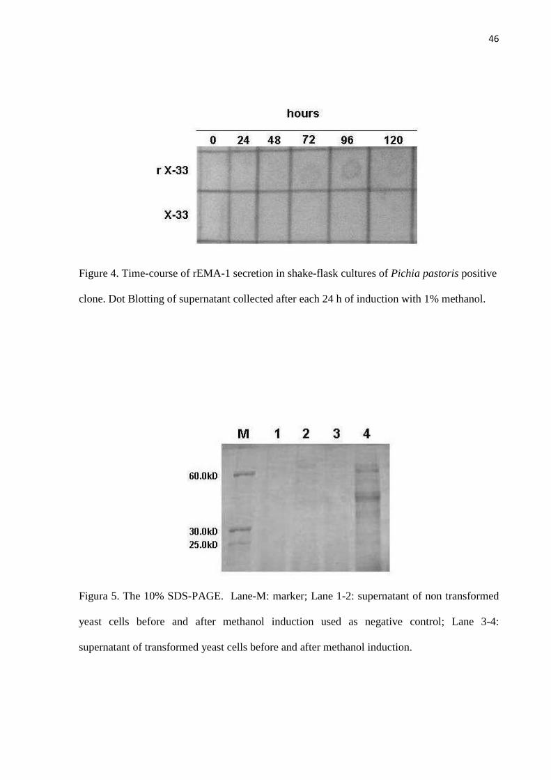
46
Figure 4. Time-course of rEMA-1 secretion in shake-flask cultures of Pichia pastoris positive
clone. Dot Blotting of supernatant collected after each 24 h of induction with 1% methanol.
Figura 5. The 10% SDS-PAGE. Lane-M: marker; Lane 1-2: supernatant of non transformed
yeast cells before and after methanol induction used as negative control; Lane 3-4:
supernatant of transformed yeast cells before and after methanol induction.

47
Figure 6. rEMA-1 Dot blot. Reactivity against different antibodies (A) anti-histidine
monoclonal antibody; (B) anti-EMA-1 monoclonal antibody; (C) sera from positive equine;
(D) sera from negative equine. In left of membranes was spotted a negative control and right
the rEMA-1 protein precipitated with ammonium sulfate.

5 ARTIGO 3
IMMUNOGENICITY AND ANTIGENICITY OF THE RECOMBINANT EMA-1
PROTEIN OF Theileria equi EXPRESSED IN THE YEAST Pichia pastoris
Artigo aceito para publicação: Revista Brasileira de Parasitologia Veterinária
Volume 18 (1-2) 2009.
Imunogenicidade e antigenicidade da proteína recombinante EMA-1 de
Theileria equi expressa em Pichia pastoris
NIZOLI, Leandro Quintana1*; CONCEIÇÃO, Fabrício Rochedo2; SILVA, Sergio Silva3;
DUMMER, Luana Alves1; SANTOS Jr, Alceu Gonçalves1; LEITE, Fábio Pereira Leivas1,4
ABSTRACT
The equine piroplasmosis caused by Theileria equi is one of the most important parasitic
diseases of the equine, causing damage to animal health and economic losses. In T. equi, two
merozoite surface proteins, equi merozoite antigen EMA-1 and EMA-2, have been identified
as the most immunodominant antigens. Thus, suggesting that these antigens might be used as
immunobiological tools. The EMA-1 of Theileria equi was cloned and expressed in the yeast
Pichia pastoris. The transformed yeast was grown at high cell density expressing up to 389
mg.L-1 of recombinant protein. The protein was concentrate, purified and detected in Dot blot.
The recombinant product was antigenically similar to the native protein as determined using
monoclonal antibodies, and polyclonal antibodies obtained from naturally infected equine
with T. equi. The immunogenicity of rEMA-1 protein was demonstrated by IFAT using sera
from recombinant protein immunized mice using aluminum hydroxide as adjuvant. All
animals vaccinated with rEMA-1 developed a high specific antibody response. This results
suggest that rEMA-1expressed in P. pastoris might be used as an antigen for immune
diagnostic as well as vaccine antigen.
Keywords: Theileria equi; recombinant vaccine; Pichia pastoris; EMA-1.
1 Centro de Biotecnologia, Universidade Federal de Pelotas (UFPel). 2 Departamento de Patologia, Universidade Federal do Rio Grande (FURG). 3 Departamento de Veterinária Preventiva, Faculdade de Veterinária, UFPel. 4 Departamento de Microbiologia e Parasitologia, Instituto de Biologia, UFPel. * Corresponding author at: Centro de Biotecnologia, Universidade Federal de Pelotas, CP 354, 96010-900 Pelotas, Brasil. Tel.: +55 53 32757350; fax: +55 53 32757555 E-mail adddress: [email protected]

50
RESUMO
A piroplasmose eqüina causada por Theileria equi é uma das mais importantes doenças
parasitárias de eqüídeos, causando danos a saúde animal e perdas econômicas. Em T. equi,
duas proteínas de superfície de merozoítos, equi merozoite antigen EMA-1 e EMA-2, têm
sido identificados como antígenos imunodominantes. Sugerindo que estes antígenos possam
ser usados como produtos imunobiológicos. O gene EMA-1 de T. equi foi clonado e
expressado na levedura Pichia pastoris. As leveduras transformadas foram cultivadas a alta
densidades celulares expressando 389 mg.L-1 de proteína recombinante. A proteína foi
concentrada, purificada e detectada em Dot blot. O produto recombinante foi antigenicamente
similar a proteína nativa quando determinado usando anticorpo monoclonal e anticorpos
policlonais obtidos de eqüinos naturalmente infectados com T. equi. A imunogenicidade da
proteína rEMA-1 foi demonstrada por RIFI usando soro de camundongos imunizados com
proteína recombinante usando hidróxido de alumínio como adjuvante. Todos animais
vacinados com rEMA-1 desenvolveram uma alta resposta específica de anticorpos. Esses
resultados sugerem que rEMA-1 expressa em P. pastoris possa ser usado como antígeno para
diagnóstico imunológico bem como antígeno para vacinas.
Palavras chaves: Theileria equi, vacina recombinante, Pichia pastoris, EMA-1
INTRODUCTION
The equine piroplasmosis is one of the most important parasitic diseases of the
equines, it causes great damage to animal health. The disease is a haemoparasitosis caused by
Theileria equi and Babesia caballi, is a tick-borne hemoprotozoan disease affecting horses
worldwide (SCHEIN, 1988). Through infecting and destroying red blood cells, it can
compromise the equine function, leading to loss of vitality and decrease in the performance of
infected animals. Equine piroplasmosis caused by T. equi is more pathogenic and widespread
in horses than that by B. caballi (DE WAAL, 1992), causes a persistent infection for which
drug therapy or vaccination is not available (SCHEIN, 1988; FRIEDHOFF, 1990). The T.
equi infection is characterized by fever, anemia, icterus, and hepato and splenomegaly (OIE,
1989).
Merozoite surface proteins are important in the pathogenesis of hemoprotozoan
diseases because of their role in parasite recognition of, attachment to, and penetration of host
51
erythrocytes (JACK & WARD, 1981; KUMAR et al., 2004). In T. equi, two kinds of
merozoite surface proteins, equi merozoite antigen EMA-1 and EMA-2, have been identified
as the most immunodominant antigens (KAPPMEYER et al., 1993; KNOWLES et al., 1991;
KNOWLES et al., 1992). EMA-1 is geographically conserved among all T. equi isolates
(KNOWLES et al., 1991; CUNHA et al., 2002) and shares significantly high homologies in
amino acid sequence with the counterpart proteins of many Theileria parasites
(KAPPMEYER et al., 1993; KNOWLES et al., 1997). Additionally, EMA-1 has glycosyl-
phosphatidylinositol (GPI) anchor-specific motifs in their sequence, suggesting that these
proteins might be expressed on the outer surface of merozoite with a GPI anchor (KNOWLES
et al., 1997). The T. equi merozoite express surface protein of molecular mass 34 kDa, which
are strongly recognized by antibodies produced in infected animals (KNOWLES et al., 1997;
CUNHA et al., 2006). Further, EMA-1/2 are members of the major piroplasm surface protein
(MPSP) family that is conserved among the genus Theileria, but the biological role of MPSP
has not yet been clarified. Therefore, functional studies on EMA-1/2 family proteins may also
provide insight into the biological significance of MPSP expression in the intra-erythrocytic
development of Theileria parasites.
The methylotrophic yeast Pichia pastoris, which is one of the dominant expression
systems in molecular biology due to its stable and high-level expression of heterologous
proteins (CREGG et al., 1993, ROMANOS et al., 1998). Over 400 proteins from prokaryotes,
eukaryotes, and viruses have now been successfully expressed in this yeast (CEREGHINO et
al., 2002). The P. pastoris expression system offers many advantages, including its ease of
usage relative to other eukaryotic expression systems, the possibility of high-level expression
of foreign proteins. The yeast is also able to introduce eukaryotic post-translational
modifications such as glycosylation and proteolytic processing (CEREGHINO & CREGG,
2000).
The present study aims to express truncated EMA-1 gene by Pichia pastoris system
and subsequently determine the immunologic and antigenic properties of recombinant EMA-1
protein.
MATERIALS AND METHODS
Transformation of P. pastoris and expression in shaken flasks
The transformation of P. pastoris and cultivation in shaken flasks were performed
according to the EasySelectTM Pichia Expression Kit (Invitrogen, Catalog No. K1740-01). A
52
DNA encoding the EMA-1 from T. equi was obtained as previously described Nicolaiewsky
et al. (2001). The EMA-1 gene (GenBank accession number AF261824) was amplified by
PCR with the following primers: forward (5′-CGGAATTCACAAAATGGAGGAGGAGAA
ACCCAAG-3′) and reverse (5′-GGGGTACCAATAGAGTAGAAAATGCAATG- 3′),
containing a EcoRI (forward) and an XbaI site (reverse), respectively. After purification and
digestion, the amplified DNA fragment was cloned into the vector. The Escherichia coli
TOP10F were used as a host for plasmid propagation. The P. pastoris wild type strain X-33
and pPICZαB vector were used as a host for expression of EMA-1 in P. pastoris. The P.
pastoris transformants were selected in YPD containing 100 mg/mL zeocin.
Precipitation of protein
Proteins in the media were precipitated by addition of solid ammonium sulfate to 80%
and incubation at 4°C for 16 hours. Precipitated protein was pelleted by centrifugation at
12.000 g for 15 min and the pellet was resuspended in buffer PBS. The solution was desalted
by overnight dialysis against buffer PBS at 4°C. Protein quantitation was determined using
the BCATM Protein Assay Kit (Pierce Chemical Company) with bovine serum albumin as a
standard.
Dot blot
Dot blot assays were performed using 7,5 µg of recombinant protein. As a negative
control, 20 µL of a membrane preparation from a P. pastoris clone transformed with the
empty expression vector. The proteins were spotted onto a nitrocellulose membrane (Hybond
C, Amersham Biosciences). The nitrocellulose membranes were blocked for 1 hour at room
temperature in PBST buffer containing 5% of non-fat milk. One membrane was incubated
with monoclonal antibody anti-His peroxidase conjugate and other membrane with
monoclonal antibody anti-EMA-1. Two membranes were incubated with serum of equines
negative and positive for infection of T. equi, respectively. Membranes were incubated with
secondary antibodies for 1 hour with goat anti-horse or anti-mouse IgG peroxidase conjugate.
The reaction was developed with diaminobenzidine (Sigma).
Immunization of mice
The recombinant purified protein was prepared for immunization of mice (Mus
muscullus), female BALB/c mice (six weeks old) were randomly divided into three groups

53
(four mice per group) and subcutaneously immunized twice at 10 days intervals. One group
(GI) was injected with EMA-1 protein (50 µg) without adjuvant. The other group (GII) was
injected with 50 µg of EMA-1 protein formulated with the adjuvant aluminum hydroxide. The
final group (GIII) was used as a negative control and injected with 100 µl of sterile PBS.
Serum samples were collected from the retro-orbital plexus immediately before immunization
and about 10 days after each of the immunizations and used in serological tests. The
experiment was approved by the UFPel Committee of Ethics in Animal Experimentation.
Immunofluorescence assay
Antibody titers of the serum samples against the recombinant protein were measured
with IFAT. The slides were prepared with infected horses erythrocytes in which are visible as
compact inclusion by IFAT staining. IFAT was performed according to the Cunha (1993).
Then polled serum from the different groups were added incubated, and applied FITC-
conjugated goat anti-mouse IgG (Invitrogen), per well diluted at 1:400 in PBS buffer. The
visualization slides were in epifluorescent microscopy (Olympus, BX-FLA).
RESULTS AND DISCUSSION
Expression and purification of rEMA-1 in P. pastoris
The P. pastoris gene expression system is an attractive method with which to produce
a variety of intercellular and extracellular proteins (CREGG et al., 1993). Since, we
previously isolated a DNA encoding this protein, we decided to clone the DNA sequence
encoding EMA-1 without its native signal peptide in the P. pastoris pPICZαB plasmid, in
frame with the yeast a-factor signal sequence, for secretion of protein containing a 6His-Tag
at its C-terminus. The pPICZαB-EMA-1 plasmid was then transformed and targeted to the P.
pastoris genome by means of homologous recombination. The presence of the EMA-1 coding
sequence in the genomic DNA isolated from Pichia transformants was confirmed by PCR. In
the P. pastoris expression system, recombinant protein expression is strictly controlled by the
AOX1 promoter. Expression was induced by addition of methanol to a final methanol
concentration of 0.5%. A protein that was not present before methanol induction was detected
by SDS-PAGE (data not shown).
One of the main advantages in producing heterologous proteins as secreted products in
P. pastoris is the easy isolation of the recombinant product from the medium in which it is
produced. Indeed, the initial purity of recombinant molecule in culture medium is high

54
because the level of endogenous secreted proteins is very low. Thus, after removal of the
yeast cells by centrifugation, proteins in the culture supernatant were precipitated with 80%
ammonium sulfate fractionation. The final yield of the purified protein was 389 mg of rEMA-
1 per liter of cell culture supernatant.
Detection of reactivity to recombinant EMA-1
Dot blot is a technique for detecting and identifying proteins, similar to the Western
blot technique but differing in that protein samples are not separated electrophoretically but
are spotted through circular templates directly onto the membrane. Antigens may be applied
directly to nitrocellulose membrane as a discrete spot (dot) to give a simple and reliable assay.
In order to evaluate the reactivity of recombinant EMA-1 with sera from T. equi
positive and negative equine, we tested by Dot blot. Positive samples were considered all of
those that developed a reaction color. Negative controls samples did not develop any color.
In Dot blots, we are able to observe that the rEMA-1 protein reacted with the serum
from naturally infected horse by T. equi, but did not with the serum of the negative horse.
The figure 1 show the reactivity of the rEMA-1 protein using anti-histidine
monoclonal antibody, monoclonal antibody anti-EMA-1, and serum of equine positive carrier
of T. equi, and did not show reactivity with serum of negative animal. No reactivity was
observed with control negative protein.
Immunogenicity of the rEMA-1 protein in mice
Mice immunized with the recombinant protein showed high antibody response, were
able to react with native EMA-1 of T. equi as observed by IFAT. Sera from the negative
control group did not react with native EMA-1, showing the specificity of the response
(Figure 2). These results indicate that immune response was generated in all the vaccinated
groups, showing that recombinant protein has capability to generated antibodies that react
with the native protein. We also observed that the antibody titers have significant difference
between the groups with without adjuvant. This worth noting that rEMA-1 expressed in P.
pastoris maintain the same three-dimensional structure as the native protein since it was
recognized by antibodies generated by naturally infected horses.
The P. pastoris expression system has been used for the production of a wide variety
of proteins (CEREGHINO & CREGG, 2000). However, to the best of our knowledge this is
the first report on the cloning and expression of EMA-1 protein in P. pastoris.

55
In conclusion, the production and purification of rEMA-1 in the methylotrophic yeast
P. pastoris was effective, permitting a high-yield production of this protein. Thus, rEMA-
1expressed in P. pastoris might be a strong candidate to be used as an antigen for immune
diagnostic as well as vaccine antigen.
ACKNOWLEDGEMENTS
This author was supported by the CAPES Foundation through the Brazilian
government.
REFERENCES
CEREGHINO, G. P. L.; CEREGHINO, J. L.; ILGEN, C.; CREGG, J. M. Production of
recombinant proteins in fermenter cultures of the yeast Pichia pastoris. Current Opinion in
Biotechnology, v. 13, n. 4, p. 329-332, 2002.
CEREGHINO, J. L.; CREGG, J. M. Heterologous protein expression in the methylotrophic
yeast Pichia pastoris. FEMS Microbiology Reviews, v. 24, n. 1, p. 45-66, 2000.
CREGG, J. M.; VEDVICK, T. S.; RASCHKE, V. Recent advances in the expression of
foreign genes in Pichia pastoris. Biotechnology, v. 11, n. 8, p. 905-910, 1993.
CUNHA, C. W. Babesiose Eqüina: Padronização da Reação de Imunofluorescência Indireta
para sorodiagnóstico em eqüinos Puro Sangue Inglês. – Brasil. 1993. 56 p. Dissertação
(Mestrado) – Universidade Federal de Pelotas, Pelotas, 1993.
CUNHA, C. W.; KAPPMEYER, L. S.; McGUIRE, T. C.; DELLAGOSTIN, O. A.;
KNOWLES, D. P. Conformational dependence and conservation of an immunodominant
epitope within the Babesia equi erythrocyte-stage surface protein equi merozoite antigen 1.
Clinical and Diagnostic Laboratory Immunology, v. 9, n. 6, p. 1301-1306, 2002.
CUNHA, C. W.; McGUIRE, T. C.; KAPPMEYER, L. S.; HINES, S. A.; LOPEZ, A. M.;
DELLAGOSTIN, O. A.; KNOWLES, D. P. Development of specific immunoglobulin Ga
(IgGa) and IgGb antibodies correlates with control of parasitemia in Babesia equi infection.
Clinical and Vaccine Immunology, v. 13, n. 2, p. 297-300, 2006.
DE WAAL, D. T. Equine piroplasmosis: a review. British Veterinary Journal, v. 148, n. 1, p.
6-14, 1992. .
FRIEDHOFF, K. T. Interaction between parasite and tick vector. International Journal for
Parasitology, v. 20, n. 4, p. 525-535, 1990.

56
JACK, R. M. & WARD, P. A. Mechanisms of entry of Plasmodium and Babesia into red
cells. In Ristic, M. and Kreier, J. P. (ed.), Babesiosis. Academic Press, Inc., New York.
1981, p. 445-458.
KAPPMEYER, L. S.; PERRYMAN, L. E.; KNOWLES, D. P. Jr. A Babesia equi gene
encodes a surface protein with homology to Theileria species. Molecular and Biochemical
Parasitology, v. 62, n. 1, p. 121-124, 1993.
KNOWLES, D. P. Jr.; PERRYMAN, L. E.; GOFF, W.L.; MILLER, C.D.; HARRINGTON,
R.D.; GORHAM, J.R.. A monoclonal antibody defines a geographically conserved surface
protein epitope of Babesia equi merozoites. Infection and Immunity, v. 59, n. 7, p. 2412-
2417, 1991.
KNOWLES, D. P. Jr., KAPPMEYER, L. S.; STILLER, D., HENNAGER, S. G.;
PERRYMAN, L. E. Antibody to a recombinant merozoite protein epitope identifies horses
infected with Babesia equi. Journal of Clinical Microbiology, v. 30, n. 12, p. 3122-3126,
1992.
KNOWLES, D. P.; KAPPMEYER, L. S.; PERRYMAN, L. E. Genetic and biochemical
analysis of erythrocyte-stage surface antigens belonging to a family of highly conserved
proteins of Babesia equi and Theileria species. Molecular and Biochemical Parasitology, v.
90, n. 1, p. 69-79, 1997.
KUMAR, S.; YOKOYAMA, N.; KIM, J. Y.; HUANG, X.; INOUE, N.; XUAN, X.;
IGARASHI, I.; SUGIMOTO, C. Expression of Babesia equi EMA-1 and EMA-2 during
merozoite developmental stages in erythrocyte and their interaction with erythrocytic
membrane skeleton. Molecular and Biochemical Parasitology, v. 133, n. 2, p. 221-227,
2004.
NICOLAIEWSKY, T. B.; RICHTER, M. F.; LUNGE, V. R.; CUNHA, C. W.;
DELAGOSTIN, O.; IKUTA, N.; FONSECA, A. S.; SILVA, S. S.; OZAKI, L. S. Detection
of Babesia equi (Laveran, 1901) by nested polymerase chain reaction. Veterinary
Parasitology, v. 101, n. 1, p. 9-21, 2001.
Office International Des Epizooties (OIE). Manual of recommended diagnostic techniques
and requirements for biological products for lists A and B diseases, v. 1, p. 1-6, 1989.
ROMANOS, M.; SCORNER, C.; SREEKRSHNA, K.; CLARE, J. The generation of
microscopy recombinant strains. Methods Molecular Biology, v. 103, p. 55-72, 1998.
SCHEIN, E. Equine babesiosis. In Ristic, M., Babesiosis of Domestic Animals and Man. CRC
Press, Boca Raton, 1988. p. 197-208.

57
FIGURES
Figure 1. rEMA-1 Dot blot. Reactivity against different antibodies (A) anti-histidine
monoclonal antibody; (B) anti-EMA-1 monoclonal antibody; (C) sera from positive equine;
(D) sera from negative equine. In left of membranes was spotted a negative control and right
the rEMA-1 protein.
Figure 2. IFAT of sera from immunized mice with rEMA-1. (A) Pool of sera from immunized
mice twice in the presence of adjuvant; (B) Mice immunized with sterile PBS; (C) IFAT
positive control (equine sera with FITC anti-equine).
6 CONSIDERAÇÕES FINAIS
Durante a interação parasito-hospedeiro, as proteínas de superfície do
parasito são as principais responsáveis pela resposta imune do hospedeiro, sendo
que, os antígenos de superfície são promissores para uso como vacinas de
subunidade e reagentes para diagnóstico.
As tentativas para controlar a epidemia da doença causada pelo protozoário
T. equi tem sido focalizadas em estudos biológicos, bioquímicos, estruturais do
parasito e no desenvolvimento de novas drogas. As proteínas de superfície do
merozoito desempenham um papel chave no estabelecimento da infecção, a
interação da EMA-1 com as células do hospedeiro é crucial para a sua infectividade
(KUMAR, et al., 2004). Sabendo-se que P. pastoris é um sistema de expressão de
proteína de grande eficiência e reproduz as melhores condições de expressão de
proteína de eucariotos superiores, neste estudo, este organismo foi selecionado
como o ideal para a produção de EMA-1 com a finalidade de utilizá-la como antígeno
vacinal recombinante.
A escolha do sistema utilizado para a expressão da EMA-1 recombinante,
assim como, a definição dos objetivos foi extremamente importante. O entendimento
de que EMA-1 seria utilizada não só para a produção de vacinas, mas também para
a padronização de um teste imunológico para diagnóstico e para teste de protocolos
terapêuticos, nos remeteu à necessidade de uma molécula extremamente
semelhante à nativa.
No entanto, a capacidade de tradução de uma seqüência de DNA seria igual
em qualquer vetor de expressão. Porém, as diferenças se baseiam principalmente
na capacidade de produzir glicosilação da proteína, e na quantidade de proteína que
o vetor é capaz de produzir. A glicosilação em proteínas pode influenciar na
bioatividade, farmacocinética, biodistribuição e imunogenicidade da molécula.
Células procariontes não conseguem adicionar carboidratos (glicosilar); leveduras
bem como outros sistemas eucarióticos são capazes de glicosilações mais simples
normalmente sem a capacidade de sialização e com baixa capacidade de

59
glicosilação ligada ao O. A escolha do modelo de expressão da EMA-1 foi baseada
nos dados citados acima.
Várias proteínas já foram expressas em P. pastoris, entretanto, até o
momento não há relato de expressão de EMA-1 de T. equi nesta levedura para ser
utilizado como um antígeno recombinante. Desta forma, este trabalho é pioneiro
quanto à utilização de P. pastoris como vetor de expressão de proteína EMA-1 de T.
equi na tentativa de ser utilizada como antígeno em produtos imunobiológicos.
Com os resultados deste estudo desenvolvemos ferramentas que poderá ser
utilizadas para melhor entender a patogenicidade deste importante parasito bem
como sua utilização em estudos de vacinas e de diagnóstico.
60
7 CONCLUSÕES GERAIS
- A levedura P. pastoris é capaz de expressar a proteína recombinante
EMA-1 de Theileria equi;
- A proteína EMA-1 expressa por P. pastoris mostrou ser antigênica e
imunogênica;
- A proteína EMA-1 é um promissor alvo á ser utilizado como imunobiológico
no desenvolvimento de testes diagnósticos e na formulação de vacinas
recombinantes.
- Primeira citação da clonagem e expressão do gene EMA-1 de T. equi em
sistema de expressão heterólogo P. pastoris;

8 REFERÊNCIAS
ALLSOPP, M. T., CAVALIER-SMITH, T., DE WAAL, D. T., ALLSOPP, B. A.
Phylogeny and evolution of the piroplasms. Parasitology , v. 108, p. 147-152, 1994.
AMBAWAT, H. K.; MALHOTRA, D. V.; KUMAR, S.; DHAR, S. Erythrocyte associated
haemato-biochemical changes in Babesia equi infection experimentally produced in
donkeys. Veterinary Parasitology , v.85, p.319-324, 1999.
AUCAN, C., TRAORE, Y., TALL, F., NACRO, B., TRAORE-LEROUX, T., FUMOUX,
F., RIHET, P. High immunoglobulin G2 (IgG2) and low IgG4 levels are associated
with human resistance to Plasmodium falciparum malaria. Infection and Immunity ,
v. 68, p. 1252-1258, 2000.
BATTSETSEG, B.; LUCERO, S.; XUAN, X.; CLAVERIA, F. G.; INOUE, N.;
ALHASSAN, A.; KANNO, T.; IGARASHI, I.; NAGASAWA, H.; MIKAMI, T.; FUJISAKI,
K. Detection of natural infection of Boophilus microplus with Babesia equi and
Babesia caballi in Brazilian horses using nested polymerase chain reaction.
Veterinary Parasitology , v.107, p.351-357, 2002.
BROWN, W. C. Molecular approaches to elucidating innate and acquired immune
responses to Babesia bovis, a protozoan parasite that causes persistent infection.
Veterinary Parasitology , v. 101, p. 233-248, 2001.
BROWN, W. C., PALMER, G. H. Designing Blood-stage Vaccines against Babesia
bovis and B. bigemina. Parasitology Today , v. 15, p. 275-281, 1999.
CAVINATO, R. A., BASTOS, K. R., SARDINHA, L. R., ELIAS, R. M., ALVAREZ, J.
M., D'IMPERIO LIMA, M. R. Susceptibility of the different developmental stages of
the asexual (schizogonic) erythrocyte cycle of Plasmodium chabaudi chabaudi to
hyperimmune serum, immunoglobulin (Ig)G1, IgG2a and F(ab')2 fragments. Parasite
Immunology , v. 23, p. 587-597, 2001.

62
CEREGHINO, L. G. P.; CREGG, J. M. Applications of yeast in biotechnology: protein
production and genetic analysis. Current Opinion in Biotechnology , v. 10, p. 422-
427, 1999.
CEREGHINO, L. J.; CREGG, J. M. Heterologous protein expression in the
methylotrophic yeast Pichia pastoris. FEMS Microbiology Reviews, v. 24, p. 45-66,
2000.
CHEN, D., COPEMAN, D. B., BURNELL, J., HUTCHINSON, G. W. Helper T cell and
antibody responses to infection of CBA mice with Babesia microti. Parasite
Immunology , v. 22, p. 81-88, 2000a.
CHEN, D., COPEMAN, D. B., HUTCHINSON, G. W., BURNELL, J. Inhibition of
growth of cultured Babesia microti by serum and macrophages in the presence or
absence of T cells. Parasitology International , v. 48, p. 223-231, 2000b.
CRIADO-FORNELIO, A., MARTINEZ-MARCOS, A., BULING-SARAÑA, A., BARBA-
CARRETERO, J. C. Molecular studies on Babesia, Theileria and Hepatozoon in
southern Europe. Part I. Epizootiological aspects. Veterinary Parasitology , v. 113,
p. 189-201, 2003a.
CRIADO-FORNELIO, A., MARTINEZ-MARCOS, A., BULING-SARAÑA, A., BARBA-
CARRETERO, J. C. Molecular studies on Babesia, Theileria and Hepatozoon in
southern Europe. Part II. Phylogenetic analysis and evolutionary history. Veterinary
Parasitology , v. 114, p. 173-194, 2003b.
CUNHA, C. W., SILVA, S. S., PIMENTEL, C. A., DAPPER, E. Avaliação da
freqüência de equinos soropositivos a Babesia equi no Joquei Clube de Pelotas e
em dois haras da zona sul do Rio Grande do Sul. Revista Brasileira de
Parasitologia Veterinária , v. 5, p. 119-122, 1996.
CUNHA, C. W., SILVA, S. S., RODRIGUES, A. L., GUERREIRO, G. Avaliação do
efeito da esplenectomia em eqüinos portadores e livres de Babesia spp. Revista
Brasileira de Parasitologia Veterinária , v. 6, p. 157-160, 1997.
CUNHA, C. W.; McGUIRE, T. C.; KAPPMEYER, L. S.; HINES, S. A.; LOPEZ, A. M.;
DELLAGOSTIN, O. A.; KNOWLES, D. P. Development of specific immunoglobulin
Ga (IgGa) and IgGb antibodies correlates with control of parasitemia in Babesia equi
infection. Clinical and Vaccine Immunology , v. 13, p. 297-300, 2006.

63
CUNHA, C. W.; SILVA, S. S.; OSÓRIO, B. L.; DUTRA, C. L. Alterações
hematológicas e sorológicas em eqüinos experimentalmente infectados com Babesia
equi. Ciência Rural, v.28, p.283-286, 1998.
DUMMER, L. A., NIZOLI, L. Q., ROCHA, A. S. R., CONCEIÇÃO, F. R., TURNES, C.
G., VIDOR, T., LEITE, F. P L. Production and characterization of recombinant Bovine
Herpesvirus type 5 Glycoprotein D expressed in Pichia pastoris. In: 8th International
Veterinary Immunology Symposium, 2007, Anais do 8th International Veterinary
Immunology Symposium , 2007.
FRIEDHOFF, K. T. Interaction between parasite and tick vector. International
Journal for Parasitology , v.20, n.4, p.525-535, 1990.
FRIEDHOFF, K. T.; TENTER, A. M.; MULLER, I. Haemoparasites of equines: impact
on international trade of horses. Revue Scientifique et Technique , v.9, p.1187-
1194, 1990.
GUIMARÃES, A. M.; LIMA, J. D.; RIBEIRO, M. F. B. Sporogony and experimental
transmission of Babesia equi by Boophilus microplus ticks. Parasitology Research ,
v.84, p.323-327, 1998b.
GUIMARÃES, A. M.; LIMA, J. D.; RIBEIRO, M. F. B.; CAMARGOS, E. R. S.; BOZZI,
I. A. Ultrastructure of sporogony in Babesia equi in salivary glands of adult female
Boophilus microplus ticks. Parasitology Research , v.84, p.69-74, 1998a.
HELMBY, H., TROYE-BLOMBERG, M. Differential immunoglobulin E and cytokine
responses in BALB/c and C57Bl/6 mice during repeated infections with blood-stage
Plasmodium chabaudi malaria. Parasite Immunology , v. 22, p. 185-190, 2000.
HEUCHERT, C. M., DE, G. V., JR., DE ATHAIDE, D. F., BOSE, R., FRIEDHOFF, K.
T. Seroepidemiologic studies on Babesia equi and Babesia caballi infections in
Brazil. Veterinary Parasitology , v. 85, p. 1-11, 1999.
IGARASHI, I., SUZUKI, R., WAKI, S., TAGAWA, Y., SENG, S., TUM, S., OMATA, Y.,
SAITO, A., NAGASAWA, H., IWAKURA, Y., SUZUKI, N., MIKAMI, T., TOYODA, Y.
Roles of CD4(+) T cells and gamma interferon in protective immunity against Babesia
microti infection in mice. Infection and Immunity , v. 67, p. 4143-4148, 1999.
JENKINS, M. C. Advances and prospects for subunit vaccines against protozoa of
veterinary importance. Veterinary Parasitology , v. 101, p. 291-310, 2001.

64
KATZER, F., MCKELLAR, S., KIRVAR, E., SHIELS, B. Phylogenetic analysis of
Theileria and Babesia equi in relation to the establishment of parasite populations
within novel host species and the development of diagnostic tests. Molecular and
Biochemical Parasitology , v. 95, p. 33-44, 1998.
KERBER, C. E., FERREIRA, F., PEREIRA, M. C. Control of equine piroplasmosis in
Brazil. Onderstepoort Journal of Veterinary Research , v.66, p.123-127, 1999.
KNOWLES, D. P., KAPPMEYER, L. S., PERRYMAN, L. E. Specific immune
responses are required to control parasitemia in Babesia equi infection. Infection
and Immunity , v. 62, p. 1909-1913, 1994.
KNOWLES, D. P., KAPPMEYER, L. S., PERRYMAN, L. E. Genetic and biochemical
analysis of erythrocyte-stage surface antigens belonging to a family of highly
conserved proteins of Babesia equi and Theileria species. Molecular and
Biochemical Parasitology , v. 90, p. 69-79, 1997.
KNOWLES, D. P., JR. Equine babesiosis (Piroplasmosis): A problem in the
international movement of horses, British Veterinary Journal , v. 152, p. 123-126,
1996
KUMAR, S.; YOKOYAMA, N.; KIM, J. Y.; HUANG, X.; INOUE, N.; XUAN, X.;
IGARASHI, I.; SUGIMOTO, C. Expression of Babesia equi EMA-1 and EMA-2 during
merozoite developmental stages in erythrocyte and their interaction with erythrocytic
membrane skeleton. Molecular and Biochemical Parasitology , v. 133, p. 221-227,
2004.
LORDING, P. M. Erythrocytes. Veterinary Clinical Equine, v. 24, p. 225-237, 2008.
MARTIN, R. Equine piroplasmosis: the temporary importation of soropositive horses
into Australia. Australian Veterinary Journal , v. 77, p. 308-309, 1999.
MEHLHORN, H., SCHEIN, E. Redescription of Babesia equi Laveran, 1901 as
Theileria equi Mehlhorn, Schein 1998. Parasitology Research , v. 84, p. 467-475,
1998.
NICOLAIEWSKY, T. B., RICHTER, M. F., LUNGE, V. R., CUNHA, C. W.,
DELAGOSTIN, O., IKUTA, N., FONSECA, A. S., DA SILVA, S. S., OZAKI, L. S.
Detection of Babesia equi (Laveran, 1901) by nested polymerase chain reaction.
Veterinary Parasitology , v. 101, p. 9-21, 2001.

65
NIZOLI, L. Q., DUMMER, L. A., CONCEIÇÃO, F. R., SILVA, S. S., SANTOS Jr, A.
G., LEITE, F. P L. Construção de vacina recombinante contra babesiose equina In:
XX Congresso Brasileiro de Parasitologia, 2007, Revista de Patologia Tropical ,
v.36, 2007.
NOGUEIRA, C. E. W.; SILVA, S. S.; NIZOLI, L. Q.; RIBAS, L. M.; ALBUQUERQUE, L. P. A. N. Efeito quimioprofilático do dipropionato imidocarb na prevenção da agudização de babesiose eqüina em cavalos portadores da infecção. A Hora Veterinária, Porto Alegre-RS, v.146, p.17-20, 2005.
OLADASU, L. A.; OLUFEMI, B. E. Hematology of experimental babesiosis and
ehrlichiosis in steroid immunosuppressed horses. Journal Veterinary Medicine B ,
v.39, p.345-352, 1992.
OLADOSU, L. A. Effects of intravenous corticosteroid on the pathogenicity of
Babesia equi infection of donkeys (Equus asinus). Journal Veterinary Medicine B ,
v.35, p.509-514, 1988.
PALMER, G. H., RURANGIRWA, F. R., KOCAN, K. M., BROWN, W. C. Molecular
basis for vaccine development against the ehrlichial pathogen Anaplasma marginale.
Parasitology Today , v. 15, p. 281-286, 1999.
PFEIFER, B. I., BOSE, R., PEYMANN, B., FRIEDHOFF, K. T. Epidemiological
aspects of equine babesioses in a herd of horses in Brazil. Veterinary Parasitology ,
v. 58, p. 1-8, 1995.
RIBEIRO, M. F., COSTA, J. O., GUIMARAES, A. M. Epidemiological aspects of
Babesia equi in horses in Minas Gerais, Brazil. Veterinary Research
Communications , v. 23, p. 385-390, 1999.
SCHEIN, E. Equine babesiosis In Ristic, M., Babesiosis of Domestic Animals and
Man CRC Press, Boca Raton, FL. 1988, p. 197-208.
SCHEIN, E., REHBEIN, G., VOIGT, W. P., ZWEYGARTH, E. Babesia equi (Laveran
1901) Development in horses and in lymphocyte culture. Tropenmed Parasitology ,
v. 32, p. 223-227, 1981.

66
SHODA, L. K., PALMER, G. H., FLORIN-CHRISTENSEN, J., FLORIN-
CHRISTENSEN, M., GODSON, D. L., BROWN, W. C. Babesia bovis-stimulated
macrophages express interleukin-1beta, interleukin-12, tumor necrosis factor alpha,
and nitric oxide and inhibit parasite replication in vitro. Infection and Immunity , v.
68, p. 5139-5145, 2000.
STILLER, D., GOFF, W. L., JOHNSON, L. W., KNOWLES, D. P. Dermacentor
variabilis and Boophilus microplus (Acari: Ixodidae): Experimental vectors of Babesia
equi to equids. Journal of Medical Entomology , v.39, p.667-670, 2002.
SU, Z., STEVENSON, M. M. Central role of endogenous gamma interferon in
protective immunity against blood-stage Plasmodium chabaudi AS infection.
Infection and Immunity , v. 68, p. 4399-4406, 2000.
TAYLOR, P. R., SEIXAS, E., WALPORT, M. J., LANGHORNE, J., BOTTO, M.
Complement contributes to protective immunity against reinfection by Plasmodium
chabaudi chabaudi parasites. Infection and Immunity , v. 69, p. 3853-3859, 2001.
TENTER, A. M., FRIEDHOFF, K. T. Serodiagnosis of experimental and natural
Babesia equi and B. caballi infections. Veterinary Parasitology , v. 20, p. 49-61,
1986.
UETI, M. W., PALMER, G. H., KAPPMEYER, L. S., STATFIELD, M., SCOLES, G. A.,
KNOWLES, D. P. Ability of the vector tick Boophilus microplus to acquire and
transmit Babesia equi following feeding on chronically infected horses with low-level
parasitemia. Journal Clinical Microbiology , v. 43, p. 3755-3759, 2005.
UETI, M. W., PALMER, G. H., SCOLES, G. A., KAPPMEYER, L. S., KNOWLES, D.
P. Persistently infected horses are reservoirs for intrastadial tick-borne transmission
of the apicomplexan parasite Babesia equi. Infection and Immunity , v. 76, p. 3525-
3529, 2008.
World Organization for Animal Health (OIE). World animal health information
database (WAHID) [database online]. Equine piroplasmosis: January 2005 –August
2008. Paris: OIE;2008. Available at: http://www.oie.int/wahid-
prod/public.php?page=disease_status_lists. Accessed 28 Aug 2008.

67
XUAN, X., LARSEN, A., IKADAI, H., TANAKA, T., IGARASHI, I., NAGASAWA, H.,
FUJISAKI, K., TOYODA, Y., SUZUKI, N., MIKAMI, T. Expression of Babesia equi
merozoite antigen 1 in insect cells by recombinant baculovirus and evaluation of its
diagnostic potential in an enzyme-linked immunosorbent assay. Journal of Clinical
Microbiology , v. 39, p. 705-709, 2001.
ZAHLER, M., RINDER, H., GOTHE, R. Genotypic status of Babesia microti within the
piroplasms. Parasitology Research , v. 86, p. 642-646, 2000a.
ZAHLER, M., RINDER, H., SCHEIN, E., GOTHE, R. Detection of a new pathogenic
Babesia microti-like species in dogs. Veterinary Parasitology , v. 89, p. 241-248,
2000b.
ZOBBA, R., ARDU, M., NICCOLINI, S., CHESSA, B., MANNA, L., COCCO, R.,
PARPAGLIAE, M. L. P. Clinical and Laboratory Findings in Equine Piroplasmosis.
Journal of Equine Veterinary Science , v. 28, 2008.
Recommended