UNIVERSIDADE FEDERAL DE UBERLÂNDIA FACULDADE DE MEDICINA PROGRAMA DE PÓS-GRADUAÇÃO EM CIÊNCIAS DA SAÚDE
ALTERAÇÕES DOS MARCADORES INFLAMATÓRIOS, BIOQUÍMICOS E APTIDÃO FÍSICA EM MULHERES EM RECUPERAÇÃO DO CÂNCER DE MAMA APÓS TREINAMENTO RESISTIDO NÃO LINEAR: UM ESTUDO
PILOTO
MARCO AURÉLIO FERREIRA DE JESUS LEITE
UBERLÂNDIA
2017

MARCO AURÉLIO FERREIRA DE JESUS LEITE
ALTERAÇÕES DOS MARCADORES INFLAMATÓRIOS, BIOQUÍMICOS E APTIDÃO FÍSICA EM MULHERES EM RECUPERAÇÃO DO CÂNCER DE MAMA APÓS TREINAMENTO RESISTIDO NÃO LINEAR: UM ESTUDO
PILOTO
Dissertação apresentada ao Programa de
Pós-Graduação em Ciências da Saúde da
Faculdade de Medicina da Universidade
Federal de Uberlândia, como requisito
parcial para a obtenção do título de Mestre
em Ciências da Saúde.
Área de concentração: Ciências da Saúde.
Orientador: Guilherme Morais Puga
Co-orientador: Carlo José Freire de Oliveira

Dados Internacionais de Catalogação na Publicação(CIP) Sistema de Bibliotecas da UFU, MG, Brasil.
L533a
2017
Leite, Marco Aurélio Ferreira de Jesus, 1994Alterações dos marcadores inflamatórios, bioquímicos e aptidão física em
mulheres em recuperação do câncer de mama após treinamento resistido não linear: um estudo piloto / Marco Aurélio Ferreira de Jesus Leite. - 2017.
61 f. : il.
Orientador: Guilherme Morais Puga.Coorientador: Carlo José Freire de Oliveira.Dissertação (mestrado) - Universidade Federal de Uberlândia, Programa de Pós-
Graduação em Ciências da Saúde.Inclui bibliografia.
1. Ciências médicas - Teses. 2. Mamas - Câncer - Teses. 3. Hormonioterapia - Teses. 4. Inflamação - Teses. I. Puga, Guilherme Morais. II. Oliveira, Carlo José Freire de. III. Universidade Federal de Uberlândia. Programa de Pós-Graduação em Ciências da Saúde. IV.

UBERLÂNDIA
2017
FOLHA DE APROVAÇÃO
Marco Aurélio Ferreira de Jesus Leite.
Alterações dos marcadores inflamatórios, bioquímicos e aptidão física em mulheres em recuperação do câncer de mama após treinamento resistido não linear: um estudo piloto.
Presidente da banca (orientador): Prof. Dr. Guilherme Morais Puga
Dissertação apresentada ao Programa de Pós-Graduação em Ciências da Saúde da Faculdade de Medicina da Universidade Federal de Uberlândia, como requisito parcial para a obtenção do título de Mestre em Ciências da Saúde.Área de concentração: Ciências da Saúde.
Banca Examinadora
Titular: Pro f Dr. Fábio Lera Orsatti
Instituição: Universidade Federal do Triângulo Mineiro
Titular: Prof. Dra. Ana Paula Magalhães Resende
Instituição: Universidade Federal de Uberlândia

DEDICATÓRIA
A Deus pela oportunidade, a minha mãe por toda a dedicação e abdicação em virtude da minha formação, a minha avó pelo amor em minha criação.

AGRADECIMENTOS
Ao Prof. Dr. Guilherme Morais Puga, pela orientação, confiança e oportunidade cedida
para realização deste trabalho. Muito obrigado!
Ao Prof. Dr. Carlo José Freire de Oliveira e Prof. Nilson Penha-Silva, por sempre
acreditar no meu potencial e seu imessuravel auxílio na execução da pesquisa. Sem sua
expertise como co-orientador não conseguíamos alcançar estes resultados!
Aos meus colegas do Laboratório de Fisiologia Cardiorespiratória e Metabólica,
Laboratório de Biofisicoquímica e Laboratório de Imunologia Aplicada pela parceria e
suporte. A todos os funcionários, professores e técnicos da Faculdade de Educação Física
e de Medicina da Universidade Federal de Uberlândia pelos auxílios prestados.
Aos amigos de laboratório Lucas, Franciel, Wener, Rodney, Hugo, Mário, Luciana, Igor,
Juliene, Jéssica, Larissa, Brenda, Jonatas, Brenda pelo aprendizado e colaboração para
elaboração desta pesquisa. A todas as voluntárias que acreditaram na minha pesquisa e
reservaram seu tempo para participar de toda intervenção.
Agradeço a minha família, primeiramente minha mãe Maria pela oportunidade, amor,
dedicação e por lutar dia a dia junto a mim em meus sonhos. Obrigado pela confiança. A
minha tia Terezinha e ao meu primo Ernando por me darem exemplaridade e apoio. Aos
meus irmãos José Victor, Natália e Carla pelo afeto e auxílios. Em especial, agradeço
minha avó Arminda, a qual perdi pela luta contra o câncer. Sua simplicidade, carinho e
amor me fizeram e me faz mais forte. Não fui capaz de lhe ajudar, mas espero que estes
estudos possam ajudar pessoas que estão passando pela mesma situação. A vocês a minha
eterna gratidão.
Aos meus amigos que Uberlândia me oportunizou a conquistar, Lucas, Pedro, Diego,
Gabriel, Matheus, Bruno, Iago, Alexandre, Luiza, Fernanda, Luan, Caio, Amanda,
Adriele e também aos meus amigos de Monte Carmelo, Marcos, Mayone e Bruno.
Obrigado por se fazerem sempre presentes e por todo carinho e cuidado que tiveram
comigo nesta jornada. O carinho de vocês foi fundamental pra esse percurso.

“Não vos aconselho o trabalho, mas a luta. Não vos aconselho a paz, mas a vitória! Seja o nosso trabalho uma luta! Seja a vossa paz uma vitória ”.
Friedrich Wilhelm Nietzsch

RESUMO
Introdução: Os tratamentos oncológicos podem propiciar vários efeitos adversos em pacientes
com câncer de mama (CM), como elevado processo inflamatório e dislipidemia. Até o momento
ainda não se sabe o efeito do treinamento resistido periodizado de forma não linear (TRNL) em
variáveis inflamatórias e bioquímicas em mulheres recuperando de CM. Objetivo: Verificar o
efeito do TRNL sobre o perfil inflamatório, bioquímico, aptidão física e composição corporal
em mulheres em recuperação do CM durante hormonioterapia. Métodos: 14 mulheres
recuperando do CM durante a hormonioterapia participaram do estudo. Foram coletadas
amostras de sangue e saliva para analises do perfil inflamatório, lipídico e imune e realizado
avaliações antropométricas, força e resistência muscular antes e após de intervenção. O TRNL
foi realizado 3x/semana em dias não consecutivos durante 3 meses. A normalidade dos dados
foi verificada pelo teste Shapiro-Wilk e as comparação das variáveis foi realizada pelo teste t
pareado e Wilcoxon. Ponto de corte foi ajustado para a = 5% para todas as análises. Resultados:
Não houve alterações significativas na concentração plasmática das citocinas analisadas (TNF
a, IL-4, IL-6, IL-10, IL-17, IL-1RA, IFN-y). Porém houve aumento significativo de
imunoglobulina A (IgA) salivar (186.29%) após intervenção. Houve também de HDL-
colesterol (17.98%), massa livre de gordura (2.99%), massa magra (2.97%), força (39.16%) e
resistência muscular (69.16%).Além disso houve reduções significativas de colesterol total (
4.81%), triglicerídeos (-14.40%), LDL-colesterol (-10.23%), plaquetas (-7.90%), monócitos (
38.89%), massa gorda (-4.53%), percentual de gordura corporal (-1.19%). Conclusão: Doze
semanas de TRNL aumentou a concentração de IgA salivar, melhorou composição corporal,
perfil lipídico e aptidão física em mulheres recuperando do BC durante hormonioterapia, força
e resistência em mulheres em recuperação do CM.
Palavras-chaves: Câncer, Treinamento de Força, Perfil Inflamatório.

ABSTRACT
Introduction: Oncological treatments can provide several adverse effects in patients with
breast cancer (BC), such as high inflammatory process and dyslipidemia. To date, the effect of
nonlinear periodized resistance training (NLRT) on inflammatory and biochemical variables in
women recovering from CM has not yet been known. Objective: To verify the effect of NLRT
on the inflammatory, biochemical profile, physical fitness and body composition in women in
CM recovery during hormone therapy. Methods: The assessments were done before and after
3 months of application of a NLRT program, consisting of three sessions of exercise per week
on non-consecutive days. The normality of the data was verified by the Shapiro-Wilk test and
the comparisons of results were performed by the paired t or Wilcoxon’s tests. Statistical
significance was set at 5% (alpha = 0.05). Results: There were no significant changes in the
plasma concentration of the analyzed cytokines (TNF-a, IL-4, IL-6, IL-10, IL-17, IL-1RA, IFN
Y), but a significant increase in salivary levels of immunoglobulin A (IgA) was observed after
the intervention (186.29%). Significant increases were also observed in HDL-cholesterol
(17.98%), fat-free mass (2.99%), lean mass (2.97%), and muscle strength (39.16%) and
endurance (65.66%). On the other hand, significant reductions were observed in the blood levels
of triglycerides (-14.40%) and total (-4.81%) and LDL- cholesterol (-10.23%), in the platelets
(-7.90%) and monocytes counts (-38.89%), in the amount of fat mass (-4.53%) and in the
percent body fat (-1.19%). Conclusion: Twelve weeks of NPRT led to an increase in salivary
IgA concentration and improvement in body composition, lipid profile and physical fitness of
women under recovery from BC treatment and under hormone therapy.
Key words: Cancer, Strength Training, Inflammatory Profile.

LISTA DE ILUSTRAÇÕES
Figura 1. Desenho experimental do estudo 40

LISTA DE TABELAS
Tabela 1. Ordem de exercícios, por dia, a partir de estímulos musculares................................ 41
Tabela 2. Características, aspectos clínicos e tratamentos dos participantes............................42
Tabela 3. Medidas antropométricas dos participantes antes (pré) e após (pós)
intervenção..................................................................................................................... 43
Tabela 4. Perfil lipídico, contagem de células imunológicas e marcadores inflamatórios antes
(pré) e após (pós) intervenção..................................................................................... 44
Tabela 5. Força e resistência muscular localizada dos participantes antes (pré) e após (pós)
a intervenção..................................................................................................................45

LISTA DE ABREVIATURAS E SÍMBOLOS
%BF
1RM
AC
AT
BC
BM
BMI
CM
CRP
CT
CT
FCmáx
FFM
FM
HC
HDL-c
HMGB1
IFN-y
IL- ip
IL-10
IL-15
IL-1Ra
IL-2
IL-2
IL-6
IL-6
IL-8
IQR
LDL-c
LM
LME
Percentage o f body fa t
1 repetition maximum test
Abdome circumference
Aerobic training
Breast cancer
Body mass
Body mass index
Câncer de mama
C-reative protein
Colesterol total
Total cholesterol
Frequência cardiacada máxima
Fat free mass
Fat mass
Hip circumference
High density lipoprotein cholesterol
High mobility group box 1
Interferon gama
Interleucina-1 beta
Interleucina-10
Interleucina-15
Antagonista de receptor de interleucina-1
Inteceulina-2
Interleukin-2
Interleucina-6
Interleukin-6
Interleucina-8
Interquartile range
Low density lipoprotein cholesterol
Lean mass
Localized muscular endure
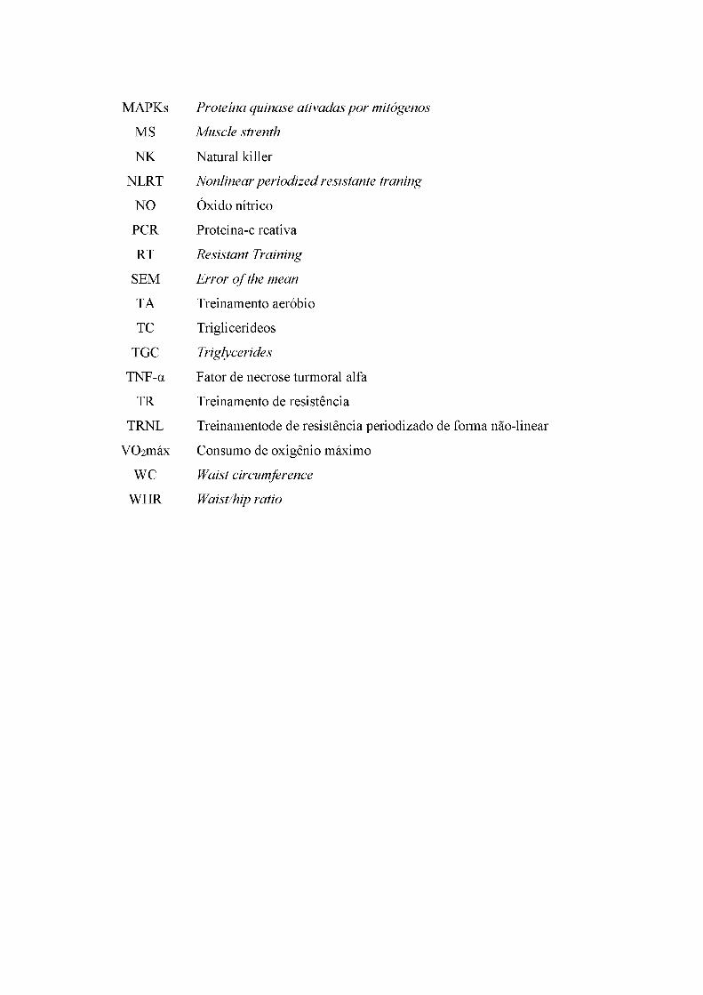
MAPKs Proteína quinase ativadas por mitógenos
MS Muscle strenth
NK Natural killer
NLRT Nonlinear periodized resistante traning
NO Óxido nítrico
PCR Proteína-c reativa
RT Resistant Training
SEM Error of the mean
TA Treinamento aeróbio
TC Triglicerídeos
TGC Triglycerides
TNF-a Fator de necrose turmoral alfa
TR Treinamento de resistência
TRNL Treinamentode de resistência periodizado de forma não-linear
VO2máx Consumo de oxigênio máximo
WC Waist circumference
WHR Waist/hip ratio

SUMÁRIO
1 INTRODUÇÃO.........................................................................................................................14
2 REVISÃO DE LITERATURA................................................................................................ 16
2.1 Processo inflamatório e efeitos adversos em pacientes oncológicos.................................. 16
2.2 Efeitos do treinamento físico em sobreviventes de câncer...................................................18
2.3 Efeitos dos treinamentos combinados e resistidos no perfil inflamatório em sobreviventes
de câncer de mama.....................................................................................................................20
2.4 Periodização do treinamento resistido.....................................................................................22
3 OBJETIVOS.............................................................................................................................. 23
4 A RTIG O .....................................................................................................................................24
REFERÊNCIAS........................................................................................................................46
APÊNDICES.............................................................................................................................. 52
Apêndice 1. Anamnese.............................................................................................................. 52
Apêndice 2. Ficha de avaliação do recordatório alimentar.................................................. 53
Apêndice 3. Ficha de avaliação dos testes de aptidão física................................................54
Apêndice 4. Termo de consentimento livre e esclarecido.................................................... 55
ANEXOS..................................................................................................................................... 56
Anexo 1. Parecer do comitê de ética em pesquisa................................................................56
Anexo 2. Questionário internacional de atividade física...................................................... 60

14
1. INTRODUÇÃO
Existemvários fatores de risco que podem desencadear a ocorrência de lesões
proliferativas benignas da glândula mamária, sendo estes classificados em intrínsecos e
extrínsecos. Os fatores de risco intrínsecos como idade, sexo e aspectos genéticos constituem
parâmetros independentes e que não são modificados ao longo da vida (KAMINSKA et al.,
2015). Os fatores extrínsecos são condicionados pelo estilo de vida, dieta e intervenções
farmacológicas (contraceptivos hormonais orais e terapia de reposição hormonal) e suas
influências sobre o processo neoplásico pode ser modificados até certo ponto (KAMINSKA et
al., 2015).
Atualmente, o câncer de mama (CM) representa um grande problema na saúde mundial
devido o seu alto resistro de incidência e mortalidade. No último levantamento mundial, em
2012, registrou-se 1,7 milhões de novos casos (25% de todos os cânceres) e 522.000 mortes
pelo CM (WORLD HEALTH ORGANIZATION, 2013). Em países desenvolvidos como
Estados Unidos, em 2016 projetou-se uma estimativa de 246.660 novos casos e 40.450 mortes
por CM (SIEGEL; MILLER; JEMAL, 2016). Essas tendências não diferem em país em
desenvolvimento, como no Brasil, que na estimativa de 2016, denotou a incidência de 57.960
novos casos (28,1% do total de câncer) em mulheres, sendo considerado o câncer mais
frequente em mulheres no país (INCA, 2016).
Os tratamentos e intervenções oncológicos atuais asseguram a sobrevida e minimizam
os riscos de mortalidade por CM (CHEN et al., 2016; STEWARD et al., 2014). Estes
contemplam abordagens cirúrgicas do tipo radical ou conservadora, esvaziamento axilar de
linfonodos (linfadenectomia), radioterapia, quimioterapia, imunoterapia, hormonioterapia,
como outros. As aplicações dessas estatrégias oncológicas dependem da especificidade do
câncer e individualidade biológica do paciente (NATIONAL CANCER INSTITUTE, 2016).
Entretanto, todos tratamentos oncológicos podem oferecer riscos de efeitos adversos.
A queda de células imunes, sobrepeso e aumento dos níveis de triglicerídeos (TC) são
alguns dos efeitos adversos após a quimioterapia, enquanto que a hormonioterapia podem induz
redução os níveis de lipoproteína de alta densidade (HDL) (ALEXOPOULOS et al., 1992;
LOVE et al., 1991; MAKARI-JUDSON et al., 2014; SEWELL et al., 1993). Algumas células
imunes, como natural killer (NK) são a primeira linha de defesa contra as células cancerosas
tumores (DEWAN et al., 2007), enquanto que o excesso de gordura corporal é provedora de
citocinas pro-inflamatórias (OST et al., 2016), assim como a própria quimioterapia (MILLS et
al., 2008). Por sua vez, o consequente aumento do perfil inflamação é associado a incidência

15
de outras comorbidades, como depressão, declínio de força, aumento de gordura (LEE et al.,
2004; SCHUBERT et al., 2007), como também a reincidência do câncer nessa população
(COUSSENS; WERB, 2002).
Atualmente, o treinamento físico é considerado como importante estratégia
complementar para pacientes com CM, principalmente por melhorar a aptidão física e perfil
inflamatório (MENESES-ECHÁVEZ et al., 2016). Porém as repostas ao treinamento físico
dependem tanto da condição de saúde do paciente quanto do tipo, intensidade e protocolo de
treinamento. Alguns estudos demonstraram que o treinamento aeróbio (TA) de intensidade
moderada diminui a gordura corporal e melhoram significativamente o perfil inflamatório em
sobreviventes de CM, como redução de proteína-c reativa (PCR), diminuição de interleucina-2
(IL-2) e aumento de interleucina-6 (IL-6) (FAIREY et al., 2005a; JANELSINS et al., 2011).
Em contrapartida, todos os estudos que aplicaram intervenções isoladas de treinamento
resistido (TR) em sobreviventes de CM até o presente momento, não observaram nenhuma
alteração significativa na composição corporal e em marcadores inflamatórios (HAGSTROM
et al., 2016; SCHMIDT et al., 2016; SIMONAVICE et al., 2014).
Intrigantemente, estes estudos que interviram e investigaram a aplicação do TR nessa
população não relataram sistematização do treinamento e/ou aplicaram periodizações
“tradicionais” . Nesta periodização o mesmo conjunto de exercício é realizado na mesma ordem
e com intensidades constantes por longos períodos (FLECK, 2011). Em sessões de TR
periodizado de forma não linear (TRNL) a intensidade e o volume do treinamento são alterados
com muito mais frequência para minimizar o tédio, evitar excessos de lesões e potencializar
ganhos de força (FLECK, 2011).
Existem poucas informações sobre o efeito do TRNL no perfil inflamatório em
indivíduos com doenças crônicas (ZANETTI et al., 2016a). Pelo crescimento da prática clínica
do treinamento físico para pacientes com CM e a falta de evidências científicas sobre os
métodos de TR nesta população, se faz necessário a investigação dos marcadores inflamatórios
após TRNL em mulheres recuperando do CM, população a qual necessita de minimizar os
efeitos adversos dos tratamentos oncológicos e otimizar os benefícios envolvidos com o
treinamento físico.

16
2. REVISÃO DE LITERATURA
2.1. Processo inflamatório e efeitos adversos em pacientes oncológicos
O ambiente pro-inflamatório desencadeado pelo câncer é devido em parte pelos fatores
diretos da progressão do crescimento tumoral e incapacidade de atuação do sistema
imunológico, quanto indiretos, advindos dos tratamentos oncológicos e seus efeitos adversos.
Leucócitos infiltrantes de tumor, assim como as vias de sinalização relacionadas com citocinas,
são componentes importantes no desenvolvimento do microambiente inflamatório tumoral
(KANG et al., 2013). Os principais leucócitos infiltrantes, como macrófagos e células NK,
atuam no combate de células tumorais, porém induzem a liberação extracelular de uma proteína
denominada high mobility group box 1 (HMBG1) a partir da apoptose dessas células (JUBE et
al., 2012).
A HMGB1 livre se liga em diferentes receptores de superfície expressos em células do
sistema imunológico, como o receptor de produtos finais com glicosilação avançada (KANG et
al., 2010), toll-like receptor 4 (YU et al., 2006), receptores de gatilho expressos em células
mielóides-1 (EL MEZAYEN et al., 2007), e CD24+ (CHEN et al., 2009). Após a interação elas
ativam vias de sinalização proteína-quinases ativadas por mitógenos (MAPKs), fator nuclear
kappa beta (NF-kP), e fosfatidilinositol 3 quinases/AKT, mediando as respostas de migração,
ativação, proliferação e diferenciação celular (TANG et al., 2011), desencadeando a cascata
inflamatória em suas múltiplas vias, entre elas, a ativação de mais macrófagos e liberação de
mais citocinas pro-inflamatórias (IL-ip, IL-2, IL-6, IL-8, TNF-a e PCR), ativação das células
endoteliais, aumento da expressão de moléculas de adesão e o aumento da expressão de
inibidor-1 do ativador do plasminogénio PAI-1 e tetradecanoílo acetato de forbol (KANG et
al., 2013; MENESES-ECHÁVEZ et al., 2016; TANG et al., 2011). Estas sinalizações regulam
a coagulação e aumentam a permeabilidade epitelial, resultando em uma respostas inflamatórias
que pode induzir graves danos teciduais e/ou até mesmo a morte (KANG et al., 2013).
Em casos da presença de necrose tumoral, o ambiente inflamatório local pode ser ainda
mais agravado devido a inibição da atuação do sistema imunitário. A necrose induz a
extravasação de íons de potássio (K+) no fluido extracelular, causando supressão profunda da
função efetora das células T (EIL et al., 2016). A elevação na concentração extracelular de K+
prejudica a atividade dos receptores de células T efetoras contra a fosforilação mediada por
Akt-mTOR (EIL et al., 2016). Assim, se o gradiente de sódio-potássio não conseguir fornecer
influxo de K+ para as células T específicas do tumor, elas serão incapazes de deter a progressão
tumoral (EIL et al., 2016).

17
Além disso, após os tratamentos oncológicos neoadjuvantes e/ou adjuvantes, o processo
inflamatório ainda pode estar presente nestes sobreviventes de câncer, sendo um dos efeitos
adversos apresentado por estes tratamentos (TSAVARIS et al., 2002). Embora possua efeitos
anti-inflamatórios após exercício físico, a IL-6 também apresenta efeitos pro-inflamatórios
quando é procedida de um patógeno e em sobreviventes de CM, é um marcador inflamatório
eminente (JONES et al., 2013; LAVOY; FAGUNDES; DANTZER, 2016; PETERSEN;
PEDERSEN, 2005; WIESELER-FRANK; MAIER; WATKINS, 2005). Nesta população, a IL-
6 é associada a dor (WIESELER-FRANK; MAIER; WATKINS, 2005) e também estimula o
aumento de hepcidina, a qual é responsável pelo bloqueio do transporte de ferro para o meio
extracelular (GRELLIER et al., 2015). Esse processo pode diminuir a eficiência de oxigenação
metabólica, favorecendo diretamente a fadiga constatada em sobreviventes de CM (LAVOY;
FAGUNDES; DANTZER, 2016). Por sua vez, este estado pode favorecer mudanças
comportamentais pós-tratamento que também pode influenciar indiretamento o estado
inflamatório.
É constato que os sobreviventes de CM aumetam o tempo de comportamento sedentário
e diminuem os níveis de atividade física (queda de até 11%) após o tratamento oncológico,
possivelmente pela fadiga incidida pelo tratamento (DIELI-CONWRIGHT; OROZCO, 2015;
LAVOY; FAGUNDES; DANTZER, 2016; PHILLIPS et al., 2015). Além da imobilização e da
inatividade física favorecer o estado sarcopenico, contribui também para redução da expressão
de citocinas anti-inflamatórias e aumenta citocinas pro-inflamatórias, principalmente em no
tecido adiposo (OST et al., 2016).
O menor gasto energético capacita o aumento da estocagem de gordura nos tecido
adiposos, resultando em hipertrofia adipocitária. O excesso deste processo pode ocasionar a
ruptura das membranas das gotículas lipídicas e a promoção da infiltração de macrófagos M1
para localidade, que por sua vez, exacerba a expressão de fator de necrose tumoral alfa (TNF
a), interceulina-1 beta (IL-1P), IL-6 e interleucina-8 (IL-8) (JUNG; CHOI, 2014), na medida
em que favorecem outros resultados deletérios para a saúde desta população (BARTON
BURKE, 2006; CHRISTIANSEN et al., 2010). , o aumento da produção de citocinas pró-
inflamatórias e hipertrofia do tecido adiposo leva ao aumento da produção de PCR no fígado e
diminuição da produção de adipocinas anti-inflamatórias, como a interleucina-10 (IL-10) e a
adiponectina, que podem contrabalançar positivamente a inflamação sistêmica (JUNG; CHOI,
2014).
Além disso, o aumento no teor de gordura corporal em sobreviventes de CM pode ser
devido à hormonioterapia. Pacientes que são positivos ao receptor de estrogênio, receptores

18
positivos à progesterona e/ou receptor-2 do factor de crescimento epidérmico humano devem
fazer o uso de tamoxifeno ou inibidores da aromatase, o que favorece uma provável diminuição
da taxa metabólica basal devido a inibição da ação hormonal (BURSTEIN et al., 2016;
JOHNSTON; DOWSETT, 2003).
Assim, uma vez que, a debilitação e efeitos adversos conduzida pelos tratamentos
elevam a expressão de marcadores inflamatórios como IL-6, CRP, IL-8 e TNF-a (BARTON
BURKE, 2006; KOZLOWSKI et al., 2003; LAMBIASE et al., 2013), torna-se evidente a
existência de um ciclo progressivo e continuo do perfil pró-inflamatório desde o diagnóstico,
crescimento tumoral, tratamento e desenvolvimento de efeitos adversos em sobreviventes de
CM. Dentro dessa lógica, parece previsível que o treinamento físico teria efeitos benéficos
associados à desaceleração e/ou inibição em algumas destas vias do ciclo inflamatórios.
2.2. Efeitos do treinamento físico em sobreviventes de câncer
O treinamento físico mostrou ser capaz de combater efeitos adversos dos tratamentos
oncológicos do CM (CASLA et al., 2015; DIELI-CONWRIGHT; OROZCO, 2015;
MENESES-ECHÁVEZ et al., 2016). Os possiveis mecanismos que conduzem melhora nestes
parâmetros adversos nos tratamentos são o aumento da capacidade respiratória
(GOLDHAMMER et al., 2005), força muscular (CASLA et al., 2015), aumento da massa
magra e redução da gordura corporal (OST et al., 2016). Como as alterações na composição
corporal são um fator determinante da inflamação sistêmica (OST et al., 2016), faz sentido que
esse benefício também possa aparecer em sobreviventes de CM.
Embora as vias de sinalização envolvidas no deslocamento do perfil inflamatório ainda
não sejam totalmente compreendidas, existe evidência de que o aumento da produção aguda de
IL-6 pela contração muscular (PETERSEN; PEDERSEN, 2005) pode conduzir à inibição das
principais citoquinas pró-inflamatórias, tais como TNF-a , o que significa que a IL-6 produzida
nas fibras musculares teria características anti-inflamatórias após exercício físico (PETERSEN;
PEDERSEN, 2005). Além deste efeito de fase aguda na produção de IL-6, o aumento da massa
muscular pelo treinamento físico a longo prazo poderia otimizar a diminuição do TNF-a
circulante. De fato, os receptores aumentados de TNF-a no tecido muscular foram observados
após o treino de resistência (HELED et al., 2005; PETERSEN; PEDERSEN, 2005), levando à
hipótese de que a hipertrofia muscular poderia permitir uma maior absorção de TNF-a
circulante além da maior expressão de citocinas anti-inflamatórias (OST et al., 2016).

19
A IL-6 produzida pelo tecido muscular também estimula a expressão de citocinas anti-
inflamatórias, tais como o antagonista do receptor de IL-1 (IL-1ra) no músculo e a IL-10 em
adipócitos (PEDERSEN; FEBBRAIO, 2012). Estas citocinas, por sua vez, são responsáveis
pela inibição da produção de IL-1B, TNF-a e IL-8 mediada pela atividade de monócitos
humanos (PETERSEN; PEDERSEN, 2005). O aumento da produção de IL-6 a partir do tecido
muscular também leva ao aumento da translocação do transportador insulino-sensível 4 para o
sarcolema, e à ativação da via de sinalização AMPK (precursor da biogênese mitocondrial e
biossíntese proteica) e lipólise no tecido adiposo (PEDERSEN; FEBBRAIO, 2012).
O treinamento físico também é capaz de aumentar a expressão de citoquinas menos
conhecidas nestas adaptações benéficas, tais como a interleucina-15 (IL-15). Esta citocina é
expressa com concentrações mais elevadas pelo músculo especificamente após TR (NIELSEN;
PEDERSEN, 2007). Sua atividade provoca um declínio na degradação protéica, sendo
considerada uma citocina "anabólica". A IL-15 também é capaz de aumentar a vida útil das
células CD4+, CD8+ e naive que sustentam atividade imuno-protetora (NIELSEN; PEDERSEN,
2007).
Além disso, a redução das reservas de gordura no tecido adiposo pelo exercício físico
diminui a infiltração de macrófagos de tipo M1 no tecido adiposo e, conseqüentemente, a
expressão de citocinas pró-inflamatórias neste local (JUNG; CHOI, 2014) e melhora da
resistência a insulina (SURMI; HASTY, 2008). A diminuição das reservas de gordura no tecido
adiposo também leva ao aumento da expressão de adipocinas anti-inflamatórias pela infiltração
de macrófagos de tipo M2 no próprio tecido, levando a um aumento das adiposinas, tal como
adiponectina e IL-10, potenciando a metabolização dos ácidos graxos (JUNG; CHOI, 2014;
SURMI; HASTY, 2008).
Assim, o treinamento físico é uma estratégia que deve ser encorajada no tratamento
complementar em sobreviventes de CM, uma vez que, o ambiente fisiológico com menores
marcadores inflamatórios reduz o risco de recorrência do câncer e até mesmo proporciona maior
proteção contra outras doenças aportunas advindas do tratamento oncológico (CASLA et al.,
2015; FAIREY et al., 2005a, 2005b; JANELSINS et al., 2011).
2.3. Efeitos do treinamentos combinados e resistidos no perfil inflamatório em
sobreviventes de câncer de mama
A partir das informações levantadas sobre o perfil inflamatório, pode-se supor que o
treinamento físico envolvendo exercício de resistência isolado ou exercício de resistência
20
combinado com exercícios aeróbios seriam mais eficientes na redução de marcadores pró-
inflamatórios em sobreviventes com CM, uma vez que, estes tipos de treinamentos levam a
redução de peso e aumento da massa muscular em uma magnitude maior do que a aplicação
exclusiva de TA em indivíduos saudáveis com sobrepeso (HO et al., 2012). Até o presente
momento, existem poucos estudos que envolveram avaliação do perfil inflamatório e aplicação
de treinamento de força em sobreviventes do CM. Alguns estudos existentes envolveram o
treinamento combinado (GÓMEZ et al., 2011; HUTNICK et al., 2005; ROGERS et al., 2013),
ou TR isolado (HAGSTROM et al., 2016; SCHMIDT et al., 2016; SIMONAVICE et al., 2014)
como intervenção nesta população. No entanto, nenhum desses estudos demonstrou a
ocorrência de melhora significativa do perfil inflamatório plasmático (citocinas séricas) em
sobreviventes CM submetidos ao treinamento.
A possível explicação para esta conclusão seria a multiplicidade de características
individuais de cada paciente, devido às diferenças no tempo de tratamentos adjuvantes (número
de sessões de quimioterapia / radioterapia aplicada / tempo de hormonioterapia) e ao período
de recuperação em que foram recrutadas para a pesquisa. O estudo de Schmidt et al. (2016) foi
o único estudo desenvolvido com sobreviventes CM que usaram TR de intensidade moderada
a alta (60-80% de 1RM) durante a radioterapia. Após 8 semanas de TR (2x / semana) em
máquinas guiadas, não foi observada melhora nos marcadores inflamatórios, mas foi
evidenciado aumento na produção de IL-6 (p = 0,010) e relação IL-6 / IL-1ra (p = 0,018) no
grupo controle (relaxamento muscular). Neste sentido, embora o tratamento tenha levado a um
aumento nos níveis de citocinas pró-inflamatórias no grupo controle, a TR foi capaz de inibir a
progressão destes marcadores no grupo intervido.
Outra hipótese para explicar a ausência de melhora no perfil inflamatório seria que as
intervenções de treinamento não teriam atingido níveis suficientes de exercício (tempo de
treinamento, intensidade, volume e estímulos) e/ou tempo suficiente para adaptações imunes
para alterar os marcadores inflamatórios. Dois dos estudos considerados neste levantamento,
interviram com dois treinos semanais durante seis meses de TR moderado a intenso (60-80%
de 1RM) (SIMONAVICE et al., 2014) e combinado com intensidades moderadas de exercícios
aeróbicos (60-75% do VO2max) (HUTNICK et al., 2005) em sobreviventes de CM após o
tratamento adjuvante de quimioterapia e radioterapia. Apesar do elevado tempo de treinamento
(6 meses), no estudo de Simonavi et al. (2014), além de não haver diminuição da PCR, também
não ocorreram alterações na composição corporal, mesmo com monitoramento nutricional e
TR de moderada. Pesquisadores especulam que a resistência à alteração da qualidade corporal
poderia ser devido à hormonioterapia (BURSTEIN et al., 2016; JOHNSTON; DOWSETT,

21
2003; SIMONAVICE et al., 2014). No estudo de Hutnick et al. (2005), a intensidade da TR não
pôde ser medida devido ao uso de bandas elásticas na aplicação dos exercícios de resistência.
A baixa intensidade de exercício neste estudo associada à ausência de progressão de intensidade
devido ao uso de bandas elásticas pode justificar a ausência de melhora nos marcadores
inflamatórios e na força dos membros inferiores (HUTNICK et al., 2005).
Um estudo recente que interveio com TR isolado também não mostrou diminuição nas
citocinas em soro sanguíneo após 16 semanas (3x / semana) de treinamento progressivo de alta
intensidade (80% de 1RM), mas houve redução na expressão de TNF-a produzida pelas células
NK e T natural killer (NKT) (HAGSTROM et al., 2016). Os pesquisadores vinculam a
diminuição deste marcador devido ao aumento dos níveis de lactato sanguíneo durante a TR. O
acúmulo de lactato devido às sessões de exercício está associado a concentrações aumentadas
de adenosina cíclica monofosfato (cAMP), que, por sua vez, é capaz de suprimir a expressão
de TNF-a em células NK e NKT (KAST; ALTSCHULER, 2005). Além disso, a diminuição da
expressão de TNF-a em células NK foi associada a um aumento na força dos membros
inferiores, sugerindo que as intervenções de TR com progressão de carga e envolvendo trabalho
de alta intensidade (80% de 1RM) podem promover maior sensibilização na redução de TNF
a expressas por células NK em conjunto com ganho de força e resistência muscular.
Outras intervenções que adotaram treinamento combinado de baixa intensidade,
envolvendo 150-160 minutos de caminhada em 48-52% da freqüência cardíaca máxima
(FCmáx) em conjunto com 8 exercícios para membros superiores e inferiores usando banda
elástica, não encontraram mudança nos marcadores quando comparados ao grupo controle ou
em relação ao tempo de intervenção (ROGERS et al., 2013). Assim, os métodos de treinamento
e suas variáveis (exercícios, cargas, amplitude e tempo de recuperação entre outros) parecem
estar intimamente relacionadas com as alterações das citocinas e otimização de celulas
imunológica não só em populações saudáveis, mas também em sobreviventes de CM.
Embora estes estudos não terem encontrado uma redução significativa nos marcadores
inflamatórios séricos após intervenção com treinamento combinado e/ou TR em sobreviventes
CM, há benefícios que devem ser destacados, já que podem aumentar a sobrevida dessa
população. Esses benefícios incluem aumento do consumo máximo de oxigênio (VO2 máx)
(HUTNICK et al., 2005; ROGERS et al., 2013), força muscular (HAGSTROM et al., 2016;
SIMONAVICE et al., 2014), maior eficácia das células imunológicas (HAGSTROM et al.,
2016; HUTNICK et al., 2005) e sono melhorado (ROGERS et al., 2013). Ainda cabe salientar
que a eficazia do treinamento físico no perfil inflamatório em sobreviventes de CM não se
restrigem em apenas aumentar parâmetros pro-inflamatórios, mas também impedir uma

22
progressão dessas citocinas. Esta premissa é justificada pela progressão de marcadores pro-
inflamatórios nos grupos controles de alguns estudos (BRADLEY et al., 2011; SCHMIDT et
al., 2016).
Por fim, também é importante destacar a ausência do controle de progressão de carga
associada à similaridade das periodizações utilizadas nos estudos citados, que foram
unicamente do tipo tradicional e/ou ausentes de periodização. A periodização do tipo
ondulatório ou não-linear é bastante difundido no esporte e no TR para atletas de alto
rendimento (FLECK, 2011), porém pouco se sabe da sua aplicação em populações com doenças
crônicas, principalmente sobreviventes de CM. Devido a ausência de informações, se faz
necessário a realização de estudos de intervenções com treinamento resistido periodizado de
forma não-linear (TRNL) em mulheres com CM.
2.4. Periodização do treinamento resistido
A periodização incide em alterações programadas das variáveis agudas e crônicas de um
programa de treinamento (BRADLEY-POPOVICH, 2001). Estas variáveis nas modalidades
esportivas contemplam em intensidade, volume, tempo de recuperação (BRADLEY-
POPOVICH, 2001). Especificamente no TR, a aplicação de periodização indica a mudanças
agudas em variáveis do próprio programa de treino, como da ordem, a escolha de exercícios,
número de séries, o número de repetições por série, tempo de pausa entre as séries e exercícios,
a intensidade do treino, volume de treino e o número de sessões por dia (AMERICAN
COLLEGE OF SPORTS MEDICINE, 2009).
O objetivo ao se periodizar o TR é em potencializar as adaptações do treinamento em
curtos (semanas e meses) à longo (anos) período. Variações sistematizadas da relação estresse
(intensidade) e recuperação (descanso) podem atenuar a estabilização dos resultados de
desempenho, garantindo maiores aumentos de força e potência (HERRICK; STONE;
METTLER, 1997), como também, desfavorece a indicencia de possiveis sobrecargas (RHEA
et al., 2003). O manuseio das diferentes formas de aplicações das variáveis e controle de
intensidade vs. volume (heterocronismo) resultou em diversas estratégias de periodização em
curto, médio e longo prazo. Entretanto, existem basicamente dois tipos principais de modelo de
periodização para o TR: periodização linear/tradicional e periodização não-linear/ondulatória.
Estes modelos são os tipos mais estudados na atualidade, como também os mais
aplicados em atletas de alto rendimento. O a periodização linear frequentemente inicia-se com
alto volume (como repetições) e baixa intensidade de treino (carga) e na medida do tempo, é

23
alterado no sentido de recressão de volume de aumento de intensidade de treino. Essa
programação leva várias semanas, cerca de 46, para alcançar o pico de intensidade e
consequentemente o auge da aptidão física (FLECK, 2011). Durante este período a ocilação de
intensidade/volume é muito restrita e fixa apenas em um sentido, sendo crescente ou
decrescente em relação a intensidade.
Na peridiozação do TRNL, o volume/intensidade são alterados com muito mais
frequência em relação ao linear (FLECK, 2011). O tipo de variação mais comum desta
periodização seria a chamada TRNL diária. Neste tipo de modelo, a relação volume/intensidade
é ajustada a cada sessão de treinamento. Um exemplo mais simples de periodização do TRNL
diária utiliza-se três zonas de treinamento, tais como 4-6 repetições máximas, seguidas de 15
20 e 8-12 repetições máximas, totalizando três sessões por semana.
Vários estudos procuraram investigar as diferenças entre os dois principais modelos de
periodização do TR. Algumas comparações mostraram significativamente maiores ganhos de
força no TRNL em relação ao linear em jovens universitários (MONTEIRO et al., 2009; RHEA
et al., 2002). Porém, outras comparações não denotaram mostram diferenças significativas entre
os dois modelos de periodização do TR (PRESTES et al., 2009). A maioria dessas comparações
envolveram jovens de ambos os sexos com pouca ou nenhuma experiência no TR, enquanto
um estudo envolvendo atletas universitários treinados relatou melhores resultados do TRNL
(HOFFMAN et al., 2009). Assim, o nível de treinamento dos indivíduos deve ser levado em
consideração antes de tomar decisões sobre qual modelo de periodização do TR será utilizado
no programa. Em suma, os estudos indicam que o TRNL são tão eficaz ou até mais eficaz do
que o TRL para ganhos de força máxima. Entretanto para melhora do desempenho motor
(HOFFMAN et al., 2009), composição corporal (MONTEIRO et al., 2009; PRESTES et al.,
2009) não existe diferença entre a aplicação destes dois modelos de periodização.
Nesse sentido, o TRNL poderia favorecer indivíduos que necessitam melhorar a aptidão
física em curto período de tempo. Em indivíduos com doenças crônicas a possibilidade de se
afastarem do exercício é grande devido a complicações da própria doença ou a efeitos adversos
dos tratamentos. Assim protocolos mais eficientes no acarreamento dos benefícios do TR
poderiam ser de grande vantagem nestas ocasiões. Entretanto até o presente momento não
existem muitos estudos que verificaram o efeito da utilização do TRNL em pacientes com
doenças crônicas.
Em um estudo de intervenção multidisciplinar realizado com adolescentes obesos, que
incluíram o TRNL aplicado 3 vezes semanais durante 24 semanas, demostrou eficazia na
melhora das respostas anti-inflamatórias e das defesas antioxidantes (NUNES et al., 2016).

24
Outros dois recentes estudos, envolvendo pacientes com vírus da imunodeficiência humana
(HIV) demonstram que o modelo de TRNL aplicado 12 semanas foi eficiente da redução de
gordura corporal (p=0.004), TC (p<0.001), LDL (p<0.001), PCR (p<0.001), TNF-a (p<0.001),
células CD4+ (p=0.004) e CD8+ (p<0.001), aumento de HDL (p<0.001) e citocina anti-
inflamatório (IL-10; p<0.001) (ZANETTI et al., 2016a, 2016b). Assim aplicação do TRNL
também pode favorecer as especificidades adversas de grupos especiais, porém até o momento
não se sabe sobre o efeito do TRNL em sobrevientes de câncer. Nesse sentido invesgitgações
sobre a aplicação de TRNL nesta população parece promissor devido aos seus possiveis
benefícios adquiridos de forma otimizada e possibilidade de adequação do treinamento em vista
de suas limitações específicas.

25
3. OBJETIVOS
Verificar o efeito do treinamento resistido não linear nos marcadores inflamatórios,
bioquímicos e aptidão física em mulheres em recuperação do câncer de mama.
Objetivos específicos
Verificar se o efeito treinamento resistido não linear na concentração plasmática de TNF-a, IL-
4, IL-6, IL-10, IL-17, IL-1RA, IFN-y e imunoglobulina-A salivar em mulheres em recuperação
do câncer de mama.
Verificar se o efeito treinamento resistido não linear no perfil lipídico em mulheres em
recuperação do câncer de mama.
Verificar se o efeito treinamento resistido não linear em células imunológicas em mulheres em
recuperação do câncer de mama.
Verificar se o efeito treinamento resistido não linear na composição corporal em mulheres em
recuperação do câncer de mama.
Verificar se o efeito treinamento resistido não linear na força e resistência muscular em
mulheres em recuperação do câncer de mama.
26
4. ARTIGO
“Alterations of inflammatory, biochemical and physical fitness markers in women under
recovery of breast cancer treatment after non-linear resistance training: A pilot study”
ABSTRACT
Introduction: The objective of this pilot study was to determine the effects of nonlinear
periodized resistance training (NLRT) on inflammatory and biochemical profiles, physical
fitness and body composition in a population of 14 women under recovery from breast cancer
(BC) treatment and under hormone therapy. Methods: The assessments were done before and
after 12 weeks of application of a NLRT program, consisting of three sessions of exercise per
week on non-consecutive days. The normality of the data was verified by the Shapiro-Wilk test
and the comparisons of results were performed by the paired t or Wilcoxon’s tests. Statistical
significance was set at 5% (alpha = 0.05). Results: There were no significant changes in the
plasma concentration of the analyzed cytokines (TNF-a, IL-4, IL-6, IL-10, IL-17, IL-1RA, IFN
Y), but a significant increase in salivary levels of immunoglobulin A (IgA) was observed after
the intervention (186.29%). Significant increases were also observed in HDL-cholesterol
(17.98%), fat-free mass (2.99%), lean mass (2.97%), and muscle strength (39.16%) and
endurance (65.66%). On the other hand, significant reductions were observed in the blood levels
of triglycerides (-14.40%) and total (-4.81%) and LDL- cholesterol (-10.23%), in the platelets
(-7.90%) and monocytes counts (-38.89%), in the amount of fat mass (-4.53%) and in the
percent body fat (-1.19%). Conclusion: Twelve weeks of NPRT led to an increase in salivary
IgA concentration and improvement in body composition, lipid profile and physical fitness of
women under recovery from BC treatment and under hormone therapy.
Key Words: CANCER; RESISTANCE TRAINING; INFLAMMATORY PROFILE;
PHYSICAL ACTIVITY AND HEALTH.

27
INTRODUCTION
The incidence of breast cancer (BC) has increased sharply in the world population in
recent years. Since 2008, the occurrence of new cases has increased by about 20%, causing
about 1.7 million cases and 522 thousand deaths in 2012 (39).
The progression of the BC death rate could be higher without oncological treatment
strategies. Despite increasing patient survival, cancer treatments induce detrimental changes in
women diagnosed with cancer. Falling in natural killer (NK) lymphocyte counts, increased
overweight and triglyceride (TGC) levels are common adverse effects after chemotherapy in
patients with BC (3, 19, 33). These consequences are quite undesirable because NK cells are
the first line of defense against cancer cells (8), while excess body fat raises the production of
pro-inflammatory cytokines (27), as well as chemotherapy itself (23). In addition, the systemic
elevation of inflammation is associated with the incidence of other adverse effects, such as
depression, declining strength, increased fat (18, 32) and also the recurrence of cancer in this
population (7).
Physical training in patients under BC recovery is considered as an important
therapeutic strategy, mainly in the improvement of physical fitness and inflammatory profile
(22). However, responses to physical training depend on the patient's health condition and the
type, intensity, and training protocol. Some studies have shown that moderate-intensity aerobic
training (AT) decreases body fat and significantly improves the inflammatory profile in BC
survivors, with reduced C-reactive protein (CRP), decreased interleukin-2 (IL-2), and Increased
interleukin-6 (IL-6) (10, 14). In contrast, to date, studies that applied isolated resistance training
(RT) interventions in BC survivors did not observe any significant changes in body composition
and inflammatory markers (12, 31, 34).
Intriguingly, all studies that intervened and investigated the application of RT in this
population did not report training systematization and/or applied only traditional periodizations,
in which the same exercise set is performed in the same order and with constant intensity for
long periods (11). In non-linear exercise or RT sessions, the intensity and volume of the training
are altered much more frequently, in order to minimize boredom, to avoid possible injuries and
to increase gains in physical fitness (11).
There is little information on the application of nonlinear resistance training (NLRT) in
individuals with chronic diseases (40) and to date, its effects on patients with BC are entirely
unknown. Due to the need for rapid improvement in general health indicators, especially in the
inflammatory profile of this population (3, 19, 23, 33), this periodization of RT should be tested

28
as a complementary intervention to cancer treatments, because of its ability to promote benefits
over shorter periods compared to traditional methods (11, 40). Thus, the aim of this study was
to verify whether the effects of NLRT on inflammatory and biochemical markers in BC survivor
women under hormone therapy.
METHODS
Participants. The study was previously approved by the local Ethics Committee under
registration 5152/2016. Fourteen BC survivor women under hormone therapy (40-62 years old)
participated in this study. Participants were recruited by voluntary demand application from
regional dissemination of this study (Uberlândia, MG, Brazil). Fifty-seven patients who had BC
and were discharged from hospital were screened through their medical records and invited to
participate in this survey. Of these, fifteen were not resident in the municipality, nine were not
located, five had no interest in participating in the study and four had already deceased.
Twenty-four patients attended the inclusion interview. Patients who met the following
criteria were included: 1) age between 40 and 65 years; 2) previous submission to
lymphadenectomy; 3) completion of chemotherapy and radiotherapy at least 6 months prior to
the study; 4) be performing hormone therapy (aromatase inhibitors and tamoxifen); 5) no
involvement in any exercise program for at least 6 months; 6) medical authorization for TR; 7)
absence of musculoskeletal disorders and / or limitations that could limit participation in the
exercise program; 8) non-smokers and non-alcoholics. Only four subjects were excluded due
to limitations in the shoulder range of motion and thus, effectively, twenty patients entered the
study. All participants signed informed consent and the study was initiated.
Procedures. Initially, the level of physical activity was evaluated by the simplified
model of the questionnaire "International Physical Activity Survey" (15) and important details
were compiled from the medical records of the participants for sample characterization. Before
and after 12 weeks of the intervention, all participants were submitted to collection of blood
and saliva samples for evaluation of blood (TNF-a, IL-6, IL-17, IL-1RA, IFN-y and IL-10) and
salivary (IgA) inflammatory markers, hematologic (immune cells) counts and biochemical
(lipid profile) analytes, as well as to anthropometric and physical fitness (muscle strength and
endurance) assessments. Collection times were always standardized to avoid circadian
variations.
To avoid interference from possible changes in eating patterns, all participants were
instructed to maintain eating habits during the study. In addition, all participants were submitted
to food consumption assessment before and after the intervention. In this evaluation,

29
nutritionists applied the food record from an individual interview in two (non-consecutive) days
of the week and one day of the weekend (35). Data on the consumption of macronutrients
(carbohydrates, lipids and proteins) and daily energy consumption were analyzed using the
Dietpro 5.7i™ (Viçosa, MG, Brazil) application.
Anthropometric Assessments. Body composition was evaluated by bioelectrical
impedance analysis (BIA) using a tetrapolar equipment (InBody230™, Biospace, Seoul, Korea),
with estimates of absolute body mass (BM), fat free mass (FFM), lean mass (LM), fat mass
(FM) and percentage of body fat (%BF). Evaluations were always made after a 10-hour fast
and 72 hours absent from physical exertion, ingestion of thermogenic foods, alcoholic
beverages and / or diuretics. Height was evaluated using a 2-meter length anthropometer
(Personal Caprice, Sanny™, São Bernardo do Campo, SP, Brazil) with 0.1 cm precision. The
waist (WC), hip (HC) and abdomen (AC) circumferences were measured using an
anthropometric tape (Sanny™, São Bernardo do Campo, SP, Brazil), always being expressed
by the mean value of three non-consecutive measures at the same location. These measures
were used to estimate waist/hip circumference ratio (WHR) and body mass index (BMI).
Inflammatory and Biochemical Markers. Blood and saliva samples were taken after
12 hours of fasting and 72 hours of physical effort restriction. The blood samples were collected
in 4 mL sterile tubes (Vacutainer™, Becton-Dickinson, Juiz de Fora, MG, Brazil), one of them
with separator gel, for collection of serum, and another with heparin, for the hematologic
analyzes. After cleaning the mouth with distilled water for 30 seconds, the saliva samples were
collected directly into conical tubes for centrifugation (Becton-Dickinson, Juiz de Fora, MG,
Brazil). After centrifugation of the biological samples at 1200 x g for 10 min, for separation of
supernatant, the serum and saliva aliquots for the determination of the inflammatory profile
were stored at -80 °C until the moment of analysis. The lipid profile (triglycerides, total
cholesterol, LDL-cholesterol and HDL-cholesterol) was determined by enzymatic colorimetric
method using specific kits (Labtest™, Lagoa Santa, MG, Brazil) in a semi-automated analyzer
(Bioplus BIO 2000™, São Paulo, SP, Brazil). The cell counts were made using automated
analyzer (Horiba ABX Diagnostics™, São Paulo, SP, Brazil). Serum (TNF-a, IL-4, IL-6, IL-
10, IL-17, IL-1RA, IFN-y e IgA) and salivary (IgA) inflammatory profiles were determined by
Enzyme Linked Immunosorbent Assay (ELISA, BD Pharmigen™, San Diego, USA),
according to the kit manufacturer's (BD Biosciences™, San Jose, CA, EUA) recommendations.
Physical Fitness. One week before the beginning of the training program, muscle
strength (MS) and localized muscular endurance (LME) were evaluated by the 1 repetition
maximum test (1RM) and by the number of repetitions completed at 50% 1RM, respectively.

30
The tests were performed in all the exercises applied in the physical training, alternately by
body segment, in order to maintain the performance of the participant in all the devices. For the
1RM evaluation, each volunteer preheated with 15 repetitions at approximately 50% of the
subjectively estimated value of 1RM and, after two minutes, with three repetitions at
approximately 70% of that subjectively estimated 1RM value (5). The volunteers then
performed simple repetitions with progressively heavier loads until the 1RM was determined
to the desired level of accuracy, with a maximum of 5 trials. The rest intervals between attempts
were 5 minutes (5). 48 hours after the completion of the 1RM test, the participants performed
the LME test, which consisted of performing the largest possible number of repetitions (up to
concentric failure), with complete and standardized range of motion, using 50% of the load
estimated in the 1RM test.
Protocol of Nonlinear Resistance Training. Prior to the beginning of the training, 4
standardized sessions of resistance exercise were performed for 2 weeks for familiarization with
the protocol. In all training days, the participants underwent different muscular stimuli
(strength, hypertrophy and resistance), starting the first exercises with high intensity and low
volume and finishing with low intensity and high volume, with variation in the order of the
exercises (by body segment) at each training day, so that all exercises were performed on all
stimuli at the end of the week, in order to generate, thus, a type of NLRT more adapted to this
specific population (11). The training was always applied three times a week on non
consecutive days, with the accomplishment of 3 series of each exercise. The training load was
adjusted daily so that the participants would always perform the repetition zones proposed for
each stimulus. There was an increase in load (5-10%) when the participant performed the
maximum repetition range without concentric failure. The details and exercises of the TRNL
protocol are presented in Table 1.
[TABLE 1]
Statistical analysis. Statistical analyzes were performed using the Statistical Package
for Social Sciences (SPSS) 21.0 software. Initially the data were expressed as mean, standard
error of the mean (SEM) and absolute variation. The normality of the data was verified by the
Shapiro-Wilk test. For variables with non-parametric distribution, normalization of data was
done through the use of square root, logarithm and exponential. The results provided were
converted back to the original unit of measure because of the ease of understanding their
meaning. The data of the variables that could not be normalized were expressed as median and
interquartile range (IQR). The values of the variables with normal or normalized distribution
and non-parametric distribution were compared with the application of the t test for paired

31
measures and the Wilcoxon test, respectively. For the validation of the p-value the effect size
was calculated by the Cohen formula (d) and the Hattie classification. Statistical significance
was set at a = 5% for all analyzes.
RESULTS
During the intervention there was 30% (n = 6) of sample loss. Three participants had
less than a 75% presence in the training, one was affected by inflammation in the breast
expander balloon, another had aggravation of rheumatoid arthritis and one sixth abandoned
training on her own. There was no incidence of lymphedema. In all, then, fourteen participants
completed all the procedures of the study (Figure 1).
[FIGURE 1]
The baseline characteristics of the participants who completed the study are presented
in table 2. The mean age was 51.71 ± 2.24 years and the prevalent pattern of physical activity
was mild to moderate. Although the predominant type of cancer was invasive carcinoma, there
was also a diagnosis of carcinoma in situ. The mean tumor size was 3.10 ± 0.35 cm and the
surgical treatment, by mastectomy or quadrantectomy, had been done between 4 months and
more than 24 months before the beginning of the intervention described in this study. All
participants were lymphadenectomized and were on hormone therapy predominantly with
tamoxifen, most of them for more than 9 months. Although the absence of concomitant diseases
has been the most prevalent condition, some of the participants had other pathological
conditions and made continuous use of other drugs.
[TABLE 2]
Before and after intervention, the consumption of carbohydrates (198.66 ± 15.62 vs.
197.74 ± 12.05; p= 0.96; d= 0.21), lipids (62.65 ± 5.64 vs. 58.93 ± 4.90; p= 0.541; d= 0.18) and
proteins (74.54 ± 6.21 vs. 70.07 ± 3.55;p= 0.502; d= 0.23), given in g/day, as well as the energy
intake (1646.32 ± 111.28 vs. 1597.21 ± 76.88; p= 0.621; d= 0.16), given in kcal/day, were not
statistically different. However, there was an increase in LM and FFM and a decrease in FM
and %BF, but without significant changes in BM, BMI, AB and WHR after the intervention
reported in this study (Table 3).
[TABLE 3]
Table 4 presents the results obtained for the analysis of the lipid, hematologic and
inflammatory variables of the participants. After intervention, there were statistically
significant reductions in the blood levels of triglycerides (TGC) and total cholesterol (CT), and
in the counts of platelets and monocytes, as well as significant increases in the blood levels of
32
the high density lipoprotein cholesterol (HDL-C) and in the basophil counts. The lymphocyte,
neutrophil and eosinophil counts did not change significantly with the exercise program.
Although serum levels of the analyzed cytokines also have not changed significantly, after the
intervention there was a significant increase in the salivary IgA concentration. In addition, we
did not obtain plasma IL-6 in the sample. It is important to note that after the intervention there
was also a reduction (p=0.021) in the concentration of low-density lipoprotein cholesterol
(LDL-C).
[TABLE 4]
Table 5 shows the variables analyzed to measure the strength and the localized muscular
endurance (LME) of the participants. After the intervention, in all exercises, there were
significant increases in the load of 1RM (force) and in the number of repetitions completed with
50% of the load of 1RM (LME). The mean percentages of increase in load and number of
repetitions in the tests of all the exercises were of 39.16% and 65.66%, respectively. This means
that for the exercises performed there were mean increases in load and number of repetitions of
1.09% and 1.82%, respectively, on each training day.
[TABLE 5]
DISCUSSION
The main objective of this pilot study was to investigate the effects of a nonlinear
resistance training (NLRT) protocol on biochemical, hematologic and inflammatory parameters
in BC survivors under hormone therapy. Although no serum cytokine showed significant
change after 12 weeks of intervention, there was a significant increase in salivary IgA,
improvement in body composition, lipid profile, increased basophils, reduced plasma and
monocytes and physical fitness of the participants. Other studies that investigated the effects of
RT isolated on inflammatory profiles in BC survivors also did not show significant changes in
serum inflammatory markers (12, 31, 34). But these studies followed a periodization model of
the linear type, with initial intensities of 60% 1RM to 80% 1RM, without control and load
readjustments. Therefore, our work was the first to report results after non-linearly periodic RT
intervention (11) in this population
Physical exercise, regardless of its type, has the capacity to provide an anti
inflammatory environment that acts against the incidence of various diseases, such as cancer
and its recurrence (7). Muscle contraction stimulates the production of IL-6 by this tissue,
neutralize the activity of circulating TNF-a (29) and stimulate adipocytes to produce IL-10 (28),
which in turn has the capacity to decrease the production of pro-inflammatory cytokines, such
as IL-2, are often elevated in patients with BC (22). Chronically, muscle and adipose tissues

33
also play a determining role in the inflammatory response (27), as is also observed in BC
survivors. In the study by Jones et al. (16), IL-6 and C-reactive protein (CRP) were positively
associated with BM and BMI. This makes sense, since an increase in body fat may play a
determining role in increasing serum levels of proinflammatory cytokines (13, 27).
To date, it has not been proven that increased muscle mass and reduced body fat content
could promote an anti-inflammatory environment in BC survivors, but it is known that such
anthropometric changes provide protection against the incidence of concomitant pathological
processes, as insulin resistance (6, 9). Interestingly, studies that investigated the influence of
RT on cancer survivors (12, 31, 34) and did not detect the occurrence of changes in serum
cytokine levels, also did not report changes in body composition. But this does not mean that
the hypothesis of improvement in the inflammatory profile from reductions in FM and increase
in LM in response to RT should not be rejected in this population. Much of the previous studies,
as well as the present study, may not have achieved sufficient time for the occurrence of the
metabolic adaptations that could influence the inflammatory pathways. In addition, there are
still many difficulties in the investigation of serum cytokines, such as laboratory limitations in
the quantification process itself, which in fact occurred with IL-6 in the present study and with
TNF-a in the study by Hagstrom et al. (2016) (12).
Surprisingly, this was the first study to report that one type of TR increases salivary IgA
concentration in patients in BC recovery and under hormone therapy (Table 4). This is very
important because a decrease in the level of salivary IgA is associated with an increase in the
incidence of diseases of the upper respiratory tract (25), which may mean that salivary IgA may
be a useful biological marker for assessing the clinical predisposition also for other diseases
(1). Paradoxically, the only other study investigating the influence of exercise on salivary
inflammatory response in BC patients observed that yoga practice one week before and four
weeks after BC surgery resulted in a significant decrease in salivary IgA concentration (30). It
is possible that this contradiction in the results is due to differences in the type, intensity and
time of training, as well as differences in the health condition of the participants. Indeed, in the
healthy elderly (n=45), the application of 12 weeks of moderate RT, three times a week, was
able to significantly increase (36.84%) the salivary IgA concentration (2). In any case, this is a
question that should be better investigated, since the tumor burden is directly related to serum
IgA levels in cancer patients (1).
It is known that cancer and oncological treatments themselves are associated with
worsening of the immune and inflammatory profile (36, 37) and that the limited information
available on the influence of RT on BC survivors does not comprise changes in the

34
subpopulations of NK lymphocytes (12). The increase in basophil count reported in this study
is a beneficial change to the population of BC survivors, as these cells play an important role
in inflammation and simple allergic responses, although they constitute only 0.5 to 1% of total
blood leukocytes (17). The decrease in circulating platelet count may be related to its use in the
hemostasis of muscle micro-lesions, caused by TRNL, and also in the angiogenesis process
(24). As the TR does not provide alterations in the population of monocytes (4), the decrease
in the monocyte count in this study could be due to the increase in the differentiation of
monocytes to macrophages rate.
In relation to the lipid profile, the influence of RT on the improvement of the lipid profile
in healthy people is well known, as a result of increased mobilization and oxidation of fatty
acids (20). In BC survivors, the application of 15 weeks of combined (aerobic and resistance)
training was able to reduce blood levels of TGC (5%), t-C (6.8%) and LDL-C (9.7%), as well
as increase HDL-C levels (4.5%) (26). The magnitudes of these changes were similar to those
described in this study, but the increase in the blood level of HDL-C reported here (17.98%)
was substantially higher, and this is very significant, because each increase of 1 mg/dL in HDL
C is equivalent to 2-3% reduction in risk of cardiovascular events and decreased chance of
recurrence of cancer (21).
The mean increase in muscle strength reported in this study (39.16%) after 12 weeks of
intervention was higher compared to the results of other intervention studies with isolated RT
after 12 (12, 31) and even after 24 weeks duration (25- 26%) in BC survivors (34). The best
efficiency in increasing muscle strength reported in this study may be associated with the
periodization model used (11). As the studies that investigated the influence of RT in this
population did not evaluate LME (12, 31, 34, 38), establishing comparisons with literature
results is not possible, but it is an interesting evaluation method to determine the physical
condition of this population, mainly due to fatigue installed after treatment.
The present study has limitations, among which it is worth mentioning the absence of a
group whose intervention was a traditional linear physical training protocol, in order to allow
an effective comparison between the protocols. But even so, this pilot study also well serves
the primary purpose of testing the applicability of NLRT in BC survivors and stimulating the
development of new research on the subject.
Anyway, the improvement in the physical fitness of BC survivors under hormone
therapy is a very relevant finding of this study, which shows that the physical limitations arising
from the oncological treatments themselves and that involve fat mass gain, lean mass loss and
fatigue, can be efficiently combated with the application of non-linear resistance training. In

35
addition, although there was no improvement in the inflammatory profile of the participants,
there were improvements in other health indicators, such as salivary IgA and lipidemia, which
are very relevant for the prevention of concomitant diseases.
CONFLICTS OF INTEREST
M. A. Leite was supported by a scholarship from the “Coordenação de Aperfeiçoamento
de Pessoal de Nível Superior” (CAPES). N. Penha-Silva was supported by a scientific
productivity grant from “Conselho Nacional de Pesquisa e Desenvolvimento” (CNPq). The
authors of the present study have no conflicts of interest to report. There was no participation
of professionals or companies that could benefit from the results of the present study. The
results of this study are unpublished and were presented clearly, honestly and without improper
data manipulation. All authors participated equally in all stages of the study.
REFERENCES
1. Ahmad S, Faruqi NA, Arif SH, Akhtar S. Serum immunoglobulin levels in neoplastic
disorder of breast. J Indian M ed Assoc 2002;100(8):495-6.
2. Akimoto T, Kumai Y, Akama T, et al. Effects of 12 months of exercise training on
salivary secretory IgA levels in elderly subjects. Br J Sports M ed 2003;37(1):76-9.
3. Alexopoulos CG, Pournaras S, Vaslamatzis M, Avgerinos A, Raptis S. Changes in
serum lipids and lipoproteins in cancer patients during chemotherapy. Cancer
Chemother Pharmacol 1992;30(5):412-6.
4. Bobeuf F, Labonté M, Khalil A, Dionne IJ. Effect of Resistance Training on
Hematological Blood Markers in Older Men and Women: A Pilot Study. ResearchGate
2009;2009(1687-7063):156820.
5. Brown LE, Weir JP. ASEP Procedures recommendation I: accurate assessment of
muscular strength and power. P ro f Exerc Physiol 2001;4(11)
6. Caan BJ, Kwan ML, Shu XO, et al. Weight Change and Survival after Breast Cancer in
the After Breast Cancer Pooling Project. Cancer Epidemiol Biomark Prev Publ Am
Assoc Cancer Res Cosponsored Am Soc Prev Oncol 2012;21(8):1260-71.
7. Coussens LM, Werb Z. Inflammation and cancer. Nature 2002;420(6917):860-7.
8. Dewan MZ, Terunuma H, Takada M, et al. Role of natural killer cells in hormone
independent rapid tumor formation and spontaneous metastasis of breast cancer cells in
vivo. Breast Cancer Res Treat 2007;104(3):267-75.

36
9. Dodson S, Baracos VE, Jatoi A, et al. Muscle wasting in cancer cachexia: clinical
implications, diagnosis, and emerging treatment strategies. Annu Rev M ed
2011;62:265-79.
10. Fairey AS, Courneya KS, Field CJ, Bell GJ, Jones LW, Mackey JR. Randomized
controlled trial of exercise and blood immune function in postmenopausal breast cancer
survivors. JApplP hysiolBethesdaM d 1985 2005;98(4):1534-40.
11. Fleck SJ. Non-Linear Periodization for General Fitness & Athletes. J Hum Kinet
2011;29A:41-5.
12. Hagstrom AD, Marshall PWM, Lonsdale C, et al. The effect of resistance training on
markers of immune function and inflammation in previously sedentary women
recovering from breast cancer: a randomized controlled trial. Breast Cancer Res Treat
2016;155(3):471-82.
13. Heled Y, Dror Y, Moran DS, et al. Physical exercise increases the expression of
TNFalpha and GLUT 1 in muscle tissue of diabetes prone Psammomys obesus. Life Sci
2005;77(23):2977-85.
14. Janelsins MC, Davis PG, Wideman L, et al. Effects of Tai Chi Chuan on Insulin and
Cytokine Levels in a Randomized Controlled Pilot Study on Breast Cancer Survivors.
Clin Breast Cancer 2011;11(3):161-70.
15. Johnson-Kozlow M, Sallis JF, Gilpin EA, Rock CL, Pierce JP. Comparative validation
of the IPAQ and the 7-Day PAR among women diagnosed with breast cancer. Int J
Behav Nutr Phys Act 2006;3:7.
16. Jones SB, Thomas GA, Hesselsweet SD, Alvarez-Reeves M, Yu H, Irwin ML. Effect
of Exercise on Markers of Inflammation in Breast Cancer Survivors: The Yale Exercise
and Survivorship Study. Cancer PrevRes PhilaPa 2013;6(2):109-18.
17. Knol EF, Olszewski M. Basophils and mast cells: Underdog in immune regulation?
Immunol Lett 2011;138(1):28-31.
18. Lee B-N, Dantzer R, Langley KE, et al. A cytokine-based neuroimmunologic
mechanism of cancer-related symptoms. Neuroimmunomodulation 2004;11(5):279-92.
19. Makari-Judson G, Braun B, Jerry DJ, Mertens WC. Weight gain following breast cancer
diagnosis: Implication and proposed mechanisms. World J Clin Oncol 2014;5(3):272-
82.
20. Mann S, Beedie C, Jimenez A. Differential Effects of Aerobic Exercise, Resistance
Training and Combined Exercise Modalities on Cholesterol and the Lipid Profile:
Review, Synthesis and Recommendations. Sports M ed Auckl Nz 2014;44(2):211-21.

37
21. McGrowder D, Riley C, Morrison EYSA, Gordon L. The Role of High-Density
Lipoproteins in Reducing the Risk of Vascular Diseases, Neurogenerative Disorders,
and Cancer [Internet]. Cholesterol 2011;2011 [cited 2017 May 12 ] Available from:
http://www.ncbi.nlm.nih.gov/pmc/articles/PMC3065895/
22. Meneses-Echâvez JF, Correa-Bautista JE, Gonzâlez-Jiménez E, et al. The Effect of
Exercise Training on Mediators of Inflammation in Breast Cancer Survivors: A
Systematic Review with Meta-analysis. Cancer Epidemiol Biomark Prev Publ Am
Assoc Cancer Res Cosponsored Am Soc Prev Oncol 2016;25(7):1009-17.
23. Mills PJ, Ancoli-Israel S, Parker B, et al. Predictors of Inflammation in Response to
Anthracycline-Based Chemotherapy for Breast Cancer. Brain Behav Immun
2008;22(1):98-104.
24. Mosca MJ, Rodeo SA. Platelet-rich plasma for muscle injuries: game over or time out?
Curr Rev Musculoskelet M ed 2015;8(2):145-53.
25. Neville V, Gleeson M, Folland JP. Salivary IgA as a risk factor for upper respiratory
infections in elite professional athletes. M edSci Sports Exerc 2008;40(7):1228-36.
26. Nuri R, Mahmudieh B, Akochakian M, Moghaddasi M, Nuri R. Effect of 15 weeks
Combination exercise training on lipid profile and fatty liver indices in Postmenopausal
women with breast cancer. Braz JBiomotricity 2012;6(4):297-303.
27. Ost M, Coleman V, Kasch J, Klaus S. Regulation of myokine expression: Role of
exercise and cellular stress. Free Radic Biol M ed 2016;98:78-89.
28. Pedersen BK, Febbraio MA. Muscles, exercise and obesity: skeletal muscle as a
secretory organ. Nat Rev Endocrinol 2012;8(8):457-65.
29. Petersen AMW, Pedersen BK. The anti-inflammatory effect of exercise. J Appl Physiol
BethesdaM d 1985 2005;98(4):1154-62.
30. Rao RM, Nagendra HR, Raghuram N, et al. Influence of yoga on mood states, distress,
quality of life and immune outcomes in early stage breast cancer patients undergoing
surgery. Int J Yoga 2008;1(1):11-20.
31. Schmidt ME, Meynkohn A, Habermann N, et al. Resistance Exercise and Inflammation
in Breast Cancer Patients Undergoing Adjuvant Radiation Therapy: Mediation Analysis
From a Randomized, Controlled Intervention Trial. Int J Radiat Oncol Biol Phys
2016;94(2):329-37.
32. Schubert C, Hong S, Natarajan L, Mills PJ, Dimsdale JE. The association between
fatigue and inflammatory marker levels in cancer patients: a quantitative review. Brain
Behav Immun 2007;21(4):413-27.

38
33. Sewell HF, Halbert CF, Robins RA, Galvin A, Chan S, Blamey RW. Chemotherapy-
induced differential changes in lymphocyte subsets and natural-killer-cell function in
patients with advanced breast cancer. Int J Cancer 1993;55(5):735-8.
34. Simonavice E, Liu P-Y, Ilich JZ, Kim J-S, Arjmandi B, Panton LB. The effects of a 6-
month resistance training and dried plum consumption intervention on strength, body
composition, blood markers of bone turnover, and inflammation in breast cancer
survivors 1. ApplPhysiolNutr Metab 2014;39(6):730-9.
35. Slater B, Philippi ST, Marchioni DML, Fisberg RM. Validation of Food Frequency
Questionnaires - FFQ: methodological considerations. Rev Bras Epidemiol
2003;6(3):200-8.
36. Tsavaris N, Kosmas C, Vadiaka M, Kanelopoulos P, Boulamatsis D. Immune changes
in patients with advanced breast cancer undergoing chemotherapy with taxanes. Br J
Cancer 2002;87(1):21-7.
37. White JP, Wilson JM, Austin KG, Greer BK, St John N, Panton LB. Effect of
carbohydrate-protein supplement timing on acute exercise-induced muscle damage. J
Int Soc Sports Nutr 2008;5:5.
38. Winters-Stone KM, Dobek J, Bennett JA, Nail LM, Leo MC, Schwartz A. The effect of
resistance training on muscle strength and physical function in older, postmenopausal
breast cancer survivors: a randomized controlled trial. J Cancer Surviv Res Pract
2012;6(2):189-99.
39. World Health Organization. International Agency for Research on Cancer. 2013;13.
40. Zanetti HR, da Cruz LG, Lourenço CL, et al. Nonlinear Resistance Training Enhances
the Lipid Profile and Reduces Inflammation Marker in People Living With HIV: A
Randomized Clinical Trial. JP hys Act Health 2016;13(7):765-70.

39
Legends of Figures
Figure 1: Stages of study sample selection.

40
FIGURE 1:

41
TABLE 1. Order of exercises, per day, from muscle stimuli._____________________________ Stimuli_____________ Monday___________ Wednesday____________ Friday
1. Strength a
2. Hypertrophy b
3. Resistance c
Chest Press Bench Press Knee Flexion Front Pull Rowing Machine Abdominal
Knee Flexion Front Pull Rowing Machine Abdominal Leg Press Chest Press
Rowing Machine Abdominal Leg Press Chest Press Knee Flexion Front Pull
a Strenght: 3 sets of 4-6 repetitions at 85% 1RM with 2-3 minutes rest between sets. b Hypertrophy: 3 sets of 8-12 repetitions at 70% 1RM with 1-1.5 minutes rest between sets c Resistance: 3 sets of 15-20 repetitions at 60% 1RM with 45 seconds rest between sets.

42
TABLE 2. Characteristics, clinical aspects and treatments of participants.Variables N Total Mean ± SEM Median Min Max
(%)Age (years)
40-55 7 50.0 51.71 ± 2.24 52.5 40 6256-65 7 50.0Physical Activity Time (min / week)
Light Intensity 13 92.9 124.64 ± 80 0 36029.36
Moderate Intensit 10 71.4 31.21 ± 31.21 110 0 345Type of Cancer
Invasive carcinoma 8 57.1 - - -Carcinoma in situ 4 42.9
Tumor size (cm)< 2 3 21.4 3.10 ± 0.35 3 1 52 - 5 11 78.6
Type of surgeryMastectomy 8 57.1Quadrantectomy 6 42.9
Time after surgery (months)4-12 4 28.613-23 4 28.6 23.5 ± 3.44 22 4 48> 24 6 42.9
Number of Lymph Nodes Removed
> 12 5 35.76 a 11 5 35.7 9.57 ± 1.96 10 1 261 a 5 4 28.6
Type of Hormone TherapyAromatase Inhibitors 2 14.3Tamoxifen 12 65.7
Hormone Therapy Time (months)1-8 4 28.69-16 5 35.3 17 ± 3.28 13 3 40>17 5 35.7
Concomitant diseasesHypertension 3 21.4Hypothyroidism 3 21.4Osteoarthritis 2 14.2Nonexistent 6 42.9
Use of other medications *Yes 9 64.3No 5 35.7
*Hydrochlorothiazide, Lozartan, Enalapril, Metformin, Simvastatin, Omeprazole, Cyntroid, Venlafaxine, Zolpidem, and Reuquinol.

43
TABLE 3. Anthropometric measures of participants before (pre) and after (post) intervention.
Variables
Pre PostChange ±
SEM
CI 95%Effect
SizeMean ±
SEM
Mean ±
SEMInf Sup
p-value
BM (kg) 64.7 ± 3.5 65.1 ± 3.5 0.33 ± 0.36 -0.45 1.10 0.383 0.24
BMI (kg/m2) 25.8 ± 1.3 25.9 ± 1.2 0.13 ± 0.14 -0.17 0.44 0.355 0.26
LM (kg) 37.1 ± 1.2 38.2 ± 1.3 1.12 ± 0.25 0.58 1.65 <0.001 0.21
FFM (kg) 39.4 ± 1.3 40.6 ± 1.4 1.17 ± 0.27 0.59 1.75 <0.001 1.18
FM (kg) 25.1 ± 2.4 24.0 ± 2.3 -0.84 ± 0.37 -1.64 -0.02 0.044 1.30
%BF 37.7 ± 1.9 36.5 ± 1.9 -1.19 ± 0.42 -2.11 -0.28 0.014 0.16
WHR (cm) 0.81 ± 0.02 0.79 ± 0.01 -0.01 ± 0.01 -0.01 0.02 0.204 0.36
AC (cm) 90.1 ± 2.8 88.8 ± 2.7 -1.30 ± 1.23 -1.37 3.97 0.312 0.09
BM: body mass; BMI: body mass index; LM: lean mass; FFM: fat free mass; FM: fat mass;
%BF: percentage of body fat; WHR: waist/hip circumference ratio; AC: Abdomen
circumference.
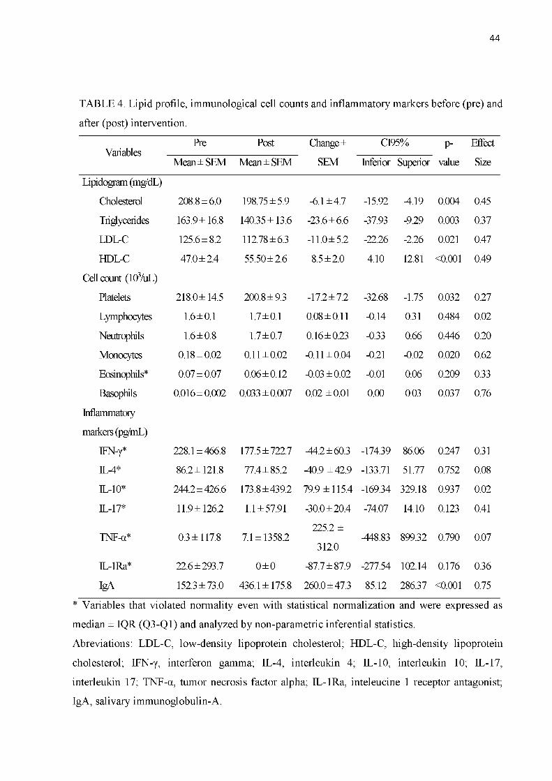
44
TABLE 4. Lipid profile, immunological cell counts and inflammatory markers before (pre) and
after (post) intervention.
VariablesPre Post Change ± CI95% p- Effect
Mean ± SEM Mean ± SEM SEM Inferior Superior value Size
Lipidogram (mg/dL)
Cholesterol 208.8 ± 6.0 198.75 ± 5.9 -6.1 ± 4.7 -15.92 -4.19 0.004 0.45
Triglycerides 163.9 ± 16.8 140.35 ± 13.6 -23.6 ± 6.6 -37.93 -9.29 0.003 0.37
LDL-C 125.6 ± 8.2 112.78 ± 6.3 -11.0 ± 5.2 -22.26 -2.26 0.021 0.47
HDL-C 47.0 ± 2.4 55.50 ± 2.6 8.5 ± 2.0 4.10 12.81 <0.001 0.49
Cell count (103/uL)
Platelets 218.0 ± 14.5 200.8 ± 9.3 -17.2 ± 7.2 -32.68 -1.75 0.032 0.27
Lymphocytes 1.6 ± 0.1 1.7 ± 0.1 0.08 ± 0.11 -0.14 0.31 0.484 0.02
Neutrophils 1.6 ± 0.8 1.7 ± 0.7 0.16 ± 0.23 -0.33 0.66 0.446 0.20
Monocytes 0.18 ± 0.02 0.11 ± 0.02 -0.11 ± 0.04 -0.21 -0.02 0.020 0.62
Eosinophils* 0.07 ± 0.07 0.06 ± 0.12 -0.03 ± 0.02 -0.01 0.06 0.209 0.33
Basophils 0.016 ± 0.002 0.033 ± 0.007 0.02 ± 0.01 0.00 0.03 0.037 0.76
Inflammatory
markers (pg/mL)
IFN-y* 228.1 ± 466.8 177.5 ± 722.7 -44.2 ± 60.3 -174.39 86.06 0.247 0.31
IL-4* 86.2 ± 121.8 77.4 ± 85.2 -40.9 ± 42.9 -133.71 51.77 0.752 0.08
IL-10* 244.2 ± 426.6 173.8 ± 439.2 79.9 ± 115.4 -169.34 329.18 0.937 0.02
IL-17* 11.9 ± 126.2 1.1 ± 57.91 -30.0 ± 20.4 -74.07 14.10 0.123 0.41
TNF-a* 0.3 ± 117.8 7.1 ± 1358.2225.2 ±
-448.83 899.32 0.790 0.07
IL-1Ra* 22.6 ± 293.7 0 ± 0
312.0
-87.7 ± 87.9 -277.54 102.14 0.176 0.36
IgA 152.3 ± 73.0 436.1 ± 175.8 260.0 ± 47.3 85.12 286.37 <0.001 0.75
* Variables that violated normality even with statistical normalization and were expressed as
median ± IQR (Q3-Q1) and analyzed by non-parametric inferential statistics.
Abreviations: LDL-C, low-density lipoprotein cholesterol; HDL-C, high-density lipoprotein
cholesterol; IFN-y, interferon gamma; IL-4, interleukin 4; IL-10, interleukin 10; IL-17,
interleukin 17; TNF-a, tumor necrosis factor alpha; IL-1Ra, inteleucine 1 receptor antagonist;
IgA, salivary immunoglobulin-A.

45
TABLE 5. Strength and localized muscular endurance of the participants before (pre) and after
(post) the intervention.
VariablesPre Post Change ± CI95% p- Effect
Mean ± SEM Mean ± SEM SEM Inf Sup value Size
1RM (kg)
Leg Press 132.1 ± 13.3 207.9 ± 16.7 12.2 ± 1.7 60.26 91.31 <0.001 1.20
Bench Press 21.9 ± 1.5 33.1 ± 2.5 11.2 ± 1.3 8.47 13.96 <0.001 1.06
Rowing
Machine34.8 ± 1.6 42.4 ± 1.9
7.2 ± 0.8
5.53 8.90<0.001
0.90
Knee Flexion 10.8 ± 0.5 13.9 ± 0.6 3.1 ± 0.4 2.19 4.10 <0.001 1.05
Front Pull 28.3 ± 1.4 38.5 ± 2.3 10.2 ± 1.4 7.23 13.20 <0.001 1.18
LME
(repetitions)
Leg press 21.1 ± 1.9 33.3 ± 1.7 12.2 ± 1.7 8.54 15.89 0.009 0.44
Bench Press 21.1 ± 1.9 29.0 ± 2.3 7.9 ± 2.6 2.09 14.07 <0.001 1.01
Rowing
Machine20.6 ± 1.4 32.3 ± 1.8
11.6 ± 1.8 7.80 15.49<0.001
1.03
Knee Flexion 22.3 ± 2.2 37.1 ± 2.8 14.8 ± 2.8 8.78 20.80 <0.001 0.47
Front Pull 26.0 ± 2.1 43.4 ± 2.5 17.4 ± 2.0 13.10 21.75 <0.001 1.06
Abdominal 18.9 ± 1.5 39.4 ± 3.9 20.6 ± 3.3 13.54 27.61 <0.001 1.08
1RM, one repetition maximum; LME, localized muscular endurance, given by the number of repetitions
completed at 50% 1RM.

46
REFERENCIAS
ALEXOPOULOS, C. G. et al. Changes in serum lipids and lipoproteins in cancer patients during chemotherapy. Cancer Chemotherapy and Pharmacology, v. 30, n. 5, p. 412-416, 1992.
AMERICAN COLLEGE OF SPORTS MEDICINE. American College of Sports Medicine position stand. Progression models in resistance training for healthy adults. Medicine and Science in Sports and Exercise, v. 41, n. 3, p. 687-708, mar. 2009.
BARTON-BURKE, M. Cancer-related fatigue and sleep disturbances. Further research on the prevalence of these two symptoms in long-term cancer survivors can inform education, policy, and clinical practice. The American Journal of Nursing, v. 106, n. 3 Suppl, p. 7277, mar. 2006.
BRADLEY, P. S. et al. Sub-maximal and maximal Yo-Yo intermittent endurance test level 2: heart rate response, reproducibility and application to elite soccer. European Journal of Applied Physiology, v. 111, n. 6, p. 969-978, jun. 2011.
BRADLEY-POPOVICH, G. E. Nonlinear Versus Linear Periodization Models. Strength & Conditioning Journal, v. 23, n. 1, p. 42, 2001.
BURSTEIN, H. J. et al. Adjuvant Endocrine Therapy for Women With Hormone ReceptorPositive Breast Cancer: American Society of Clinical Oncology Clinical Practice Guideline Update on Ovarian Suppression. Journal of Clinical Oncology, p. JCO659573, 16 fev. 2016.
CASLA, S. et al. Running away from side effects: physical exercise as a complementary intervention for breast cancer patients. Clinical & Translational Oncology: Official Publication of the Federation of Spanish Oncology Societies and of the National Cancer Institute of Mexico, v. 17, n. 3, p. 180-196, mar. 2015.
CHEN, G.-Y. et al. CD24 and Siglec-10 selectively repress tissue damage-induced immune responses. Science (New York, N.Y.), v. 323, n. 5922, p. 1722-1725, 27 mar. 2009.
CHEN, Z.-L. et al. The efficiency and safety of trastuzumab and lapatinib added to neoadjuvant chemotherapy in Her2-positive breast cancer patients: a randomized metaanalysis. OncoTargets and therapy, v. 9, p. 3233-3247, 30 maio 2016.
CHRISTIANSEN, T. et al. Exercise training versus diet-induced weight-loss on metabolic risk factors and inflammatory markers in obese subjects: a 12-week randomized intervention study. American Journal of Physiology - Endocrinology and Metabolism, v. 298, n. 4, p. E824-E831, 1 abr. 2010.
COUSSENS, L. M.; WERB, Z. Inflammation and cancer. Nature, v. 420, n. 6917, p. 860867, 19 dez. 2002.
DEWAN, M. Z. et al. Role of natural killer cells in hormone-independent rapid tumor formation and spontaneous metastasis of breast cancer cells in vivo. Breast Cancer Research and Treatment, v. 104, n. 3, p. 267-275, 1 set. 2007.
DIELI-CONWRIGHT, C.; OROZCO, B. Exercise after breast cancer treatment: current perspectives. Breast Cancer: Targets and Therapy, p. 353, out. 2015.

47
EIL, R. et al. Ionic immune suppression within the tumour microenvironment limits T cell effector function. Nature, v. 537, n. 7621, p. 539-543, 14 set. 2016.
EL MEZAYEN, R. et al. Endogenous signals released from necrotic cells augment inflammatory responses to bacterial endotoxin. Immunology Letters, v. 111, n. 1, p. 36-44, 31 jul. 2007.
FAIREY, A. S. et al. Randomized controlled trial of exercise and blood immune function in postmenopausal breast cancer survivors. Journal of Applied Physiology (Bethesda, Md.: 1985), v. 98, n. 4, p. 1534-1540, abr. 2005a.
FAIREY, A. S. et al. Effect of exercise training on C-reactive protein in postmenopausal breast cancer survivors: a randomized controlled trial. Brain, Behavior, and Immunity, v.19, n. 5, p. 381-388, set. 2005b.
FLECK, S. J. Non-Linear Periodization for General Fitness & Athletes. Journal of Human Kinetics, v. 29A, p. 41-45, 4 out. 2011.
GOLDHAMMER, E. et al. Exercise training modulates cytokines activity in coronary heart disease patients. International Journal of Cardiology, v. 100, n. 1, p. 93-99, 8 abr. 2005.
GOMEZ, A. M. et al. Exercise Training and Cytokines in Breast Cancer Survivors. International Journal of Sports Medicine, v. 32, n. 06, p. 461-467, jun. 2011.
GRELLIER, N. et al. Carence martiale fonctionnelle, inflammation et fatigue après radiothérapie. Bulletin du Cancer, v. 102, n. 9, p. 780-785, set. 2015.
HAGSTROM, A. D. et al. The effect of resistance training on markers of immune function and inflammation in previously sedentary women recovering from breast cancer: a randomized controlled trial. Breast Cancer Research and Treatment, v. 155, n. 3, p. 471482, fev. 2016.
HELED, Y. et al. Physical exercise increases the expression of TNFalpha and GLUT 1 in muscle tissue of diabetes prone Psammomys obesus. Life Sciences, v. 77, n. 23, p. 29772985, 21 out. 2005.
HERRICK, A. B.; STONE, W. J.; METTLER, M. M. Stages of change, decisional balance, and self-efficacy across four health behaviors in a worksite environment. American journal of health promotion: AJHP, v. 12, n. 1, p. 49-56, out. 1997.
HO, S. S. et al. The effect of 12 weeks of aerobic, resistance or combination exercise training on cardiovascular risk factors in the overweight and obese in a randomized trial. BMC Public Health, v. 12, p. 704, 28 ago. 2012.
HOFFMAN, J. R. et al. Comparison between different off-season resistance training programs in Division III American college football players. Journal of Strength and Conditioning Research, v. 23, n. 1, p. 11-19, jan. 2009.
HUTNICK, N. A. et al. Exercise and Lymphocyte Activation following Chemotherapy for Breast Cancer: Medicine & Science in Sports & Exercise, v. 37, n. 11, p. 1827-1835, nov. 2005.

48
INCA. Instituto Nacional de Câncer: Estimativa de novos casos de câncer em 2016. 2 jul. 2016.
JANELSINS, M. C. et al. Effects of Tai Chi Chuan on Insulin and Cytokine Levels in a Randomized Controlled Pilot Study on Breast Cancer Survivors. Clinical Breast Cancer, v. 11, n. 3, p. 161-170, jun. 2011.
JOHNSTON, S. R. D.; DOWSETT, M. Aromatase inhibitors for breast cancer: lessons from the laboratory. Nature Reviews Cancer, v. 3, n. 11, p. 821-831, nov. 2003.
JONES, S. B. et al. Effect of Exercise on Markers of Inflammation in Breast Cancer Survivors: The Yale Exercise and Survivorship Study. Cancer prevention research (Philadelphia, Pa.), v. 6, n. 2, p. 109-18, 2013.
JUBE, S. et al. Cancer cell secretion of the DAMP protein HMGB1 supports progression in malignant mesothelioma. Cancer Research, v. 72, n. 13, p. 3290-3301, 1 jul. 2012.
JUNG, U. J.; CHOI, M.-S. Obesity and Its Metabolic Complications: The Role of Adipokines and the Relationship between Obesity, Inflammation, Insulin Resistance, Dyslipidemia and Nonalcoholic Fatty Liver Disease. International Journal of Molecular Sciences, v. 15, n. 4, p. 6184-6223, 11 abr. 2014.
KAMINSKA, M. et al. Breast cancer risk factors. Przeglad Menopauzalny = MenopauseReview, v. 14, n. 3, p. 196-202, set. 2015.
KANG, R. et al. The receptor for advanced glycation end products (RAGE) sustains autophagy and limits apoptosis, promoting pancreatic tumor cell survival. Cell Death and Differentiation, v. 17, n. 4, p. 666-676, abr. 2010.
KANG, R. et al. HMGB1 in cancer: good, bad, or both? Clinical Cancer Research: An Official Journal of the American Association for Cancer Research, v. 19, n. 15, p. 40464057, 1 ago. 2013.
KAST, R. E.; ALTSCHULER, E. L. Anti-apoptosis function of TNF-alpha in chronic lymphocytic leukemia: lessons from Crohn’s disease and the therapeutic potential of bupropion to lower TNF-alpha. Archivum Immunologiae Et Therapiae Experim ental, v.53, n. 2, p. 143-147, abr. 2005.
KOZLOWSKI, L. et al. Concentration of interleukin-6 (IL-6), interleukin-8 (IL-8) and interleukin-10 (IL-10) in blood serum of breast cancer patients. Roczniki Akademii Medycznej W Bialymstoku (1995), v. 48, p. 82-84, 2003.
LAMBIASE, M. J. et al. Temporal relationships between physical activity and sleep in older women. Medicine and science in sports and exercise, v. 45, n. 12, dez. 2013.
LAVOY, E. C. P.; FAGUNDES, C. P.; DANTZER, R. Exercise, inflammation, and fatigue in cancer survivors. Exercise immunology review, v. 22, p. 82-93, 2016.
LEE, B.-N. et al. A cytokine-based neuroimmunologic mechanism of cancer-related symptoms. Neuroimmunomodulation, v. 11, n. 5, p. 279-292, 2004.
49
LOVE, R. R. et al. Effects of tamoxifen on cardiovascular risk factors in postmenopausal women. Annals of Internal Medicine, v. 115, n. 11, p. 860-864, 1 dez. 1991.
MAKARI-JUDSON, G. et al. Weight gain following breast cancer diagnosis: Implication and proposed mechanisms. World Journal of Clinical Oncology, v. 5, n. 3, p. 272-282, 10 ago. 2014.
MENESES-ECHÁVEZ, J. F. et al. The Effect of Exercise Training on Mediators of Inflammation in Breast Cancer Survivors: A Systematic Review with Meta-analysis. Cancer Epidemiology, Biomarkers & Prevention: A Publication of the American Association for Cancer Research, Cosponsored by the American Society of Preventive Oncology, v. 25,n. 7, p. 1009-1017, jul. 2016.
MILLS, P. J. et al. Predictors of Inflammation in Response to Anthracycline-Based Chemotherapy for Breast Cancer. Brain, behavior, and immunity, v. 22, n. 1, p. 98-104, jan. 2008.
MONTEIRO, A. G. et al. Nonlinear periodization maximizes strength gains in split resistance training routines. Journal of Strength and Conditioning Research, v. 23, n. 4, p. 13211326, jul. 2009.
NATIONAL CANCER INSTITUTE. Adjuvant and Neoadjuvant Therapy for Breast Cancer. cgvFactSheet. Disponível em: <http://www.cancer.gov/types/breast/adjuvant-fact- sheet>. Acesso em: 28 fev. 2016.
NIELSEN, A. R.; PEDERSEN, B. K. The biological roles of exercise-induced cytokines: IL- 6, IL-8, and IL-15. Applied Physiology, Nutrition, and Metabolism, v. 32, n. 5, p. 833-839, 14 ago. 2007.
NUNES, J. E. D. et al. Interdisciplinary therapy changes superoxide dismutase activity and adiponectin in obese adolescents: a randomised controlled trial. Journal of Sports Sciences, v. 34, n. 10, p. 945-950, 18 maio 2016.
OST, M. et al. Regulation of myokine expression: Role of exercise and cellular stress. Free Radical Biology & Medicine, v. 98, p. 78-89, set. 2016.
PEDERSEN, B. K.; FEBBRAIO, M. A. Muscles, exercise and obesity: skeletal muscle as a secretory organ. Nature Reviews. Endocrinology, v. 8, n. 8, p. 457-465, 3 abr. 2012.
PETERSEN, A. M. W.; PEDERSEN, B. K. The anti-inflammatory effect of exercise. Journal of Applied Physiology (Bethesda, Md.: 1985), v. 98, n. 4, p. 1154-1162, abr. 2005.
PHILLIPS, S. M. et al. Physical activity and sedentary behavior in breast cancer survivors: New insight into activity patterns and potential intervention targets. Gynecologic Oncology, v. 138, n. 2, p. 398-404, ago. 2015.
PRESTES, J. et al. Comparison between linear and daily undulating periodized resistance training to increase strength. Journal of Strength and Conditioning Research, v. 23, n. 9, p.2437-2442, dez. 2009.

50
RHEA, M. R. et al. A comparison of linear and daily undulating periodized programs with equated volume and intensity for strength. Journal of Strength and Conditioning Research,v. 16, n. 2, p. 250-255, maio 2002.
RHEA, M. R. et al. A comparison of linear and daily undulating periodized programs with equated volume and intensity for local muscular endurance. Journal of Strength and Conditioning Research, v. 17, n. 1, p. 82-87, fev. 2003.
ROGERS, L. Q. et al. Effects of a Physical Activity Behavior Change Intervention on Inflammation and Related Health Outcomes in Breast Cancer Survivors: Pilot Randomized Trial. Integrative Cancer Therapies, v. 12, n. 4, p. 323-335, 1 jul. 2013.
SCHMIDT, M. E. et al. Resistance Exercise and Inflammation in Breast Cancer Patients Undergoing Adjuvant Radiation Therapy: Mediation Analysis From a Randomized,Controlled Intervention Trial. International Journal of Radiation Oncology, Biology, Physics, v. 94, n. 2, p. 329-337, 1 fev. 2016.
SCHUBERT, C. et al. The association between fatigue and inflammatory marker levels in cancer patients: a quantitative review. Brain, Behavior, and Immunity, v. 21, n. 4, p. 413427, maio 2007.
SEWELL, H. F. et al. Chemotherapy-induced differential changes in lymphocyte subsets and natural-killer-cell function in patients with advanced breast cancer. International Journal of Cancer, v. 55, n. 5, p. 735-738, 11 nov. 1993.
SIEGEL, R. L.; MILLER, K. D.; JEMAL, A. Cancer statistics, 2016. CA: A Cancer Journal for Clinicians, v. 66, n. 1, p. 7-30, 1 jan. 2016.
SIMONAVICE, E. et al. The effects of a 6-month resistance training and dried plum consumption intervention on strength, body composition, blood markers of bone turnover, and inflammation in breast cancer survivors 1. Applied Physiology, Nutrition, and Metabolism, v. 39, n. 6, p. 730-739, jun. 2014.
STEWARD, L. T. et al. Impact of radiation therapy on survival in patients with triplenegative breast cancer. Oncology Letters, v. 7, n. 2, p. 548-552, fev. 2014.
SURMI, B. K.; HASTY, A. H. Macrophage infiltration into adipose tissue. Future lipidology, v. 3, n. 5, p. 545-556, 2008.
TANG, D. et al. High-mobility group box 1, oxidative stress, and disease. Antioxidants & Redox Signaling, v. 14, n. 7, p. 1315-1335, 1 abr. 2011.
TSAVARIS, N. et al. Immune changes in patients with advanced breast cancer undergoing chemotherapy with taxanes. British Journal of Cancer, v. 87, n. 1, p. 21-27, 1 jul. 2002.
WIESELER-FRANK, J.; MAIER, S. F.; WATKINS, L. R. Immune-to-brain communication dynamically modulates pain: Physiological and pathological consequences. Brain, Behavior, and Immunity, v. 19, n. 2, p. 104-111, mar. 2005.
WORLD HEALTH ORGANIZATION. International Agency for Research on Cancer. p. 13, Disponível a partir de: http: //www.iarc.fr/en/mediacentre/ pr/ /pdfs/pr223_E.pdf 2013.

51
YU, M. et al. HMGB1 signals through toll-like receptor (TLR) 4 and TLR2. Shock (Augusta,Ga.), v. 26, n. 2, p. 174-179, ago. 2006.
ZANETTI, H. R. et al. Nonlinear Resistance Training Enhances the Lipid Profile and Reduces Inflammation Marker in People Living With HIV: A Randomized Clinical Trial. Journal of Physical Activity & Health, v. 13, n. 7, p. 765-770, jul. 2016a.
ZANETTI, H. R. et al. Non-linear resistance training reduces inflammatory biomarkers in persons living with HIV: A randomized controlled trial. European Journal of Sport Science, v. 16, n. 8, p. 1232-1239, nov. 2016b.

52
APÊNDICES
Apêndice 1 - Anamnese

53
Apêndice 2 - Ficha de avaliação do recordatório alimentar
Universidade Federal de Uberlândia Faculdade de Educação Física
Registro Alimentar
N u m e d u p a r t i c i p a n t e : ID :
Q u a n t i d a d e C o n s u m id a : ( ) U s u a l ( ) M e n o r q u e u s u a l ( ) M a i o r q u e u s u a l D a ta d o c o n s u m o :
N o m ee n t r e v i s t a d o r :
N ú m e r o d a v is i t a : D a ta d a e n t r e v i s t a : D ia d o c o n s u m o :
Anote bebidas e alimentos (ingredientes de preparações) consumidos no dia anterior e os horários. Anote o nome da refeição Não esqueça das marcas comerciais, tipo de preparo, medidas caseiras, utensílios (tipo de colher, copo, prato, etc.), tamanho das porções (pequena, média, grande).HORÁRIO E LOCAL
NOME DA REFEIÇÃO ALIMENTOS, BEBIDAS E PREPARAÇÕES
TIPO/FORMA DE PREPARO
QUANTIDADE MARCA

54
Apêndice 3 - Ficha de avaliação dos testes de aptidão física
ID :___ Código:
Nome:
Universidade Federal de Uberlândia Faculdade de Educação Física
Data RM: / / Data RML: / / Momento da Avaliação ( )Pré ( )Pós
___________ Avaliador:________________ Grupo: ( ) Caso ( ) Controle
AVALIAÇAO DE APTIDAO FÍSICA 1RM-DATA / /
TESTE DE 1RM LEG PRESS TESTE DE 1 RM POSTERIOR DE COXA
TENTTIVAS CARGAS PESO TENTTIVAS CARGAS PESO1 12 23 34 45 5TESTE DE 1 RM SUPINO TESTE DE 1 RM PUXADA FRONTAL
TENTTIVAS CARGAS PESO TENTTIVAS CARGAS PESO1 12 23 34 45 5
TESTE DE 1 RM REMADA MAQUINA TESTE DE 1 RM ABDOMINALTENTTIVAS CARGAS PESO TENTTIVAS CARGAS PESO
1 12 23 34 45 5
RML - DATA: / /
TESTE DE RML LEG PRESS TESTE DE RML POSTERIOR DE COXACARGA 50% 1RM N° DE REPETIÇÕES CARGA 50% 1RM N“ DE REPETIÇÕES
TESTE DE RML SUPINO TESTE DE RML PUXADA FRONTALCARGA 50% 1RM N° DE REPETIÇÕES CARGA 50% 1RM N° DE REPETIÇÕES
TESTE DE RML REMADA MAQUINA TESTE DE RML ABDOMINALCARGA 50% 1RM N° DE REPETIÇÕES CARGA 50% 1RM N° DE REPETIÇÕES
55
Apêndice 4 - Termo de concentimento livre e esclarecido
TERMO DE CONSENTIMENTO LIVRE E ESCLARECIDO
A senhora está sendo convidada para participar da pesquisa intitulada "Efeito do Treinamento Resistido não Linear após Linfedenectomia em Sobreviventes de Câncer de Mama: Ensaio Clínico Randomizado" sob a responsabilidade do pesquisador e professor Dr. Guilherme Morais Puga. Nesta pesquisa nós estamos verificando os benefícios do exercício resistido na melhora da sua saúde durante a terapia hormonal adjuvante.
O Termo de Consentimento Livre e Esclarecido será obtido pelo discente Marco Aurélio Ferreira de Jesus Leite durante a primeira visita na academia da Faculdade de Educação Física e Fisioterapia (FAEFI) da Universidade Federal de Uberlândia. Sua inserção no projeto consiste em participar de 12 semanas de exercício físico em dias não consecutivos e ser avaliada quanto a fadiga muscular, sono, aptidões físicas, qualidade de vida por meio de questionários e ainda será avaliado pressão arterial e frequência cardíaca, composição corporal e avaliações sanguíneas, que será realizadas por profissionais altamente qualificados. Após análise, o material biológico será descartado.
Em nenhum momento você será identificada. Os resultados da pesquisa serão publicados e ainda assim a sua identidade será preservada. Você não terá nenhum gasto e ganho financeiro por participar na pesquisa. Alguns possíveis riscos em participar são desenvolvimento de quadros de hipoglicemia, tonturas, cansaço, fadiga muscular, câimbras e dores musculares. Os benefícios serão a melhora da capacidade física (força e resistência muscular), resistência imunológica, diminuição de gordura, normatização da pressão arterial, maior resistência imunológica e melhora da qualidade de vida.
Você é livre para deixar de participar da pesquisa a qualquer momento sem nenhum prejuízo ou coação. Uma via original deste Termo de Consentimento Livre e Esclarecido ficará com você. Qualquer dúvida a respeito da pesquisa, você poderá entrar em contato com: Prof. Dr. Guilherme Morais Puga, vinculado no Programa de Pós-graduação em Ciências da Saúde (FAMED-UFU). Poderá também entrar em contato com o Comitê de Ética na Pesquisa com Seres-Humanos - Universidade Federal de Uberlândia: Av. João Naves de Ávila, n2 2121, bloco A, sala 224, Campus Santa Mônica - Uberlândia -MG, CEP: 38408-100; fone: 34-32394131
Uberlândia,......d e .........de 20.......
Discente Marco Aurélio F. de Jesus Leite Prof. Dr. Guilherme Morais Puga
Eu aceito participar do projeto citado acima, voluntariamente, após ter sido devidamente esclarecido.
Participante da pesquisa

56
ANEXOS
Anexo 1 - Parecer do comitê de ética em pesquisa
Apresentação do Projeto:Conforme apresenta a emenda:
Trata-se de um experimento com dois grupos de observação - controle e treinamento resistido não linear,
os participantes serão recrutados a partir da divulgação do projeto na comunidade, avaliação e seleção dos
participantes. Pretende-se com esse projeto analisar os efeitos dos exercícios físicos para pacientes que sofreram câncer de mama, trata-se de uma proposta inovadora uma vez que foge aos estereótipos comuns para o tratamento deste tipo de doenças.
Objetivo da Pesquisa:“...verificar o efeito de 12 semanas de treinamento resistido não linear (TRNL) no perfil inflamatório em pacientes sobreviventes de câncer (SCM) após linfadenectomia em tratamento hormonal (TH)."
Avaliação dos Riscos e Benefícios:Previstos no projeto, inclusive (para o caso de riscos) assessoria de profissionais qualificados em caso de
algum tipo de transtorno de saúde.
Comentários e Considerações sobre a Pesquisa:Trata-se de emenda para inclusão de novo membro na pesquisa.
Endereço: Av. João Naves de Ávila 2121- Bloco "1A", sala 224 - Campus Sta. Mônica Bairro: Santa Mônica CEP: 38.408-144UF: MG Município: UBERLÂNDIATelefone: (34)3239-4131 Fax: (34)3239-4335 E-mail: [email protected]
Página 01 de 04

57
Continuação do Parecer: 1.936.496
Considerações sobre os Termos de apresentação obrigatória:Foram apresentados.
Recomendações:Não há.
Conclusões ou Pendências e Lista de Inadequações:Notificou-se a entrada de novo pesquisador à proposta, ela está justificada na comprovada experiência do professor que se reuniu ao grupo.
De acordo com as atribuições definidas na Resolução CNS 466/12, o CEP manifesta-se pela aprovação da emenda.
A emenda não apresenta problemas de ética nas condutas de pesquisa com seres humanos, nos limites da redação e da metodologia apresentadas.
Considerações Finais a critério do CEP:OBS.: O CEP/UFU LEMBRA QUE QUALQUER MUDANÇA NO PROTOCOLO DEVE SER INFORMADA IMEDIATAMENTE AO CEP PARA FINS DE ANÁLISE E APROVAÇÃO DA MESMA.
O CEP/UFU lembra que:a- segundo a Resolução 466/12, o pesquisador deverá arquivar por 5 anos o relatório da pesquisa e os
Termos de Consentimento Livre e Esclarecido, assinados pelo sujeito de pesquisa, b- poderá, por escolha aleatória, visitar o pesquisador para conferência do relatório e documentação pertinente ao projeto.
c- a aprovação do protocolo de pesquisa pelo CEP/UFU dá-se em decorrência do atendimento a Resolução CNS 466/12, não implicando na qualidade científica do mesmo.
Orientações ao pesquisador:
• O sujeito da pesquisa tem a liberdade de recusar-se a participar ou de retirar seu consentimento em
qualquer fase da pesquisa, sem penalização alguma e sem prejuízo ao seu cuidado (Res. CNS 466/12 ) e deve receber uma via original do Termo de Consentimento Livre e Esclarecido, na íntegra, por ele assinado.
• O pesquisador deve desenvolver a pesquisa conforme delineada no protocolo aprovado e descontinuar o
estudo somente após análise das razões da descontinuidade pelo CEP que o
Endereço: Av. João Naves de Ávila 2121- Bloco "1A", sala 224 - Campus Sta. Mônica Bairro: Santa Mônica CEP: 38.408-144UF: MG Município: UBERLÂNDIATelefone: (34)3239-4131 Fax: (34)3239-4335 E-mail: [email protected]
Página 02 de 04

58
Continuação do Parecer: 1.936.496
aprovou (Res. CNS 466/12), aguardando seu parecer, exceto quando perceber risco ou dano não previsto
ao sujeito participante ou quando constatar a superioridade de regime oferecido a um dos grupos da
pesquisa que requeiram ação imediata.• O CEP deve ser informado de todos os efeitos adversos ou fatos relevantes que alterem o curso normal do
estudo (Res. CNS 466/12). É papel de o pesquisador assegurar medidas imediatas adequadas frente a
evento adverso grave ocorrido (mesmo que tenha sido em outro centro) e enviar notificação ao CEP e à Agência Nacional de Vigilância Sanitária - ANVISA - junto com seu posicionamento.
• Eventuais modificações ou emendas ao protocolo devem ser apresentadas ao CEP de forma clara e sucinta, identificando a parte do protocolo a ser modificada e suas justificativas. Em caso de projetos do Grupo I ou II apresentados anteriormente à ANVISA, o pesquisador ou patrocinador deve enviá-las também à mesma, junto com o parecer aprobatório do CEP, para serem juntadas ao protocolo inicial (Res.251/97, item III.2.e).
Este parecer foi elaborado baseado nos documentos abaixo relacionadosTipo Documento Arquivo Postagem Autor Situação
Informações Básicas do Proieto
PB INFORMAÇÕES BASICAS 861854 E1.pdf
07/02/201712:40:05
Aceito
Outros Cartajustificativa.pdf 07/02/201711:20:33
Marco Aurélio Ferreira de Jesus Leite
Aceito
Outros declaracao_novo_integrante.pdf 06/02/201715:19:39
Marco Aurélio Ferreira de Jesus Leite
Aceito
Outros LinksPesquisadores.docx 06/02/201715:05:08
Marco Aurélio Ferreira de Jesus Leite
Aceito
Projeto Detalhado /BrochuraInvestiaador
Projeto_Modelo_CEP.docx 28/09/201618:08:21
Marco Aurélio Ferreira de Jesus Leite
Aceito
Outros Pedencias_respostas2.doc 28/09/201618:06:52
Marco Aurélio Ferreira de Jesus Leite
Aceito
TCLE / Termos de Assentimento / Justificativa de Ausência
TCLE_APENDICE_A.docx 28/09/201618:05:01
Marco Aurélio Ferreira de Jesus Leite
Aceito
Folha de Rosto FolhaDeRosto.pdf 28/09/201618:04:24
Marco Aurélio Ferreira de Jesus
Aceito
Endereço: Av. João Naves de Ávila 2121- Bloco "1A", sala 224 - Campus Sta. Mônica Bairro: Santa Mônica CEP: 38.408-144UF: MG Município: UBERLÂNDIATelefone: (34)3239-4131 Fax: (34)3239-4335 E-mail: [email protected]
Página 03 de 04

59
IDUFU UNIVERSIDADE FEDERAL DE UBERLÂNDIA/MG
C > PlofeoPof mo ^ ^ r a / i l
C o mité dc E bica i
Continuação do Parecer: 1.936.496
Folha de Rosto FolhaDeRosto.pdf 28/09/201618:04:24
Leite Aceito
Outros ApendiceB.doc 13/07/201617:00:10
Marco Aurélio Ferreira de Jesus Leite
Aceito
Outros AnexoB.docx 11/07/201614:49:19
Marco Aurélio Ferreira de Jesus Leite
Aceito
Outros ApendiceC.docx 11/07/201614:44:44
Marco Aurélio Ferreira de Jesus Leite
Aceito
Outros SolicitacaoDoPesquisadorParalnstituicao_folha2.pdf
14/05/201619:12:37
Marco Aurélio Ferreira de Jesus Leite
Aceito
Outros SolicitacaoDoPesquisadorParalnstituica o_folha1 .pdf
14/05/201619:11:15
Marco Aurélio Ferreira de Jesus Leite
Aceito
Declaração de Pesquisadores
TermoSeCompromissoDaEquipeExecutora.pdf
14/05/201619:06:32
Marco Aurélio Ferreira de Jesus Leite
Aceito
Outros apendiceD.docx 14/05/201618:59:50
Marco Aurélio Ferreira de Jesus Leite
Aceito
Outros FichaDeColeta.docx 14/05/201618:57:54
Marco Aurélio Ferreira de Jesus Leite
Aceito
Outros ANEXOC.docx 14/05/201618:57:29
Marco Aurélio Ferreira de Jesus Leite
Aceito
Outros ANEXOA.docx 14/05/201618:56:46
Marco Aurélio Ferreira de Jesus Leite
Aceito
Situação do Parecer:Aprovado
Necessita Apreciação da CONEP:Não
UBERLÂNDIA, 21 de Fevereiro de 2017
Assinado por:Sandra Terezinha de Farias Furtado
(Coordenador)
Endereço: Av. João Naves de Ávila 2121- Bloco "1A", sala 224 - Campus Sta. MônicaBairro: Santa Mônica CEP: 38.408-144UF: MG Município: UBERLÂNDIATelefone: (34)3239-4131 Fax: (34)3239-4335 E-mail: [email protected]

60
Anexo 2 - Questionário Internacional de Atividade Física
QUESTIONÁRIO INTERNACIONAL DE ATIVIDADE FÍSICA - VERSÃO CURTA -
Nome:_______________________________________________________Data:______/ _______/ ______ Idade : ______ Sexo: F ( ) M ( )
Nós estamos interessados em saber que tipos de atividade física as pessoas fazem como parte do seu dia a dia. Este projeto faz parte de um grande estudo que está sendo feito em diferentes países ao redor do mundo. Suas respostas nos ajudarão a entender que tão ativos nós somos em relação à pessoas de outros países. As perguntas estão relacionadas ao tempo que você gasta fazendo atividade física na ÚLTIMA semana. As perguntas incluem as atividades que você faz no trabalho, para ir de um lugar a outro, por lazer, por esporte, por exercício ou como parte das suas atividades em casa ou no jardim. Suas respostas são MUITO importantes. Por favor responda cada questão mesmo que considere que não seja ativo. Obrigado pela sua participação !
Para responder as questões lembre que:> atividades físicas VIGOROSAS são aquelas que precisam de um grande
esforço físico e que fazem respirar MUITO mais forte que o normal> atividades físicas MODERADAS são aquelas que precisam de algum esforço
físico e que fazem respirar UM POUCO mais forte que o normal
Para responder as perguntas pense somente nas atividades que você realiza por pelo menos 10 minutos contínuos de cada vez.
1a Em quantos dias da última semana você CAMINHOU por pelo menos 10 minutos contínuos em casa ou no trabalho, como forma de transporte para ir de um lugar para outro, por lazer, por prazer ou como forma de exercício?
dias_____por SEMANA ( ) Nenhum
1b Nos dias em que você caminhou por pelo menos 10 minutos contínuos quanto tempo no total você gastou caminhando por dia?
horas:______Minutos:_____
2a. Em quantos dias da última semana, você realizou atividades MODERADAS por pelo menos 10 minutos contínuos, como por exemplo pedalar leve na bicicleta, nadar, dançar, fazer ginástica aeróbica leve, jogar vôlei recreativo, carregar pesos leves, fazer serviços domésticos na casa, no quintal ou no jardim como varrer, aspirar, cuidar do jardim, ou qualquer atividade que fez aumentar
CENTRO COORDENADOR DO IPAQ NO BRASIL- CELAFISCS - INFORMAÇÕES ANÁLISE, CLASSIFICAÇÃO E COMPARAÇÃO DE RESULTADOS NO BRASIL
Tel-Fax: - 011-42298980 ou 42299643. E-mail: [email protected] Home Page: www.celafiscs.com.br IPAQ Internacional: www.ipaq.ki.se
Activity Questionnaire

61
moderadamente sua respiração ou batimentos do coração (POR FAVOR NAO INCLUA CAMINHADA)
dias_____por SEMANA ( ) Nenhum
2b. Nos dias em que você fez essas atividades moderadas por pelo menos 10 minutos contínuos, quanto tempo no total você gastou fazendo essas atividades por dia?
horas:______Minutos:_____
3a Em quantos dias da última semana, você realizou atividades VIGOROSAS por pelo menos 10 minutos contínuos, como por exemplo correr, fazer ginástica aeróbica, jogar futebol, pedalar rápido na bicicleta, jogar basquete, fazer serviços domésticos pesados em casa, no quintal ou cavoucar no jardim, carregar pesos elevados ou qualquer atividade que fez aumentar MUITO sua respiração ou batimentos do coração.
dias_____por SEMANA ( ) Nenhum
3b Nos dias em que você fez essas atividades vigorosas por pelo menos 10 minutos contínuos quanto tempo no total você gastou fazendo essas atividades por dia?
horas:______ Minutos:_____
Estas últimas questões são sobre o tempo que você permanece sentado todo dia, no trabalho, na escola ou faculdade, em casa e durante seu tempo livre. Isto inclui o tempo sentado estudando, sentado enquanto descansa, fazendo lição de casa visitando um amigo, lendo, sentado ou deitado assistindo TV. Não inclua o tempo gasto sentando durante o transporte em ônibus, trem, metrô ou carro.
4a. Quanto tempo no total você gasta sentado durante um dia de semana? ______horas____minutos
4b. Quanto tempo no total você gasta sentado durante em um dia de final de semana?
______horas____minutos
PERGUNTA SOMENTE PARA O ESTADO DE SÃO PAULO
5. Você já ouviu falar do Programa Agita São Paulo? ( ) Sim ( ) Não 6 . . Você sabe o objetivo do Programa? ( ) Sim ( ) Não
CENTRO COORDENADOR DO IPAQ NO BRASIL- CELAFISCS - INFORMAÇÕES ANÁLISE, CLASSIFICAÇÃO E COMPARAÇÃO DE RESULTADOS NO BRASIL
Tel-Fax: - 011-42298980 ou 42299643. E-mail: [email protected] Home Page: www.celafiscs.com.br IPAQ Internacional: www.ipaq.ki.se
Recommended