7/25/2019 ZnO - SiO2 Photolityc
http://slidepdf.com/reader/full/zno-sio2-photolityc 1/5
Ali et al. Int. J. Res. Chem. Environ. Vol.4 Issue 2 April 2014(161-165)
[161]
International Journal of Research in Chemistry and EnvironmentVol . 4 I ssue 2 Apr i l 2014(161-165)
ISSN 2248-9649
Research Paper
Synthesis and Characterization of ZnO/SiO2 Core-Shell Microparticles and
Photolytic Studies in Methylene Blue
Ali İmran Vaizoğullar, Ahmet Balcı Department of Chemistry, Mugla University, TURKEY
(Received 11th
December 2013, Accepted 19th
March 2014)
Available online at: www.ijrce.org
Abstract: ZnO and ZnO/SiO2 particles were synthesized. ZnO surface coated with SiO2. Thecharacterization of these particles was obtained by Fourier transform infrared spectroscopy (FT-IR), X-
ray diffraction (XRD), Scanning electron microscopy (SEM), and optical microscope. Compared
photoactivity pure ZnO and ZnO/SiO2 particles using Methylene Blue solutions in alkaline conditions, we
observed bare ZnO particles slightly degraded Metylen Blue but ZnO/SiO2 particles showed enhanced
photoactivity at the end of 90 minutes. This result that in the removal of organic pollutants, changing of
surface properties plays an important role.
Keywords: Nano synthesis, Fourier transform infrared spectroscopy, X-ray diffraction (XRD), Scanning electron
microscopy (SEM), optical microscope
Introduction
In recent years the synthesis andcharacterization of semiconductor particles attractedgreat attention because of their usege in many areassuch as biomedicine, luminescence, photocatalysis,
solar cells, display panels, single-electron transistors[1-
2]. The synthesis of such particles and the geometric
consepts more relevant for its applications[3]
. Although
ZnO is very good photocatalyst, there are some problems in practice. ZnO is amphoteric, so it turns
Zn+2
and H2O in acidic medium[4-5]
, and formedzincates in the alkaline medium
[6].
So the photo-activity of ZnO varies dependingon the pH value in aqeous solution. For example
Daneshevar et al.[7]
found that %65 removaling ofdiazinon in neutral medium and they provided that
%49 removaling of diazinon in acidic medium(pH=3).
The coating of nanoparticles to enhance the
surface chemical and physical properties is the key for
the successful applications of nanomaterials[8]
. Forexample, Posthumus et al.
[9], modified various oxidic
particles using 3-methacryloxypropyltrimethoxysilaneand improved that association of modified particles
with organic materials. Min et al.[10]
, stored ZnO rods
on the conformal Al2O3 and provided that Al2O3 cylindrical shells surrounds the ZnO rods. Grasset et
al.[11]
coated commercial ZnO nanoparticles with
aminopropyltriethoxysilane under varying conditions
and found that the coating was controllable. Manystudies on the synthesis of composites, i.e. TiO2
[12],
CaCO3[13]
, Fe2O3[14]
covered with SiO2 have been
reported. SiO2 is a most studied shell candidate due toits relative ease in preparation, good environmentalstability and compatibility with other materials, which
motivated us to prepare the core/shell structuredcomposite of ZnO and SiO2 and expected to achieve
novel properties resulting from the synergic interaction
of these two chemical components[15]
.
In this study we synthesized core/shell
structured ZnO/SiO2 microcomposites via a facilechemical route. The obtained samples were
characterized by transform infrared spectroscopy (FT-IR), X-ray diffraction (XRD), scanning electron
microscopy (SEM). The photocatalytic performance ofsamples were also studied with Methylenee blue.
Material and MethodsZinc acetate (Zn(CH3COO)2·2H2O), di-
ethylene glycol (C4H10O3), polyethylene glycol (400),
tetraethoxysilane (C8H20O4Si) and anhydrous ethanol(C2H5OH) were all bought from Merck. Methylene
Blue was purchased from Aldrich. All solutions were
prepared with distilled water.
7/25/2019 ZnO - SiO2 Photolityc
http://slidepdf.com/reader/full/zno-sio2-photolityc 2/5
Ali et al. Int. J. Res. Chem. Environ. Vol.4 Issue 2 April 2014(161-165)
[162]
Preparation of ZnO microparticles: 1.5 g zinc
acetate were dispersed for 30 minutes in 100 ml DEG.0.2 g of PEG and 100 ml ethanol was dispersed in a
separate place, each mixture was rapidly stirred in 500ml beaker and it was stirred for 24 hours at 100
oC.
White precipitates were washed with ethanol for three
times and centrifugation. Finally, the precursors weredried 24 h in oven and then ZnO microparticles were
obtained
Preparation of ZnO/SiO2 core/shell structured
nanoparticles: ZnO nanoparticles (obtained from theabove preparation in Section 2.2) were dispersed into
50 ml of ethanol and then slowly transferred into the
500 ml round bottom flask. An appropriate amount ofTEOS (5 ml) together with 25 ml of ethanol, 100 ml
water and 20 ml NH3·H2O were then added into thereaction flask. The mixture containing the ZnO,
TEOS, solvent, and NH3·H2O was stirred for 20h.
Products washed with ethanol for several times, andthen dried in oven at 120 ◦C for 4h.
Characterization of ZnO/SiO2 core/shell structurednanoparticles: The particles were characterized usingFourier transform infrared spectroscopy (FT-IR,Thermo Scientific Nicolet-İS10-ATR), X-ray
diffraction (XRD, Rikagu-Smart Lab), Scanning
electron microscopy (SEM, JEOL JSM 7600-F) andtransmission electron microscope (JEOL JEM 2100F
HRTEM).
Photocatalytic activity experimentThe photocatalytic degradations of Methylene Bluesolutions using prepared ZnO and ZnO/SiO2 particles
were investigated with the following process:
photocatalysts were added into Methylene bluesolution (0,1gr, for 50ml), and the provided suspension
was kept in a dark environment with stirring for 30min to allow the physical adsorption of MethyleneBlue on photocatalyst particles and then starting
photocatalytic working.
After that, the mixture was taken into the photoreactor for photocatalytic degradation. We
measured the UV – vis absorption of the clarified
solution at the wavelength for 600nm. Finally, the photocatalytic degradation was calculated using C/C0,where C is concentration of Methylene Blue in
sampled solution, C0 is concentration of MethyleneBlue in original solution.
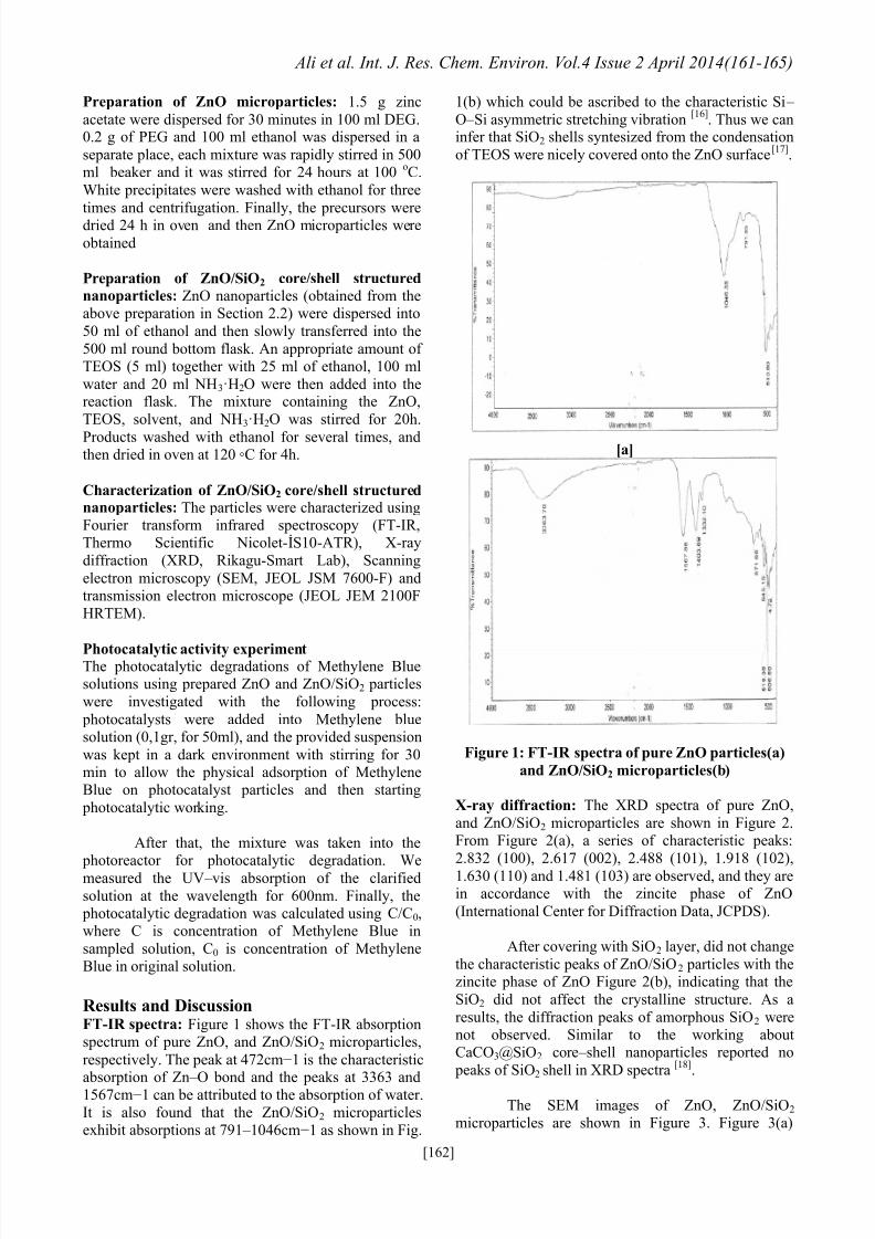
Results and DiscussionFT-IR spectra: Figure 1 shows the FT-IR absorptionspectrum of pure ZnO, and ZnO/SiO2 microparticles,
respectively. The peak at 472cm−1 is the characteristicabsorption of Zn – O bond and the peaks at 3363 and1567cm−1 can be attributed to the absorption of water.It is also found that the ZnO/SiO2 microparticlesexhibit absorptions at 791 – 1046cm−1 as shown in Fig.
1(b) which could be ascribed to the characteristic Si – O – Si asymmetric stretching vibration
[16]. Thus we can
infer that SiO2 shells syntesized from the condensation
of TEOS were nicely covered onto the ZnO surface[17]
.
[a]
Figure 1: FT-IR spectra of pure ZnO particles(a)
and ZnO/SiO2 microparticles(b)
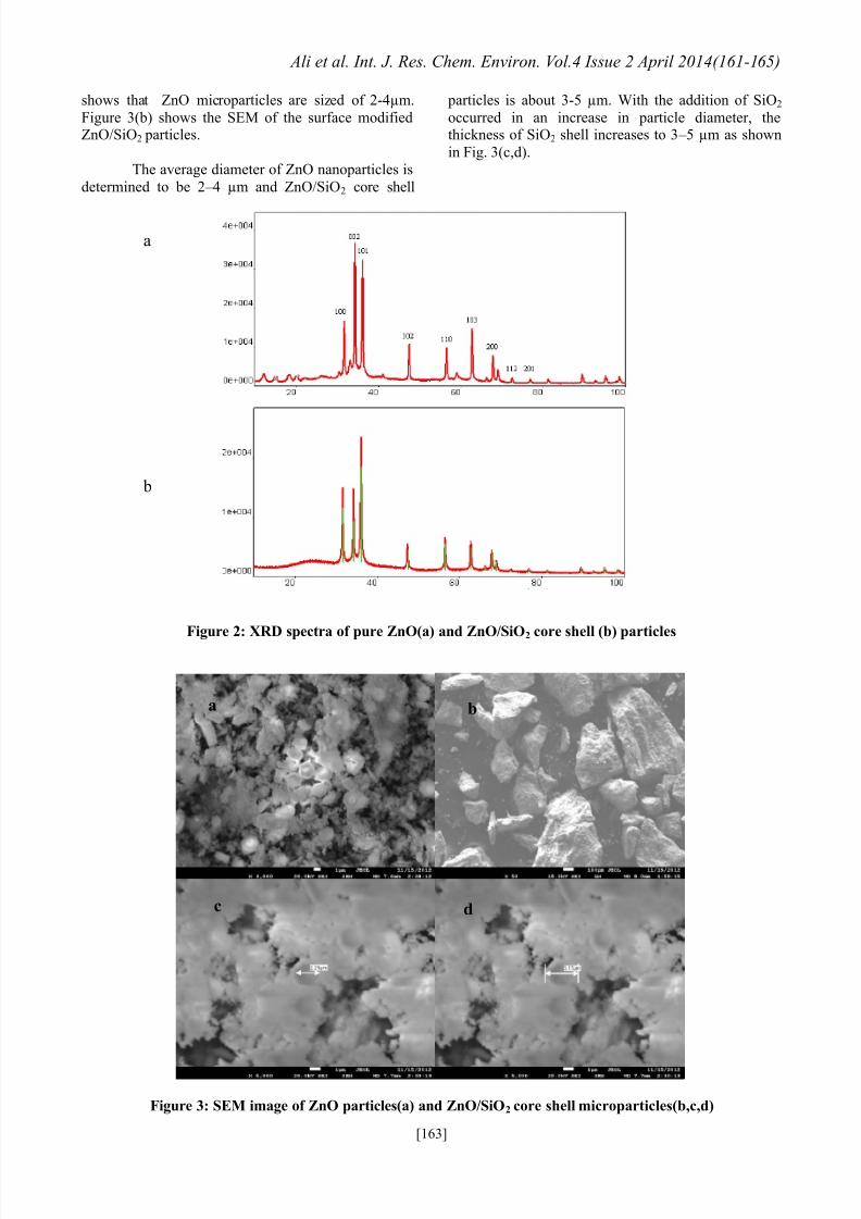
X-ray diffraction: The XRD spectra of pure ZnO,
and ZnO/SiO2 microparticles are shown in Figure 2.From Figure 2(a), a series of characteristic peaks:
2.832 (100), 2.617 (002), 2.488 (101), 1.918 (102),
1.630 (110) and 1.481 (103) are observed, and they arein accordance with the zincite phase of ZnO
(International Center for Diffraction Data, JCPDS).
After covering with SiO2 layer, did not change
the characteristic peaks of ZnO/SiO2 particles with thezincite phase of ZnO Figure 2(b), indicating that the
SiO2 did not affect the crystalline structure. As a
results, the diffraction peaks of amorphous SiO2 werenot observed. Similar to the working aboutCaCO3@SiO2 core – shell nanoparticles reported no
peaks of SiO2 shell in XRD spectra[18]
.
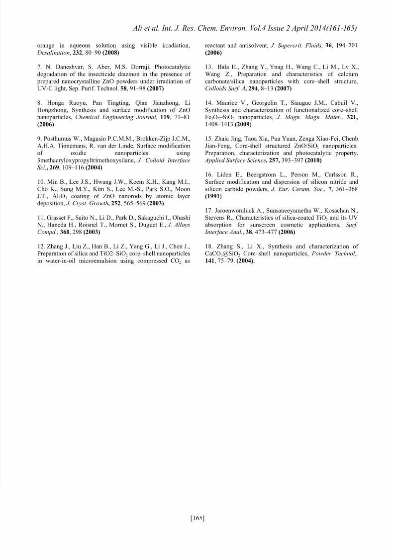
The SEM images of ZnO, ZnO/SiO2 microparticles are shown in Figure 3. Figure 3(a)
7/25/2019 ZnO - SiO2 Photolityc
http://slidepdf.com/reader/full/zno-sio2-photolityc 3/5
Ali et al. Int. J. Res. Chem. Environ. Vol.4 Issue 2 April 2014(161-165)
[163]
shows that ZnO microparticles are sized of 2-4µm.
Figure 3(b) shows the SEM of the surface modifiedZnO/SiO2 particles.
The average diameter of ZnO nanoparticles is
determined to be 2 – 4 µm and ZnO/SiO2 core shell
particles is about 3-5 µm. With the addition of SiO2
occurred in an increase in particle diameter, thethickness of SiO2 shell increases to 3 – 5 µm as shown
in Fig. 3(c,d).
Figure 2: XRD spectra of pure ZnO(a) and ZnO/SiO2 core shell (b) particles
Figure 3: SEM image of ZnO particles(a) and ZnO/SiO2 core shell microparticles(b,c,d)
a
b
a b
c d

7/25/2019 ZnO - SiO2 Photolityc
http://slidepdf.com/reader/full/zno-sio2-photolityc 4/5
Ali et al. Int. J. Res. Chem. Environ. Vol.4 Issue 2 April 2014(161-165)
[164]
Figure 4: TEM image of ZnO/SiO2 core shell microparticles (a,b)
PHOTOCATALYTİC REMOVAL OF MM
0
10
20
30
40
50
60
70
80
0 20 40 60 80 100 120
İRRADİATİON TİME
P E R C E N
T A G E
O F
R E M O
V A L İ N G
ZnO
ZnO/SiO2
Figure 5: Photocatalytic Degradation of Methylene Blue for ZnO and ZnO/SiO2 particles
The TEM images of ZnO/SiO2 microparticles
are shown in Figure 4. Figure 4 shows that ZnO particles are in core and SiO2 particles are in surface.
We also that SiO2 particles are distributed on ZnO
surface homogenously.
Photocatalytic Degradation: ZnO, and ZnO/SiO2
microparticles were used as photocatalystsrespectively to degrade Methylene Blue dissolved in
water. Figure 5 shows the relationship between C/C0 and irradiation time for Methylene Blue in basicsolution photocatalyzed by ZnO and ZnO/SiO2
microparticles. Within 90 minutes, the photocatalyticdegradation of Methylene Blue is 13 and 68% in the
presence of ZnO, and ZnO/SiO2 microparticles. Thismeans that the modified ZnO/SiO2 microparticles have
higher photocatalytic activity. Although ZnOnanoparticles is a quite active photocatalyst, ZnOreactions with excessive OH
- in alkaline medium,
threrefore leading to the failure of photocatalytic
degradation of Methylene Blue. After ZnO wasmodified with SiO2, the stability of core/shell
structured ZnO/SiO2 microparticles was improved dueto the reduced contact of ZnO with [OH]
−[15].
ConclusionZnO and ZnO/SiO2 microparticles have been
prepared via a simple chemical method. FT-IR,
analysis showed that the successful covering of SiO2 on ZnO surface. The core/shell structure has beenverified by SEM images. The uncoated ZnO showed
weak photocatalytic activity in basic solution of
Methylene Blue. ZnO/SiO2 microparticles consisted ofZnO core and SiO2 shell showed a better
photocatalytic performance in alkaline solutions of
Methylene blue, because of the improved stabilitymodified by the SiO2 shell. This is thought to be a
candidate particles in removing pollutants.
References1. Liao Min-Hung, Hsu Chih-Hsiung, Chen Dong-Hwang,Preparation and properties of amorphous titania-coated zinc
oxide nanoparticles, Journal of Solid State Chemistry, 179,
2020 – 2026 (2006)
2. Kim S., Fisher B., Eisler H.J., Bawendi M., Type-IIquantum dots: CdTe/CdSe(core/shell) and
CdSe/ZnTe(core/shell) heterostructures, J. Am. Chem. Soc.,
125, 11466 – 11467 (2003)
3.Zhong C.J., Maye M.M., Core-Shell assembled
nanoparticales as catalysts, Adv. Mater., 13(19), 1507 – 1511
(2001)
4. Kislov N., Lahiri J., Verma H., Stefanakos D., Batzill M.,
Photocatalytic degradation of methyl orange over single
crystalline ZnO: orientation dependence of photoactivity and photostability of ZnO, Langmuir , 25, 3310 – 3315 (2009)
5. Daneshvar N., Salari D., Khataee A.R., Photocatalytic
degradation of azo dye acid red 14 in water on ZnO as an
alternative catalyst to TiO2, J. Photochem. Photobiol . A:Chem., 162, 317 – 322 (2004)
6. Pare B., Jonnalagadda S.B., Tomar H., Singh P., Bhagwat
V.W., ZnO assisted photocatalytic degradation of acridine
7/25/2019 ZnO - SiO2 Photolityc
http://slidepdf.com/reader/full/zno-sio2-photolityc 5/5
Ali et al. Int. J. Res. Chem. Environ. Vol.4 Issue 2 April 2014(161-165)
[165]
orange in aqueous solution using visible irradiation,
Desalination, 232, 80 – 90 (2008)
7. N. Daneshvar, S. Aber, M.S. Dorraji, Photocatalytic
degradation of the insecticide diazinon in the presence of
prepared nanocrystalline ZnO powders under irradiation ofUV-C light, Sep. Purif. Technol. 58, 91 – 98 (2007)
8. Honga Ruoyu, Pan Tingting, Qian Jianzhong, Li
Hongzhong, Synthesis and surface modification of ZnOnanoparticles, Chemical Engineering Journal , 119, 71 – 81
(2006)
9. Posthumus W., Magusin P.C.M.M., Brokken-Zijp J.C.M.,
A.H.A. Tinnemans, R. van der Linde, Surface modification
of oxidic nanoparticles using
3methacryloxypropyltrimethoxysilane, J. Colloid Interface
Sci., 269, 109 – 116 (2004)
10. Min B., Lee J.S., Hwang J.W., Keem K.H., Kang M.I.,
Cho K., Sung M.Y., Kim S., Lee M.-S., Park S.O., Moon
J.T., Al2O3 coating of ZnO nanorods by atomic layerdeposition, J. Cryst. Growth, 252, 565 – 569 (2003)
11. Grasset F., Saito N., Li D., Park D., Sakaguchi I., Ohashi N., Haneda H., Roisnel T., Mornet S., Duguet E., J. Alloys
Compd., 360, 298 (2003)
12. Zhang J., Liu Z., Han B., Li Z., Yang G., Li J., Chen J.,Preparation of silica and TiO2 – SiO2 core – shell nanoparticles
in water-in-oil microemulsion using compressed CO2 as
reactant and antisolvent, J. Supercrit. Fluids, 36, 194 – 201
(2006)
13. Bala H., Zhang Y., Ynag H., Wang C., Li M., Lv X.,
Wang Z., Preparation and characteristics of calcium
carbonate/silica nanoparticles with core – shell structure,Colloids Surf. A, 294, 8 – 13 (2007)
14. Maurice V., Georgelin T., Siaugue J.M., Cabuil V.,
Synthesis and characterization of functionalized core – shellFe2O3 – SiO2 nanoparticles, J. Magn. Magn. Mater., 321,
1408 – 1413 (2009)
15. Zhaia Jing, Taoa Xia, Pua Yuan, Zenga Xiao-Fei, Chenb
Jian-Feng, Core-shell structured ZnO/SiO2 nanoparticles:
Preparation, characterization and photocatalytic property,
Applied Surface Science, 257, 393 – 397 (2010)
16. Liden E., Beergstrom L., Person M., Carlsson R.,
Surface modification and dispersion of silicon nitride and
silicon carbide powders, J. Eur. Ceram. Soc., 7, 361 – 368
(1991)
17. Jaroenworaluck A., Sunsaneeyametha W., Kosachan N.,
Stevens R., Characteristics of silica-coated TiO2 and its UVabsorption for sunscreen cosmetic applications, Surf.
Interface Anal., 38, 473 – 477 (2006)
18. Zhang S., Li X., Synthesis and characterization ofCaCO3@SiO2 Core – shell nanoparticles, Powder Technol.,
141, 75 – 79. (2004).
Recommended