Embed Size (px)
Citation preview

AHMED LABIB


General rules for X-ray exposure :
Explain to the patient about the procedures being undertaken and Place the lead apron on the patient
Examine the patient’s mouth for any deviation from the normal which may require some adjustment of the standard radiographic technique.
Remove any object from the patient’s mouth as removable PD. or orthodontic appliances.
Set the exposure factors.
Set the approximate the X-ray tube angel.
Adjust the patient head in correctly positioned.
Place the film into the patient mouth properly according to the X-ray technique used.
Re-Check the position of the patient’s head.
The operator must stand behind a lead screen.
Remove the film from patient’s mouth after exposure and to prevent leakage, dry the film before processing.


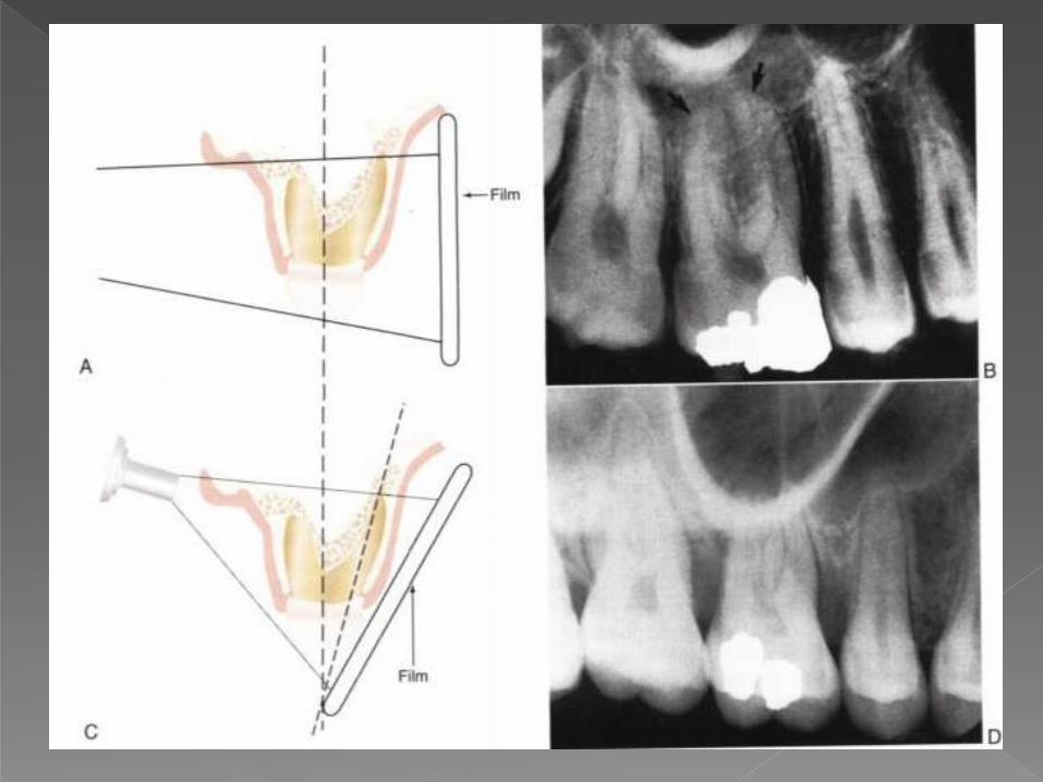
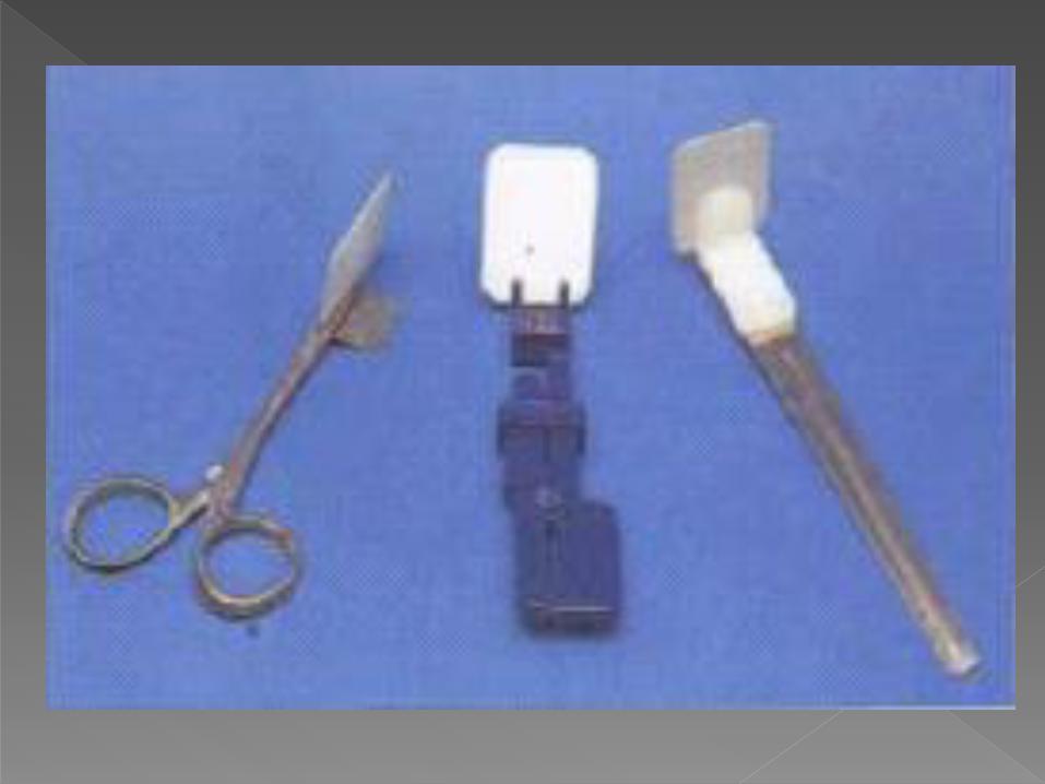
- The dental films must be placed parallel to the long axis of the tooth to be examined.
- The central ray will be directed at a right angle to the tooth and to the film.
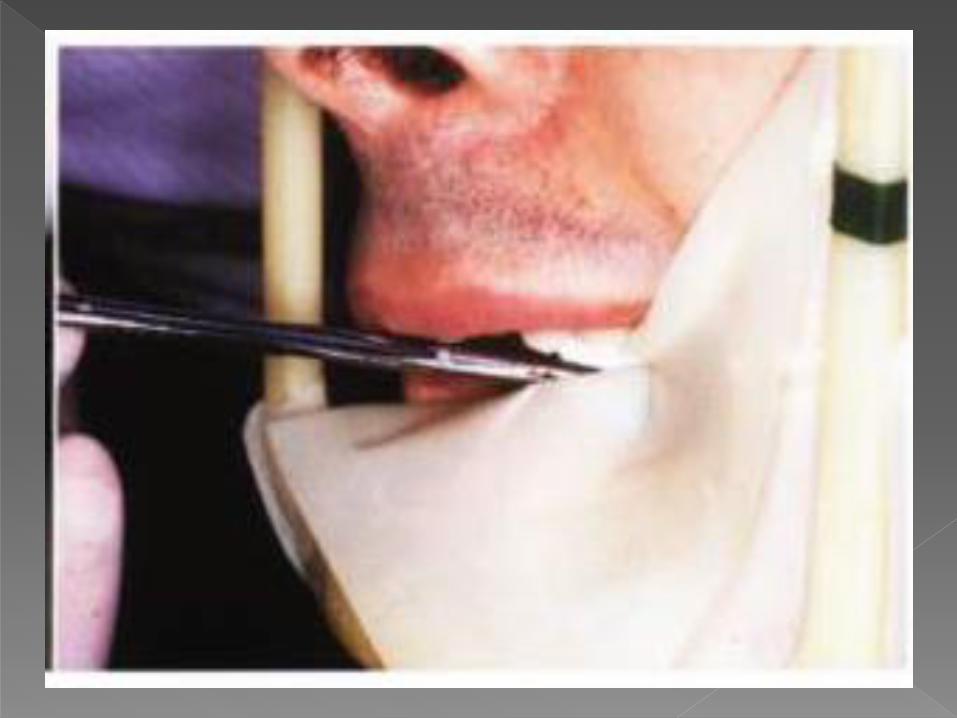
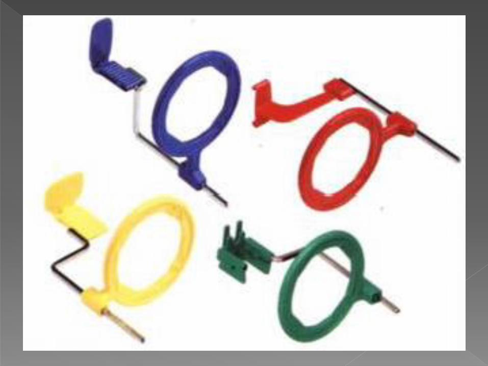
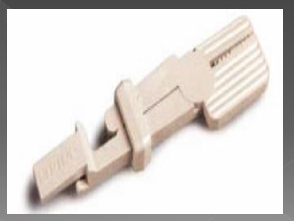
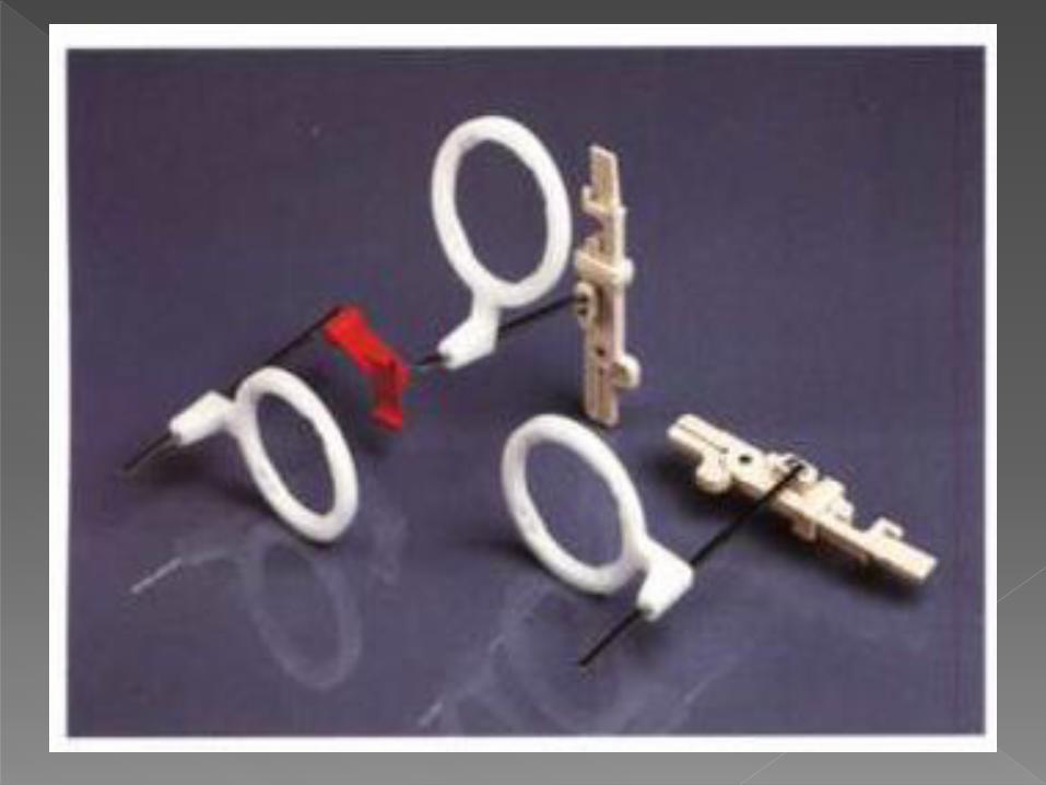
- It requires film holders, which hold the film in a parallel position to the long axis of the tooth.


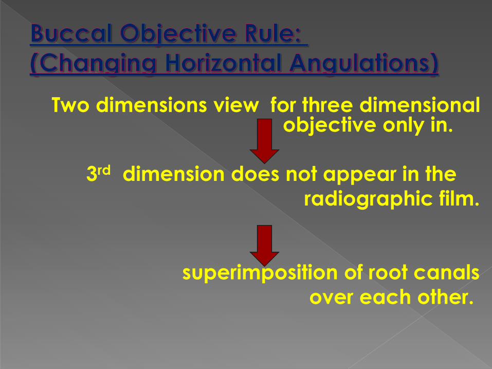
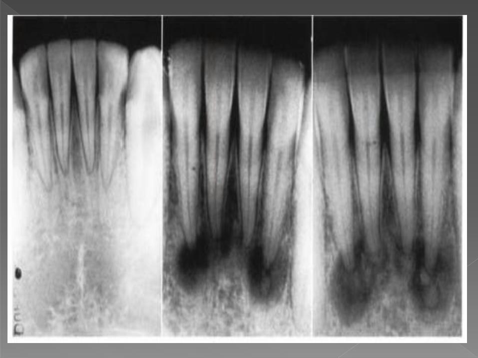
Two dimensions view for three dimensional objective only in.
3rd dimension does not appear in the
radiographic film.
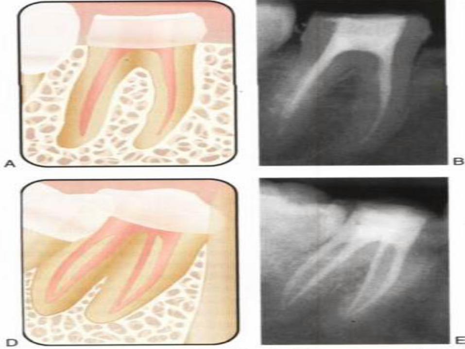
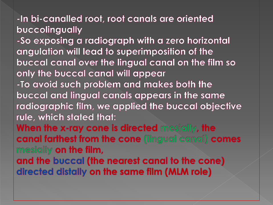
superimposition of root canals
over each other.

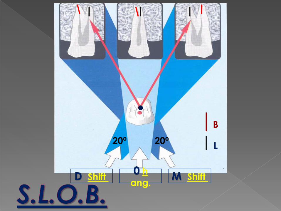
0 h
ang.M Shift D Shift
B
L20⁰20⁰