Embed Size (px)
Citation preview
Histology of Endocrine System
UMAR AHMAD
MODERATOR: PROF. DR. FAUZIAH OTHMAN
Department of Human Anatomy
Faculty of Medicine & Health Sciences
Universiti Putra Malaysia.
PREMEDIC MESIR 2013
Apr 12, 2023 Umar Ahmad 1
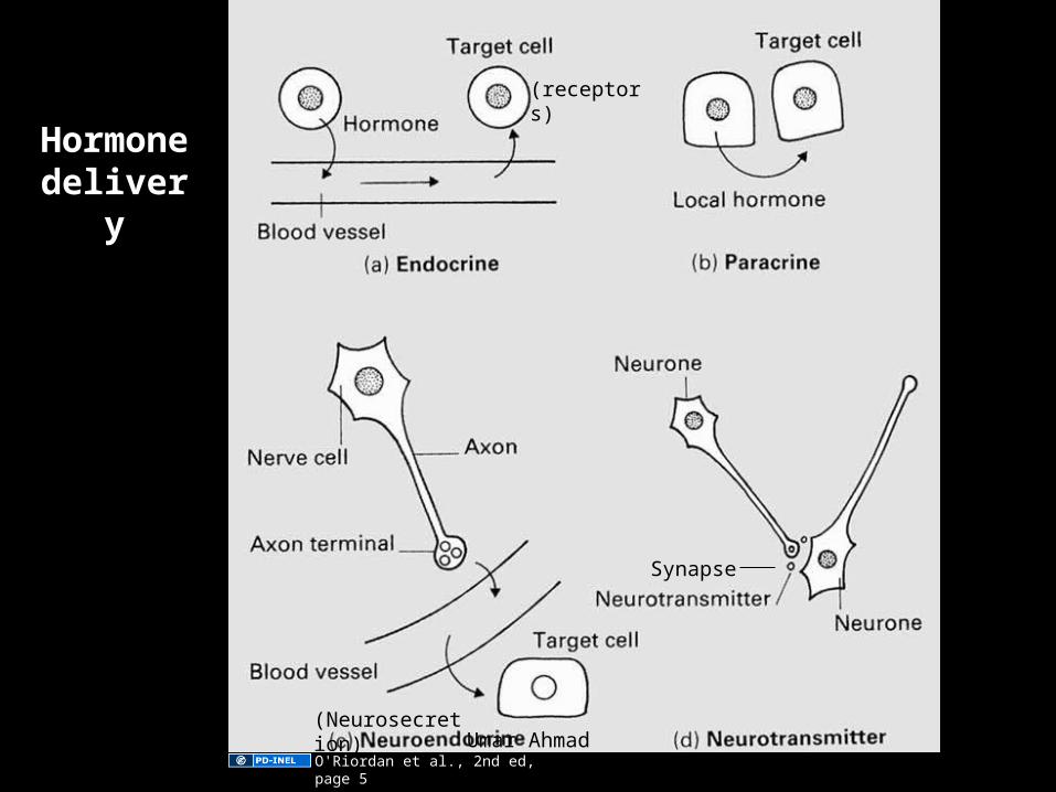
Hormone delivery
(receptors)
(Neurosecretion)
Synapse
O'Riordan et al., 2nd ed, page 5Apr 12, 2023 Umar Ahmad 2
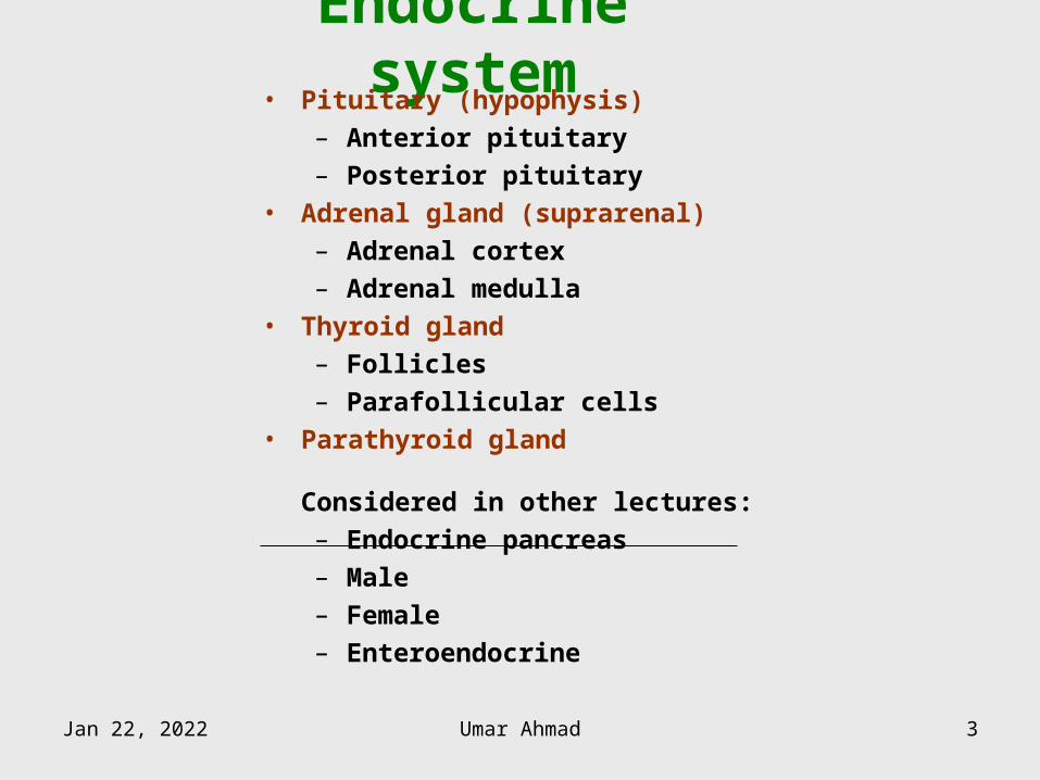
Endocrine system• Pituitary (hypophysis)
– Anterior pituitary
– Posterior pituitary
• Adrenal gland (suprarenal)
– Adrenal cortex
– Adrenal medulla
• Thyroid gland
– Follicles
– Parafollicular cells
• Parathyroid gland
Considered in other lectures:
– Endocrine pancreas
– Male
– Female
– Enteroendocrine
Apr 12, 2023 Umar Ahmad 3
PITUITARY
Apr 12, 2023 Umar Ahmad 4
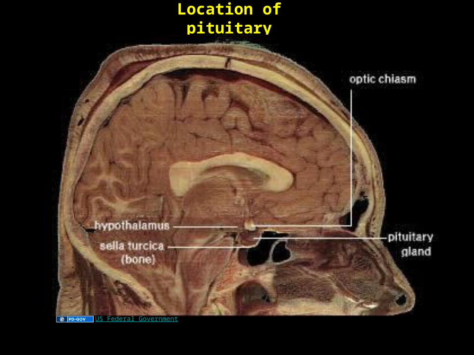
Location of pituitary
US Federal Government
Apr 12, 2023 Umar Ahmad 5
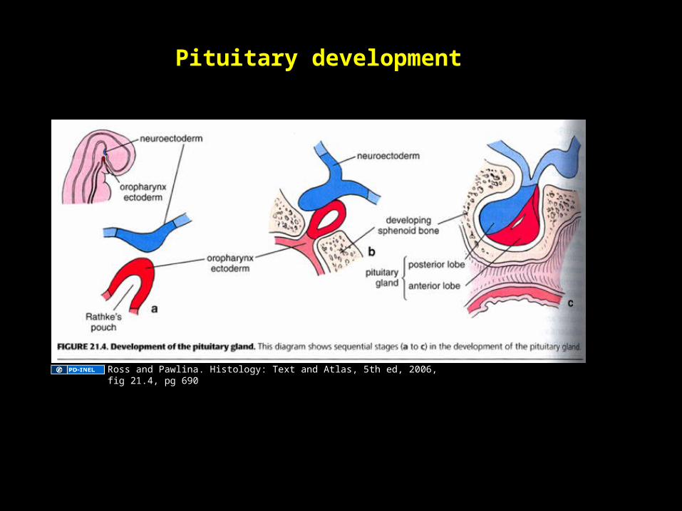
Pituitary development
Ross and Pawlina. Histology: Text and Atlas, 5th ed, 2006, fig 21.4, pg 690
Apr 12, 2023 Umar Ahmad 6
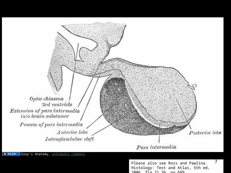
Pituitary nomenclature
Pituitary nomenclature
Please also see Ross and Pawlina. Histology: Text and Atlas, 5th ed, 2006, fig 21.3b, pg 689
Gray’s Anatomy, wikimedia commons
Apr 12, 2023 Umar Ahmad 7
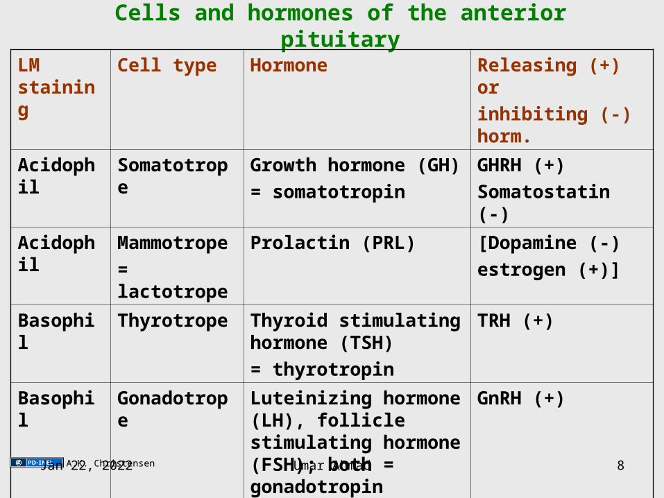
Cells and hormones of the anterior pituitary
LM staining
Cell type Hormone Releasing (+) or
inhibiting (-) horm.
Acidophil Somatotrope Growth hormone (GH)
= somatotropin
GHRH (+)
Somatostatin (-)
Acidophil Mammotrope
= lactotrope
Prolactin (PRL) [Dopamine (-)
estrogen (+)]
Basophil Thyrotrope Thyroid stimulating hormone (TSH)
= thyrotropin
TRH (+)
Basophil Gonadotrope Luteinizing hormone (LH), follicle stimulating hormone (FSH); both = gonadotropin
GnRH (+)
Basophil
(human)
Corticotrope Adrenocorticotropin
(ACTH) = corticotropin
CRH (+)
A.K. ChristensenApr 12, 2023 Umar Ahmad 8
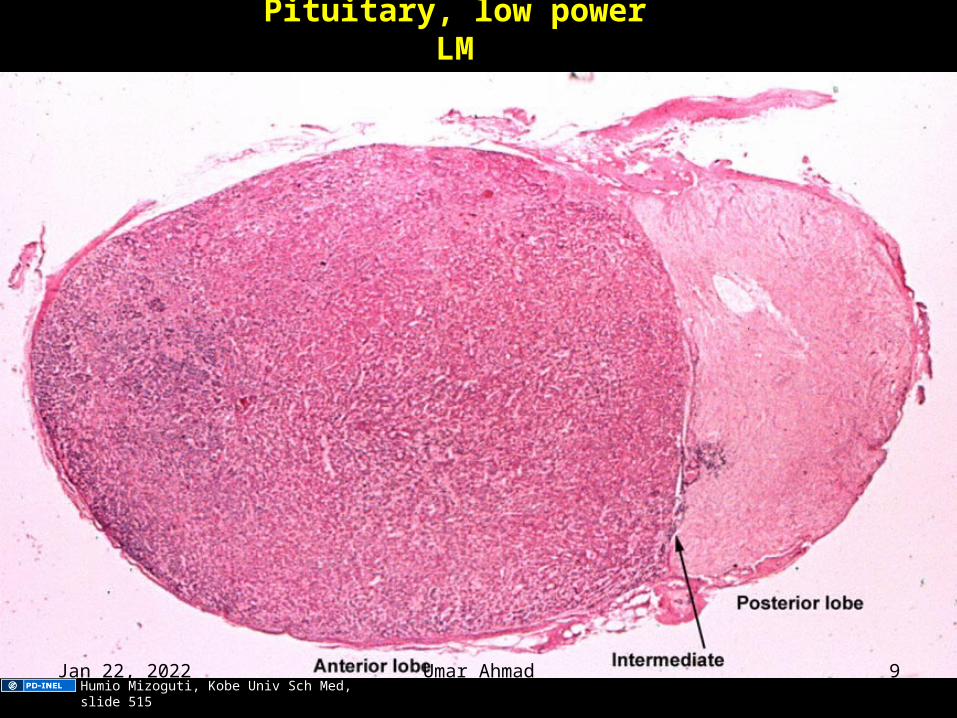
Pituitary, low power LM
Humio Mizoguti, Kobe Univ Sch Med, slide 515Apr 12, 2023 Umar Ahmad 9
Anterior pituitary, LM drawing
Image of cords of cells in anterior
pituitary removed. Original here:
Bailey's textbook of histology.
72(700)6
Apr 12, 2023 Umar Ahmad 10
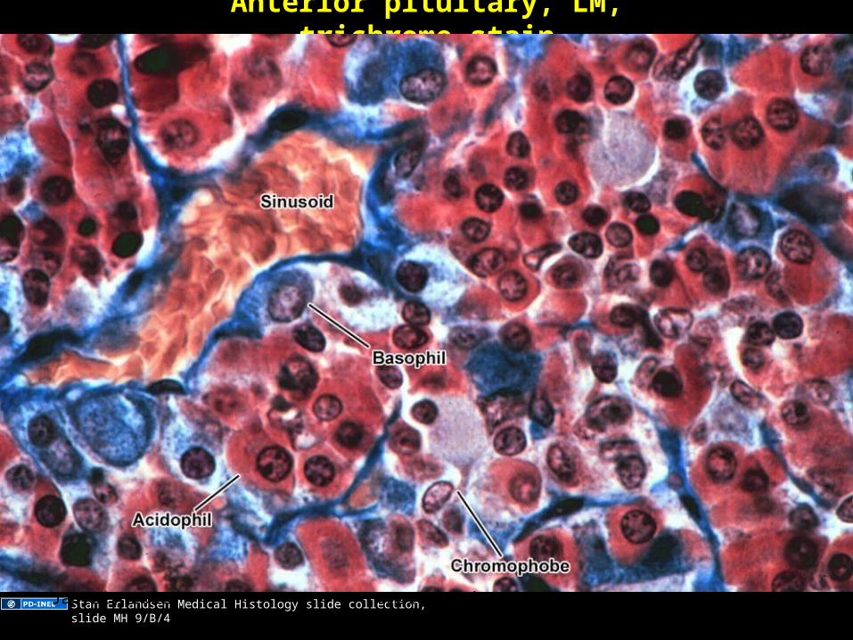
Anterior pituitary, LM, trichrome stain
Stan Erlandsen Medical Histology slide collection, slide MH 9/B/4Apr 12, 2023 Umar Ahmad 11
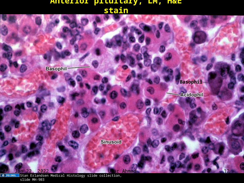
Anterior pituitary, LM, H&E stain
Basophil
Stan Erlandsen Medical Histology slide collection, slide MH-9B3Apr 12, 2023 Umar Ahmad 12
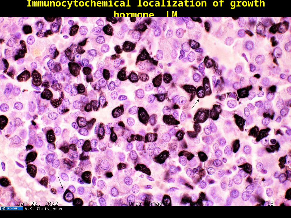
Immunocytochemical localization of growth hormone, LM
A.K. ChristensenApr 12, 2023 Umar Ahmad 13
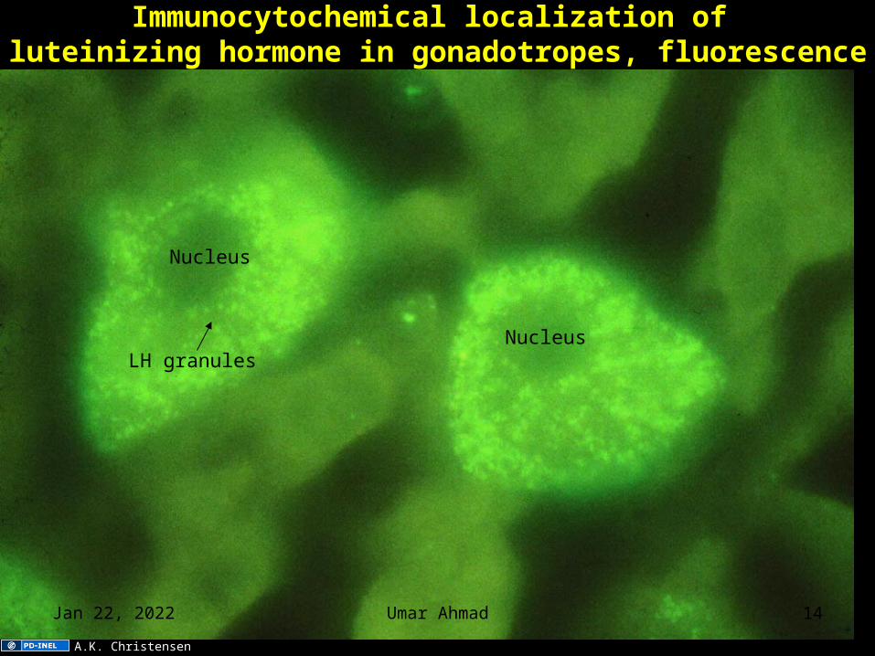
Immunocytochemical localization of luteinizing hormone in gonadotropes, fluorescence
Nucleus
NucleusLH granules
A.K. Christensen
Apr 12, 2023 Umar Ahmad 14
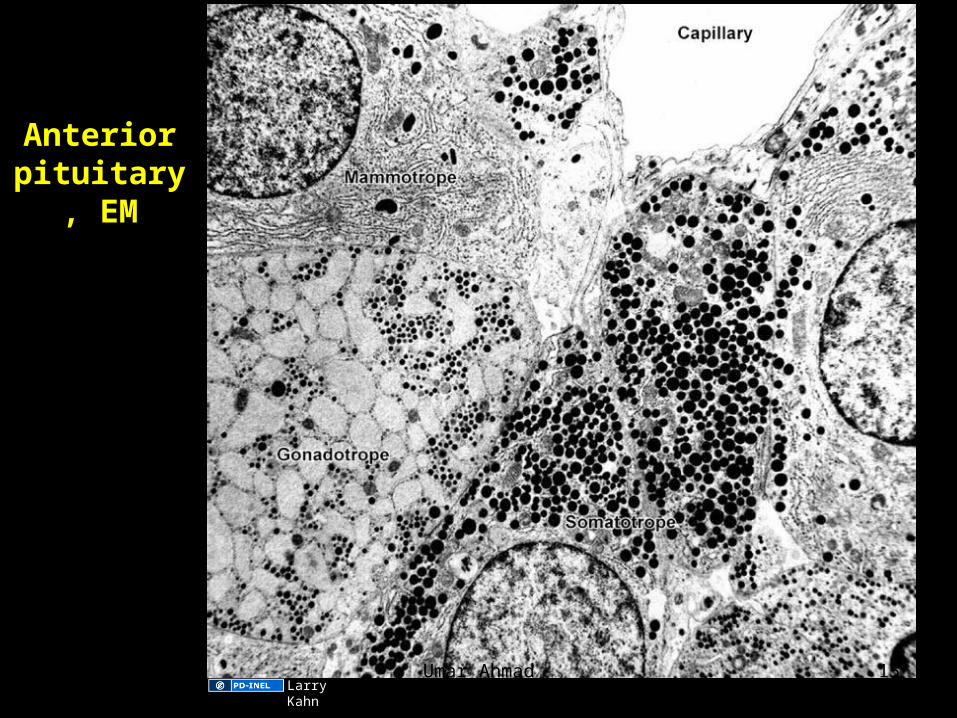
Anterior pituitary,
EM
Larry KahnApr 12, 2023 Umar Ahmad 15
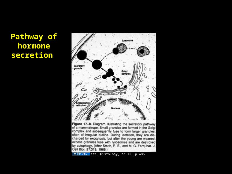
Pathway of hormone secretion
Fawcett. Histology, ed 11, p 486
Apr 12, 2023 Umar Ahmad 16
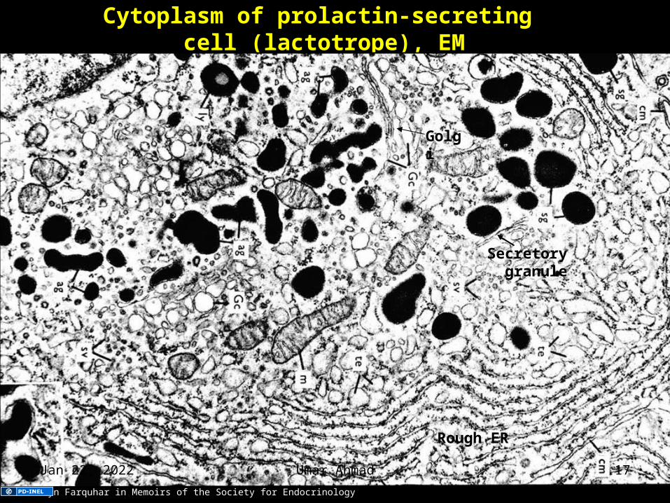
Cytoplasm of prolactin-secreting cell (lactotrope), EM
Secretory granule
Golgi
Rough ER
Marilyn Farquhar in Memoirs of the Society for Endocrinology
Apr 12, 2023 Umar Ahmad 17
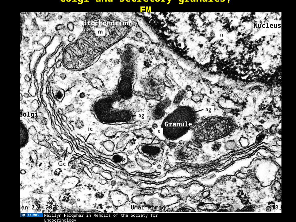
Golgi and secretory granules, EM
Golgi
Granule
Mitochondrion Nucleus
Marilyn Farquhar in Memoirs of the Society for Endocrinology
Apr 12, 2023 Umar Ahmad 18
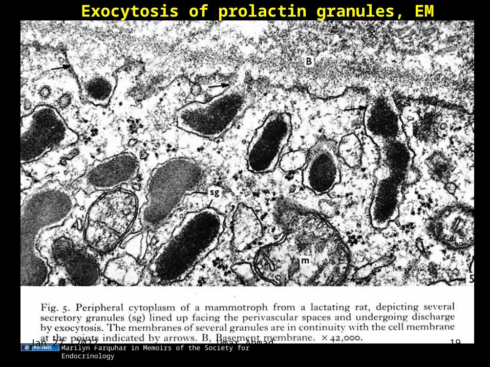
Exocytosis of prolactin granules, EM
Marilyn Farquhar in Memoirs of the Society for EndocrinologyApr 12, 2023 Umar Ahmad 19
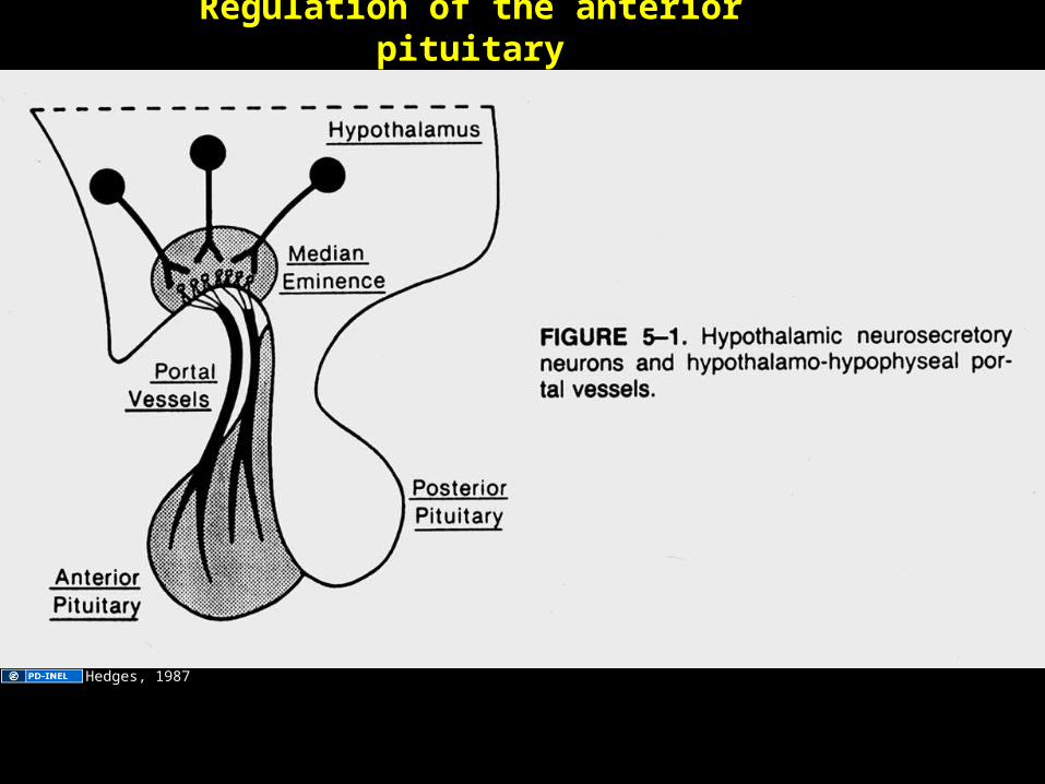
Regulation of the anterior pituitary
Hedges, 1987
Apr 12, 2023 Umar Ahmad 20
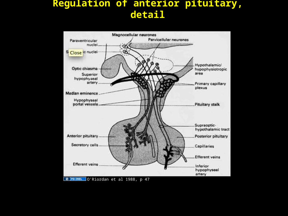
Regulation of anterior pituitary, detail
O'Riordan et al 1988, p 47
Apr 12, 2023 Umar Ahmad 21
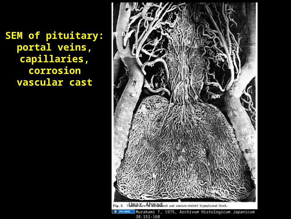
SEM of pituitary: portal veins,
capillaries, corrosion vascular cast
Murakami T, 1975, Archivum Histologicum Japanicum 38:151-168
Apr 12, 2023 Umar Ahmad 22
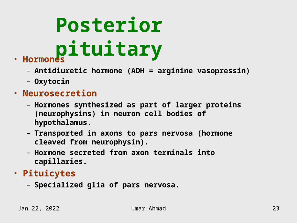
Posterior pituitary• Hormones
– Antidiuretic hormone (ADH = arginine vasopressin)– Oxytocin
• Neurosecretion– Hormones synthesized as part of larger proteins
(neurophysins) in neuron cell bodies of hypothalamus.– Transported in axons to pars nervosa (hormone cleaved
from neurophysin).– Hormone secreted from axon terminals into capillaries.
• Pituicytes– Specialized glia of pars nervosa.
Apr 12, 2023 Umar Ahmad 23
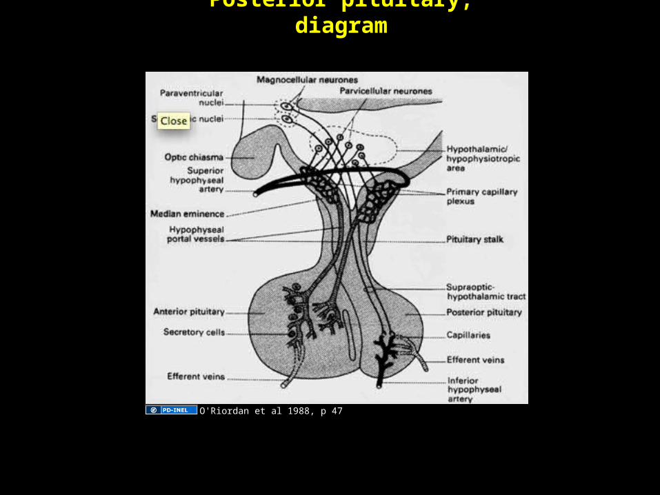
Posterior pituitary, diagram
O'Riordan et al 1988, p 47
Apr 12, 2023 Umar Ahmad 24
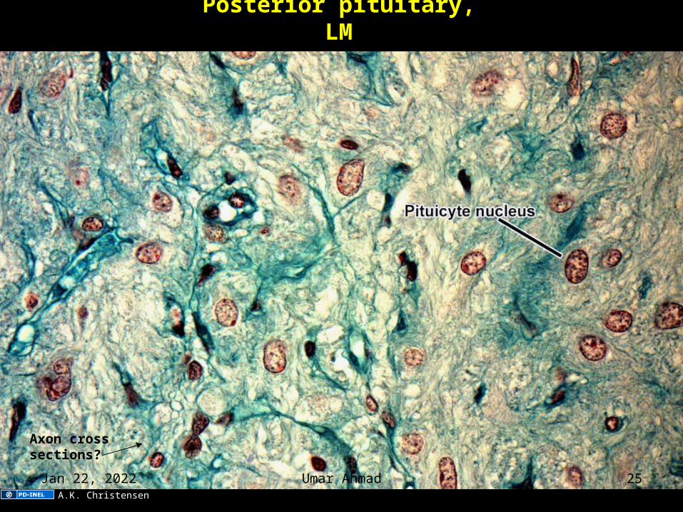
Posterior pituitary, LM
Axon cross sections?
A.K. Christensen
Apr 12, 2023 Umar Ahmad 25
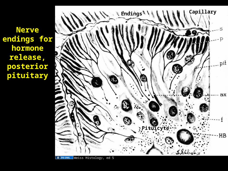
Nerve endings for
hormone release,
posterior pituitary
CapillaryEndings
Pituicyte
Weiss Histology, ed 5Apr 12, 2023 Umar Ahmad 26
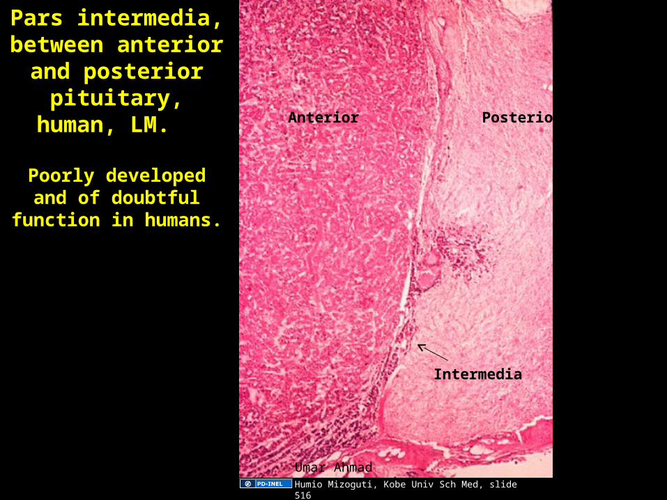
Pars intermedia, between anterior
and posterior pituitary, human,
LM.
Poorly developed and of doubtful function in
humans.
Intermedia
Anterior Posterior
Humio Mizoguti, Kobe Univ Sch Med, slide 516
Apr 12, 2023 Umar Ahmad 27
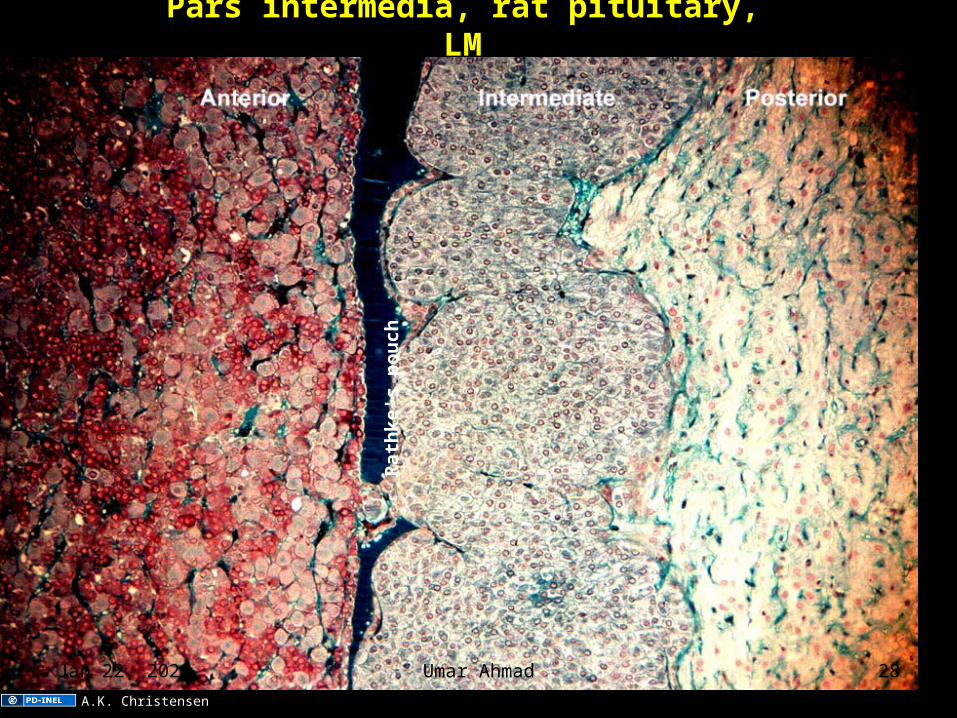
Pars intermedia, rat pituitary, LM
Rat
hk
e's
po
uch
A.K. Christensen
Apr 12, 2023 Umar Ahmad 28
ADRENAL GLAND
Apr 12, 2023 Umar Ahmad 29
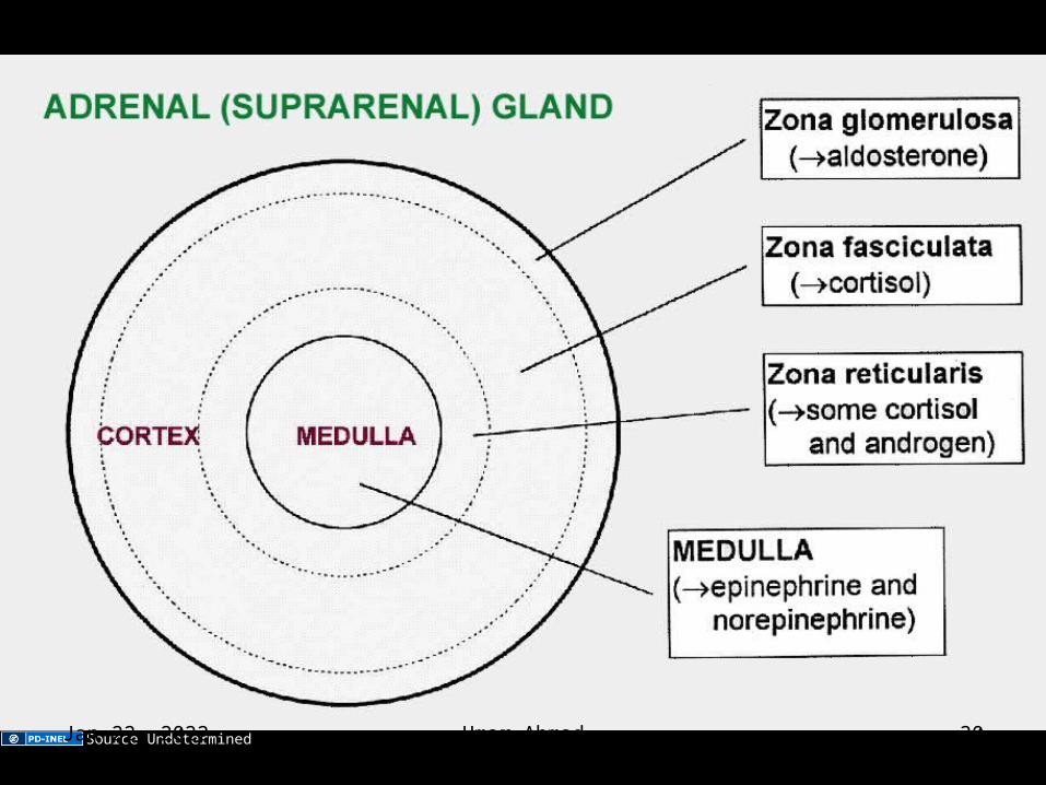
Adrenal (suprarenal) gland
Source UndeterminedApr 12, 2023 Umar Ahmad 30
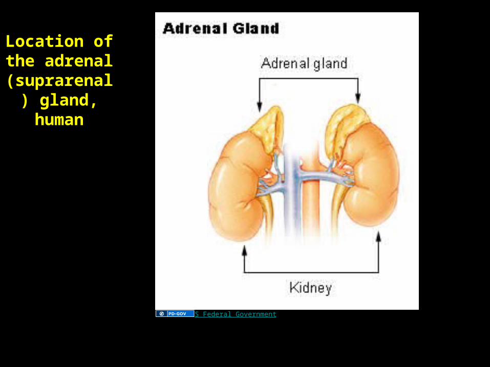
Location of the adrenal (suprarenal)
gland, human
US Federal Government
Apr 12, 2023 Umar Ahmad 31
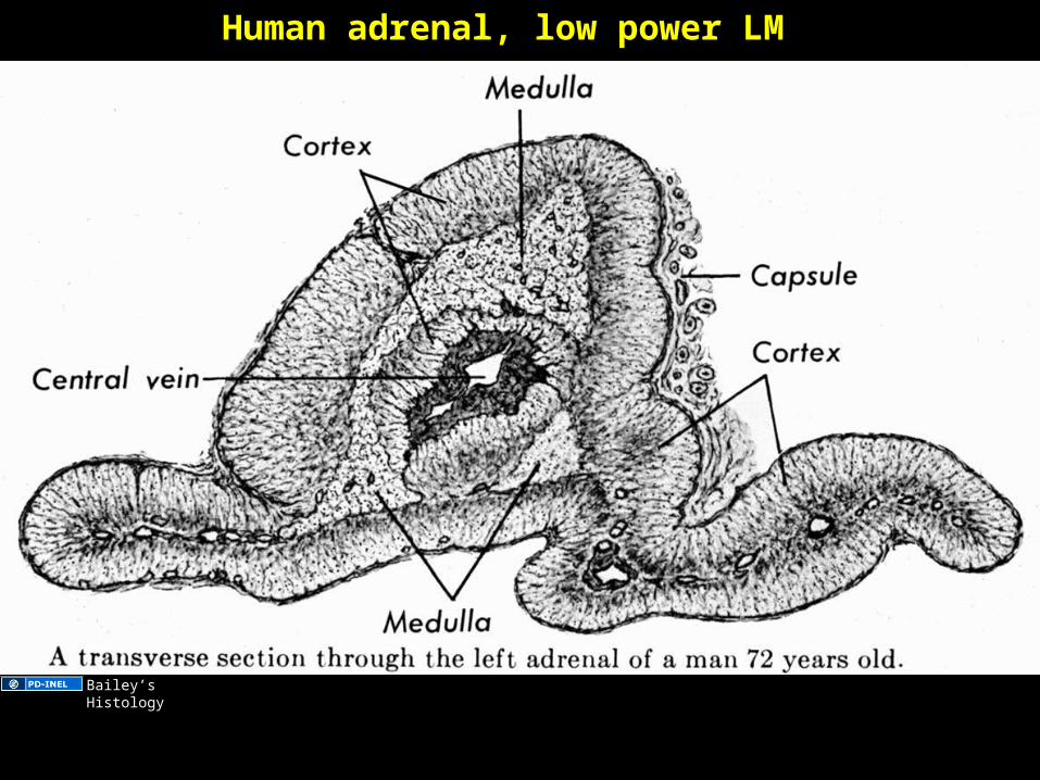
Human adrenal, low power LM
Bailey’s Histology
Apr 12, 2023 Umar Ahmad 32
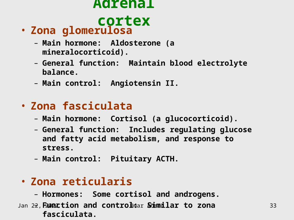
Adrenal cortex• Zona glomerulosa
– Main hormone: Aldosterone (a mineralocorticoid).– General function: Maintain blood electrolyte balance.– Main control: Angiotensin II.
• Zona fasciculata– Main hormone: Cortisol (a glucocorticoid).– General function: Includes regulating glucose and fatty
acid metabolism, and response to stress.– Main control: Pituitary ACTH.
• Zona reticularis– Hormones: Some cortisol and androgens.– Function and control: Similar to zona fasciculata.
Apr 12, 2023 Umar Ahmad 33
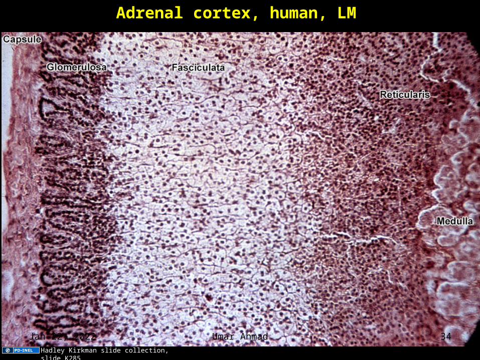
Adrenal cortex, human, LM
Hadley Kirkman slide collection, slide K285
Apr 12, 2023 Umar Ahmad 34
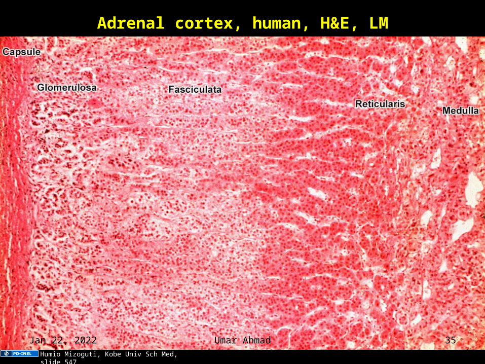
Adrenal cortex, human, H&E, LM
Humio Mizoguti, Kobe Univ Sch Med, slide 547
Apr 12, 2023 Umar Ahmad 35
Adrenal blood vessels
Image of adrenal gland vasculature removed. Original here: Junqueira
and Carneiro, 10th ed., 2003, page 414, fig 21-2.
Apr 12, 2023 Umar Ahmad 36
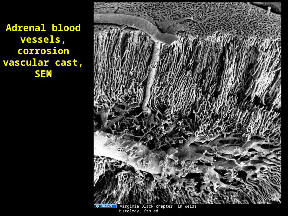
Adrenal blood vessels,
corrosion vascular cast,
SEM
Virginia Black chapter, in Weiss Histology, 6th ed Apr 12, 2023 Umar Ahmad 37
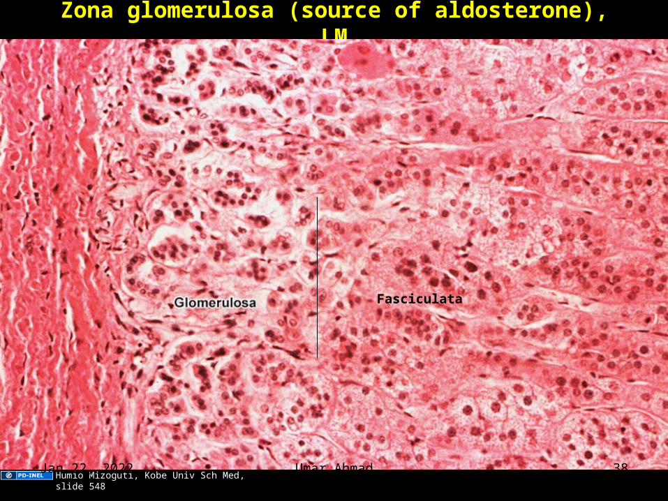
Zona glomerulosa (source of aldosterone), LM
Fasciculata
Humio Mizoguti, Kobe Univ Sch Med, slide 548 Apr 12, 2023 Umar Ahmad 38
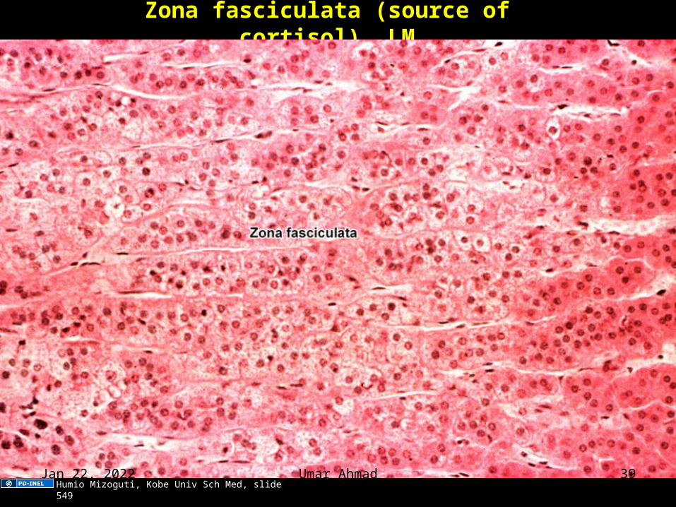
Zona fasciculata (source of cortisol), LM
Humio Mizoguti, Kobe Univ Sch Med, slide 549 Apr 12, 2023 Umar Ahmad 39
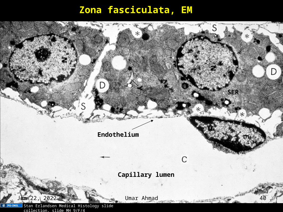
Zona fasciculata, EM
Capillary lumen
Endothelium
SER
Stan Erlandsen Medical Histology slide collection, slide MH 9/F/4
Apr 12, 2023 Umar Ahmad 40
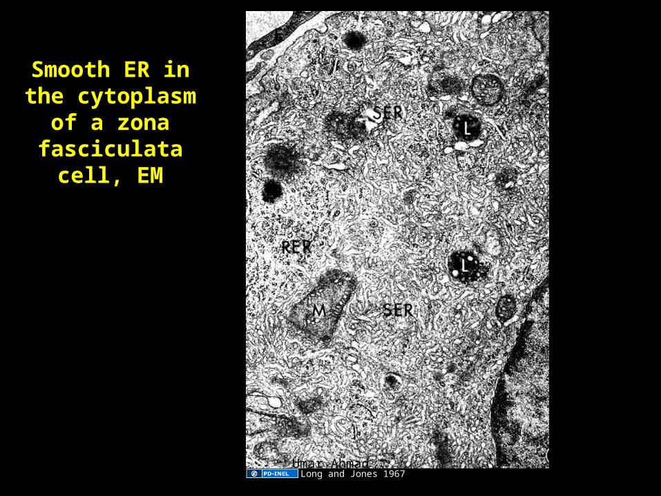
Smooth ER in the cytoplasm of a
zona fasciculata cell, EM
Long and Jones 1967Apr 12, 2023 Umar Ahmad 41
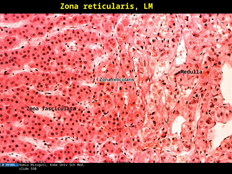
Zona reticularis, LM
Medulla
Zona fasciculata
Humio Mizoguti, Kobe Univ Sch Med, slide 550Apr 12, 2023 Umar Ahmad 42
Adrenal medulla• Hormones
– Epinephrine (adrenalin) and norepinephrine (noradrenalin), both catecholamines. Two cell types, one for E and one for N.
– General function: Acute response to stress.– Main control: Preganglionic sympathetic innervation.
• Embryonic source– From neural crest cells, same as postganglionic sympathetic
neurons. Although adrenal medulla cells do not have dendrites or axons, they behave like postganglionic sympathetic neurons, releasing norepinephrine/epinephrine in response to preganglionic sympathetic stimulation.
• Also called "chromaffin cells"– Cells of the adrenal medulla are examples of "chromaffin
cells," containing catecholamine granules that stain brown with potassium dichromate. Neurons of sympathetic ganglia are also chromaffin cells. The term is used in pathology.Apr 12, 2023 Umar Ahmad 43
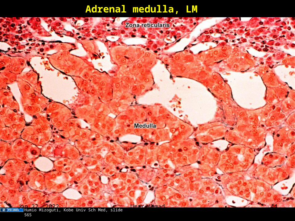
Adrenal medulla, LM
Humio Mizoguti, Kobe Univ Sch Med, slide 565Apr 12, 2023 Umar Ahmad 44
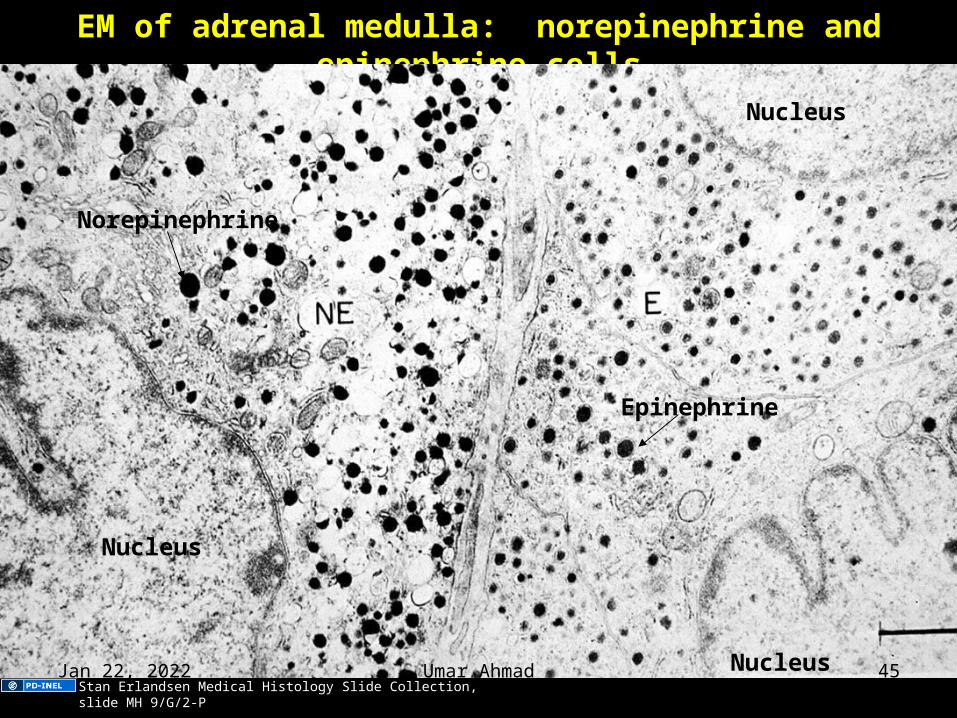
EM of adrenal medulla: norepinephrine and epinephrine cells
Nucleus
Nucleus
Nucleus
Norepinephrine
Epinephrine
Stan Erlandsen Medical Histology Slide Collection, slide MH 9/G/2-PApr 12, 2023 Umar Ahmad 45
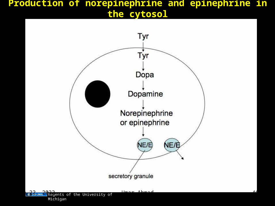
Production of norepinephrine and epinephrine in the cytosol
Regents of the University of MichiganApr 12, 2023 Umar Ahmad 46
THYROID GLAND
Apr 12, 2023 Umar Ahmad 47
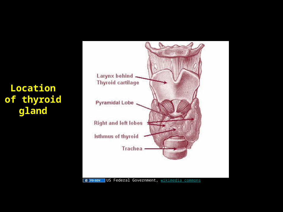
Location of thyroid gland
US Federal Government, wikimedia commons
Apr 12, 2023 Umar Ahmad 48
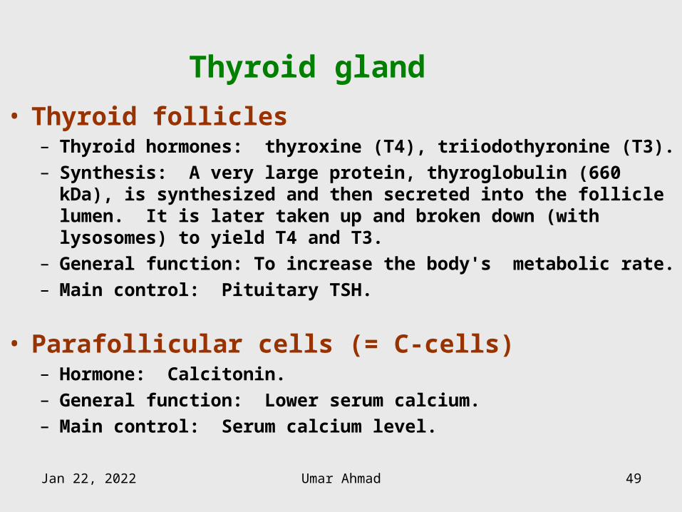
Thyroid gland
• Thyroid follicles– Thyroid hormones: thyroxine (T4), triiodothyronine (T3).– Synthesis: A very large protein, thyroglobulin (660 kDa), is
synthesized and then secreted into the follicle lumen. It is later taken up and broken down (with lysosomes) to yield T4 and T3.
– General function: To increase the body's metabolic rate.– Main control: Pituitary TSH.
• Parafollicular cells (= C-cells)– Hormone: Calcitonin.– General function: Lower serum calcium.– Main control: Serum calcium level.
Apr 12, 2023 Umar Ahmad 49
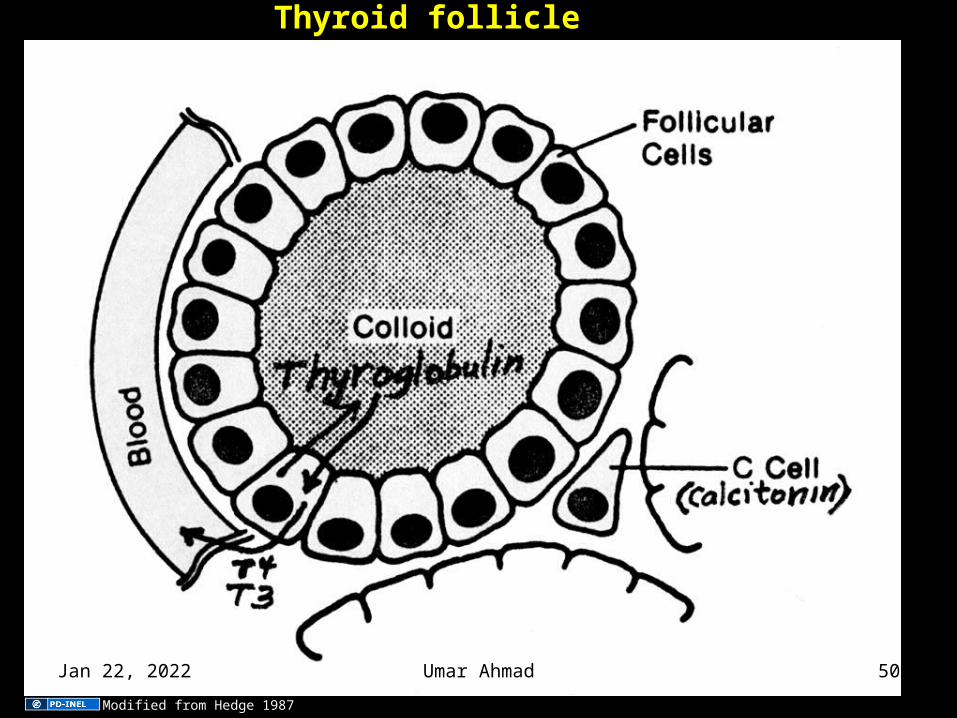
Thyroid follicle
Modified from Hedge 1987
Apr 12, 2023 Umar Ahmad 50
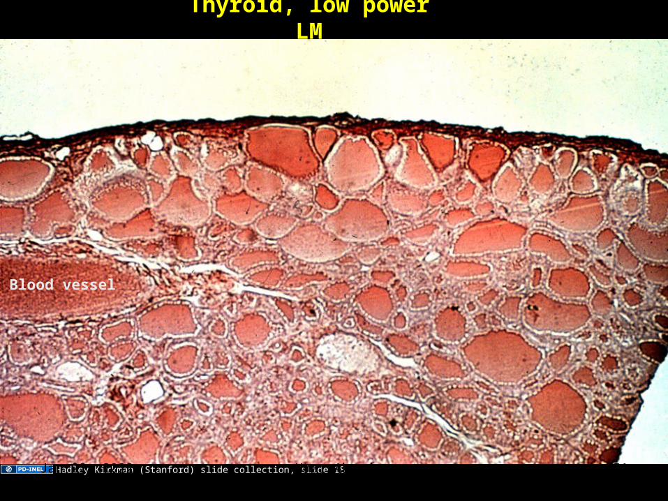
Thyroid, low power LM
Blood vessel
Hadley Kirkman (Stanford) slide collection, slide 18Apr 12, 2023 Umar Ahmad 51
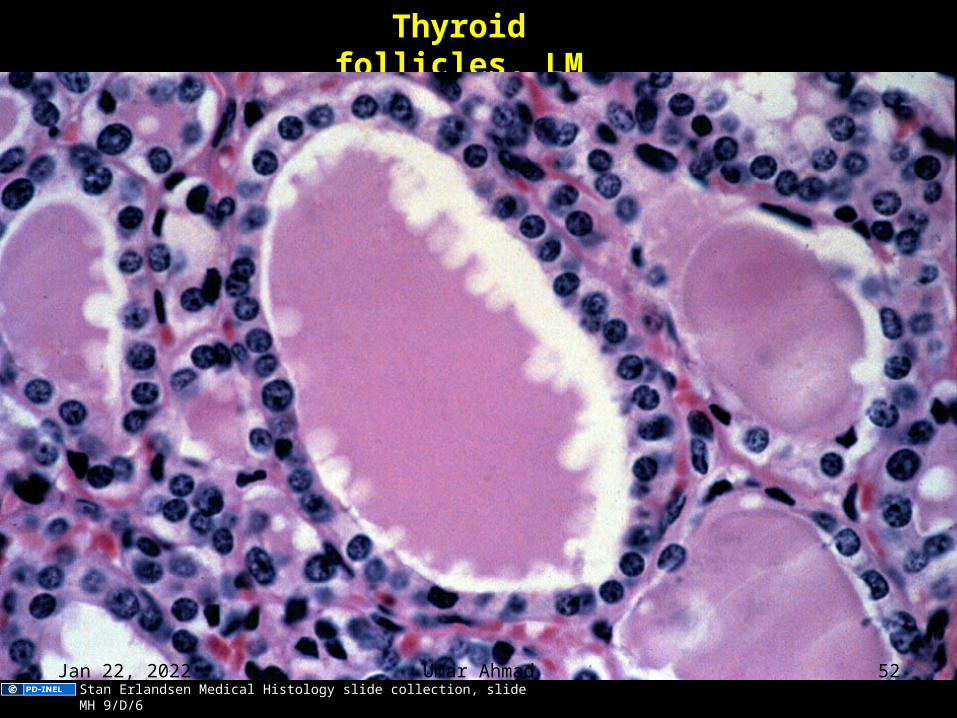
Thyroid follicles, LM
Stan Erlandsen Medical Histology slide collection, slide MH 9/D/6Apr 12, 2023 Umar Ahmad 52
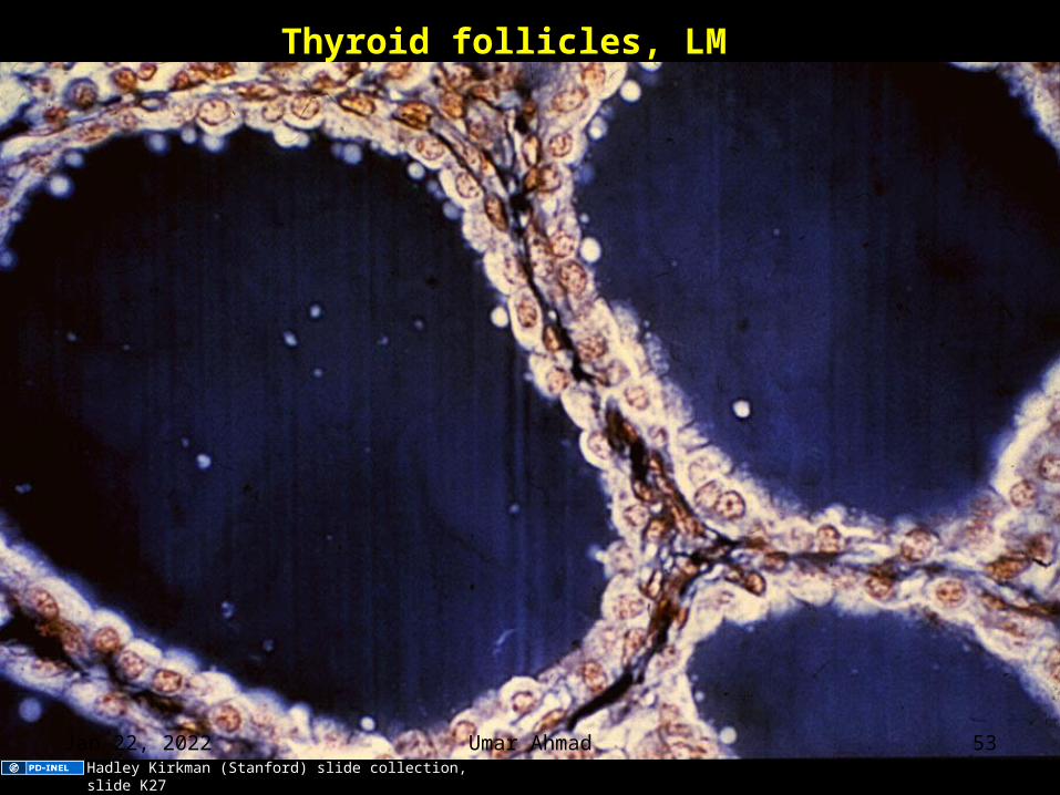
Thyroid follicles, LM
Hadley Kirkman (Stanford) slide collection, slide K27
Apr 12, 2023 Umar Ahmad 53
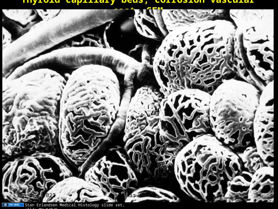
Thyroid capillary beds, corrosion vascular cast, SEM
Stan Erlandsen Medical Histology slide set, slide MH 9/D/5
Apr 12, 2023 Umar Ahmad 54
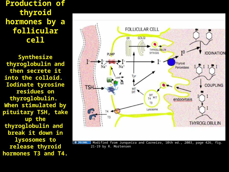
Production of thyroid hormones by a follicular cell
Synthesize thyroglobulin and then
secrete it into the colloid. Iodinate
tyrosine residues on thyroglobulin. When
stimulated by pituitary TSH, take up the
thyroglobulin and break it down in
lysosomes to release thyroid hormones T3
and T4.
Colloid
Modified from Junqueira and Carneiro, 10th ed., 2003, page 426, fig. 21-19 by R. Mortensen
Apr 12, 2023 Umar Ahmad 55
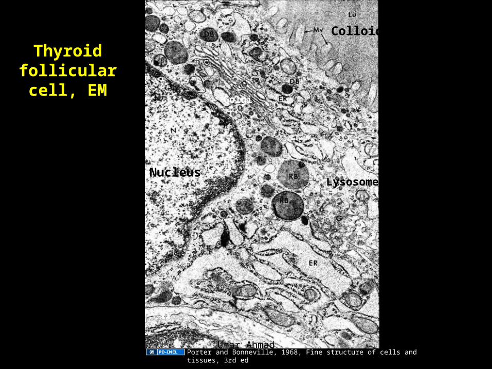
Thyroid follicular cell, EM
Nucleus
Colloid
Lysosome
Golgi
Porter and Bonneville, 1968, Fine structure of cells and tissues, 3rd edApr 12, 2023 Umar Ahmad 56

Causes of goiter
(increase in thyroid size)
Rugh and Patton 1965, Physiology and biophysics, 19th edApr 12, 2023 Umar Ahmad 57
Functional states of thyroid follicles
NormalUnderactive = hypoactiveOveractive = hyperactive
Normal
Image of thyroid follicles removed.
Original here: 0'Riordan, 2nd ed,
p 160.
Apr 12, 2023 Umar Ahmad 58
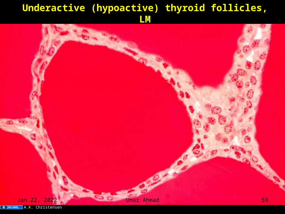
Underactive (hypoactive) thyroid follicles, LM
A.K. Christensen
Apr 12, 2023 Umar Ahmad 59
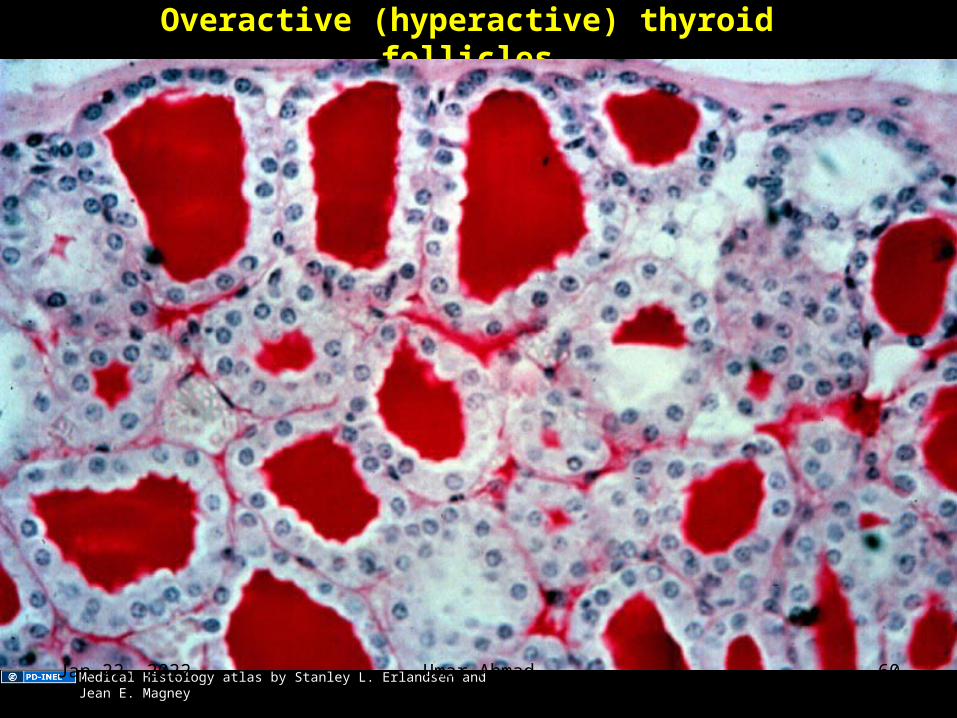
Overactive (hyperactive) thyroid follicles
Medical Histology atlas by Stanley L. Erlandsen and Jean E. MagneyApr 12, 2023 Umar Ahmad 60
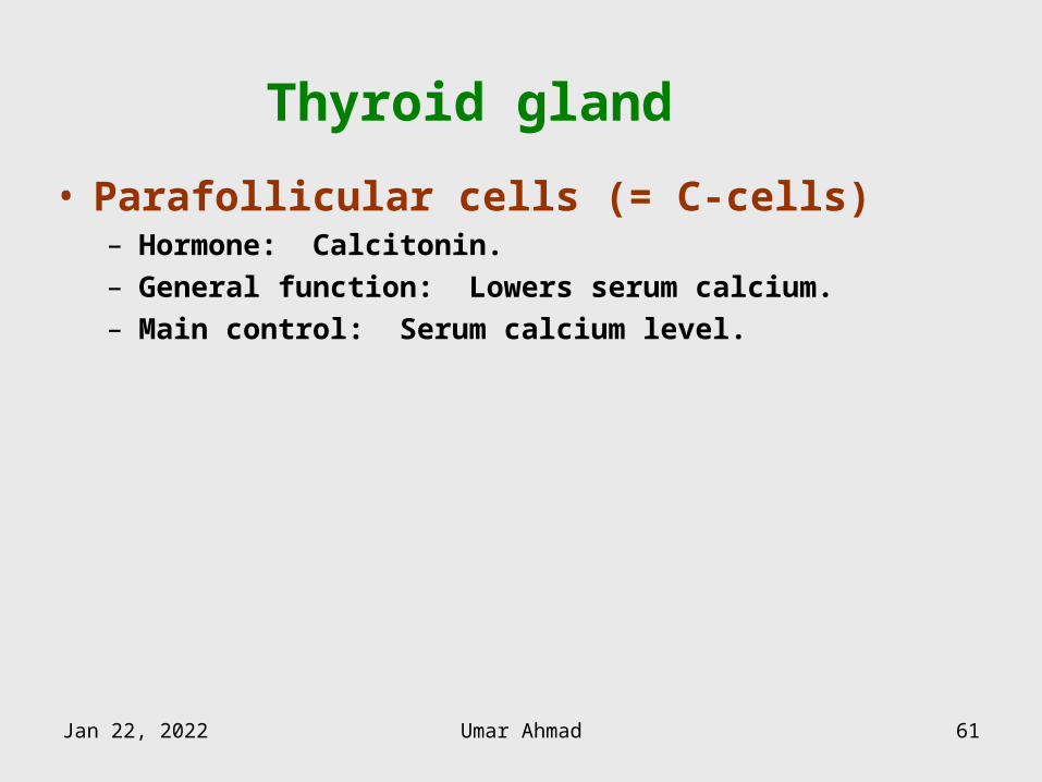
Thyroid gland
• Parafollicular cells (= C-cells)– Hormone: Calcitonin.– General function: Lowers serum calcium.– Main control: Serum calcium level.
Apr 12, 2023 Umar Ahmad 61
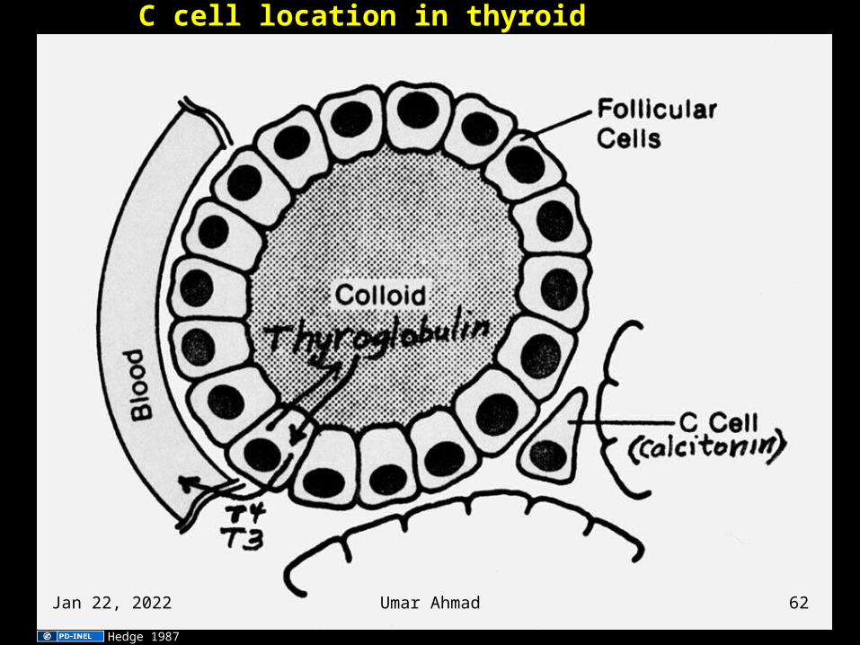
C cell location in thyroid
Hedge 1987
Apr 12, 2023 Umar Ahmad 62
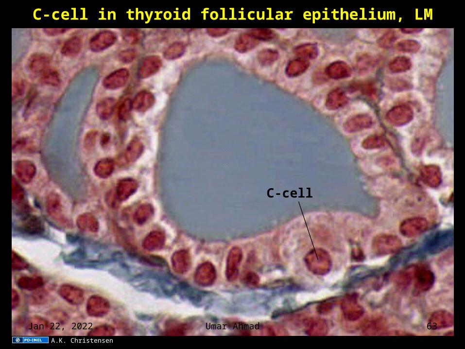
C-cell in thyroid follicular epithelium, LM
C-cell
A.K. Christensen
Apr 12, 2023 Umar Ahmad 63
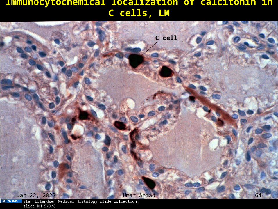
Immunocytochemical localization of calcitonin in C cells, LM
C cell
Stan Erlandsen Medical Histology slide collection, slide MH 9/D/8
Apr 12, 2023 Umar Ahmad 64
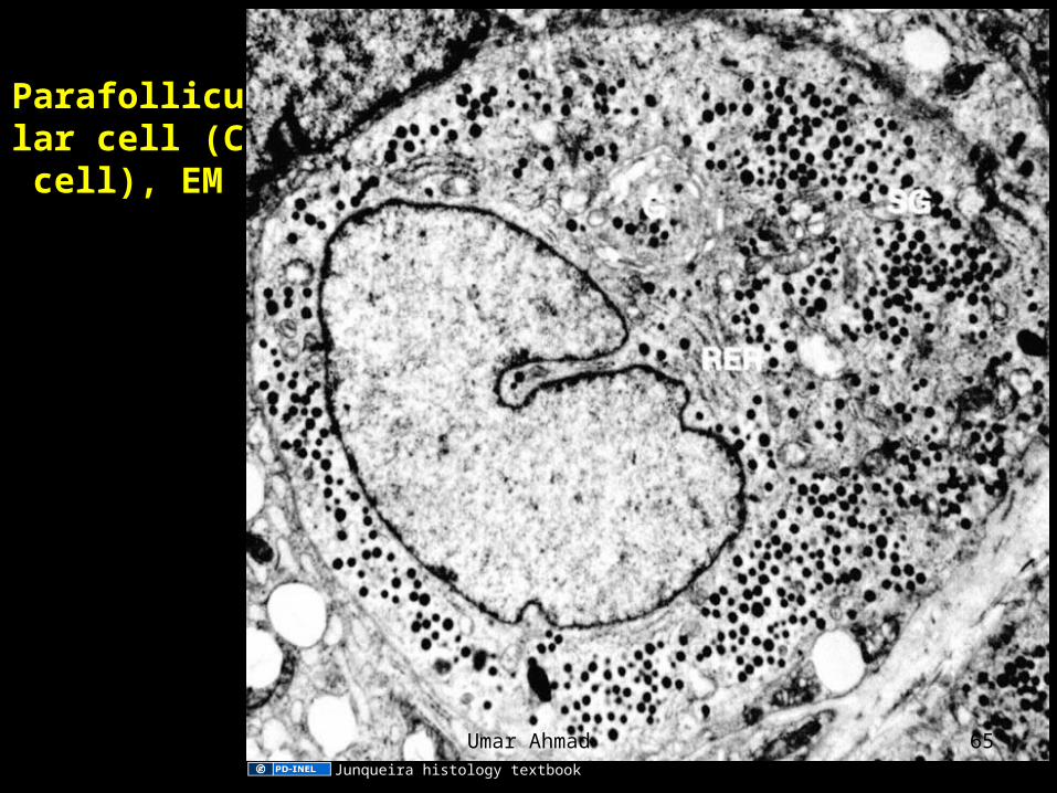
Parafollicular cell (C cell),
EM
Junqueira histology textbook
Apr 12, 2023 Umar Ahmad 65
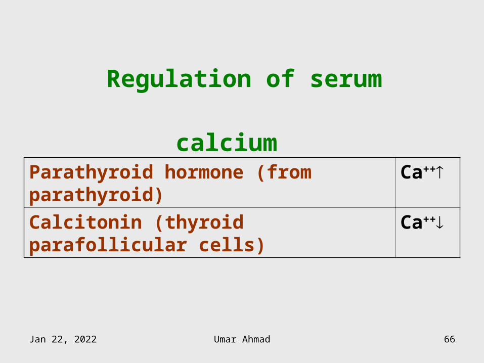
Regulation of serum calcium Parathyroid hormone (from parathyroid) Ca++
Calcitonin (thyroid parafollicular cells) Ca++
Apr 12, 2023 Umar Ahmad 66
PARATHYROID GLAND
Apr 12, 2023 Umar Ahmad 67
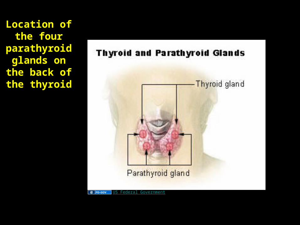
Location of the four
parathyroid glands on the
back of the thyroid
US Federal Government
Apr 12, 2023 Umar Ahmad 68
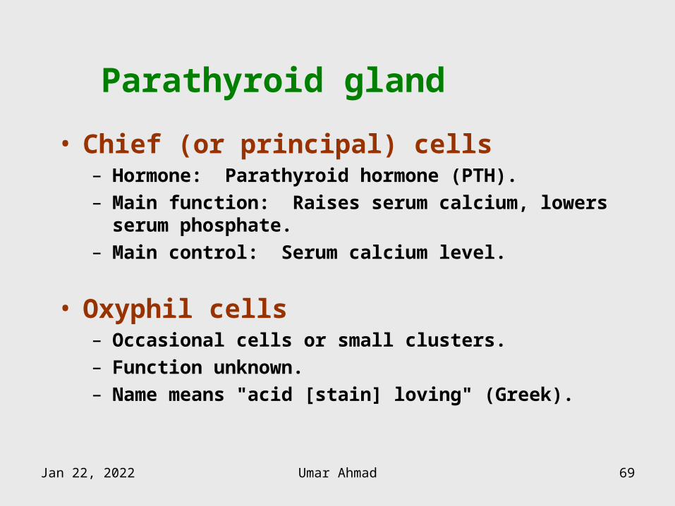
Parathyroid gland
• Chief (or principal) cells– Hormone: Parathyroid hormone (PTH).– Main function: Raises serum calcium, lowers serum
phosphate.– Main control: Serum calcium level.
• Oxyphil cells– Occasional cells or small clusters.– Function unknown.– Name means "acid [stain] loving" (Greek).
Apr 12, 2023 Umar Ahmad 69
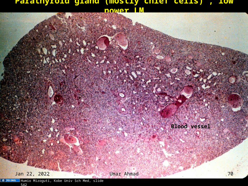
Parathyroid gland (mostly chief cells) , low power LM
Blood vessel
Humio Mizoguti, Kobe Univ Sch Med, slide 542
Apr 12, 2023 Umar Ahmad 70
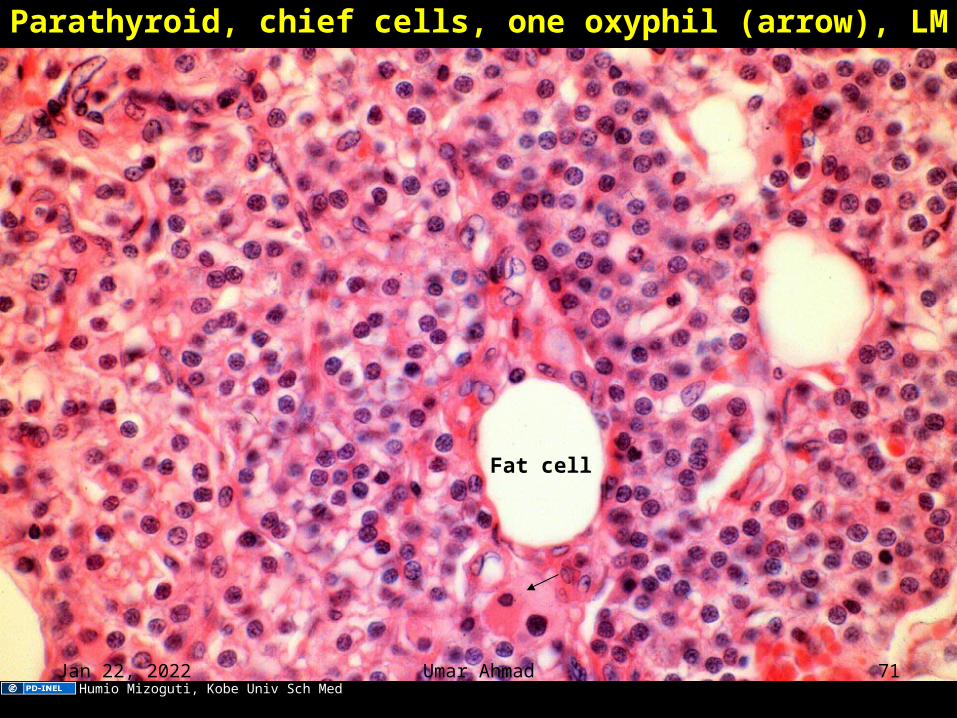
Parathyroid, chief cells, one oxyphil (arrow), LM
Fat cell
Humio Mizoguti, Kobe Univ Sch MedApr 12, 2023 Umar Ahmad 71
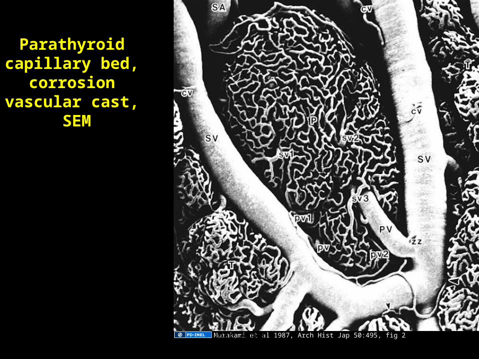
Parathyroid capillary bed,
corrosion vascular cast,
SEM
Murakami et al 1987, Arch Hist Jap 50:495, fig 2Apr 12, 2023 Umar Ahmad 72
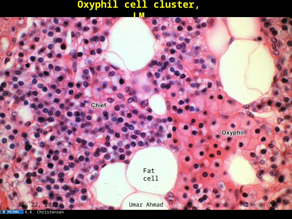
Oxyphil cell cluster, LM
Fatcell
A.K. Christensen
Apr 12, 2023 Umar Ahmad 73
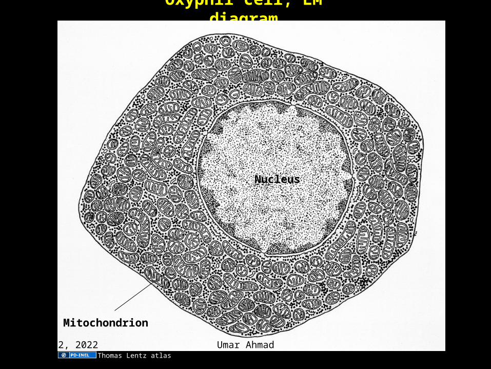
Oxyphil cell, EM diagram
Nucleus
Mitochondrion
Thomas Lentz atlas
Apr 12, 2023 Umar Ahmad 74

Apr 12, 2023 Umar Ahmad 75


![090713 jc caderno_empresas_negocios_empreendimentos_domesticos_10[1]](https://img.pdfslide.tips/doc/110x75/55d15d12bb61ebe9228b457b/090713-jc-cadernoempresasnegociosempreendimentosdomesticos101.jpg)