Embed Size (px)
Citation preview
c h a p t e r 1Principles of Acid-Base Physiology
Introduction�������������������������������������������������������������������������������������������� 3Objectives������������������������������������������������������������������������������������������������ 4
P A R T A CHEMISTRY OF H+ ��������������������������������������������������������������������������� 5H+ and the regeneration of ATP��������������������������������������������������������� 5Concentration of H+������������������������������������������������������������������������������ 6Risks associated with H+ ��������������������������������������������������������������������� 6
P A R T B DAILY BALANCE OF H+������������������������������������������������������������������� 6Production and removal of H+������������������������������������������������������������ 6
Acid balance ��������������������������������������������������������������������������������������������8Base balance �������������������������������������������������������������������������������������������9
Buffering of H+������������������������������������������������������������������������������������� 10Binding of H+ to proteins ���������������������������������������������������������������������10Bicarbonate buffer system ��������������������������������������������������������������������11
Role of the kidney in acid-base balance������������������������������������������ 15Reabsorption of filtered HCO3
− ������������������������������������������������������������15Net acid excretion ���������������������������������������������������������������������������������19Excretion of ammonium �����������������������������������������������������������������������20
Urine pH and kidney stone formation������������������������������������������� 28
P A R T C INTEGRATIVE PHYSIOLOGY������������������������������������������������������ 29Why is the normal blood pH 7.40?������������������������������������������������� 29Metabolic buffering of H+ during a sprint ������������������������������������ 31Discussion of questions��������������������������������������������������������������������� 34
Our goal in this chapter is to describe the physiology of hydrogen ions (H+). It quickly becomes apparent that many issues are difficult to understand unless one stops frequently to examine the “big pic-ture.” For example, based on the chemistry, H+ are the smallest ions (atomic weight 1) and their concentration in body fluids is tiny (a millionfold lower than that of HCO3
−, their major partner). On the other hand, H+ are extremely powerful in that they are intimately involved in the capture of energy from fuel oxidation in a form that permits humans to survive by driving the production of adenosine triphosphate (ATP). In this context, the electrical charge on the proton is far more important than its chemical concentration.
Introduction
�
ACID-BASE�
Definitions
ACiDeMiA VeRsUs ACiDosis• Acidemiadescribesanincreased
concentrationofH+inplasma.• Acidosisisaprocessinwhich
thereisanadditionofH+tothebody;thismayormaynotcauseacidemia.
ACiDs AnD BAsesAcidsarecompoundsthatarecapableofdonatingH+;basesarecompoundsthatarecapableofacceptingH+.Whenanacid(HA)dissociates,ityieldsH+anditsconjugatebaseoranions(A−).
HA⇌H++A−
VALenCeValenceisthenetelectricalchargeonacompoundoranelement.
ACiD-BAse teRMsConcentration of H+:Thenormalvalueinplasmais40±2-nmol/L.pH:Thisisthenegativelogarithmofthe[H+];itsnormalvalueinplasmais7.40±0.02.HCO3
−:HCO3−,theconjugate
baseofcarbonicacid,isthe“H+remover”ofthebicarbonatebuffersystem;itsconcentrationinplasmaiscloseto25mmol/L,buttherearelargefluctuationsthroughouttheday(22to31mmol/L).Pco2:Themajorcarbonwasteproductoffueloxidationiscarbondioxide.Itsconcentration(orPco2,whichisitspartialpressure)mustbelowinvenousbloodtoensurethatthebicarbonatebuffersys-temcanminimizechangesintheconcentrationofH+.ThenormalarterialPco2is40±2mmHg.ThePco2inblooddrainingskeletalmusclesis~6mmHg>thearterialPco2atrest.
OBJECTIVES
n To describe the major processes that lead to acid and to base balance.
Achieve Acid Balance 1. Production of acids: This is revealed when new anions are
found in the body and/or in excreted fluids. 2. Buffering of H+: This should minimize H+ binding to proteins
in vital organs (i.e., the brain and the heart). To do so, H+ must react with HCO3
−. The vast majority of HCO3− in the body
is in the interstitial and intracellular compartments of skeletal muscle. The key to achieving this function is to have a low Pco2 in the arterial blood and in the capillaries of skeletal muscle.
3. Kidneys add new HCO3− to the body: This occurs primarily
when NH4+ are excreted in the urine.
Achieve Base Balance 1. Input of alkali: This occurs primarily when fruit and vegetables
are ingested because they contain the K+ salts of organic acids that are metabolized to yield HCO3
−.
The concentration of H+ in body fluids must be maintained in a very narrow range. If this concentration rises, H+ will bind to very important compounds (proteins), and this changes their charge, shape, and possibly their function, with potentially dire consequences. Accordingly, it is not surprising that there is a H+ removal process (the bicarbonate buffer sys-tem) that reacts with H+ even if the concentration of H+ is not elevated. The strategy that permits this H+ removal system to function is that a low Pco2 obliges H+ to react with HCO3
− (see equation at the end of this paragraph). Therefore, a high H+ concentration stimulates breathing and thereby ensures that there is a lower concentration of CO2 in each liter of alveolar air and hence in the arterial blood. As we stress throughout this chapter, the Pco2 that is directly related to the majority of the bicarbon-ate buffer system is the Pco2 in capillaries of skeletal muscle rather than the arterial Pco2; the former is revealed by measuring the brachial or femoral venous Pco2.
H + HCO CO + H O+3 2 2
−
As shown in the preceding equation, this safe way to remove H+ leads to a deficit of HCO3
−. Accordingly, one must have another system that adds new HCO3
− to the body as long as acidemia per-sists; this is the task of the kidney. The most important component is the excretion of ammonium ions (NH4
+) because the kidney makes NH4
+ + HCO3− in the same metabolic process (see equation).
Glutamine 2NH 2HCO4 3→ −+ +
The diet also produces an alkali load, which the kidneys eliminate. The chemical form to excrete this alkali must be such that it does not lead to the formation of precipitates in the urine. This is achieved by excreting alkali as a family of organic anions rather than as HCO3
−, because the latter raises the urine pH, which may lead to the pre-cipitation of calcium and phosphate. Hence, another theme in this chapter is that the H+ concentration in the urine is regulated in a safe range to minimize the risk of forming kidney stones. Maintaining a urine pH of about 6 will permit a high rate of excretion of NH4
+, and this diminishes the risk of precipitation or uric acid.
ABBReViAtionsATP,adenosinetriphosphateADP,adenosinediphosphate
1 : PRINCIPLES OF ACID-BASE PHYSIOLOGY �
P A R T ACHEMISTRY OF H+
H+ AND THE REGENERATION OF ATP
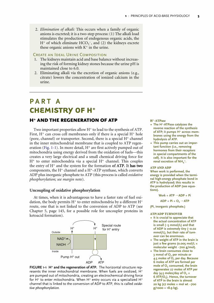
Two important properties allow H+ to lead to the synthesis of ATP. First, H+ can cross cell membranes only if there is a special H+ hole (pore, channel) or transporter. Second, there is a special H+ channel in the inner mitochondrial membrane that is coupled to ATP regen-eration (Fig. 1-1). In more detail, H+ are first actively pumped out of mitochondria using energy derived from the oxidation of fuels—this creates a very large electrical and a small chemical driving force for H+ to enter mitochondria via a special H+ channel. This couples the entry of H+ and the system for the formation of ATP. It has twoof ATP. It has twohas two components, the H+ channel and a H+-ATP synthase, which converts ADP plus inorganic phosphate to ATP (this process is called oxidative phosphorylation; see margin note).
Uncoupling of oxidative phosphorylation
At times, when it is advantageous to have a faster rate of fuel oxi-dation, the body permits H+ to enter mitochondria by a different H+ route, one that is not linked to the conversion of ADP to ATP (see Chapter 5, page 145, for a possible role for uncoupler proteins in ketoacid formation).
2. Elimination of alkali: This occurs when a family of organic anions is excreted; it is a two-step process: (1) The alkali load stimulates the production of endogenous organic acids, the H+ of which eliminate HCO3
−, and (2) the kidneys excrete these organic anions with K+ in the urine.
Create an Ideal Urine Composition 1. The kidneys maintain acid and base balance without increas-
ing the risk of forming kidney stones because the urine pH is maintained close to 6.0.
2. Eliminating alkali via the excretion of organic anions (e.g., citrate) lowers the concentration of ionized calcium in the urine.
H+-AtPase• TheH+-ATPasecatalyzesthe
reversereactionofthesynthesisofATP;itpumpsH+acrossmem-branesusingtheenergyfromthehydrolysisofATP.
• Thispumpcarriesoutanimpor-tantfunction(i.e.,removinghormonesfromtheirreceptorsinspecialcompartmentsofthecell).ItisalsoimportantfortherenalexcretionofNH4
+.
AtP AnD ADPWhenworkisperformed,theenergyisprovidedwhenthetermi-nalhigh-energyphosphatebondinATPishydrolyzed;thisresultsintheproductionofADP(seeequa-tions).
Work+ATP→ADP+Pi
ADP+Pi+O2→ATP
(Pi,inorganicphosphate.)
AtP/ADP tURnoVeR• Itiscrucialtoappreciatethat
theactualconcentrationofATPissmall(∼5mmol/L)andthatofADPisextremelytiny(∼0.02mmol/L),buttheirrateofturn-overcanbeenormous.
• TheweightofATPinthebrainisjustafewgrams(0.005mol/L×molecularweight∼700g/mol).
• Thebrainconsumescloseto3mmolofO2perminuteor4.5molesofO2perday.Because6molesofATPareformedpermoleofO2consumed,thebrainregenerates27molesofATPperday(4.5moles/dayofO2×6ATP/O2).Hence,theturnoverofATPinthebrainisalmost20kg(27moles×molwt∼700g/1000=18.9kg).
Outside
Inside
NAD+
H+ H+
Pump H+ out
Special routefor H+ entry
NADH
ADP ATP
FIGURE1-1 H+andtheregenerationofATP. The horizontal structure rep-resents the inner mitochondrial membrane. When fuels are oxidized, H+ are pumped out of mitochondria, creating an electrochemical driving force for H+ to enter mitochondria. When H+ entry occurs via a specialized H+ channel that is linked to the conversion of ADP to ATP, this is called oxida-tive phosphorylation.
ACID-BASE�
CONCENTRATION OF H+
Think of the concentration of H+ from the following three perspectives. 1. In relation to the concentration of other ions in the extracel-
lular fluid (ECF) compartment, the normal concentration of H+ is extremely small (0.000040 mmol/L, or 40 ± 2 nmol/L). In fact, the concentration of its partner, HCO3
− (∼25 mmol/L), is almost one millionfold higher than that of H+.
2. The concentration of H+ should be examined relative to the affinity of H+ for chemical groups on organic or inorganic com-pounds in the body. This comparison provides insights as to whether H+ will be bound or remain free.
3. A quantitative perspective is obtained by examining the rates of production and removal of H+. An enormous number of H+ are formed and removed each day (>70,000,000 nmol versus the amount of free H+ in the body at any one time [∼3000 nmol]; see margin note); small discrepancies between the rate of formation versus the rate of removal of H+ can result in major changes in concentration of H+ if this is sustained.
RISKS ASSOCIATED WITH H+
Proteins have an “ideal” shape that maintains their structural integrity and permits them to carry out essential functions (enzymes, transporters, contractile elements). H+ have a high affinity to bind to the amino acid histidine in proteins at the pH in cells. When the concentration of H+ rises, these bound H+ change the charge, shape, and possibly the function of these proteins. Accordingly, there must be a mechanism to keep the concentration of H+ very low. Moreover, this mechanism must be very efficient because of the enormous daily turnover of H+. Nevertheless, there are examples when this binding of H+ to proteins has important biologic functions (see margin note).
QUESTIONS
(Discussions on page 34)1-1 In certain locations in the body, H+ remain free and are not bound.
What is the advantage in having such a high concentration of H+?
1-2 What is the rationale for the statement, “In biology only weak acids kill”?
P A R T BDAILY BALANCE OF H+
PRODUCTION AND REMOVAL OF H+
• H+ production: H+ are generated when neutral compounds are converted to anions.
• H+ removal: H+ are removed when anions are converted to neutral products.
To determine whether H+ are produced or removed during metabolism, we use a “metabolic process” analysis (see margin note).
AMoUnt of H+ in tHe BoDYExtracellularfluid(ECF):15L×40nmol/L=600nmolIntracellularfluid(ICF):30L×80nmol/L=2400nmol
Benefit of H+ BinDinG to HeMoGLoBinWhenH+bindtohemoglobininsystemiccapillaries,hemoglobincanoff-loadoxygen(O2)atahigherPo2toimprovethediffusionofO2tocells.Incontrast,whenH+dissociatefromhemoglobininthecapillariesinthelungs(drivenbyahigherPo2),thisleadstoagreateruptakeofO2fromalveolarairforagivenalveolarPo2(seeChapter8,page235formorediscussionofthistopic).
MetABoLiC PRoCess• Ametabolicprocessanalysis
isawayofexaminingtheoverallimpactofmetabolismbyconsideringonlythesubstratesandtheproductswhileignoringallintermediates(seeChapter5,page113formoredetails).
• Ametabolicprocessismadeupofaseriesofmetabolicpathwaysthatcarryoutaspecificfunction.Theyareusuallylocatedinmorethanoneorgan.
• Examples:
1. Dietfuel→ATP+CO2+H2O
2.Dietfuel→storagecompounds
3.Storagecompounds→ATP+CO2 +H2O
1 : PRINCIPLES OF ACID-BASE PHYSIOLOGY �
To establish the balance for H+ in a metabolic process, one need only examine the valences of all of its substrates and products. If the sum of all of these valences is equal, there is no net production or removal of H+. When the products of a metabolic process have a greater anionic charge than its substrates, H+ are produced (e.g., incomplete oxida-tion of the major energy fuels, carbohydrates, and fats). Conversely, when the products of a metabolic process have a lesser anionic charge than its substrates, H+ are removed.
In a typical Western diet, 85% of kilocalories are in the form of carbohydrates and fat. Although their metabolism produces a large amount of H+, this is not a net H+ load because subsequent metabolic steps remove these H+ as fast as they are formed (see following equa-tions for the oxidation of carbohydrates {glucose} and fat [triglyc-erides]). The only time there is a net H+ load is when the complete oxidation of these fuels does not occur. Faster production of H+ from oxidation of glucose occurs during hypoxia (see Chapter 6, page 165 for more discussion), whereas faster production of H+ from neutral fat (triglycerides) occurs during states with a net lack of insulin (see Chapter 5, page 117 for more discussion).
Glucose -lactate +H CO +H O+2 2→ →−L
Triglycerides ketoacid anions H CO H O→ + → +− +2 2
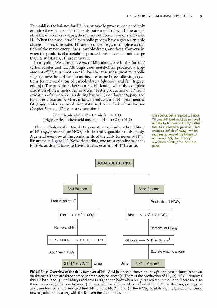
The metabolism of certain dietary constituents leads to the addition of H+ (e.g., proteins) or HCO3
– (fruits and vegetables) to the body. A general overview of the components of the daily turnover of H+ is illustrated in Figure 1-2. Notwithstanding, one must examine balances for both acids and bases to have a true assessment of H+ balance.
DisPosAL of H+ fRoM A MeALThisnetH+loadmustberemovedinitiallybybindingtoHCO3
–ratherthantointracellularproteins.ThiscreatesadeficitofHCO3
–,whichrequiresactionsofthekidneytoaddnewHCO3
–tothebody(excretionofNH4
+forthemostpart).
ACID-BASE BALANCE
Acid Balance Base Balance
+
+
Production of HCO3�Production of H
Urine Urine
Diet 2 H + SO42� +Diet 3 K + 3 HCO3
�
+Removal of HCO3
�Removal of H
Add “new” HCO3�
2 NH4+ + SO4
2�
Excrete organic anions
+ 3�
3�2
3 K + Citrate
+Glucose 3 H + Citrate+ 2 CO + 2 H2O2 H + HCO3
�
FIGURE1-2 OverviewofthedailyturnoverofH+. Acid balance is shown on the left, and base balance is shown on the right. There are three components to acid balance: (1) There is the production of H+, (2) HCO3
− removes this H+ load, and (3) the kidneys add new HCO3
− to the body when NH4+ is excreted in the urine. There are also
three components to base balance: (1) The alkali load of the diet is converted to HCO3− in the liver, (2) organic
acids are formed in the liver and their H+ remove HCO3−, and (3) the HCO3
− load drives the excretion of these new organic anions along with the K+ from the diet in the urine.
ACID-BASE�
Acid balance
• H+ are produced when the valence of the products of a meta-bolic process have a net negative charge as compared to its substrates.
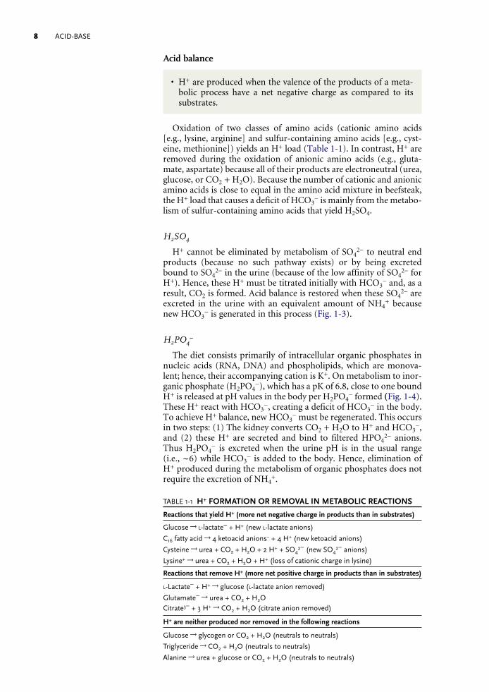
Oxidation of two classes of amino acids (cationic amino acids [e.g., lysine, arginine] and sulfur-containing amino acids [e.g., cyst-eine, methionine]) yields an H+ load (Table 1-1). In contrast, H+ are removed during the oxidation of anionic amino acids (e.g., gluta-mate, aspartate) because all of their products are electroneutral (urea, glucose, or CO2 + H2O). Because the number of cationic and anionic amino acids is close to equal in the amino acid mixture in beefsteak, the H+ load that causes a deficit of HCO3
− is mainly from the metabo-lism of sulfur-containing amino acids that yield H2SO4.
H2SO4
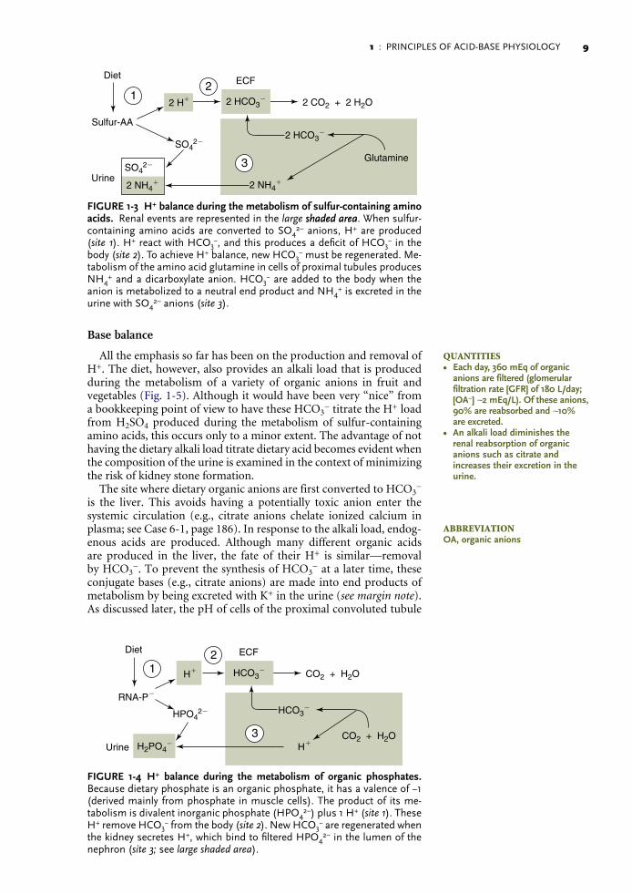
H+ cannot be eliminated by metabolism of SO42‐ to neutral end
products (because no such pathway exists) or by being excreted bound to SO4
2‐ in the urine (because of the low affinity of SO42‐ for
H+). Hence, these H+ must be titrated initially with HCO3− and, as a
result, CO2 is formed. Acid balance is restored when these SO42‐ are
excreted in the urine with an equivalent amount of NH4+ because
new HCO3− is generated in this process (Fig. 1-3).
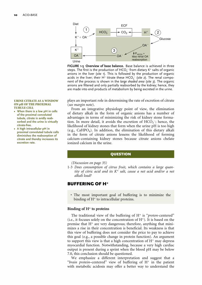
H2PO4–
The diet consists primarily of intracellular organic phosphates in nucleic acids (RNA, DNA) and phospholipids, which are monova-lent; hence, their accompanying cation is K+. On metabolism to inor-ganic phosphate (H2PO4
−), which has a pK of 6.8, close to one bound H+ is released at pH values in the body per H2PO4
− formed ((Fig. 1-4).. These H+ react with HCO3
−, creating a deficit of HCO3− in the body.
To achieve H+ balance, new HCO3− must be regenerated. This occurs
in two steps: (1) The kidney converts CO2 + H2O to H+ and HCO3−,
and (2) these H+ are secreted and bind to filtered HPO42‐ anions.
Thus H2PO4− is excreted when the urine pH is in the usual range
(i.e., ∼ 6) while HCO3− is added to the body. Hence, elimination of
H+ produced during the metabolism of organic phosphates does not require the excretion of NH4
+.
TABLE 1-1 H+FORMATIONORREMOVALINMETABOLICREACTIONS
ReactionsthatyieldH+(morenetnegativechargeinproductsthaninsubstrates)
Glucose → l-lactate‐ + H+ (new l-lactate anions)
C16 fatty acid → 4 ketoacid anions− + 4 H+ (new ketoacid anions)
Cysteine → urea + CO2 + H2O + 2 H+ + SO42‐ (new SO4
2‐ anions)
Lysine+ → urea + CO2 + H2O + H+ (loss of cationic charge in lysine)
ReactionsthatremoveH+(morenetpositivechargeinproductsthaninsubstrates)
l-Lactate‐ + H+ → glucose (l-lactate anion removed)
Glutamate‐ → urea + CO2 + H2OCitrate3‐ + 3 H+ → CO2 + H2O (citrate anion removed)
H+areneitherproducednorremovedinthefollowingreactions
Glucose → glycogen or CO2 + H2O (neutrals to neutrals)
Triglyceride → CO2 + H2O (neutrals to neutrals)
Alanine → urea + glucose or CO2 + H2O (neutrals to neutrals)
1 : PRINCIPLES OF ACID-BASE PHYSIOLOGY �
Base balance
All the emphasis so far has been on the production and removal of H+. The diet, however, also provides an alkali load that is produced during the metabolism of a variety of organic anions in fruit and vegetables (Fig. 1-5). Although it would have been very “nice” from a bookkeeping point of view to have these HCO3
− titrate the H+ load from H2SO4 produced during the metabolism of sulfur-containing amino acids, this occurs only to a minor extent. The advantage of not having the dietary alkali load titrate dietary acid becomes evident when the composition of the urine is examined in the context of minimizing the risk of kidney stone formation.
The site where dietary organic anions are first converted to HCO3−
is the liver. This avoids having a potentially toxic anion enter the systemic circulation (e.g., citrate anions chelate ionized calcium in plasma; see Case 6-1, page 186). In response to the alkali load, endog-enous acids are produced. Although many different organic acids are produced in the liver, the fate of their H+ is similar—removal by HCO3
−. To prevent the synthesis of HCO3− at a later time, these
conjugate bases (e.g., citrate anions) are made into end products of metabolism by being excreted with K+ in the urine (see margin note). As discussed later, the pH of cells of the proximal convoluted tubule
ECFDiet
Sulfur-AA
2 HCO3�
2 HCO3�
2 H�
2 NH4� 2 NH4
�
SO42�
SO42�
2 CO2 + 2 H2O
Glutamine
Urine
2
3
1
FIGURE1-3 H+balanceduringthemetabolismofsulfur-containingaminoacids. Renal events are represented in the large shaded areashaded area. When sulfur- containing amino acids are converted to SO4
2– anions, H+ are produced (site 1). H+ react with HCO3
−, and this produces a deficit of HCO3− in the
body (site 2). To achieve H+ balance, new HCO3− must be regenerated. Me-
tabolism of the amino acid glutamine in cells of proximal tubules produces NH4
+ and a dicarboxylate anion. HCO3− are added to the body when the
anion is metabolized to a neutral end product and NH4+ is excreted in the
urine with SO42– anions (site 3).
ECFDiet
RNA-P
HCO3�
HCO3�
H�
H�
H2PO4�
HPO42�
�
CO2 + H2O
CO2 + H2OUrine
2
3
1
FIGURE 1-4 H+ balance during the metabolism of organic phosphates. Because dietary phosphate is an organic phosphate, it has a valence of −1 (derived mainly from phosphate in muscle cells). The product of its me-tabolism is divalent inorganic phosphate (HPO4
2–) plus 1 H+ (site 1). These H+ remove HCO3
− from the body (site 2). New HCO3− are regenerated when
the kidney secretes H+, which bind to filtered HPO42– in the lumen of the
nephron (site 3; see large shaded area).
QUAntities• Eachday,360mEqoforganic
anionsarefiltered(glomerularfiltrationrate[GFR]of180L/day;[OA−]∼2mEq/L).Oftheseanions,90%arereabsorbedand∼10%areexcreted.
• Analkaliloaddiminishestherenalreabsorptionoforganicanionssuchascitrateandincreasestheirexcretionintheurine.
ABBReViAtionOA,organicanions
ACID-BASE10
plays an important role in determining the rate of excretion of citrate (see margin note).
From an integrative physiology point of view, the elimination of dietary alkali in the form of organic anions has a number of advantages in terms of minimizing the risk of kidney stone forma-tion. In more detail, it avoids the excretion of HCO3
−; hence, the likelihood of kidney stones that form when the urine pH is too high (e.g., CaHPO4). In addition, the elimination of this dietary alkali in the form of citrate anions lessens the likelihood of forming calcium-containing kidney stones because citrate anions chelate ionized calcium in the urine.
QUESTION
(Discussion on page 35)1-3 Does consumption of citrus fruit, which contains a large quan
tity of citric acid and its K+ salt, cause a net acid and/or a net alkali load?
BUFFERING OF H+
• The most important goal of buffering is to minimize the binding of H+ to intracellular proteins.
Binding of H+ to proteins
The traditional view of the buffering of H+ is “proton-centered” (i.e., it focuses solely on the concentration of H+). It is based on the premise that H+ are very dangerous; therefore, anything that mini-mizes a rise in their concentration is beneficial. Its weakness is that this view of buffering does not consider the price to pay to achieve this goal (e.g., a possible change in protein function). An argument to support this view is that a high concentration of H+ may depress myocardial function. Notwithstanding, because a very high cardiac output is present during a sprint when the blood pH may be below 7.0, this conclusion should be questioned.
We emphasize a different interpretation and suggest that a “brain protein–centered” view of buffering of H+ in the patient with metabolic acidosis may offer a better way to understand the
ECFDiet
K�� OA�
CO2�HCO3 H�
OA�
OA�
Glucose
PHCO3 =less renal OA�
reabsorption
2
3
1
�OA
K�
Urine
FIGURE1-5 Overviewofbasebalance. Base balance is achieved in three steps. The first is the production of HCO3
− from dietary K+ salts of organic anions in the liver (site 1). This is followed by the production of organic acids in the liver; their H+ titrate these HCO3
− (site 2). The renal compo-nent of the process is shown in the large shaded area (site 3). The organic anions are filtered and only partially reabsorbed by the kidney; hence, they are made into end products of metabolism by being excreted in the urine.
URine CitRAte As A WinDoW on pH of tHe PRoXiMAL tUBULe CeLL• WhenthereisalowpHincells
oftheproximalconvolutedtubule,citrateisavidlyreab-sorbedandtheurineisvirtuallycitrate-free.
• AhighintracellularpHinproximalconvolutedtubulecellsdiminishesthereabsorptionofcitrateandtherebyincreasesitsexcretionrate.
1 : PRINCIPLES OF ACID-BASE PHYSIOLOGY 11
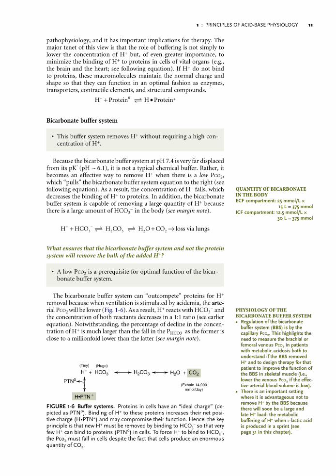
pathophysiology, and it has important implications for therapy. The major tenet of this view is that the role of buffering is not simply to lower the concentration of H+ but, of even greater importance, to minimize the binding of H+ to proteins in cells of vital organs (e.g., the brain and the heart; see following equation). If H+ do not bind to proteins, these macromolecules maintain the normal charge and shape so that they can function in an optimal fashion as enzymes, transporters, contractile elements, and structural compounds.
H Protein H Protein+ ++ •0
Bicarbonate buffer system
• This buffer system removes H+ without requiring a high con-centration of H+.
Because the bicarbonate buffer system at pH 7.4 is very far displaced from its pK′ (pH ∼ 6.1), it is not a typical chemical buffer. Rather, it becomes an effective way to remove H+ when there is a low Pco2, which “pulls” the bicarbonate buffer system equation to the right (see following equation). As a result, the concentration of H+ falls, which decreases the binding of H+ to proteins. In addition, the bicarbonate buffer system is capable of removing a large quantity of H+ because there is a large amount of HCO3
− in the body (see margin note).
H HCO H CO H O CO loss via lungs3+ −+ + → 2 3 2 2
What ensures that the bicarbonate buffer system and not the protein system will remove the bulk of the added H+?
• A low Pco2 is a prerequisite for optimal function of the bicar-bonate buffer system.
The bicarbonate buffer system can “outcompete” proteins for H+ removal because when ventilation is stimulated by acidemia, the arte-arte-rial Pco2 will be lower (Fig. 1-6). As a result, H+ reacts with HCO3
− and the concentration of both reactants decreases in a 1:1 ratio (see earlier equation). Notwithstanding, the percentage of decline in the concen-tration of H+ is much larger than the fall in the PHCO3 as the former is close to a millionfold lower than the latter (see margin note).
QUAntitY of BiCARBonAte in tHe BoDYECFcompartment:25mmol/L×
15L=375mmolICFcompartment:12.5mmol/L×
30L=375mmol
PHYsioLoGY of tHe BiCARBonAte BUffeR sYsteM• Regulationofthebicarbonate
buffersystem(BBS)isbythecapillaryPco2.ThishighlightstheneedtomeasurethebrachialorfemoralvenousPco2inpatientswithmetabolicacidosisbothtounderstandiftheBBSremovedH+andtodesigntherapyforthatpatienttoimprovethefunctionoftheBBSinskeletalmuscle(i.e.,lowerthevenousPco2iftheeffec-tivearterialbloodvolumeislow).
• ThereisanimportantsettingwhereitisadvantageousnottoremoveH+bytheBBSbecausetherewillsoonbealargeandlateH+load:themetabolicbufferingofH+whenl-lacticacidisproducedinasprint(seepage31inthischapter).
(Tiny) (Huge)
(Exhale 14,000mmol/day)
H + HCO3�
H•PTN
PTN0
H2CO3 H2O + CO2 �
�
FIGURE1-6 Buffersystems. Proteins in cells have an “ideal charge” (de-picted as PTN0). Binding of H+ to these proteins increases their net posi-tive charge (H•PTN+) and may compromise their function. Hence, the key principle is that new H+ must be removed by binding to HCO3
− so that very few H+ can bind to proteins (PTN0) in cells. To force H+ to bind to HCO3
−, the Pco2 must fall in cells despite the fact that cells produce an enormous quantity of CO2.
ACID-BASE12
Which Pco2 is important for the bicarbonate buffer system to function optimally?
Arterial Pco2
• The arterial Pco2 reflects, but is not equal to, the Pco2 in brain cells; it sets a minimum possible value for the Pco2 in capillar-ies of all other organs in the body.
The only cells in the body that have the same Pco2 as in arterial blood are the red blood cells that are located in the arterial blood volume. Therefore, the arterial Pco2 does not reveal whether the bicarbonate buffer system has operated efficiently in the vast majority of the ICF and ECF compartments. Notwithstanding, the arterial Pco2 sets the lower limit for the Pco2 in capillaries. Furthermore, the arterial Pco2 is important because it reflects the Pco2 in brain cells but in an indirect fashion. In more detail, the rate of production of CO2 in the brain is relatively constant because cerebral oxygen consumption does not vary appreciably. The brain produces 1 mmol CO2 per mmol O2 it consumes, as the brain oxidizes glucose as its primary fuel. In addition, the rate of blood flow to the brain undergoes only minimal variation throughout the day owing to autoregulation. Accordingly, the arterial Pco2 reflects the Pco2 in brain cells in the absence of a marked degree of contraction of the effective arterial blood volume during which the brain fails to autoregulate its rate of blood flow.
The process to lower the arterial Pco2 begins with stimulation of the respiratory center in the brain. It is an ideal response because it ensures that the brain will always have an “ideal” Pwill always have an “ideal” Palways have an “ideal” Pco2 in its ECF and ICF compartments so that there is only a minimal binding of H+ to intracellular proteins during metabolic acidosis, which decreases pos-sible detrimental effects on neuronal function (see margin note).
Capillary Pco2
The capillary Pco2 is higher than the arterial Pco2 because cells in the body consume O2 and add CO2 to their capillary blood. CO2 diffuses rapidly because distances are short and time is not a limiting factor; thus, the Pco2 in capillaries is virtually identical to the Pco2 in cells and in the interstitial compartment of the ECF in a given region. Therefore, the capillary Pco2 reveals whether the bicarbonate buffer system has operated efficiently in the vast majority of the ICF and ECF compartments in the body (Table 1-2). The capillary Pco2 is influenced by arterial Pco2 and the rate of addition of CO2 to capillary blood in individual organs of the body. In more detail, if most of the oxygen in each liter of blood delivered to a certain area is consumed, the Pco2 in its capillary blood rises appreciably (see margin note). There are two conditions is which most of the O2 delivered in a liter of blood is consumed: (1) a rise in the rate of metabolism without a change in the rate of blood flow, or (2) a decrease in the rate of blood flow with no change in the rate of O2 consumption.
Venous Pco2
Although the capillary Pco2 reveals whether the bicarbonate buffer system has operated efficiently in the vast majority of the ICF and ECF compartments in the area drained by this capillary bed, one can-not measure it directly. The venous Pco2, however, closely reflects the
ConCentRAtion of H+ in BRAin CeLLsInresponsetoacidemia,alveolarventilationisstimulatedandthearterialPco2falls.Nevertheless,thiscannotbringthepHinbraincellsbacktonormal—otherwisetherewouldnotbeapersistingstimulustoalveolarventilation.Hence,therewillalwaysbeaminimaldegreeofH+bindingtoproteinsinbraincellsduringacidemia.
Co2 ADDeD PeR LiteR of BLooD fLoW• Closeto3mmol/minofO2
areextractedinskeletalmuscle,whichreceivescloseto1L/minofbloodflow.ThisresultsinanaveragecapillaryPco2thatis6mmHghigherthanthearterialPco2.
• Whenskeletalmusclecellsextractmorethan3mmolO2perliterofbloodflow,moreCO2isaddedtoeachliterofcapillarybloodanditsPco2ishigher.Thiscompromisestheeffectivenessofthebicarbonatebuffersystem.
1 : PRINCIPLES OF ACID-BASE PHYSIOLOGY 1�
capillary Pco2 in its drainage bed. There is, however, one caveat—if an appreciable quantity of blood shunts from the arterial to the venous circulation and bypasses cells, this venous Pco2 does not reflect the Pco2 in the interstitial space and in cells in its drainage bed.
Which venous Pco2 should be measured in clinical medicine to assess the effectiveness of the bicarbonate buffer system?
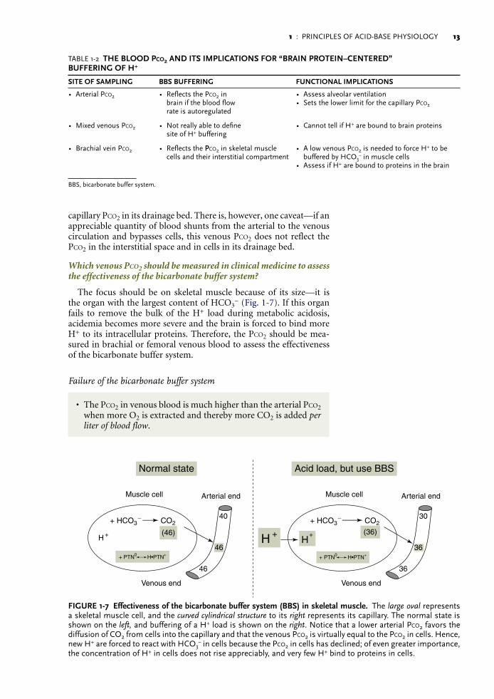
The focus should be on skeletal muscle because of its size—it is the organ with the largest content of HCO3
− (Fig. 1-7). If this organ fails to remove the bulk of the H+ load during metabolic acidosis, acidemia becomes more severe and the brain is forced to bind more H+ to its intracellular proteins. Therefore, the Pco2 should be mea-sured in brachial or femoral venous blood to assess the effectiveness of the bicarbonate buffer system.
Failure of the bicarbonate buffer system
• The Pco2 in venous blood is much higher than the arterial Pco2 when more O2 is extracted and thereby more CO2 is added per liter of blood flow.
TABLE 1-2 THEBLOODPco2ANDITSIMPLICATIONSFOR“BRAINPROTEIN–CENTERED”BUFFERINGOFH+
SITEOFSAMPLING BBSBUFFERING FUNCTIONALIMPLICATIONS
• Arterial Pco2 • Reflects the Pco2 in brain if the blood flow rate is autoregulated
• Assess alveolar ventilation • Sets the lower limit for the capillary Pco2
• Mixed venous Pco2 • Not really able to define site of H+ buffering
• Cannot tell if H+ are bound to brain proteins
• Brachial vein Pco2 • Reflects the PPco2 in skeletal muscle cells and their interstitial compartment
• A low venous Pco2 is needed to force H+ to be buffered by HCO3
− in muscle cells • Assess if H+ are bound to proteins in the brain
BBS, bicarbonate buffer system.
+H
+ PTN0 H•PTN++ PTN0 H•PTN+
+ HCO3� CO2 + HCO3
� CO240
46
46
(46)
Muscle cell
Venous end
Arterial end
+H
+H
30
36
36
(36)
Muscle cell
Acid load, but use BBSNormal state
Venous end
Arterial end
FIGURE1-7 Effectivenessofthebicarbonatebuffersystem(BBS)inskeletalmuscle. The large oval represents a skeletal muscle cell, and the curved cylindrical structure to its right represents its capillary. The normal state is shown on the left, and buffering of a H+ load is shown on the right. Notice that a lower arterial Pco2 favors the diffusion of CO2 from cells into the capillary and that the venous Pco2 is virtually equal to the Pco2 in cells. Hence, new H+ are forced to react with HCO3
− in cells because the Pco2 in cells has declined; of even greater importance, the concentration of H+ in cells does not rise appreciably, and very few H+ bind to proteins in cells.
ACID-BASE1�
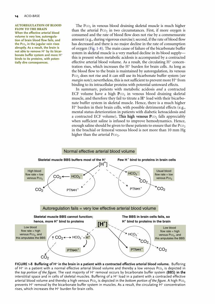
The Pco2 in venous blood draining skeletal muscle is much higher than the arterial Pco2 in two circumstances. First, if more oxygen is consumed and the rate of blood flow does not rise by a commensurate amount (e.g., during vigorous exercise); second, if the rate of blood flow has decreased and there is no major decline in the rate of consumption of oxygen (Fig. 1-8). The main cause of failure of the bicarbonate buffer system in skeletal muscle is a very marked decline in its blood supply—this is present when metabolic acidosis is accompanied by a contracted effective arterial blood volume. As a result, the circulating H+ concen-tration rises, which increases the H+ burden for brain cells. As long as the blood flow to the brain is maintained by autoregulation, its venous Pco2 does not rise and it can still use its bicarbonate buffer system (see margin note); nevertheless, this is not sufficient to prevent more H+ from binding to its intracellular proteins with potential untoward effects.
In summary, patients with metabolic acidosis and a contracted ECF volume have a high PPco2 in venous blood draining skeletal muscle, and therefore they fail to titrate a HH+ load with their bicarbo-nate buffer system in skeletal muscle. Hence, there is a much higher H+ burden in their brain cells, with possible detrimental effects (e.g., mental status deterioration in patients with diabetic ketoacidosis and a contracted ECF volume). This high venous P. This high venous PPco2 falls appreciably when sufficient saline is infused to improve hemodynamics. Hence, enough saline should be given to these patients to ensure that the Pco2 in the brachial or femoral venous blood is not more than 10 mm Hg higher than the arterial Pco2.
+PTN•H
+PTN•H
+PTN•H
+PTN•H
Skeletal muscle BBS buffers most of the H+ Few H bind to proteins in brain cells
High blood flow rate = low venous PCO2
CO2CO2
CO2
CO2
CO2CO2
HCO3�
HCO3�CO2
HCO3�
HCO3�
+
+[H ]
Usual blood flow rate = low venous PCO2
Low blood flow rate = high
venous PCO2, andthis amputates the BBS
Low blood flow rate = high
venous PCO2, andthis amputates the BBS
+
Skeletal muscle BBS cannot function;hence, more H+ bind to proteins
The BBS in brain cells fails, so H+ bind to proteins in the brain
Autoregulation fails = very low effective arterial blood volume
Normal effective arterial blood volume
+ H
[H+]
CO2
FIGURE1-8 BufferingofH+inthebraininapatientwithacontractedeffectivearterialbloodvolume. Buffering of H+ in a patient with a normal effective arterial blood volume and thereby a low venous Pco2 is depicted in the top portion of the figure. The vast majority of H+ removal occurs by bicarbonate buffer system (BBS) in the(BBS) in thein the interstitial space and in cells of skeletal muscles. Buffering of a H+ load in a patient with a contracted effective arterial blood volume and thereby a high venous Pco2 is depicted in the bottom portion of the figure. A high Pco2 prevents H+ removal by the bicarbonate buffer system in muscles. As a result, the circulating H+ concentration rises, which increases the H+ burden for brain cells.
AUtoReGULAtion of BLooD fLoW to tHe BRAinWhentheeffectivearterialbloodvolumeisverylow,autoregula-tionofbrainbloodflowfails,andthePco2inthejugularveinrisesabruptly.Asaresult,thebrainisnotabletoremoveH+byitsbicar-bonatebuffersystemandmoreH+bindstoitsproteins,withpoten-tiallydireconsequences.
1 : PRINCIPLES OF ACID-BASE PHYSIOLOGY 1�
Buffering of H+ during a sprint
Buffering of H+ produced during a sprint requires a new H+ accep-tor that binds a large quantity of H+ without causing the formation of CO2. Although the bicarbonate buffer system cannot operate in this setting because the venous Pco2 is very high, it does become very important just after exercise ceases (see the discussion of metabolic buffering in Part C, page 31).
QUESTIONS
(Discussions on page 35)1-4 Why is the llactic acidosis observed during cardiogenic shock so
much more devastating than the llactic acidosis observed during a sprint if the Pllactate, arterial pH, and PHCO3 are identical?
1-5 The heart extracts close to 70% of the oxygen from each liter of coronary artery blood. What conclusions can you draw about buffering of H+ in the heart? Might there be advantages owing to this high extraction of O2 per liter of blood flow?
ROLE OF THE KIDNEY IN ACID-BASE BALANCE
The kidneys must perform two tasks to maintain acid balance (for base balance, see page 9). First, the kidney must reabsorb virtually 100% of the filtered HCO3
−; this is achieved primarily by H+ secretion in the proximal convoluted tubule. Second, the kidneys must add new HCO3
− to the body when there is an acid load; this is achieved princi-pally by excreting more NH4
+.
Reabsorption of filtered HCO3−
• The kidneys must prevent the excretion of the very large quan-tity of filtered HCO3
−. • This is primarily the task of the proximal convoluted tubule. • Performing this function does not raise the quantity of HCO3
− in the body.
It is important at the outset to recognize that a huge amount of HCO3
− is filtered and reabsorbed each day (GFR of 180 L/day × PHCO33 25 mmol/L = 4500 mmol). Approximately 90% of this HCO3
− (~4000 mmol/day) is reabsorbed in an indirect fashion by the proxi-mal convoluted tubule (Table 1-3).
Reabsorption of NaHCO3 in the proximal convoluted tubule
The bulk of filtered HCO3− is reabsorbed in an indirect fashion
in the proximal convoluted tubule as a result of H+ secretion, which causes HCO3
− to disappear from the tubular lumen and reappear in the blood (Fig. 1-9). There is no direct acid-base impact of this reab-sorption of HCO3
− because it does not result in a positive balance of HCO3
−. Nevertheless, should this process fail, there is an initial loss of NaHCO3 in the urine and the development of metabolic acidosis (this disorder is called proximal renal tubular acidosis; it is discussed on page 85 in Chapter 4).
BAse BALAnCeThisisdescribedinmoredetailonpage9.
ACID-BASE1�
The events in the proximal convoluted tubule result in conser-vation of both Na+ and HCO3
− and can be viewed as two parallel stories—one dealing with the reabsorption of Na+ and the other deal-ing with the secretion of H+.
The Na+ story
• This story has three components: (1) at the luminal membrane, (2) in the cell, and (3) at the basolateral membrane.
• In general, this process is regulated by signals related to the need to reabsorb Na+ (higher when the effective arterial blood volume is low).
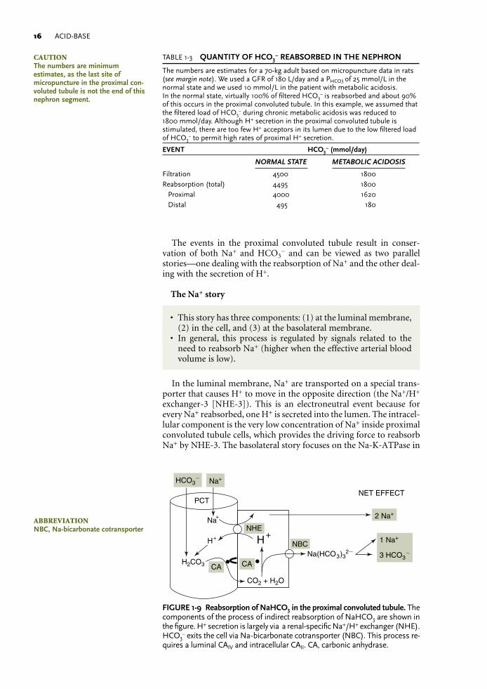
In the luminal membrane, Na+ are transported on a special trans-porter that causes H+ to move in the opposite direction (the Na+/H+ exchanger-3 [NHE-3]). This is an electroneutral event because for every Na+ reabsorbed, one H+ is secreted into the lumen. The intracel-lular component is the very low concentration of Na+ inside proximal convoluted tubule cells, which provides the driving force to reabsorb Na+ by NHE-3. The basolateral story focuses on the Na-K-ATPase in
Na
H
NHE
H
H2CO3
CO2 + H2O
PCT
+
+ +
Na(HCO )32�
3
HCO3�
2 Na+
3 HCO3�
1 Na+
Na+
NET EFFECT
CA CA
NBC
• •
FIGURE1-9 ReabsorptionofNaHCO3intheproximalconvolutedtubule. The components of the process of indirect reabsorption of NaHCO3 are shown in the figure. H+ secretion is largely via a renal-specific Na+/H+ exchanger (NHE). HCO3
− exits the cell via Na-bicarbonate cotransporter (NBC). This process re-quires a luminal CAIV and intracellular CAII. CA, carbonic anhydrase.
TABLE 1-3 QUANTITYOFHco3−REABSORBEDINTHENEPHRON
The numbers are estimates for a 70-kg adult based on micropuncture data in rats (see margin note). We used a GFR of 180 L/day and a PHCO3 of 25 mmol/L in the normal state and we used 10 mmol/L in the patient with metabolic acidosis. In the normal state, virtually 100% of filtered HCO3
− is reabsorbed and about 90% of this occurs in the proximal convoluted tubule. In this example, we assumed that the filtered load of HCO3
− during chronic metabolic acidosis was reduced to 1800 mmol/day. Although H+ secretion in the proximal convoluted tubule is stimulated, there are too few H+ acceptors in its lumen due to the low filtered load of HCO3
− to permit high rates of proximal H+ secretion.
EVENT HCO3−(mmol/day)
NORMAL STATE METABOLIC ACIDOSIS
Filtration 4500 1800Reabsorption (total) 4495 1800
Proximal 4000 1620Distal 495 180
CAUtionThenumbersareminimumestimates,asthelastsiteofmicropunctureintheproximalcon-volutedtubuleisnottheendofthisnephronsegment.
ABBReViAtionNBC,Na-bicarbonatecotransporter
1 : PRINCIPLES OF ACID-BASE PHYSIOLOGY 1�
the basolateral membrane. This ion transporter provides the driving force for the overall process—maintaining the low concentration of Na+ in these cells; it transports three Na+ out of the cell in conjunc-tion with the entry of two K+ (see margin note).
The H+ story
• This story has components in three locations: (1) at the luminal membrane, (2) in the cell, and (3) at the basolateral membrane.
• This process is limited by the filtered load of HCO3− and stim-
ulated by a high concentration of H+ in proximal convoluted tubular cells.
On the luminal membrane, there are two unique features: the NHE-3 and the luminal carbonic anhydrase (CAIV). The latter en-zyme hydrolyzes carbonic acid that is formed in the lumen to CO2 and H2O. The enzyme-catalyzed rate of hydrolysis of H2CO3 is virtu-ally as fast as the rate of generation of H2CO3. If rapid catalysis did not occur, indirect reabsorption of HCO3
− would be retarded and urinary excretion of HCO3
− would ensue.Inside the cell, there are two important components of the H+
story. First, there is a different carbonic anhydrase enzyme (CAII), which prevents the accumulation of OH− (in other words, it makes HCO3
− available inside cells). Second, there is a modifier site on NHE-3 to which H+ bind, and this activates this ion exchanger.
On the basolateral membrane, there is a unique transport system for the exit of HCO3
− from these cells, which permits an ion complex of one Na+ and the equivalent of three HCO3
− to exit as a divalent anion, Na(HCO3
−)32−—this is called the Nabicarbonate cotransporter.
Regulation of proximal H+ secretion
• NHE-3 has a high capacity, but the “leaky” tight junctions in the proximal convoluted tubule prevent the generation of steep concentration gradients for H+.
• Regulators of NaHCO3 reabsorption in the proximal convoluted tubule include the filtered load of HCO3
−, the luminal concentra-tion of H+; the intracellular concentration of H+; stimuli for Na+ reabsorption; and, importantly, the hormone angiotensin II.
The filtered load of HCO3−. The proximal convoluted tubule reab-
sorbs close to 90% of the 4500 mmol of HCO3− that are filtered each
day (see Table 1-3). In this quantitative example, lowering the PHCO3 to 10 mmol/L during metabolic acidosis reduces the filtered load of HCO3
− to 1800 mmol/day. Even though H+ secretion in the proximal convoluted tubule will be stimulated by the high concentration of H+ in proximal convoluted tubule cells, the reabsorption of NaHCO3 must be reduced by more than 50% because there are no other lumi-nal H+ acceptors of quantitative importance (see margin note).
Luminal [H+]. A higher concentration of H+ in the lumen of the proximal convoluted tubule inhibits H+ secretion in patients with metabolic acidosis (see Table 1-3). This same scenario occurs when a patient is given acetazolamide, a drug that inhibits luminal carbonic anhydrase. In this case, H+ secretion is diminished because of the rise in the concentration of carbonic acid (H2CO3) and thereby H+ in the lumen; hence, less of the filtered HCO3
− is reclaimed.
nBC AnD na+ tRAnsPoRt• Closeto5%ofNa+exitsproxi-
malconvolutedtubulecellsasaNa(HCO3
−)32−ioncomplex,
whichmovespassivelydownitselectrochemicalgradient.ThisNa+isreabsorbedwithoutrequiringthehydrolysisofATP(itbypassestheNa-K-ATPaseinproximalconvolutedtubulecells).
• Thisprocessaccountsforthereabsorptionof1260mmolofNa+perdaywithastoichiometryof1Na+per3HCO3
–ontheNBCexitstepandaGFRof180L/day(i.e.,21mmolofHCO3
–perliterofGFRarereab-sorbedintheproximalconvo-lutedtubule).
nHe AnD seCRetion of nH4
+ in tHe PRoXiMAL ConVoLUteD tUBULe• Inapatientwithchronic
metabolicacidosis,thereislessfilteredHCO3
−;hencethistransporterhasadiminishedroleinthereabsorptionofNaHCO3.
• NH4+excretionisimportantin
thissetting.ActivationofNHE-3byintracellularacidosisshouldshouldallowfortheentryofmoreNH4
+intothelumenoftheproximalconvolutedtubuleonthistransporter.
ACID-BASE1�
Concentration of H+ in proximal convoluted tubule cells. A rise in the concentration of H+ in proximal convoluted tubule cells stimulates the secretion of H+ for two reasons. First, the higher concentration of H+ may cause more H+ secretion by NHE-3. Second, and of greater importance, the binding of H+ to a modifier site on NHE-3 activates this cation exchanger. As shown in Table 1-3, this activation is not very important during metabolic acidosis because of the small filtered load of HCO3
−. Intracellular acidosis can help explain why the PHCO3 is elevated in patients with hypokalemia or chronic respiratory acidosis.hypokalemia or chronic respiratory acidosis.
Avidity for the reabsorption of Na+
• The most important regulator of the reabsorption of NaHCO3 in the proximal convoluted tubule is angiotensin II.
When the effective arterial blood volume is contracted, proximal Na+ reabsorption (and thereby H+ secretion) rises because of the higher con-centration of angiotensin II. In contrast, there is inhibition of the reab-sorption of NaHCO3 in the proximal convoluted tubule when NaHCO3 is given. Part of the mechanism involves a fall in the concentration of H+ in the ICF, which diminishes flux through the NHE-3 despite an increase in the number of H+ acceptors in the lumen. In addition, the Na+ load expands the effective arterial blood volume, leading to lower levels of angiotensin II and thereby inhibition of the reabsorption of NaHCO3 in the proximal convoluted tubule. This seems to negate the direct effect of the increased filtered load of HCO3
− to stimulate the reabsorption of HCO3
− in the proximal convoluted tubule; thus, there is a prompt excretion of the excess HCO3
−.Minor factors. Hypercalcemia and/or a low parathyroid hormone
level stimulate proximal H+ secretion by NHE-3.
Renal threshold for reabsorption of HCO3−
• There is no renal threshold for the reabsorption of NaHCO3 in the proximal convoluted tubule as long as the effective arterial blood volume is not expanded, which prevents a fall in the level of angiotensin II.
If there were a renal threshold for the reabsorption of NaHCO3 in the kidney, it would represent the highest PHCO3 at which H+ secretion is sufficient to “reclaim” virtually all of the filtered HCO3
− (Fig. 1-10). Although this has been demonstrated experimentally, one must ask, “What were the conditions in these experiments?” The demonstration of this renal threshold for the reabsorption of NaHCO3 occurs only when experimental animals or humans were given an infusion of NaHCO3. This, however, expands the effec-tive arterial blood volume owing to the load of Na+, which lowers angiotensin II levels and thereby depresses the reabsorption of the extra NaHCO3. In addition, the alkali load lowers the concentration of H+ in proximal convoluted tubule cells and thereby removes this stimulator for NHE-3. In conclusion, because this does not represent a physiologic setting where the PHCO33 is increased, it may not be the correct experimental setting to define normal physiology.
In fact, there is no renal threshold for the reabsorption of NaHCO3 when the PHCO33 is raised without expanding the effective arterial blood volume (see Fig. 1-10). The best example of this in normal
AnGiotensin ii• AngiotensinIIisavasoconstric-
tor.ItalsostimulatesthereleaseofaldosteroneandincreasestheindirectreabsorptionofNaHCO3intheproximalconvolutedtubule.
• AngiotensinIIactsbyactivatingproteinkinaseC,whichleadstophosphorylationofNHE-3.
1 : PRINCIPLES OF ACID-BASE PHYSIOLOGY 1�
physiology is the alkaline tide owing to secretion of HCl into the lumen of the stomach (see Fig. 7-2, page 198). Electroneutrality is maintained because there is an exchange of Cl− for HCO3
− in the ECF compartment with a 1:1 stoichiometry. After this occurs, the PHCO33 rises toward 30 mmol/L, but the urine contains very little HCO3
−. Hence, subjects on a typical Western diet have an effective arterial blood volume that leads to angiotensin II levels that are sufficient to stimulate proximal H+ secretion despite the presence of alkalemia.
Reabsorption of NaHCO3 in the loop of Henle
It is not clear how much filtered HCO3− leaves the proximal convo-
luted tubule, but approximately 100 mmol enters the distal convoluted tubule. Hence close to 300 mmol of HCO3
− are removed either in the pars recta of the proximal tubule or in the thick ascending limb of the loop of Henle, where H+ secretion is via an NHE-3 (see margin note).
Reabsorption of NaHCO3 in the distal nephron
A small quantity of HCO3− is delivered to the distal nephron in
normal physiology, and this is reabsorbed when H+ are secreted by an H+-ATPase in the α-intercalated cells. When the H+—derived from the dissociation of H2O—are secreted, intracellular OH− are formed; OH− are removed instantaneously by combining with CO2 to form HCO3
−, and the process is catalyzed by carbonic anhydrase II. HCO3−
exit via a Cl−/HCO3− anion exchanger in the basolateral membrane.
Net acid excretion
• The net acid excretion formula ignores the major form of elimination of alkali (excretion of urinary organic anions or “potential HCO3
−”); hence, it measures only the excretion of acid.
Gain of HCO3� and loss of Cl�Administer NaHCO3
0
25
50
Rea
bsor
bed
HC
O3�
Filtered HCO3�
Rea
bsor
bed
HC
O3�
Filtered HCO3�
0
25
50
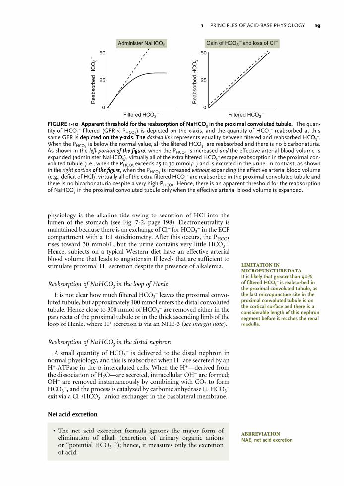
FIGURE1-10 ApparentthresholdforthereabsorptionofNaHCO3intheproximalconvolutedtubule. The quan-tity of HCO3
− filtered (GFR × PHCO3) is depicted on the x-axis, and the quantity of HCO3− reabsorbed at this
same GFR is depicted on the y-axis. Thedepicted on the y-axis. The on the y-axis. The dashed line represents equality between filtered and reabsorbed HCO3−.
When the PHCO3 is below the normal value, all the filtered HCO3− are reabsorbed and there is no bicarbonaturia.
As shown in the left portion of the figureof the figure, when the PHCO3 is increased and the effective arterial blood volume is expanded (administer NaHCO3), virtually all of the extra filtered HCO3
− escape reabsorption in the proximal con-voluted tubule (i.e., when the PHCO3 exceeds 25 to 30 mmol/L) and is excreted in the urine. In contrast, as shown in the right portion of the figureof the figure, when the PHCO3 is increased without expanding the effective arterial blood volume (e.g., deficit of HCl), virtually all of the extra filtered HCO3
− are reabsorbed in the proximal convoluted tubule and there is no bicarbonaturia despite a very high PHCO3. Hence, there is an apparent threshold for the reabsorption of NaHCO3 in the proximal convoluted tubule only when the effective arterial blood volume is expanded.
LiMitAtion in MiCRoPUnCtURe DAtAItislikelythatgreaterthan90%offilteredHCO3
–isreabsorbedintheproximalconvolutedtubule,asthelastmicropuncturesiteintheproximalconvolutedtubuleisonthecorticalsurfaceandthereisaconsiderablelengthofthisnephronsegmentbeforeitreachestherenalmedulla.
ABBReViAtionNAE,netacidexcretion
ACID-BASE20
When faced with an acid or alkali load, the kidney must eliminate the resulting H+ and/or the HCO3
−. There is no bicarbonaturia, because the urine pH is usually close to 6 for most of the 24-hour period; thus, the NAE formula fails to consider the renal component of base balance (see Fig. 1-5). Therefore, this formula must be revised as described in the following equations:
Old version: NAE U U UH PO NH HCO= + −2 4 4 3
Revised version: NAE U U U UH PO NH HCO OA= + − −2 4 4 3
Excretion of ammonium
• For every NH4+ excreted in the urine, one new HCO3
− is added to the body.
The metabolic process that leads to acid balance is described in Figure 1-11. Oxidation of sulfur-containing amino acids in proteins results in a H+ load. These H+ are eliminated after reacting with HCO3
− in the body. The kidneys must replace this deficit of HCO3−
in the body by forming new HCO3− and NH4
+ in a 1:1 stoichiometry and excreting the NH4
+ in the urine to make it an end product of metabolism (see margin note; Fig. 1-12).
Quantities: The usual rate of excretion of NH4+ is 20 to 40 mmol/day.
In chronic acidosis, the kidney can excrete close to 3 mmol NH4+/kg
body weight/day in children and close to 200 mmol/day in adults.
Biochemistry of NH4+ production
The first segment of this pathway has three steps (glutamine entry into mitochondria of the proximal convoluted tubule, its hydrolysis by glutaminase to form glutamate, and the conversion of glutamate
3
Methionine + Glutamine
Proteins
EndogenousExogenous
Reabsorb
NH
2 NH4+ + SO4
2�
H+
+
4
2 HCO3�
2 CO2 + 2 H2O
2
13
+2 H+ SO42�
2 NH4+
NH4+
NH4+
2 HCO3�
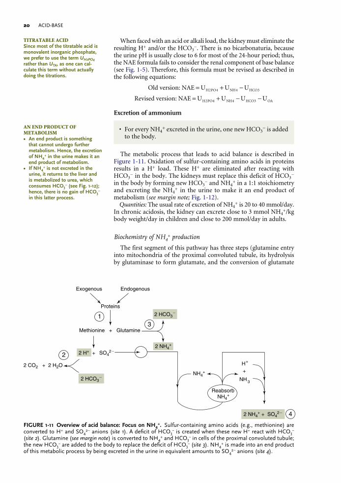
FIGURE1-11 Overviewofacidbalance:FocusonNH4+. Sulfur-containing amino acids (e.g., methionine) are
converted to H+ and SO42- anions (site 1). A deficit of HCO3
− is created when these new H+ react with HCO3−
(site 2). Glutamine (see margin note) is converted to NH4+ and HCO3
− in cells of the proximal convoluted tubule; the new HCO3
− are added to the body to replace the deficit of HCO3− (site 3). NH4
+ is made into an end product of this metabolic process by being excreted in the urine in equivalent amounts to SO4
2- anions (site 4).
An enD PRoDUCt of MetABoLisM • Anendproductissomething
thatcannotundergofurthermetabolism.Hence,theexcretionofNH4
+intheurinemakesitanendproductofmetabolism.
• IfNH4+isnotexcretedinthe
urine,itreturnstotheliverandismetabolizedtourea,whichconsumesHCO3
−(seeFig.1-12);hence,thereisnogainofHCO3
−inthislatterprocess.
titRAtABLe ACiDSincemostofthetitratableacidismonovalentinorganicphosphate,weprefertousethetermUH2PO4ratherthanUTA,asonecancal-culatethistermwithoutactuallydoingthetitrations.
1 : PRINCIPLES OF ACID-BASE PHYSIOLOGY 21
to 2-oxoglutarate2−). The final products are two NH4+ and the
anion 2-oxoglutarate2−. The next segment is the conversion of 2- oxoglutarate2− to the neutral end products CO2 and glucose, which results in the formation of the new HCO3
−. All of these products plus the new HCO3
− are added to the renal venous blood. The function of the last segment of the process is to maintain this gain of new HCO3
−; it is accomplished when NH4
+ is excreted in the urine (see Fig. 1-12).
Signals to augment the oxidation of glutamine in the proximal convoluted tubule
• Because the overall function of this metabolic process is to add new HCO3
− to the body when there is a deficit of HCO3−, it is not
surprising that it is activated when there is a chronic H+ load.
The signal to synthesize more NH4+ (a sustained high concentra-
tion of H+) has to be recognized in a location that is central to the overall process, in cells of the proximal convoluted tubule. Just how intracellular acidosis in the proximal convoluted tubule stimulates the production of NH4
+ and HCO3− is not known with certainty. Possible
mechanisms are listed in the margin (see margin note). Because a high H+ concentration in proximal convoluted tubule cells is also present during chronic hypokalemia, renal ammoniagenesis is augmented in this setting (see margin note). The converse is also true; hyperkalemia is the most common cause of metabolic acidosis owing to a dimin-ished rate of production of NH4
+ because it causes a lower H+ concen-tration (higher pH) in cells of the proximal convoluted tubule.
Selection of fuels for oxidation in the proximal convoluted tubule
• There is a lag period before glutamine is “selected” as the fuel for oxidation so that NH4
+ production does not rise apprecia-bly during acute and transient acidosis.
When there is a large H+ load owing to the overproduction of l-lactic acid, there is no need to augment the oxidation of glutamine to eliminate this H+ load because the l-lactate anion is metabolized
GLUtAMine• Glutamineisthemostabun-
dantaminoacidinproteins;itcanalsobemadeintheliverandskeletalmuscle;hence,itsavailabilityisnotlikelytolimitrenalammoniagenesisexceptinseverelymalnourishedpatients.
• Glutamineisanimportantfuelfortheintestinaltract,anditisalsoaprecursorofreducedglu-tathione,animportantmitochon-drialantioxidant(seeChapter6,page183).
2�Glutamine
ADPReabsorbfiltered Na
ATP
Urea
2-Oxoglutarate + 2 NH4+
2 HCO3�
to body
+
2 NH4+
in urine
Liver
2 NH4+
2 HCO3�
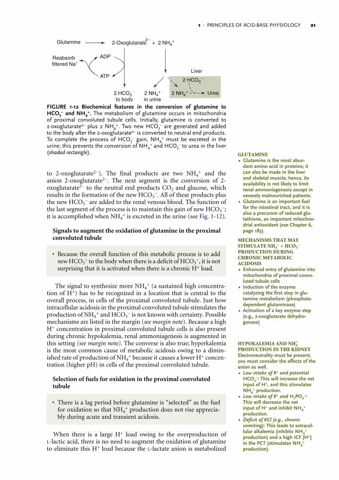
FIGURE 1-12 Biochemical features in the conversion of glutamine toHCO3
−andNH4+. The metabolism of glutamine occurs in mitochondria
of proximal convoluted tubule cells. Initially, glutamine is converted to 2-oxoglutarate2− plus 2 NH4
+. Two new HCO3− are generated and added
to the body after the 2-oxoglutarate2− is converted to neutral end products. To complete the process of HCO3
− gain, NH4+ must be excreted in the
urine; this prevents the conversion of NH4+ and HCO3
− to urea in the liver (shaded rectangle).
MeCHAnisMs tHAt MAY stiMULAte nH4
+ + HCo3−
PRoDUCtion DURinG CHRoniC MetABoLiC ACiDosis• Enhancedentryofglutamineinto
mitochondriaofproximalconvo-lutedtubulecells
• Inductionoftheenzymecatalyzingthefirststepinglu-taminemetabolism(phosphate-dependentglutaminase)
• Activationofakeyenzymestep(e.g.,2-oxoglutaratedehydro-genase)
HYPoKALeMiA AnD nH4+
PRoDUCtion in tHe KiDneYElectroneutralitymustbepresent;youmustconsidertheeffectsoftheanionaswell.• Low intake of K+ and potential
HCO3−:Thiswillincreasethenet
inputofH+,andthisstimulatesNH4
+production.• Low intake of K+ and H2PO4
−:ThiswilldecreasethenetinputofH+andinhibitNH4
+production.
• Deficit of KCl (e.g., chronic vomiting):Thisleadstoextracel-lularalkalemia(inhibitsNH4
+production)andahighICF[H+]inthePCT(stimulatesNH4
+production).
ACID-BASE22
and new HCO3− are produced in a relatively short period of time.
An obvious example of this scenario is the acute and severe l-lactic acidosis during a sprint. Hence, it is not surprising that the proximal convoluted tubule cells continue to oxidize fuels of carbohydrate (l-lactate−) or fat origin (fatty acids) in this setting. This lag period offers a biologic advantage because the excretion of NH4
+ would cause the catabolism of lean body mass to provide the substrate, glutamine, for NH4
+ production during the first days of fasting.
Diminished availability of ADP decreases the conversion of glutamine to NH4
+
• ADP is generated when renal work is performed.
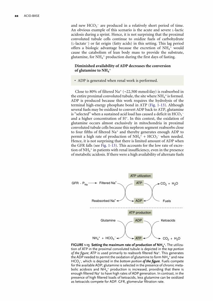
Close to 80% of filtered Na+ (∼22,500 mmol/day) is reabsorbed in the entire proximal convoluted tubule, the site where NH4
+ is formed. ADP is produced because this work requires the hydrolysis of the terminal high-energy phosphate bond in ATP (Fig. 1-13). Although several fuels may be oxidized to convert ADP back to ATP, glutamine is “selected” when a sustained acid load has caused a deficit in HCO3
− and a higher concentration of H+. In this context, the oxidation of glutamine occurs almost exclusively in mitochondria in proximal convoluted tubule cells because this nephron segment reabsorbs close to four fifths of filtered Na+ and thereby generates enough ADP to permit a high rate of production of NH4
+ + HCO3− when needed.
Hence, it is not surprising that there is limited amount of ADP when the GFR falls (see Fig. 1-13). This accounts for the low rate of excre-tion of NH4
+ in patients with renal insufficiency, even in the presence of metabolic acidosis. If there were a high availability of alternate fuels
Glutamine
GFR • PNa
ATP production
ATP utilization
KetoacidsADP
ATP 2 2NH4+ + HCO3
� CO + H O
Filtered Na
Fuels
ATP+
Reabsorbed Na+
2 2CO + H O
ADP
FIGURE1-13 SettingthemaximumrateofproductionofNH4+. The utiliza-
tion of ATP in the proximal convoluted tubule is depicted in the top portion of the figure; ATP is used primarily to reabsorb filtered Na+. This generates the ADP needed to permit the oxidation of glutamine to form NH4
+ and new HCO3
−, which is depicted in the bottom portion of the figureof the figure. Fuels compete for the available ADP; glutamine is selected in the presence of chronic meta-bolic acidosis and NH4
+ production is increased, providing that there is enough filtered Na+ to have high rates of ADP generation. In contrast, in the presence of high filtered loads of ketoacids, less glutamine can be oxidized as ketoacids compete for ADP. GFR, glomerular filtration rate.
1 : PRINCIPLES OF ACID-BASE PHYSIOLOGY 2�
competing with glutamine for this limited amount of ADP, there could be a lower than expected rate of production of NH4
+. One example is the ketoacidosis of chronic starvation where the goal is to have equal excretion rates of NH4
+ and ketoacid anions. This requires a lower rate of excretion of NH4
+ in this setting of metabolic acidosis owing to the oxidation of some of the reabsorbed ketoacids (oxida-tion of β-hydroxybutyrate and glutamine require almost identical amounts of ADP; see Fig. 1-13).
Transport of NH4+ into the lumen of the proximal convoluted
tubule
• In quantitative terms, virtually 100% of the NH4+ to be excreted
is added to the luminal fluid that exits from the proximal con-voluted tubule in the rat with chronic metabolic acidosis.
The major mechanism for the entry of NH4+ into the lumen of
proximal convoluted tubule is by NH4+ replacing H+ on NHE-3, thus
making it a Na+/NH4+ cation exchanger. The remainder of the NH4
+ exits the kidney via the renal vein.
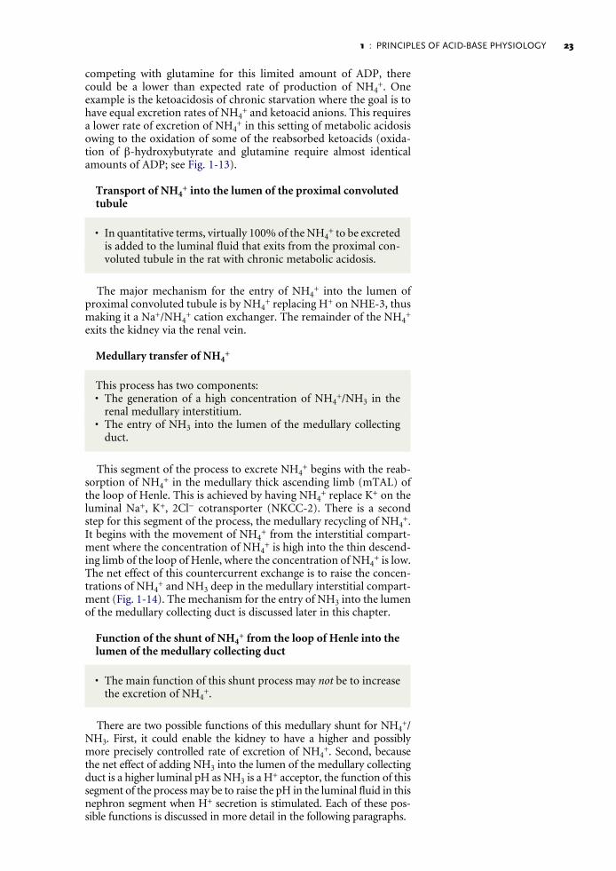
Medullary transfer of NH4+
This process has two components: • The generation of a high concentration of NH4
+/NH3 in the renal medullary interstitium.
• The entry of NH3 into the lumen of the medullary collecting duct.
This segment of the process to excrete NH4+ begins with the reab-
sorption of NH4+ in the medullary thick ascending limb (mTAL) of
the loop of Henle. This is achieved by having NH4+ replace K+ on the
luminal Na+, K+, 2Cl− cotransporter (NKCC-2). There is a second step for this segment of the process, the medullary recycling of NH4
+. It begins with the movement of NH4
+ from the interstitial compart-ment where the concentration of NH4
+ is high into the thin descend-ing limb of the loop of Henle, where the concentration of NH4
+ is low. The net effect of this countercurrent exchange is to raise the concen-trations of NH4
+ and NH3 deep in the medullary interstitial compart-ment (Fig. 1-14). The mechanism for the entry of NH3 into the lumen of the medullary collecting duct is discussed later in this chapter.
Function of the shunt of NH4+ from the loop of Henle into the
lumen of the medullary collecting duct
• The main function of this shunt process may not be to increase the excretion of NH4
+.
There are two possible functions of this medullary shunt for NH4+/
NH3. First, it could enable the kidney to have a higher and possibly more precisely controlled rate of excretion of NH4
+. Second, because the net effect of adding NH3 into the lumen of the medullary collecting duct is a higher luminal pH as NH3 is a H+ acceptor, the function of this segment of the process may be to raise the pH in the luminal fluid in this nephron segment when H+ secretion is stimulated. Each of these pos-sible functions is discussed in more detail in the following paragraphs.
ACID-BASE2�
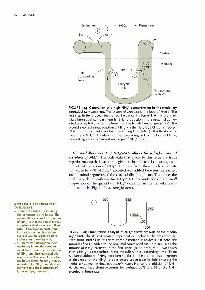
The medullary shunt of NH4+/NH3 allows for a higher rate of
excretion of NH4+. The only data that speak to this issue are from
experiments carried out in rats given a chronic acid load to augment the rate of excretion of NH4
+. The data from these studies indicate that close to 75% of NH4
+ excreted was added between the earliest and terminal segments of the cortical distal nephron. Therefore, the medullary shunt pathway for NH4
+/NH3 accounts for only a small proportion of the quantity of NH4
+ excretion in the rat with meta-bolic acidosis (Fig. 1-15; see margin note).
Na
RecycleNH4
+
Cortex
MedullaNa ,2 Cl
Thindescendinglimb
Glutamine Renal vein
3 2
1
�
+
+
+Competeswith K
NH4+
HCO3�
NH4+
NH4+
FIGURE1-14 GenerationofahighNH4+concentration in themedullary
interstitialcompartment. The U-shaped structure is the loop of Henle. The first step in the process that raises the concentration of NH4
+ in the med-ullary interstitial compartment is NH4
+ production in the proximal convo-luted tubule; NH4
+ enter the lumen on the Na+/H+ exchanger (site 1). The second step is the reabsorption of NH4
+ via the Na+, K+, 2 Cl− cotransporter (NKCC-2) in the medullary thick ascending limb (site 2). The third step is the entry of NH4
+ ultimately into the descending limb of the loop of Henle, completing a countercurrent exchange of NH4
+ (site 3).
1000 333
667
250
750
1000
FIGURE1-15 QuantitativeanalysisofNH4+excretion:Roleofthemedul-
laryshunt. The stylized structure represents a nephron. The data were de-rived from studies in rats with chronic metabolic acidosis. Of note, the amount of NH4
+ added to the proximal convoluted tubule is similar to the amount of NH4
+ excreted in the final urine (1000 nmol/min); two thirds of this NH4
+ is reabsorbed in the medullary thick ascending limb. There is a large addition of NH4
+ into luminal fluid in the cortical distal nephron so that most of the NH4
+ to be excreted are present in fluid entering the medullary collecting duct (see margin note). Hence, the addition of NH4
+ via the medullary shunt accounts for perhaps 20% to 25% of the NH4
+ excreted in these rats.
APPLYinG DAtA fRoM RAts to HUMAns• Thereisadangerinassuming
thatahumanisa70-kgrat.ThemajordifferencefortheexcretionofNH4
+isthatthedietoftheratsupplies10-foldmorealkalithanacid.Therefore,themostimpor-tantacid-basefunctionintheratistoexcreteorganicanionsratherthantoexcreteNH4
+.• Humanswithdamagetotheir
medullaryinterstitialcompart-menthavealowrateofexcretionofNH4
+anddevelopmetabolicacidosisonthisbasis.HencethemedullaryshuntforNH4
+maybeimportantforNH4
+excretioninhumans(seethediscussionofQuestion4-1,page108).

1 : PRINCIPLES OF ACID-BASE PHYSIOLOGY 2�
The medullary shunt of NH4+/NH3 adjusts the
urine pH to close to 6.0
• The renal medullary shunt pathway for NH3 is part of a system that maintains the urine pH close to 6.0, without compromis-ing the ability of the kidney to achieve acid-base balance.
One way to evaluate the function of the medullary NH4+/NH3 shunt
pathway is to observe what happens to the rate of excretion of NH4+
and the urine pH when this NH4+ shunt pathway is inhibited. Blocking
the reabsorption of NH4+ in the loop of Henle in rats with chronic met-
abolic acidosis and a high rate of excretion of NH4+ did decrease the
medullary interstitial concentration of NH4+, but it did not decrease the
rate of excretion of NH4+. Hence, this shunt process does not appear
to be critical for excreting NH4+ in rats. On the other hand, when this
shunt was inhibited, the urine pH declined significantly; this suggests that this medullary shunt pathway may be important to raise the urine pH toward 6.0 by adding NH3 into the inner medullary collecting duct when distal H+ secretion is stimulated by the metabolic acidosis. The secondary effect is to have a small increase in the net addition of NH4
+ to the urine. Therefore, the renal medullary shunt pathway is part of a system that maintains the urine pH close to 6.0, without compromis-ing the role of the kidney in achieving acid-base balance. In fact, the highest rates of NH4
+ excretion in a person with chronic metabolic acidosis occur while the urine pH is close to 6.0 (Fig. 1-16).
Diffusion of NH3 in the renal medulla: A more detailed examination
Historical note. The main process for transport of NH3 into the lumen of the medullary collecting duct has been described as “diffusion trapping.” In essence, distal H+ secretion lowers the urine pH below 6.0, which decreases the NH3 concentration in the lumen of the medul-lary collecting duct to permit more NH3 to diffuse from the medullary interstitial compartment to the lumen of the medullary collecting duct. The question, however, is, “Does the decline in the NH3 concentration
100
05.0 6.0
Urine pH
NH
4+ e
xcre
tion
(µm
ol/m
in)
NH
4+ e
xcre
tion
(µm
ol/m
in)
Acute acidosis
300
1005.06.0
Urine pH
Chronic acidosis
H+ + NH3 → NH4+ H+ + NH3 → NH4
+
FIGURE1-16 UrinepHandtheexcretionofNH4+. In acute acidosis (shown
on the left), distal H+ secretion is stimulated, but there is a lag period be-fore a high rate of ammoniagenesis is achieved. The urine pH is low (tem-porarily), and there is only a modest rise in the rate of NH4
+ excretion. In chronic metabolic acidosis (shown on the right), both the H+ secretory rate and the NH3 availability are greatly increased. The increase in NH3 avail-ability is relatively larger than the increment in H+ secretion. The medullary shunt of NH4
+/NH3 adjusts the urine pH to a value of close to 6.0.
NotethedifferenceinscaleforNH4
+onthey-axisofeachpanel.
ACID-BASE2�
in the lumen of the medullary collecting duct aid the diffusion of NH3 appreciably in the physiologic range of urine pH values (∼ 6.0)?”
Synopsis of diffusion. Diffusion is a passive process. To be effi-cient, a large concentration difference is required for a substance to diffuse, the distance for diffusion must be short enough, and barriers for diffusion must be absent or bypassed. We examine the diffusion of NH3 from the medullary interstitial compartment into the lumen of the medullary collecting duct in this context.1. Concentration difference for NH3: To understand whether distal
H+ secretion influences this concentration difference for NH3 in the medullary collecting duct in a major way, one must examine this process in quantitative terms (see margin note). As shown in Table 1-4, lowering the luminal H+ concentration by distal H+ secretion is potentially important in this context if the urine pH is greater than 6.3. In contrast, lowering the urine pH below 6.3 has a progressively minor additional effect to raise the concentration difference for NH3; hence, it cannot augment the driving force for NH3 diffusion by an appreciable amount. Nevertheless, ongoing H+ secretion must match the addition of NH3 into the lumen of the medullary collecting duct to prevent a rise in the urine pH to a range that could compromise additional diffusion of NH3.
2. Remove the barrier for diffusion of NH3: There are two competing theories concerning the mechanism for the transfer NH3 from the loop of Henle to the lumen of the medullary collecting duct (Fig. 1-17). The important physiologic fact is that the net effect is identical—the entry of NH3 into the luminal fluid of the medul-lary collecting duct (see margin note).
QUESTION
(Discussion on page 36)1-6 What changes would you expect to find in the urine if the transport
system that adds NH3 to the lumen of the inner medullary collecting duct were to “disappear” (be knocked out) in a rodent?
Secretion of H+ in the distal nephron
H+ pumps. H+ pumps are located primarily in the luminal mem-branes of the mitochondria-rich α-intercalated cells (Fig. 1-18). There are two major H+ pumps in this segment, an H+-ATPase (in contrast to the NHE-3 of the proximal nephron) and an H+/K+-ATPase, but only
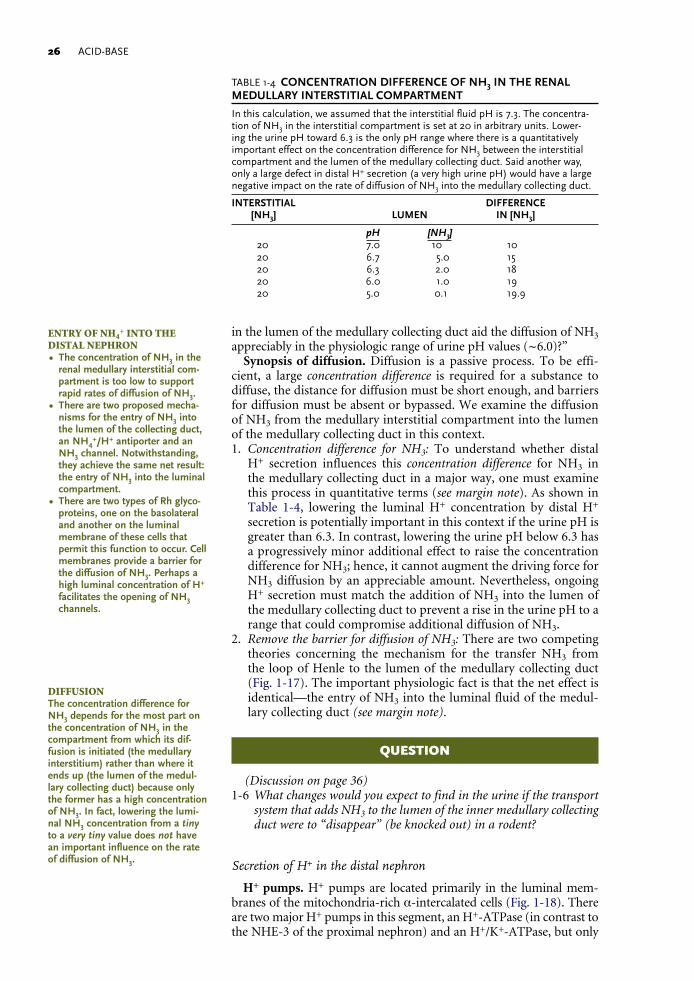
TABLE 1-4 CONCENTRATIONDIFFERENCEOFNH3INTHERENALMEDULLARYINTERSTITIALCOMPARTMENT
In this calculation, we assumed that the interstitial fluid pH is 7.3. The concentra-tion of NH3 in the interstitial compartment is set at 20 in arbitrary units. Lower-ing the urine pH toward 6.3 is the only pH range where there is a quantitatively important effect on the concentration difference for NH3 between the interstitial compartment and the lumen of the medullary collecting duct. Said another way, only a large defect in distal H+ secretion (a very high urine pH) would have a large negative impact on the rate of diffusion of NH3 into the medullary collecting duct.
INTERSTITIAL[NH3] LUMEN
DIFFERENCEIN[NH3]
pH [NH3]20 7.0 10 1020 6.7 5.0 1520 6.3 2.0 1820 6.0 1.0 1920 5.0 0.1 19.9
entRY of nH4+ into tHe
DistAL nePHRon• TheconcentrationofNH3inthe
renalmedullaryinterstitialcom-partmentistoolowtosupportrapidratesofdiffusionofNH3.
• Therearetwoproposedmecha-nismsfortheentryofNH3intothelumenofthecollectingduct,anNH4
+/H+antiporterandanNH3channel.Notwithstanding,theyachievethesamenetresult:theentryofNH3intotheluminalcompartment.
• TherearetwotypesofRhglyco-proteins,oneonthebasolateralandanotherontheluminalmembraneofthesecellsthatpermitthisfunctiontooccur.CellmembranesprovideabarrierforthediffusionofNH3.PerhapsahighluminalconcentrationofH+facilitatestheopeningofNH3channels.
DiffUsionTheconcentrationdifferenceforNH3dependsforthemostpartontheconcentrationofNH3inthecompartmentfromwhichitsdif-fusionisinitiated(themedullaryinterstitium)ratherthanwhereitendsup(thelumenofthemedul-larycollectingduct)becauseonlytheformerhasahighconcentrationofNH3.Infact,loweringthelumi-nalNH3concentrationfromatinytoavery tinyvaluedoesnothaveanimportantinfluenceontherateofdiffusionofNH3.
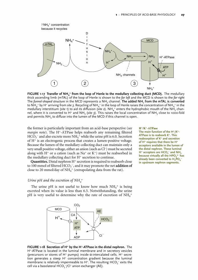
1 : PRINCIPLES OF ACID-BASE PHYSIOLOGY 2�
the former is particularly important from an acid-base perspective (see margin note). The H+-ATPase helps reabsorb any remaining filtered HCO3
− and also excrete more NH4+ while the urine pH is 6.0. Secretion
of H+ is an electrogenic process that creates a lumen-positive voltage. Because the lumen of the medullary collecting duct can maintain only a very small positive voltage, either an anion (such as Cl–) must be secreted along with H+ or a cation (such as Na+ or K+) must be reabsorbed in the medullary collecting duct for H+ secretion to continue.
Quantities. Distal nephron H+ secretion is required to reabsorb close to 100 mmol of filtered HCO3
−, and it may promote the net addition ofaddition of close to 20 mmol/day of NH4
+ (extrapolating data from the rat).
Urine pH and the excretion of NH4+
The urine pH is not useful to know how much NH4+ is being
excreted when its value is less than 6.5. Notwithstanding, the urine pH is very useful to determine why the rate of excretion of NH4
+
Diffusion
+
+
NH3
HNHATP
H+-ATPaseNH3
3
3
+
H
H
mTAL
? NH4+ concentration
because it recycles
NH channels
NH4+ MCD
1 2 3
NH4+
NH4+
FIGURE1-17 TransferofNH4+fromtheloopofHenletothemedullarycollectingduct(MCD). The medullary
thick ascending limb (mTAL) of the loop of Henle is shown to the far left and the MCD is shown to the far right The funnel-shaped structure in the MCD represents a NH3 channel. The added NHThe added NH3 from the mTAL is convertedfrom the mTAL is converted to NH4
+ by H+ arriving from site 3. Recycling of NH4+ in the loop of Henle raises the concentration of NH4
+ in the medullary interstitium (site 1) to aid its diffusion (site 2). NH4
+ enters the hydrophobic mouth of the NH3 chan-nel, where it is converted to H+ and NH3 (site 3). This raises the local concentration of NH3 close to 1000-fold and permits NH3 to diffuse into the lumen of the MCD if this channel is open.
H+/K+-AtPaseThemainfunctionoftheH+/K+-ATPaseistoreabsorbK+.ThisreabsorptionofK+andsecretionofH+requiresthattherebeH+acceptorsavailableinthelumenofthedistalnephron.TheseluminalH+acceptorsareHCO3
−andNH3becausevirtuallyalltheHPO4
2–hasalreadybeenconvertedtoH2PO4
−inupstreamnephronsegments.
Cl
MCD
H
CO
CA
ADP
+
ATP
H+HCO3
�
2
�
AE
FIGURE1-18 SecretionofH+bytheH+-ATPaseinthedistalnephron. The H+-ATPase is located in the luminal membrane and in secretory vesicles (precursors or stores of H+ pumps) inside α-intercalated cells. H+ secre-tion generates a steep H+ concentration gradient because the luminal membrane is relatively impermeable to H+. The resulting HCO3
− exits the cell via a basolateral HCO3
−/Cl− anion exchanger (AE).
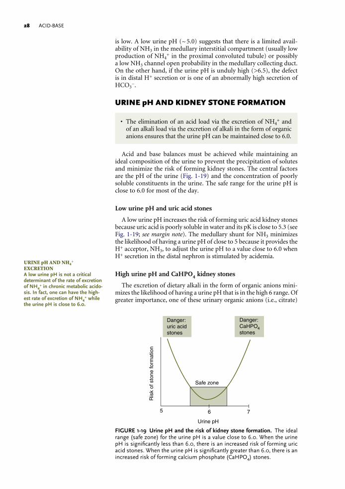
ACID-BASE2�
is low. A low urine pH (~ 5.0) suggests that there is a limited avail-ability of NH3 in the medullary interstitial compartment (usually low production of NH4
+ in the proximal convoluted tubule) or possibly a low NH3 channel open probability in the medullary collecting duct. On the other hand, if the urine pH is unduly high (>6.5), the defect is in distal H+ secretion or is one of an abnormally high secretion of HCO3
−.
URINE pH AND KIDNEY STONE FORMATION
• The elimination of an acid load via the excretion of NH4+ and
of an alkali load via the excretion of alkali in the form of organic anions ensures that the urine pH can be maintained close to 6.0.
Acid and base balances must be achieved while maintaining an ideal composition of the urine to prevent the precipitation of solutes and minimize the risk of forming kidney stones. The central factors are the pH of the urine (Fig. 1-19) and the concentration of poorly soluble constituents in the urine. The safe range for the urine pH is close to 6.0 for most of the day.
Low urine pH and uric acid stones
A low urine pH increases the risk of forming uric acid kidney stones because uric acid is poorly soluble in water and its pK is close to 5.3 (see Fig. 1-19; see margin note). The medullary shunt for NH3 minimizes the likelihood of having a urine pH of close to 5 because it provides the H+ acceptor, NH3, to adjust the urine pH to a value close to 6.0 when H+ secretion in the distal nephron is stimulated by acidemia.
High urine pH and CaHPO4 kidney stones
The excretion of dietary alkali in the form of organic anions mini-mizes the likelihood of having a urine pH that is in the high 6 range. Of greater importance, one of these urinary organic anions (i.e., citrate)
5
4
Urine pH
Safe zone
Danger:uric acidstones
Danger:CaHPOstones
Ris
k of
sto
ne fo
rmat
ion
6 7
FIGURE1-19 UrinepHandtheriskofkidneystoneformation. The ideal range (safe zone) for the urine pH is a value close to 6.0. When the urine pH is significantly less than 6.0, there is an increased risk of forming uric acid stones. When the urine pH is significantly greater than 6.0, there is an increased risk of forming calcium phosphate (CaHPO4) stones.
URine pH AnD nH4+
eXCRetionAlowurinepHisnotacriticaldeterminantoftherateofexcretionofNH4
+inchronicmetabolicacido-sis.Infact,onecanhavethehigh-estrateofexcretionofNH4
+whiletheurinepHiscloseto6.0.
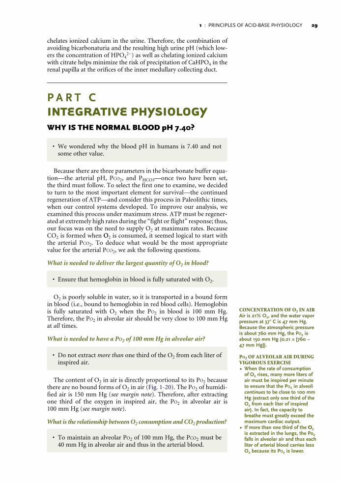
1 : PRINCIPLES OF ACID-BASE PHYSIOLOGY 2�
chelates ionized calcium in the urine. Therefore, the combination of avoiding bicarbonaturia and the resulting high urine pH (which low-ers the concentration of HPO4
2−) as well as chelating ionized calcium with citrate helps minimize the risk of precipitation of CaHPO4 in the renal papilla at the orifices of the inner medullary collecting duct.
P A R T CINTEGRATIVE PHYSIOLOGYWHY IS THE NORMAL BLOOD pH �.�0?
• We wondered why the blood pH in humans is 7.40 and not some other value.
Because there are three parameters in the bicarbonate buffer equa-tion—the arterial pH, Pco2, and PHCO3—once two have been set, the third must follow. To select the first one to examine, we decided to turn to the most important element for survival—the continued regeneration of ATP—and consider this process in Paleolithic times, when our control systems developed. To improve our analysis, we examined this process under maximum stress. ATP must be regener-ated at extremely high rates during the “fight or flight” response; thus, our focus was on the need to supply O2 at maximum rates. Because CO2 is formed when OO2 is consumed, it seemed logical to start with the arterial Pco2. To deduce what would be the most appropriate value for the arterial Pco2, we ask the following questions.
What is needed to deliver the largest quantity of O2 in blood?
• Ensure that hemoglobin in blood is fully saturated with O2.
O2 is poorly soluble in water, so it is transported in a bound form in blood (i.e., bound to hemoglobin in red blood cells). Hemoglobin is fully saturated with O2 when the Po2 in blood is 100 mm Hg. Therefore, the Po2 in alveolar air should be very close to 100 mm Hg at all times.
What is needed to have a Po2 of 100 mm Hg in alveolar air?
• Do not extract more than one third of the O2 from each liter of inspired air.
The content of O2 in air is directly proportional to its Po2 because there are no bound forms of O2 in air (Fig. 1-20). The Po2 of humidi-fied air is 150 mm Hg (see margin note). Therefore, after extracting one third of the oxygen in inspired air, the Po2 in alveolar air is 100 mm Hg (see margin note).
What is the relationship between O2 consumption and CO2 production?
• To maintain an alveolar Po2 of 100 mm Hg, the Pco2 must be 40 mm Hg in alveolar air and thus in the arterial blood.
ConCentRAtion of o2 in AiRAiris21%O2,andthewatervaporpressureat37°Cis47mmHg.Becausetheatmosphericpressureisabout760mmHg,thePo2isabout150mmHg(0.21×[760−47mmHg]).
Po2 of ALVeoLAR AiR DURinG ViGoRoUs eXeRCise• Whentherateofconsumption
ofO2rises,manymorelitersofairmustbeinspiredperminutetoensurethatthePo2inalveolicontinuestobecloseto100mmHg(extractonlyonethirdoftheO2fromeachliterofinspiredair).Infact,thecapacitytobreathemustgreatlyexceedthemaximumcardiacoutput.
• IfmorethanonethirdoftheOO2isextractedinthelungs,thePo2fallsinalveolarairandthuseachliterofarterialbloodcarrieslessO2becauseitsPo2islower.
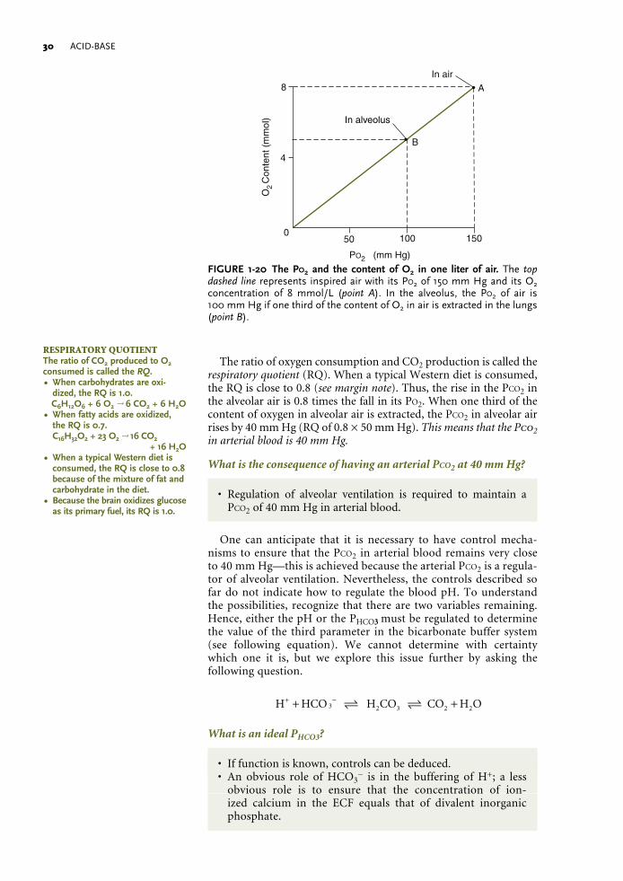
ACID-BASE�0
The ratio of oxygen consumption and CO2 production is called the respiratory quotient (RQ). When a typical Western diet is consumed, the RQ is close to 0.8 (see margin note). Thus, the rise in the Pco2 in the alveolar air is 0.8 times the fall in its Po2. When one third of the content of oxygen in alveolar air is extracted, the Pco2 in alveolar air rises by 40 mm Hg (RQ of 0.8 × 50 mm Hg). This means that the Pco2 in arterial blood is 40 mm Hg.
What is the consequence of having an arterial Pco2 at 40 mm Hg?
• Regulation of alveolar ventilation is required to maintain a Pco2 of 40 mm Hg in arterial blood.
One can anticipate that it is necessary to have control mecha-nisms to ensure that the Pco2 in arterial blood remains very close to 40 mm Hg—this is achieved because the arterial Pco2 is a regula-tor of alveolar ventilation. Nevertheless, the controls described so far do not indicate how to regulate the blood pH. To understand the possibilities, recognize that there are two variables remaining. Hence, either the pH or the PHCO33 must be regulated to determine the value of the third parameter in the bicarbonate buffer system (see following equation). We cannot determine with certainty which one it is, but we explore this issue further by asking the following question.
H HCO H CO CO H O3+ −+ + 2 3 2 2
What is an ideal PHCO3?
• If function is known, controls can be deduced. • An obvious role of HCO3
− is in the buffering of H+; a less obvious role is to ensure that the concentration of ion-ized calcium in the ECF equals that of divalent inorganic phosphate.
ResPiRAtoRY QUotientTheratioofCO2producedtoO2consumediscalledtheRQ.• Whencarbohydratesareoxi-
dized,theRQis1.0.C6H12O6+6O2→6CO2+6H2O
• Whenfattyacidsareoxidized,theRQis0.7.C16H32O2+23O2→16CO2
+16H2O• WhenatypicalWesterndietis
consumed,theRQiscloseto0.8becauseofthemixtureoffatandcarbohydrateinthediet.
• Becausethebrainoxidizesglucoseasitsprimaryfuel,itsRQis1.0.
•
• A
B
In air
In alveolus
O C
onte
nt (
mm
ol)
8
4
050 100 150
PO2 (mm Hg)2
FIGURE 1-20 The Po2 and the content of O2 in one liter of air. The top dashed line represents inspired air with its Po2 of 150 mm Hg and its O2 concentration of 8 mmol/L (point A). In the alveolus, the Po2 of air is 100 mm Hg if one third of the content of O2 in air is extracted in the lungs (point B).
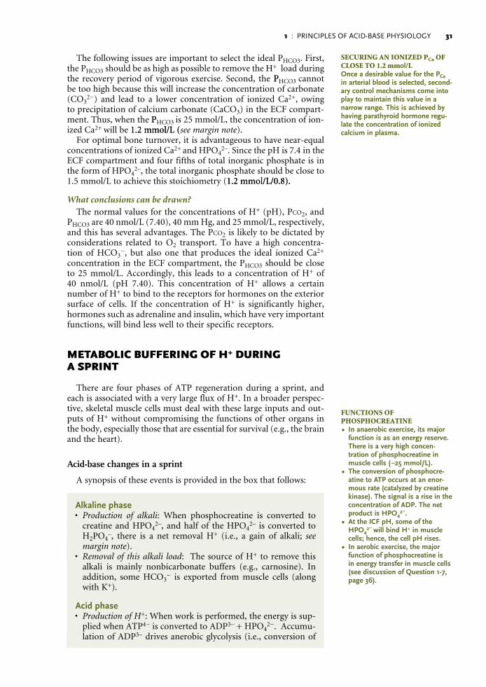
1 : PRINCIPLES OF ACID-BASE PHYSIOLOGY �1
The following issues are important to select the ideal PHCO3. First, the PHCO3 should be as high as possible to remove the H+ load during the recovery period of vigorous exercise. Second, the PPHCO3
cannot be too high because this will increase the concentration of carbonate (CO3
2−) and lead to a lower concentration of ionized Ca2+, owing to precipitation of calcium carbonate (CaCO3) in the ECF compart-ment. Thus, when the PPHCO3
is 25 mmol/L, the concentration of ion-ized Ca2+ will be 1.2 mmol/L (1.2 mmol/L (see margin note).
For optimal bone turnover, it is advantageous to have near-equal concentrations of ionized Ca2+ and HPO4
2–. Since the pH is 7.4 in the ECF compartment and four fifths of total inorganic phosphate is in the form of HPO4
2–, the total inorganic phosphate should be close to 1.5 mmol/L to achieve this stoichiometry (1.2 mmol/L/0.8).1.2 mmol/L/0.8).
What conclusions can be drawn?The normal values for the concentrations of H+ (pH), Pco2, and
PHCO3 are 40 nmol/L (7.40), 40 mm Hg, and 25 mmol/L, respectively, and this has several advantages. The Pco2 is likely to be dictated by considerations related to O2 transport. To have a high concentra-tion of HCO3
−, but also one that produces the ideal ionized Ca2+ concentration in the ECF compartment, the PHCO3 should be close to 25 mmol/L. Accordingly, this leads to a concentration of H+ of 40 nmol/L (pH 7.40). This concentration of H+ allows a certain number of H+ to bind to the receptors for hormones on the exterior surface of cells. If the concentration of H+ is significantly higher, hormones such as adrenaline and insulin, which have very important functions, will bind less well to their specific receptors.
METABOLIC BUFFERING OF H+ DURING A SPRINT
There are four phases of ATP regeneration during a sprint, and each is associated with a very large flux of H+. In a broader perspec-tive, skeletal muscle cells must deal with these large inputs and out-puts of H+ without compromising the functions of other organs in the body, especially those that are essential for survival (e.g., the brain and the heart).
Acid-base changes in a sprint
A synopsis of these events is provided in the box that follows:
Alkalinephase • Production of alkali: When phosphocreatine is converted to
creatine and HPO42–, and half of the HPO4
2– is converted to H2PO4
–, there is a net removal H+ (i.e., a gain of alkali; see margin note).
• Removal of this alkali load: The source of H+ to remove this alkali is mainly nonbicarbonate buffers (e.g., carnosine). In addition, some HCO3
− is exported from muscle cells (along with K+).
Acidphase • Production of H+: When work is performed, the energy is sup-
plied when ATP4− is converted to ADP3− + HPO42−. Accumu-
lation of ADP3– drives anerobic glycolysis (i.e., conversion of
seCURinG An ioniZeD PCa of CLose to 1.2 mmol/LOnceadesirablevalueforthePCainarterialbloodisselected,second-arycontrolmechanismscomeintoplaytomaintainthisvalueinanarrowrange.Thisisachievedbyhavingparathyroidhormoneregu-latetheconcentrationofionizedcalciuminplasma.
fUnCtions of PHosPHoCReAtine• Inanaerobicexercise,itsmajor
functionisasanenergyreserve.Thereisaveryhighconcen-trationofphosphocreatineinmusclecells(∼25mmol/L).
• Theconversionofphosphocre-atinetoATPoccursatanenor-mousrate(catalyzedbycreatinekinase).ThesignalisariseintheconcentrationofADP.ThenetproductisHPO4
2–.• AttheICFpH,someofthe
HPO42–willbindH+inmuscle
cells;hence,thecellpHrises.• Inaerobicexercise,themajor
functionofphosphocreatineisinenergytransferinmusclecells(seediscussionofQuestion1-7,page36).
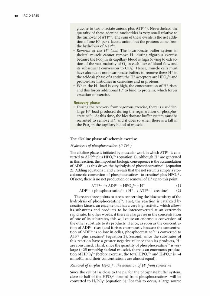
ACID-BASE�2
glucose to two l-lactate anions plus ATP4−). Nevertheless, the quantity of these adenine nucleotides is very small relative to the turnover of ATP4−. The sum of these events is the net addi-tion of one H+ per l-lactate anion, but the protons come from the hydrolysis of ATP4−.
• Removal of the H+ load: The bicarbonate buffer system in skeletal muscle cannot remove H+ during vigorous exercise because the Pco2 in its capillary blood is high (owing to extrac-tion of the vast majority of O2 in each liter of blood flow and its subsequent conversion to CO2). Hence, muscle cells must have abundant nonbicarbonate buffers to remove these H+ in the acidosis phase of a sprint; the H+ acceptors are HPO4
2– and proton-free histidines in carnosine and in proteins.
• When the H+ load is very high, the concentration of H+ rises, and this forces additional H+ to bind to proteins, which forces cessation of exercise.
Recoveryphase • During the recovery from vigorous exercise, there is a sudden,
large H+ load produced during the regeneration of phospho-creatine2–. At this time, the bicarbonate buffer system must be recruited to remove H+, and it does so when there is a fall in the Pco2 in the capillary blood of muscle.
The alkaline phase of ischemic exercise
Hydrolysis of phosphocreatine (P-Cr2–)
The alkaline phase is initiated by muscular work in which ATP4– is con-verted to ADP3– plus HPO4
2– (equation 1). Although H+ are generated in this reaction, the important biologic consequence is the accumulation of ADP3–, as this drives the hydrolysis of phosphocreatine2– (equation 2). Adding equations 1 and 2 reveals that the net result is simply a stoi-chiometric conversion of phosphocreatine2– to creatine0 plus HPO4
2–. Of note, there is no net production or removal of H+ up to this point.
ATP ADP HPO H4 34
2− − − +→ + + (1)
ADP phosphocreatine H ATP creatine3 2 4 0− − + −+ + → + (2)
There are three points to stress concerning the biochemistry of the hydrolysis of phosphocreatine2–. First, the reaction is catalyzed by creatine kinase, an enzyme that has a very high activity, which allows its substrates and products to be interconverted at an extremely rapid rate. In other words, if there is a large rise in the concentration of one of its substrates, this will cause an enormous conversion of the other substrate to its products. Hence, as soon as the concentra-tion of ADP3– rises (and it rises enormously because the concentra-tion of ADP3– is so low in cells), phosphocreatine2– is converted to ATP4– plus creatine0 (equation 2). Second, since the substrates of this reaction have a greater negative valence than its products, H+ are consumed. Third, since the quantity of phosphocreatine2– is very large (~25 mmol/kg skeletal muscle), there is an enormous produc-tion of HPO4
2– (before exercise, the total HPO42– and H2PO4
– is ~4 mmol/L, and their concentrations are almost equal).
Removal of surplus HPO42–, the donation of H+ from carnosine
Since the cell pH is close to the pK for the phosphate buffer system, close to half of the HPO4
2– formed from phosphocreatine2– will be converted to H2PO4
– (equation 3). For this to occur, a large source
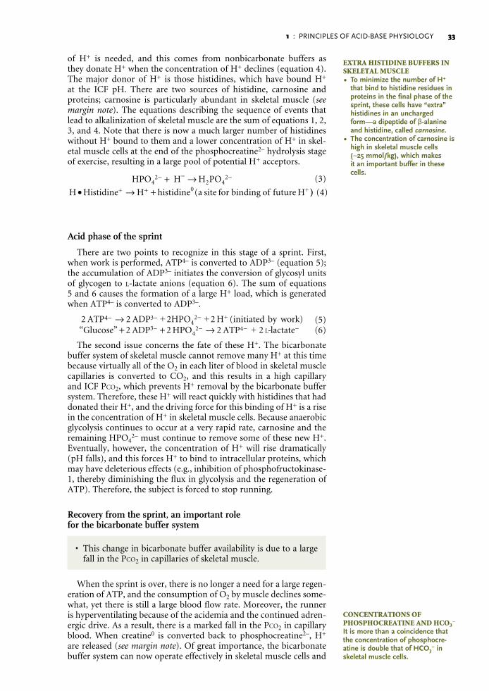
1 : PRINCIPLES OF ACID-BASE PHYSIOLOGY ��
of H+ is needed, and this comes from nonbicarbonate buffers as they donate H+ when the concentration of H+ declines (equation 4). The major donor of H+ is those histidines, which have bound H+ at the ICF pH. There are two sources of histidine, carnosine and proteins; carnosine is particularly abundant in skeletal muscle (see margin note). The equations describing the sequence of events that lead to alkalinization of skeletal muscle are the sum of equations 1, 2, 3, and 4. Note that there is now a much larger number of histidines without H+ bound to them and a lower concentration of H+ in skel-etal muscle cells at the end of the phosphocreatine2– hydrolysis stage of exercise, resulting in a large pool of potential H+ acceptors.
HPO H PO4 2 42 2− − −+ → H (3)
H Histidine H histidine (a site for binding of future H0• → +++ + )) (4)
Acid phase of the sprint
There are two points to recognize in this stage of a sprint. First, when work is performed, ATP4– is converted to ADP3– (equation 5); the accumulation of ADP3– initiates the conversion of glycosyl units of glycogen to l-lactate anions (equation 6). The sum of equations 5 and 6 causes the formation of a large H+ load, which is generated when ATP4– is converted to ADP3–.
2 ATP 2 ADP 2HPO 2 H (initiated by work)34
24 ++ +− − −→ (5)“Glucose” 2 ADP 2 HPO 2 ATP 2 -lactate3
42 4 L+ + →− − − − + (6)
The second issue concerns the fate of these H+. The bicarbonate buffer system of skeletal muscle cannot remove many H+ at this time because virtually all of the O2 in each liter of blood in skeletal muscle capillaries is converted to CO2, and this results in a high capillary and ICF Pco2, which prevents H+ removal by the bicarbonate buffer system. Therefore, these H+ will react quickly with histidines that had donated their H+, and the driving force for this binding of H+ is a rise in the concentration of H+ in skeletal muscle cells. Because anaerobic glycolysis continues to occur at a very rapid rate, carnosine and the remaining HPO4
2– must continue to remove some of these new H+. Eventually, however, the concentration of H+ will rise dramatically (pH falls), and this forces H+ to bind to intracellular proteins, which may have deleterious effects (e.g., inhibition of phosphofructokinase-1, thereby diminishing the flux in glycolysis and the regeneration of ATP). Therefore, the subject is forced to stop running.
Recovery from the sprint, an important role for the bicarbonate buffer system
• This change in bicarbonate buffer availability is due to a large fall in the Pco2 in capillaries of skeletal muscle.
When the sprint is over, there is no longer a need for a large regen-eration of ATP, and the consumption of O2 by muscle declines some-what, yet there is still a large blood flow rate. Moreover, the runner is hyperventilating because of the acidemia and the continued adren-ergic drive. As a result, there is a marked fall in the Pco2 in capillary blood. When creatine0 is converted back to phosphocreatine2–, H+ are released (see margin note). Of great importance, the bicarbonate buffer system can now operate effectively in skeletal muscle cells and
eXtRA HistiDine BUffeRs in sKeLetAL MUsCLe• TominimizethenumberofH+
thatbindtohistidineresiduesinproteinsinthefinalphaseofthesprint,thesecellshave“extra”histidinesinanunchargedform—adipeptideofβ-alanineandhistidine,calledcarnosine.
• Theconcentrationofcarnosineishighinskeletalmusclecells(∼25mmol/kg),whichmakesitanimportantbufferinthesecells.
ConCentRAtions of PHosPHoCReAtine AnD HCo3
−
Itismorethanacoincidencethattheconcentrationofphosphocre-atineisdoublethatofHCO3
−inskeletalmusclecells.
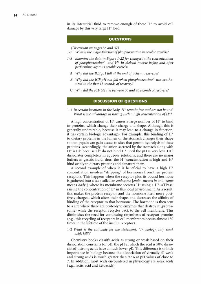
ACID-BASE��
in its interstitial fluid to remove enough of these H+ to avoid cell damage by this very large H+ load.
QUESTIONS
(Discussion on pages 36 and 37)17 What is the major function of phosphocreatine in aerobic exercise?
18 Examine the data in Figure 122 for changes in the concentrations of phosphocreatine2− and H+ in skeletal muscle before and after performing vigorous aerobic exercise.
A Why did the ICF pH fall at the end of ischemic exercise?
B Why did the ICF pH not fall when phosphocreatine2– was synthesized in the first 15 seconds of recovery?
C Why did the ICF pH rise between 30 and 45 seconds of recovery?
DISCUSSION OF QUESTIONS
1-1 In certain locations in the body, H+ remain free and are not bound. What is the advantage in having such a high concentration of H+?
A high concentration of H+ causes a large number of H+ to bind to proteins, which change their charge and shape. Although this is generally undesirable, because it may lead to a change in function, it has certain biologic advantages. For example, this binding of H+ to dietary proteins in the lumen of the stomach changes their shape so that pepsin can gain access to sites that permit hydrolysis of these proteins. Accordingly, the anion secreted by the stomach along with H+ is Cl− because Cl− do not bind H+ until the pH is very low. HCl dissociates completely in aqueous solutions, and there are no major buffers in gastric fluid; thus, the H+ concentration is high and H+ bind avidly to dietary proteins and denature them.
A second example of when it is beneficial to have a high H+ concentration involves “stripping” of hormones from their protein receptors. This happens when the receptor plus its bound hormone is gathered into a sac (called an endosome [endo means in and some means body]) where its membrane secretes H+ using a H+-ATPase, raising the concentration of H+ in this local environment. As a result, this makes the protein receptor and the hormone itself more posi-tively charged, which alters their shape, and decreases the affinity of binding of the receptor to that hormone. The hormone is then sent to a site where there are proteolytic enzymes that destroy it (protea-some) while the receptor recycles back to the cell membrane. This diminishes the need for continuing resynthesis of receptor proteins (e.g., this recycling of receptors in cell membranes occurs almost 180 times in the lifetime of the insulin receptor).
1-2 What is the rationale for the statement, “In biology only weak acids kill”?
Chemistry books classify acids as strong or weak based on their dissociation constants (or pK, the pH at which the acid is 50% disso-ciated); strong acids have a much lower pK. This difference is of little importance in biology because the dissociation of virtually all weak and strong acids is much greater than 99% at pH values of close to 7. In addition, most acids encountered in physiology are weak acids (e.g., lactic acid and ketoacids).
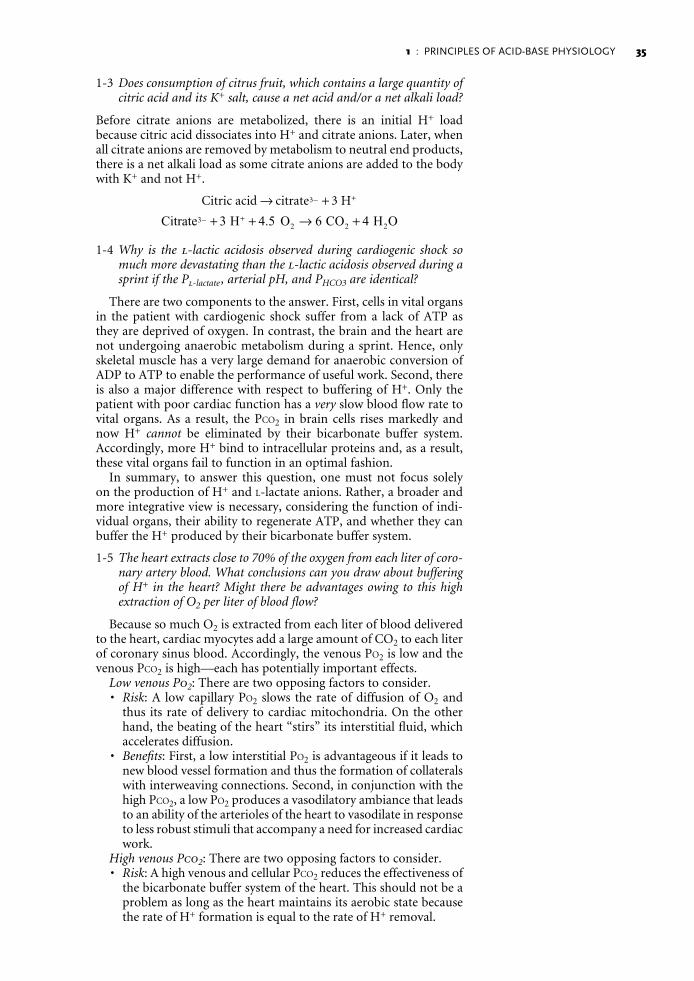
1 : PRINCIPLES OF ACID-BASE PHYSIOLOGY ��
1-3 Does consumption of citrus fruit, which contains a large quantity of citric acid and its K+ salt, cause a net acid and/or a net alkali load?
Before citrate anions are metabolized, there is an initial H+ load because citric acid dissociates into H+ and citrate anions. Later, when all citrate anions are removed by metabolism to neutral end products, there is a net alkali load as some citrate anions are added to the body with K+ and not H+.
Citric acid citrate 3 H→ +− +3
Citrate H O CO H O32 2 23 4 5 6 4− ++ + → +.
1-4 Why is the llactic acidosis observed during cardiogenic shock so much more devastating than the llactic acidosis observed during a sprint if the Pllactate , arterial pH, and PHCO3 are identical?
There are two components to the answer. First, cells in vital organs in the patient with cardiogenic shock suffer from a lack of ATP as they are deprived of oxygen. In contrast, the brain and the heart are not undergoing anaerobic metabolism during a sprint. Hence, only skeletal muscle has a very large demand for anaerobic conversion of ADP to ATP to enable the performance of useful work. Second, there is also a major difference with respect to buffering of H+. Only the patient with poor cardiac function has a very slow blood flow rate to vital organs. As a result, the Pco2 in brain cells rises markedly and now H+ cannot be eliminated by their bicarbonate buffer system. Accordingly, more H+ bind to intracellular proteins and, as a result, these vital organs fail to function in an optimal fashion.
In summary, to answer this question, one must not focus solely on the production of H+ and l-lactate anions. Rather, a broader and more integrative view is necessary, considering the function of indi-vidual organs, their ability to regenerate ATP, and whether they can buffer the H+ produced by their bicarbonate buffer system.
1-5 The heart extracts close to 70% of the oxygen from each liter of coronary artery blood. What conclusions can you draw about buffering of H+ in the heart? Might there be advantages owing to this high extraction of O2 per liter of blood flow?
Because so much O2 is extracted from each liter of blood delivered to the heart, cardiac myocytes add a large amount of CO2 to each liter of coronary sinus blood. Accordingly, the venous Po2 is low and the venous Pco2 is high—each has potentially important effects.
Low venous Po2: There are two opposing factors to consider. • Risk: A low capillary Po2 slows the rate of diffusion of O2 and
thus its rate of delivery to cardiac mitochondria. On the other hand, the beating of the heart “stirs” its interstitial fluid, which accelerates diffusion.
• Benefits: First, a low interstitial Po2 is advantageous if it leads to new blood vessel formation and thus the formation of collaterals with interweaving connections. Second, in conjunction with the high Pco2, a low Po2 produces a vasodilatory ambiance that leads to an ability of the arterioles of the heart to vasodilate in response to less robust stimuli that accompany a need for increased cardiac work.
High venous Pco2: There are two opposing factors to consider. • Risk: A high venous and cellular Pco2 reduces the effectiveness of
the bicarbonate buffer system of the heart. This should not be a problem as long as the heart maintains its aerobic state because the rate of H+ formation is equal to the rate of H+ removal.
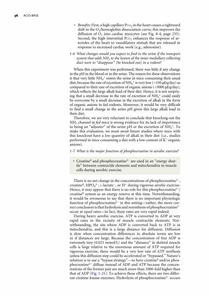
ACID-BASE��
• Benefits: First, a high capillary Pco2 in the heart causes a rightward shift in the O2/hemoglobin dissociation curve; this improves the diffusion of O2 into cardiac myocytes (see Fig. 8-4, page 235). Second, the high interstitial Pco2 enhances the response of ar-terioles of the heart to vasodilatory stimuli that are released in response to increased cardiac work (e.g., adenosine).
1-6 What changes would you expect to find in the urine if the transport system that adds NH3 to the lumen of the inner medullary collecting duct were to “disappear” (be knocked out) in a rodent?
When this experiment was performed, there was little if any change in the pH in the blood or in the urine. The reason for these observations is that very little NH4
+ enters the urine in mice consuming their usual diet, because the rate of excretion of NH4
+ is very low (∼150 μEq/day) as compared to their rate of excretion of organic anions (∼3000 μEq/day), which reflects the large alkali load of their diet. Hence, it is not surpris-ing that a small decrease in the rate of excretion of NH4
+ could easily be overcome by a small decrease in the excretion of alkali in the form of organic anions in fed rodents. Moreover, it would be very difficult to find a small change in the urine pH given this large alkali load in their diet.
Therefore, we are very reluctant to conclude that knocking out the NH3 channel in fed mice is strong evidence for its lack of importance in being an “adjuster” of the urine pH or the excretion of NH4
+. To make this evaluation, we must await future studies where mice with this knockout have a low quantity of alkali in their diet (i.e., studies performed in mice consuming a diet with a low content of K+ organic anions).
1-7 What is the major function of phosphocreatine in aerobic exercise?
• Creatine0 and phosphocreatine2− are used in an “energy shut-tle” between contractile elements and mitochondria in muscle cells during aerobic exercise.
There is no net change in the concentrations of phosphocreatine2−, creatine0, HPO4
2–, l-lactate−, or H+ during vigorous aerobic exercise. Hence, it may appear that there is no role for this phosphocreatine2−/creatine0 system as an energy reserve at this time. Notwithstanding, it would be erroneous to say that there is no important physiologic function of phosphocreatine2− in this setting—rather, the more cor-rect conclusion is that hydrolysis and resynthesis of phosphocreatine2− occur at equal rates—in fact, these rates are very rapid indeed.
During heavy aerobic exercise, ATP is converted to ADP at very rapid rates in the vicinity of muscle contractile elements. Not-withstanding, the site where ADP is converted back to ATP is in mitochondria, and this is a large distance for diffusion. Diffusion is slow when concentration differences in absolute terms are low or if distances are large. Because the concentration of free ADP is extremely tiny (0.025 mmol/L) and the “distance” in skeletal muscle cells is large relative to the enormous amount of ATP required for vigorous exercise, there would be a very low rate of ATP synthesis unless this diffusion step could be accelerated or “bypassed.” Nature’s solution is to use a “bypass strategy”—to have creatine0 and/or phos-phocreatine2− diffuse instead of ADP and ATP because the concen-trations of the former pair are much more than 1000-fold higher than that of ADP (Fig. 1-21). To achieve these effects, there are two differ-ent creatine kinase enzymes. Hydrolysis of phosphocreatine2− occurs
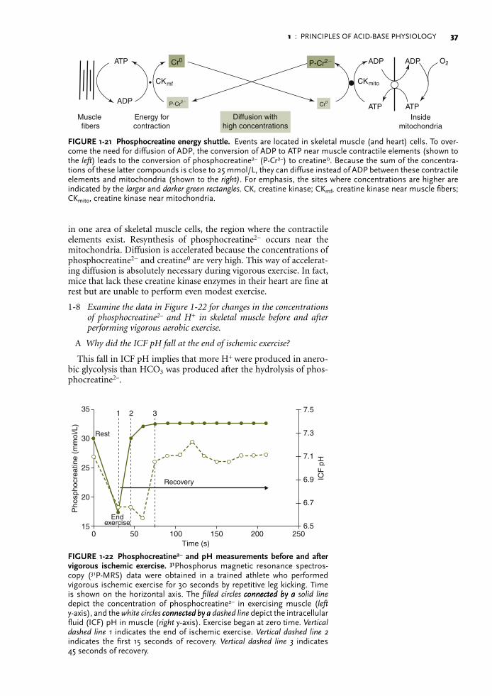
1 : PRINCIPLES OF ACID-BASE PHYSIOLOGY ��
in one area of skeletal muscle cells, the region where the contractile elements exist. Resynthesis of phosphocreatine2− occurs near the mitochondria. Diffusion is accelerated because the concentrations of phosphocreatine2− and creatine0 are very high. This way of accelerat-ing diffusion is absolutely necessary during vigorous exercise. In fact, mice that lack these creatine kinase enzymes in their heart are fine at rest but are unable to perform even modest exercise.
1-8 Examine the data in Figure 122 for changes in the concentrations of phosphocreatine2– and H+ in skeletal muscle before and after performing vigorous aerobic exercise.
A Why did the ICF pH fall at the end of ischemic exercise?
This fall in ICF pH implies that more H+ were produced in anero-bic glycolysis than HCO3 was produced after the hydrolysis of phos-phocreatine2–.
• •ADP
ATP ATP
ADP ADP OATP
P-Cr2� Cr0
CK CK
Cr0 P-Cr2�
Diffusion withhigh concentrations
Insidemitochondria
Muscle fibers
Energy forcontraction
2
mf mito
FIGURE1-21 Phosphocreatineenergyshuttle. Events are located in skeletal muscle (and heart) cells. To over-come the need for diffusion of ADP, the conversion of ADP to ATP near muscle contractile elements (shown to the left) leads to the conversion of phosphocreatine2− (P-Cr2−) to creatine0. Because the sum of the concentra-tions of these latter compounds is close to 25 mmol/L, they can diffuse instead of ADP between these contractile elements and mitochondria (shown to the right). For emphasis, the sites where concentrations are higher are indicated by the larger and darker green rectangles. CK, creatine kinase; CKmf, creatine kinase near muscle fibers; CKmito, creatine kinase near mitochondria.
35 1 2 3
Recovery
Endexercise
Rest
ICF
pH
Pho
spho
crea
tine
(mm
ol/L
)
30
25
20
150 50 100 150 200
Time (s)
7.5
7.3
7.1
6.9
6.7
6.5250
FIGURE1-22 Phosphocreatine2–andpHmeasurementsbeforeandaftervigorous ischemic exercise. 31Phosphorus magnetic resonance spectros-copy (31P-MRS) data were obtained in a trained athlete who performed vigorous ischemic exercise for 30 seconds by repetitive leg kicking. Time is shown on the horizontal axis. The filled circles connected by aconnected by a solid line depict the concentration of phosphocreatine2– in exercising muscle (left y-axis), and the white circles connected by aconnected by a dashed line depict the intracellular fluid (ICF) pH in muscle (right y-axis). Exercise began at zero time. Vertical dashed line 1 indicates the end of ischemic exercise. Vertical dashed line 2 indicates the first 15 seconds of recovery. Vertical dashed line 3 indicates 45 seconds of recovery.
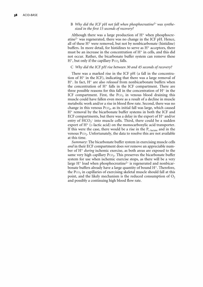
ACID-BASE��
B Why did the ICF pH not fall when phosphocreatine2– was synthesized in the first 15 seconds of recovery?
Although there was a large production of H+ when phosphocre-atine2– was regenerated, there was no change in the ICF pH. Hence, all of these H+ were removed, but not by nonbicarbonate (histidine) buffers. In more detail, for histidines to serve as H+ acceptors, there must be an increase in the concentration of H+ in cells, and this did not occur. Rather, the bicarbonate buffer system can remove these H+, but only if the capillary Pco2 falls.
C Why did the ICF pH rise between 30 and 45 seconds of recovery?
There was a marked rise in the ICF pH (a fall in the concentra-tion of H+ in the ICF), indicating that there was a large removal of H+. In fact, H+ are also released from nonbicarbonate buffers when the concentration of H+ falls in the ICF compartment. There are three possible reasons for this fall in the concentration of H+ in the ICF compartment. First, the Pco2 in venous blood draining this muscle could have fallen even more as a result of a decline in muscle metabolic work and/or a rise in blood flow rate. Second, there was no change in this venous Pco2, as its initial fall was large, which caused H+ removal by the bicarbonate buffer systems in both the ICF and ECF compartments, but there was a delay in the export of H+ and/or entry of HCO3
– into muscle cells. Third, there could be a sudden export of H+ (l-lactic acid) on the monocarboxylic acid transporter. If this were the case, there would be a rise in the Pl-lactate and in the venous Pco2. Unfortunately, the data to resolve this are not available at this time.
Summary: The bicarbonate buffer system in exercising muscle cells and in their ECF compartment does not remove an appreciable num-ber of H+ during ischemic exercise, as both areas are exposed to the same very high capillary Pco2. This preserves the bicarbonate buffer system for use when ischemic exercise stops, as there will be a very large H+ load when phosphocreatine2– is regenerated and nonbicar-bonate buffers already have a large quantity of bound H+. Therefore, the Pco2 in capillaries of exercising skeletal muscle should fall at this point, and the likely mechanism is the reduced consumption of O2 and possibly a continuing high blood flow rate.