Embed Size (px)
Citation preview

Interstitial Lung Disease
호흡기알레르기내과
실습 2조 강천지, 구윤수

Definition
Heterogenous group of lower respiratory tract disorders
Many potential causes
Common features
Exertional dyspnea, restrictive pattern on PFT, airflow
obstruction, decreased DLCO, increased alveolar-arterial
oxygen difference
absence of pulmonary infection or neoplasm
ILD = misnomer
In pathology, Not restricted to the interstitium
Gas exchange unit + beyond gas exchange unit are
involved

Clinical classification
Idiopathic interstitial pneumonias : 40%
IPF(idiopathic pulmonary fibrosis) – m/c, at least 30% of ILD
nonspecific pulmonary fibrosis – 10%
Respiratory bronchiolitis-associated ILD – 6%
Desquamative interstitial pneumonia
ILD associated with collagen vascular disease : RA, SLE, DM, PM, AS
Hypersensitivity pneumonitis : 26%, occupational, environmental
Drug-induced and iatrogenic ILD : cepha., aspirin, amodarone, CBZ
Alveolar filling disorders : DAH, Goodpasture’s, pul. hemosiderosis
ILD associated with pulmonary vasculitis
Wegener’s syndrome, Churg-strauss syndrome
Other specific forms of ILD
sarcoidosis, lymphangioleiomyomatosis, histiocytosis X
Inherited forms of ILD
Neurofibromatosis, tuberous sclerosis
Epidemiology
Prevalence in US
Man : 81/100,000
Woman : 61/100,000
Overall incidence in US : man > woman
Man : 31.5/100,000
Woman : 26.1/100,000
Pathobiology
Unknown tissue injury + attempted repair
Fibroproliferattion ⇒ honeycombing
⇒ pulmonary vascular resistance↑ + 2nd pulmonary HTN
Highly heterogeneous findings
Normal, inflammation, fibrosis, granuloma, vasculitis,
secondary vascular change
Gene : hTERT, hTR(telomerase), MUC5B promotor

Clinical manifestations
Progressive dyspnea : typically Sx.
Nonproductive cough, fatigue : common Sx.
Pleuritic chest pain : collagen vascular, drug-induced
Pneumothorax : LAM, neurofibromatosis, Histiocytosis X
Hemoptysis : diffuse alveolar hemorrhagic synd., SLE,
LAM, Wegener, Goodpasture
Coarse rale, crackle in ILD / wheezing is not common
Cyanosis, clubbing

Diagnosis : History
Age, sex, smoking
IPF : >50 yrs, 75% smoking
Sarcoidosis : young, middle-aged adult
Langerhans cell histiocytosis : smoking, young man
LAM : women of childbearing age
Respiratory bronchiolitis associated ILD : smoking, all ages
Environmental, occupational factor
Framing, avian antigen, mold, mining, grinding, welding
Medication, drug
Immunosuppression, HIV, transplant
Onset, duration, progression
Extra-pul. Sx.
dysphagia, arthritis, m. weakness, rec. sinusitis, hematuria

Diagnosis : Physical examination
Rarely helpful on respiratory exam
Rhonchi, rale, digital clubbing = non-specific
Findings On cardiac exam, in Pts with advanced lung disease
P2, tricuspid insufficiency, pulmonary hypertension, corpulmonale
Extra-thoracic findings
Sarcoidosis : skin abNL, peripheral lymphadenopathy, hepatosplenomegaly, arthritis
Polymyositis : m. tenderness, proximal m. weakness
Collagen vascular disease : arthritis

Diagnosis : Laboratory testing
Not specific : CBC, chemistry panel, U/A
Hypoxemia in moderate to severe ILD
SLE : ANA
RA : RF, anti-citrullinated peptide
Scleroderma : Scl 70
DM, PM : anti-Jo-1, aldolase, creatine kinase
Wegener’s granulomatosis : ANCA
Goodpasture’s syndrome: anti-BM

Diagnosis : Chest radiograph
Normal in 10% pts of ILD
Most ILDs
Infiltration in the lower lung zones
No hilar / mediastinal adenopathy
Decreased lung volume
Diffuse GGO -> reticulonodular infiltration
ill-defined nodules w/ air bronchograms
Infiltration : recurrent, migratory
Pleural thickening, pleural effusion, pneumothorax

Diagnosis : HRCT
Essential in both the diagnosis and staging of ILD
Earlier diagnosis than CXR
Help narrow differential diagnosis
Aid selecting the site for BAL, lung biopsy
Assist in choosing among therapeutic options
Assist in estimating the response to treatment

Diagnosis : Pulmonary function test
Monitoring the progression, prognosis
Restrictive lung defect
Decreased : DLCO, FVC, FEV1, total lung capacity,
lung volume
DDx
Obstructive-restrictive : CSS, allergic bronchopulmonary
aspergillosis, endobronchial sarcoidosis, hypersensitivity
pneumonitis
Respiratory m. weakness : PM, systemic sclerosis, SLE
Diagnosis : Exercise testing
Increased PAO2-PaO2 ∝ severity of disease, degree
of fibrosis
Decreased work rate, maximal oxygen consumption
Abnormally high minute ventilation
Decreased peak minute ventilation
Failure of tidal volumes to increase
6-min walk test : quantitative date on exercise
capacity and oxygen desaturation with exercise

Diagnosis : Invasive evaluation
Bronchoalveolar lavage
Lymphocyte, eosinophil, asbestos body, staining
surfactants
Transbronchial lung biopsy
Non-caseating granuloma, giant cell, smooth m.
proliferation
Video-assisted thoracoscopic biopsy
Open lung biopsy
Most IPF do not need to have biopsy to confirm the
diagnosis

Treatment
Avoidance of the inciting agent
Systemic corticosteroid
Unclear dosage and duration
Supportive oxygen supplementation
Treatments for pulmonary hypertension
Lung transplantation
End-stage ILD : significant pul. Fibrosis, pul. Hypertension

2014-07-1116
Idiopathic Interstitial Pneumonias
Idiopathic pulmonary fibrosis
Nonspecific interstitial pneumonia
Respiratory bronchiolitis-associated ILD
Desquamative interstitial pneumonia
Acute interstitial pneumonia
Cryptogenic organizing pneumonia
Lymphoid and lymphocytic interstitial pneumonia

Idiopathic Interstitial Pneumonias
Unknown etiology
Dyspnea, nonproductive cough, chest pain, wt. loss,
fatigue, crackle, clubbing
HRCT, better than CXR

Idiopathic Pulmonary Fibrosis
2014-07-1118
Epidemiology
50~60% of IIP
Mean age at onset : 62 yrs(older than 50 yrs)
Clinical manifestation
Cigarette smoking, coexisting emphysema
Gradual dyspnea, restrictive pattern on PFT
HRCT : bilateral pulmonary fibrosis
Median survival : 3~5 yrs after diagnosis
Acute exacerbation : 5~10%, admission & ICU

Diagnosis
2014-07-1119
CXR : basal, reticular pattern, decreased lung volume
HRCT
basilar patchy intra-lobular reticulation
Subpleural honeycomb cysts
Traction bronchiectasis : advanced disease
PFT : restrictive pattern
Mild disease : normal lung volume, small decreased DLCO,
normal PFT
BAL : nonspecific / neutrophil, eosinophil
Histopathology : honeycombing, fibrosis, inflammatory
cell, collagen deposition, normal lung

Diagnosis
2014-07-1120

Treatment
2014-07-1121
No treatment to improve survival rate
All treatments are experimental
Oral prednisone + azathioprine + N-acetylcysteine Better preservation of PFT
Pirfenidone + anti-fibrotic agent Loss of lung function↓, progression-free survival↑
Empirical IV corticosteroid
Not beneficial INF-ɣ1b , cyclophosphamide, colchicine, D-penicillamine, oral
corticosteroid + immunosuppressive agent
Supplemental O2, Tx of infection, PE, pul. HTN, GERD
Immunization for influenza, herpes zoster, pneumococcus
Lung transplantation : 2/3 of IPF is a contraindication

Prognosis
2014-07-1122
Progressive impairment of lung function, gas exchange
Median survival : 3~5 yrs
Longer survival
Less fibrosis, less functional impairment, no pul. HTN,
no significant oxygen desaturation on the 6min walk test
Shorter survival
Emphysema, pul. HTN, acute exacerbation
Nonspecific Interstitial Pneumonia
2014-07-1123
Two subgroup : cellular, fibrotic
Fibrotic NIP is similar to early IPF
Onset age : 50 yrs
Diagnosis
CXR : bilateral patchy infiltration in LLF
HRCT
Cellular : bilateral GGO, consolidation, subpleural reticulation, loss of lower lobe volume
Fibrotic : bilateral architectural derangement in lower lobe
Bx.
Cellular : chronic lymphoplasmacytic infiltration
Fibrotic : dense interstitial fibrosis
Treatment : corticosteroid
Prognosis : better than IPF

RESPIRATORY BRONCHIOLITIS–
ASSOCIATED ILD
2014-07-1124
Epidemiology : Smoking, 40th or 50th yrs
Diagnosis
PFTs
airway obstruction, mildly decreased or preserved TLC, Dlco ↓
CXR
bronchial wall thickening, GGO
BAL
brown-pigmented alveolar macrophages w/ neutrophils
HRCT
Biopsy

Diagnosis
2014-07-1125

Treatment and Prognosis
2014-07-1126
cessation of smoking
low-dose corticosteroids
(e.g., prednisone, 10 to 20 mg/day) for a few months

DESQUAMATIVE INTERSTITIAL
PNEUMONIA
2014-07-1127
more extensive form of RB-ILD
More affected in smoker
Diagnosis
PFTs
restrictive pattern, Dlco ↓ w/ or w/o obstruction
CXR
patchy basal consolidation w/ a lower lobe & periphery
BAL
pigmented alveolar Mø, frequently w/ neutrophils ↑
HRCT
Biopsy
Diagnosis
2014-07-1128

Treatment and Prognosis
2014-07-1129
cessation of smoking
oral corticosteroid therapy
lung transplantation for selected patients
Good therapeutic outcome, survival rate : 70% at 10 yrs

Treatment and Prognosis
2014-07-1130

Treatment and Prognosis
2014-07-1131

ACUTE INTERSTITIAL PNEUMONIA
2014-07-1132
Healthy person
after viral URI
mimics the ARDS
Dx
CT scan

CRYPTOGENIC ORGANIZING
PNEUMONIA (idiopathic BOOP)
2014-07-1133
flulike illness w/ nonproductive cough, followed by exertional dyspnea
DX
PFTs : restrictive defect
CXR : peripheral or recurrent migratory alveolar opacities
BAL : nonspecific
HRCT
LLF consolidation(subpleural or peribronchial),
small nodules along bronchovascular bundles & GGO
Biopsy : granulation tissue ↑↑
within the small airways & alveolar ducts

Treatment and Prognosis
2014-07-1134
oral corticosteroids
adjunct immunosuppressive agents (azathioprine)
Spontaneous remissions can occur

LYMPHOID AND LYMPHOCYTIC
INTERSTITIAL PNEUMONIA
2014-07-1135
Epidemiology
women > men, especially in the 50th
Clinical manifestation
Concurrent collagen vascular dz. or an autoimmune dis (esp. Sjogren’s synd.)
gradual onset of cough & exertional dyspnea
Dx
CXR : Lower lung field reticular or reticulonodular pattern
BAL : lymphocytes ↑
HRCT : bilateral GGO, small or large nodules & scattered cysts
perivascular honeycombing & reticular abnormalities
Biopsy : dense interstitial lymphocytic infiltrate

Treatment and Prognosis
2014-07-1136
oral corticosteroids
more than 1/3 of pts progressing to diffuse pul. fibrosis

ILD Associated with
Collagen Vascular Disease
2014-07-1137
Progressive systemic sclerosis
Rheumatoid arthritis
Systemic lupus erythematosus
Dermatomyositis and polymyositis
Sjogren’s syndrome
Mixed connective tissue disease
Ankylosing spondylitis

Progressive systemic sclerosis
2014-07-1138
most frequently associated with ILD
nonspecific interstitial pneumonia
Clinical manifestation
Pulmonary Sx → cutaneous or digital Sx
chronic pulmonary fibrosis : bronchogenic ca. ↑
Tx
cyclophosphamide for 1 year

Rheumatoid arthritis
2014-07-1139
more common in men (3:1 ratio) at 50th to 60th
Clinical manifestation
bronchiectasis, bronchiolitis,
idiopathic interstitial pneumonias
pleural effusions or pleural thickening
lymphocytic infiltrate ↑ -> fibrous tissue, honeycomb
Tx
underlying RA

Systemic lupus erythematosus
2014-07-1140
Pleural disease (m/c) or pleural effusions
widespread GGO w/ consolidation, or DAH
Shrinking lung
Result of diaphragmatic weakness→Progressive lung
restriction →resistant to Tx
Tx
underlying SLE

Dermatomyositis and Polymyositis
2014-07-1141
anti-Jo-1 antibody
Clinical manifestation
acute interstitial pneumonia–like syndrome
ILD → muscular manifestations
The severity of the muscular dis. does not corr. with ILD
Tx
underlying DM PM

Sjogren’s syndrome
2014-07-1142
ILD is primary form
Lymphocytic interstitial pneumonia
Respiratory infections & bronchiectasis
common in advanced stages
Tx response is usually good

Mixed connective tissue disease
2014-07-1143
overlap syndrome
Pulmonary disease is common
but most often subclinical & identified only radiographically
Tx
underlying disease

Ankylosing spondylitis
2014-07-1144
upper lobe, bilateral reticulonodular infiltrates
w/ cyst formation
Tx : no known effective therapy
for apical fibrobullous disease

Hypersensitivity Pneumonitis
2014-07-1145

Hypersensitivity Pneumonitis
2014-07-1146
extrinsic allergic alveolitis
Pathogenesis
repeated inhalation of specific antigens
inflammatory cell infiltration : bronchioles, alveoli & interstitium
noncaseating, epithelioid cell granulomas
A history of exposure is essential to Diagnosis & Tx

2014-07-1147
Diagnosis CXR : focal patchy consolidation or diffuse GGO
→ micronodular & reticular shadowing → upper lung zone reticulation with honeycombing
BAL : lymphocytes & plasma cells ↑↑
HRCT : small centrilobular ill-defined nodules of GGO Biopsy : granulation ↑↑ (small airways & alveolar ducts)
& chronic inflammation in the surrounding alveoli
Tx & Px avoidance of exposure to the antigen Corticosteroids
Continued exposure : chronic HP & irreversible fibrosis

Occupational ILDs
2014-07-1148
silicosis inhalation of silica in crystalline form
or silicon dioxide,
sandblasting & working w/ granite
coal workers’
pneumoconiosis
inhalation of coal dust
asbestosis deposition of fibers
during mining, milling
welding & working in a shipyard
berylliosis seen in aerospace workers
& in electronic industries

Drug-Induced ILD
2014-07-1149
alveolar and interstitial abnormalities
granulomatous pneumonitis
chronic nitrofurantoin-induced ILD

Drug-Induced ILD
2014-07-1150
Nonspecific bilateral
alveolar & interstitial
inflammatory
& fibrotic abnormalities
sarcoid-like
granulomatous ILD

Alveolar Filling Disorders
2014-07-1151
IDIOPATHIC PULMONARY HEMOSIDEROSIS
CHRONIC EOSINOPHILIC PNEUMONIA

IDIOPATHIC PULMONARY
HEMOSIDEROSIS
2014-07-1152
Epidemiology
children and young adults : rare
DAH w/o vasculitis, inflammation, granulomas, necrosis
Dx
BAL : Hemosiderin-laden macrophages
CXR
diffuse, bilateral alveolar infiltrates
hilar & mediastinal adenopathy
Tx
Systemic corticosteroids

CHRONIC EOSINOPHILIC PNEUMONIA
2014-07-1153
20th ~ 40th women, Peripheral blood eosinophilia : common, usually 10 to 40%
Sx
fevers, sweats, weight loss, fatigue, dyspnea, cough
Cardinal feature
CXR & HRCT : peripheral multifocal consolidation
predominantly in the upper and mid lung zones
BAL : eosinophils > 40% during exacerbations
Dx & Tx: corticosteroids
“photographic negative of pulmonary edema,”
Px: Relapse rate is high

ILD associated
with Pulmonary Vasculitides
2014-07-1154
WEGENER’S GRANULOMATOSIS
CHURG-STRAUSS SYNDROME
IDIOPATHIC PULMONARY CAPILLARITIS

ILD associated
with Pulmonary Vasculitides
2014-07-1155
WEGENER’S
GRANULOMATOSIS
CHURG-STRAUSS
SYNDROME
IDIOPATHIC
PULMONARY
CAPILLARITIS
m/c form of vasculitis
Destruction of the
nasal cartilage
→ septal perforation
→ cavitating nodules
Vasculitis of both
respiratory tracts
allergic disorders,
eosinophilia, IgE ↑
bronchospasm
pulmonary vasculature
within the alveolar walls
Manifestation like ILD
CXR multiple nodular or
cavitating infiltrates
bilateral patchy,
diffuse nodular
infiltrates, diffuse
reticulonodular dis.
Dx: ACNA Histopathologic
examination
subclinical alveolar
hemorrhage associated
w/ p- ACNA
Tx cyclophosphamide
→ Rituximab
corticosteroid unclear
Corticosteroids
cyclophosphamide
or rituximab

Other Forms of ILD
2014-07-1156
SARCOIDOSIS
PULMONARY LANGERHANS CELL HISTIOCYTOSIS
LYMPHANGIOLEIOMYOMATOSIS

SARCOIDOSIS
2014-07-1157
Idiopathic chronic multi- system granulomatous dis.
Epidemiology : 20~ 40th , women
Pathogenesis
T helper 1 cell ↑↑ → non caseating granuloma
Clinincal manifestation : Lung, Liver, skin, eye
Lung : ILD, BHL

PULMONARY LANGERHANS CELL
HISTIOCYTOSIS
2014-07-1158
pulmonary histiocytosis X or eosinophilic granuloma of the lung,
idiopathic, granulomatous ILD 20 ~ 30th, male, smoke
DIAGNOSIS
CXR : diffuse symmetrical reticulonodular opacities + multiple small cysts (upper & mid lung)
HRCT : subpleural nodules, scattered GGO, irregular cysts in both lungs, with spare lung bases
PFTs : a mixed restrictive & obstructive pattern
BAL : Langerhans cells (atypical histiocytes)
TBLB. Open lung bx
interstitial and peribronchiolar (histiocytes, eosinophils, and lymphocytes)
peribronchiolar nodules + cysts + central stellate fibrosis.
immunostaining : CD1, S-100
Tx & Px
prognosis is favorable
discontinue smoking (75% of patients improving or stabilizing)
corticosteroids
w/ or w/o vincristine, cyclosporine, cyclophosphamide, azathioprine
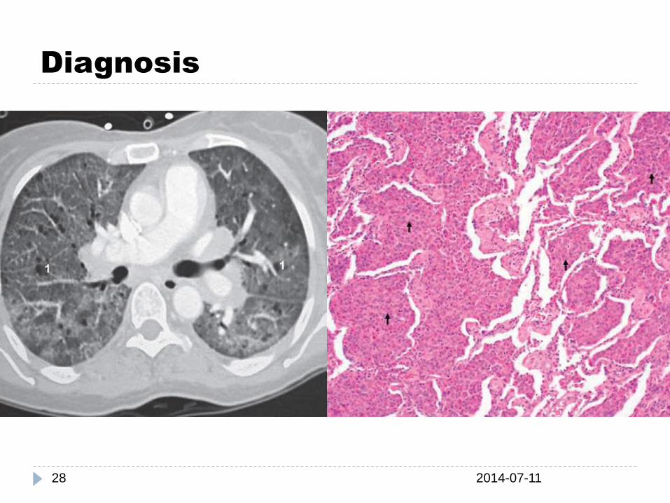
LYMPHANGIOLEIOMYOMATOSIS
2014-07-1159
Women>men, childbearing age.
Sx
Hemoptysis, pneumothorax(rupture of subpleural cysts) chylothorax (lymphatic ob.)
CXR: Coarse reticulonodular infiltrates, often w/ bilat’ cysts or bullae, lung vol. ↑
HRCT : diffuse thin-walled cysts <2 cm
BAL : occult alveolar hemorrhage
Lung bx : abNL sm. cells in airways, lymphatics, bv,
w/ concurrent airflow obstruction & replace parenchyma with cysts
Tx & Px
Sirolimus(RCT) progesterone or Tamoxifen(non-RCT)
10 yrs survival after the onset of symptoms

Inherited Disorders
2014-07-1160
AD tuberous sclerosis indistinguishable from LAM
both radiographically & histopathologically
neurofibromatosis bilateral lower lobe fibrosis
& bullae or cystic changes
AR Gaucher’s disease interstitial infiltration
w/ fibrosis, alveolar consolidation,
& filling of alveolar spaces
Niemann-Pick disease infiltration of the characteristic "foam cell"
throughout the pulmonary lymphatics,
the pulmonary arteries,
& the pulmonary alveoli
Hermansky-Pudlak
syndrome
Pulmonary fibrosis;
onset in the 30th~ 40th
slowly progressive

경청해주셔서 감사합니다.