Embed Size (px)
DESCRIPTION
This market leading Ophthamology title provides the ultimate foundations in ophthalmology for trainees and keeps experienced practitioners abreast on current practice and evolving techniques for diagnosing and treating ophthalmic disorders. It provides a pictorial, templated and bulleted approach presenting the key information needed to understand important eye disorders encountered in daily practice, giving the busy clinician a handle on a diagnostic problem in quick time by using either the book or a web version. This is also an excellent resource for for preparing for the exams. To purchase this title, please visit www.asia.elsevierhealth.com
Citation preview
Q
Chapter
13 Retinal Vascular Disease
RETINAL CIRCULATION 534
DIABETIC RETINOPATHY 534Introduction 534Pathogenesis 535Classification 536Signs 536Treatment 543Advanced diabetic eye disease 549
RETINAL VENOUS OCCLUSIVE DISEASE 551Pathogenesis 551Predisposing factors 551Systemic assessment 552Branch retinal vein occlusion 552Impending central retinal vein
occlusion 555Non-ischaemic central retinal vein
occlusion 555Ischaemic central retinal vein
occlusion 557Papillophlebitis 558Hemiretinal vein occlusion 559Systemic treatment in retinal vein
occlusion 559
RETINAL ARTERIAL OCCLUSIVE DISEASE 559Aetiology 559Systemic assessment 561
Amaurosis fugax 562Branch retinal artery occlusion 562Central retinal artery occlusion 563Cilioretinal artery occlusion 564Treatment of acute retinal artery
occlusion 564Systemic prophylaxis following retinal
artery occlusion 565Asymptomatic retinal embolus 566
OCULAR ISCHAEMIC SYNDROME 566
HYPERTENSIVE DISEASE 567Retinopathy 567Choroidopathy 568
SICKLE-CELL RETINOPATHY 569Sickling haemoglobinopathies 569Proliferative retinopathy 570Non-proliferative retinopathy 572Anterior segment features 572
RETINOPATHY OF PREMATURITY 573Pathogenesis 573Active disease 573Cicatricial disease 576
RETINAL ARTERY MACROANEURYSM 576
PRIMARY RETINAL TELANGIECTASIA 580Idiopathic macular telangiectasia 581Coats disease 582
EALES DISEASE 583
RADIATION RETINOPATHY 586
PURTSCHER RETINOPATHY 586
BENIGN IDIOPATHIC HAEMORRHAGIC RETINOPATHY 586
VALSALVA RETINOPATHY 588
LIPAEMIA RETINALIS 588
RETINOPATHY IN BLOOD DISORDERS 589Leukaemia 589Anaemia 589Hyperviscosity 590
CONGENITAL VASCULAR ANOMALIES 591Retinal macrovessel 591Arteriovenous communications 592
Kanski_Chapter 13_main.indd 533 2/11/2011 10:42:58 AM
Q
534Clinical OphthalmologyA S y S T e m A T I C A P P R o A C H
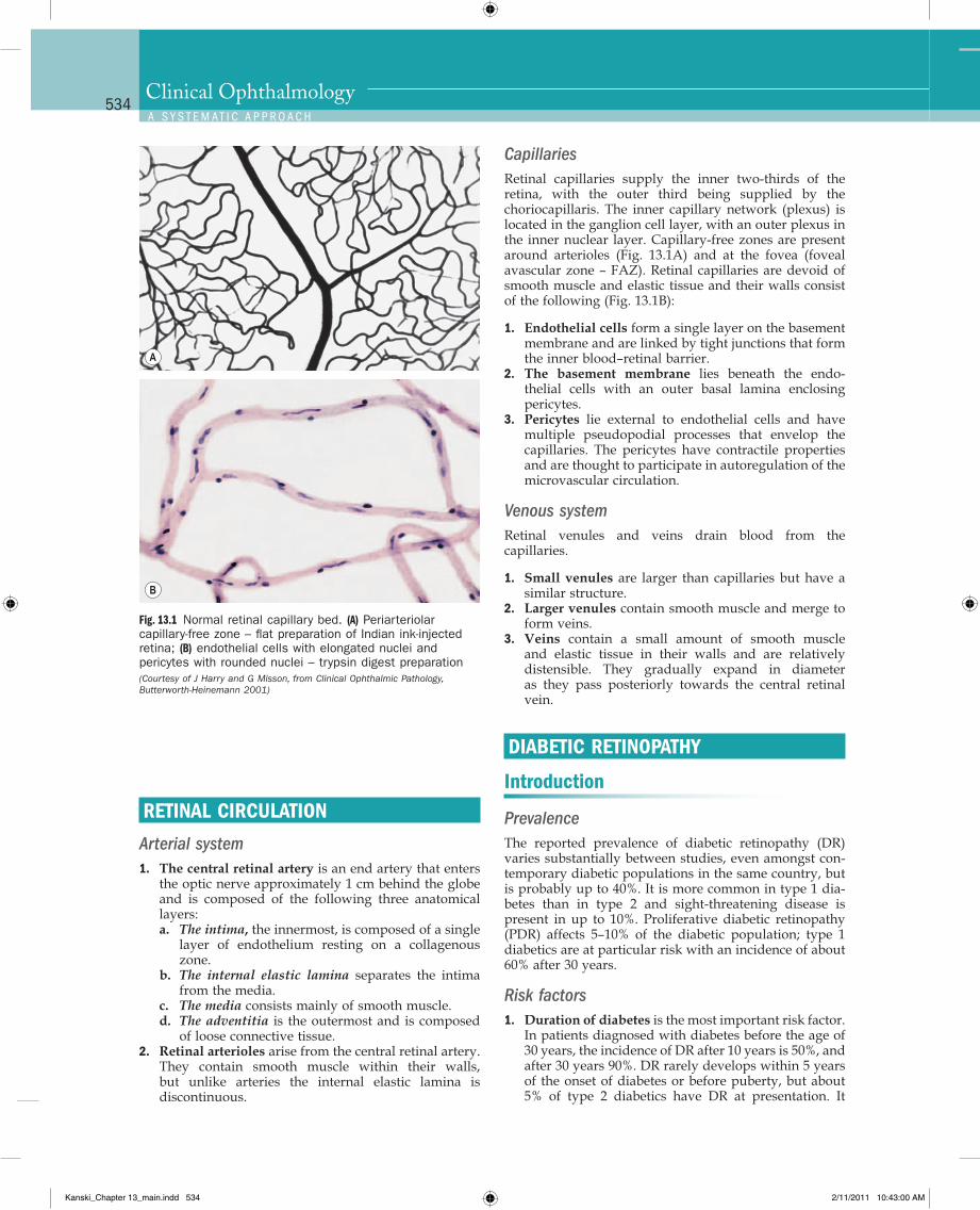
CapillariesRetinal capillaries supply the inner two-thirds of the retina, with the outer third being supplied by the choriocapillaris. The inner capillary network (plexus) is located in the ganglion cell layer, with an outer plexus in the inner nuclear layer. Capillary-free zones are present around arterioles (Fig. 13.1A) and at the fovea (foveal avascular zone – FAZ). Retinal capillaries are devoid of smooth muscle and elastic tissue and their walls consist of the following (Fig. 13.1B):
1. Endothelial cells form a single layer on the basement membrane and are linked by tight junctions that form the inner blood–retinal barrier.
2. The basement membrane lies beneath the endo-thelial cells with an outer basal lamina enclosing pericytes.
3. Pericytes lie external to endothelial cells and have multiple pseudopodial processes that envelop the capillaries. The pericytes have contractile properties and are thought to participate in autoregulation of the microvascular circulation.
Venous systemRetinal venules and veins drain blood from the capillaries.
1. Small venules are larger than capillaries but have a similar structure.
2. Larger venules contain smooth muscle and merge to form veins.
3. Veins contain a small amount of smooth muscle and elastic tissue in their walls and are relatively disten sible. They gradually expand in diameter as they pass posteriorly towards the central retinal vein.
DIABETIC RETINOPATHY
Introduction
PrevalenceThe reported prevalence of diabetic retinopathy (DR) varies substantially between studies, even amongst con-temporary diabetic populations in the same country, but is probably up to 40%. It is more common in type 1 dia-betes than in type 2 and sight-threatening disease is present in up to 10%. Proliferative diabetic retinopathy (PDR) affects 5–10% of the diabetic population; type 1 diabetics are at particular risk with an incidence of about 60% after 30 years.
Risk factors1. Duration of diabetes is the most important risk factor.
In patients diagnosed with diabetes before the age of 30 years, the incidence of DR after 10 years is 50%, and after 30 years 90%. DR rarely develops within 5 years of the onset of diabetes or before puberty, but about 5% of type 2 diabetics have DR at presentation. It
RETINAL CIRCULATION
Arterial system1. The central retinal artery is an end artery that enters
the optic nerve approximately 1 cm behind the globe and is composed of the following three anatomical layers:a. The intima, the innermost, is composed of a single
layer of endothelium resting on a collagenous zone.
b. The internal elastic lamina separates the intima from the media.
c. The media consists mainly of smooth muscle.d. The adventitia is the outermost and is composed
of loose connective tissue.2. Retinal arterioles arise from the central retinal artery.
They contain smooth muscle within their walls, but unlike arteries the internal elastic lamina is discontinuous.
Fig. 13.1 Normal retinal capillary bed. (A) Periarteriolar capillary-free zone – flat preparation of Indian ink-injected retina; (B) endothelial cells with elongated nuclei and pericytes with rounded nuclei – trypsin digest preparation (Courtesy of J Harry and G Misson, from Clinical Ophthalmic Pathology, Butterworth-Heinemann 2001)
A
B
Kanski_Chapter 13_main.indd 534 2/11/2011 10:43:00 AM
Q
535Retinal Vascular Disease 13C H A P T e R
appears that duration is a stronger predictor for pro-liferative disease than for maculopathy.
2. Poor control of diabetes. It has been shown that tight blood glucose control, particularly when instituted early, can prevent or delay the development or pro-gression of DR. However, a sudden improvement in control may be associated with progression of retin-opathy in the near term. Type 1 diabetic patients appear to obtain greater benefit from good control than those with type 2. Raised HbA1c is associated with an increased risk of proliferative disease.
3. Pregnancy is sometimes associated with rapid pro-gression of DR. Predicating factors include greater pre-pregnancy severity of retinopathy, poor pre-pregnancy control of diabetes, control exerted too rapidly during the early stages of pregnancy, and the development of pre-eclampsia and fluid imbalance. The risk of progression is related to the severity of DR in the first trimester. If substantial DR is present, fre-quency of review should reflect the individual risk, and can be up to monthly. Diabetic macular oedema usually resolves spontaneously after pregnancy and need not be treated if it develops in later pregnancy.
4. Hypertension, which is very common in patients with type 2 diabetes, should be rigorously controlled (<140/80). Tight control appears to be particularly beneficial in type 2 diabetics with maculopathy. Car-diovascular disease and previous stroke are also predictive.
5. Nephropathy, if severe, is associated with worsening of DR. Conversely, treatment of renal disease (e.g. renal transplantation) may be associated with impro-vement of retinopathy and a better response to photocoagulation.
6. Other risk factors include hyperlipidaemia, smoking, cataract surgery, obesity and anaemia.
Pathogenesis
DR is predominantly a microangiopathy in which small blood vessels are particularly vulnerable to damage from hyperglycaemia. Direct hyperglycaemic effects on retinal cells are also likely to play a role.
1. Mechanisms of cellular damage include intracellular sorbitol accumulation, oxidative stress due to free radical excess, accumulation of advanced glycation end products and excessive activation of several protein kinase C isoforms. Disruption of ion channel function is an important early feature.
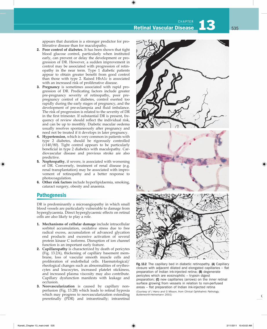
2. Capillaropathy is characterized by death of pericytes (Fig. 13.2A), thickening of capillary basement mem-brane, loss of vascular smooth muscle cells and proliferation of endothelial cells. Haematological/rheological changes such as abnormalities of erythro-cytes and leucocytes, increased platelet stickiness, and increased plasma viscosity may also contribute. Capillary dysfunction manifests with leakage and occlusion.
3. Neovascularization is caused by capillary non-perfusion (Fig. 13.2B) which leads to retinal hypoxia which may progress to neovascularization extending preretinally (PDR) and intraretinally; intraretinal
Fig. 13.2 The capillary bed in diabetic retinopathy. (A) Capillary closure with adjacent dilated and elongated capillaries – flat preparation of Indian ink-injected retina; (B) degenerate pericytes which are eosinophilic – trypsin digest preparation; (C) new capillaries (arrows) on the inner retinal surface growing from vessels in relation to non-perfused areas – flat preparation of Indian ink-injected retina (Courtesy of J Harry and G Misson, from Clinical Ophthalmic Pathology, Butterworth-Heinemann 2001)
A
B
C
Kanski_Chapter 13_main.indd 535 2/11/2011 10:43:02 AM
Q
536Clinical OphthalmologyA S y S T e m A T I C A P P R o A C H
microvascular abnormalities (IRMA) are shunts that run within the retina from arterioles to venules. New vessel growth is thought to be caused by an imbalance between the elaboration of angiogenic and anti-angiogenic factors, putatively in an attempt to re-vascularize hypoxic retina.
Many angiogenic stimulators have been identified; vascu-lar endothelial growth factor (VEGF), especially VEGF-A, appears to be of particular importance. Others include platelet-derived growth factor and hepatocyte growth factor. Similarly, several endogenous inhibitors of angio-genesis have also been reported such as endostatin, angi-ostatin and pigment epithelium-derived factor. It has been hypothesized that a key determinant of the activity of retinopathy is the net balance between VEGF and endostatin.
Classification
The classification used in the Early Treatment Diabetic Retinopathy Study (the modified Airlie House classifica-tion) is widely used internationally. An abbreviated version is set out in Table 13.1, in conjunction with man-agement guidelines. The following descriptive categories are also in widespread use in clinical practice:
1. Background diabetic retinopathy (BDR) is character-ized by microaneurysms, dot and blot haemorrhages and exudates. Generally the earlier signs of DR, although persisting as more advanced lesions appear.
2. Diabetic maculopathy strictly refers to the presence of any retinopathy at the macula, but commonly reserved for significant changes, particularly vision-threatening oedema and ischaemia.
3. Preproliferative diabetic retinopathy (PPDR) mani-fests cotton wool spots, venous changes, intraretinal microvascular anomalies (IRMA) and often deep retinal haemorrhages. PPDR indicates progressive retinal ischaemia, with a heightened risk of progres-sion to retinal neovascularization.
4. PDR is characterized by neovascularization on or within one disc diameter of the disc (NVD) and/or new vessels elsewhere (NVE) in the fundus.
4. Advanced diabetic eye disease is characterized by tractional retinal detachment, significant persistent vitreous haemorrhage and neovascular glaucoma.
Signs
Figure 13.3 shows the location of lesions in background diabetic retinopathy.
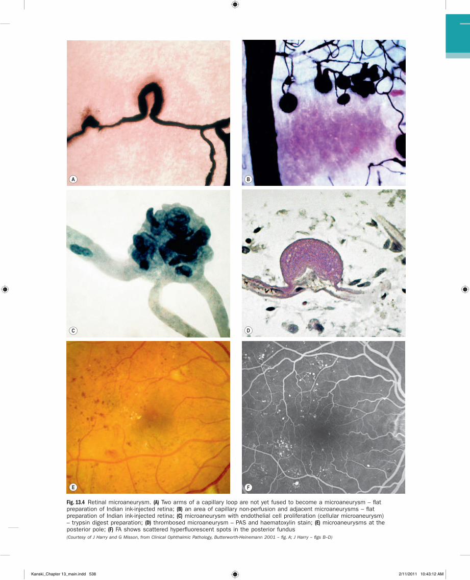
MicroaneurysmsMicroaneurysms are localized out-pouchings, mainly sac-cular, of the capillary wall that may form either by focal dilatation of the capillary wall where pericytes are absent, or by fusion of two arms of a capillary loop (Fig. 13.4A). Most develop in the inner capillary plexus (inner nuclear
layer) frequently in relation to areas of capillary non-perfusion (Fig. 13.4B). Loss of pericytes may also lead to endothelial cell proliferation with the formation of ‘cel-lular’ microaneurysms (Fig. 13.4C). Microaneurysms may leak plasma constituents into the retina as a result of breakdown in the blood–retinal barrier, or become throm-bosed (Fig. 13.4D).
1. Signs. Tiny red dots, often initially temporal to the fovea; tend to be the earliest signs of DR (Fig. 13.4E). They may be indistinguishable from dot haemorrhages.
2. Fluorescein angiography (FA). Early frames show tiny hyperfluorescent dots (Fig. 13.4F), representing non-thrombosed microaneurysms, typically more numerous than visible clinically. Late frames show diffuse hyperfluorescence due to leakage.
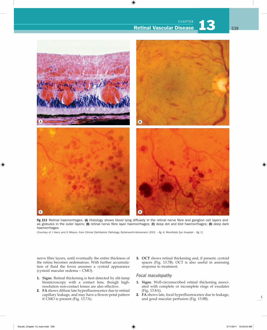
Retinal haemorrhagesFigure 13.5A is a histological section showing the location of blood.
1. Retinal nerve fibre layer haemorrhages arise from the larger superficial pre-capillary arterioles and because of the architecture of the retinal nerve fibres are flame-shaped (Fig. 13.5B).
2. Intraretinal haemorrhages arise from the venous end of capillaries and are located in the compact middle layers of the retina with a resultant red ‘dot/blot’ con-figuration (Fig. 13.5C).
3. Deeper dark round haemorrhages represent haemor-rhagic retinal infarcts and are located within the middle retinal layers (Fig. 13.5D). The extent of involvement is a significant marker of the likelihood of progression to retinal neovascularization.
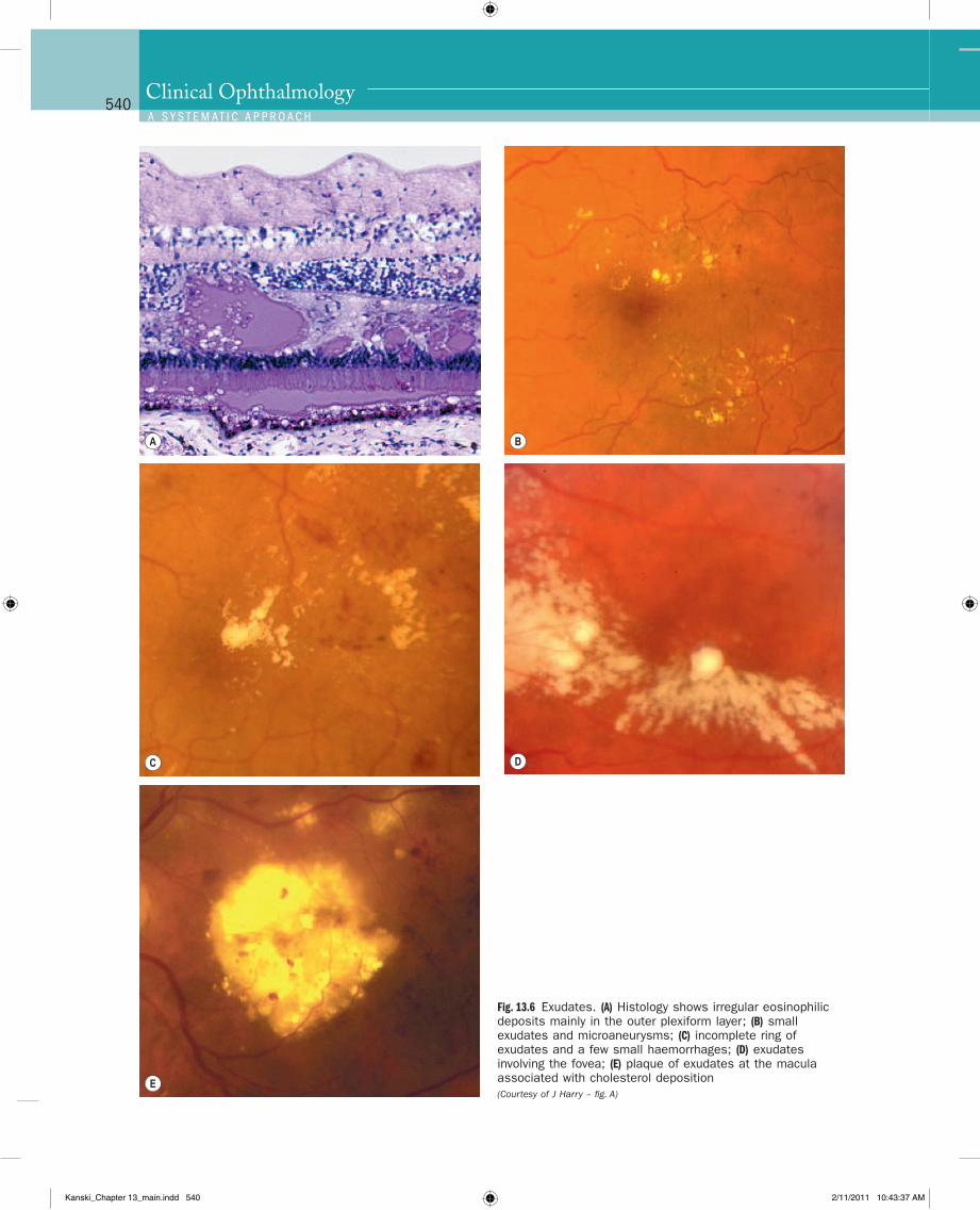
ExudatesExudates, sometimes termed ‘hard’ exudates to distin-guish from the older term of ‘soft’ exudates for cotton wool spots, are caused by chronic localized retinal oedema and develop at the junction of normal and oedematous retina. They are composed of lipoprotein and lipid-filled
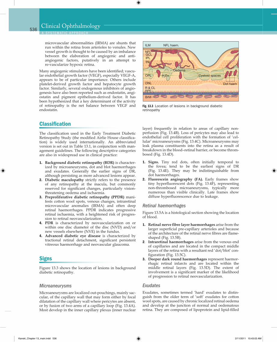
Fig. 13.3 Location of lesions in background diabetic retinopathy
Oedema
NFLGCLIPLINL
OPL H.Ex. Cystoid spaces
Dot haemONL
R & CLRPE
BrM
ILM NFL haem.
Kanski_Chapter 13_main.indd 536 2/11/2011 10:43:03 AM
Q
537Retinal Vascular Disease 13C H A P T e R
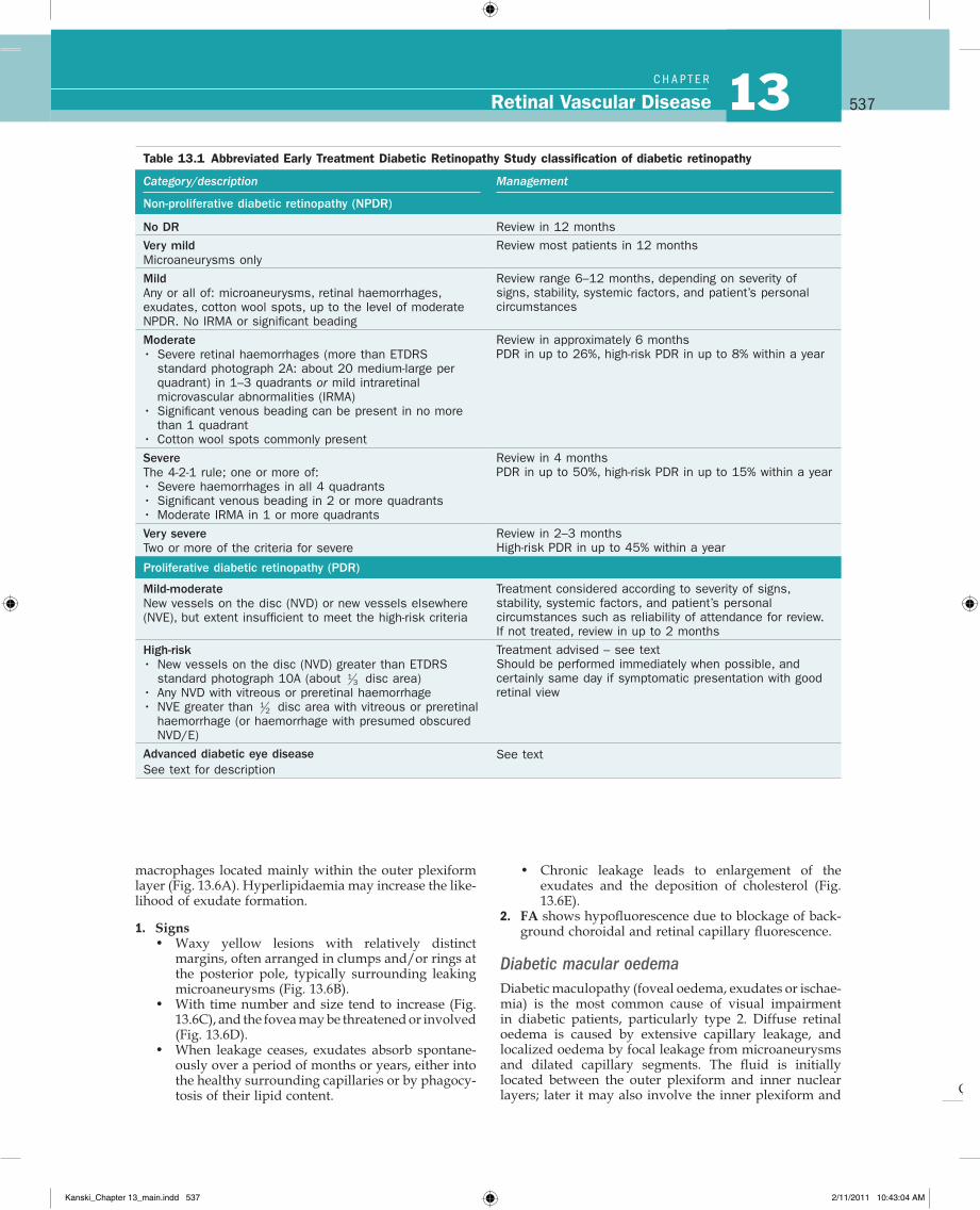
Table 13.1 Abbreviated Early Treatment Diabetic Retinopathy Study classification of diabetic retinopathy
Category/description Management
Non-proliferative diabetic retinopathy (NPDR)
No DR Review in 12 monthsVery mild Review most patients in 12 monthsMicroaneurysms onlyMild Review range 6–12 months, depending on severity of
signs, stability, systemic factors, and patient’s personal circumstances
Any or all of: microaneurysms, retinal haemorrhages, exudates, cotton wool spots, up to the level of moderate NPDR. No IRMA or significant beadingModerate Review in approximately 6 months
PDR in up to 26%, high-risk PDR in up to 8% within a year• Severe retinal haemorrhages (more than ETDRS standard photograph 2A: about 20 medium-large per quadrant) in 1–3 quadrants or mild intraretinal microvascular abnormalities (IRMA)
• Significant venous beading can be present in no more than 1 quadrant
• Cotton wool spots commonly presentSevere Review in 4 months
PDR in up to 50%, high-risk PDR in up to 15% within a yearThe 4-2-1 rule; one or more of:• Severe haemorrhages in all 4 quadrants• Significant venous beading in 2 or more quadrants• Moderate IRMA in 1 or more quadrantsVery severe Review in 2–3 months
High-risk PDR in up to 45% within a yearTwo or more of the criteria for severe
Proliferative diabetic retinopathy (PDR)
Mild-moderate Treatment considered according to severity of signs, stability, systemic factors, and patient’s personal circumstances such as reliability of attendance for review. If not treated, review in up to 2 months
New vessels on the disc (NVD) or new vessels elsewhere (NVE), but extent insufficient to meet the high-risk criteria
High-risk Treatment advised – see textShould be performed immediately when possible, and certainly same day if symptomatic presentation with good retinal view
• New vessels on the disc (NVD) greater than ETDRS standard photograph 10A (about 1
3 disc area)• Any NVD with vitreous or preretinal haemorrhage• NVE greater than 1
2 disc area with vitreous or preretinal haemorrhage (or haemorrhage with presumed obscured NVD/E)
Advanced diabetic eye disease See textSee text for description
macrophages located mainly within the outer plexiform layer (Fig. 13.6A). Hyperlipidaemia may increase the like-lihood of exudate formation.
1. Signs• Waxy yellow lesions with relatively distinct
margins, often arranged in clumps and/or rings at the posterior pole, typically surrounding leaking microaneurysms (Fig. 13.6B).
• With time number and size tend to increase (Fig. 13.6C), and the fovea may be threatened or involved (Fig. 13.6D).
• When leakage ceases, exudates absorb spontane-ously over a period of months or years, either into the healthy surrounding capillaries or by phagocy-tosis of their lipid content.
• Chronic leakage leads to enlargement of the exudates and the deposition of cholesterol (Fig. 13.6E).
2. FA shows hypofluorescence due to blockage of back-ground choroidal and retinal capillary fluorescence.
Diabetic macular oedemaDiabetic maculopathy (foveal oedema, exudates or ischae-mia) is the most common cause of visual impairment in diabetic patients, particularly type 2. Diffuse retinal oedema is caused by extensive capillary leakage, and localized oedema by focal leakage from microaneurysms and dilated capillary segments. The fluid is initially located between the outer plexiform and inner nuclear layers; later it may also involve the inner plexiform and
Kanski_Chapter 13_main.indd 537 2/11/2011 10:43:04 AM
Q
Fig. 13.4 Retinal microaneurysm. (A) Two arms of a capillary loop are not yet fused to become a microaneurysm – flat preparation of Indian ink-injected retina; (B) an area of capillary non-perfusion and adjacent microaneurysms – flat preparation of Indian ink-injected retina; (C) microaneurysm with endothelial cell proliferation (cellular microaneurysm) – trypsin digest preparation; (D) thrombosed microaneurysm – PAS and haematoxylin stain; (E) microaneurysms at the posterior pole; (F) FA shows scattered hyperfluorescent spots in the posterior fundus (Courtesy of J Harry and G Misson, from Clinical Ophthalmic Pathology, Butterworth-Heinemann 2001 – fig. A; J Harry – figs B–D)
B
C
A
D
E F
Kanski_Chapter 13_main.indd 538 2/11/2011 10:43:12 AM
Q
539Retinal Vascular Disease 13C H A P T e R
Fig. 13.5 Retinal haemorrhages. (A) Histology shows blood lying diffusely in the retinal nerve fibre and ganglion cell layers and as globules in the outer layers; (B) retinal nerve fibre layer haemorrhages; (C) deep dot and blot haemorrhages; (D) deep dark haemorrhages (Courtesy of J Harry and G Misson, from Clinical Ophthalmic Pathology, Butterworth-Heinemann 2001 – fig. A; Moorfields Eye Hospital – fig. C)
A B
C D
nerve fibre layers, until eventually the entire thickness of the retina becomes oedematous. With further accumula-tion of fluid the fovea assumes a cystoid appearance (cystoid macular oedema – CMO).
1. Signs. Retinal thickening is best detected by slit-lamp biomicroscopy with a contact lens, though high-resolution non-contact lenses are also effective.
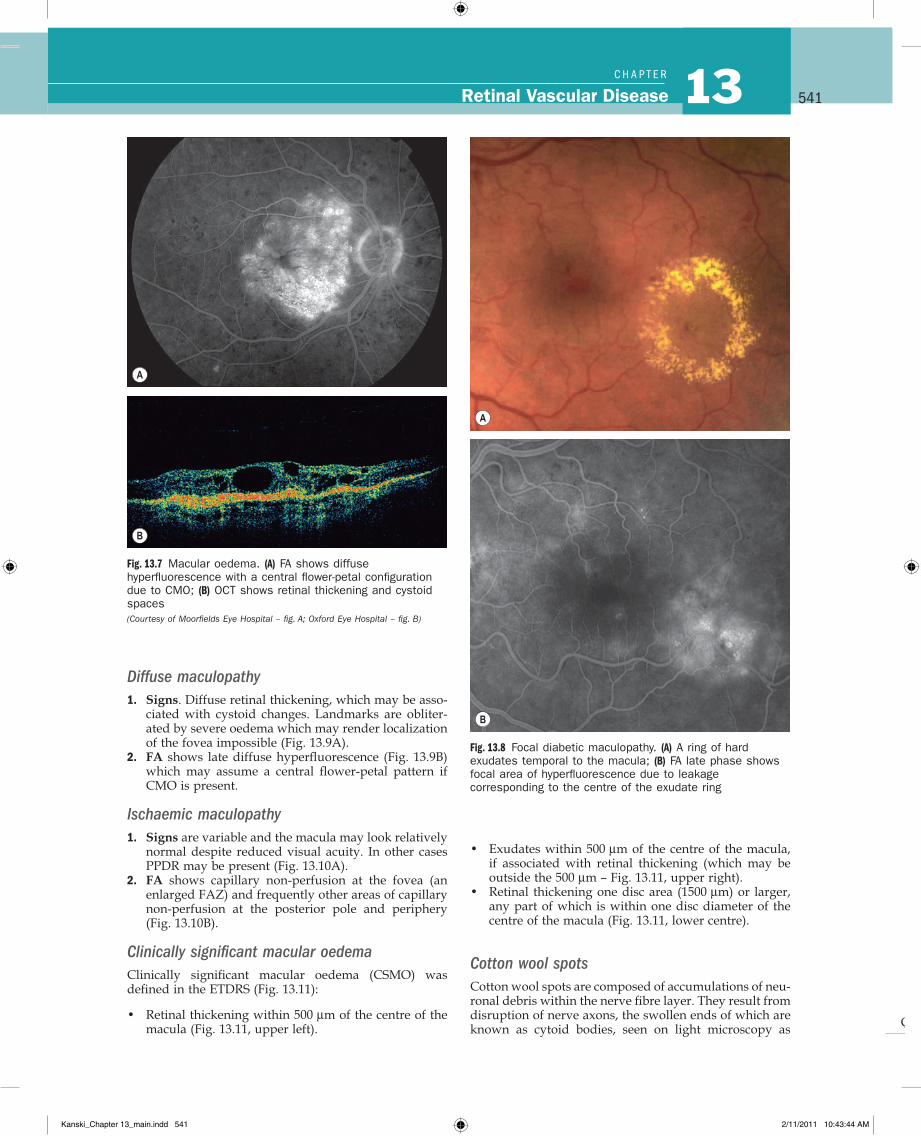
2. FA shows diffuse late hyperfluorescence due to retinal capillary leakage, and may have a flower-petal pattern if CMO is present (Fig. 13.7A).
3. OCT shows retinal thickening and, if present, cystoid spaces (Fig. 13.7B). OCT is also useful in assessing response to treatment.
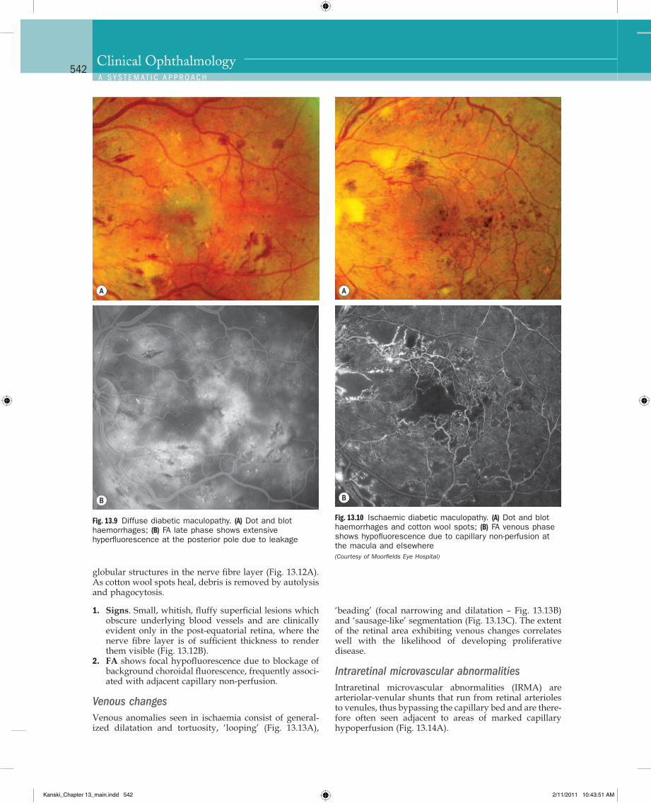
Focal maculopathy1. Signs. Well-circumscribed retinal thickening associ-
ated with complete or incomplete rings of exudates (Fig. 13.8A).
2. FA shows late, focal hyperfluorescence due to leakage, and good macular perfusion (Fig. 13.8B).
Kanski_Chapter 13_main.indd 539 2/11/2011 10:43:24 AM
Q
540Clinical OphthalmologyA S y S T e m A T I C A P P R o A C H
Fig. 13.6 Exudates. (A) Histology shows irregular eosinophilic deposits mainly in the outer plexiform layer; (B) small exudates and microaneurysms; (C) incomplete ring of exudates and a few small haemorrhages; (D) exudates involving the fovea; (E) plaque of exudates at the macula associated with cholesterol deposition (Courtesy of J Harry – fig. A)
C
E
A B
D
Kanski_Chapter 13_main.indd 540 2/11/2011 10:43:37 AM
Q
541Retinal Vascular Disease 13C H A P T e R
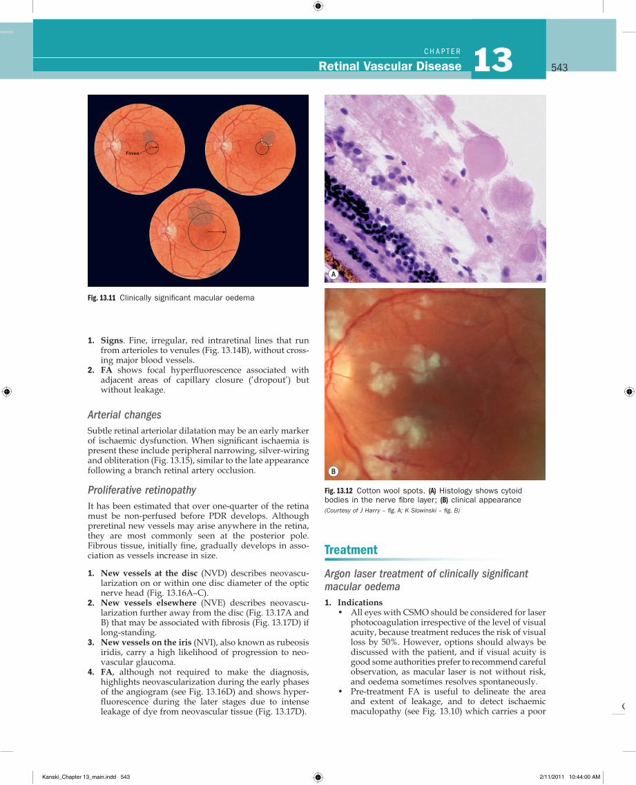
• Exudates within 500 µm of the centre of the macula, if associated with retinal thickening (which may be outside the 500 µm – Fig. 13.11, upper right).
• Retinal thickening one disc area (1500 µm) or larger, any part of which is within one disc diameter of the centre of the macula (Fig. 13.11, lower centre).
Cotton wool spotsCotton wool spots are composed of accumulations of neu-ronal debris within the nerve fibre layer. They result from disruption of nerve axons, the swollen ends of which are known as cytoid bodies, seen on light microscopy as
Diffuse maculopathy1. Signs. Diffuse retinal thickening, which may be asso-
ciated with cystoid changes. Landmarks are obliter-ated by severe oedema which may render localization of the fovea impossible (Fig. 13.9A).
2. FA shows late diffuse hyperfluorescence (Fig. 13.9B) which may assume a central flower-petal pattern if CMO is present.
Ischaemic maculopathy1. Signs are variable and the macula may look relatively
normal despite reduced visual acuity. In other cases PPDR may be present (Fig. 13.10A).
2. FA shows capillary non-perfusion at the fovea (an enlarged FAZ) and frequently other areas of capillary non-perfusion at the posterior pole and periphery (Fig. 13.10B).
Clinically significant macular oedemaClinically significant macular oedema (CSMO) was defined in the ETDRS (Fig. 13.11):
• Retinal thickening within 500 µm of the centre of the macula (Fig. 13.11, upper left).
Fig. 13.7 Macular oedema. (A) FA shows diffuse hyperfluorescence with a central flower-petal configuration due to CMO; (B) OCT shows retinal thickening and cystoid spaces (Courtesy of Moorfields Eye Hospital – fig. A; Oxford Eye Hospital – fig. B)
A
B
Fig. 13.8 Focal diabetic maculopathy. (A) A ring of hard exudates temporal to the macula; (B) FA late phase shows focal area of hyperfluorescence due to leakage corresponding to the centre of the exudate ring
A
B
Kanski_Chapter 13_main.indd 541 2/11/2011 10:43:44 AM
Q
542Clinical OphthalmologyA S y S T e m A T I C A P P R o A C H
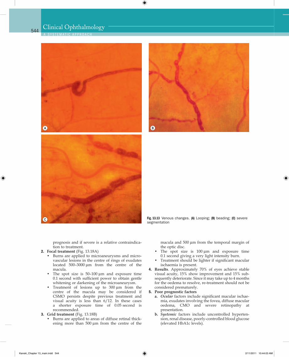
‘beading’ (focal narrowing and dilatation – Fig. 13.13B) and ‘sausage-like’ segmentation (Fig. 13.13C). The extent of the retinal area exhibiting venous changes correlates well with the likelihood of developing proliferative disease.
Intraretinal microvascular abnormalitiesIntraretinal microvascular abnormalities (IRMA) are arteriolar-venular shunts that run from retinal arterioles to venules, thus bypassing the capillary bed and are there-fore often seen adjacent to areas of marked capillary hypoperfusion (Fig. 13.14A).
globular structures in the nerve fibre layer (Fig. 13.12A). As cotton wool spots heal, debris is removed by autolysis and phagocytosis.
1. Signs. Small, whitish, fluffy superficial lesions which obscure underlying blood vessels and are clinically evident only in the post-equatorial retina, where the nerve fibre layer is of sufficient thickness to render them visible (Fig. 13.12B).
2. FA shows focal hypofluorescence due to blockage of background choroidal fluorescence, frequently associ-ated with adjacent capillary non-perfusion.
Venous changesVenous anomalies seen in ischaemia consist of general-ized dilatation and tortuosity, ‘looping’ (Fig. 13.13A),
Fig. 13.9 Diffuse diabetic maculopathy. (A) Dot and blot haemorrhages; (B) FA late phase shows extensive hyperfluorescence at the posterior pole due to leakage
A
B
Fig. 13.10 Ischaemic diabetic maculopathy. (A) Dot and blot haemorrhages and cotton wool spots; (B) FA venous phase shows hypofluorescence due to capillary non-perfusion at the macula and elsewhere (Courtesy of Moorfields Eye Hospital)
A
B
Kanski_Chapter 13_main.indd 542 2/11/2011 10:43:51 AM
Q
543Retinal Vascular Disease 13C H A P T e R
Treatment
Argon laser treatment of clinically significant macular oedema1. Indications
• All eyes with CSMO should be considered for laser photocoagulation irrespective of the level of visual acuity, because treatment reduces the risk of visual loss by 50%. However, options should always be discussed with the patient, and if visual acuity is good some authorities prefer to recommend careful observation, as macular laser is not without risk, and oedema sometimes resolves spontaneously.
• Pre-treatment FA is useful to delineate the area and extent of leakage, and to detect ischaemic maculopathy (see Fig. 13.10) which carries a poor
Fig. 13.11 Clinically significant macular oedema
Fovea
Fig. 13.12 Cotton wool spots. (A) Histology shows cytoid bodies in the nerve fibre layer; (B) clinical appearance (Courtesy of J Harry – fig. A; K Slowinski – fig. B)
A
B
1. Signs. Fine, irregular, red intraretinal lines that run from arterioles to venules (Fig. 13.14B), without cross-ing major blood vessels.
2. FA shows focal hyperfluorescence associated with adjacent areas of capillary closure (’dropout’) but without leakage.
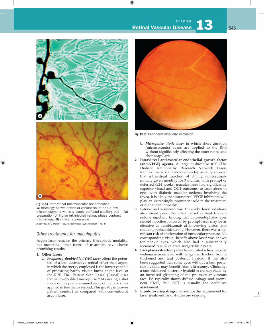
Arterial changesSubtle retinal arteriolar dilatation may be an early marker of ischaemic dysfunction. When significant ischaemia is present these include peripheral narrowing, silver-wiring and obliteration (Fig. 13.15), similar to the late appearance following a branch retinal artery occlusion.
Proliferative retinopathyIt has been estimated that over one-quarter of the retina must be non-perfused before PDR develops. Although preretinal new vessels may arise anywhere in the retina, they are most commonly seen at the posterior pole. Fibrous tissue, initially fine, gradually develops in asso-ciation as vessels increase in size.
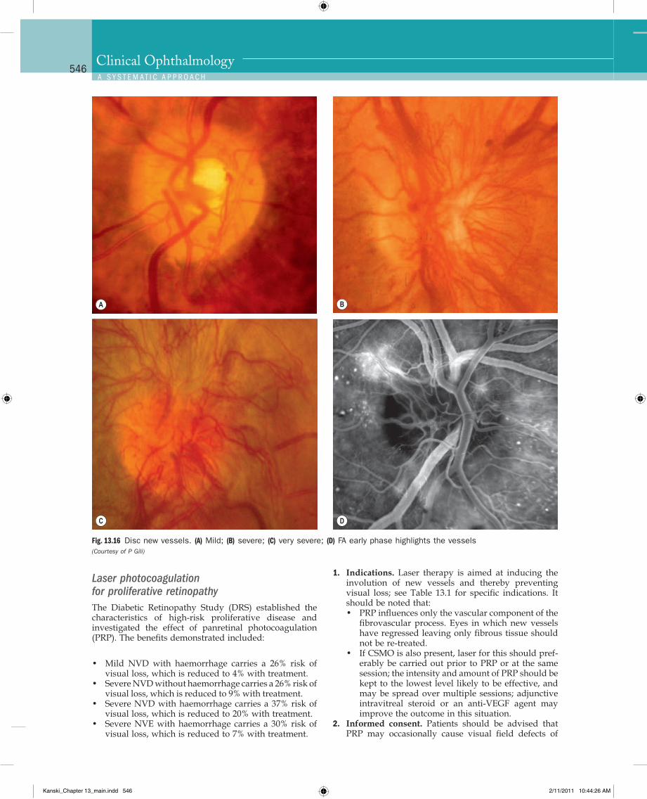
1. New vessels at the disc (NVD) describes neovascu-larization on or within one disc diameter of the optic nerve head (Fig. 13.16A–C).
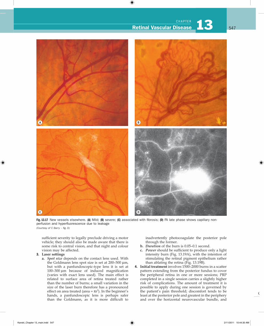
2. New vessels elsewhere (NVE) describes neovascu-larization further away from the disc (Fig. 13.17A and B) that may be associated with fibrosis (Fig. 13.17D) if long-standing.
3. New vessels on the iris (NVI), also known as rubeosis iridis, carry a high likelihood of progression to neo-vascular glaucoma.
4. FA, although not required to make the diagnosis, highlights neovascularization during the early phases of the angiogram (see Fig. 13.16D) and shows hyper-fluorescence during the later stages due to intense leakage of dye from neovascular tissue (Fig. 13.17D).
Kanski_Chapter 13_main.indd 543 2/11/2011 10:44:00 AM
Q
544Clinical OphthalmologyA S y S T e m A T I C A P P R o A C H
macula and 500 µm from the temporal margin of the optic disc.
• The spot size is 100 µm and exposure time 0.1 second giving a very light intensity burn.
• Treatment should be lighter if significant macular ischaemia is present.
4. Results. Approximately 70% of eyes achieve stable visual acuity, 15% show improvement and 15% sub-sequently deteriorate. Since it may take up to 4 months for the oedema to resolve, re-treatment should not be considered prematurely.
5. Poor prognostic factorsa. Ocular factors include significant macular ischae-
mia, exudates involving the fovea, diffuse macular oedema, CMO and severe retinopathy at presentation.
b. Systemic factors include uncontrolled hyperten-sion, renal disease, poorly-controlled blood glucose (elevated HbA1c levels).
prognosis and if severe is a relative contraindica-tion to treatment.
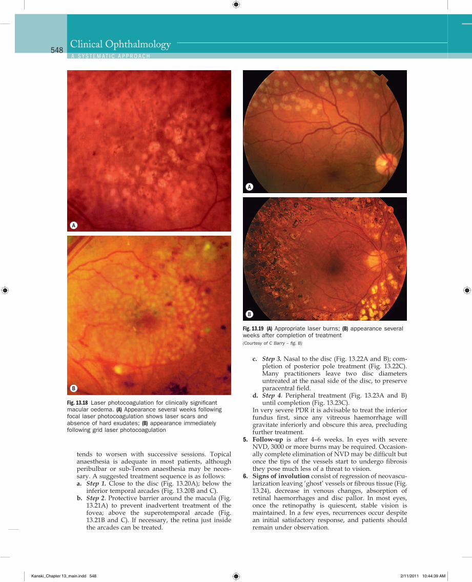
2. Focal treatment (Fig. 13.18A)• Burns are applied to microaneurysms and micro-
vascular lesions in the centre of rings of exudates located 500–3000 µm from the centre of the macula.
• The spot size is 50–100 µm and exposure time 0.1 second with sufficient power to obtain gentle whitening or darkening of the microaneurysm.
• Treatment of lesions up to 300 µm from the centre of the macula may be considered if CSMO persists despite previous treatment and visual acuity is less than 6/12. In these cases a shorter exposure time of 0.05 second is recommended.
3. Grid treatment (Fig. 13.18B)• Burns are applied to areas of diffuse retinal thick-
ening more than 500 µm from the centre of the
Fig. 13.13 Venous changes. (A) Looping; (B) beading; (C) severe segmentation
A B
C
Kanski_Chapter 13_main.indd 544 2/11/2011 10:44:03 AM
Q
545Retinal Vascular Disease 13C H A P T e R
b. Micropulse diode laser in which short duration (microseconds) burns are applied to the RPE without significantly affecting the outer retina and choriocapillaris.
2. Intravitreal anti-vascular endothelial growth factor (anti-VEGF) agents. A large multicentre trial (The Diabetic Retinopathy Research Network Laser- Ranibizumab-Triamcinolone Study) recently showed that intravitreal injection of 0.5 mg ranibizumab, initially given monthly for 3 months, with prompt or deferred (≥24 weeks) macular laser had significantly superior visual and OCT outcomes to laser alone in eyes with diabetic macular oedema involving the fovea. It is likely that intravitreal VEGF inhibitors will play an increasingly prominent role in the treatment of diabetic retinopathy.
3. Intravitreal triamcinolone. The study described above also investigated the effect of intravitreal triamci-nolone injection, finding that in pseudophakic eyes steroid injection followed by prompt laser may be as effective as ranibizumab at improving vision and reducing retinal thickening. However, there was a sig-nificant risk of an elevation of intraocular pressure. No corresponding visual benefit above laser was shown for phakic eyes, which also had a substantially increased rate of cataract surgery by 2 years.
4. Pars plana vitrectomy may be indicated when macular oedema is associated with tangential traction from a thickened and taut posterior hyaloid. It has also been suggested that some eyes without a taut poste-rior hyaloid may benefit from vitrectomy. Clinically, a taut thickened posterior hyaloid is characterized by an increased glistening of the pre-macular vitreous face. FA typically shows diffuse leakage and promi-nent CMO, but OCT is usually the definitive assessment.
5. Lipid-lowering drugs may reduce the requirement for laser treatment, and studies are ongoing.
Other treatments for maculopathyArgon laser remains the primary therapeutic modality, but numerous other forms of treatment have shown promising results.
1. Other lasersa. Frequency-doubled Nd:YAG laser offers the poten-
tial of a less destructive retinal effect than argon, in which the energy employed is the lowest capable of producing barely visible burns at the level of the RPE. The ‘Pattern Scan Laser’ (Pascal) uses frequency-doubled micropulse YAG in single shot mode or in a predetermined array of up to 56 shots applied in less than a second. This greatly improves patient comfort as compared with conventional argon laser.
Fig. 13.14 Intraretinal microvascular abnormalities. (A) Histology shows arteriolar-venular shunt and a few microaneurysms within a poorly perfused capillary bed – flat preparation of Indian ink-injected retina; phase contrast microscopy; (B) clinical appearance (Courtesy of J Harry – fig. A; Moorfields Eye Hospital – fig. B)
A
B
Fig. 13.15 Peripheral arteriolar occlusion
Kanski_Chapter 13_main.indd 545 2/11/2011 10:44:10 AM
Q
546Clinical OphthalmologyA S y S T e m A T I C A P P R o A C H
1. Indications. Laser therapy is aimed at inducing the involution of new vessels and thereby preventing visual loss; see Table 13.1 for specific indications. It should be noted that:• PRP influences only the vascular component of the
fibrovascular process. Eyes in which new vessels have regressed leaving only fibrous tissue should not be re-treated.
• If CSMO is also present, laser for this should pref-erably be carried out prior to PRP or at the same session; the intensity and amount of PRP should be kept to the lowest level likely to be effective, and may be spread over multiple sessions; adjunctive intravitreal steroid or an anti-VEGF agent may improve the outcome in this situation.
2. Informed consent. Patients should be advised that PRP may occasionally cause visual field defects of
Laser photocoagulation for proliferative retinopathyThe Diabetic Retinopathy Study (DRS) established the characteristics of high-risk proliferative disease and investigated the effect of panretinal photocoagulation (PRP). The benefits demonstrated included:
• Mild NVD with haemorrhage carries a 26% risk of visual loss, which is reduced to 4% with treatment.
• Severe NVD without haemorrhage carries a 26% risk of visual loss, which is reduced to 9% with treatment.
• Severe NVD with haemorrhage carries a 37% risk of visual loss, which is reduced to 20% with treatment.
• Severe NVE with haemorrhage carries a 30% risk of visual loss, which is reduced to 7% with treatment.
Fig. 13.16 Disc new vessels. (A) Mild; (B) severe; (C) very severe; (D) FA early phase highlights the vessels (Courtesy of P Gili)
A B
C D
Kanski_Chapter 13_main.indd 546 2/11/2011 10:44:26 AM
Q
547Retinal Vascular Disease 13C H A P T e R
inadvertently photocoagulate the posterior pole through the former.
b. Duration of the burn is 0.05–0.1 second.c. Power should be sufficient to produce only a light
intensity burn (Fig. 13.19A), with the intention of stimulating the retinal pigment epithelium rather than ablating the retina (Fig. 13.19B).
4. Initial treatment involves 1500–2000 burns in a scatter pattern extending from the posterior fundus to cover the peripheral retina in one or more sessions; PRP completed in a single session carries a slightly higher risk of complications. The amount of treatment it is possible to apply during one session is governed by the patient’s pain threshold; discomfort tends to be least at the posterior pole and greatest in the periphery and over the horizontal neurovascular bundle, and
sufficient severity to legally preclude driving a motor vehicle; they should also be made aware that there is some risk to central vision, and that night and colour vision may be affected.
3. Laser settingsa. Spot size depends on the contact lens used. With
the Goldmann lens spot size is set at 200–500 µm, but with a panfundoscopic-type lens it is set at 100–300 µm because of induced magnification (varies with exact lens used). The main effect is related to surface area of retina treated rather than the number of burns; a small variation in the size of the laser burn therefore has a pronounced effect on area treated (area = πr2). In the beginner’s hands, a panfundoscopic lens is perhaps safer than the Goldmann, as it is more difficult to
Fig. 13.17 New vessels elsewhere. (A) Mild; (B) severe; (C) associated with fibrosis; (D) FA late phase shows capillary non-perfusion and hyperfluorescence due to leakage (Courtesy of C Barry – fig. D)
A B
C D
Kanski_Chapter 13_main.indd 547 2/11/2011 10:44:30 AM
Q
548Clinical OphthalmologyA S y S T e m A T I C A P P R o A C H
tends to worsen with successive sessions. Topical anaesthesia is adequate in most patients, although peribulbar or sub-Tenon anaesthesia may be neces-sary. A suggested treatment sequence is as follows:a. Step 1. Close to the disc (Fig. 13.20A); below the
inferior temporal arcades (Fig. 13.20B and C).b. Step 2. Protective barrier around the macula (Fig.
13.21A) to prevent inadvertent treatment of the fovea; above the superotemporal arcade (Fig. 13.21B and C). If necessary, the retina just inside the arcades can be treated.
Fig. 13.18 Laser photocoagulation for clinically significant macular oedema. (A) Appearance several weeks following focal laser photocoagulation shows laser scars and absence of hard exudates; (B) appearance immediately following grid laser photocoagulation
A
B
Fig. 13.19 (A) Appropriate laser burns; (B) appearance several weeks after completion of treatment (Courtesy of C Barry – fig. B)
A
B
c. Step 3. Nasal to the disc (Fig. 13.22A and B); com-pletion of posterior pole treatment (Fig. 13.22C). Many practitioners leave two disc diameters untreated at the nasal side of the disc, to preserve paracentral field.
d. Step 4. Peripheral treatment (Fig. 13.23A and B) until completion (Fig. 13.23C).
In very severe PDR it is advisable to treat the inferior fundus first, since any vitreous haemorrhage will gravitate inferiorly and obscure this area, precluding further treatment.
5. Follow-up is after 4–6 weeks. In eyes with severe NVD, 3000 or more burns may be required. Occasion-ally complete elimination of NVD may be difficult but once the tips of the vessels start to undergo fibrosis they pose much less of a threat to vision.
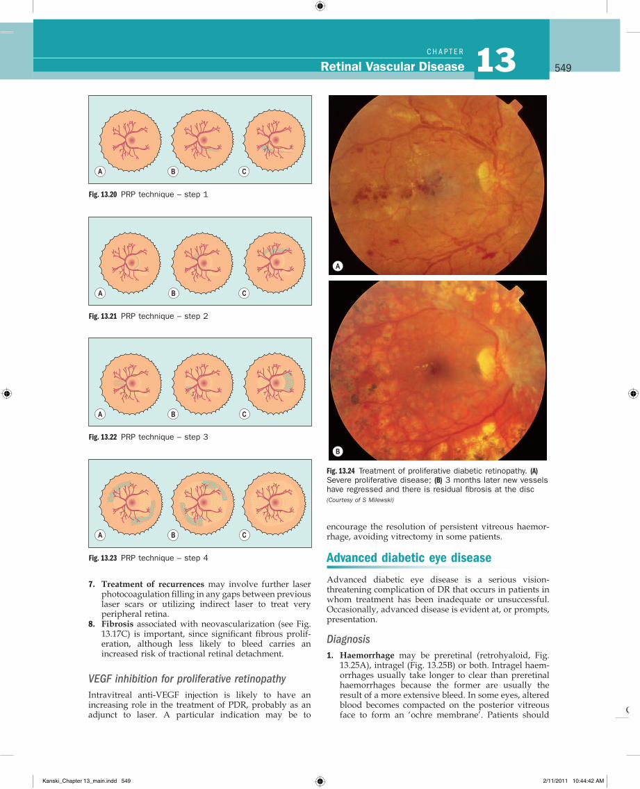
6. Signs of involution consist of regression of neovascu-larization leaving ‘ghost’ vessels or fibrous tissue (Fig. 13.24), decrease in venous changes, absorption of retinal haemorrhages and disc pallor. In most eyes, once the retinopathy is quiescent, stable vision is maintained. In a few eyes, recurrences occur despite an initial satisfactory response, and patients should remain under observation.
Kanski_Chapter 13_main.indd 548 2/11/2011 10:44:39 AM
Q
549Retinal Vascular Disease 13C H A P T e R
Fig. 13.20 PRP technique – step 1
A B C
Fig. 13.21 PRP technique – step 2
A B C
encourage the resolution of persistent vitreous haemor-rhage, avoiding vitrectomy in some patients.
Advanced diabetic eye disease
Advanced diabetic eye disease is a serious vision-threatening complication of DR that occurs in patients in whom treatment has been inadequate or unsuccessful. Occasionally, advanced disease is evident at, or prompts, presentation.
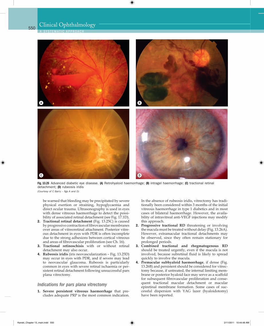
Diagnosis1. Haemorrhage may be preretinal (retrohyaloid, Fig.
13.25A), intragel (Fig. 13.25B) or both. Intragel haem-orrhages usually take longer to clear than preretinal haemorrhages because the former are usually the result of a more extensive bleed. In some eyes, altered blood becomes compacted on the posterior vitreous face to form an ‘ochre membrane’. Patients should
Fig. 13.22 PRP technique – step 3
A B C
Fig. 13.23 PRP technique – step 4
A B C
Fig. 13.24 Treatment of proliferative diabetic retinopathy. (A) Severe proliferative disease; (B) 3 months later new vessels have regressed and there is residual fibrosis at the disc (Courtesy of S Milewski)
A
B
7. Treatment of recurrences may involve further laser photocoagulation filling in any gaps between previous laser scars or utilizing indirect laser to treat very peripheral retina.
8. Fibrosis associated with neovascularization (see Fig. 13.17C) is important, since significant fibrous prolif-eration, although less likely to bleed carries an increased risk of tractional retinal detachment.
VEGF inhibition for proliferative retinopathyIntravitreal anti-VEGF injection is likely to have an increasing role in the treatment of PDR, probably as an adjunct to laser. A particular indication may be to
Kanski_Chapter 13_main.indd 549 2/11/2011 10:44:42 AM
Q
550Clinical OphthalmologyA S y S T e m A T I C A P P R o A C H
In the absence of rubeosis iridis, vitrectomy has tradi-tionally been considered within 3 months of the initial vitreous haemorrhage in type 1 diabetics and in most cases of bilateral haemorrhage. However, the availa-bility of intravitreal anti-VEGF injections may modify this approach.
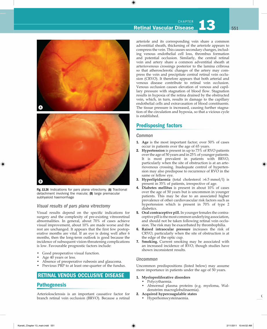
2. Progressive tractional RD threatening or involving the macula must be treated without delay (Fig. 13.26A). However, extramacular tractional detachments may be observed, since they often remain stationary for prolonged periods.
3. Combined tractional and rhegmatogenous RD should be treated urgently, even if the macula is not involved, because subretinal fluid is likely to spread quickly to involve the macula.
4. Premacular subhyaloid haemorrhage, if dense (Fig. 13.26B) and persistent should be considered for vitrec-tomy because, if untreated, the internal limiting mem-brane or posterior hyaloid face may serve as a scaffold for subsequent fibrovascular proliferation and conse-quent tractional macular detachment or macular epiretinal membrane formation. Some cases of suc-cessful dispersion with YAG lazer (hyaloidotomy) have been reported.
be warned that bleeding may be precipitated by severe physical exertion or straining, hypoglycaemia and direct ocular trauma. Ultrasonography is used in eyes with dense vitreous haemorrhage to detect the possi-bility of associated retinal detachment (see Fig. 17.1D).
2. Tractional retinal detachment (Fig. 13.25C) is caused by progressive contraction of fibrovascular membranes over areas of vitreoretinal attachment. Posterior vitre-ous detachment in eyes with PDR is often incomplete due to the strong adhesions between cortical vitreous and areas of fibrovascular proliferation (see Ch. 16).
3. Tractional retinoschisis with or without retinal detachment may also occur.
4. Rubeosis iridis (iris neovascularization – Fig. 13.25D) may occur in eyes with PDR, and if severe may lead to neovascular glaucoma. Rubeosis is particularly common in eyes with severe retinal ischaemia or per-sistent retinal detachment following unsuccessful pars plana vitrectomy.
Indications for pars plana vitrectomy1. Severe persistent vitreous haemorrhage that pre-
cludes adequate PRP is the most common indication.
Fig. 13.25 Advanced diabetic eye disease. (A) Retrohyaloid haemorrhage; (B) intragel haemorrhage; (C) tractional retinal detachment; (D) rubeosis iridis (Courtesy of C Barry – figs A and D)
A B
C D
Kanski_Chapter 13_main.indd 550 2/11/2011 10:44:48 AM
Q
551Retinal Vascular Disease 13C H A P T e R
arteriole and its corresponding vein share a common adventitial sheath, thickening of the arteriole appears to compress the vein. This causes secondary changes, includ-ing venous endothelial cell loss, thrombus formation and potential occlusion. Similarly, the central retinal vein and artery share a common adventitial sheath at arteriovenous crossings posterior to the lamina cribrosa so that atherosclerotic changes of the artery may com-press the vein and precipitate central retinal vein occlu-sion (CRVO). It therefore appears that both arterial and venous disease contribute to retinal vein occlusion. Venous occlusion causes elevation of venous and capil-lary pressure with stagnation of blood flow. Stagnation results in hypoxia of the retina drained by the obstructed vein, which, in turn, results in damage to the capillary endothelial cells and extravasation of blood constituents. The tissue pressure is increased, causing further stagna-tion of the circulation and hypoxia, so that a vicious cycle is established.
Predisposing factors
Common1. Age is the most important factor; over 50% of cases
occur in patients over the age of 65 years.2. Hypertension is present in up to 73% of RVO patients
over the age of 50 years and in 25% of younger patients. It is most prevalent in patients with BRVO, particularly when the site of obstruction is at an arte-riovenous crossing. Inadequate control of hyperten-sion may also predispose to recurrence of RVO in the same or fellow eye.
3. Hyperlipidaemia (total cholesterol >6.5 mmol/l) is present in 35% of patients, irrespective of age.
4. Diabetes mellitus is present in about 10% of cases over the age of 50 years but is uncommon in younger patients. This may be due to an associated higher prevalence of other cardiovascular risk factors such as hypertension which is present in 70% of type 2 diabetics.
5. Oral contraceptive pill. In younger females the contra-ceptive pill is the most common underlying asso ciation, and should not be taken following retinal vein occlu-sion. The risk may be exacerbated by thrombophilia.
6. Raised intraocular pressure increases the risk of CRVO, particularly when the site of obstruction is at the edge of the optic cup.
7. Smoking. Current smoking may be associated with an increased incidence of RVO, though studies have shown inconsistent results.
UncommonUncommon predispositions (listed below) may assume more importance in patients under the age of 50 years.
1. Myeloproliferative disorders• Polycythaemia.• Abnormal plasma proteins (e.g. myeloma, Wal-
denström macroglobulinaemia).2. Acquired hypercoagulable states
• Hyperhomocysteinaemia.
Visual results of pars plana vitrectomyVisual results depend on the specific indications for surgery and the complexity of pre-existing vitreoretinal abnormalities. In general, about 70% of cases achieve visual improvement, about 10% are made worse and the rest are unchanged. It appears that the first few postop-erative months are vital. If an eye is doing well after 6 months, then the long-term outlook is good because the incidence of subsequent vision-threatening complications is low. Favourable prognostic factors include:
• Good preoperative visual function.• Age 40 years or less.• Absence of preoperative rubeosis and glaucoma.• Previous PRP to at least one-quarter of the fundus.
RETINAL VENOUS OCCLUSIVE DISEASE
Pathogenesis
Arteriolosclerosis is an important causative factor for branch retinal vein occlusion (BRVO). Because a retinal
Fig. 13.26 Indications for pars plana vitrectomy. (A) Tractional detachment involving the macula; (B) large premacular subhyaloid haemorrhage
A
B
Kanski_Chapter 13_main.indd 551 2/11/2011 10:44:52 AM
Q
552Clinical OphthalmologyA S y S T e m A T I C A P P R o A C H
1. Chest X-ray. Sarcoidosis, tuberculosis, left ventricular hypertrophy in hypertension.
2. C-reactive protein (CRP). Sensitive indicator of inflammation.
3. ’Thrombophilia screen’. By convention refers to herit-able thrombophilias; might typically include thrombin time, prothrombin time and activated partial throm-boplastin time, antithrombin functional assay, protein C, protein S, activated protein C resistance, factor V Leiden mutation, prothrombin G20210A mutation; anticardiolipin antibody (IgG and IgM), lupus anticoagulant.
4. Autoantibodies. Rheumatoid factor, anti-nuclear anti-body, anti-DNA antibody.
5. Serum angiotensin-converting enzyme (ACE). Sarcoidosis.
6. Fasting plasma homocysteine level. To exclude hyperhomocysteinaemia.
7. Treponemal serology. Local testing preference should be discussed with the microbiology team.
8. Carotid duplex imaging to exclude ocular ischaemic syndrome.
Branch retinal vein occlusion
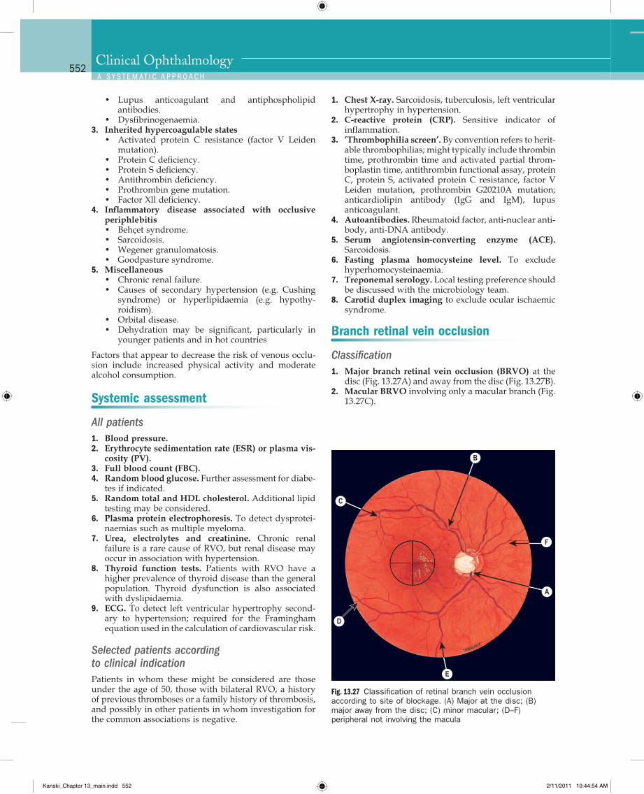
Classification1. Major branch retinal vein occlusion (BRVO) at the
disc (Fig. 13.27A) and away from the disc (Fig. 13.27B).2. Macular BRVO involving only a macular branch (Fig.
13.27C).
• Lupus anticoagulant and antiphospholipid antibodies.
• Dysfibrinogenaemia.3. Inherited hypercoagulable states
• Activated protein C resistance (factor V Leiden mutation).
• Protein C deficiency.• Protein S deficiency.• Antithrombin deficiency.• Prothrombin gene mutation.• Factor Xll deficiency.
4. Inflammatory disease associated with occlusive periphlebitis• Behçet syndrome.• Sarcoidosis.• Wegener granulomatosis.• Goodpasture syndrome.
5. Miscellaneous• Chronic renal failure.• Causes of secondary hypertension (e.g. Cushing
syndrome) or hyperlipidaemia (e.g. hypothy-roidism).
• Orbital disease.• Dehydration may be significant, particularly in
younger patients and in hot countries
Factors that appear to decrease the risk of venous occlu-sion include increased physical activity and moderate alcohol consumption.
Systemic assessment
All patients1. Blood pressure.2. Erythrocyte sedimentation rate (ESR) or plasma vis-
cosity (PV).3. Full blood count (FBC).4. Random blood glucose. Further assessment for diabe-
tes if indicated.5. Random total and HDL cholesterol. Additional lipid
testing may be considered.6. Plasma protein electrophoresis. To detect dysprotei-
naemias such as multiple myeloma.7. Urea, electrolytes and creatinine. Chronic renal
failure is a rare cause of RVO, but renal disease may occur in association with hypertension.
8. Thyroid function tests. Patients with RVO have a higher prevalence of thyroid disease than the general population. Thyroid dysfunction is also associated with dyslipidaemia.
9. ECG. To detect left ventricular hypertrophy second-ary to hypertension; required for the Framingham equation used in the calculation of cardiovascular risk.
Selected patients according to clinical indicationPatients in whom these might be considered are those under the age of 50, those with bilateral RVO, a history of previous thromboses or a family history of thrombosis, and possibly in other patients in whom investigation for the common associations is negative.
Fig. 13.27 Classification of retinal branch vein occlusion according to site of blockage. (A) Major at the disc; (B) major away from the disc; (C) minor macular; (D–F) peripheral not involving the macula
A
E
D
C
B
F
Kanski_Chapter 13_main.indd 552 2/11/2011 10:44:54 AM
Q
553Retinal Vascular Disease 13C H A P T e R
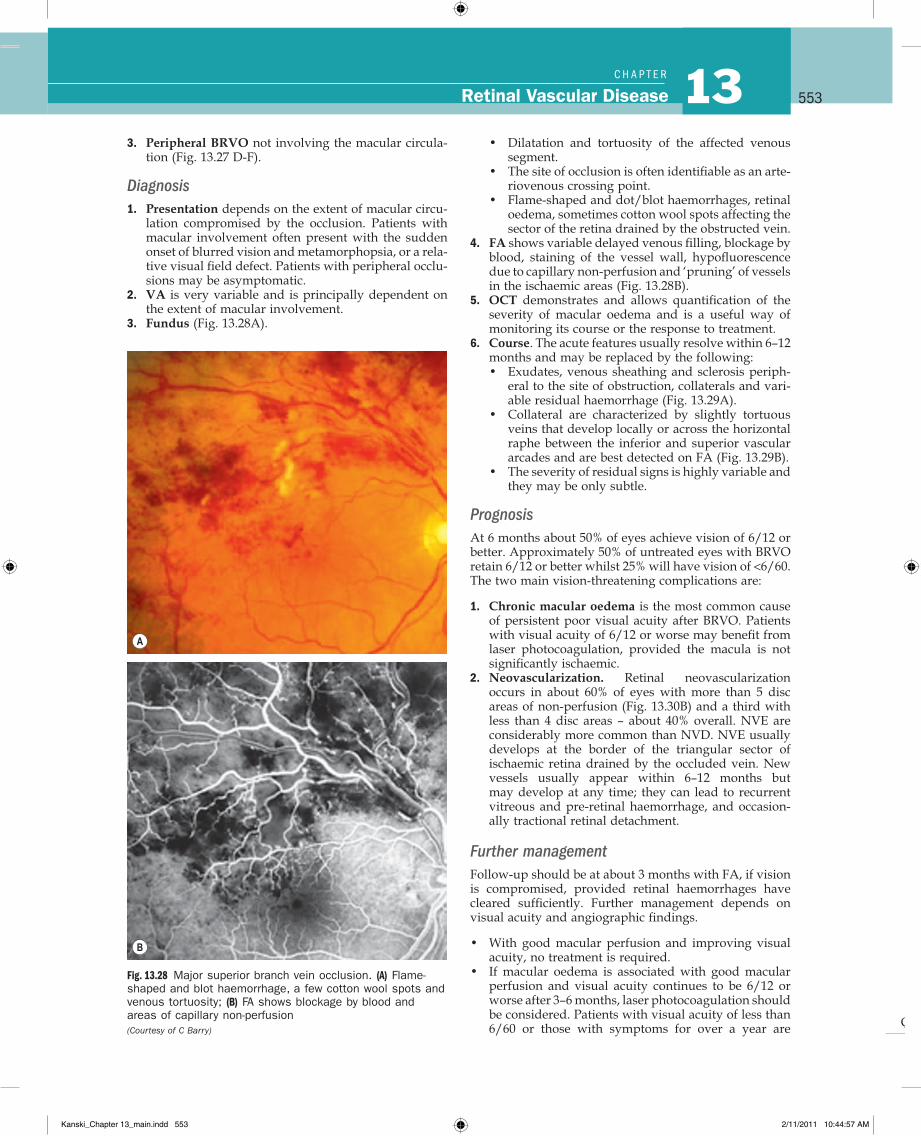
Fig. 13.28 Major superior branch vein occlusion. (A) Flame-shaped and blot haemorrhage, a few cotton wool spots and venous tortuosity; (B) FA shows blockage by blood and areas of capillary non-perfusion (Courtesy of C Barry)
A
B
• Dilatation and tortuosity of the affected venous segment.
• The site of occlusion is often identifiable as an arte-riovenous crossing point.
• Flame-shaped and dot/blot haemorrhages, retinal oedema, sometimes cotton wool spots affecting the sector of the retina drained by the obstructed vein.
4. FA shows variable delayed venous filling, blockage by blood, staining of the vessel wall, hypofluorescence due to capillary non-perfusion and ‘pruning’ of vessels in the ischaemic areas (Fig. 13.28B).
5. OCT demonstrates and allows quantification of the severity of macular oedema and is a useful way of monitoring its course or the response to treatment.
6. Course. The acute features usually resolve within 6–12 months and may be replaced by the following:• Exudates, venous sheathing and sclerosis periph-
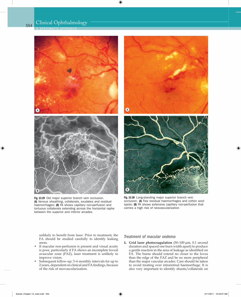
eral to the site of obstruction, collaterals and vari-able residual haemorrhage (Fig. 13.29A).
• Collateral are characterized by slightly tortuous veins that develop locally or across the horizontal raphe between the inferior and superior vascular arcades and are best detected on FA (Fig. 13.29B).
• The severity of residual signs is highly variable and they may be only subtle.
PrognosisAt 6 months about 50% of eyes achieve vision of 6/12 or better. Approximately 50% of untreated eyes with BRVO retain 6/12 or better whilst 25% will have vision of <6/60. The two main vision-threatening complications are:
1. Chronic macular oedema is the most common cause of persistent poor visual acuity after BRVO. Patients with visual acuity of 6/12 or worse may benefit from laser photocoagulation, provided the macula is not significantly ischaemic.
2. Neovascularization. Retinal neovascularization occurs in about 60% of eyes with more than 5 disc areas of non-perfusion (Fig. 13.30B) and a third with less than 4 disc areas – about 40% overall. NVE are considerably more common than NVD. NVE usually develops at the border of the triangular sector of ischaemic retina drained by the occluded vein. New vessels usually appear within 6–12 months but may develop at any time; they can lead to recurrent vitreous and pre-retinal haemorrhage, and occasion-ally tractional retinal detachment.
Further managementFollow-up should be at about 3 months with FA, if vision is compromised, provided retinal haemorrhages have cleared sufficiently. Further management depends on visual acuity and angio graphic findings.
• With good macular perfusion and improving visual acuity, no treatment is required.
• If macular oedema is associated with good macular perfusion and visual acuity continues to be 6/12 or worse after 3–6 months, laser photocoagulation should be considered. Patients with visual acuity of less than 6/60 or those with symptoms for over a year are
3. Peripheral BRVO not involving the macular circula-tion (Fig. 13.27 D-F).
Diagnosis1. Presentation depends on the extent of macular circu-
lation compromised by the occlusion. Patients with macular involvement often present with the sudden onset of blurred vision and metamorphopsia, or a rela-tive visual field defect. Patients with peripheral occlu-sions may be asymptomatic.
2. VA is very variable and is principally dependent on the extent of macular involvement.
3. Fundus (Fig. 13.28A).
Kanski_Chapter 13_main.indd 553 2/11/2011 10:44:57 AM
Q
554Clinical OphthalmologyA S y S T e m A T I C A P P R o A C H
Fig. 13.30 Long-standing major superior branch vein occlusion. (A) Few residual haemorrhages and cotton wool spots; (B) FA shows extensive capillary non-perfusion that carries a high risk of neovascularization
A
B
Fig. 13.29 Old major superior branch vein occlusion. (A) Venous sheathing, collaterals, exudates and residual haemorrhages; (B) FA shows capillary non-perfusion and tortuous collaterals extending across the horizontal raphe between the superior and inferior arcades
A
B
unlikely to benefit from laser. Prior to treatment, the FA should be studied carefully to identify leaking areas.
• If macular non-perfusion is present and visual acuity is poor, particularly if FA shows an incomplete foveal avascular zone (FAZ), laser treatment is unlikely to improve vision.
• Subsequent follow-up: 3–6 monthly intervals for up to 2 years, dependent on clinical and FA findings, because of the risk of neovascularization.
Treatment of macular oedema1. Grid laser photocoagulation (50–100 µm, 0.1 second
duration and spaced one burn width apart) to produce a gentle reaction in the area of leakage as identified on FA. The burns should extend no closer to the fovea than the edge of the FAZ and be no more peripheral than the major vascular arcades. Care should be taken to avoid treating over intraretinal haemorrhage. It is also very important to identify shunts/collaterals on
Kanski_Chapter 13_main.indd 554 2/11/2011 10:45:07 AM
Q
555Retinal Vascular Disease 13C H A P T e R
Impending central retinal vein occlusion
Impending (partial) CRVO is a relatively poorly-defined condition which may resolve or progress to complete obstruction.
1. Presentation is with mild blurring of vision which is characteristically worse on waking and improves during the day.
2. Signs. Mild venous dilatation and tortuosity with a few widely scattered flame-shaped haemorrhages (Fig. 13.32).
3. FA shows increased retinal circulation time.4. OCT may facilitate a degree of objective monitoring
of the macular course, if CMO is present.5. Treatment is aimed at preventing progression to com-
plete occlusion by correcting any predisposing sys-temic conditions, avoiding dehydration, and lowering intraocular pressure (e.g. systemic carbonic anhydrase inhibitors) to improve perfusion. Antiplatelet agents may be of benefit, and in some circumstances such as monocularity in an otherwise healthy patient it may be appropriate to consider other options such as anti-coagulants, fibrinolytics or haemodilution.
Non-ischaemic central retinal vein occlusion
Non-ischaemic CRVO is the most common type, account-ing for about 75%.
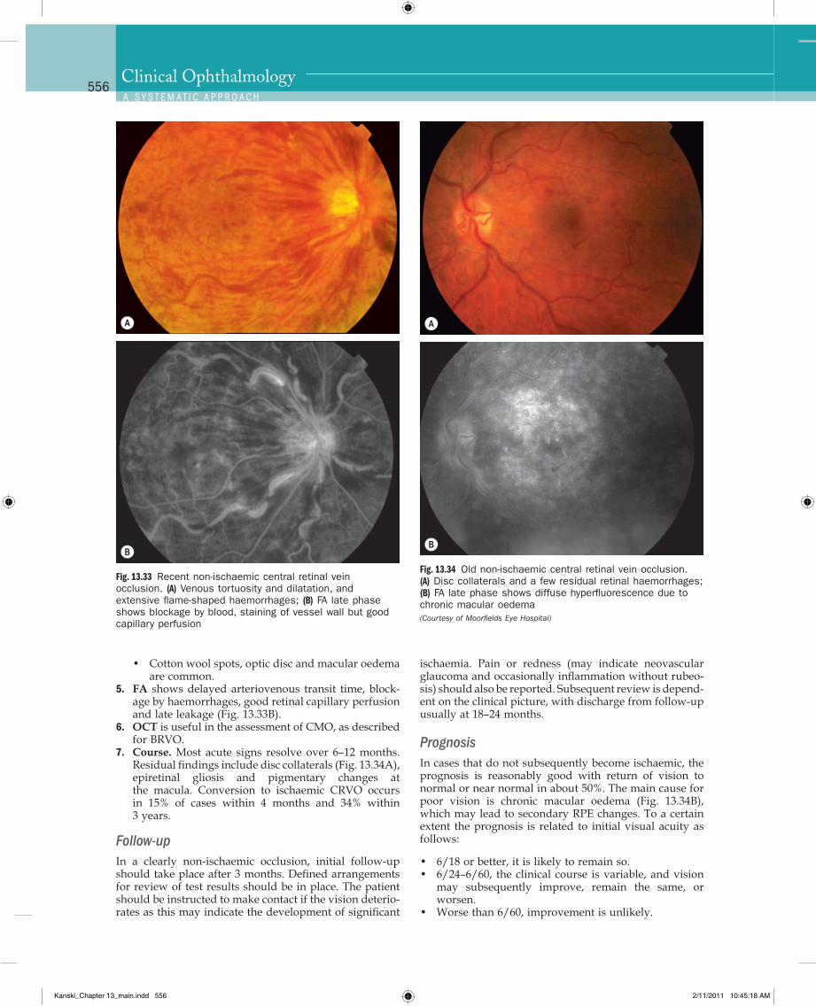
Diagnosis1. Presentation is with sudden, unilateral blurred vision.2. VA is impaired to a moderate-severe degree.3. Relative afferent pupillary defect (RAPD) is absent
or mild (in contrast to ischaemic CRVO).4. Fundus (Fig. 13.33A)
• Tortuosity and dilatation of all branches of the central retinal vein, dot/blot and flame-shaped haemorrhages, throughout all four quadrants and most numerous in the periphery.
FA, which do not leak fluorescein, because they must not be treated. Follow-up should take place after three months. If macular oedema persists, re-treatment may be considered although the results are frequently disappointing.
2. Intravitreal triamcinolone (IVT) is as effective as laser in eyes with macular oedema, but may cause cataract and elevation of intraocular pressure. An average of 2 injections of 1 mg are given in the first year.
3. Periocular steroid injection is less invasive, although probably less effective, than the intravitreal route.
4. Intravitreal anti-VEGF agents. Bevacizumab (Avastin) 0.05 mL/1.25 mg) in a regimen of 2–3 injections over 5–6 months has shown promising effects on macular oedema and vision, including in patients resistant to laser.
5. Arteriovenous sheathotomy. Some positive results have been reported both for sheathotomy and for vit-rectomy alone; a randomized controlled trial showed similar benefit from IVT.
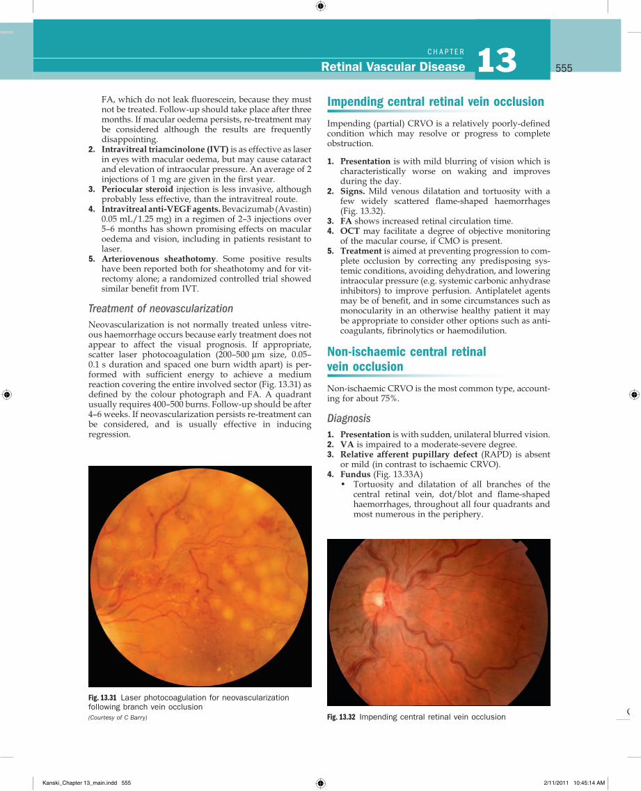
Treatment of neovascularizationNeovascularization is not normally treated unless vitre-ous haemorrhage occurs because early treatment does not appear to affect the visual prognosis. If appropriate, scatter laser photocoagulation (200–500 µm size, 0.05–0.1 s duration and spaced one burn width apart) is per-formed with sufficient energy to achieve a medium reaction covering the entire involved sector (Fig. 13.31) as defined by the colour photograph and FA. A quadrant usually requires 400–500 burns. Follow-up should be after 4–6 weeks. If neovascularization persists re-treatment can be considered, and is usually effective in inducing regression.
Fig. 13.31 Laser photocoagulation for neovascularization following branch vein occlusion(Courtesy of C Barry) Fig. 13.32 Impending central retinal vein occlusion
Kanski_Chapter 13_main.indd 555 2/11/2011 10:45:14 AM
Q
556Clinical OphthalmologyA S y S T e m A T I C A P P R o A C H
ischaemia. Pain or redness (may indicate neovascular glaucoma and occasionally inflammation without rubeo-sis) should also be reported. Subsequent review is depend-ent on the clinical picture, with discharge from follow-up usually at 18–24 months.
PrognosisIn cases that do not subsequently become ischaemic, the prognosis is reasonably good with return of vision to normal or near normal in about 50%. The main cause for poor vision is chronic macular oedema (Fig. 13.34B), which may lead to secondary RPE changes. To a certain extent the prognosis is related to initial visual acuity as follows:
• 6/18 or better, it is likely to remain so.• 6/24–6/60, the clinical course is variable, and vision
may subsequently improve, remain the same, or worsen.
• Worse than 6/60, improvement is unlikely.
• Cotton wool spots, optic disc and macular oedema are common.
5. FA shows delayed arteriovenous transit time, block-age by haemorrhages, good retinal capillary perfusion and late leakage (Fig. 13.33B).
6. OCT is useful in the assessment of CMO, as described for BRVO.
7. Course. Most acute signs resolve over 6–12 months. Residual findings include disc collaterals (Fig. 13.34A), epiretinal gliosis and pigmentary changes at the macula. Conversion to ischaemic CRVO occurs in 15% of cases within 4 months and 34% within 3 years.
Follow-upIn a clearly non-ischaemic occlusion, initial follow-up should take place after 3 months. Defined arrangements for review of test results should be in place. The patient should be instructed to make contact if the vision deterio-rates as this may indicate the development of significant
Fig. 13.33 Recent non-ischaemic central retinal vein occlusion. (A) Venous tortuosity and dilatation, and extensive flame-shaped haemorrhages; (B) FA late phase shows blockage by blood, staining of vessel wall but good capillary perfusion
A
B
Fig. 13.34 Old non-ischaemic central retinal vein occlusion. (A) Disc collaterals and a few residual retinal haemorrhages; (B) FA late phase shows diffuse hyperfluorescence due to chronic macular oedema (Courtesy of Moorfields Eye Hospital)
A
B
Kanski_Chapter 13_main.indd 556 2/11/2011 10:45:18 AM
Q
557Retinal Vascular Disease 13C H A P T e R
8. Course. Most acute signs resolve over 9–12 months. Residual findings include disc collaterals and macular epiretinal gliosis and pigmentary changes. Rarely sub-retinal fibrosis resembling that associated with exuda-tive age-related macular degeneration may develop.
PrognosisThe prognosis is extremely poor due to macular ischae-mia. Rubeosis iridis develops in about 50% of eyes, usually between 2 and 4 months (100-day glaucoma), and there is a high risk of neovascular glaucoma. The development of opticociliary shunts (retinochoroidal collateral veins) may protect the eye from anterior segment neovasculari-zation and probably indicates a dramatic reduction in risk. Retinal neovascularization occurs in about 5% of eyes and is therefore much less common than with BRVO.
Follow upWhere possible, patients with ischaemic CRVO should be seen monthly for 6 months to detect the onset of anterior segment neovascularization. Angle neovascularization (Fig. 13.36A), while not synonymous with progression to
Treatment of macular oedemaLaser photocoagulation for macular oedema is not benefi-cial. Some of the following novel therapies have exhibited apparent significant benefit and may play an increasing role in management.
1. Intravitreal steroid. The SCORE study showed an improvement in the vision of 3 or more lines at one year in over 25% of patients treated with an average of 2 injections of 1 mg triamcinolone versus 7% of con-trols. A trial (GENEVA) of a 0.7 mg dexamethasone sustained-release biodegradeable intravitreal implant (Ozurdex®) showed substantial visual improvement over the first 2 months following a single implantation, though this declined to baseline by 6 months.
2. Intravitreal anti-VEGF agents. Ranibizumab showed a significant visual benefit when used for CMO. Injec-tions were given monthly for 6 months and subse-quently less intensively. Several uncontrolled case series suggest that approximately 50% of patients improve 2 or more lines with intravitreal bevacizu-mab, with 90% of eyes achieving stabilization of vision by 12 months. Pegaptanib also shows promising results.
3. Experimental treatments include chorioretinal anas-tomosis, vitrectomy with radial optic neurotomy or tissue plasminogen activator (rTPA) local infusion.
Ischaemic central retinal vein occlusion
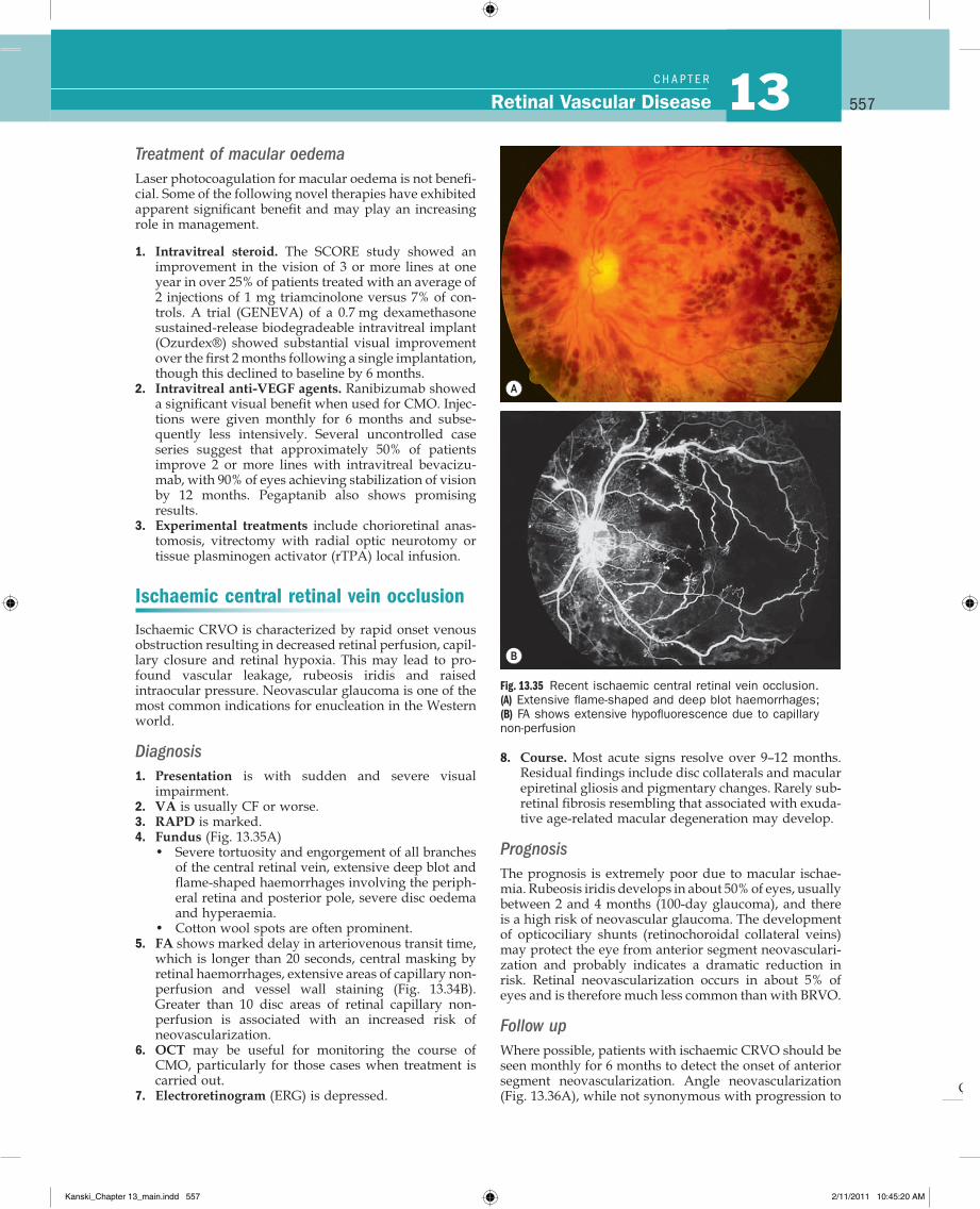
Ischaemic CRVO is characterized by rapid onset venous obstruction resulting in decreased retinal perfusion, capil-lary closure and retinal hypoxia. This may lead to pro-found vascular leakage, rubeosis iridis and raised intraocular pressure. Neovascular glaucoma is one of the most common indications for enucleation in the Western world.
Diagnosis1. Presentation is with sudden and severe visual
impairment.2. VA is usually CF or worse.3. RAPD is marked.4. Fundus (Fig. 13.35A)
• Severe tortuosity and engorgement of all branches of the central retinal vein, extensive deep blot and flame-shaped haemorrhages involving the periph-eral retina and posterior pole, severe disc oedema and hyperaemia.
• Cotton wool spots are often prominent.5. FA shows marked delay in arteriovenous transit time,
which is longer than 20 seconds, central masking by retinal haemorrhages, extensive areas of capillary non- perfusion and vessel wall staining (Fig. 13.34B). Greater than 10 disc areas of retinal capillary non-perfusion is associated with an increased risk of neovascularization.
6. OCT may be useful for monitoring the course of CMO, particularly for those cases when treatment is carried out.
7. Electroretinogram (ERG) is depressed.
Fig. 13.35 Recent ischaemic central retinal vein occlusion. (A) Extensive flame-shaped and deep blot haemorrhages; (B) FA shows extensive hypofluorescence due to capillary non-perfusion
A
B
Kanski_Chapter 13_main.indd 557 2/11/2011 10:45:20 AM
Q
558Clinical OphthalmologyA S y S T e m A T I C A P P R o A C H
prior to mydriasis. Prophylactic PRP is generally not rec-ommended even with marked ischaemia unless iris new vessels develop, though may be considered in patients unlikely to attend scheduled review. Subsequent review should usually be for up to 2 years to detect significant ischaemia and macular oedema.
Treatment of neovascularizationLaser PRP should be performed without delay in eyes with angle neovascularization or rubeosis iridis. This involves the application of 1500–3000 burns (0.5–0.1 second, spaced one burn width apart), with sufficient energy to produce a moderate reaction in the periphery but avoiding areas of haemorrhage (Fig. 13.36C). Some cases require further treatment if rubeosis fails to regress or continues to progress. Intravitreal anti-VEGF injections may be used adjunctivally in selected cases.
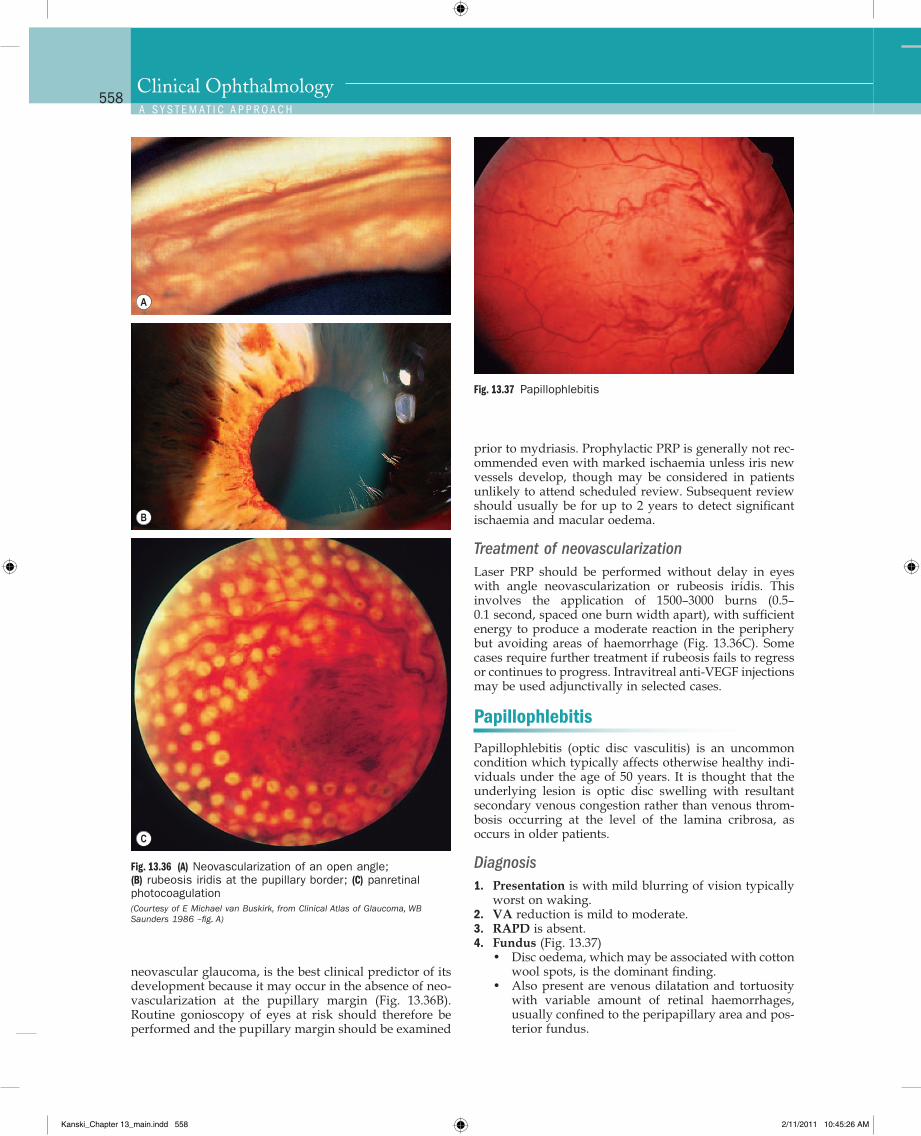
Papillophlebitis
Papillophlebitis (optic disc vasculitis) is an uncommon condition which typically affects otherwise healthy indi-viduals under the age of 50 years. It is thought that the underlying lesion is optic disc swelling with resultant secondary venous congestion rather than venous throm-bosis occurring at the level of the lamina cribrosa, as occurs in older patients.
Diagnosis1. Presentation is with mild blurring of vision typically
worst on waking.2. VA reduction is mild to moderate.3. RAPD is absent.4. Fundus (Fig. 13.37)
• Disc oedema, which may be associated with cotton wool spots, is the dominant finding.
• Also present are venous dilatation and tortuosity with variable amount of retinal haemorrhages, usually confined to the peripapillary area and pos-terior fundus.
neovascular glaucoma, is the best clinical predictor of its development because it may occur in the absence of neo-vascularization at the pupillary margin (Fig. 13.36B). Routine gonioscopy of eyes at risk should therefore be performed and the pupillary margin should be examined
Fig. 13.36 (A) Neovascularization of an open angle; (B) rubeosis iridis at the pupillary border; (C) panretinal photocoagulation (Courtesy of E Michael van Buskirk, from Clinical Atlas of Glaucoma, WB Saunders 1986 –fig. A)
A
B
C
Fig. 13.37 Papillophlebitis
Kanski_Chapter 13_main.indd 558 2/11/2011 10:45:26 AM
Q
559Retinal Vascular Disease 13C H A P T e R
5. Blind spot is enlarged on perimetry.6. FA shows mild delay in arteriovenous transit time,
hyperfluorescence due to leakage and good capillary perfusion.
7. OCT may show CMO.
PrognosisThe prognosis is excellent despite the lack of treatment. Eighty per cent of cases achieve a final visual acuity of 6/12 or better. The remainder suffer significant and per-manent visual impairment as a result of macular oedema.
Hemiretinal vein occlusion
Hemiretinal vein occlusion is generally regarded as a variant of CRVO and may be ischaemic or non-ischaemic. It is less common than both BRVO and CRVO and involves occlusion of the superior or inferior branch of the CRV. A hemispheric occlusion blocks a major branch of the CRV at or near the optic disc. A hemicentral occlusion, which is less common, involves one trunk of a dual-trunked CRV, which persists in the anterior part of the optic nerve head as a congenital variant.
1. Presentation is with a sudden onset altitudinal visual field defect.
2. VA reduction is variable.3. Fundus shows the features of BRVO, involving the
superior or inferior hemisphere (Fig. 13.38A).4. FA shows masking by haemorrhages, hyperfluores-
cence due to leakage and variable capillary non-perfusion (Fig. 13.38B).
5. Treatment depends on the severity of retinal ischae-mia. Extensive retinal ischaemia carries the risk of neovascular glaucoma and should be managed in the same way as ischaemic CRVO. Macular oedema usually responds poorly to grid laser due to extensive foveal capillary shutdown; newer treatments may be effective in some cases.
Systemic treatment in retinal vein occlusion
1. Control of systemic risk factors. This will also amel-iorate adverse systemic vascular outcomes, because retinal vein occlusions are associated with cerebro- and cardiovascular causes of death.
2. Antiplatelet therapy with aspirin or an alternative agent should be considered according to systemic risk factors, and might reduce the risk of further venous occlusion.
3. Hormone replacement therapy (HRT). The risk of HRT remains undefined. Most authorities would avoid commencing oestrogen-containing HRT follow-ing RVO in women not already taking this.
4. Isovolaemic haemodilution. The results of trials are inconsistent, though benefit has been shown by some.
5. Other. A range of treatment modalities (e.g. plas-mapheresis) has been employed to try to improve visual outcomes in RVO, but as yet clear evidence for benefit is lacking.
Fig. 13.38 (A) Inferior hemiretinal vein occlusion; (B) FA late phase shows extensive hypofluorescence due to capillary non-perfusion and mild perivascular hyperfluorescence (Courtesy of C Barry)
A
B
RETINAL ARTERIAL OCCLUSIVE DISEASE
Aetiology
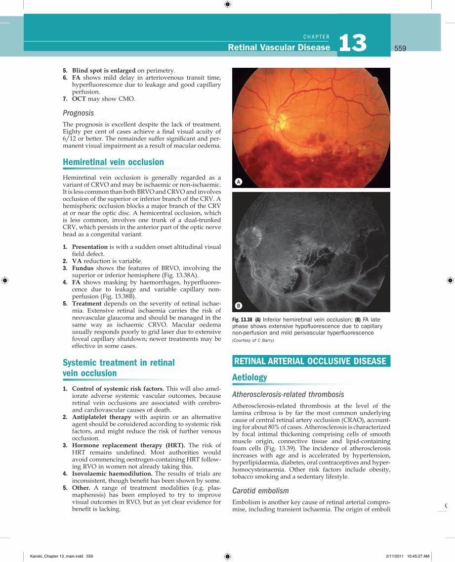
Atherosclerosis-related thrombosisAtherosclerosis-related thrombosis at the level of the lamina cribrosa is by far the most common underlying cause of central retinal artery occlusion (CRAO), account-ing for about 80% of cases. Atherosclerosis is characterized by focal intimal thickening comprising cells of smooth muscle origin, connective tissue and lipid-containing foam cells (Fig. 13.39). The incidence of atherosclerosis increases with age and is accelerated by hypertension, hyperlipidaemia, diabetes, oral contraceptives and hyper-homocysteinaemia. Other risk factors include obesity, tobacco smoking and a sedentary lifestyle.
Carotid embolismEmbolism is another key cause of retinal arterial compro-mise, including transient ischaemia. The origin of emboli
Kanski_Chapter 13_main.indd 559 2/11/2011 10:45:27 AM
Q
560Clinical OphthalmologyA S y S T e m A T I C A P P R o A C H
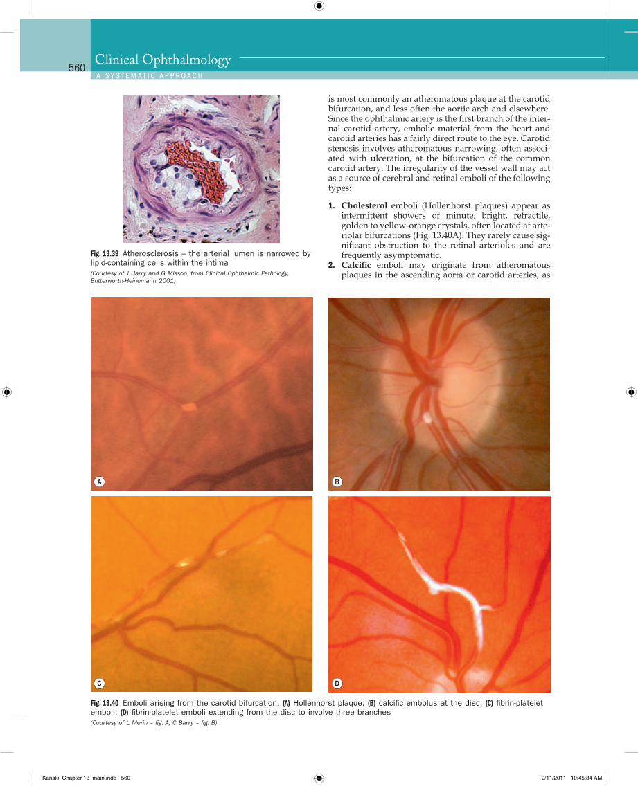
is most commonly an atheromatous plaque at the carotid bifurcation, and less often the aortic arch and elsewhere. Since the ophthalmic artery is the first branch of the inter-nal carotid artery, embolic material from the heart and carotid arteries has a fairly direct route to the eye. Carotid stenosis involves atheromatous narrowing, often associ-ated with ulceration, at the bifurcation of the common carotid artery. The irregularity of the vessel wall may act as a source of cerebral and retinal emboli of the following types:
1. Cholesterol emboli (Hollenhorst plaques) appear as intermittent showers of minute, bright, refractile, golden to yellow-orange crystals, often located at arte-riolar bifurcations (Fig. 13.40A). They rarely cause sig-nificant obstruction to the retinal arterioles and are frequently asymptomatic.
2. Calcific emboli may originate from atheromatous plaques in the ascending aorta or carotid arteries, as
Fig. 13.40 Emboli arising from the carotid bifurcation. (A) Hollenhorst plaque; (B) calcific embolus at the disc; (C) fibrin-platelet emboli; (D) fibrin-platelet emboli extending from the disc to involve three branches (Courtesy of L Merin – fig. A; C Barry – fig. B)
A B
C D
Fig. 13.39 Atherosclerosis – the arterial lumen is narrowed by lipid-containing cells within the intima (Courtesy of J Harry and G Misson, from Clinical Ophthalmic Pathology, Butterworth-Heinemann 2001)
Kanski_Chapter 13_main.indd 560 2/11/2011 10:45:34 AM
Q
561Retinal Vascular Disease 13C H A P T e R
well as from calcified heart valves. They are usually single, white, non-scintillating and often on or close to the disc (Fig. 13.40B). When located on the disc itself, they may be easily overlooked as they tend to merge with the disc. They may cause permanent occlusion of the central retinal artery or one of its main branches.
3. Fibrin-platelet emboli are dull grey, elongated parti-cles which are usually multiple (Fig. 13.40C) and occa-sionally fill the entire lumen (Fig. 13.40D). They may cause a retinal transient ischaemic attack (TIA), with resultant amaurosis fugax, and occasionally complete obstruction.
Uncommon causes1. Giant cell (temporal) arteritis (GCA) is a common
cause of anterior ischaemic optic neuropathy but iso-lated CRAO is rare.
2. Cardiac embolism from the heart and its valves may consist of calcific material, vegetations in bacterial endocarditis, thrombus from the left side of the heart and rarely myxomatous material from an atrial myxoma.
3. Periarteritis associated with dermatomyositis, sys-temic lupus erythematosus, polyarteritis nodosa, Wegener granulomatosis and Behçet syndrome may occasionally be responsible for branch retinal artery occlusion (BRAO), which may be multiple and bilat-eral (Fig. 13.41A and B).
4. Thrombophilic disorders that may be associated with retinal artery occlusion, especially in younger individuals, include hyperhomocysteinaemia, anti-phospholipid antibody syndrome and inherited defects of natural anticoagulants.
5. Sickling haemoglobinopathies.6. Retinal migraine may very rarely be responsible for
retinal artery occlusion in young individuals. However, the diagnosis should be made only after other more common causes have been excluded.
7. Susac syndrome (retinocochleocerebral vasculopathy) is a microangiopathy characterized by the triad of retinal artery occlusion, sensorineural deafness and encephalopathy.
Systemic assessment
All patientsMany patients will have a history of vascular disease; enquiry should be made about smoking.
1. Symptoms of GCA such as headache, jaw claudica-tion, scalp tenderness, limb girdle pain, weight loss and existing polymyalgia rheumatica (see Ch. 19).
2. Pulse, particularly to detect atrial fibrillation.3. Blood pressure.4. Cardiac auscultation.5. Carotid examination.
a. Palpation of severe or complete stenosis is associated with a diminished or absent carotid pulse.
b. Auscultation over a partial stenosis gives rise to a bruit, best detected with the bell of the stethoscope. It is important to perform auscultation along the
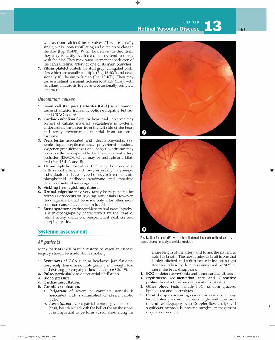
Fig. 13.41 (A) and (B) Multiple bilateral branch retinal artery occlusions in polyarteritis nodosa
A
B
entire length of the artery and to ask the patient to hold his breath. The most ominous bruit is one that is high-pitched and soft because it indicates tight stenosis. When the lumen is narrowed by 90% or more, the bruit disappears.
6. ECG to detect arrhythmia and other cardiac disease.7. Erythrocyte sedimentation rate and C-reactive
protein to detect the remote possibility of GCA.8. Other blood tests include FBC, random glucose,
lipids, urea and electrolytes.9. Carotid duplex scanning is a non-invasive screening
test involving a combination of high-resolution real-time ultrasonography with Doppler flow analysis. If significant stenosis is present, surgical management may be considered.
Kanski_Chapter 13_main.indd 561 2/11/2011 10:45:38 AM
Q
562Clinical OphthalmologyA S y S T e m A T I C A P P R o A C H
Selected patientsThe following additional tests can be considered on a targeted basis in some patients, particularly if younger and with no known cardiovascular risk factors.
1. Further carotid imaging (see Ch. 19)2. Cranial MRI or CT may be indicated to rule out intra-
cranial or orbital pathology; may be required prior to fibrinolysis.
3. Echocardiography. Usually performed if there is a spe-cific indication such as a history of rheumatoid fever, known cardiac valvular disease, or intravenous drug use.
4. Chest X-ray. Sarcoidosis, tuberculosis, left ventricular hypertrophy in hypertension.
5. 24-hour ECG (Holter monitor) to exclude intermittent arrhythmia.
6. Additional blood testsa. Fasting plasma homocysteine level to exclude
hyperhomocysteinaemia.b. ’Thrombophilia screen’. By convention refers to
heritable thrombophilias, which have predomi-nantly been implicated in venous rather than arterial thromboses.
c. Plasma protein electrophoresis to detect dyspro-teinaemias such as multiple myeloma.
d. Thyroid function tests, especially if atrial fibril-lation is present; may be associated with dyslipidaemia.
e. Autoantibodies. Rheumatoid factor, anti-nuclear antibody, anti-DNA antibody.
f. Blood cultures.
Amaurosis fugax
Amaurosis fugax is characterized by painless transient monocular loss of vision, often described as a curtain coming down over the eye, usually from top to bottom, but occasionally vice versa; it is common for patients to be unaware of whether transient unilateral visual loss affects one eye or the ipsilateral hemifield (cerebral ischae-mia) of both. Visual loss, which may be complete, usually lasts a few minutes. Recovery is in the same pattern as the initial loss, although usually more gradual. Frequency of attacks may vary from several times a day to once every few months. The attacks may sometimes be accompanied by ipsilateral cerebral TIA, with contralateral neurological features. Investigation and systemic management is carried out as for persistent arterial occlusion.
Branch retinal artery occlusion
Diagnosis1. Presentation is with sudden and profound painless
altitudinal or sectoral visual field loss. It can sometimes go unnoticed, particularly if central vision is spared.
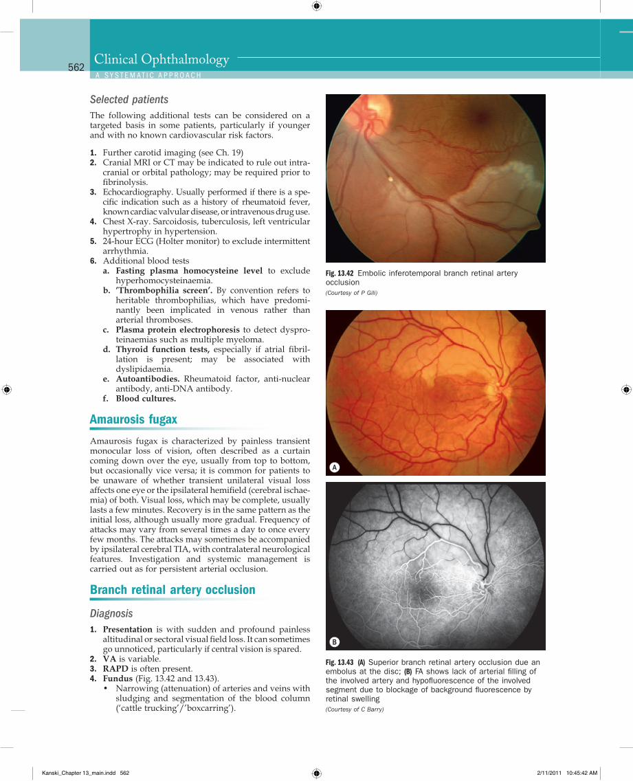
2. VA is variable.3. RAPD is often present.4. Fundus (Fig. 13.42 and 13.43).
• Narrowing (attenuation) of arteries and veins with sludging and segmentation of the blood column (‘cattle trucking’/’boxcarring’).
Fig. 13.42 Embolic inferotemporal branch retinal artery occlusion (Courtesy of P Gili)
Fig. 13.43 (A) Superior branch retinal artery occlusion due an embolus at the disc; (B) FA shows lack of arterial filling of the involved artery and hypofluorescence of the involved segment due to blockage of background fluorescence by retinal swelling (Courtesy of C Barry)
A
B
Kanski_Chapter 13_main.indd 562 2/11/2011 10:45:42 AM
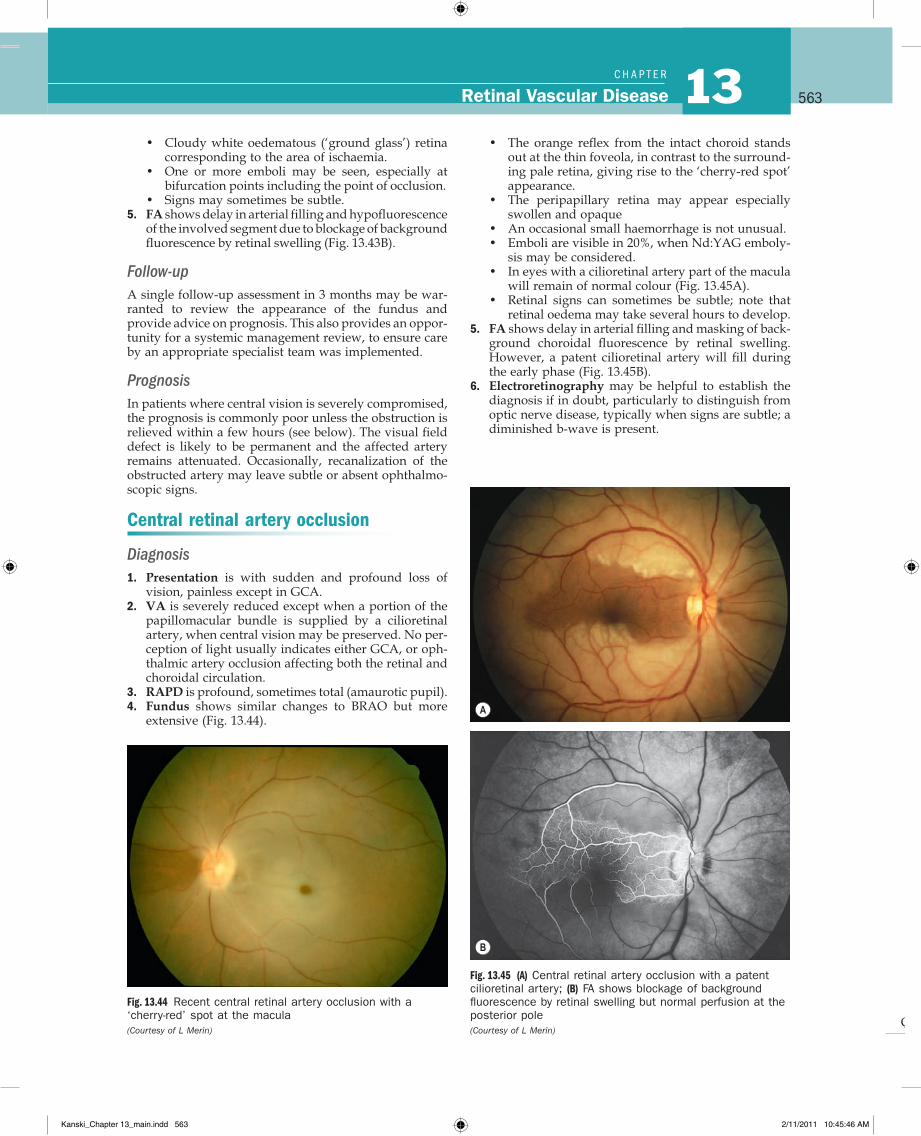
Q
563Retinal Vascular Disease 13C H A P T e R
• The orange reflex from the intact choroid stands out at the thin foveola, in contrast to the surround-ing pale retina, giving rise to the ‘cherry-red spot’ appearance.
• The peripapillary retina may appear especially swollen and opaque
• An occasional small haemorrhage is not unusual.• Emboli are visible in 20%, when Nd:YAG emboly-
sis may be considered.• In eyes with a cilioretinal artery part of the macula
will remain of normal colour (Fig. 13.45A).• Retinal signs can sometimes be subtle; note that
retinal oedema may take several hours to develop.5. FA shows delay in arterial filling and masking of back-
ground choroidal fluorescence by retinal swelling. However, a patent cilioretinal artery will fill during the early phase (Fig. 13.45B).
6. Electroretinography may be helpful to establish the diagnosis if in doubt, particularly to distinguish from optic nerve disease, typically when signs are subtle; a diminished b-wave is present.
• Cloudy white oedematous (‘ground glass’) retina corresponding to the area of ischaemia.
• One or more emboli may be seen, especially at bifurcation points including the point of occlusion.
• Signs may sometimes be subtle.5. FA shows delay in arterial filling and hypofluorescence
of the involved segment due to blockage of background fluorescence by retinal swelling (Fig. 13.43B).
Follow-upA single follow-up assessment in 3 months may be war-ranted to review the appearance of the fundus and provide advice on prognosis. This also provides an oppor-tunity for a systemic management review, to ensure care by an appropriate specialist team was implemented.
PrognosisIn patients where central vision is severely compromised, the prognosis is commonly poor unless the obstruction is relieved within a few hours (see below). The visual field defect is likely to be permanent and the affected artery remains attenuated. Occasionally, recanalization of the obstructed artery may leave subtle or absent ophthalmo-scopic signs.
Central retinal artery occlusion
Diagnosis1. Presentation is with sudden and profound loss of
vision, painless except in GCA.2. VA is severely reduced except when a portion of the
papillomacular bundle is supplied by a cilioretinal artery, when central vision may be preserved. No per-ception of light usually indicates either GCA, or oph-thalmic artery occlusion affecting both the retinal and choroidal circulation.
3. RAPD is profound, sometimes total (amaurotic pupil).4. Fundus shows similar changes to BRAO but more
extensive (Fig. 13.44).
Fig. 13.44 Recent central retinal artery occlusion with a ‘cherry-red’ spot at the macula (Courtesy of L Merin)
Fig. 13.45 (A) Central retinal artery occlusion with a patent cilioretinal artery; (B) FA shows blockage of background fluorescence by retinal swelling but normal perfusion at the posterior pole (Courtesy of L Merin)
A
B
Kanski_Chapter 13_main.indd 563 2/11/2011 10:45:46 AM
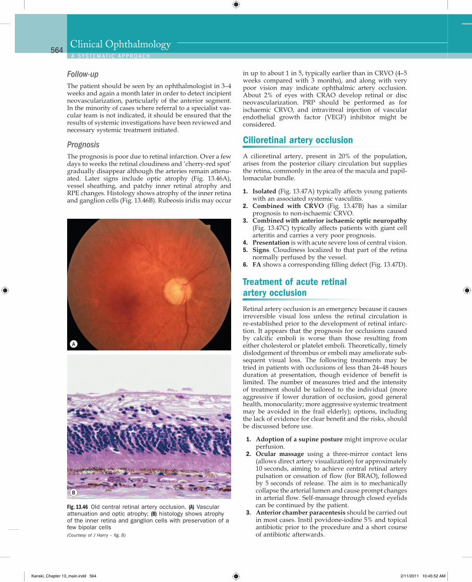
Q
564Clinical OphthalmologyA S y S T e m A T I C A P P R o A C H
in up to about 1 in 5, typically earlier than in CRVO (4–5 weeks compared with 3 months), and along with very poor vision may indicate ophthalmic artery occlusion. About 2% of eyes with CRAO develop retinal or disc neovascularization. PRP should be performed as for ischaemic CRVO, and intravitreal injection of vascular endothelial growth factor (VEGF) inhibitor might be considered.
Cilioretinal artery occlusion
A cilioretinal artery, present in 20% of the population, arises from the posterior ciliary circulation but supplies the retina, commonly in the area of the macula and papil-lomacular bundle.
1. Isolated (Fig. 13.47A) typically affects young patients with an associated systemic vasculitis.
2. Combined with CRVO (Fig. 13.47B) has a similar prognosis to non-ischaemic CRVO.
3. Combined with anterior ischaemic optic neuropathy (Fig. 13.47C) typically affects patients with giant cell arteritis and carries a very poor prognosis.
4. Presentation is with acute severe loss of central vision.5. Signs. Cloudiness localized to that part of the retina
normally perfused by the vessel.6. FA shows a corresponding filling defect (Fig. 13.47D).
Treatment of acute retinal artery occlusion
Retinal artery occlusion is an emergency because it causes irreversible visual loss unless the retinal circulation is re-established prior to the development of retinal infarc-tion. It appears that the prognosis for occlusions caused by calcific emboli is worse than those resulting from either cholesterol or platelet emboli. Theoretically, timely dislodgement of thrombus or emboli may ameliorate sub-sequent visual loss. The following treatments may be tried in patients with occlusions of less than 24–48 hours duration at presentation, though evidence of benefit is limited. The number of measures tried and the intensity of treatment should be tailored to the individual (more aggressive if lower duration of occlusion, good general health, monocularity; more aggressive systemic treatment may be avoided in the frail elderly); options, including the lack of evidence for clear benefit and the risks, should be discussed before use.
1. Adoption of a supine posture might improve ocular perfusion.
2. Ocular massage using a three-mirror contact lens (allows direct artery visualization) for approximately 10 seconds, aiming to achieve central retinal artery pulsation or cessation of flow (for BRAO), followed by 5 seconds of release. The aim is to mechanically collapse the arterial lumen and cause prompt changes in arterial flow. Self-massage through closed eyelids can be continued by the patient.
3. Anterior chamber paracentesis should be carried out in most cases. Instil povidone-iodine 5% and topical antibiotic prior to the procedure and a short course of antibiotic afterwards.
Follow-upThe patient should be seen by an ophthalmologist in 3–4 weeks and again a month later in order to detect incipient neovascularization, particularly of the anterior segment. In the minority of cases where referral to a specialist vas-cular team is not indicated, it should be ensured that the results of systemic investigations have been reviewed and necessary systemic treatment initiated.
PrognosisThe prognosis is poor due to retinal infarction. Over a few days to weeks the retinal cloudiness and ‘cherry-red spot’ gradually disappear although the arteries remain attenu-ated. Later signs include optic atrophy (Fig. 13.46A), vessel sheathing, and patchy inner retinal atrophy and RPE changes. Histology shows atrophy of the inner retina and ganglion cells (Fig. 13.46B). Rubeosis iridis may occur
Fig. 13.46 Old central retinal artery occlusion. (A) Vascular attenuation and optic atrophy; (B) histology shows atrophy of the inner retina and ganglion cells with preservation of a few bipolar cells (Courtesy of J Harry – fig. B)
A
B
Kanski_Chapter 13_main.indd 564 2/11/2011 10:45:52 AM

Q
565Retinal Vascular Disease 13C H A P T e R
10. Thrombolysis. Extrapolating from successful treat-ment of stroke and myocardial infarction, various strategies have been used to deliver thrombolytic agents to the ophthalmic artery, including local arte-rial (internal carotid and ophthalmic) and intrave-nous infusion. Many studies suggest that this may improve the visual outcomes in CRAO, with a low risk of serious complications. However, no benefit over conservative treatment has been conclusively demonstrated, though there is evidence that improve-ment is more likely if commenced within the first 6 hours of presentation.
Systemic prophylaxis following retinal arterial occlusion
The risk of stroke is relatively high in the first few days following retinal artery occlusion or amaurosis fugax, and ‘fast-track’ referral to a specialist stroke clinic is advisable.
4. Topical apraclonidine 1%, timolol 0.5% and intra-venous acetazolamide 500 mg to achieve a more sustained lowering of intraocular pressure.
5. Sublingual isosorbide dinitrate to induce vasodilation.
6. ‘Rebreathing’ into a paper bag in order to elevate blood carbon dioxide and respiratory acidosis has been advocated, as this may promote vasodilation.
7. Breathing a high oxygen (95%) and carbon dioxide (5%) mixture, ‘carbogen’, has been advocated for a possible dual effect of retarding ischaemia and vasodilation.
8. Hyperosmotic agents. Mannitol or glycerol have been used for their possibly more rapid IOP-lowering effect as well as increased intravascular volume.
9. Transluminal Nd:YAG laser embolysis has been advocated for BRAO or CRAO in which an occluding embolus is visible; shots of 0.5–1.0 mJ or higher are applied directly to the embolus using a fundus contact lens. Embolectomy has been said to occur if the embolus is ejected into the vitreous via a hole in the arteriole. The main complication is vitreous haemorrhage.
Fig. 13.47 Cilioretinal artery occlusion. (A) Isolated; (B) combined with central retinal vein occlusion; (C) combined with anterior ischaemic optic neuropathy; (D) FA shows hypofluorescence at the macula due to lack of filling and masking by retinal swelling (Courtesy of S Milewski – fig. D)
A B
C D
Kanski_Chapter 13_main.indd 565 2/11/2011 10:45:56 AM
Q
566Clinical OphthalmologyA S y S T e m A T I C A P P R o A C H
1. General risk factors as discussed above should be addressed and smoking should be discontinued. Urgent referral to an appropriate physician is manda-tory for significant cardiac arrhythmia.
2. Antiplatelet therapy is commenced provided there are no contraindications; an immediate loading dose of 600 mg may be given; alternative/additional agents include dipyridamole and clopidogrel. If fibrinolysis (see above) is being considered, this should be dis-cussed with a physician prior to starting antiplatelet treatment.
3. Oral anticoagulation (e.g. warfarin) may be pre-scribed for some patients, particularly those with atrial fibrillation.
4. Carotid endarterectomy may be indicated in patients with symptomatic stenosis greater than 70%.
Asymptomatic retinal embolus
It is not uncommon to identify a retinal embolus on routine examination of an asymptomatic older patient. This indi-cates a substantially increased risk of stroke and ischaemic heart disease, and management should consist of evalua-tion and treatment of risk factors discussed above. A higher threshold for carotid surgery is appropriate.
OCULAR ISCHAEMIC SYNDROME
PathogenesisOcular ischaemic syndrome (OIS) is an uncommon condi-tion which is the result of chronic ocular hypoperfusion secondary to severe ipsilateral atherosclerotic carotid steno-sis of more than 90%, resulting in a 50% reduction of ipsilat-eral perfusion pressure. It typically affects patients during the 7th decade and may be associated with diabetes, hyper-tension, ischaemic heart disease and cerebro vascular disease. The male : female ratio is about 2 : 1. Five year mor-tality is in the order of 40%, most frequently from cardiac disease. Patients with ocular ischaemic syndrome may also give a history of amaurosis fugax due to retinal embolism.
DiagnosisOIS is unilateral in 80% of cases. It affects both anterior and posterior segments. The signs are variable and may be subtle such that the condition is missed or misdiagnosed.
1. Presentation is usually with gradual loss of vision over several weeks or months although occasionally visual loss may be sudden or fleeting (amaurosis fugax). Ocular and periocular pain may also be present (40%). Patients may notice unusually persistent after-images, or worsening of vision with sudden exposure to bright light (‘bright light amaurosis fugax’), with slow adap-tation. The prognosis for vision is often very poor, though patients with better acuity at presentation are more likely to retain this. About 25% will deteriorate to ‘light perception’ by the end of 1 year.
2. Anterior segment• Diffuse episcleral injection and corneal oedema.• Aqueous flare with few if any cells (ischaemic
pseudoiritis).
• Iris atrophy and a mid-dilated and poorly reacting pupil.
• Rubeosis iridis is common, developing in up to 90%, and often progresses to neovascular glaucoma; the IOP may remain low due to poor ocular perfusion.
• Cataract in advanced cases.3. Fundus
• Venous dilatation, arteriolar narrowing, and haemorrhages and occasionally disc oedema (Fig. 13.48A) and cotton wool spots.
• Proliferative retinopathy with NVD and occasion-ally NVE.
• Spontaneous arterial pulsation, most pronounced near the optic disc, is present in most cases or may be easily induced by exerting gentle pressure on the globe (digital ophthalmodynamometry).
• Macular oedema can occur.• In diabetic patients retinopathy may be more
severe ipsilateral to carotid stenosis.4. FA
• Early phase shows delayed choroidal filling and prolonged arteriovenous transit time (Fig. 13.48B and C).
• Late phase shows disc and perivascular hyperfluo-rescence, and leakage at the posterior pole (Fig. 13.48D).
5. Carotid imaging may involve duplex scanning, digital subtraction angiography, MR or CT angiography.
Management1. Anterior segment manifestations are treated with
topical steroids and mydriatics.2. Neovascular glaucoma may be treated medically or
surgically (see Ch. 10).3. Proliferative retinopathy can be treated with PRP
although the outcome is considerably less certain than in proliferative diabetic retinopathy.
4. Macular oedema may respond to intravitreal steroid or anti-VEGF agents, or to carotid surgery.
5. Carotid surgery. Endarterectomy or stenting may be performed to reduce the risk of stroke, may be benefi-cial for proliferative retinopathy and neovascular glau-coma, and may help to stabilize vision. Endarterectomy cannot be performed where there is total obstruction; extracranial-intracranial arterial bypass surgery is sometimes carried out. It should be noted that an increase in ocular perfusion following surgery can sometimes be associated with both a rise in intraocular pressure and exacerbation of neovascularization.
6. Investigation and management of cardiovascular risk factors in conjunction with the appropriate medical specialists is essential. OIS is occasionally the only manifestation of marked systemic vascular disease. Full investigation should be carried out, broadly similar to that for retinal arterial occlusion.
Differential diagnosis1. Non-ischaemic CRVO also exhibits unilateral retinal
haemorrhages, venous dilatation and cotton wool spots. However, haemorrhages are more numerous and mainly flame-shaped, and disc oedema is often
Kanski_Chapter 13_main.indd 566 2/11/2011 10:45:56 AM
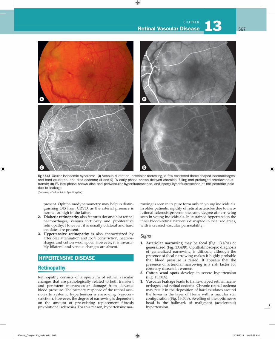
Q
567Retinal Vascular Disease 13C H A P T e R
rowing is seen in its pure form only in young individuals. In older patients, rigidity of retinal arterioles due to invo-lutional sclerosis prevents the same degree of narrowing seen in young individuals. In sustained hypertension the inner blood–retinal barrier is disrupted in localized areas, with increased vascular permeability.
Signs1. Arteriolar narrowing may be focal (Fig. 13.49A) or
generalized (Fig. 13.49B). Ophthalmoscopic diagnosis of generalized narrowing is difficult, although the presence of focal narrowing makes it highly probable that blood pressure is raised. It appears that the presence of arteriolar narrowing is a risk factor for coronary disease in women.
2. Cotton wool spots develop in severe hypertension (Fig. 13.50A).
3. Vascular leakage leads to flame-shaped retinal haem-orrhages and retinal oedema. Chronic retinal oedema may result in the deposition of hard exudates around the fovea in the layer of Henle with a macular star configuration (Fig. 13.50B). Swelling of the optic nerve head is the hallmark of malignant (accelerated) hypertension.
present. Ophthalmodynamometry may help in distin-guishing OIS from CRVO, as the arterial pressure is normal or high in the latter.
2. Diabetic retinopathy also features dot and blot retinal haemorrhages, venous tortuosity and proliferative retinopathy. However, it is usually bilateral and hard exudates are present.
3. Hypertensive retinopathy is also characterized by arteriolar attenuation and focal constriction, haemor-rhages and cotton wool spots. However, it is invaria-bly bilateral and venous changes are absent.
HYPERTENSIVE DISEASE
Retinopathy
Retinopathy consists of a spectrum of retinal vascular changes that are pathologically related to both transient and persistent microvascular damage from elevated blood pressure. The primary response of the retinal arte-rioles to systemic hypertension is narrowing (vasocon-striction). However, the degree of narrowing is dependent on the amount of pre-existing replacement fibrosis (involutional sclerosis). For this reason, hypertensive nar-
Fig. 13.48 Ocular ischaemic syndrome. (A) Venous dilatation, arteriolar narrowing, a few scattered flame-shaped haemorrhages and hard exudates, and disc oedema; (B and C) FA early phase shows delayed choroidal filling and prolonged arteriovenous transit; (D) FA late phase shows disc and perivascular hyperfluorescence, and spotty hyperfluorescence at the posterior pole due to leakage (Courtesy of Moorfields Eye Hospital)
A B
C D
Kanski_Chapter 13_main.indd 567 2/11/2011 10:45:58 AM
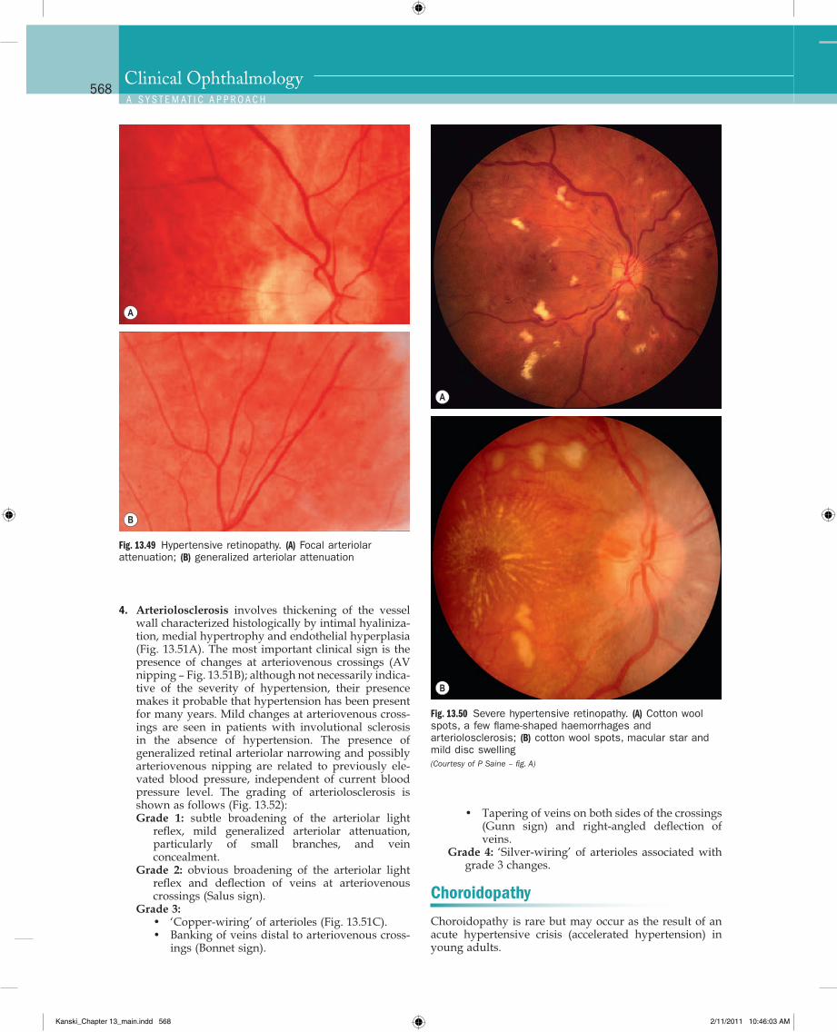
Q
568Clinical OphthalmologyA S y S T e m A T I C A P P R o A C H
Fig. 13.49 Hypertensive retinopathy. (A) Focal arteriolar attenuation; (B) generalized arteriolar attenuation
A
B
Fig. 13.50 Severe hypertensive retinopathy. (A) Cotton wool spots, a few flame-shaped haemorrhages and arteriolosclerosis; (B) cotton wool spots, macular star and mild disc swelling (Courtesy of P Saine – fig. A)
A
B
4. Arteriolosclerosis involves thickening of the vessel wall characterized histologically by intimal hyaliniza-tion, medial hypertrophy and endothelial hyperplasia (Fig. 13.51A). The most important clinical sign is the presence of changes at arteriovenous crossings (AV nipping – Fig. 13.51B); although not necessarily indica-tive of the severity of hypertension, their presence makes it probable that hypertension has been present for many years. Mild changes at arteriovenous cross-ings are seen in patients with involutional sclerosis in the absence of hypertension. The presence of generalized retinal arteriolar narrowing and possibly arteriovenous nipping are related to previously ele-vated blood pressure, independent of current blood pressure level. The grading of arteriolosclerosis is shown as follows (Fig. 13.52):Grade 1: subtle broadening of the arteriolar light
reflex, mild generalized arteriolar attenuation, particularly of small branches, and vein concealment.
Grade 2: obvious broadening of the arteriolar light reflex and deflection of veins at arteriovenous crossings (Salus sign).
Grade 3:• ‘Copper-wiring’ of arterioles (Fig. 13.51C).• Banking of veins distal to arteriovenous cross-
ings (Bonnet sign).
• Tapering of veins on both sides of the crossings (Gunn sign) and right-angled deflection of veins.
Grade 4: ‘Silver-wiring’ of arterioles associated with grade 3 changes.
Choroidopathy
Choroidopathy is rare but may occur as the result of an acute hypertensive crisis (accelerated hypertension) in young adults.
Kanski_Chapter 13_main.indd 568 2/11/2011 10:46:03 AM
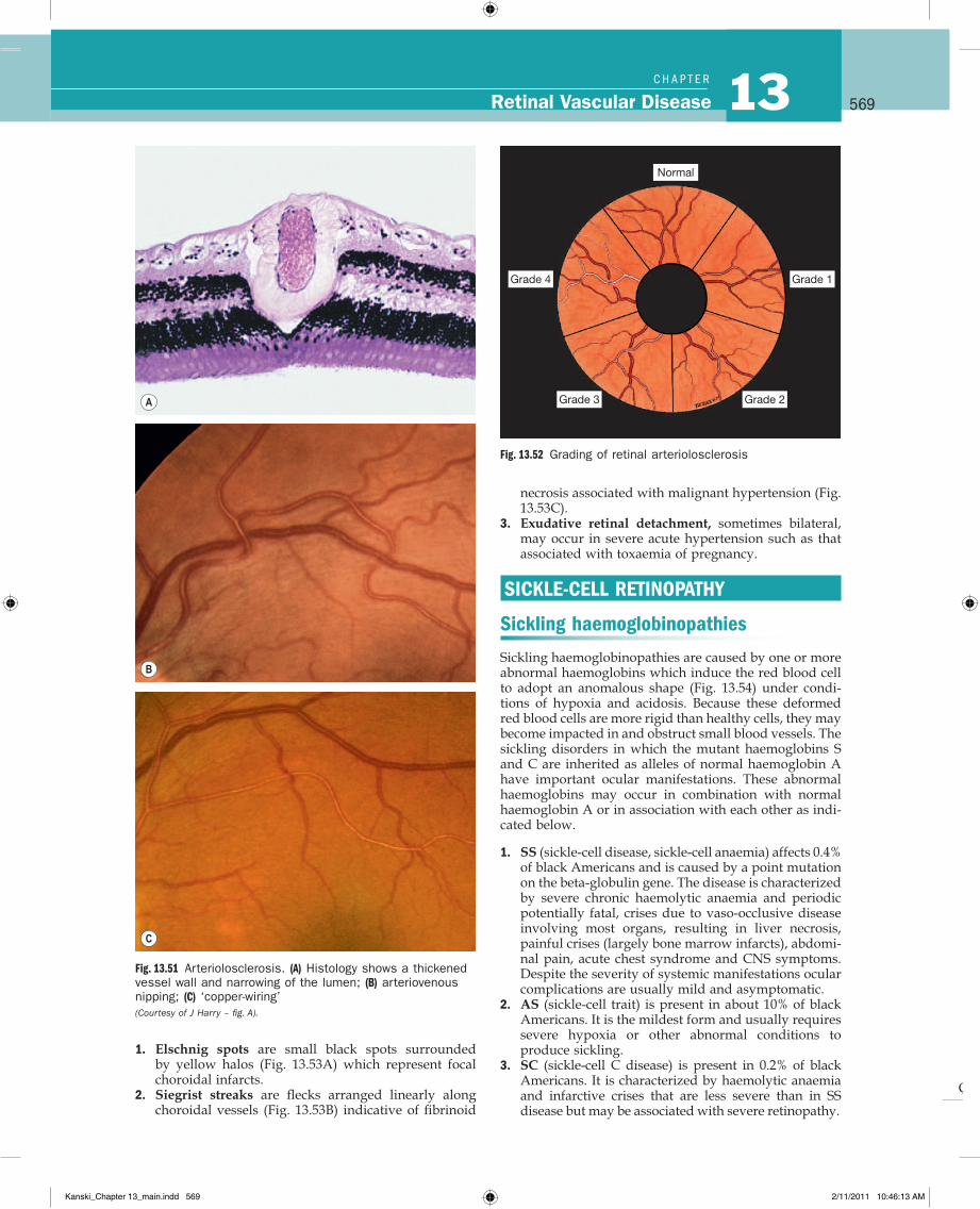
Q
569Retinal Vascular Disease 13C H A P T e R
Fig. 13.51 Arteriolosclerosis. (A) Histology shows a thickened vessel wall and narrowing of the lumen; (B) arteriovenous nipping; (C) ‘copper-wiring’ (Courtesy of J Harry – fig. A).
A
B
C
Fig. 13.52 Grading of retinal arteriolosclerosis
Grade 4 Grade 1
Grade 3 Grade 2
Normal
1. Elschnig spots are small black spots surrounded by yellow halos (Fig. 13.53A) which represent focal choroidal infarcts.
2. Siegrist streaks are flecks arranged linearly along choroidal vessels (Fig. 13.53B) indicative of fibrinoid
necrosis associated with malignant hypertension (Fig. 13.53C).
3. Exudative retinal detachment, sometimes bilateral, may occur in severe acute hypertension such as that associated with toxaemia of pregnancy.
SICKLE-CELL RETINOPATHY
Sickling haemoglobinopathies
Sickling haemoglobinopathies are caused by one or more abnormal haemoglobins which induce the red blood cell to adopt an anomalous shape (Fig. 13.54) under condi-tions of hypoxia and acidosis. Because these deformed red blood cells are more rigid than healthy cells, they may become impacted in and obstruct small blood vessels. The sickling disorders in which the mutant haemoglobins S and C are inherited as alleles of normal haemoglobin A have important ocular manifestations. These abnormal haemoglobins may occur in combination with normal haemoglobin A or in association with each other as indi-cated below.
1. SS (sickle-cell disease, sickle-cell anaemia) affects 0.4% of black Americans and is caused by a point mutation on the beta-globulin gene. The disease is characterized by severe chronic haemolytic anaemia and periodic potentially fatal, crises due to vaso-occlusive disease involving most organs, resulting in liver necrosis, painful crises (largely bone marrow infarcts), abdomi-nal pain, acute chest syndrome and CNS symptoms. Despite the severity of systemic manifestations ocular complications are usually mild and asymptomatic.
2. AS (sickle-cell trait) is present in about 10% of black Americans. It is the mildest form and usually requires severe hypoxia or other abnormal conditions to produce sickling.
3. SC (sickle-cell C disease) is present in 0.2% of black Americans. It is characterized by haemolytic anaemia and infarctive crises that are less severe than in SS disease but may be associated with severe retinopathy.
Kanski_Chapter 13_main.indd 569 2/11/2011 10:46:13 AM
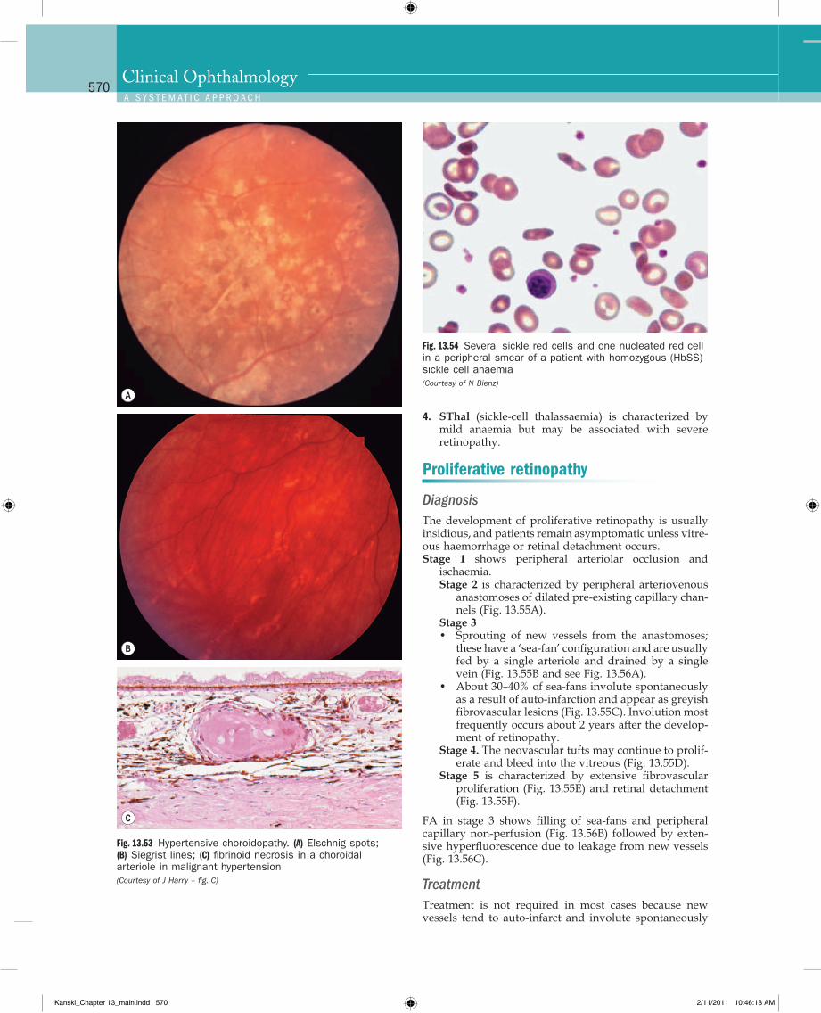
Q
570Clinical OphthalmologyA S y S T e m A T I C A P P R o A C H
Fig. 13.53 Hypertensive choroidopathy. (A) Elschnig spots; (B) Siegrist lines; (C) fibrinoid necrosis in a choroidal arteriole in malignant hypertension (Courtesy of J Harry – fig. C)
A
B
C
Fig. 13.54 Several sickle red cells and one nucleated red cell in a peripheral smear of a patient with homozygous (HbSS) sickle cell anaemia (Courtesy of N Bienz)
4. SThal (sickle-cell thalassaemia) is characterized by mild anaemia but may be associated with severe retinopathy.
Proliferative retinopathy
DiagnosisThe development of proliferative retinopathy is usually insidious, and patients remain asymptomatic unless vitre-ous haemorrhage or retinal detachment occurs.Stage 1 shows peripheral arteriolar occlusion and
ischaemia.Stage 2 is characterized by peripheral arteriovenous
anastomoses of dilated pre-existing capillary chan-nels (Fig. 13.55A).
Stage 3• Sprouting of new vessels from the anastomoses;
these have a ‘sea-fan’ configuration and are usually fed by a single arteriole and drained by a single vein (Fig. 13.55B and see Fig. 13.56A).
• About 30–40% of sea-fans involute spontaneously as a result of auto-infarction and appear as greyish fibrovascular lesions (Fig. 13.55C). Involution most frequently occurs about 2 years after the develop-ment of retinopathy.
Stage 4. The neovascular tufts may continue to prolif-erate and bleed into the vitreous (Fig. 13.55D).
Stage 5 is characterized by extensive fibrovascular proliferation (Fig. 13.55E) and retinal detachment (Fig. 13.55F).
FA in stage 3 shows filling of sea-fans and peripheral capillary non-perfusion (Fig. 13.56B) followed by exten-sive hyperfluorescence due to leakage from new vessels (Fig. 13.56C).
TreatmentTreatment is not required in most cases because new vessels tend to auto-infarct and involute spontaneously
Kanski_Chapter 13_main.indd 570 2/11/2011 10:46:18 AM
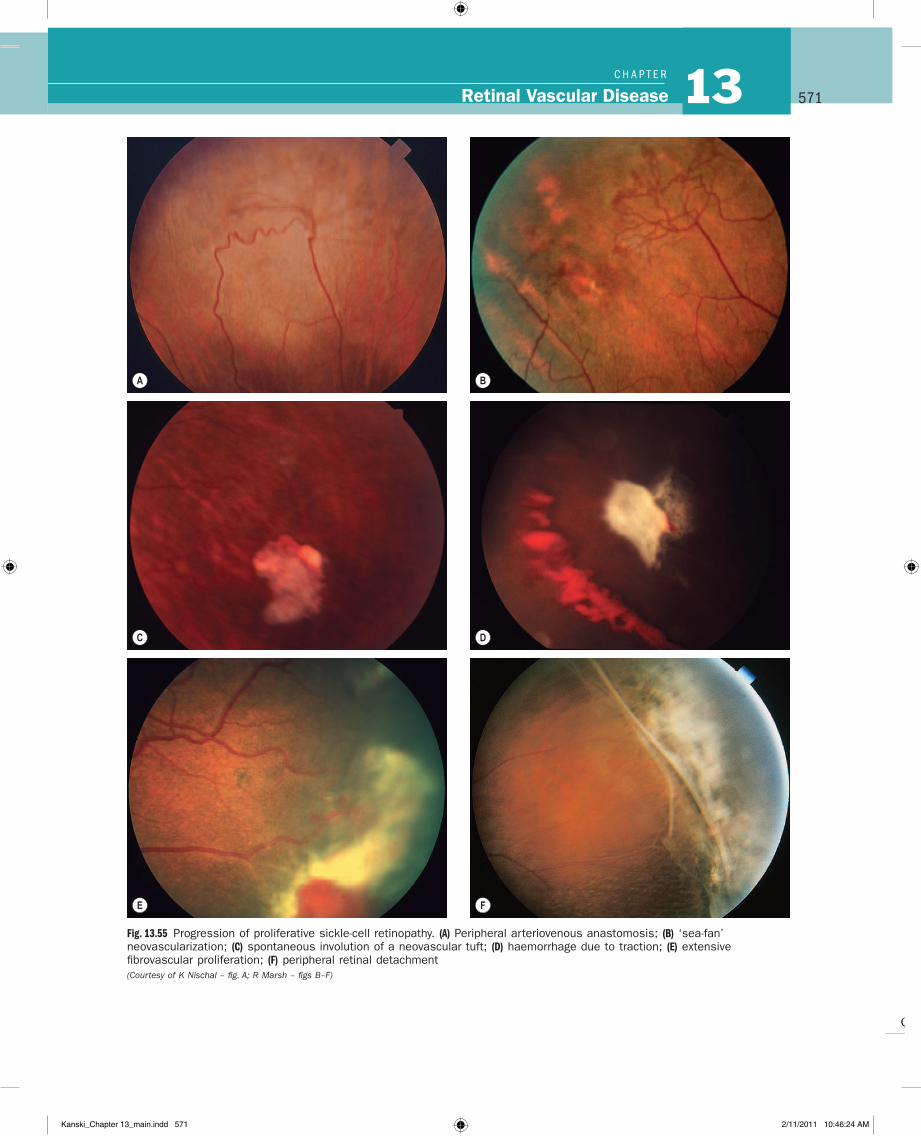
Q
571Retinal Vascular Disease 13C H A P T e R
Fig. 13.55 Progression of proliferative sickle-cell retinopathy. (A) Peripheral arteriovenous anastomosis; (B) ‘sea-fan’ neovascularization; (C) spontaneous involution of a neovascular tuft; (D) haemorrhage due to traction; (E) extensive fibrovascular proliferation; (F) peripheral retinal detachment (Courtesy of K Nischal – fig. A; R Marsh – figs B–F)
BA
C D
E FF
Kanski_Chapter 13_main.indd 571 2/11/2011 10:46:24 AM
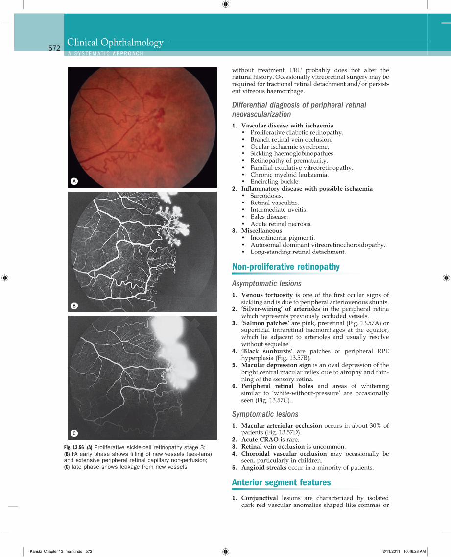
Q
572Clinical OphthalmologyA S y S T e m A T I C A P P R o A C H
Fig. 13.56 (A) Proliferative sickle-cell retinopathy stage 3; (B) FA early phase shows filling of new vessels (sea-fans) and extensive peripheral retinal capillary non-perfusion; (C) late phase shows leakage from new vessels
A
B
C
without treatment. PRP probably does not alter the natural history. Occasionally vitreoretinal surgery may be required for tractional retinal detachment and/or persist-ent vitreous haemorrhage.
Differential diagnosis of peripheral retinal neovascularization1. Vascular disease with ischaemia
• Proliferative diabetic retinopathy.• Branch retinal vein occlusion.• Ocular ischaemic syndrome.• Sickling haemoglobinopathies.• Retinopathy of prematurity.• Familial exudative vitreoretinopathy.• Chronic myeloid leukaemia.• Encircling buckle.
2. Inflammatory disease with possible ischaemia• Sarcoidosis.• Retinal vasculitis.• Intermediate uveitis.• Eales disease.• Acute retinal necrosis.
3. Miscellaneous• Incontinentia pigmenti.• Autosomal dominant vitreoretinochoroidopathy.• Long-standing retinal detachment.
Non-proliferative retinopathy
Asymptomatic lesions1. Venous tortuosity is one of the first ocular signs of
sickling and is due to peripheral arteriovenous shunts.2. ‘Silver-wiring’ of arterioles in the peripheral retina
which represents previously occluded vessels.3. ‘Salmon patches’ are pink, preretinal (Fig. 13.57A) or
superficial intraretinal haemorrhages at the equator, which lie adjacent to arterioles and usually resolve without sequelae.
4. ‘Black sunbursts’ are patches of peripheral RPE hyperplasia (Fig. 13.57B).
5. Macular depression sign is an oval depression of the bright central macular reflex due to atrophy and thin-ning of the sensory retina.
6. Peripheral retinal holes and areas of whitening similar to ‘white-without-pressure’ are occasionally seen (Fig. 13.57C).
Symptomatic lesions1. Macular arteriolar occlusion occurs in about 30% of
patients (Fig. 13.57D).2. Acute CRAO is rare.3. Retinal vein occlusion is uncommon.4. Choroidal vascular occlusion may occasionally be
seen, particularly in children.5. Angioid streaks occur in a minority of patients.
Anterior segment features
1. Conjunctival lesions are characterized by isolated dark red vascular anomalies shaped like commas or
Kanski_Chapter 13_main.indd 572 2/11/2011 10:46:28 AM
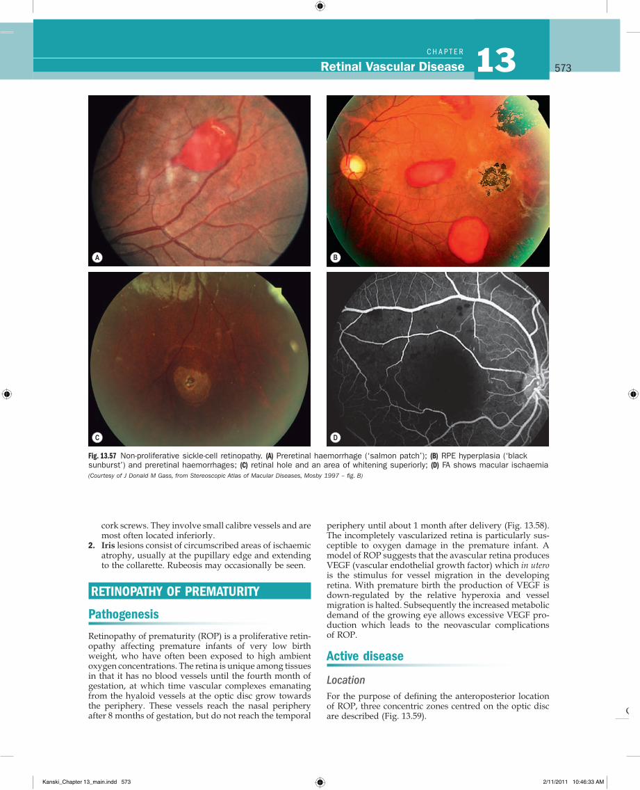
Q
573Retinal Vascular Disease 13C H A P T e R
Fig. 13.57 Non-proliferative sickle-cell retinopathy. (A) Preretinal haemorrhage (‘salmon patch’); (B) RPE hyperplasia (‘black sunburst’) and preretinal haemorrhages; (C) retinal hole and an area of whitening superiorly; (D) FA shows macular ischaemia (Courtesy of J Donald M Gass, from Stereoscopic Atlas of Macular Diseases, Mosby 1997 – fig. B)
A
BAC D
B
cork screws. They involve small calibre vessels and are most often located inferiorly.
2. Iris lesions consist of circumscribed areas of ischaemic atrophy, usually at the pupillary edge and extending to the collarette. Rubeosis may occasionally be seen.
RETINOPATHY OF PREMATURITY
Pathogenesis
Retinopathy of prematurity (ROP) is a proliferative retin-opathy affecting premature infants of very low birth weight, who have often been exposed to high ambient oxygen concentrations. The retina is unique among tissues in that it has no blood vessels until the fourth month of gestation, at which time vascular complexes emanating from the hyaloid vessels at the optic disc grow towards the periphery. These vessels reach the nasal periphery after 8 months of gestation, but do not reach the temporal
periphery until about 1 month after delivery (Fig. 13.58). The incompletely vascularized retina is particularly sus-ceptible to oxygen damage in the premature infant. A model of ROP suggests that the avascular retina produces VEGF (vascular endothelial growth factor) which in utero is the stimulus for vessel migration in the developing retina. With premature birth the production of VEGF is down-regulated by the relative hyperoxia and vessel migration is halted. Subsequently the increased metabolic demand of the growing eye allows excessive VEGF pro-duction which leads to the neovascular complications of ROP.
Active disease
LocationFor the purpose of defining the anteroposterior location of ROP, three concentric zones centred on the optic disc are described (Fig. 13.59).
Kanski_Chapter 13_main.indd 573 2/11/2011 10:46:33 AM
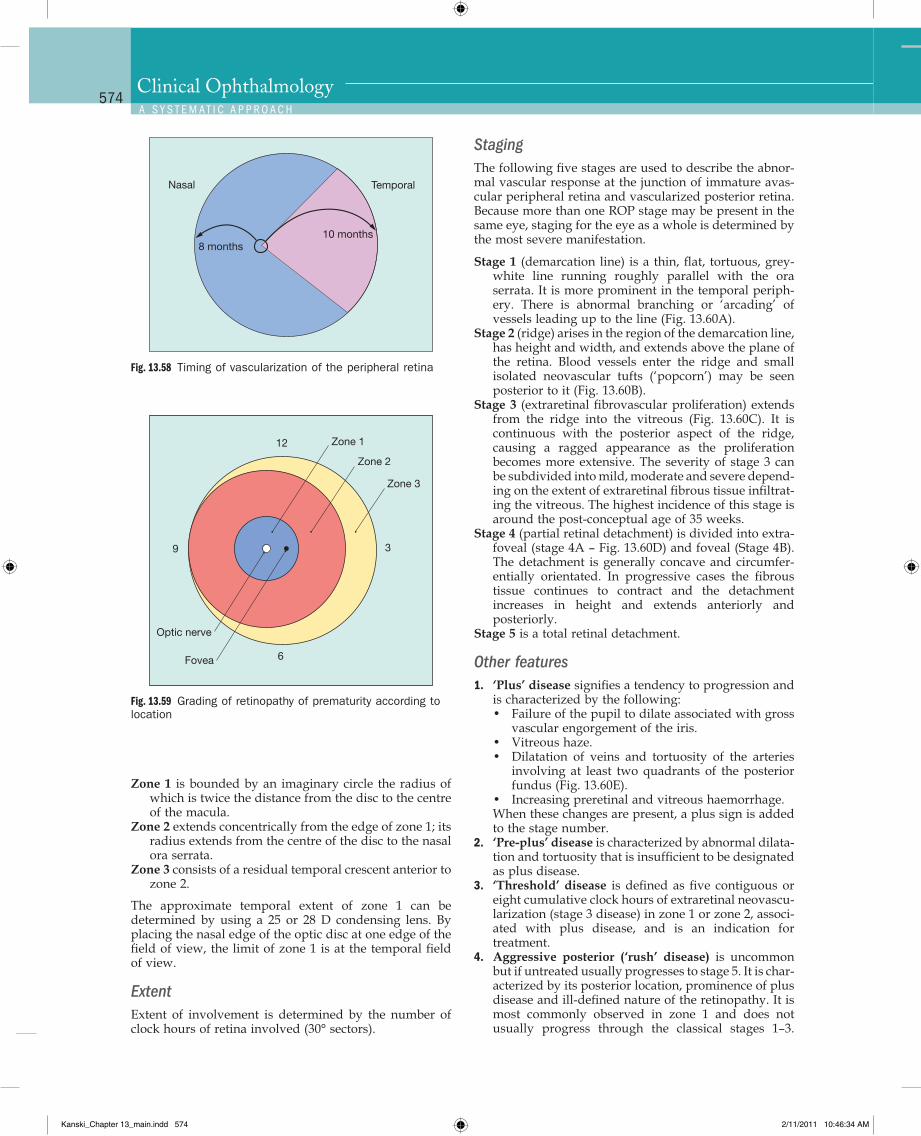
Q
574Clinical OphthalmologyA S y S T e m A T I C A P P R o A C H
Fig. 13.58 Timing of vascularization of the peripheral retina
Nasal Temporal
8 months10 months
Fig. 13.59 Grading of retinopathy of prematurity according to location
Zone 1
Zone 2
Zone 3
12
3
6
9
Optic nerve
Fovea
Zone 1 is bounded by an imaginary circle the radius of which is twice the distance from the disc to the centre of the macula.
Zone 2 extends concentrically from the edge of zone 1; its radius extends from the centre of the disc to the nasal ora serrata.
Zone 3 consists of a residual temporal crescent anterior to zone 2.
The approximate temporal extent of zone 1 can be determined by using a 25 or 28 D condensing lens. By placing the nasal edge of the optic disc at one edge of the field of view, the limit of zone 1 is at the temporal field of view.
ExtentExtent of involvement is determined by the number of clock hours of retina involved (30° sectors).
StagingThe following five stages are used to describe the abnor-mal vascular response at the junction of immature avas-cular peripheral retina and vascularized posterior retina. Because more than one ROP stage may be present in the same eye, staging for the eye as a whole is determined by the most severe manifestation.
Stage 1 (demarcation line) is a thin, flat, tortuous, grey-white line running roughly parallel with the ora serrata. It is more prominent in the temporal periph-ery. There is abnormal branching or ‘arcading’ of vessels leading up to the line (Fig. 13.60A).
Stage 2 (ridge) arises in the region of the demarcation line, has height and width, and extends above the plane of the retina. Blood vessels enter the ridge and small isolated neovascular tufts (‘popcorn’) may be seen posterior to it (Fig. 13.60B).
Stage 3 (extraretinal fibrovascular proliferation) extends from the ridge into the vitreous (Fig. 13.60C). It is continuous with the posterior aspect of the ridge, causing a ragged appearance as the proliferation becomes more extensive. The severity of stage 3 can be subdivided into mild, moderate and severe depend-ing on the extent of extraretinal fibrous tissue infiltrat-ing the vitreous. The highest incidence of this stage is around the post-conceptual age of 35 weeks.
Stage 4 (partial retinal detachment) is divided into extra-foveal (stage 4A – Fig. 13.60D) and foveal (Stage 4B). The detachment is generally concave and circumfer-entially orientated. In progressive cases the fibrous tissue continues to contract and the detachment increases in height and extends anteriorly and posteriorly.
Stage 5 is a total retinal detachment.
Other features1. ‘Plus’ disease signifies a tendency to progression and
is characterized by the following:• Failure of the pupil to dilate associated with gross
vascular engorgement of the iris.• Vitreous haze.• Dilatation of veins and tortuosity of the arteries
involving at least two quadrants of the posterior fundus (Fig. 13.60E).
• Increasing preretinal and vitreous haemorrhage.When these changes are present, a plus sign is added to the stage number.
2. ‘Pre-plus’ disease is characterized by abnormal dilata-tion and tortuosity that is insufficient to be designated as plus disease.
3. ‘Threshold’ disease is defined as five contiguous or eight cumulative clock hours of extraretinal neovascu-larization (stage 3 disease) in zone 1 or zone 2, associ-ated with plus disease, and is an indication for treatment.
4. Aggressive posterior (‘rush’ disease) is uncommon but if untreated usually progresses to stage 5. It is char-acterized by its posterior location, prominence of plus disease and ill-defined nature of the retinopathy. It is most commonly observed in zone 1 and does not usually progress through the classical stages 1–3.
Kanski_Chapter 13_main.indd 574 2/11/2011 10:46:34 AM
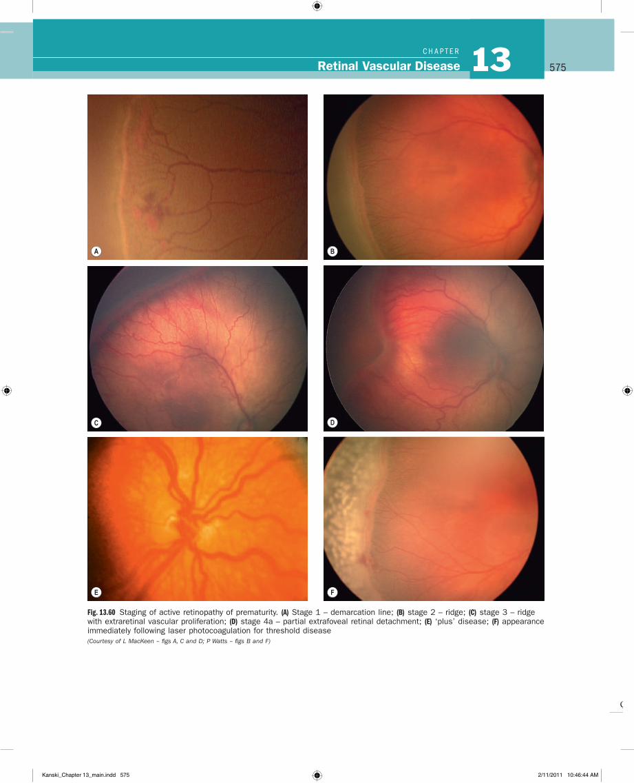
Q
575Retinal Vascular Disease 13C H A P T e R
Fig. 13.60 Staging of active retinopathy of prematurity. (A) Stage 1 – demarcation line; (B) stage 2 – ridge; (C) stage 3 – ridge with extraretinal vascular proliferation; (D) stage 4a – partial extrafoveal retinal detachment; (E) ‘plus’ disease; (F) appearance immediately following laser photocoagulation for threshold disease (Courtesy of L MacKeen – figs A, C and D; P Watts – figs B and F)
A B
C D
E F
Kanski_Chapter 13_main.indd 575 2/11/2011 10:46:44 AM
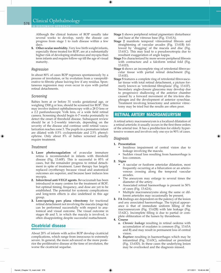
Q
576Clinical OphthalmologyA S y S T e m A T I C A P P R o A C H
Stage 1 shows peripheral retinal pigmentary disturbance and haze at the vitreous base (Fig. 13.61A).
Stage 2 manifests temporal vitreoretinal fibrosis and straightening of vascular arcades (Fig. 13.61B) fol-lowed by ‘dragging’ of the macula and disc (Fig. 13.61C). This may lead to a pseudoexotropia due to resultant exaggeration of angle kappa.
Stage 3 is characterized by more severe peripheral fibrosis with contracture and a falciform retinal fold (Fig. 13.61D).
Stage 4 shows an incomplete ring of retrolental fibrovas-cular tissue with partial retinal detachment (Fig. 13.61E).
Stage 5 features a complete ring of retrolental fibrovascu-lar tissue with total retinal detachment, a picture for-merly known as ‘retrolental fibroplasia’ (Fig. 13.61F). Secondary angle-closure glaucoma may develop due to progressive shallowing of the anterior chamber caused by a forward movement of the iris-lens dia-phragm and the development of anterior synechiae. Treatment involving lensectomy and anterior vitrec-tomy may be tried but the results are often poor.
RETINAL ARTERY MACROANEURYSMA retinal artery macroaneurysm is a localized dilatation of a retinal arteriole; it usually occurs in the first three orders of the arterial tree. It has a predilection for elderly hyper-tensive women and involves only one eye in 90% of cases.
Diagnosis1. Presentation
• Insidious impairment of central vision due to leakage involving the macula.
• Sudden visual loss resulting from haemorrhage is less common.
2. Signs• A saccular or fusiform arteriolar dilatation, most
frequently occurring at a bifurcation or an arterio-venous crossing along the temporal vascular arcades.
• The aneurysm may enlarge to several times the diameter of the artery.
• Associated retinal haemorrhage is present in 50% of cases (Fig. 13.62A).
• Multiple macroaneurysms along the same or dif-ferent arterioles may occasionally be present.
3. FA findings are dependent on the patency of the lesion and any associated haemorrhage. The typical appear-ance is that of immediate uniform filling of the macroaneurysm (Fig. 13.62B) with late leakage (Fig. 13.62C). Incomplete filling is due to partial or com-plete obliteration of the lumen by thrombosis.
4. Coursea. Chronic leakage resulting in retinal oedema with
accumulation of exudates is common (Fig. 13.63A and B) and may result in permanent loss of central vision.
b. Rupture resulting in haemorrhages which may be intraretinal (Fig. 13.63C), subretinal or preretinal (Fig. 13.63D). In these cases the underlying lesion may be overlooked and the diagnosis missed.
Although the clinical features of ROP usually take several weeks to develop, rarely the disease can progress from stage 1 to rush disease within a few days.
5. Other ocular morbidity. Very low birth weight infants, especially those treated for ROP, are at a substantially higher risk of developing strabismus and myopia than term infants and require follow-up till the age of visual maturity.
RegressionIn about 80% of cases ROP regresses spontaneously by a process of involution, or by evolution from a vasoprolif-erative to fibrotic phase leaving few if any residua. Spon-taneous regression may even occur in eyes with partial retinal detachments.
ScreeningBabies born at or before 31 weeks gestational age, or weighing 1500 g or less, should be screened for ROP. This may involve indirect ophthalmoscopy with a 28 D lens or a 2.2 panfunduscopic Volk lens, or a wide field retinal camera. Screening should begin 4–7 weeks postnatally to detect the onset of threshold disease. Subsequent review should be at 1–2-weekly intervals, depending on the severity of the disease and continue until retinal vascu-larization reaches zone 3. The pupils in a premature infant are dilated with 0.5% cyclopentolate and 2.5% phenyl-ephrine. Only about 8% of babies screened actually require treatment.
Treatment1. Laser photocoagulation of avascular immature
retina is recommended in infants with threshold disease (Fig. 13.60F). This is successful in 85% of cases, but the remainder progress to retinal detach-ment in spite of treatment. Laser therapy has largely replaced cryotherapy because visual and anatomical outcomes are superior, and because laser induces less myopia.
2. Intravitreal anti-VEGF agents. Bevacizumab has been introduced in many centres for the treatment of ROP, but optimal timing, frequency, and dose are yet to be established. The potential for systemic complications and long-term effects is also undefined in this age group.
3. Lens-sparing pars plana vitrectomy for tractional retinal detachment not involving the macula (stage 4a) can be performed successfully with respect to ana-tomical and visual outcome. The visual outcome in stages 4b and 5, in which the macula is involved, is often disappointing despite successful reattachment.
Cicatricial disease
About 20% of infants with active ROP develop cicatricial complications, which range from innocuous to extremely severe. In general, the more advanced or the more poste-rior the proliferative disease at the time of involution, the worse the cicatricial sequelae.
Kanski_Chapter 13_main.indd 576 2/11/2011 10:46:44 AM
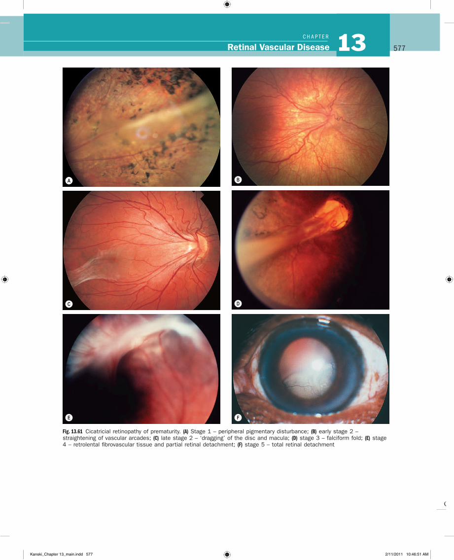
Q
577Retinal Vascular Disease 13C H A P T e R
Fig. 13.61 Cicatricial retinopathy of prematurity. (A) Stage 1 – peripheral pigmentary disturbance; (B) early stage 2 – straightening of vascular arcades; (C) late stage 2 – ‘dragging’ of the disc and macula; (D) stage 3 – falciform fold; (E) stage 4 – retrolental fibrovascular tissue and partial retinal detachment; (F) stage 5 – total retinal detachment
F
A B
C D
E
Kanski_Chapter 13_main.indd 577 2/11/2011 10:46:51 AM
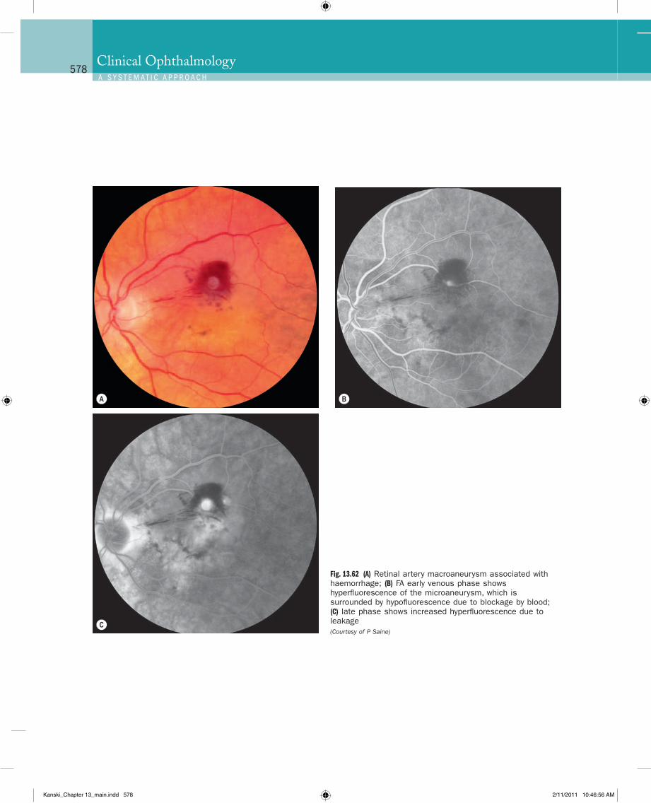
Q
578Clinical OphthalmologyA S y S T e m A T I C A P P R o A C H
A B
C
Fig. 13.62 (A) Retinal artery macroaneurysm associated with haemorrhage; (B) FA early venous phase shows hyperfluorescence of the microaneurysm, which is surrounded by hypofluorescence due to blockage by blood; (C) late phase shows increased hyperfluorescence due to leakage (Courtesy of P Saine)
Kanski_Chapter 13_main.indd 578 2/11/2011 10:46:56 AM
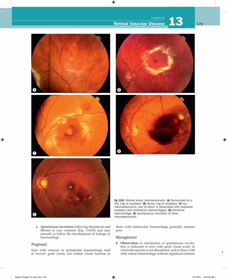
Q
579Retinal Vascular Disease 13C H A P T e R
those with submacular haemorrhage generally remains poor.
Management1. Observation in anticipation of spontaneous involu-
tion is indicated in eyes with good visual acuity in which the macula is not threatened, and in those with mild retinal haemorrhage without significant oedema
c. Spontaneous involution following thrombosis and fibrosis is very common (Fig. 13.63E) and may precede or follow the development of leakage or haemorrhage.
PrognosisEyes with vitreous or premacular haemorrhage tend to recover good vision, but central visual function in
Fig. 13.63 Retinal artery macroaneurysm. (A) Surrounded by a fine ring of exudates; (B) dense ring of exudates; (C) two macroaneurysms, one of which is associated with extensive exudates and intraretinal haemorrhages; (D) preretinal haemorrhage; (E) spontaneous involution of three macroaneurysms
A B
CD
E
Kanski_Chapter 13_main.indd 579 2/11/2011 10:47:05 AM
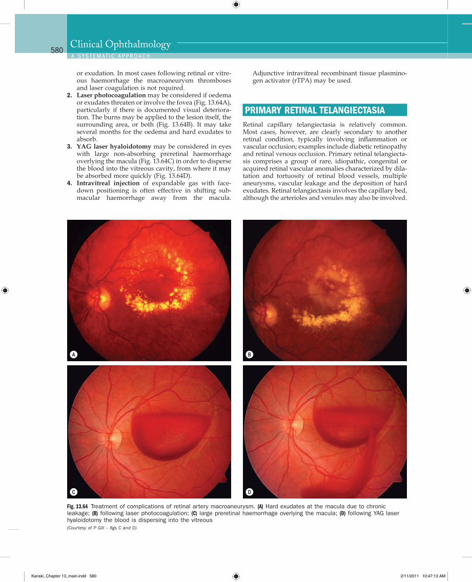
Q
580Clinical OphthalmologyA S y S T e m A T I C A P P R o A C H
Adjunctive intravitreal recombinant tissue plasmino-gen activator (rTPA) may be used.
PRIMARY RETINAL TELANGIECTASIARetinal capillary telangiectasia is relatively common. Most cases, however, are clearly secondary to another retinal condition, typically involving inflammation or vascular occlusion; examples include diabetic retinopathy and retinal venous occlusion. Primary retinal telangiecta-sis comprises a group of rare, idiopathic, congenital or acquired retinal vascular anomalies characterized by dila-tation and tortuosity of retinal blood vessels, multiple aneurysms, vascular leakage and the deposition of hard exudates. Retinal telangiectasis involves the capillary bed, although the arterioles and venules may also be involved.
or exudation. In most cases following retinal or vitre-ous haemorrhage the macroaneurysm thromboses and laser coagulation is not required.
2. Laser photocoagulation may be considered if oedema or exudates threaten or involve the fovea (Fig. 13.64A), particularly if there is documented visual deteriora-tion. The burns may be applied to the lesion itself, the surrounding area, or both (Fig. 13.64B). It may take several months for the oedema and hard exudates to absorb.
3. YAG laser hyaloidotomy may be considered in eyes with large non-absorbing preretinal haemorrhage overlying the macula (Fig. 13.64C) in order to disperse the blood into the vitreous cavity, from where it may be absorbed more quickly (Fig. 13.64D).
4. Intravitreal injection of expandable gas with face-down positioning is often effective in shifting sub-macular haemorrhage away from the macula.
Fig. 13.64 Treatment of complications of retinal artery macroaneurysm. (A) Hard exudates at the macula due to chronic leakage; (B) following laser photocoagulation; (C) large preretinal haemorrhage overlying the macula; (D) following YAG laser hyaloidotomy the blood is dispersing into the vitreous (Courtesy of P Gili – figs C and D)
A B
C D
Kanski_Chapter 13_main.indd 580 2/11/2011 10:47:13 AM
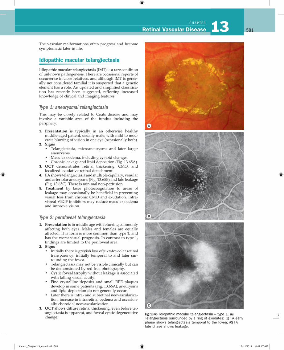
Q
581Retinal Vascular Disease 13C H A P T e R
The vascular malformations often progress and become symptomatic later in life.
Idiopathic macular telangiectasia
Idiopathic macular telangiectasia (IMT) is a rare condition of unknown pathogenesis. There are occasional reports of occurrence in close relatives, and although IMT is gener-ally not considered familial it is suspected that a genetic element has a role. An updated and simplified classifica-tion has recently been suggested, reflecting increased knowledge of clinical and imaging features.
Type 1: aneurysmal telangiectasiaThis may be closely related to Coats disease and may involve a variable area of the fundus including the periphery.
1. Presentation is typically in an otherwise healthy middle-aged patient, usually male, with mild to mod-erate blurring of vision in one eye (occasionally both).
2. Signs• Telangiectasia, microaneurysms and later larger
aneurysms.• Macular oedema, including cystoid changes.• Chronic leakage and lipid deposition (Fig. 13.65A).
3. OCT demonstrates retinal thickening, CMO, and localized exudative retinal detachment.
4. FA shows telangiectasia and multiple capillary, venular and arteriolar aneurysms (Fig. 13.65B) and late leakage (Fig. 13.65C). There is minimal non-perfusion.
5. Treatment by laser photocoagulation to areas of leakage may occasionally be beneficial in preventing visual loss from chronic CMO and exudation. Intra-vitreal VEGF inhibitors may reduce macular oedema and improve vision.
Type 2: perafoveal telangiectasia1. Presentation is in middle age with blurring commonly
affecting both eyes. Males and females are equally affected. This form is more common than type 1, and has the worst visual prognosis. In contrast to type 1, findings are limited to the perifoveal area.
2. Signs• Initially there is greyish loss of juxtafoveolar retinal
transparency, initially temporal to and later sur-rounding the fovea.
• Telangiectasia may not be visible clinically but can be demonstrated by red-free photography.
• Cystic foveal atrophy without leakage is associated with falling visual acuity.
• Fine crystalline deposits and small RPE plaques develop in some patients (Fig. 13.66A); aneurysms and lipid deposition do not generally occur.
• Later there is intra- and subretinal neovasculariza-tion, increase in intraretinal oedema and occasion-ally choroidal neovascularization.
3. OCT shows diffuse retinal thickening, even before tel-angiectasia is apparent, and foveal cystic degenerative change.
Fig. 13.65 Idiopathic macular telangiectasia – type 1. (A) Telangiectasis surrounded by a ring of exudates; (B) FA early phase shows telangiectasia temporal to the fovea; (C) FA late phase shows leakage.
A
B
C
Kanski_Chapter 13_main.indd 581 2/11/2011 10:47:17 AM
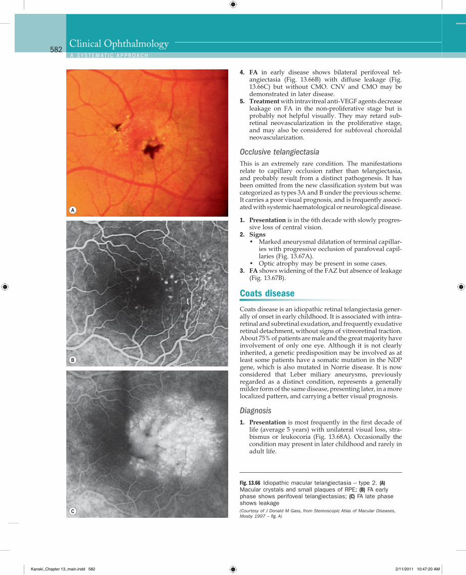
Q
582Clinical OphthalmologyA S y S T e m A T I C A P P R o A C H
4. FA in early disease shows bilateral perifoveal tel-angiectasia (Fig. 13.66B) with diffuse leakage (Fig. 13.66C) but without CMO. CNV and CMO may be demonstrated in later disease.
5. Treatment with intravitreal anti-VEGF agents decrease leakage on FA in the non-proliferative stage but is probably not helpful visually. They may retard sub-retinal neovascularization in the proliferative stage, and may also be considered for subfoveal choroidal neovascularization.
Occlusive telangiectasiaThis is an extremely rare condition. The manifestations relate to capillary occlusion rather than telangiectasia, and probably result from a distinct pathogenesis. It has been omitted from the new classification system but was categorized as types 3A and B under the previous scheme. It carries a poor visual prognosis, and is frequently associ-ated with systemic haematological or neurological disease.
1. Presentation is in the 6th decade with slowly progres-sive loss of central vision.
2. Signs• Marked aneurysmal dilatation of terminal capillar-
ies with progressive occlusion of parafoveal capil-laries (Fig. 13.67A).
• Optic atrophy may be present in some cases.3. FA shows widening of the FAZ but absence of leakage
(Fig. 13.67B).
Coats disease
Coats disease is an idiopathic retinal telangiectasia gener-ally of onset in early childhood. It is associated with intra-retinal and subretinal exudation, and frequently exudative retinal detachment, without signs of vitreoretinal traction. About 75% of patients are male and the great majority have involvement of only one eye. Although it is not clearly inherited, a genetic predisposition may be involved as at least some patients have a somatic mutation in the NDP gene, which is also mutated in Norrie disease. It is now considered that Leber miliary aneurysms, previously regarded as a distinct condition, represents a generally milder form of the same disease, presenting later, in a more localized pattern, and carrying a better visual prognosis.
Diagnosis1. Presentation is most frequently in the first decade of
life (average 5 years) with unilateral visual loss, stra-bismus or leukocoria (Fig. 13.68A). Occasionally the condition may present in later childhood and rarely in adult life.
Fig. 13.66 Idiopathic macular telangiectasia – type 2. (A) Macular crystals and small plaques of RPE; (B) FA early phase shows perifoveal telangiectasias; (C) FA late phase shows leakage (Courtesy of J Donald M Gass, from Stereoscopic Atlas of Macular Diseases, Mosby 1997 – fig. A)
A
B
C
Kanski_Chapter 13_main.indd 582 2/11/2011 10:47:20 AM
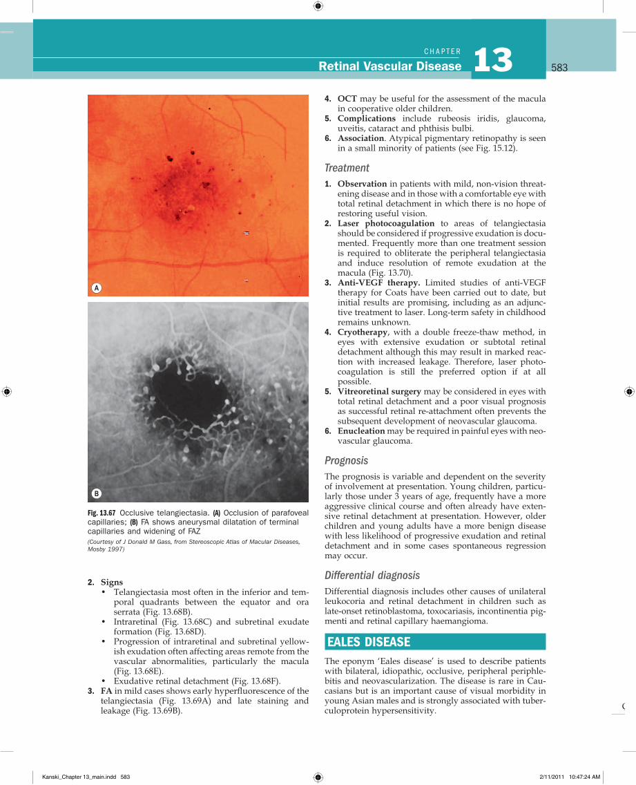
Q
583Retinal Vascular Disease 13C H A P T e R
Fig. 13.67 Occlusive telangiectasia. (A) Occlusion of parafoveal capillaries; (B) FA shows aneurysmal dilatation of terminal capillaries and widening of FAZ (Courtesy of J Donald M Gass, from Stereoscopic Atlas of Macular Diseases, Mosby 1997)
A
B
4. OCT may be useful for the assessment of the macula in cooperative older children.
5. Complications include rubeosis iridis, glaucoma, uveitis, cataract and phthisis bulbi.
6. Association. Atypical pigmentary retinopathy is seen in a small minority of patients (see Fig. 15.12).
Treatment1. Observation in patients with mild, non-vision threat-
ening disease and in those with a comfortable eye with total retinal detachment in which there is no hope of restoring useful vision.
2. Laser photocoagulation to areas of telangiectasia should be considered if progressive exudation is docu-mented. Frequently more than one treatment session is required to obliterate the peripheral telangiectasia and induce resolution of remote exudation at the macula (Fig. 13.70).
3. Anti-VEGF therapy. Limited studies of anti-VEGF therapy for Coats have been carried out to date, but initial results are promising, including as an adjunc-tive treatment to laser. Long-term safety in childhood remains unknown.
4. Cryotherapy, with a double freeze-thaw method, in eyes with extensive exudation or subtotal retinal detachment although this may result in marked reac-tion with increased leakage. Therefore, laser photo-coagulation is still the preferred option if at all possible.
5. Vitreoretinal surgery may be considered in eyes with total retinal detachment and a poor visual prognosis as successful retinal re-attachment often prevents the subsequent development of neovascular glaucoma.
6. Enucleation may be required in painful eyes with neo-vascular glaucoma.
PrognosisThe prognosis is variable and dependent on the severity of involvement at presentation. Young children, particu-larly those under 3 years of age, frequently have a more aggressive clinical course and often already have exten-sive retinal detachment at presentation. However, older children and young adults have a more benign disease with less likelihood of progressive exudation and retinal detachment and in some cases spontaneous regression may occur.
Differential diagnosisDifferential diagnosis includes other causes of unilateral leukocoria and retinal detachment in children such as late-onset retinoblastoma, toxocariasis, incontinentia pig-menti and retinal capillary haemangioma.
EALES DISEASEThe eponym ‘Eales disease’ is used to describe patients with bilateral, idiopathic, occlusive, peripheral periphle-bitis and neovascularization. The disease is rare in Cau-casians but is an important cause of visual morbidity in young Asian males and is strongly associated with tuber-culoprotein hypersensitivity.
2. Signs• Telangiectasia most often in the inferior and tem-
poral quadrants between the equator and ora serrata (Fig. 13.68B).
• Intraretinal (Fig. 13.68C) and subretinal exudate formation (Fig. 13.68D).
• Progression of intraretinal and subretinal yellow-ish exudation often affecting areas remote from the vascular abnormalities, particularly the macula (Fig. 13.68E).
• Exudative retinal detachment (Fig. 13.68F).3. FA in mild cases shows early hyperfluorescence of the
telangiectasia (Fig. 13.69A) and late staining and leakage (Fig. 13.69B).
Kanski_Chapter 13_main.indd 583 2/11/2011 10:47:24 AM
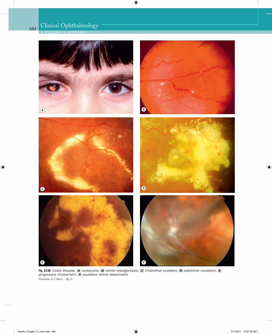
Q
584Clinical OphthalmologyA S y S T e m A T I C A P P R o A C H
Fig. 13.68 Coats disease. (A) Leukocoria; (B) retinal telangiectasis; (C) intraretinal exudates; (D) subretinal exudation; (E) progressive involvement; (F) exudative retinal detachment (Courtesy of C Barry – fig. E)
A B
C D
E F
Kanski_Chapter 13_main.indd 584 2/11/2011 10:47:35 AM
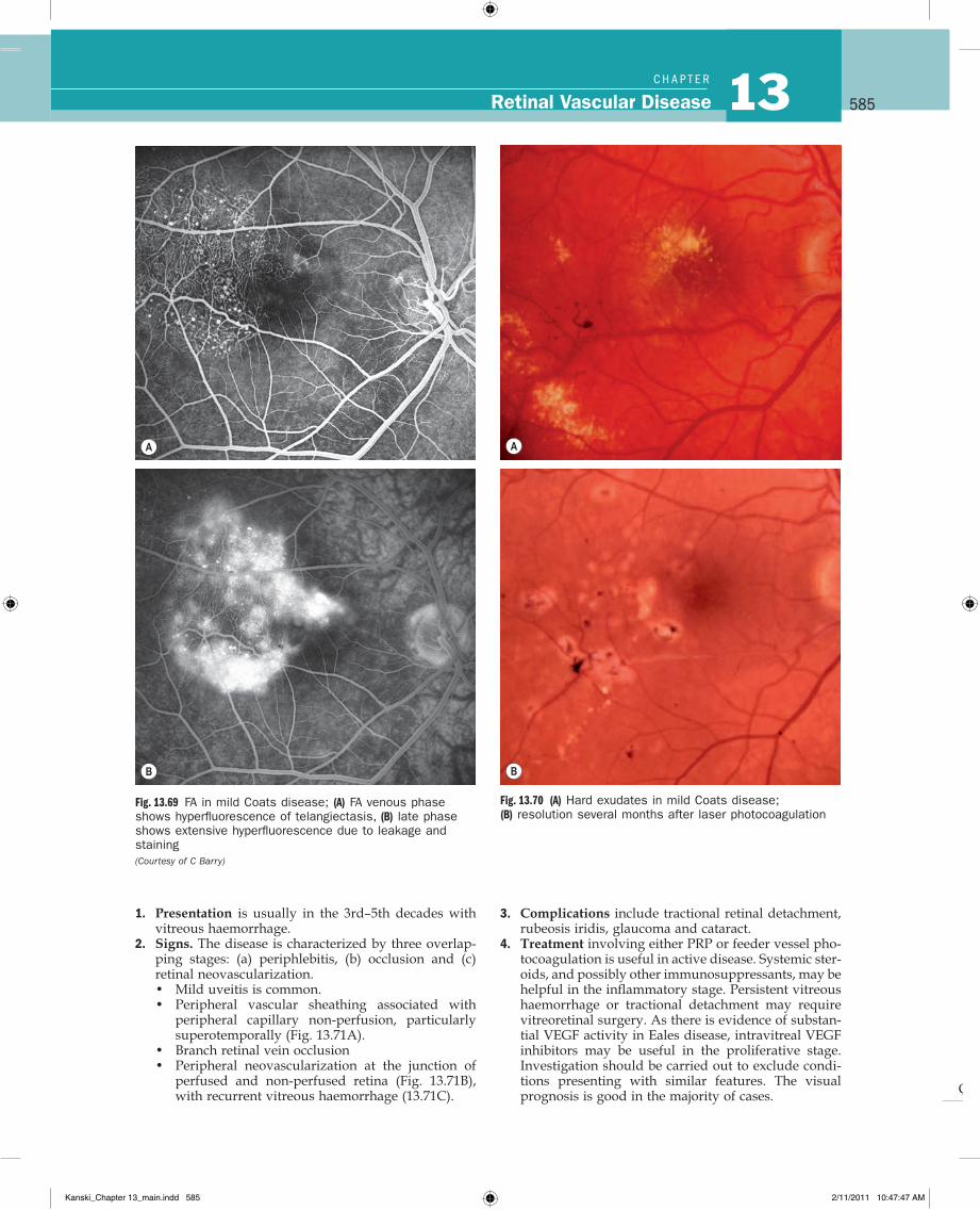
Q
585Retinal Vascular Disease 13C H A P T e R
3. Complications include tractional retinal detachment, rubeosis iridis, glaucoma and cataract.
4. Treatment involving either PRP or feeder vessel pho-tocoagulation is useful in active disease. Systemic ster-oids, and possibly other immunosuppressants, may be helpful in the inflammatory stage. Persistent vitreous haemorrhage or tractional detachment may require vitreoretinal surgery. As there is evidence of substan-tial VEGF activity in Eales disease, intravitreal VEGF inhibitors may be useful in the proliferative stage. Investigation should be carried out to exclude condi-tions presenting with similar features. The visual prognosis is good in the majority of cases.
1. Presentation is usually in the 3rd–5th decades with vitreous haemorrhage.
2. Signs. The disease is characterized by three overlap-ping stages: (a) periphlebitis, (b) occlusion and (c) retinal neovascularization.• Mild uveitis is common.• Peripheral vascular sheathing associated with
peripheral capillary non-perfusion, particularly superotemporally (Fig. 13.71A).
• Branch retinal vein occlusion• Peripheral neovascularization at the junction of
perfused and non-perfused retina (Fig. 13.71B), with recurrent vitreous haemorrhage (13.71C).
Fig. 13.69 FA in mild Coats disease; (A) FA venous phase shows hyperfluorescence of telangiectasis, (B) late phase shows extensive hyperfluorescence due to leakage and staining (Courtesy of C Barry)
A
B
Fig. 13.70 (A) Hard exudates in mild Coats disease; (B) resolution several months after laser photocoagulation
A
B
Kanski_Chapter 13_main.indd 585 2/11/2011 10:47:47 AM
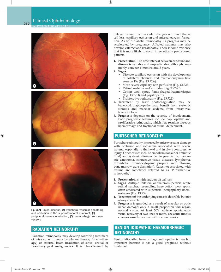
Q
586Clinical OphthalmologyA S y S T e m A T I C A P P R o A C H
delayed retinal microvascular changes with endothelial cell loss, capillary occlusion and microaneurysm forma-tion. As with diabetic retinopathy its progress may be accelerated by pregnancy. Affected patients may also develop cataract and keratopathy. There is some evidence that it is more likely to occur in genetically predisposed patients.
1. Presentation. The time interval between exposure and disease is variable and unpredictable, although com-monly between 6 months and 3 years.
2. Signs• Discrete capillary occlusion with the development
of collateral channels and microaneurysms, best seen on FA (Fig. 13.72A).
• More severe capillary non-perfusion (Fig. 13.72B).• Retinal oedema and exudates (Fig. 13.72C).• Cotton wool spots, flame-shaped haemorrhages
(Fig. 13.72D) and papillopathy.• Proliferative retinopathy (Fig. 13.72E).
3. Treatment by laser photocoagulation may be beneficial. Papillopathy may benefit from systemic steroids and macular oedema from intravitreal triamcinolone.
4. Prognosis depends on the severity of involvement. Poor prognostic features include papillopathy and proliferative retinopathy, which may result in vitreous haemorrhage and tractional retinal detachment.
PURTSCHER RETINOPATHYPurtscher retinopathy is caused by microvascular damage with occlusion and ischaemia associated with severe trauma, especially to the head and in chest compressive injury. Other causes include embolism (fat, air or amniotic fluid) and systemic diseases (acute pancreatitis, pancre-atic carcinoma, connective tissue diseases, lymphoma, thrombotic thrombocytopenic purpura and following bone marrow transplantation). Cases not associated with trauma are sometimes referred to as ‘Purtscher-like retinopathy’.
1. Presentation is with sudden visual loss.2. Signs. Multiple unilateral or bilateral superficial white
retinal patches, resembling large cotton wool spots, often associated with superficial peripapillary haem-orrhages (Fig. 13.73).
3. Treatment of the underlying cause is desirable but not always possible.
4. Prognosis is guarded as a result of macular or optic nerve damage; only a small proportion will regain normal vision. At least 50% achieve spontaneous visual recovery of two lines or more. The acute fundus changes usually resolve within a few weeks.
BENIGN IDIOPATHIC HAEMORRHAGIC RETINOPATHY
Benign idiopathic haemorrhagic retinopathy is rare but important because it has a good prognosis without treatment.
RADIATION RETINOPATHYRadiation retinopathy may develop following treatment of intraocular tumours by plaque therapy (brachyther-apy) or external beam irradiation of sinus, orbital or nasopharyngeal malignancies. It is characterized by
Fig. 13.71 Eales disease. (A) Peripheral vascular sheathing and occlusion in the superotemporal quadrant; (B) peripheral neovascularization; (C) haemorrhage from new vessels
A
B
C
Kanski_Chapter 13_main.indd 586 2/11/2011 10:47:49 AM
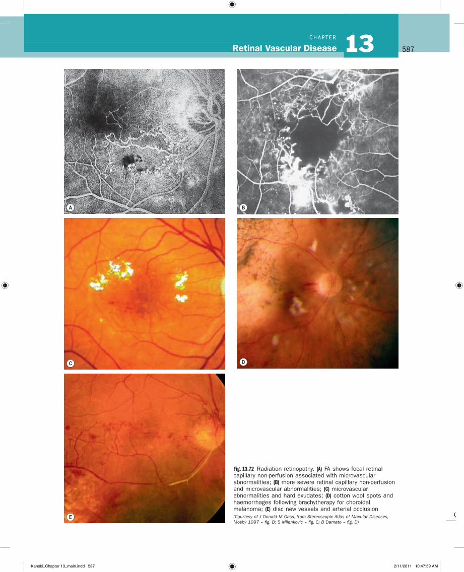
Q
587Retinal Vascular Disease 13C H A P T e R
Fig. 13.72 Radiation retinopathy. (A) FA shows focal retinal capillary non-perfusion associated with microvascular abnormalities; (B) more severe retinal capillary non-perfusion and microvascular abnormalities; (C) microvascular abnormalities and hard exudates; (D) cotton wool spots and haemorrhages following brachytherapy for choroidal melanoma; (E) disc new vessels and arterial occlusion (Courtesy of J Donald M Gass, from Stereoscopic Atlas of Macular Diseases, Mosby 1997 – fig. B; S Milenkovic – fig. C; B Damato – fig. D)
A B
C D
E
Kanski_Chapter 13_main.indd 587 2/11/2011 10:47:59 AM
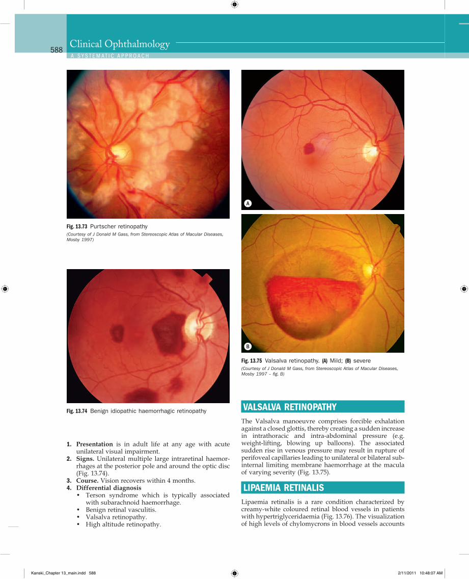
Q
588Clinical OphthalmologyA S y S T e m A T I C A P P R o A C H
VALSALVA RETINOPATHYThe Valsalva manoeuvre comprises forcible exhalation against a closed glottis, thereby creating a sudden increase in intrathoracic and intra-abdominal pressure (e.g. weight-lifting, blowing up balloons). The associated sudden rise in venous pressure may result in rupture of perifoveal capillaries leading to unilateral or bilateral sub-internal limiting membrane haemorrhage at the macula of varying severity (Fig. 13.75).
LIPAEMIA RETINALISLipaemia retinalis is a rare condition characterized by creamy-white coloured retinal blood vessels in patients with hypertriglyceridaemia (Fig. 13.76). The visualization of high levels of chylomycrons in blood vessels accounts
1. Presentation is in adult life at any age with acute unilateral visual impairment.
2. Signs. Unilateral multiple large intraretinal haemor-rhages at the posterior pole and around the optic disc (Fig. 13.74).
3. Course. Vision recovers within 4 months.4. Differential diagnosis
• Terson syndrome which is typically associated with subarachnoid haemorrhage.
• Benign retinal vasculitis.• Valsalva retinopathy.• High altitude retinopathy.
Fig. 13.73 Purtscher retinopathy (Courtesy of J Donald M Gass, from Stereoscopic Atlas of Macular Diseases, Mosby 1997)
Fig. 13.74 Benign idiopathic haemorrhagic retinopathy
Fig. 13.75 Valsalva retinopathy. (A) Mild; (B) severe (Courtesy of J Donald M Gass, from Stereoscopic Atlas of Macular Diseases, Mosby 1997 – fig. B)
A
B
Kanski_Chapter 13_main.indd 588 2/11/2011 10:48:07 AM
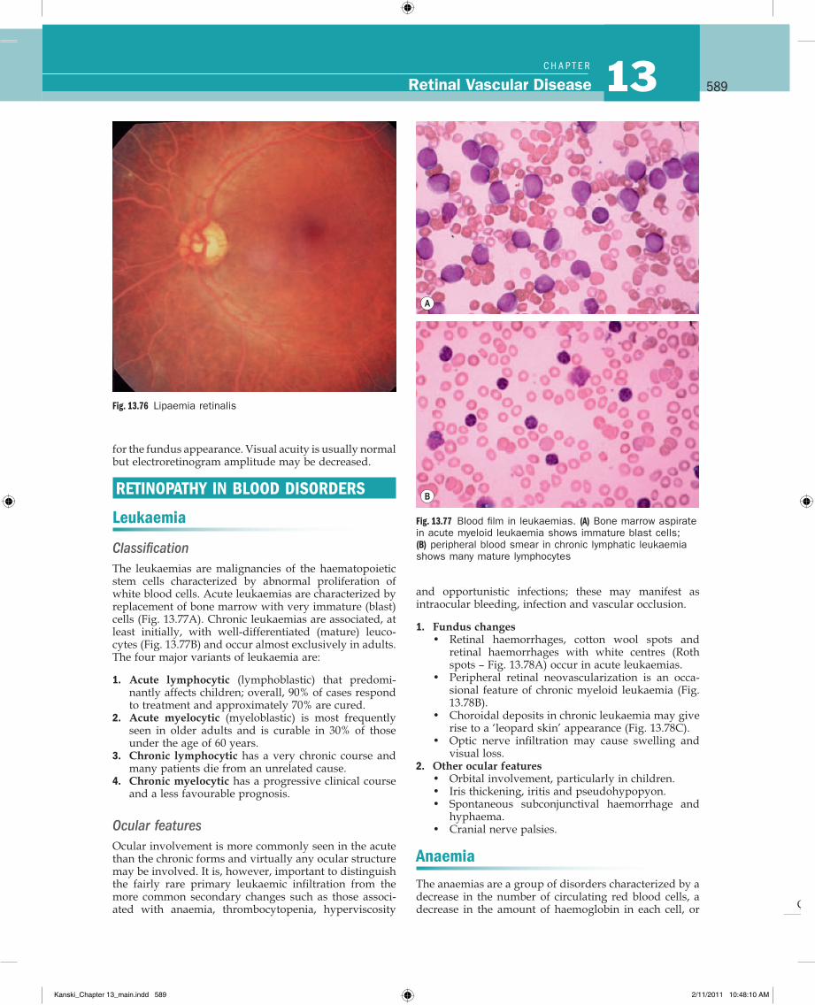
Q
589Retinal Vascular Disease 13C H A P T e R
and opportunistic infections; these may manifest as intraocular bleeding, infection and vascular occlusion.
1. Fundus changes• Retinal haemorrhages, cotton wool spots and
retinal haemorrhages with white centres (Roth spots – Fig. 13.78A) occur in acute leukaemias.
• Peripheral retinal neovascularization is an occa-sional feature of chronic myeloid leukaemia (Fig. 13.78B).
• Choroidal deposits in chronic leukaemia may give rise to a ‘leopard skin’ appearance (Fig. 13.78C).
• Optic nerve infiltration may cause swelling and visual loss.
2. Other ocular features• Orbital involvement, particularly in children.• Iris thickening, iritis and pseudohypopyon.• Spontaneous subconjunctival haemorrhage and
hyphaema.• Cranial nerve palsies.
Anaemia
The anaemias are a group of disorders characterized by a decrease in the number of circulating red blood cells, a decrease in the amount of haemoglobin in each cell, or
Fig. 13.76 Lipaemia retinalis
Fig. 13.77 Blood film in leukaemias. (A) Bone marrow aspirate in acute myeloid leukaemia shows immature blast cells; (B) peripheral blood smear in chronic lymphatic leukaemia shows many mature lymphocytes
A
B
for the fundus appearance. Visual acuity is usually normal but electroretinogram amplitude may be decreased.
RETINOPATHY IN BLOOD DISORDERS
Leukaemia
ClassificationThe leukaemias are malignancies of the haematopoietic stem cells characterized by abnormal proliferation of white blood cells. Acute leukaemias are characterized by replacement of bone marrow with very immature (blast) cells (Fig. 13.77A). Chronic leukaemias are associated, at least initially, with well-differentiated (mature) leuco-cytes (Fig. 13.77B) and occur almost exclusively in adults. The four major variants of leukaemia are:
1. Acute lymphocytic (lymphoblastic) that predomi-nantly affects children; overall, 90% of cases respond to treatment and approximately 70% are cured.
2. Acute myelocytic (myeloblastic) is most frequently seen in older adults and is curable in 30% of those under the age of 60 years.
3. Chronic lymphocytic has a very chronic course and many patients die from an unrelated cause.
4. Chronic myelocytic has a progressive clinical course and a less favourable prognosis.
Ocular featuresOcular involvement is more commonly seen in the acute than the chronic forms and virtually any ocular structure may be involved. It is, however, important to distinguish the fairly rare primary leukaemic infiltration from the more common secondary changes such as those associ-ated with anaemia, thrombocytopenia, hyperviscosity
Kanski_Chapter 13_main.indd 589 2/11/2011 10:48:10 AM
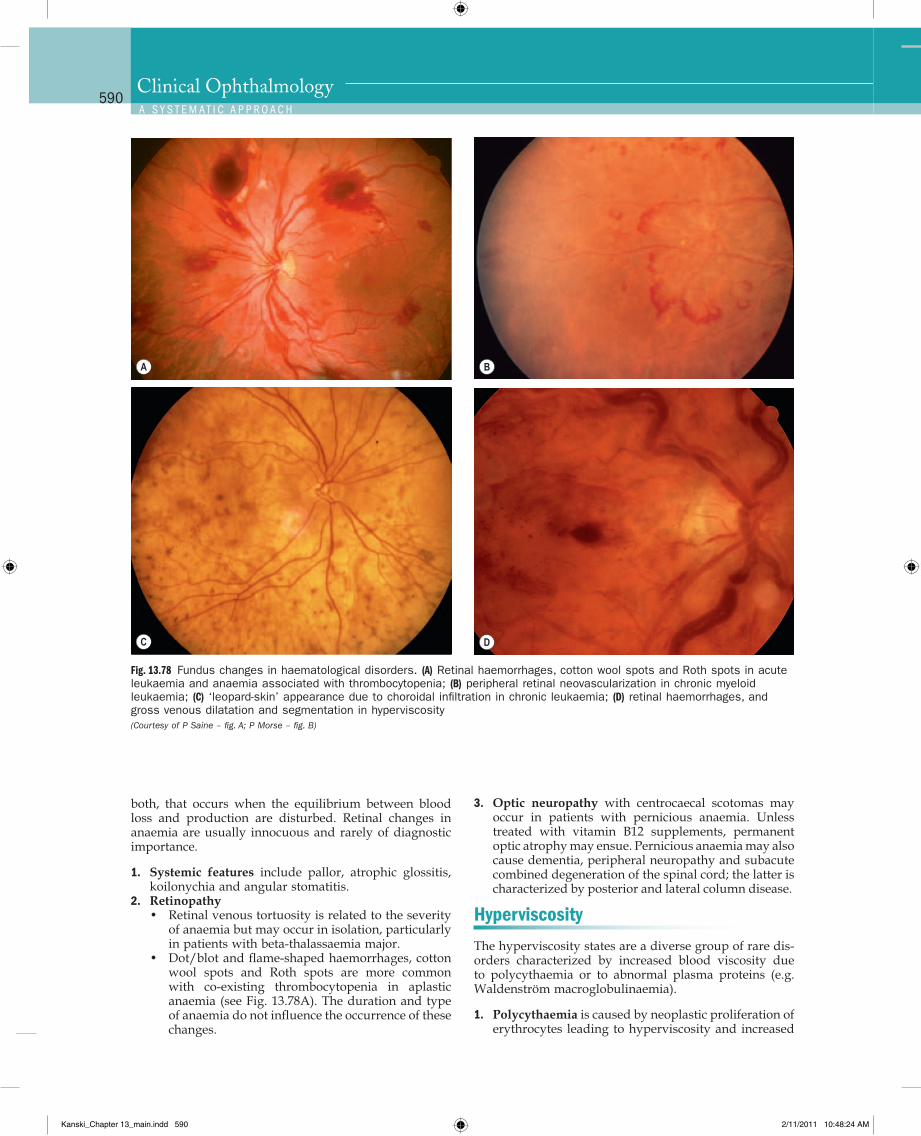
Q
590Clinical OphthalmologyA S y S T e m A T I C A P P R o A C H
3. Optic neuropathy with centrocaecal scotomas may occur in patients with pernicious anaemia. Unless treated with vitamin B12 supplements, permanent optic atrophy may ensue. Pernicious anaemia may also cause dementia, peripheral neuropathy and sub acute combined degeneration of the spinal cord; the latter is characterized by posterior and lateral column disease.
Hyperviscosity
The hyperviscosity states are a diverse group of rare dis-orders characterized by increased blood viscosity due to polycythaemia or to abnormal plasma proteins (e.g. Waldenström macroglobulinaemia).
1. Polycythaemia is caused by neoplastic proliferation of erythrocytes leading to hyperviscosity and increased
both, that occurs when the equilibrium between blood loss and production are disturbed. Retinal changes in anaemia are usually innocuous and rarely of diagnostic importance.
1. Systemic features include pallor, atrophic glossitis, koilonychia and angular stomatitis.
2. Retinopathy• Retinal venous tortuosity is related to the severity
of anaemia but may occur in isolation, particularly in patients with beta-thalassaemia major.
• Dot/blot and flame-shaped haemorrhages, cotton wool spots and Roth spots are more common with co-existing thrombocytopenia in aplastic anaemia (see Fig. 13.78A). The duration and type of anaemia do not influence the occurrence of these changes.
Fig. 13.78 Fundus changes in haematological disorders. (A) Retinal haemorrhages, cotton wool spots and Roth spots in acute leukaemia and anaemia associated with thrombocytopenia; (B) peripheral retinal neovascularization in chronic myeloid leukaemia; (C) ‘leopard-skin’ appearance due to choroidal infiltration in chronic leukaemia; (D) retinal haemorrhages, and gross venous dilatation and segmentation in hyperviscosity (Courtesy of P Saine – fig. A; P Morse – fig. B)
B
C D
A
Kanski_Chapter 13_main.indd 590 2/11/2011 10:48:24 AM
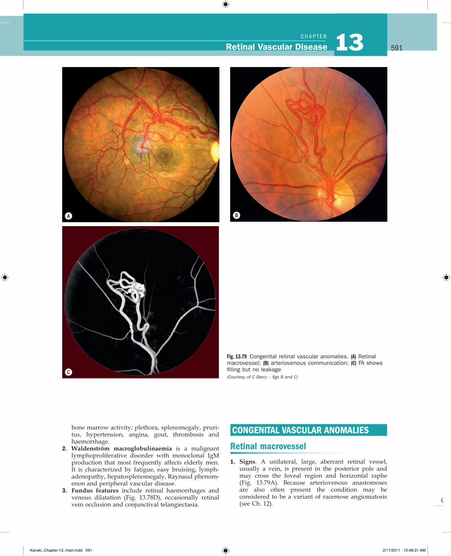
Q
591Retinal Vascular Disease 13C H A P T e R
CONGENITAL VASCULAR ANOMALIES
Retinal macrovessel
1. Signs. A unilateral, large, aberrant retinal vessel, usually a vein, is present in the posterior pole and may cross the foveal region and horizontal raphe (Fig. 13.79A). Because arteriovenous anastomoses are also often present the condition may be considered to be a variant of racemose angiomatosis (see Ch. 12).
bone marrow activity; plethora, splenomegaly, pruri-tus, hypertension, angina, gout, thrombosis and haemorrhage.
2. Waldenström macroglobulinaemia is a malignant lymphoproliferative disorder with monoclonal IgM production that most frequently affects elderly men. It is characterized by fatigue, easy bruising, lymph-adenopathy, hepatosplenomegaly, Raynaud phenom-enon and peripheral vascular disease.
3. Fundus features include retinal haemorrhages and venous dilatation (Fig. 13.78D), occasionally retinal vein occlusion and conjunctival telangiectasia.
Fig. 13.79 Congenital retinal vascular anomalies. (A) Retinal macrovessel; (B) arteriovenous communication; (C) FA shows filling but no leakage (Courtesy of C Barry – figs B and C)
A B
C
Kanski_Chapter 13_main.indd 591 2/11/2011 10:48:31 AM
Q
592Clinical OphthalmologyA S y S T e m A T I C A P P R o A C H
2. FA may show early filling and delayed emptying of the vessel; a dilated capillary bed surrounding the macrovessel is often present. Areas of capillary non-perfusion and foveal cysts may also be seen.
Arteriovenous communications
Congenital arteriovenous communications usually present on routine examination, with unilateral involve-ment in single or multiple sites of the same fundus. They have a predilection for the papillomacular bundle and the superotemporal quadrant. Occasionally reported compli-cations include haemorrhage, exudation and vascular
occlusion. Some patients may harbour similar systemic lesions. The malformations can be divided into the fol-lowing three types on the basis of severity.
Group 1 consists of an anastomosis between a small arte-riole and venule with the interposition of an abnormal capillary or arteriolar plexus. It is non-progressive and associated with good visual acuity.
Group 2 demonstrates direct arteriovenous communica-tions between a branch retinal artery and vein (Fig. 13.79B and C).
Group 3 consists of diffuse marked dilatation of the vascular tree with many large-calibre anastomosing channels.
Kanski_Chapter 13_main.indd 592 2/11/2011 10:48:32 AM