Embed Size (px)
Citation preview
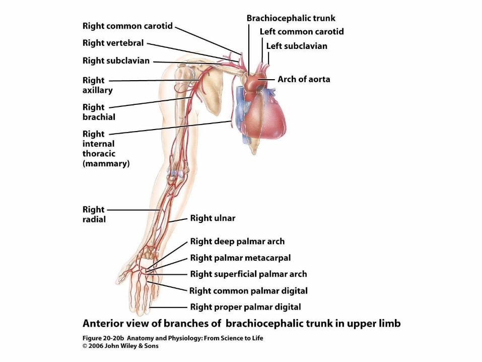
Figure 20.20b
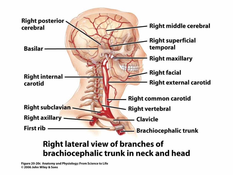
Figure 20.20c
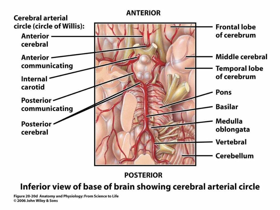
Figure 20.20d
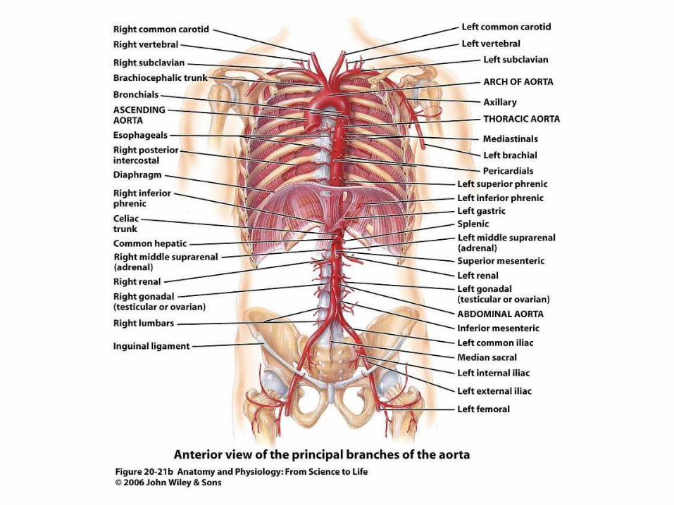
Figure 20.21b
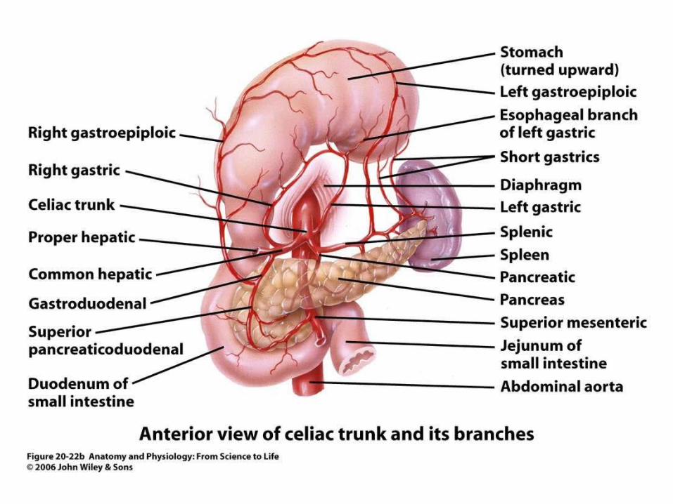
Figure 20.22b
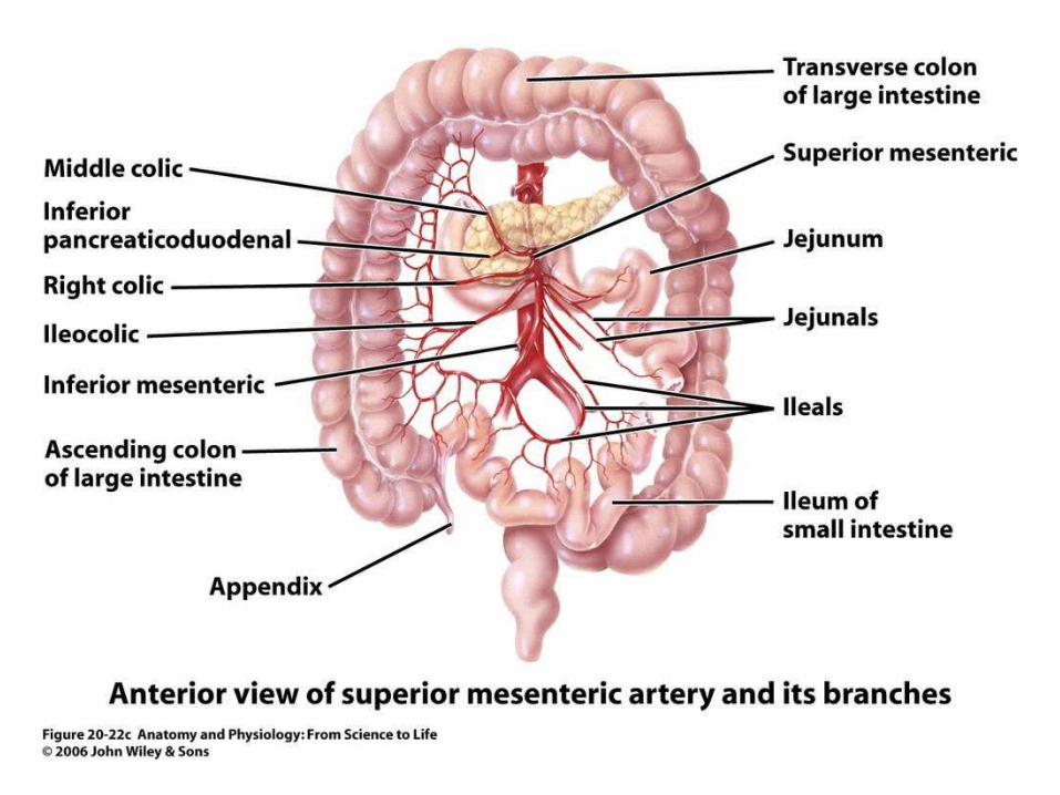
Figure 20.22c
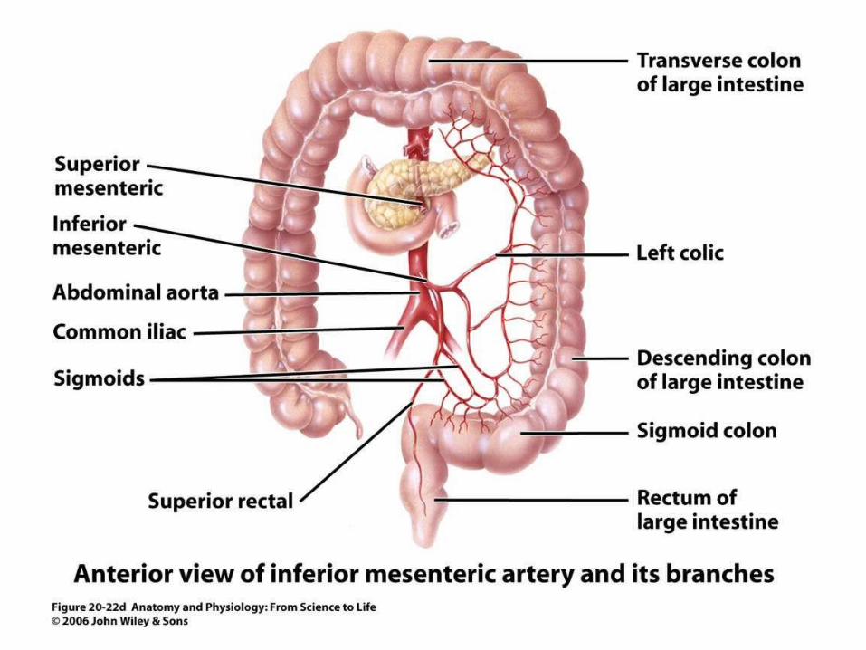
Figure 20.22d
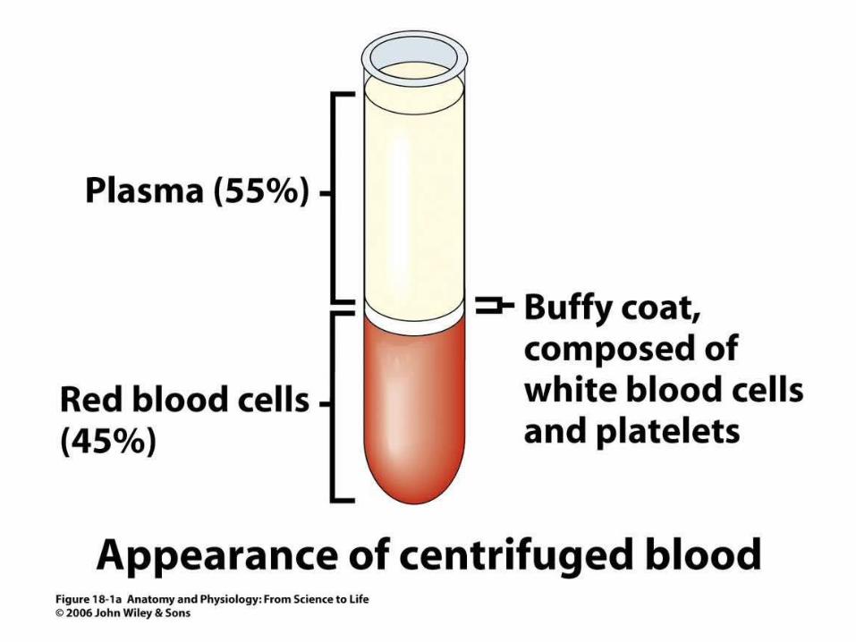
Figure 18.1a
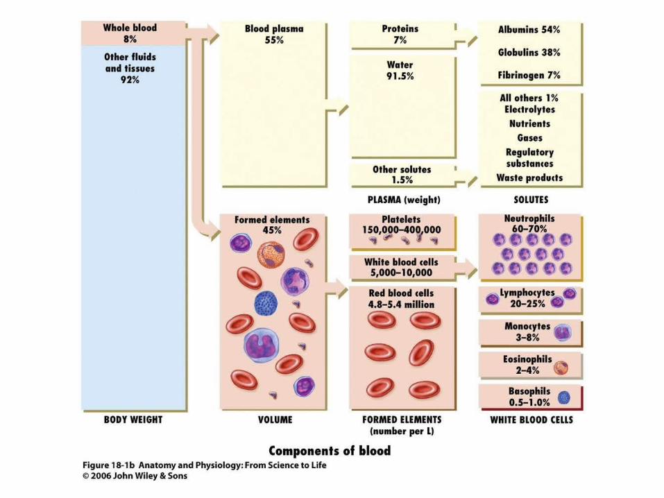
Figure 18.1b
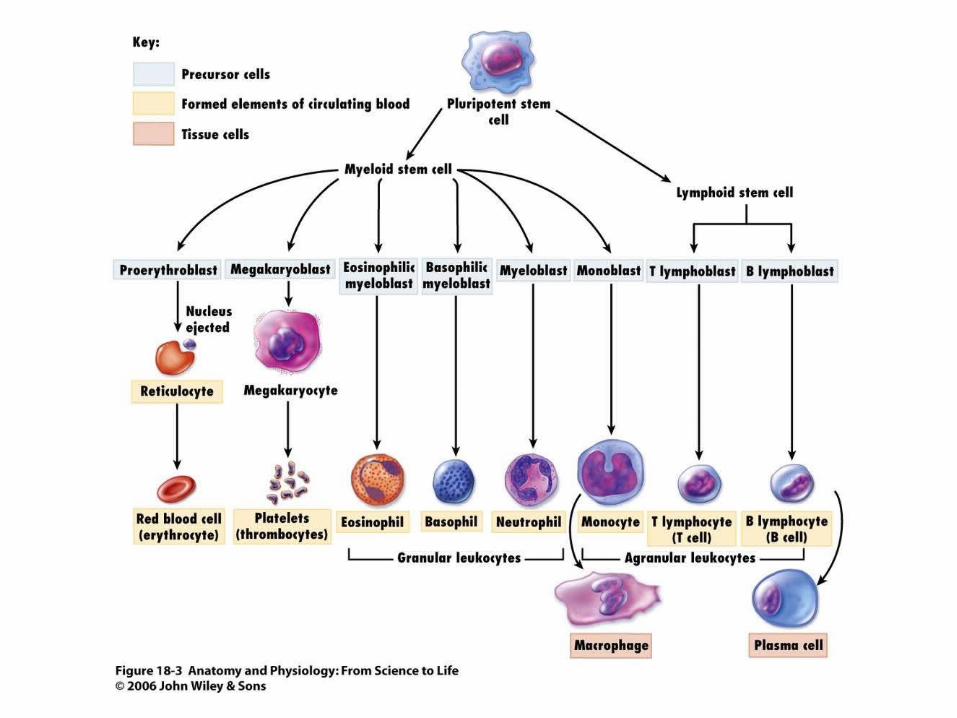
Figure 18.3
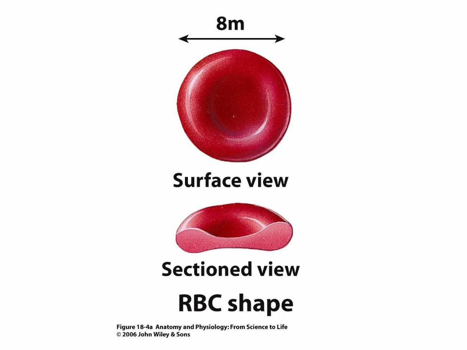
Figure 18.4a
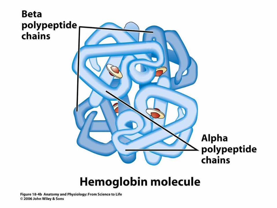
Figure 18.4b
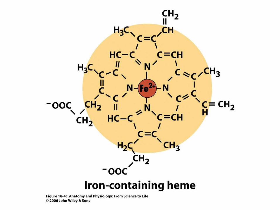
Figure 18.4c
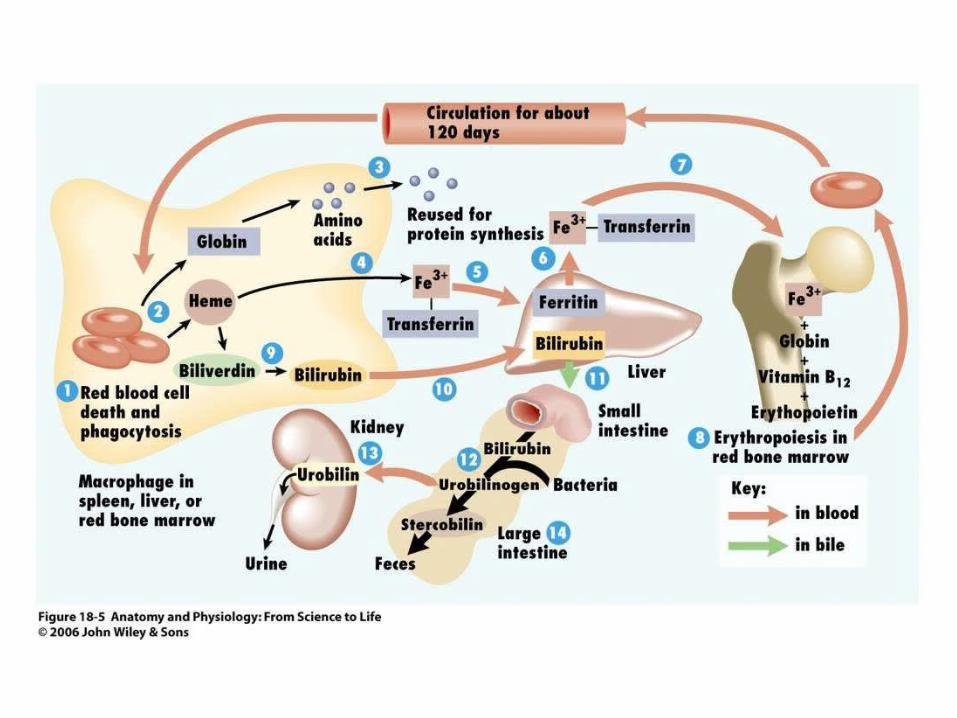
Figure 18.5
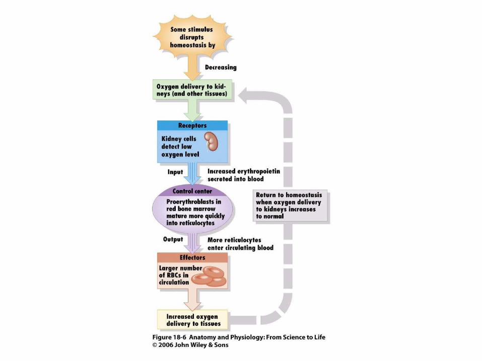
Figure 18.6
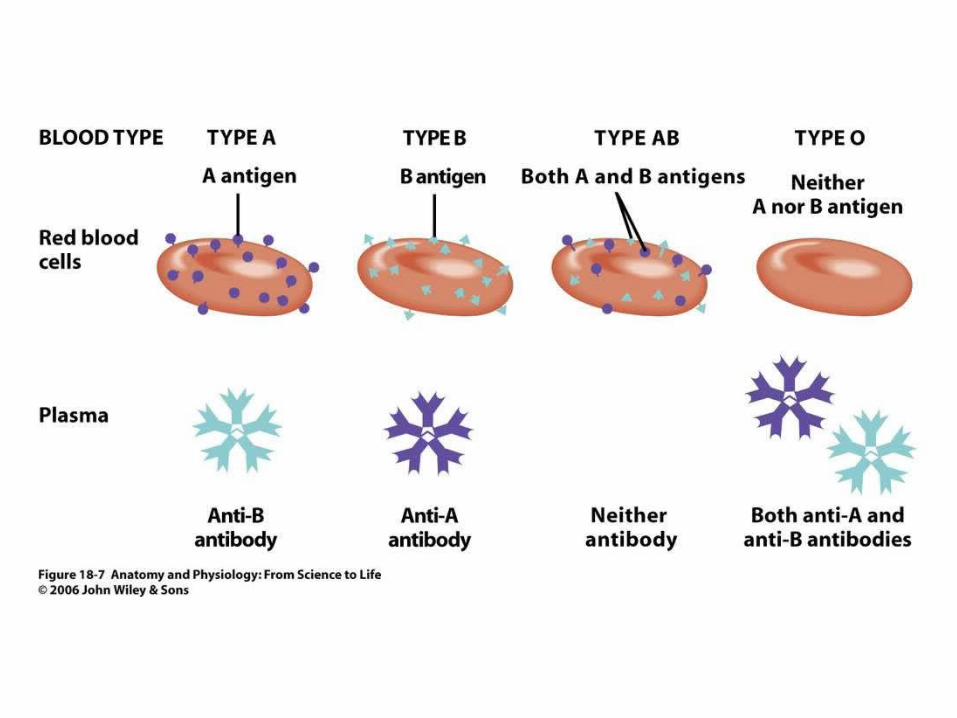
Figure 18.7

Figure 18.3
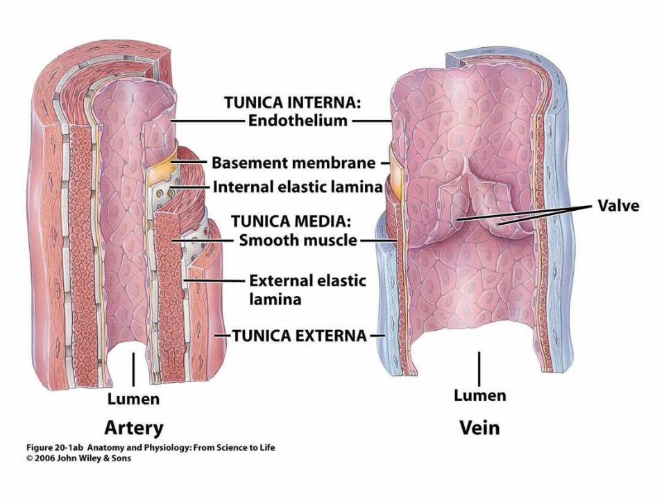
Figure 20.1ab
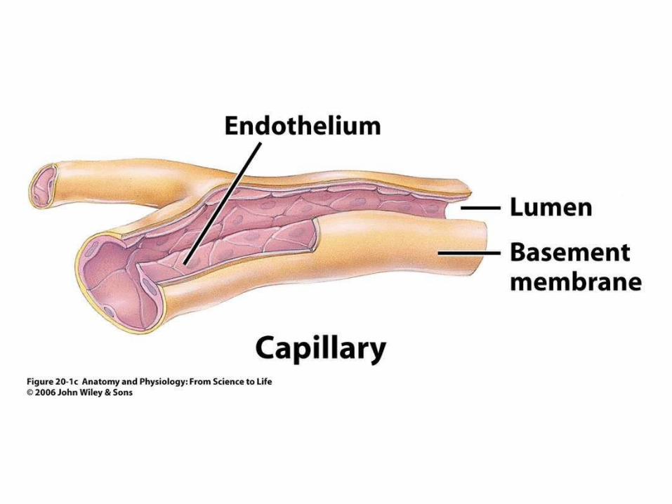
Figure 20.1c
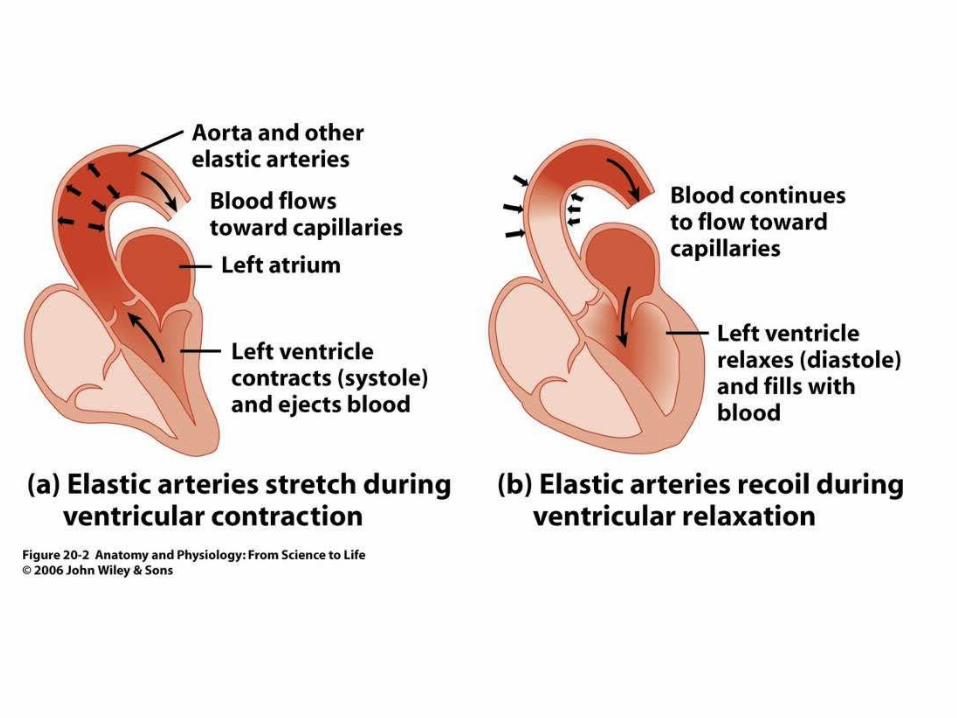
Figure 20.2

Figure 20.3
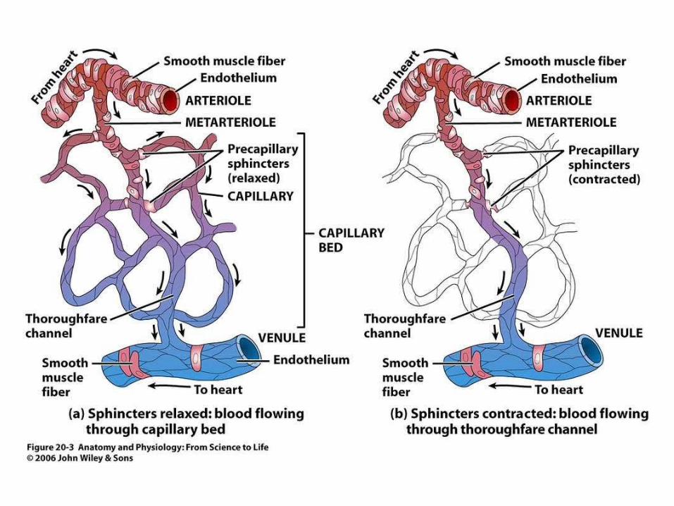
Figure 20.3
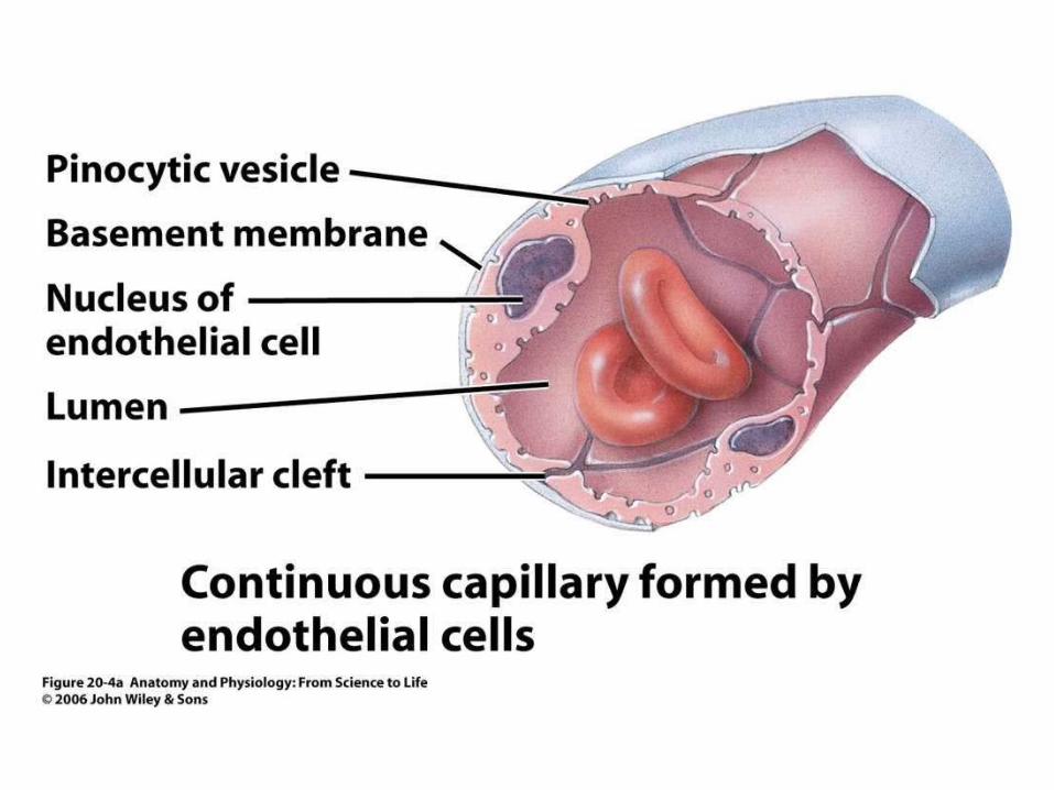
Figure 20.4a
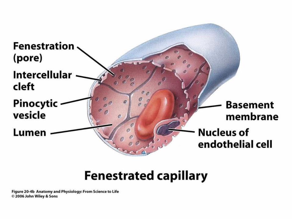
Figure 20.4b
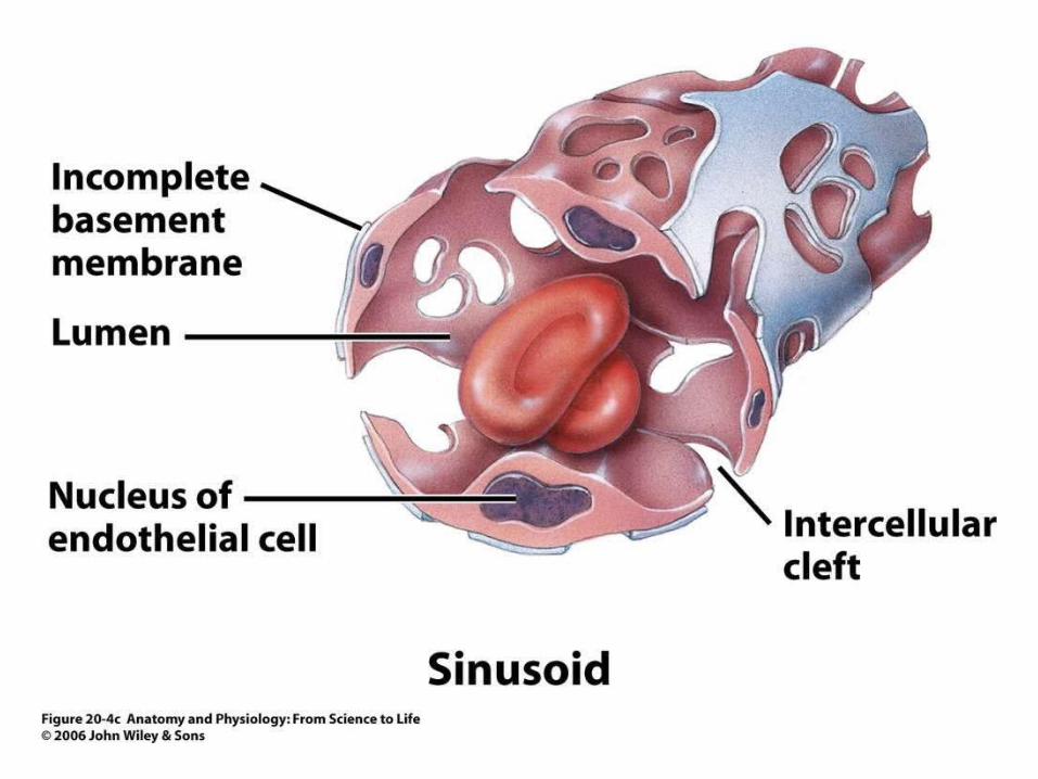
Figure 20.4c
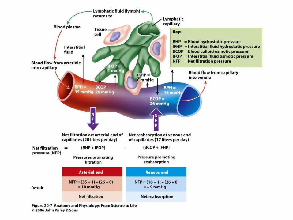
Figure 20.7
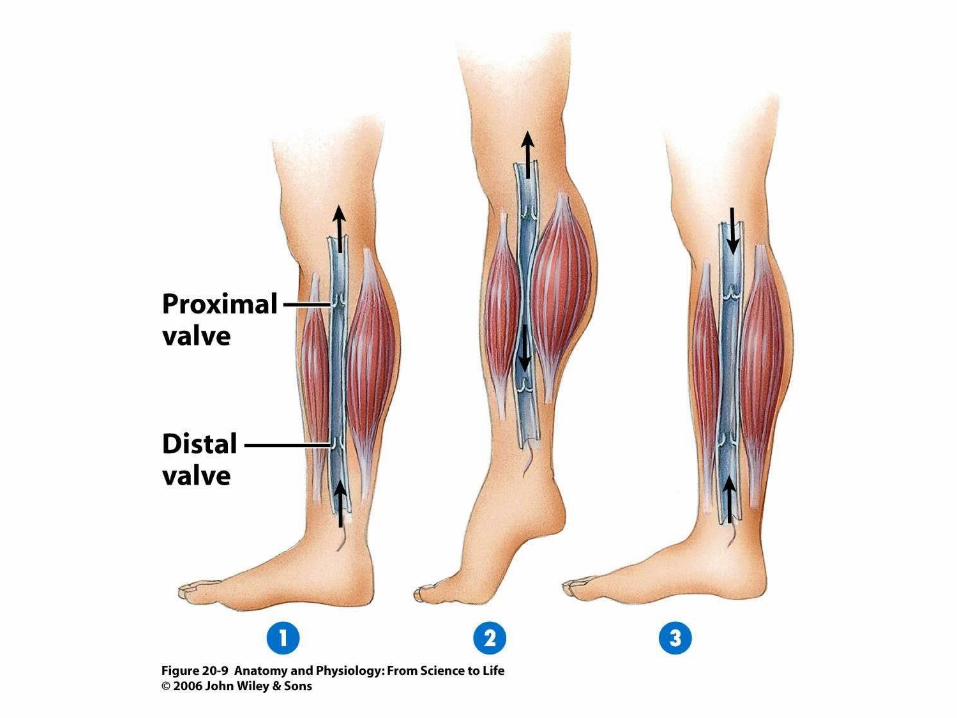
Figure 20.9
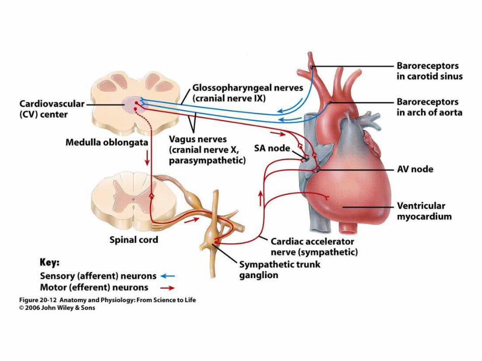
Figure 20.12

Figure 20.17a
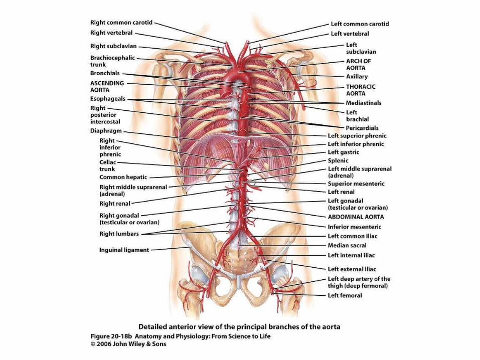
Figure 20.18b
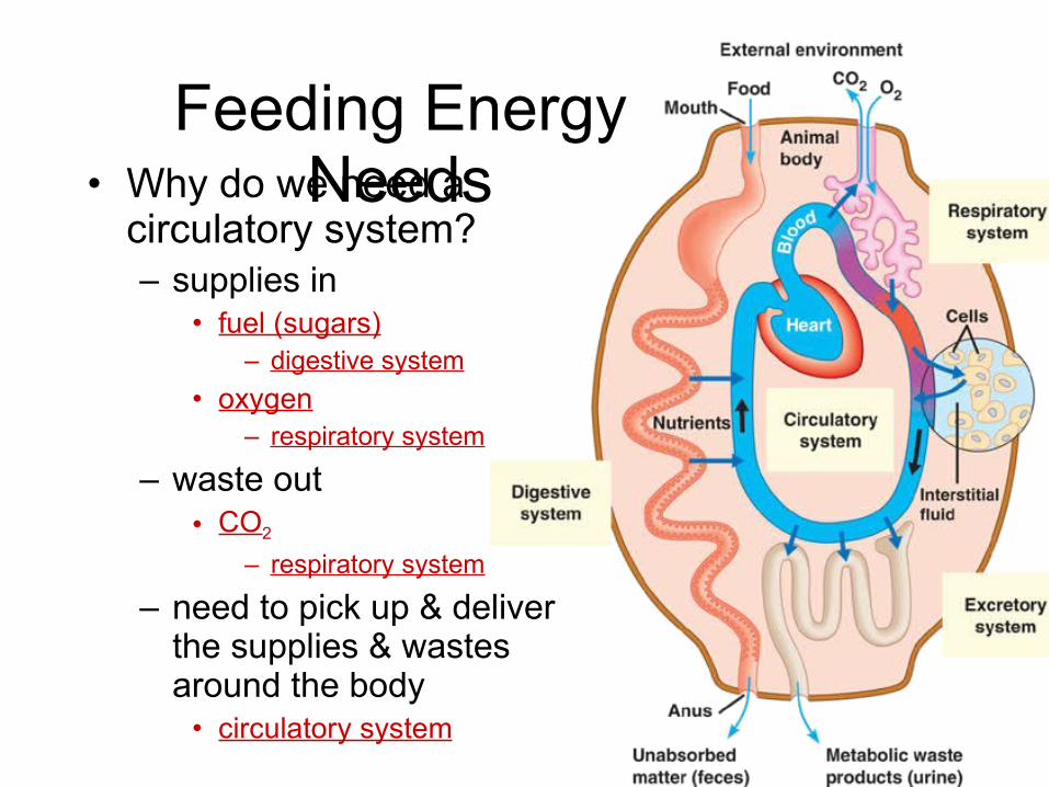
• Why do we need a circulatory system?– supplies in
• fuel (sugars)– digestive system
• oxygen– respiratory system
– waste out• CO2
– respiratory system
– need to pick up & deliver the supplies & wastes around the body
• circulatory system
Feeding Energy Needs
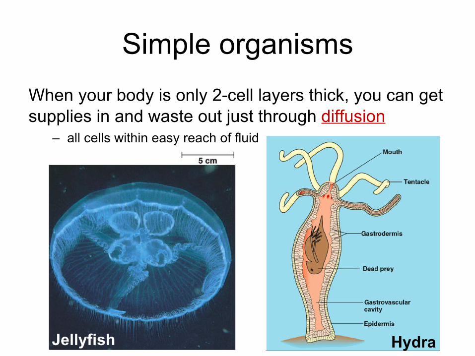
Simple organisms
When your body is only 2-cell layers thick, you can get supplies in and waste out just through diffusion
– all cells within easy reach of fluid
HydraJellyfish
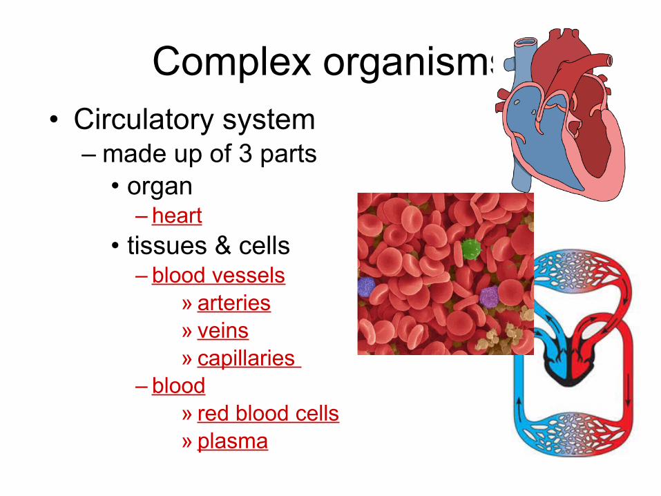
Complex organisms• Circulatory system
– made up of 3 parts• organ
– heart
• tissues & cells– blood vessels
» arteries» veins» capillaries
– blood» red blood cells» plasma
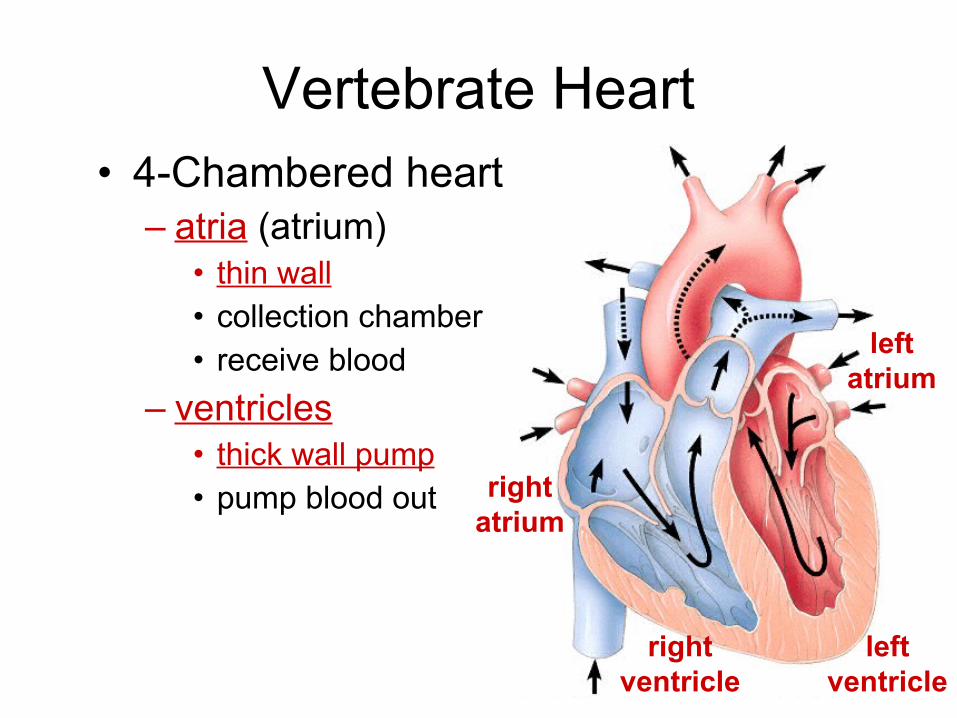
Vertebrate Heart• 4-Chambered heart
– atria (atrium)• thin wall• collection chamber• receive blood
– ventricles • thick wall pump• pump blood out right
atrium
leftatrium
rightventricle
leftventricle
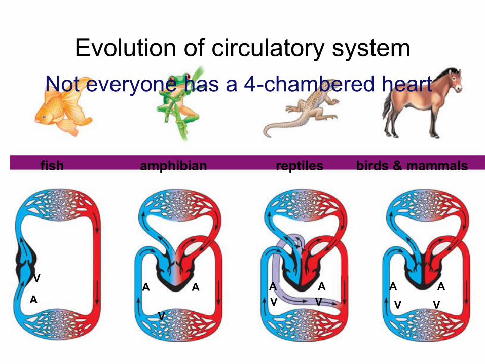
Evolution of circulatory system
fish amphibian reptiles birds & mammals
A A
VV V VV
A AAAA
V
2 chamber 3 chamber 3 chamber 4 chamber
Not everyone has a 4-chambered heart
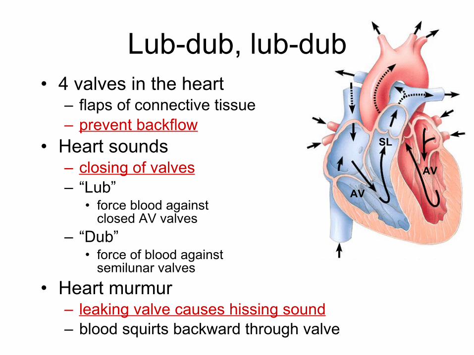
AV
SL
AV
Lub-dub, lub-dub• 4 valves in the heart
– flaps of connective tissue– prevent backflow
• Heart sounds – closing of valves– “Lub”
• force blood against closed AV valves
– “Dub”• force of blood against
semilunar valves
• Heart murmur– leaking valve causes hissing sound – blood squirts backward through valve
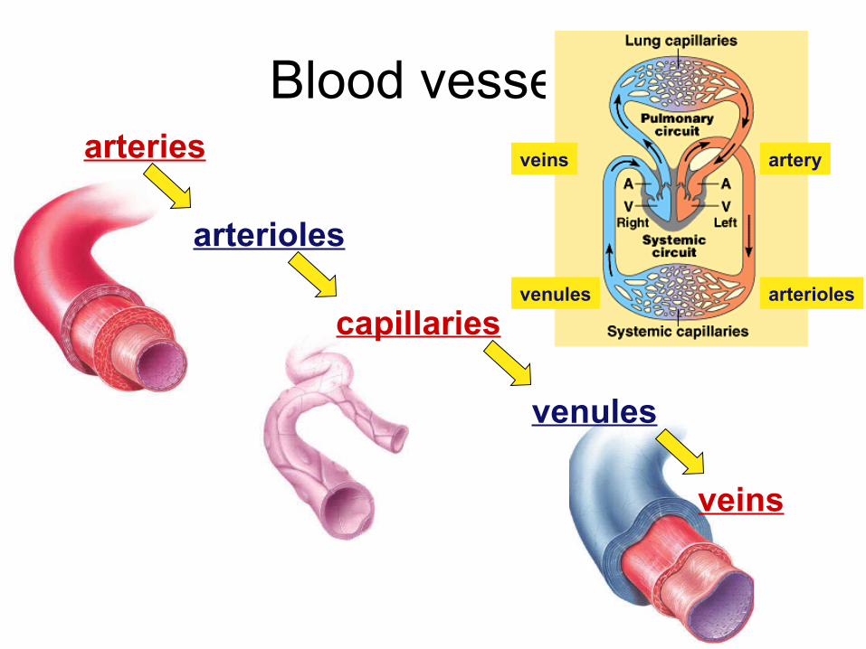
Blood vesselsarteries
arterioles
capillaries
venules
veins
artery
arteriolesvenules
veins
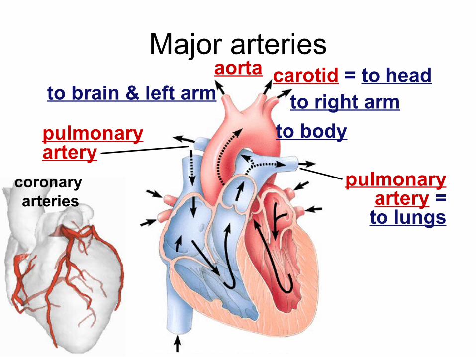
Major arteries
pulmonaryartery
pulmonaryartery =to lungs
aorta carotid = to headto brain & left arm to right arm
coronary arteries
to body
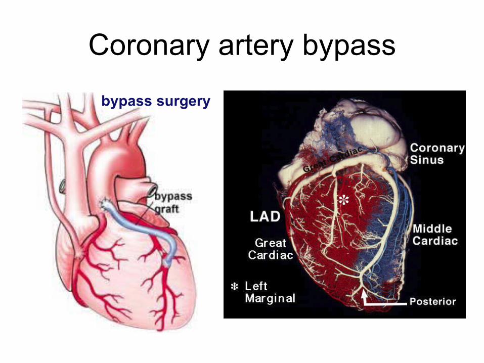
Coronary artery bypass
bypass surgery
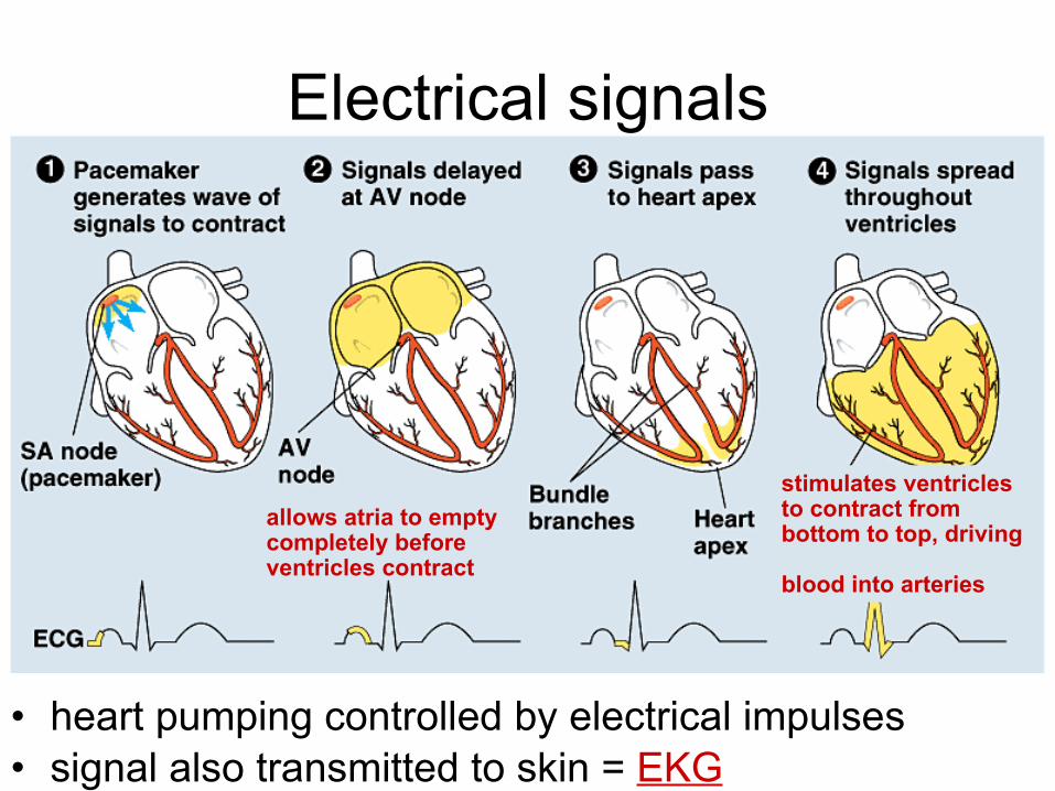
Electrical signals
allows atria to empty completely before ventricles contract
stimulates ventricles to contract from bottom to top, driving
blood into arteries
• heart pumping controlled by electrical impulses • signal also transmitted to skin = EKG
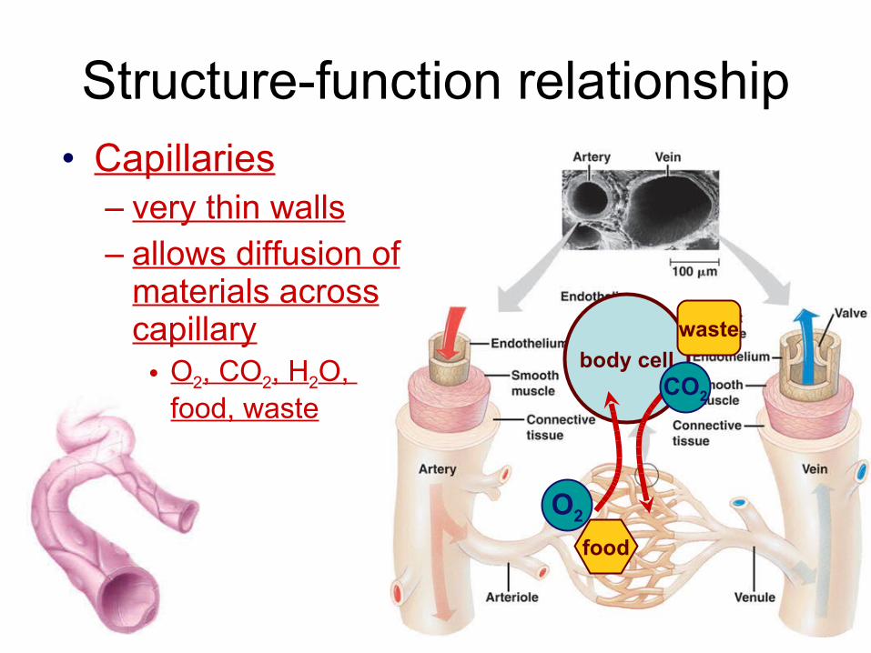
Structure-function relationship• Capillaries
– very thin walls – allows diffusion of
materials across capillary
• O2, CO2, H2O, food, waste
body cell
O2
food
waste
CO2
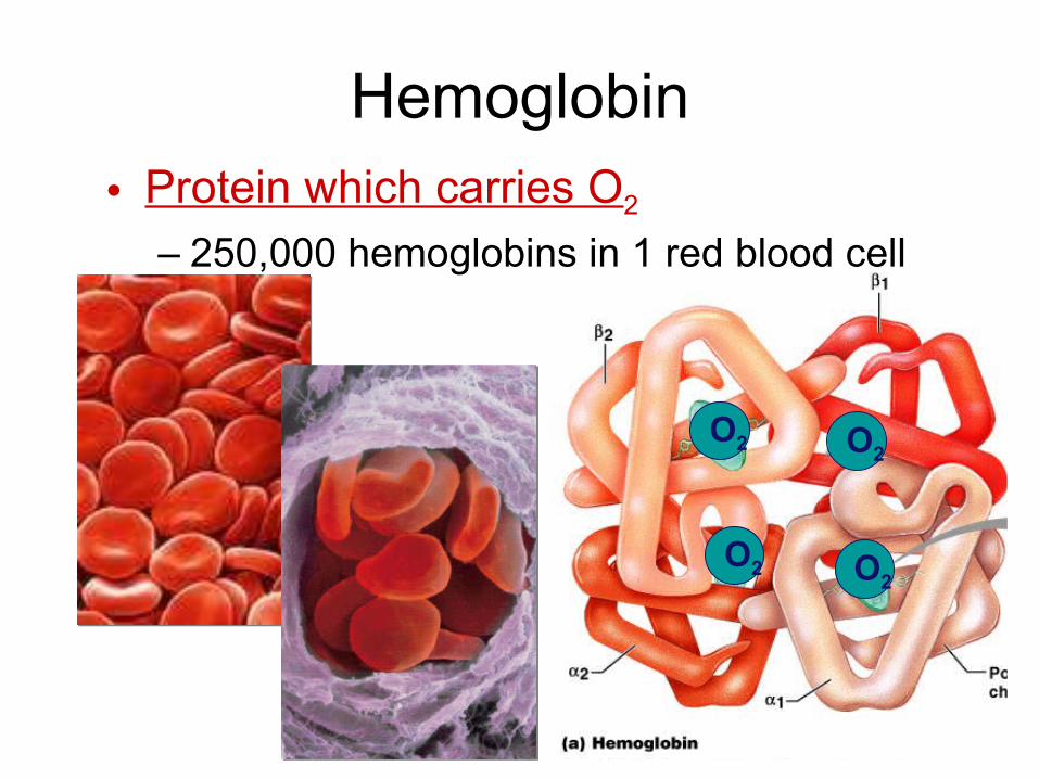
Hemoglobin
• Protein which carries O2
– 250,000 hemoglobins in 1 red blood cell
O2
O2O2
O2
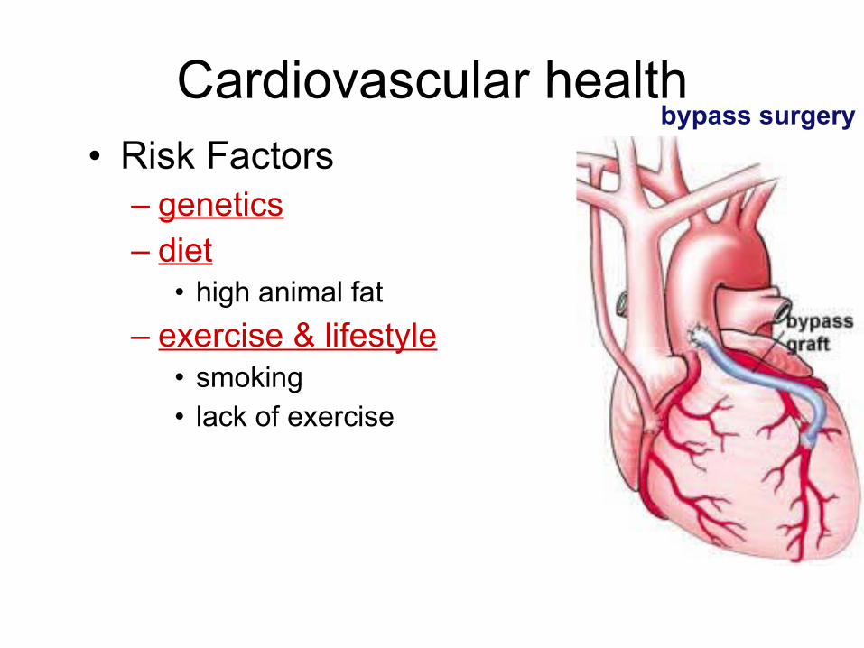
Cardiovascular healthbypass surgery
• Risk Factors– genetics– diet
• high animal fat
– exercise & lifestyle• smoking• lack of exercise
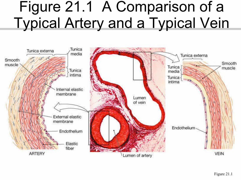
Figure 21.1
Figure 21.1 A Comparison of a Typical Artery and a Typical Vein
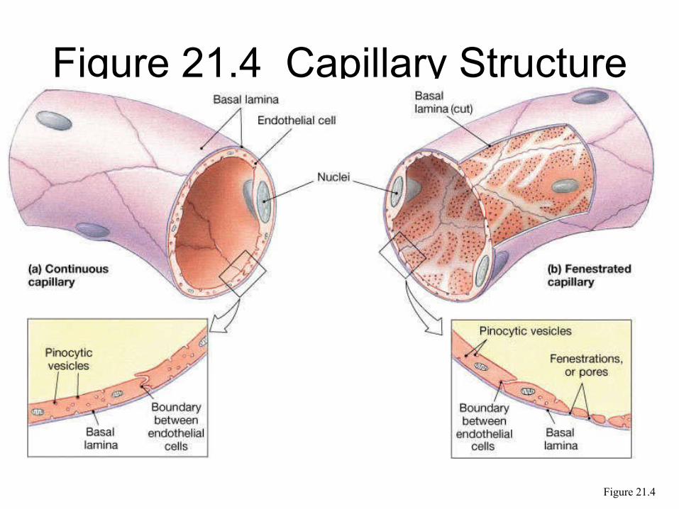
Figure 21.4 Capillary Structure
Figure 21.4
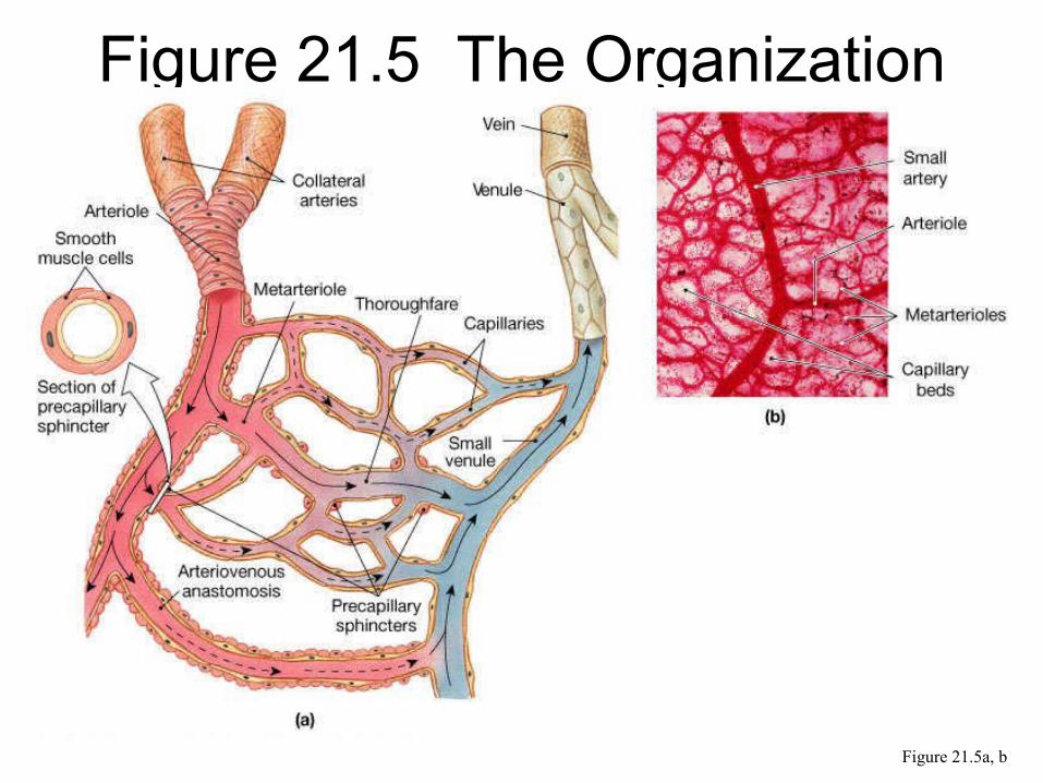
Figure 21.5 The Organization of a Capillary Bed
Figure 21.5a, b
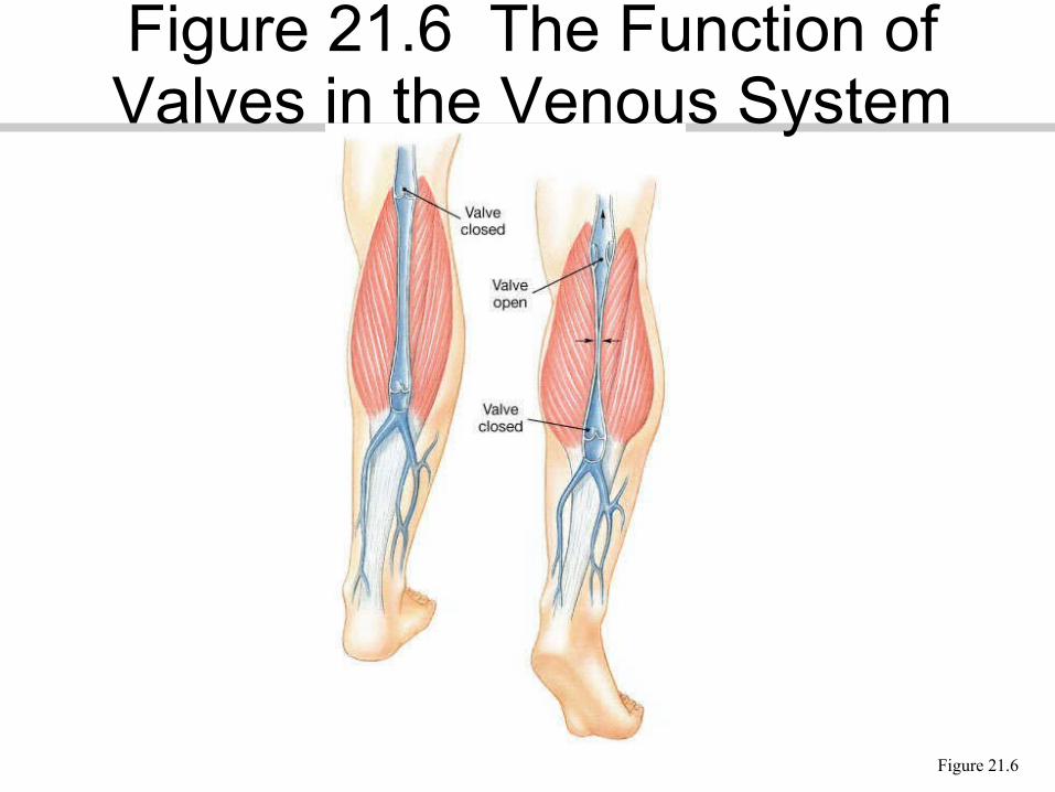
Figure 21.6
Figure 21.6 The Function of Valves in the Venous System
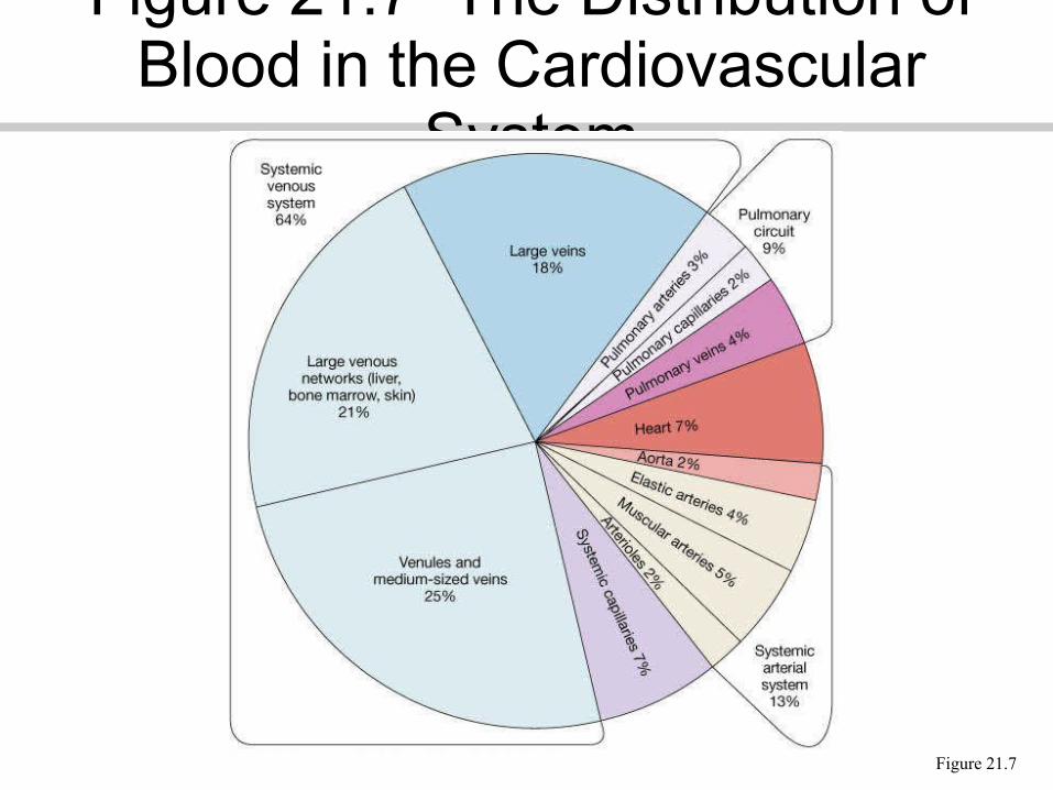
Figure 21.7
Figure 21.7 The Distribution of Blood in the Cardiovascular
System
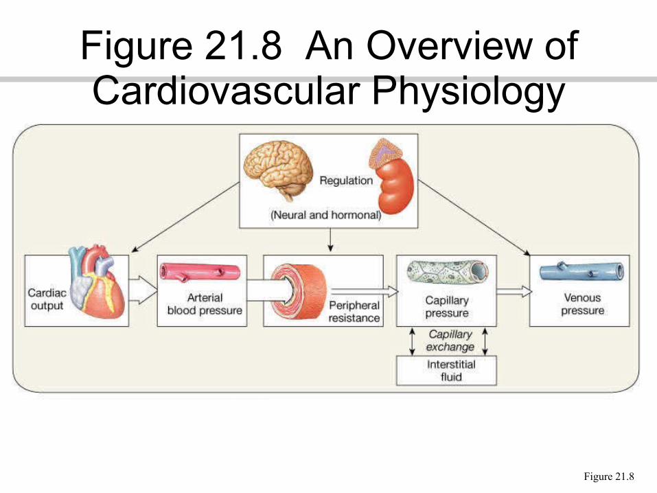
Figure 21.8
Figure 21.8 An Overview of Cardiovascular Physiology
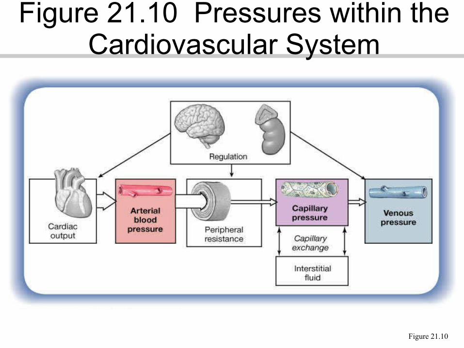
Figure 21.10
Figure 21.10 Pressures within the Cardiovascular System
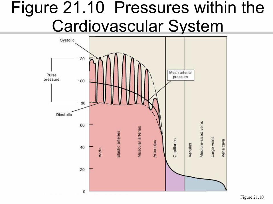
Figure 21.10
Figure 21.10 Pressures within the Cardiovascular System
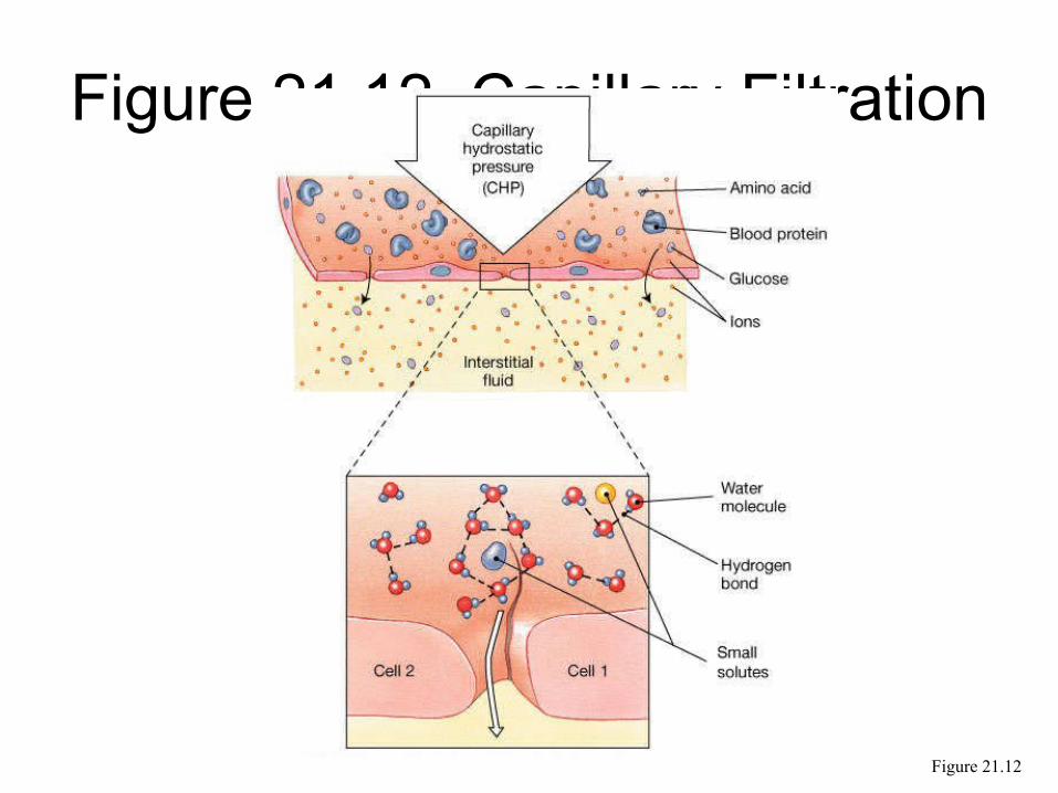
Figure 21.12 Capillary Filtration
Figure 21.12
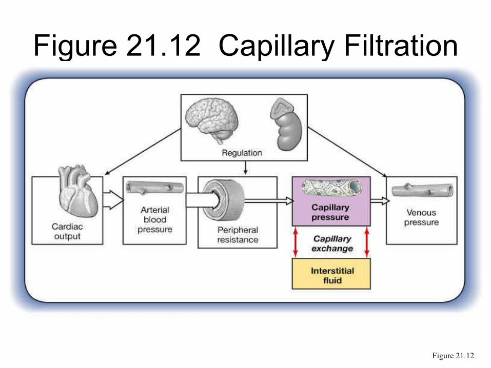
Figure 21.12 Capillary Filtration
Figure 21.12
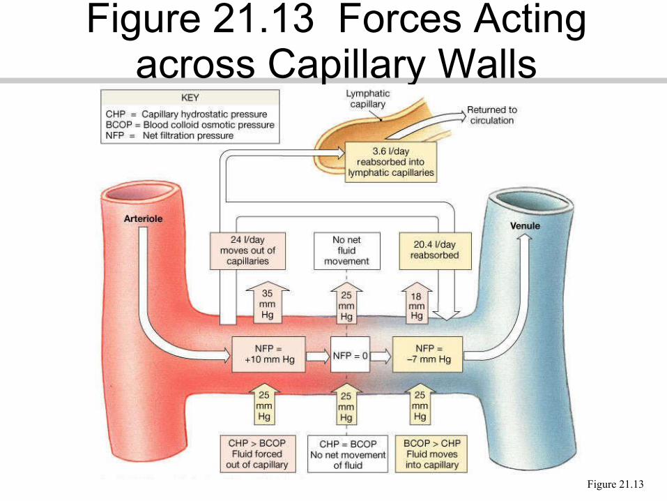
Figure 21.13
Figure 21.13 Forces Acting across Capillary Walls

Figure 21.14
Figure 21.14 Homeostatic Adjustments that Compensate for
a Reduction in Blood Pressure and Blood Flow
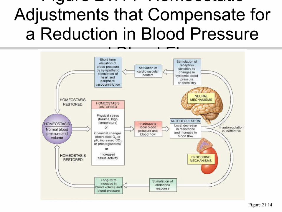
Figure 21.14
Figure 21.14 Homeostatic Adjustments that Compensate for
a Reduction in Blood Pressure and Blood Flow
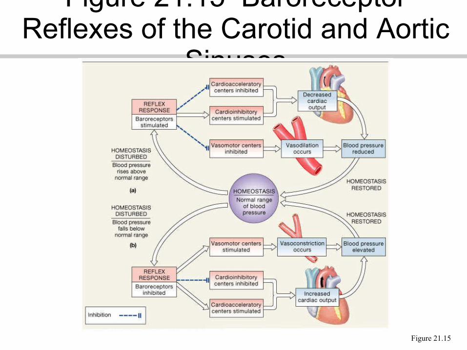
Figure 21.15
Figure 21.15 Baroreceptor Reflexes of the Carotid and Aortic
Sinuses
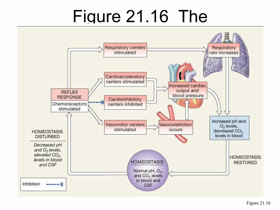
Figure 21.16 The Chemoreceptor Reflexes
Figure 21.16
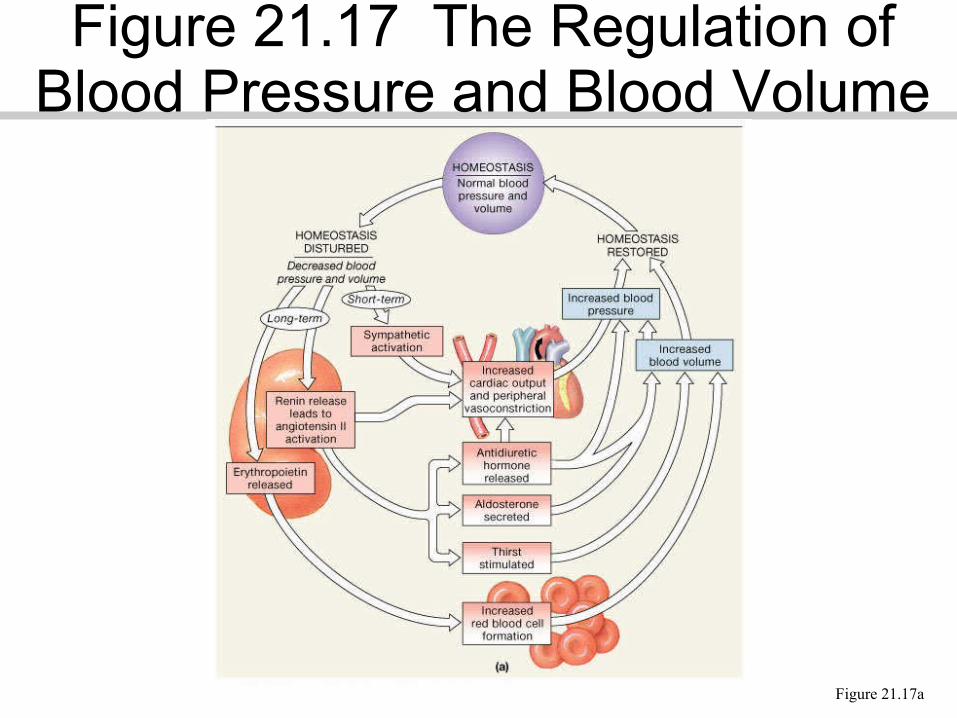
Figure 21.17a
Figure 21.17 The Regulation of Blood Pressure and Blood Volume
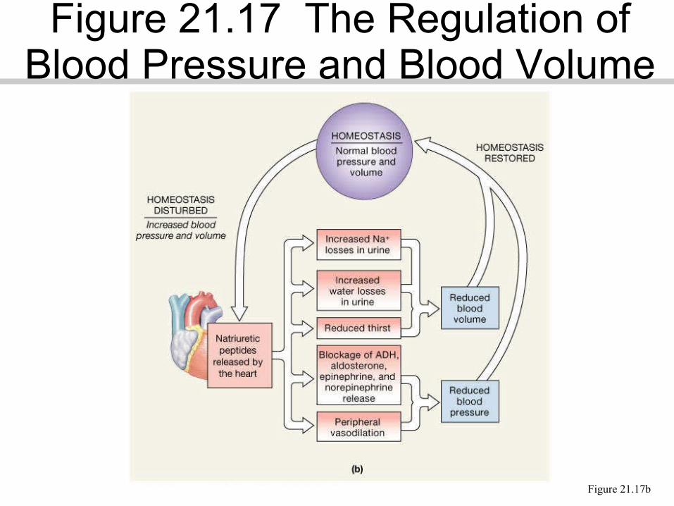
Figure 21.17b
Figure 21.17 The Regulation of Blood Pressure and Blood Volume
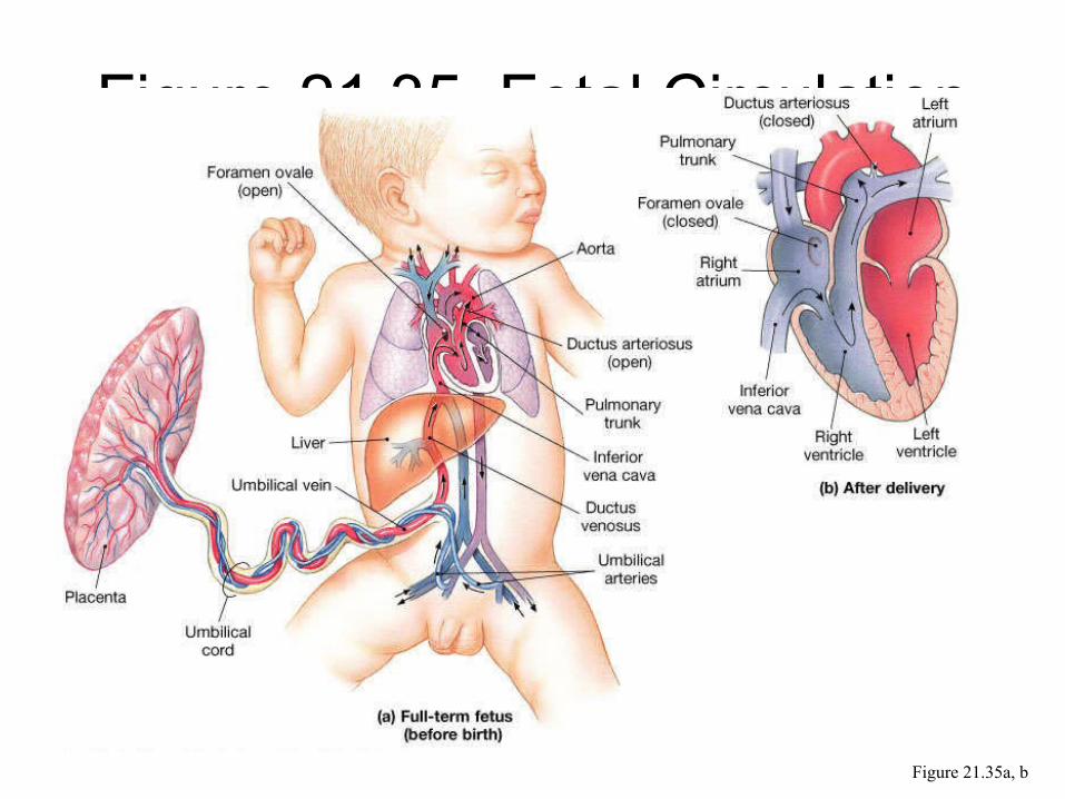
Figure 21.35a, b
Figure 21.35 Fetal Circulation


![[123doc.vn] - chuong-3-he-thong-tuan-hoan-cac-nguyen-to-hoa-hoc-pps.doc](https://img.pdfslide.tips/doc/110x75/55cf8e9e550346703b93fec0/123docvn-chuong-3-he-thong-tuan-hoan-cac-nguyen-to-hoa-hoc-ppsdoc.jpg)
![[Bài giảng, ngực bụng] tuan hoan](https://img.pdfslide.tips/doc/110x75/559b7ba61a28ab00258b4732/bai-giang-nguc-bung-tuan-hoan.jpg)