Embed Size (px)
Citation preview

Everybody Has Atherosclerosis
The Question Is Who Has
Vulnerable PlaqueParadigm Shift in Cardiology
“Magnetic Resonance Imaging of Plaque Inflammation”CIMIT Vulnerable Plaque Program
February 11th, 2002Morteza Naghavi, MD
University of Texas Houston and Texas Heart Institute

Sudden Cardiac DeathAcute MI
VulnerablePlaque(s)
Stop Vulnerable Plaque, Prevent Heart Attack!
On the basis of his studies of the triggers of MI, James E. Muller et al described “coronary occlusive thrombi occurs when atherosclerotic plaques become vulnerable to rupture … during vulnerable periods…" 5 years later in 1994 he coined the term of
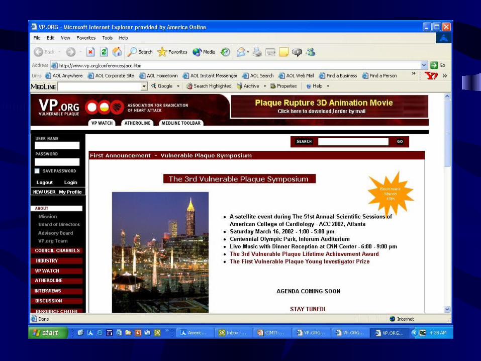
History of Atherosclerosis from www.VP.org
Who Brought the Name of “Vulnerable Plaque” to Medical Literature?
Muller JE, Abela GS, Nesto RW, Tofler GH.
Triggers, acute risk factors and vulnerable plaques: the lexicon of a new frontier.
J Am Coll Cardiol. 1994 Mar 1;23(3):809-13. Review.
Vulnerable Plaque.
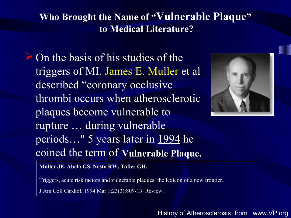
Vulnerable Plaque, the youngest creature in the land of cardiologyhas just turned in 8 y!
VPiologists call him VP!!
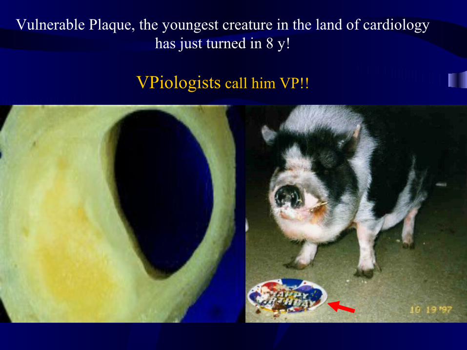
Carl von Rokitansky (1804-1878)
Rokitansky gave early detailed descriptions of arterial disease. He is
alleged to have performed 30,000 autopsies.
Rokitansky in 1841 championed the Thrombogenic Theory. He proposed that the deposits observed in the inner layer of the arterial wall derived primarily from fibrin and other blood elements rather than being the result of a purulent process. Subsequently, the atheroma
resulted from the degeneration of the fibrin and other blood proteins as a result of a preexisting crasis of the blood, and finally these deposits were modified toward a pulpy mass containing
cholesterol crystals and fatty globules.
This theory came under attack by Virchow
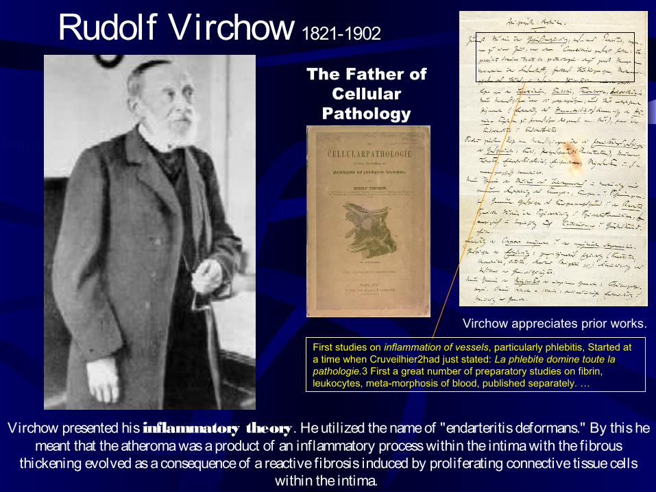
First studies on inflammation of vessels, particularly phlebitis, Started at a time when Cruveilhier2had just stated: La phlebite domine toute la pathologie.3 First a great number of preparatory studies on fibrin, leukocytes, meta-morphosis of blood, published separately. …
Rudolf Virchow 1821-1902
The Father of Cellular
Pathology
Virchow appreciates prior works.
Virchow presented his inflammatory theory. He utilized the name of "endarteritis deformans." By this he meant that the atheroma was a product of an inflammatory process within the intima with the fibrous
thickening evolved as a consequence of a reactive fibrosis induced by proliferating connective tissue cells within the intima.
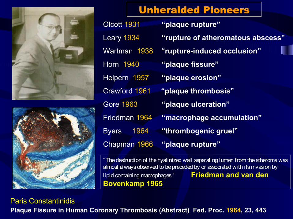
Olcott 1931 “plaque rupture”
Leary 1934 “rupture of atheromatous abscess”
Wartman 1938 “rupture-induced occlusion”
Horn 1940 “plaque fissure”
Helpern 1957 “plaque erosion”
Crawford 1961 “plaque thrombosis”
Gore 1963 “plaque ulceration”
Friedman 1964 “macrophage accumulation”
Byers 1964 “thrombogenic gruel”
Chapman 1966 “plaque rupture”
Plaque Fissure in Human Coronary Thrombosis (Abstract) Fed. Proc. 1964, 23, 443 Paris Constantinidis
“The destruction of the hyalinized wall separating lumen from the atheroma was almost always observed to be preceded by or associated with its invasion by lipid containing macrophages.” Friedman and van den Bovenkamp 1965
Unheralded Pioneers
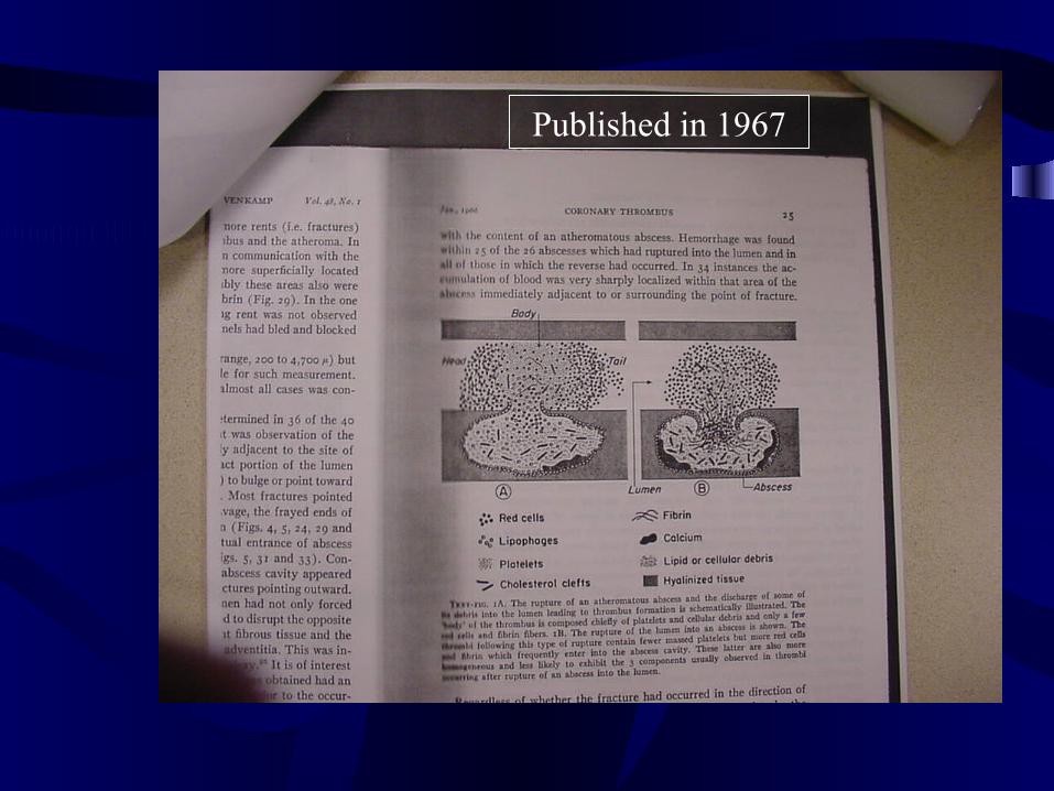
Published in 1967
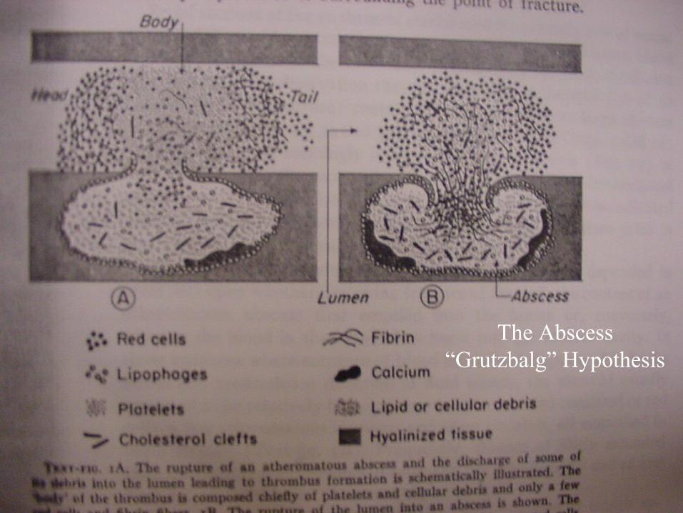
The Abscess “Grutzbalg” Hypothesis
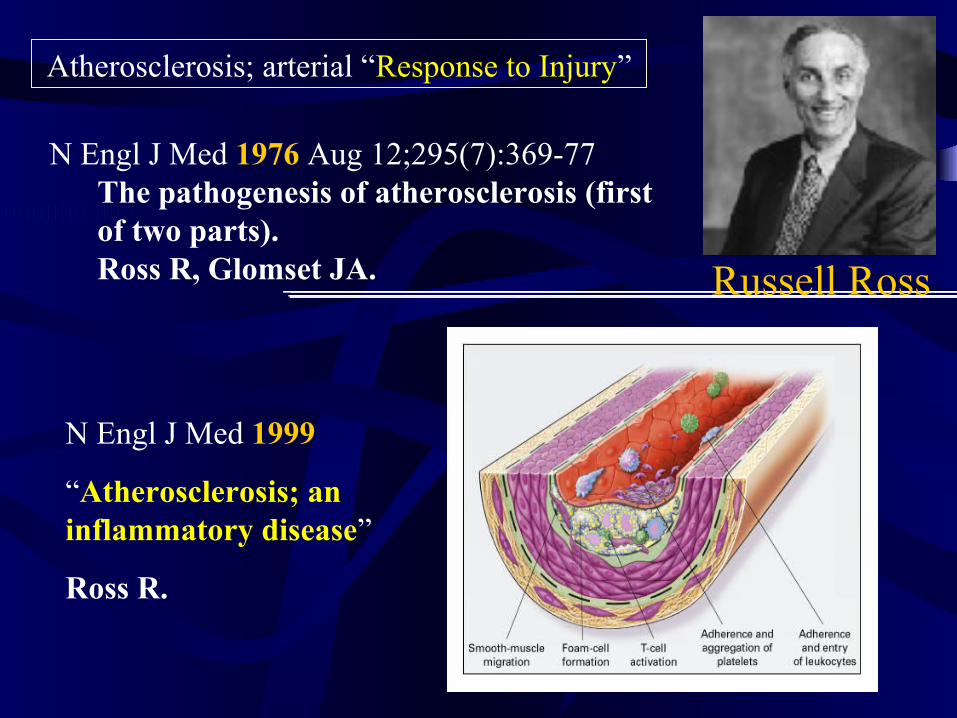
N Engl J Med 1999
“Atherosclerosis; an inflammatory disease”
Ross R.
Russell Ross
Atherosclerosis; arterial “Response to Injury”
N Engl J Med 1976 Aug 12;295(7):369-77 The pathogenesis of atherosclerosis (first of two parts).Ross R, Glomset JA.
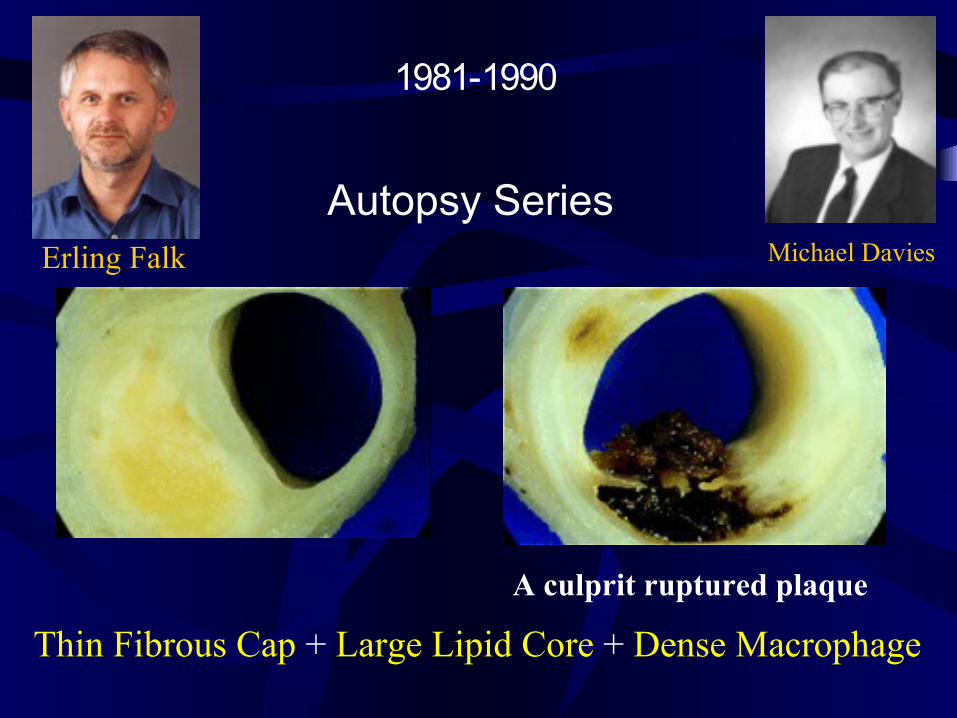
Erling Falk Michael Davies
Autopsy Series
Thin Fibrous Cap + Large Lipid Core + Dense Macrophage
A culprit ruptured plaque
1981-1990
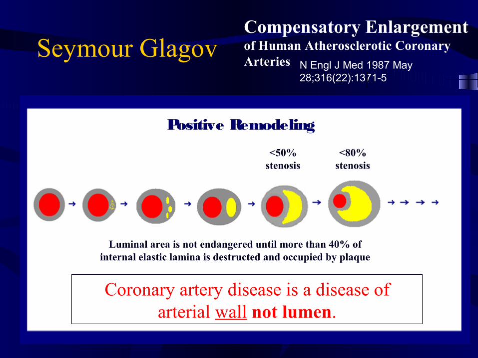
Seymour GlagovCompensatory Enlargement of Human Atherosclerotic Coronary Arteries N Engl J Med 1987 May
28;316(22):1371-5
<50% stenosis
Luminal area is not endangered until more than 40% of internal elastic lamina is destructed and occupied by plaque
Coronary artery disease is a disease of arterial wall not lumen.
Positive Remodeling
<80% stenosis

Angiographic progression of coronary artery disease and the development of myocardial infarction.Ambrose JA, Tannenbaum MA, Alexopoulos D, Hjemdahl-Monsen CE, Leavy J, Weiss M, Borrico S, Gorlin R, Fuster V.
Department of Medicine, New York Cardiac Center, Mount Sinai Medical Center, New York 10029.
Simultaneously, Little et al, Haft et al reported that majority of culprit lesions are found on previously non-critical stenosis plaques.
Conclusion: “Myocardial infarction frequently develops from non-severe lesions.”
J Am Coll Cardiol 1988 Jul;12(1):56-62
Ambrose, Fuster, and colleagues
X-Ray Angiographically Invisible Plaques
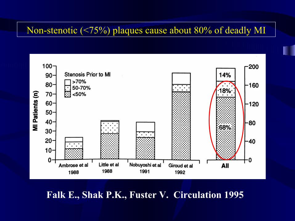
Falk E., Shak P.K., Fuster V. Circulation 1995
Non-stenotic (<75%) plaques cause about 80% of deadly MI
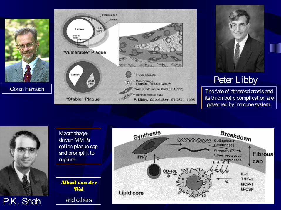
Macrophage- driven MMPs soften plaque cap and prompt it to rupture
P.K. Shah
Peter LibbyThe fate of atherosclerosis and its thrombotic complication are governed by immune system.
Goran Hansson
Allard van der Wal
and others
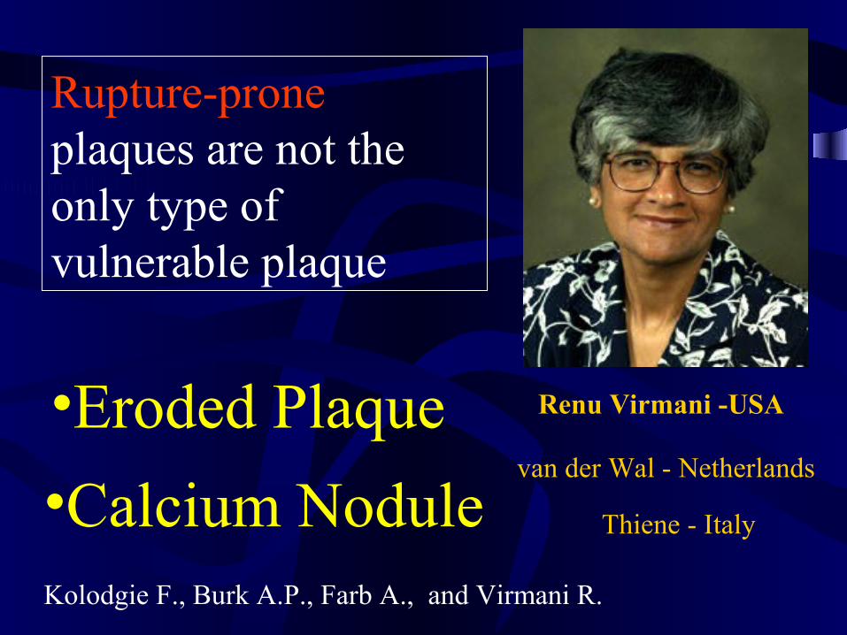
•Eroded Plaque
Rupture-prone plaques are not the only type of vulnerable plaque
•Calcium Nodulevan der Wal - Netherlands
Renu Virmani -USA
Thiene - Italy
Kolodgie F., Burk A.P., Farb A., and Virmani R.
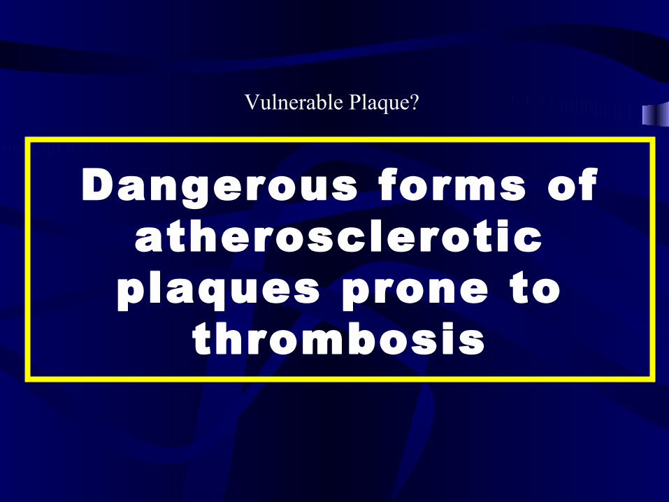
Dangerous forms of atherosclerotic
plaques prone to thrombosis
Vulnerable Plaque?
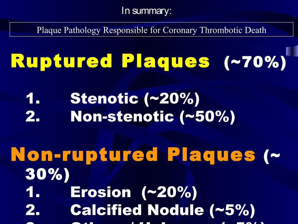
Ruptured Plaques (~70%)
1. Stenotic (~20%)2. Non-stenotic (~50%)
Non-ruptured Plaques (~ 30%)1. Erosion (~20%)2. Calcified Nodule (~5%)3. Others / Unknown (~5%)
Plaque Pathology Responsible for Coronary Thrombotic Death
In summary:
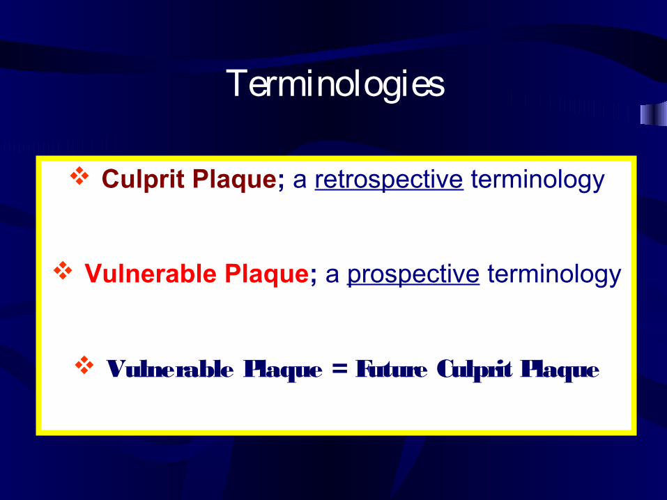
Culprit Plaque; a retrospective terminology
Vulnerable Plaque; a prospective terminology
Vulnerable Plaque = Future Culprit Plaque
Terminologies

Natural History ofVulnerable Plaques
Illustrated:
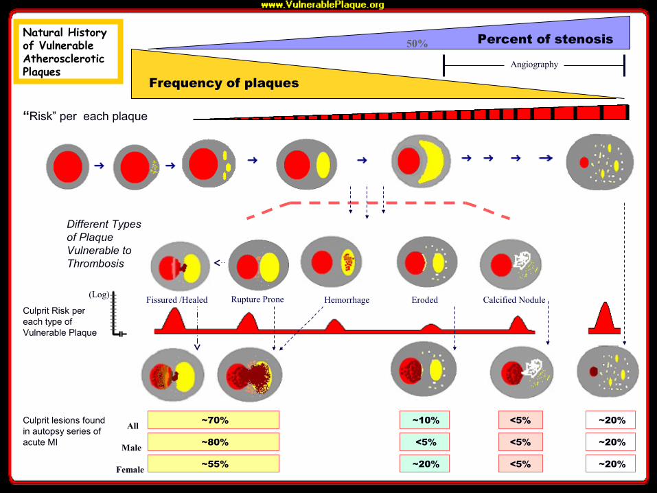
~70%
Percent of stenosis
Frequency of plaques
“Risk” per each plaque
Culprit Risk per each type of Vulnerable Plaque
(Log)
Culprit lesions found in autopsy series of acute MI
Different Types of Plaque Vulnerable to Thrombosis
All
Male
Female
~10% <5% ~20%
50%
Angiography
~80% <5% ~20%
~55% ~20%
<5%
<5% ~20%
Rupture Prone Eroded Calcified NoduleHemorrhage
Positive Remodeling
Fissured /Healed
Natural History of Vulnerable Atherosclerotic Plaques
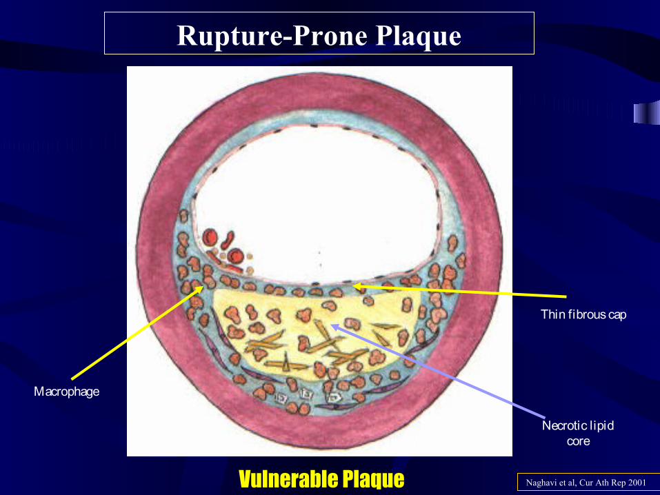
Rupture-Prone Plaque
Vulnerable Plaque Naghavi et al, Cur Ath Rep 2001
Macrophage
Necrotic lipid core
Thin fibrous cap
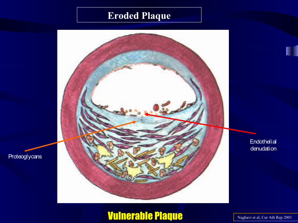
Eroded Plaque
Vulnerable Plaque Naghavi et al, Cur Ath Rep 2001
Endothelial denudation
Proteoglycans
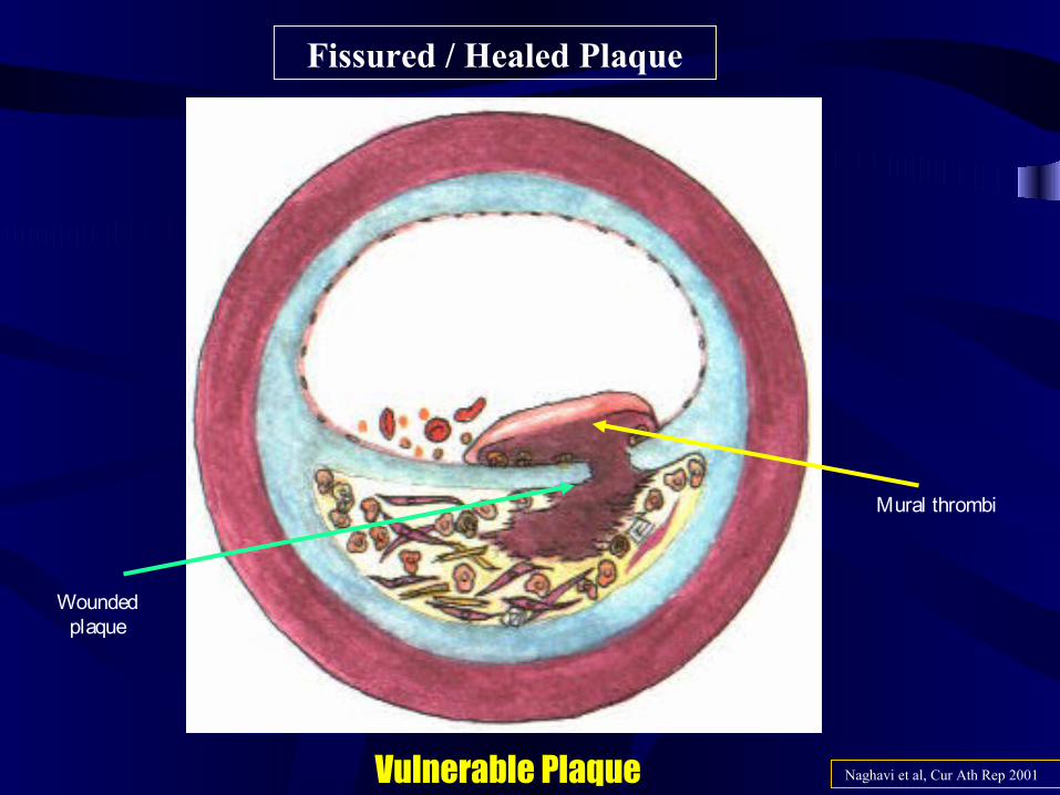
Fissured / Healed Plaque
Vulnerable Plaque Naghavi et al, Cur Ath Rep 2001
Mural thrombi
Wounded plaque
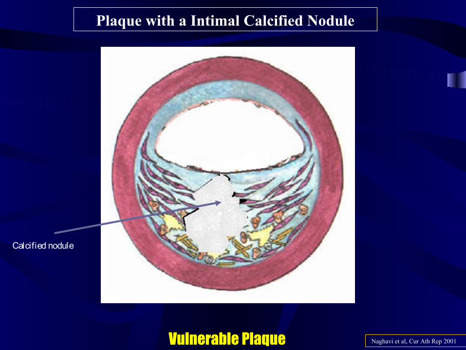
Plaque with a Intimal Calcified Nodule
Vulnerable Plaque Naghavi et al, Cur Ath Rep 2001
Calcified nodule
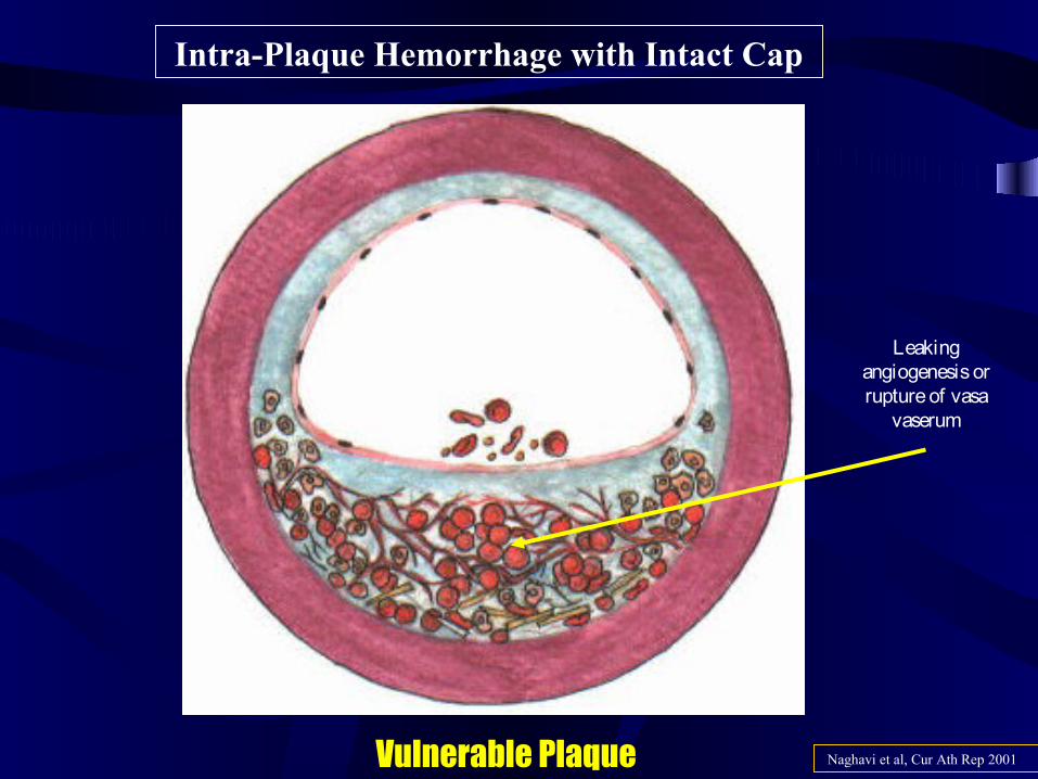
Intra-Plaque Hemorrhage with Intact Cap
Vulnerable Plaque Naghavi et al, Cur Ath Rep 2001
Leaking angiogenesis or rupture of vasa
vaserum
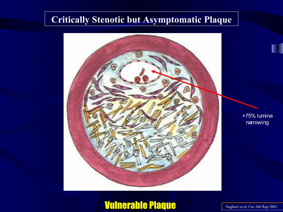
Critically Stenotic but Asymptomatic Plaque
Naghavi et al, Cur Ath Rep 2001Vulnerable Plaque
>75% lumina narrowing
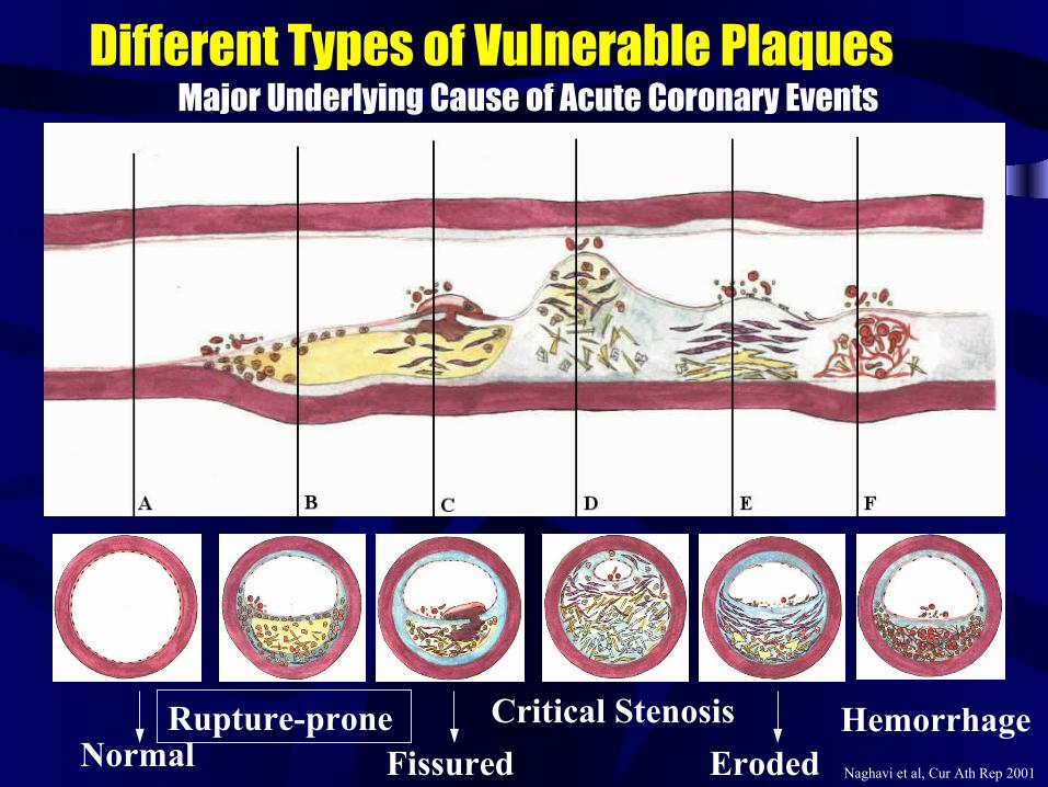
Different Types of Vulnerable Plaques Major Underlying Cause of Acute Coronary Events
NormalRupture-prone
Fissured Eroded
Critical Stenosis Hemorrhage Naghavi et al, Cur Ath Rep 2001
Emerging Techniques for
Detection of Vulnerable Plaque
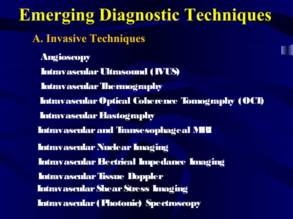
Emerging Diagnostic Techniques A. Invasive Techniques
Angioscopy
Intravascular Ultrasound (IVUS)
Intravascular Thermography
Intravascular Optical Coherence Tomography (OCT)
Intravascular Elastography
Intravascular and Transesophageal MRI
Intravascular Nuclear Imaging
Intravascular Electrical Impedance Imaging
Intravascular Tissue DopplerIntravascular Shear Stress Imaging
Intravascular (Photonic) Spectroscopy
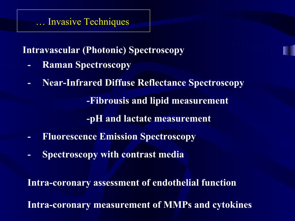
- Raman Spectroscopy
- Near-Infrared Diffuse Reflectance Spectroscopy
-Fibrousis and lipid measurement
-pH and lactate measurement
- Fluorescence Emission Spectroscopy
- Spectroscopy with contrast media
… Invasive Techniques
Intravascular (Photonic) Spectroscopy
Intra-coronary assessment of endothelial function
Intra-coronary measurement of MMPs and cytokines
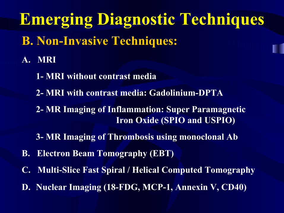
Emerging Diagnostic TechniquesB. Non-Invasive Techniques:
A. MRI
1- MRI without contrast media
2- MRI with contrast media: Gadolinium-DPTA
2- MR Imaging of Inflammation: Super Paramagnetic Iron Oxide (SPIO and USPIO)
3- MR Imaging of Thrombosis using monoclonal Ab
B. Electron Beam Tomography (EBT)
C. Multi-Slice Fast Spiral / Helical Computed Tomography
D. Nuclear Imaging (18-FDG, MCP-1, Annexin V, CD40)
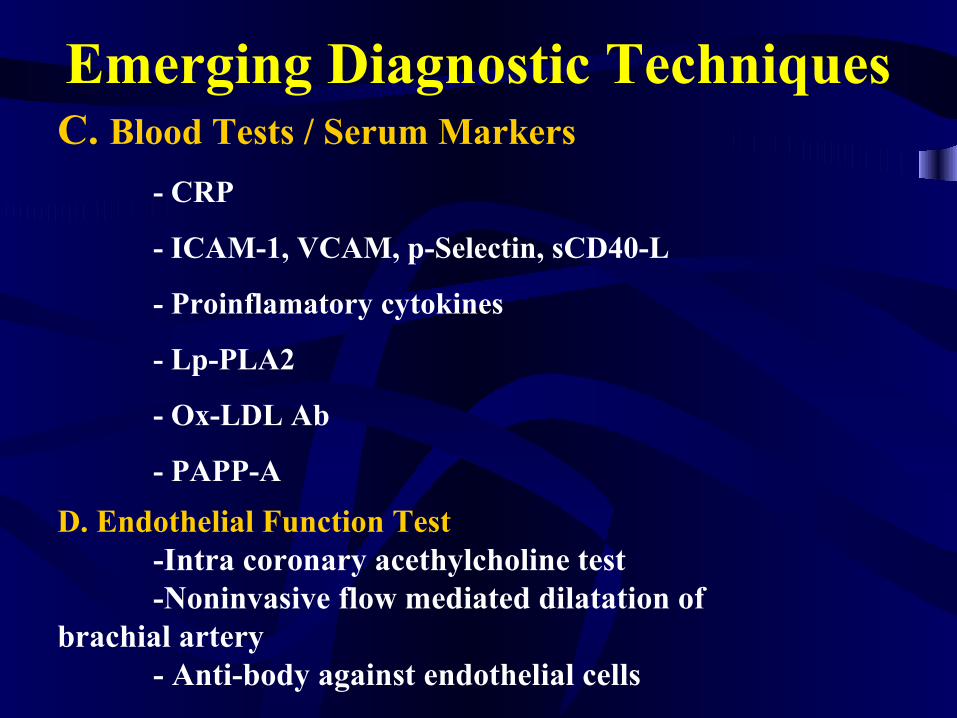
Emerging Diagnostic TechniquesC. Blood Tests / Serum Markers
- CRP
- ICAM-1, VCAM, p-Selectin, sCD40-L
- Proinflamatory cytokines
- Lp-PLA2
- Ox-LDL Ab
- PAPP-A
D. Endothelial Function Test-Intra coronary acethylcholine test-Noninvasive flow mediated dilatation of
brachial artery- Anti-body against endothelial cells
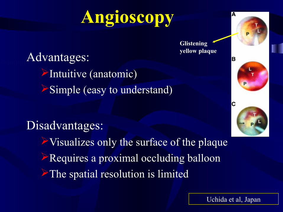
Angioscopy
Advantages:Intuitive (anatomic) Simple (easy to understand)
Disadvantages:Visualizes only the surface of the plaqueRequires a proximal occluding balloonThe spatial resolution is limited
Glistening yellow plaque
Uchida et al, Japan
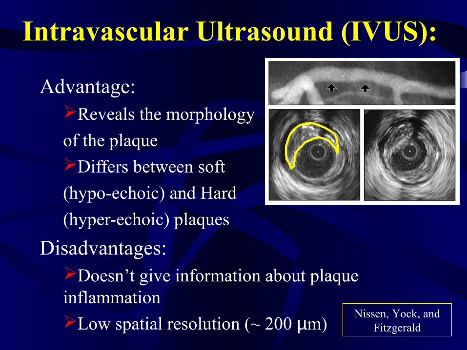
Intravascular Ultrasound (IVUS):
Advantage:Reveals the morphology
of the plaqueDiffers between soft
(hypo-echoic) and Hard
(hyper-echoic) plaques
Disadvantages:Doesn’t give information about plaque inflammationLow spatial resolution (~ 200 µm)
Nissen, Yock, and Fitzgerald
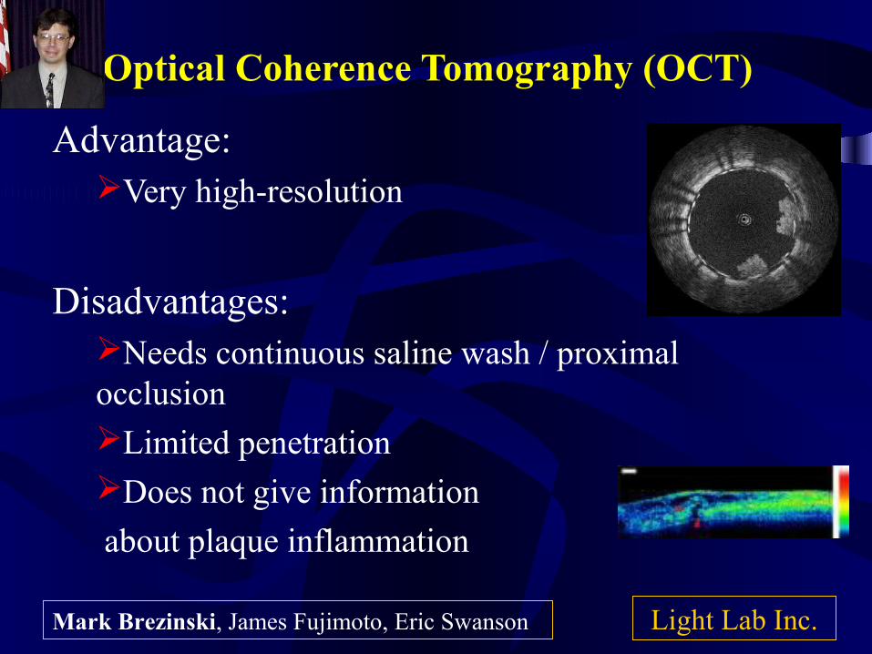
Optical Coherence Tomography (OCT) Advantage:
Very high-resolution
Disadvantages:Needs continuous saline wash / proximal occlusion Limited penetration Does not give information
about plaque inflammation
Light Lab Inc.Mark Brezinski, James Fujimoto, Eric Swanson
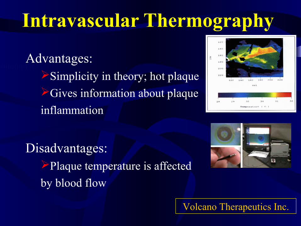
Intravascular Thermography
Advantages:Simplicity in theory; hot plaque Gives information about plaque
inflammation
Disadvantages:Plaque temperature is affected
by blood flow
Volcano Therapeutics Inc.
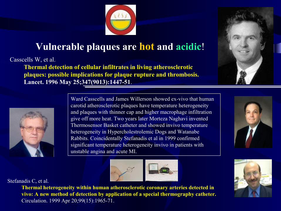
Casscells W, et al.Thermal detection of cellular infiltrates in living atherosclerotic plaques: possible implications for plaque rupture and thrombosis.Lancet. 1996 May 25;347(9013):1447-51.
Vulnerable plaques are hot and acidic!
Ward Casscells and James Willerson showed ex-vivo that human carotid atherosclerotic plaques have temperature heterogeneity and plaques with thinner cap and higher macrophage infiltration give off more heat. Two years later Morteza Naghavi invented Thermosensor Basket catheter and showed invivo temperature heterogeneity in Hypercholestrolemic Dogs and Watanabe Rabbits. Coincidentally Stefanadis et al in 1999 confirmed significant temperature heterogeneity invivo in patients with unstable angina and acute MI.
Stefanadis C, et al.Thermal heterogeneity within human atherosclerotic coronary arteries detected in vivo: A new method of detection by application of a special thermography catheter.Circulation. 1999 Apr 20;99(15):1965-71.
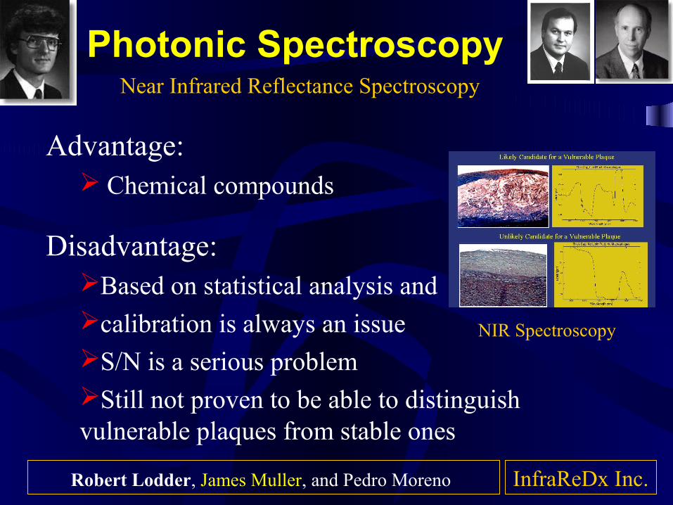
Photonic Spectroscopy
Advantage: Chemical compounds
Disadvantage:Based on statistical analysis and calibration is always an issueS/N is a serious problem Still not proven to be able to distinguish vulnerable plaques from stable ones
Near Infrared Reflectance Spectroscopy
InfraReDx Inc.
NIR Spectroscopy
Robert Lodder, James Muller, and Pedro Moreno
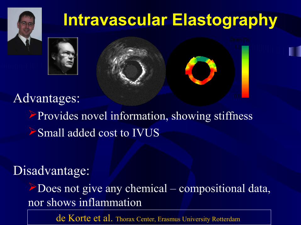
Intravascular Elastography
Advantages:Provides novel information, showing stiffness Small added cost to IVUS
Disadvantage:Does not give any chemical – compositional data, nor shows inflammation
de Korte et al. Thorax Center, Erasmus University Rotterdam
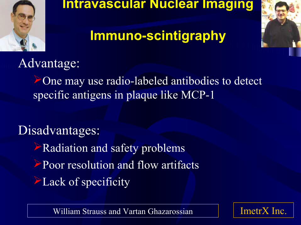
Intravascular Nuclear Imaging
Immuno-scintigraphy
Advantage:One may use radio-labeled antibodies to detect specific antigens in plaque like MCP-1
Disadvantages:Radiation and safety problems Poor resolution and flow artifacts Lack of specificity
ImetrX Inc.William Strauss and Vartan Ghazarossian
Magnetic Resonance ImagingPlaque Characterization and Angiography
Advantages:Lack of ionizing radiation Non-invasive Provides enormous information about flow as well as plaqueEnhancement by contrast agents and NMR spectroscopy
Disadvantages:Ineligibility of patients with metal prosthesesHigh costLonger time for adoption by cardiologists
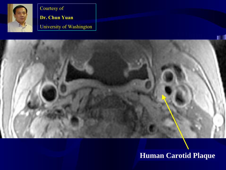
Human Carotid Plaque
Courtesy of
Dr. Chun Yuan
University of Washington
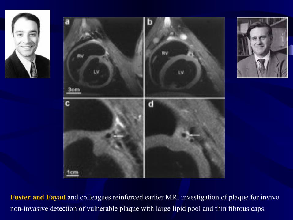
Fuster and Fayad and colleagues reinforced earlier MRI investigation of plaque for invivo
non-invasive detection of vulnerable plaque with large lipid pool and thin fibrous caps.
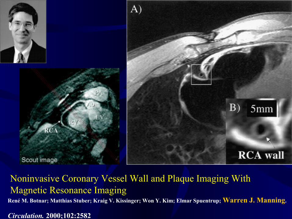
Noninvasive Coronary Vessel Wall and Plaque Imaging With Magnetic Resonance Imaging
René M. Botnar; Matthias Stuber; Kraig V. Kissinger; Won Y. Kim; Elmar Spuentrup; Warren J. Manning.
Circulation. 2000;102:2582
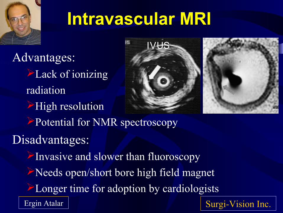
Intravascular MRI
Advantages:Lack of ionizing
radiation High resolution Potential for NMR spectroscopy
Disadvantages:Invasive and slower than fluoroscopyNeeds open/short bore high field magnetLonger time for adoption by cardiologists
Surgi-Vision Inc.Ergin Atalar
IVUS
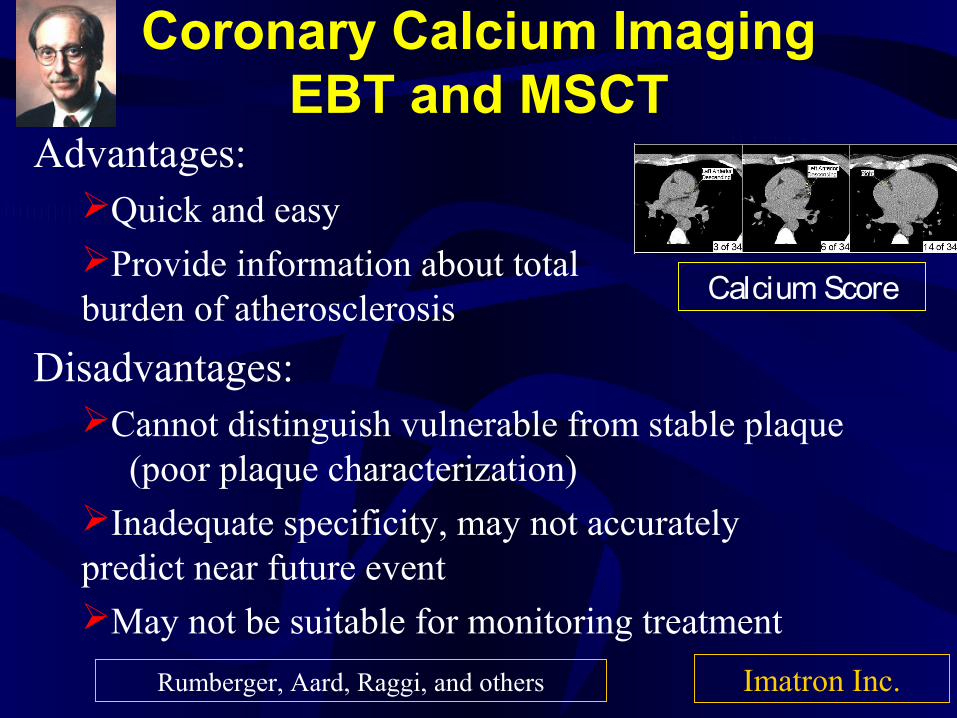
Coronary Calcium ImagingEBT and MSCT
Advantages:Quick and easy Provide information about total burden of atherosclerosis
Disadvantages:Cannot distinguish vulnerable from stable plaque
(poor plaque characterization)Inadequate specificity, may not accurately predict near future eventMay not be suitable for monitoring treatment
Calcium Score
Imatron Inc.Rumberger, Aard, Raggi, and others
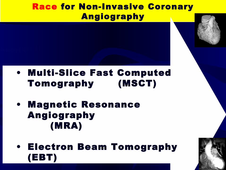
Race for Non-Invasive Coronary Angiography
• Multi-Slice Fast Computed Tomography (MSCT)
• Magnetic Resonance Angiography
(MRA)
• Electron Beam Tomography (EBT)
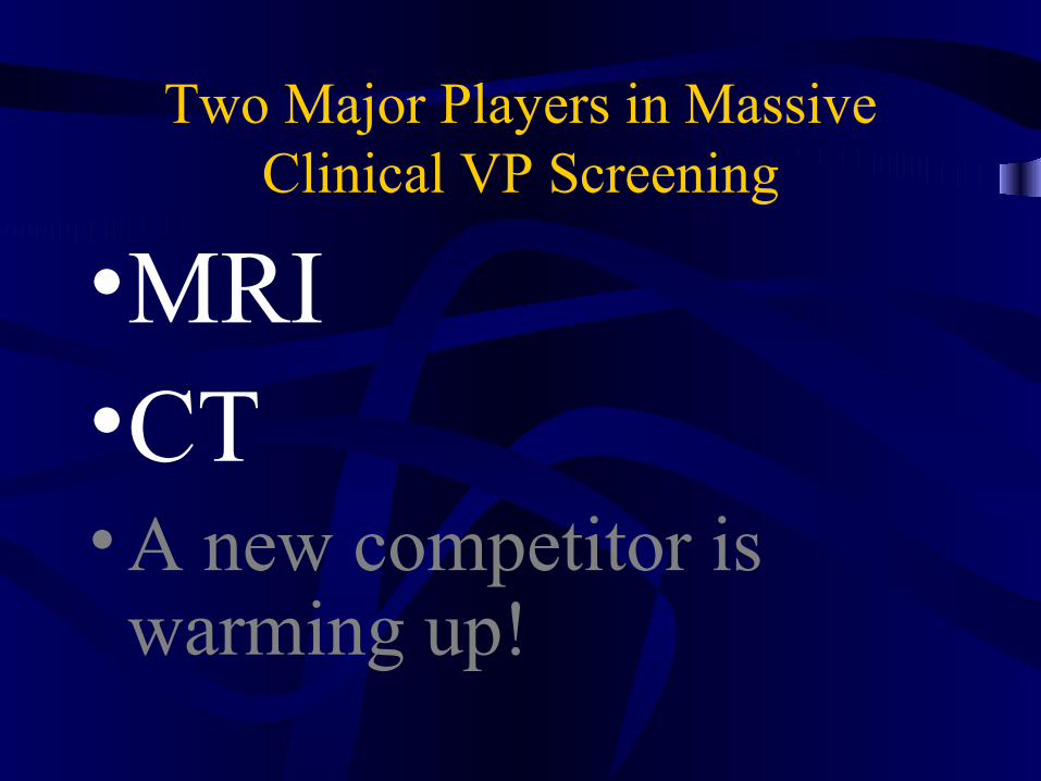
Two Major Players in Massive Clinical VP Screening
•MRI•CT• A new competitor is warming up!
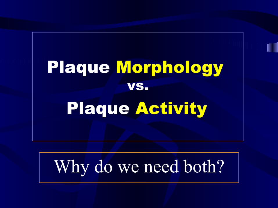
Plaque Morphology vs.
Plaque Activity
Why do we need both?
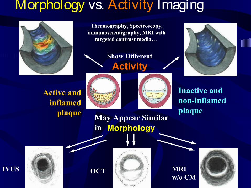
Morphology vs. Activity Imaging
Inactive and non-inflamed plaque
Active and inflamed
plaqueMay Appear Similar in
IVUS OCT MRI w/o CM
Morphology
Show Different
Activity
Thermography, Spectroscopy, immunoscientigraphy, MRI with
targeted contrast media…
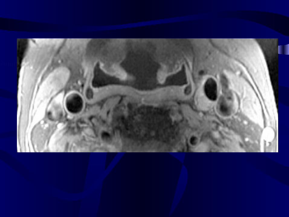
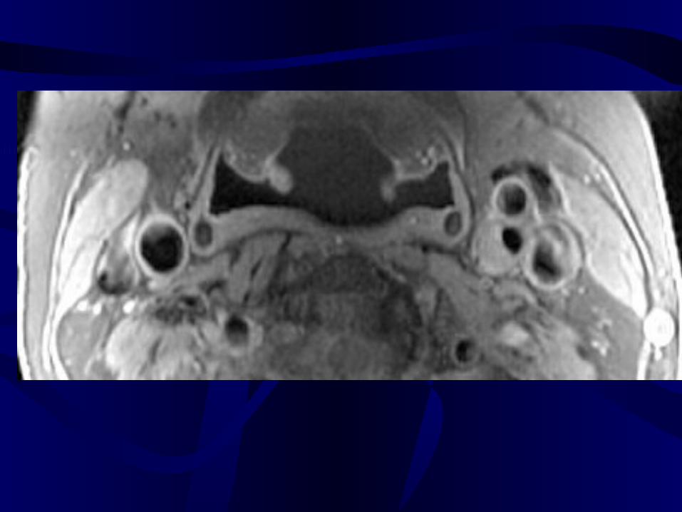
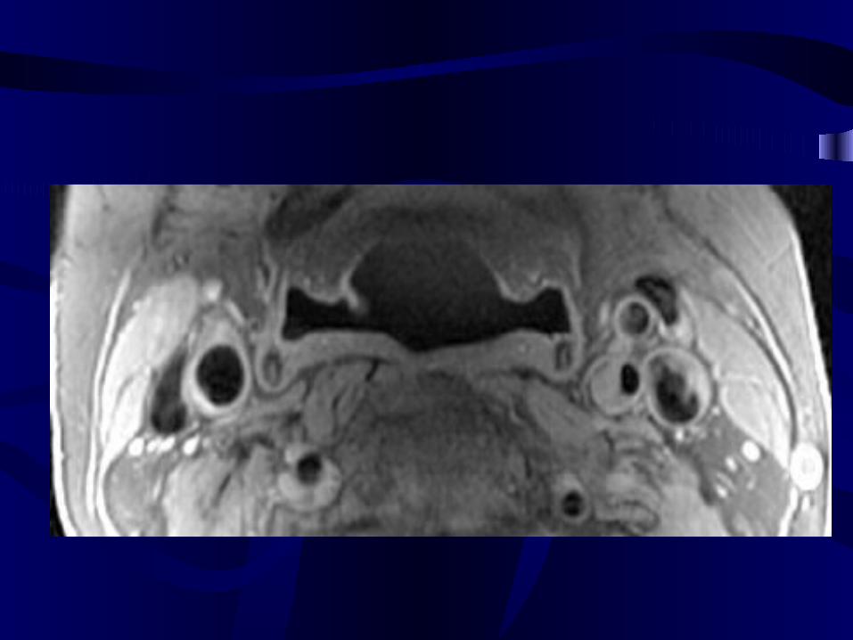
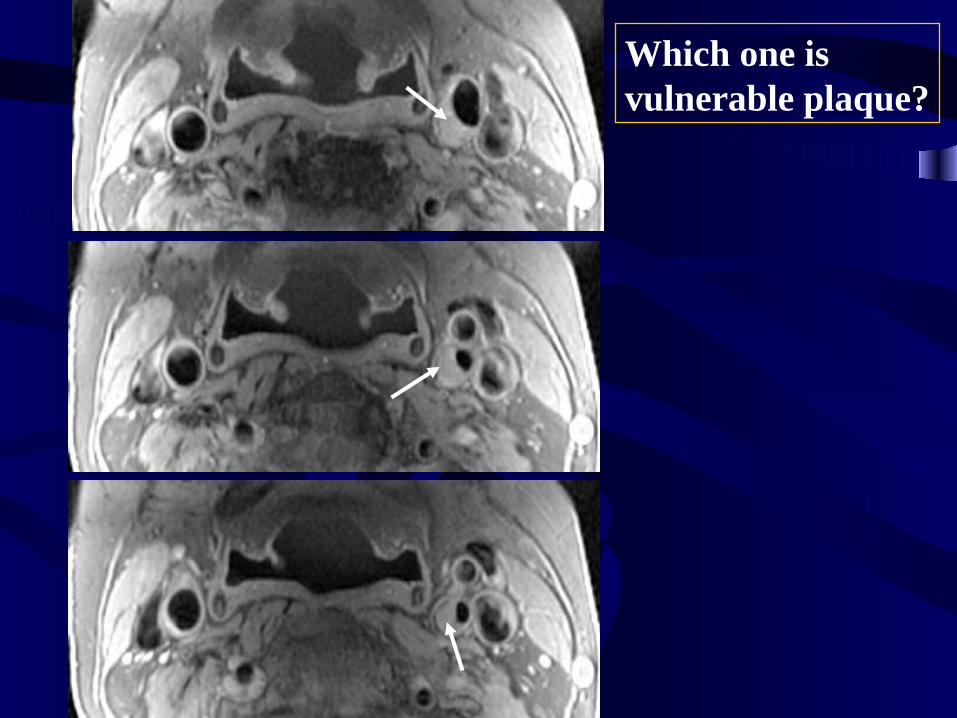
Which one is vulnerable plaque?
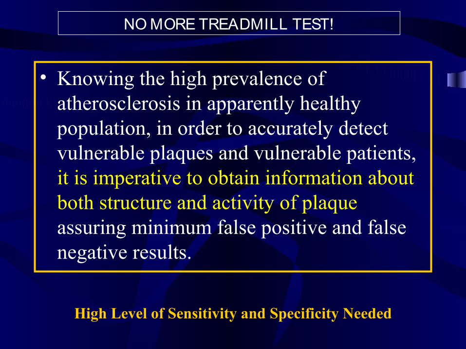
High Level of Sensitivity and Specificity Needed
• Knowing the high prevalence of atherosclerosis in apparently healthy population, in order to accurately detect vulnerable plaques and vulnerable patients, it is imperative to obtain information about both structure and activity of plaque assuring minimum false positive and false negative results.
NO MORE TREADMILL TEST!
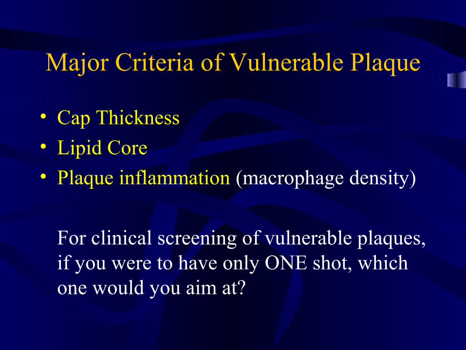
Major Criteria of Vulnerable Plaque
• Cap Thickness
• Lipid Core
• Plaque inflammation (macrophage density)
For clinical screening of vulnerable plaques, if you were to have only ONE shot, which one would you aim at?
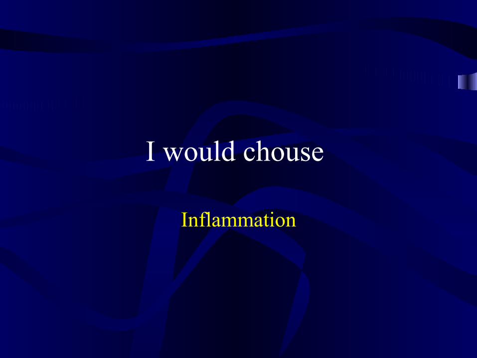
I would chouse
Inflammation

Good News!
• MRI can give us more than one choice, indeed it can provide all of them.
• CT is promising too, but there is more homework for CT fans.
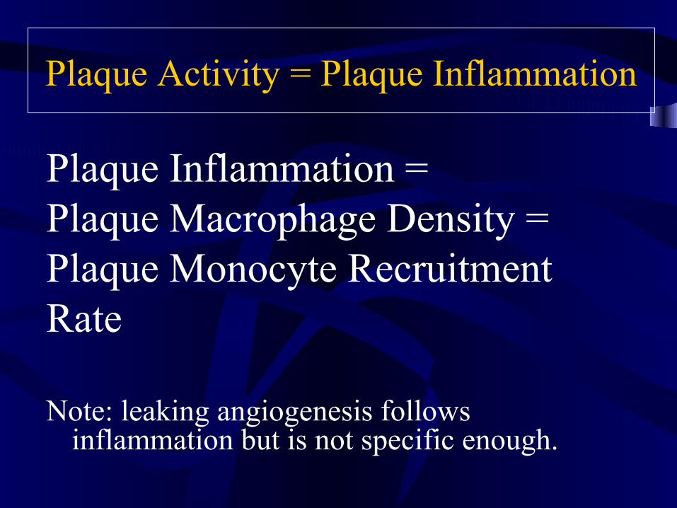
Plaque Activity = Plaque Inflammation
Plaque Inflammation = Plaque Macrophage Density =Plaque Monocyte Recruitment Rate
Note: leaking angiogenesis follows inflammation but is not specific enough.
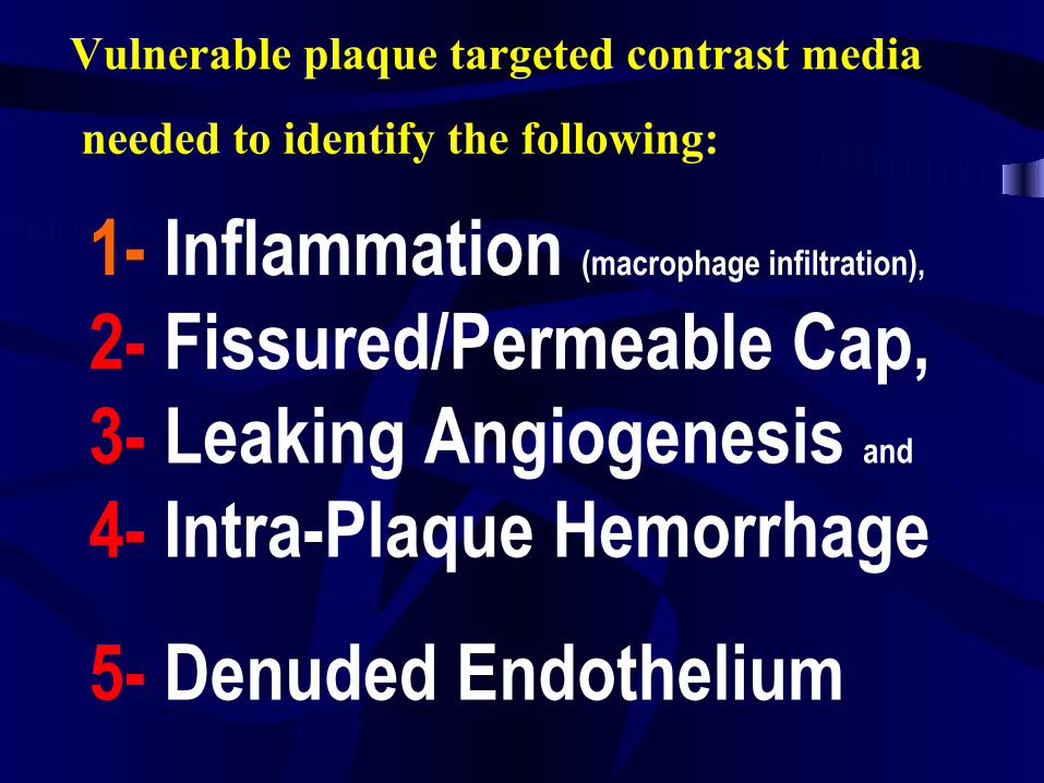
Vulnerable plaque targeted contrast media
needed to identify the following:
1- Inflammation (macrophage infiltration),
2- Fissured/Permeable Cap, 3- Leaking Angiogenesis and
4- Intra-Plaque Hemorrhage
5- Denuded Endothelium
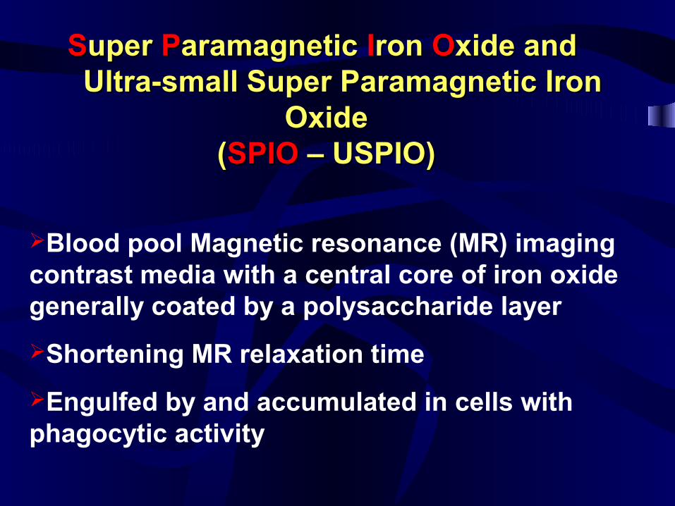
SSuper uper PParamagnetic aramagnetic IIron ron OOxide and xide and Ultra-small Super Paramagnetic Iron Ultra-small Super Paramagnetic Iron
OxideOxide((SPIOSPIO – USPIO) – USPIO)
Blood pool Magnetic resonance (MR) imaging contrast media with a central core of iron oxide generally coated by a polysaccharide layer
Shortening MR relaxation time
Engulfed by and accumulated in cells with phagocytic activity
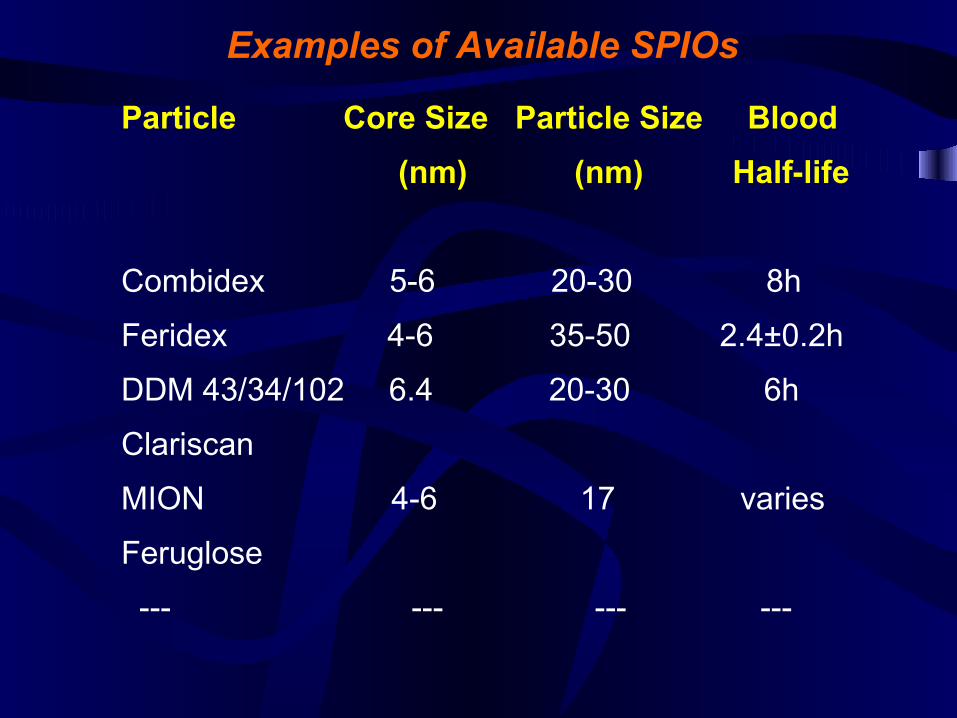
Particle Core Size Particle Size Blood
(nm) (nm) Half-life
Combidex 5-6 20-30 8h
Feridex 4-6 35-50 2.4±0.2h
DDM 43/34/102 6.4 20-30 6h
Clariscan
MION 4-6 17 varies
Feruglose
--- --- --- ---
Examples of Available SPIOs
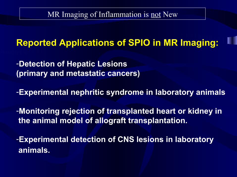
Reported Applications of SPIO in MR Imaging:
-Detection of Hepatic Lesions (primary and metastatic cancers)
-Experimental nephritic syndrome in laboratory animals
-Monitoring rejection of transplanted heart or kidney in the animal model of allograft transplantation.
-Experimental detection of CNS lesions in laboratory animals.
MR Imaging of Inflammation is not New
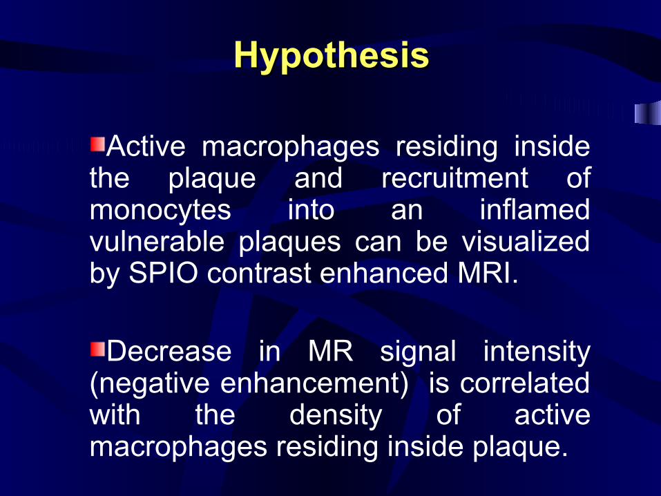
HypothesisHypothesis
Active macrophages residing inside the plaque and recruitment of monocytes into an inflamed vulnerable plaques can be visualized by SPIO contrast enhanced MRI.
Decrease in MR signal intensity (negative enhancement) is correlated with the density of active macrophages residing inside plaque.
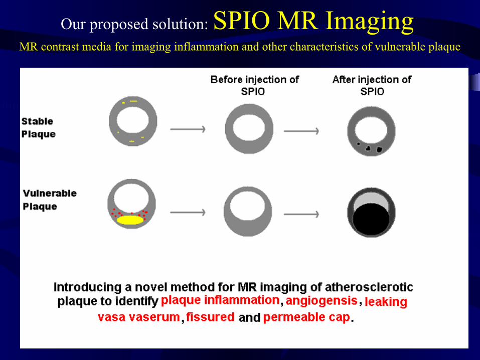
Our proposed solution: SPIO MR Imaging MR contrast media for imaging inflammation and other characteristics of vulnerable plaque
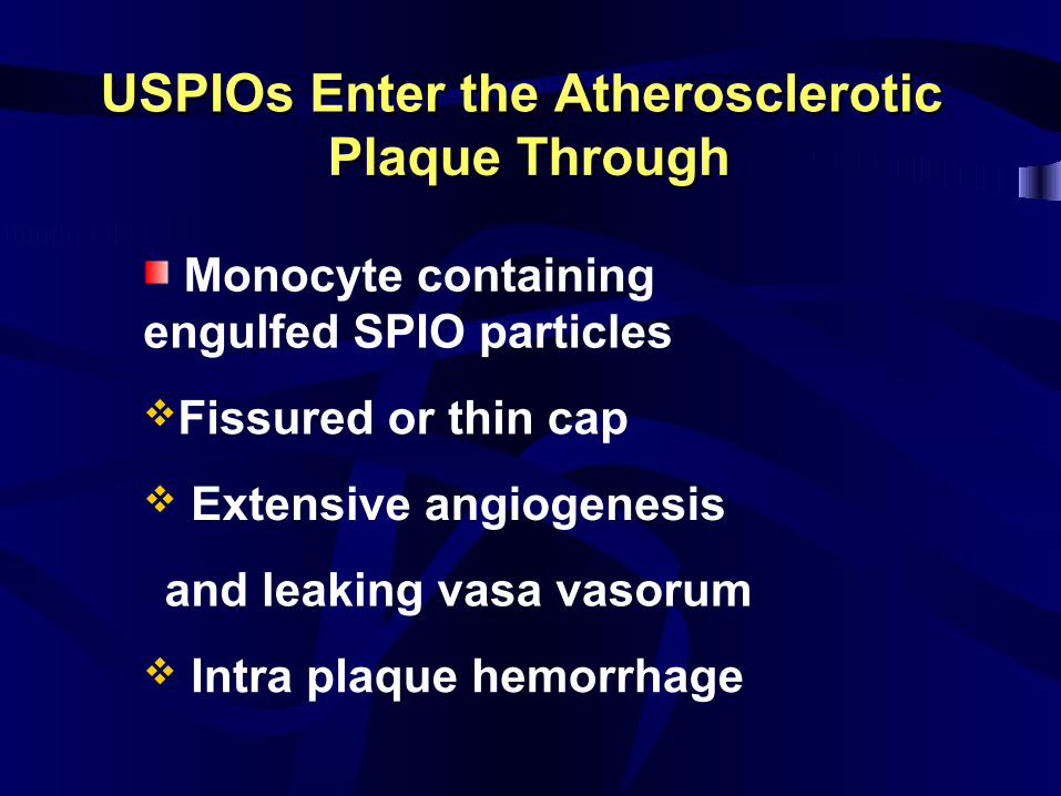
USPIOs Enter the AtheroscleroticUSPIOs Enter the Atherosclerotic Plaque ThroughPlaque Through
Monocyte containing engulfed SPIO particles
Fissured or thin cap
Extensive angiogenesis
l and leaking vasa vasorum
Intra plaque hemorrhage
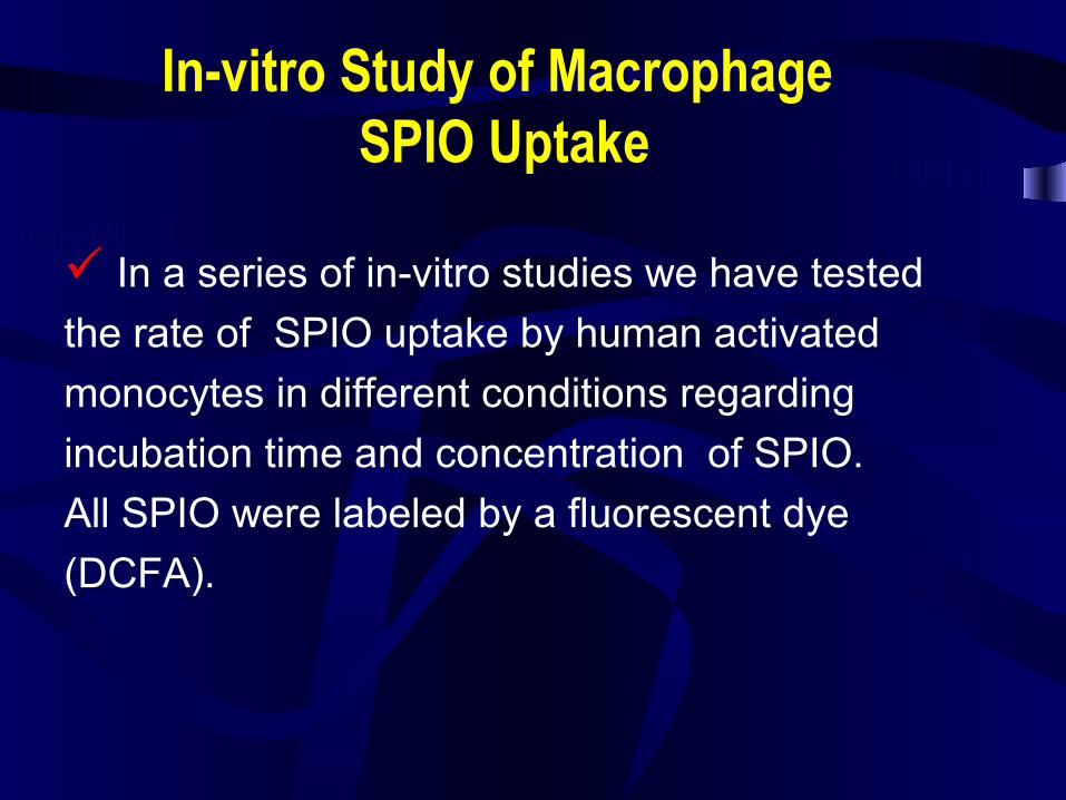
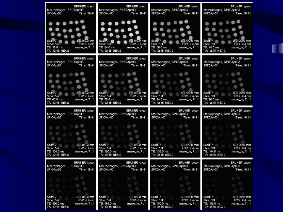
In-vitro Study of Macrophage SPIO Uptake
In a series of in-vitro studies we have tested
the rate of SPIO uptake by human activated
monocytes in different conditions regarding
incubation time and concentration of SPIO.
All SPIO were labeled by a fluorescent dye
(DCFA).
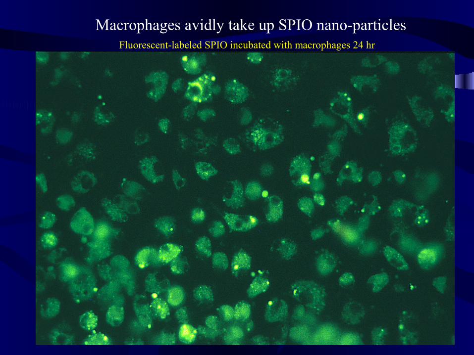
Fluorescent-labeled SPIO incubated with macrophages 24 hr
Macrophages avidly take up SPIO nano-particles
SPIO and T2 Effect
In-vitro relaxation study shows the effect of SPIO dose and incubation time on intra-macrophage SPIO negative enhancement
0
10
20
30
40
50
60
70
80
90
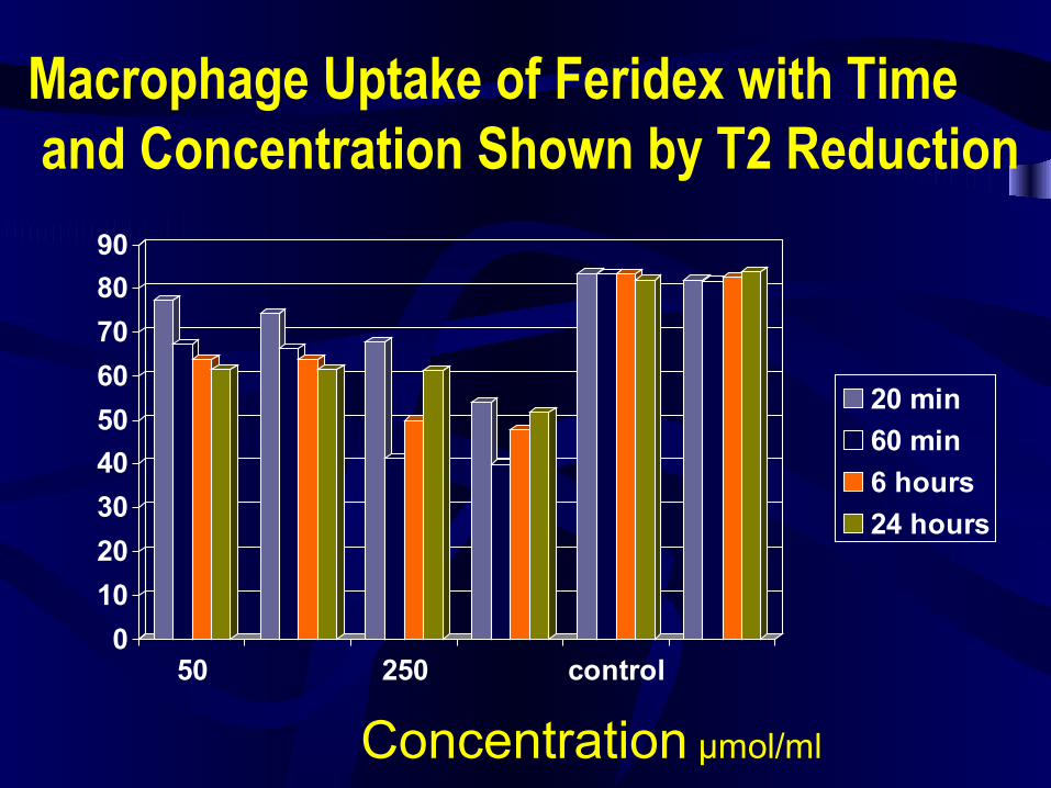
50 250 control
20 min
60 min
6 hours
24 hours
Macrophage Uptake of Feridex with Time and Concentration Shown by T2 Reduction
Concentration µmol/ml
In-vivo distribution of SPIO in ApoE deficient and wild type mice:
•For the initial study, we use the mouse model of atherosclerosis.• •ApoE deficient mouse has similar atherosclerotic lesions to human and the lesions are more common in the aortic arch and thoracic aorta.
• We used ApoE deficient mice and normal variant (C57BL mice) as control.
•The SPIO that we used was Feridex (Berlex) injectable solution.
•Animals were sacrificed on day 3 and 5 after injection.
Pre and Post-SPIO Enhanced Magnetic Resonance Imaging of ApoE K/O and Wild Type Mice:
We used 4.7 tesla MRI unit in our study.
After baseline MR imaging with respiratory gating, we injected 1mMolFe/kg super paramagnetic iron oxide to six ApoE deficient and two C57bl mice through the tail vein.
Post-contrast MR imaging were performed in day 5 with the same parameters (TR=2.5 sec, TE=0.012 sec, FOV=6.6 cm, slice thickness=2.0mm, flip angle (orient)=trans, and matrices=256x256).
We selected the aorta at the level of kidney for comparison of the baseline and post-contrast images.
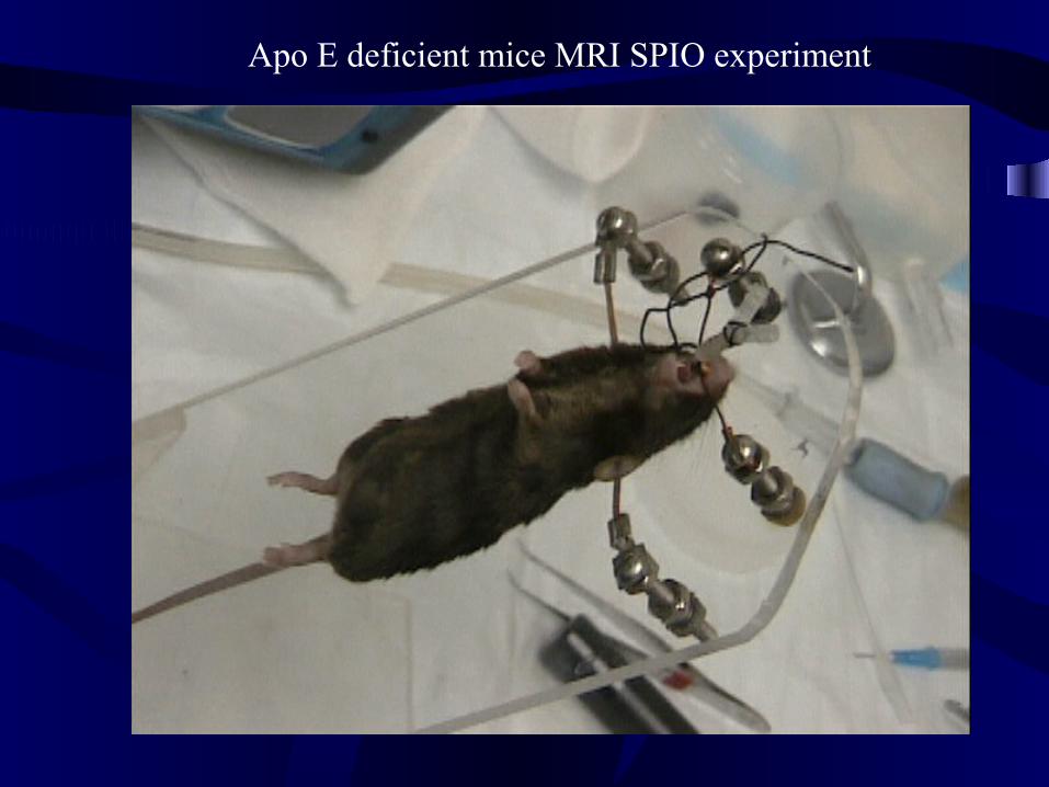
Apo E deficient mice MRI SPIO experiment
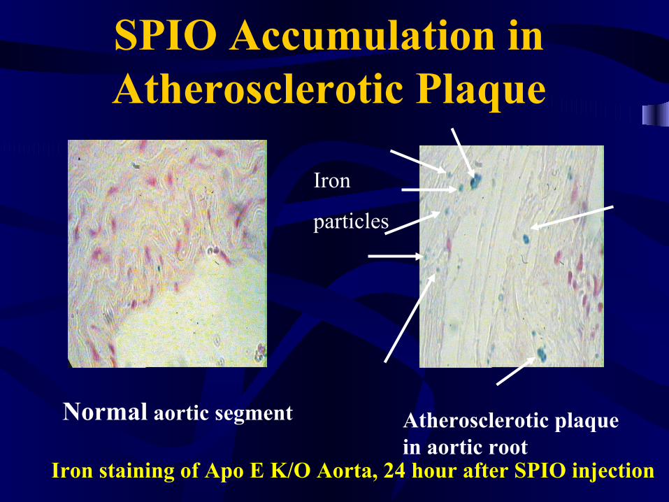
SPIO Accumulation in Atherosclerotic Plaque
Atherosclerotic plaque in aortic root
Normal aortic segment
Iron staining of Apo E K/O Aorta, 24 hour after SPIO injection
Iron
particles
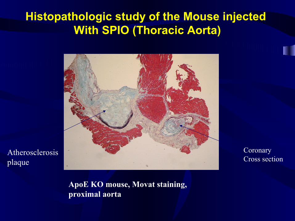
Histopathologic study of the Mouse injected With SPIO (Thoracic Aorta)
ApoE KO mouse, Movat staining, proximal aorta
Coronary Cross section
Atherosclerosisplaque

Histopathologic study of ApoE KO Mouse injected With SPIO (Thoracic Aorta)
CD68 staining(aortic plaque)
Iron Staining (aortic plaque) Iron Staining (coronary section)
Iron particles Iron particles
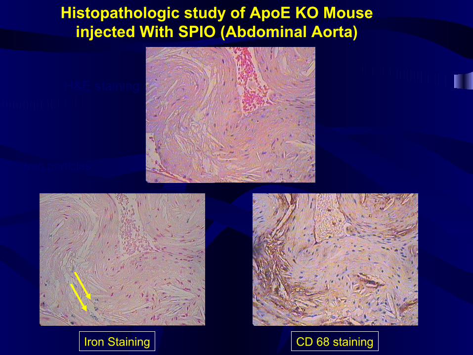
Histopathologic study of ApoE KO Mouse injected With SPIO (Abdominal Aorta)
H&E staining
Iron Staining CD 68 staining
Iron particles
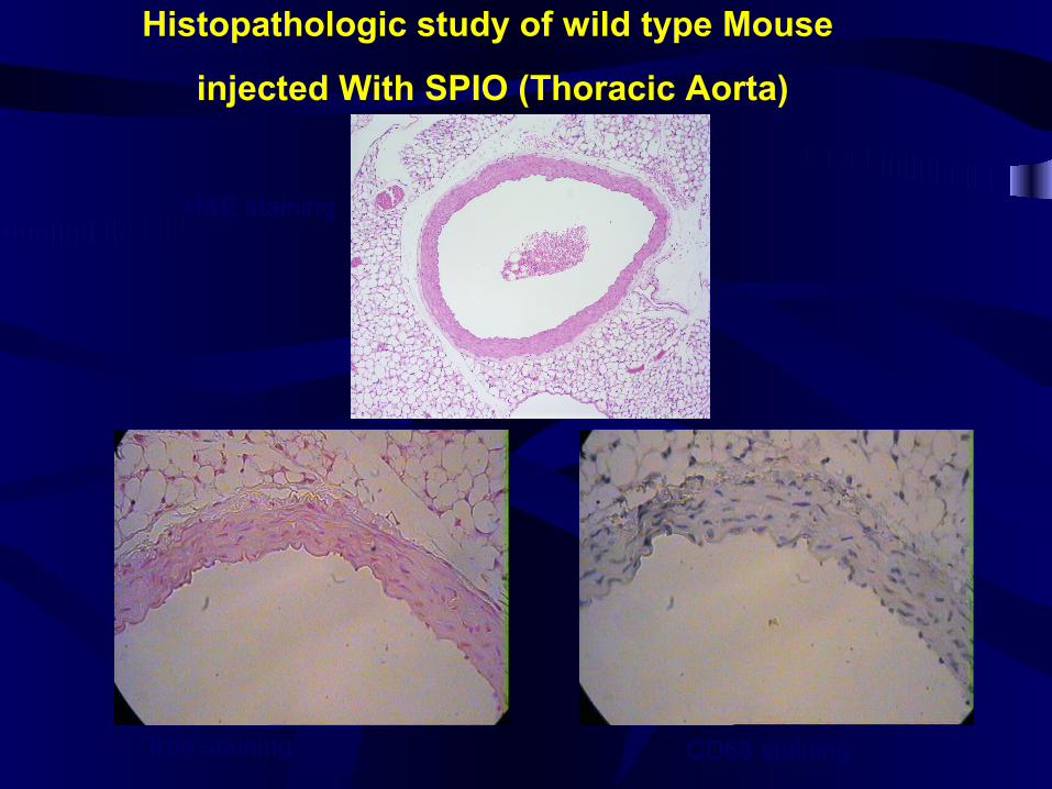
Histopathologic study of wild type Mouse
injected With SPIO (Thoracic Aorta)
H&E staining
CD68 stainingIron staining
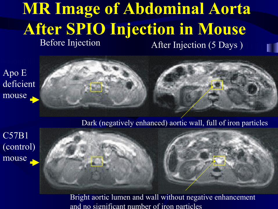
MR Image of Abdominal Aorta After SPIO Injection in Mouse
Apo E deficient mouse
C57B1 (control) mouse
Before Injection After Injection (5 Days )
Dark (negatively enhanced) aortic wall, full of iron particles
Bright aortic lumen and wall without negative enhancement and no significant number of iron particles
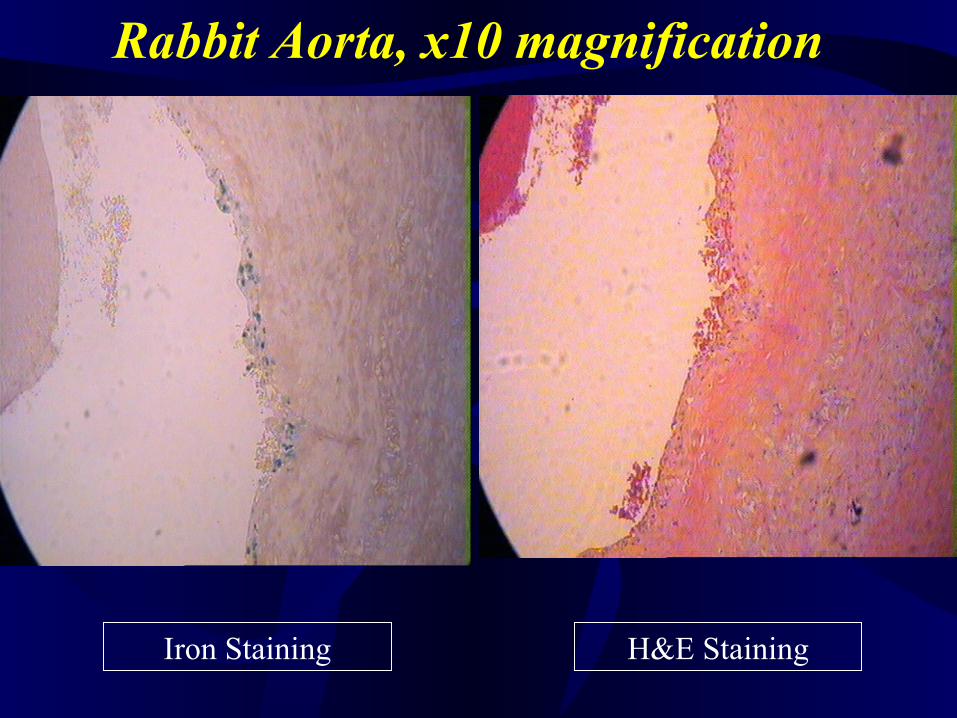
Rabbit Aorta, x10 magnification
Iron Staining H&E Staining
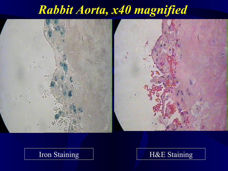
Rabbit Aorta, x40 magnified
Iron Staining H&E Staining
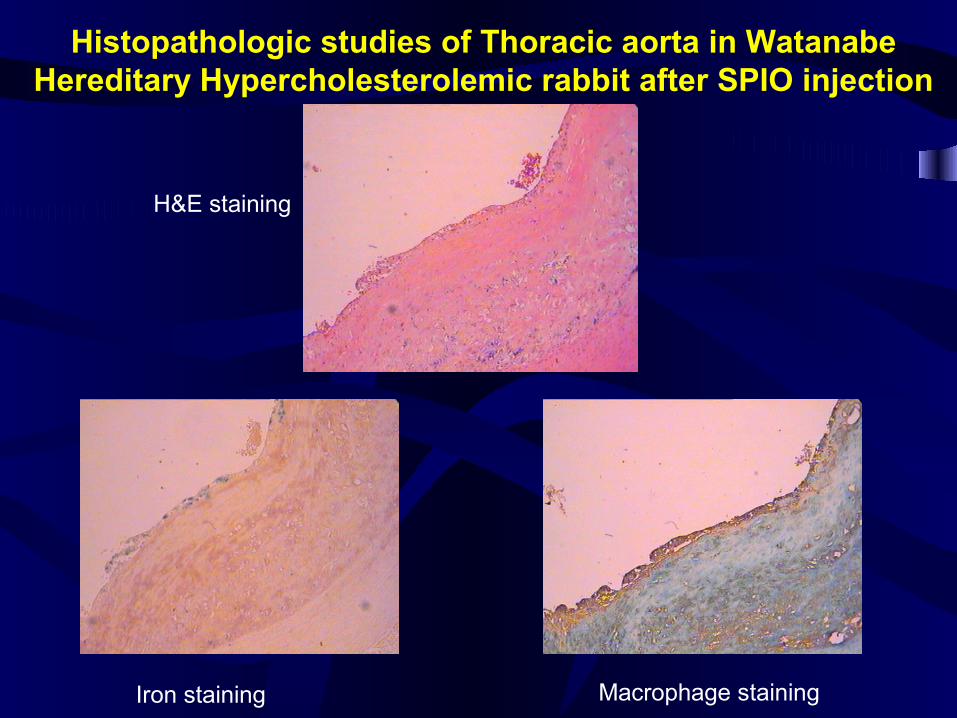
Histopathologic studies of Thoracic aorta in WatanabeHereditary Hypercholesterolemic rabbit after SPIO injection
H&E staining
Iron staining Macrophage staining
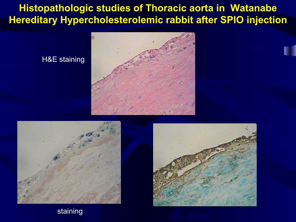
Histopathologic studies of Thoracic aorta in Watanabe Hereditary Hypercholesterolemic rabbit after SPIO injection
H&E staining
Iron stainingIron staining
Iron particles
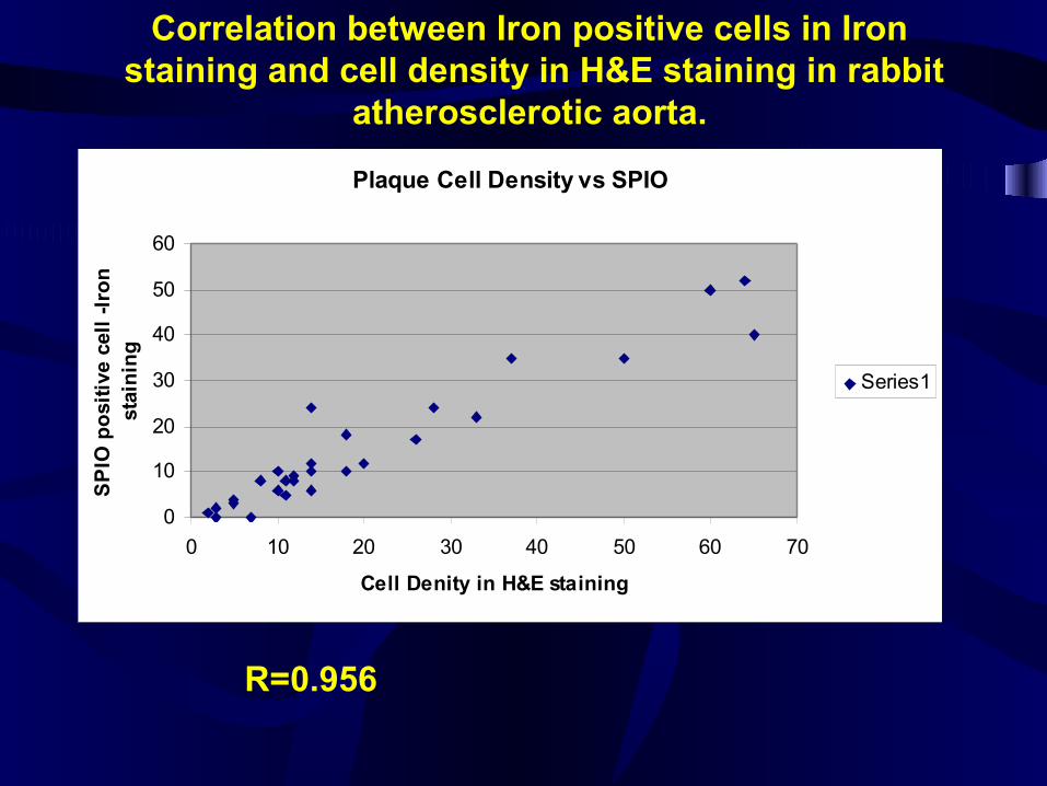
Plaque Cell Density vs SPIO
0
10
20
30
40
50
60
0 10 20 30 40 50 60 70
Cell Denity in H&E staining
SP
IO p
osi
tive
cel
l -I
ron
st
ain
ing
Series1
R=0.956
Correlation between Iron positive cells in Iron staining and cell density in H&E staining in rabbit
atherosclerotic aorta.
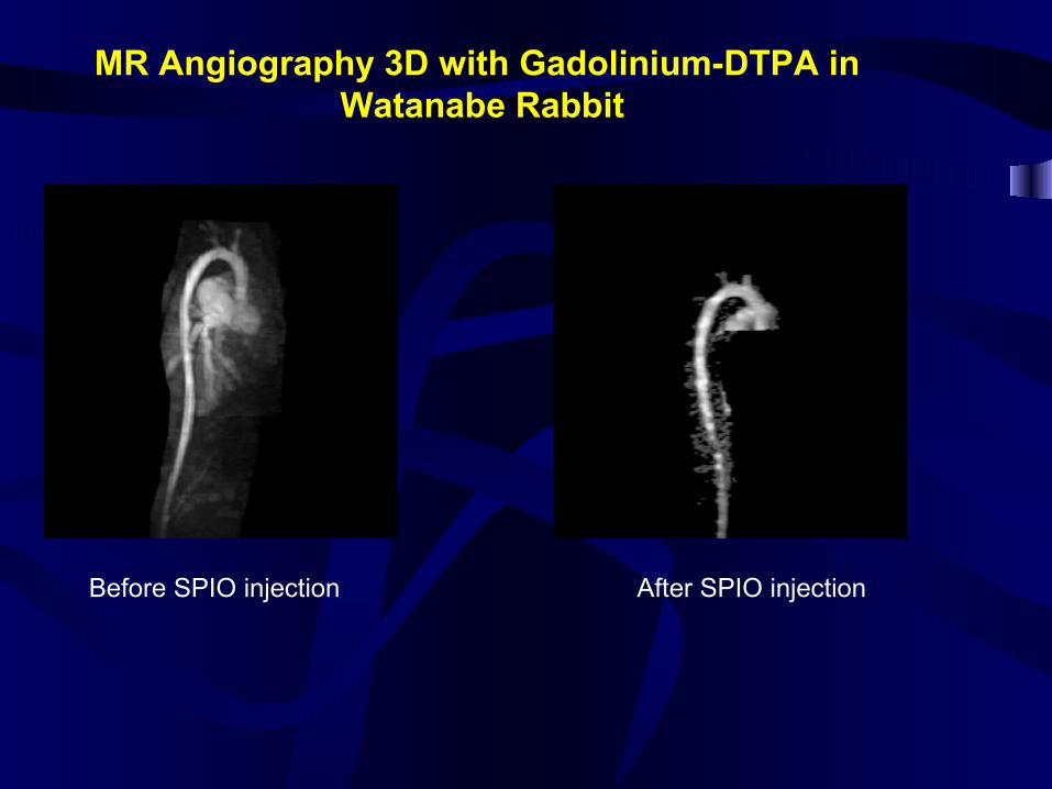
MR Angiography 3D with Gadolinium-DTPA in Watanabe Rabbit
Before SPIO injection After SPIO injection
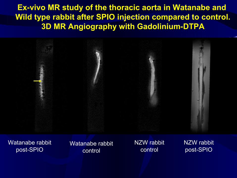
Ex-vivo MR study of the thoracic aorta in Watanabe and Wild type rabbit after SPIO injection compared to control.
3D MR Angiography with Gadolinium-DTPA
Watanabe rabbitpost-SPIO
Watanabe rabbitcontrol
NZW rabbitcontrol
NZW rabbitpost-SPIO
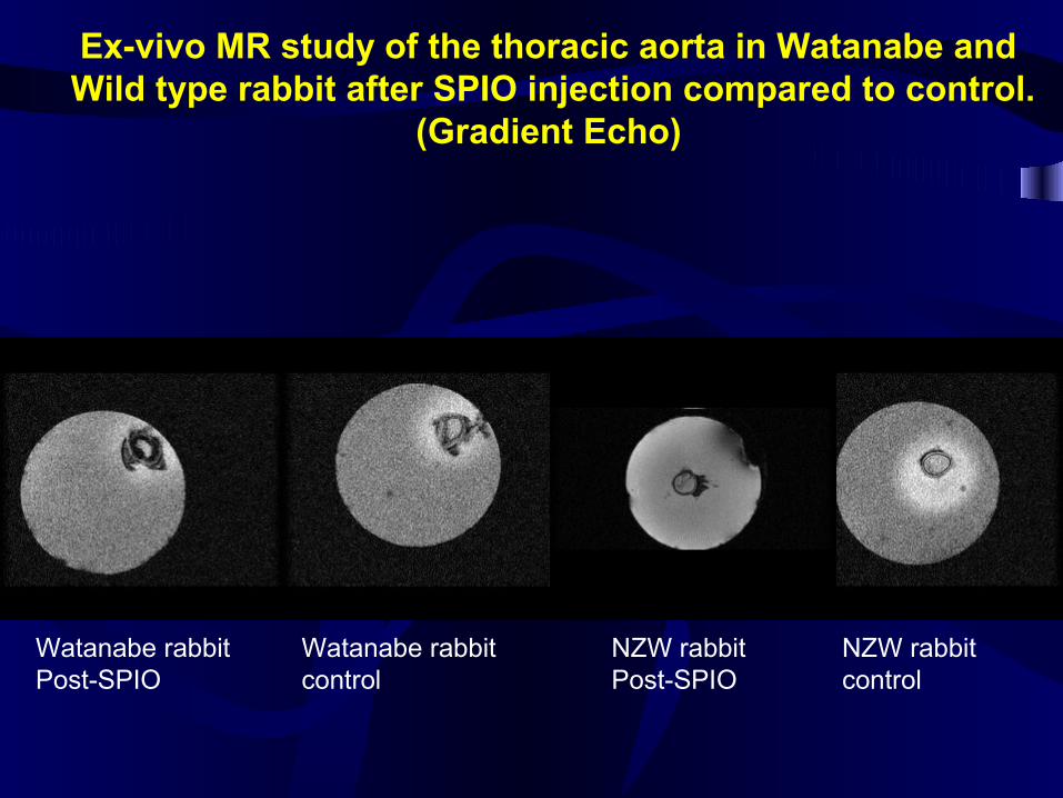
Ex-vivo MR study of the thoracic aorta in Watanabe and Wild type rabbit after SPIO injection compared to control.
(Gradient Echo)
Watanabe rabbitPost-SPIO
Watanabe rabbitcontrol
NZW rabbitPost-SPIO
NZW rabbitcontrol
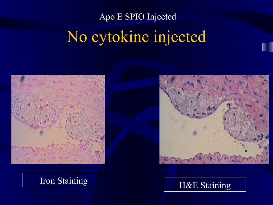
No cytokine injectedApo E SPIO Injected
Iron Staining H&E Staining
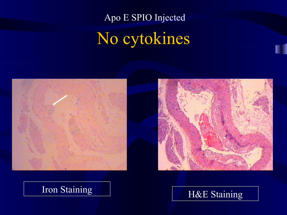
No cytokinesApo E SPIO Injected
Iron Staining H&E Staining
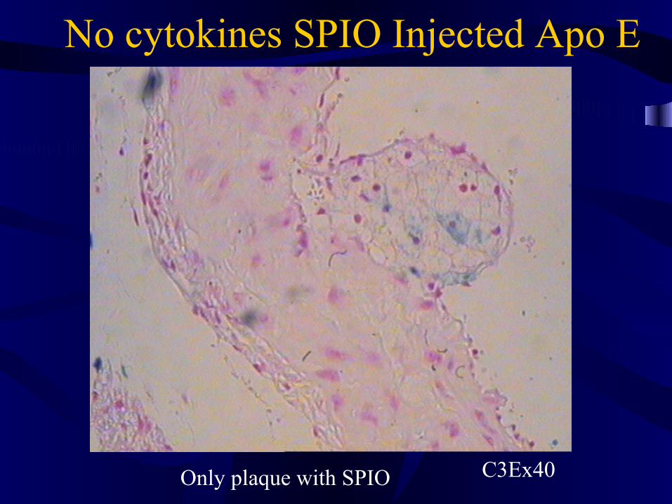
No cytokines SPIO Injected Apo E
Only plaque with SPIO C3Ex40
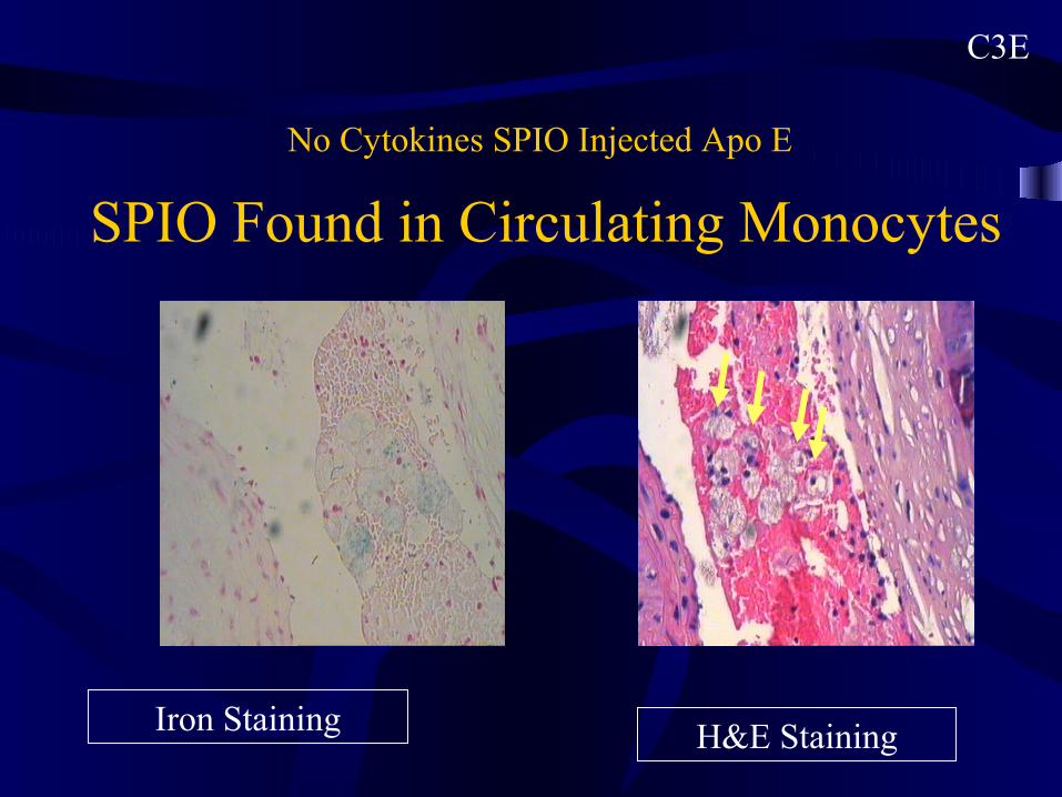
No Cytokines SPIO Injected Apo E
C3E
SPIO Found in Circulating Monocytes
Iron Staining H&E Staining
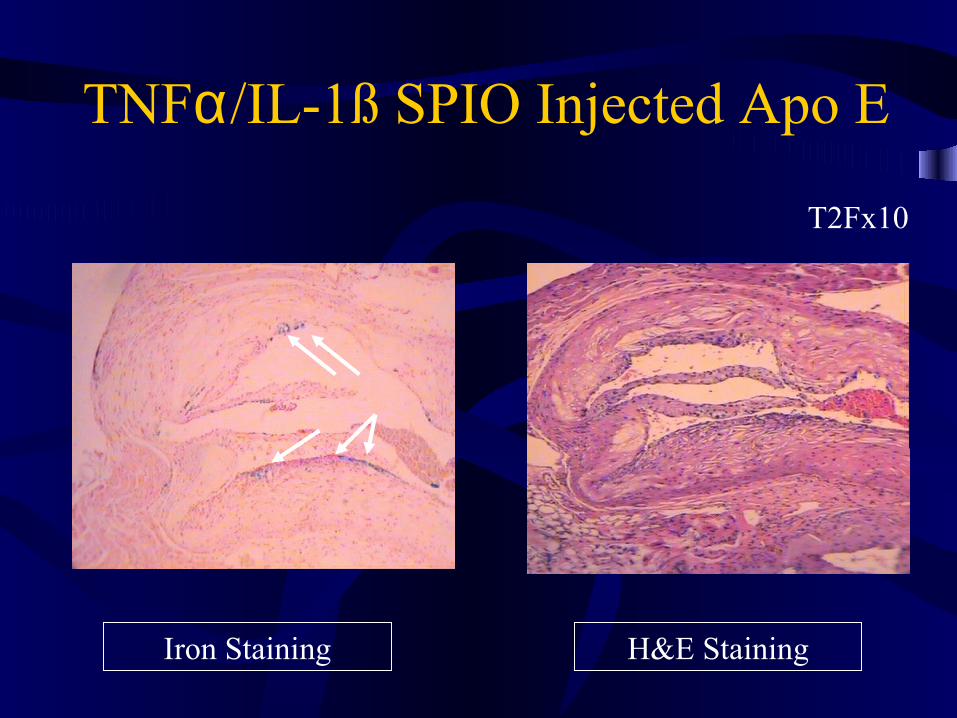
T2Fx10
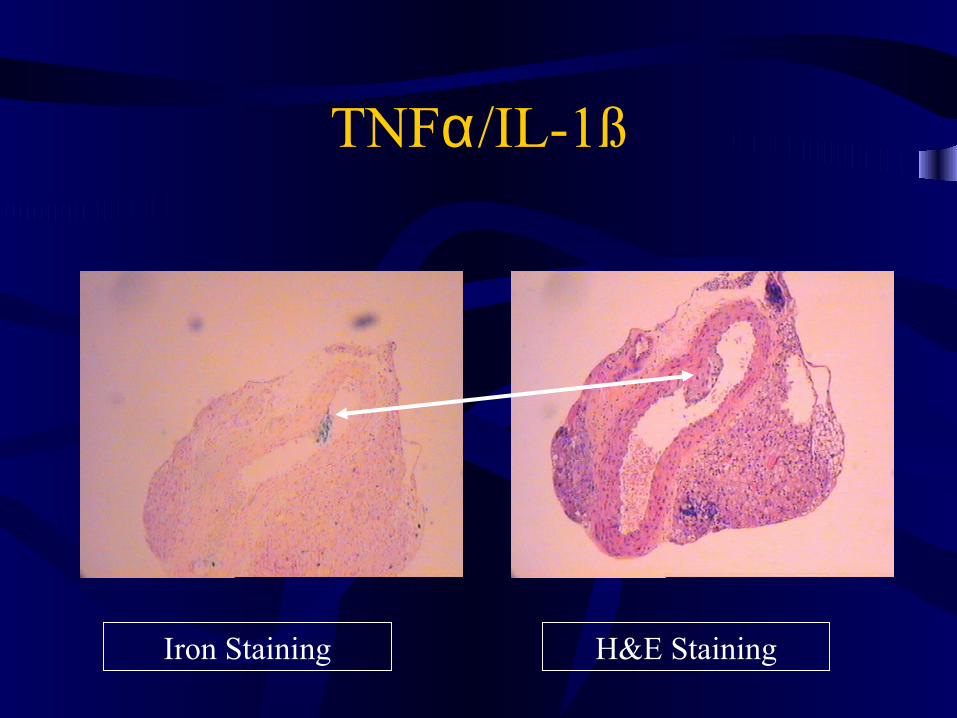
TNFα/IL-1ß SPIO Injected Apo E
Iron Staining H&E Staining

TNFα/IL-1ß SPIO Injected Apo E
H&E StainingH&E Staining
Increased Superficial Macrophage Density
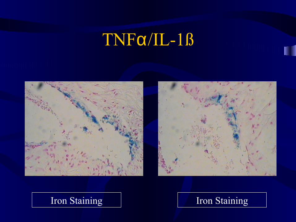
TNFα/IL-1ß
Iron Staining Iron Staining
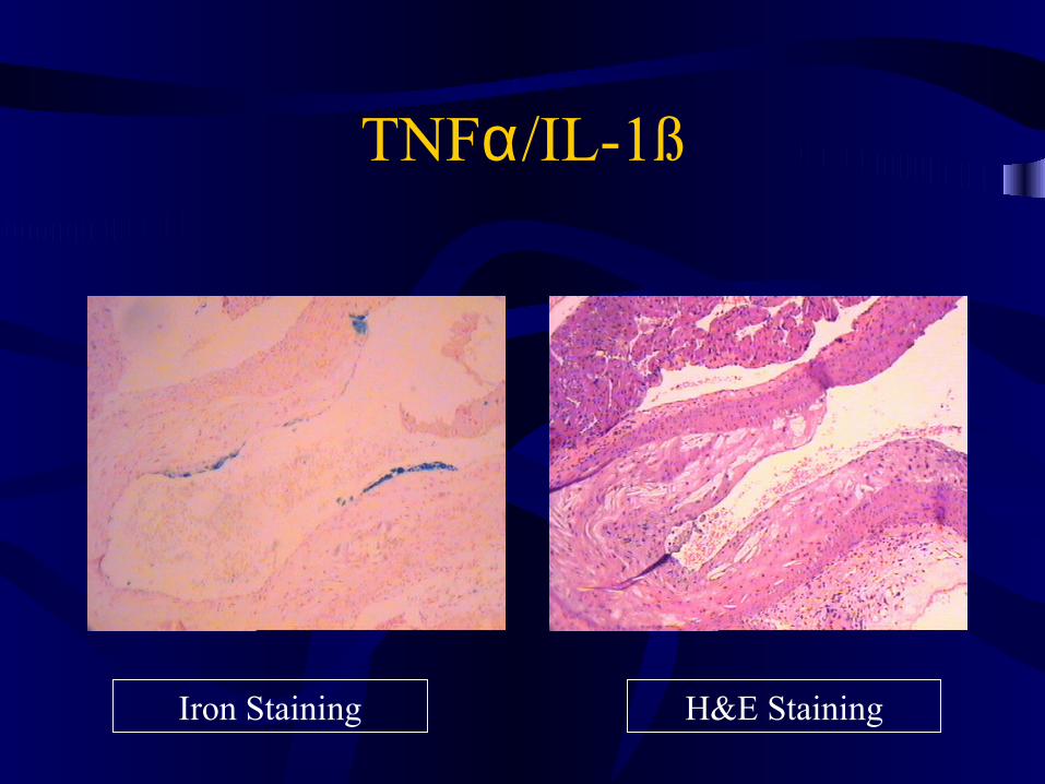
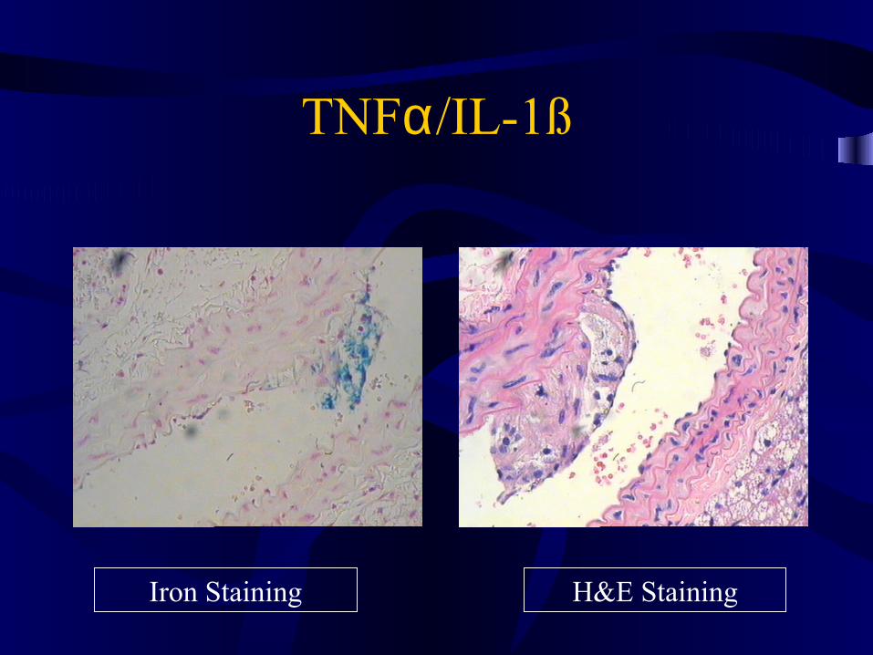
TNFα/IL-1ß
Iron Staining H&E Staining
TNFα/IL-1ß
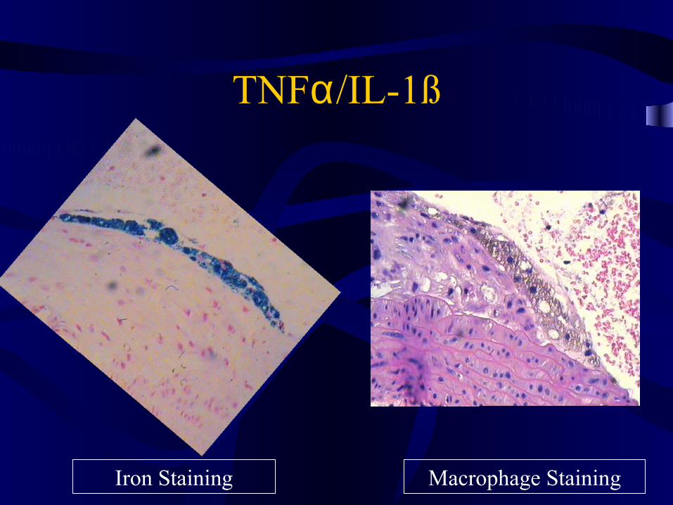
Iron Staining Macrophage Staining
TNFα/IL-1ß
Iron Staining H&E Staining
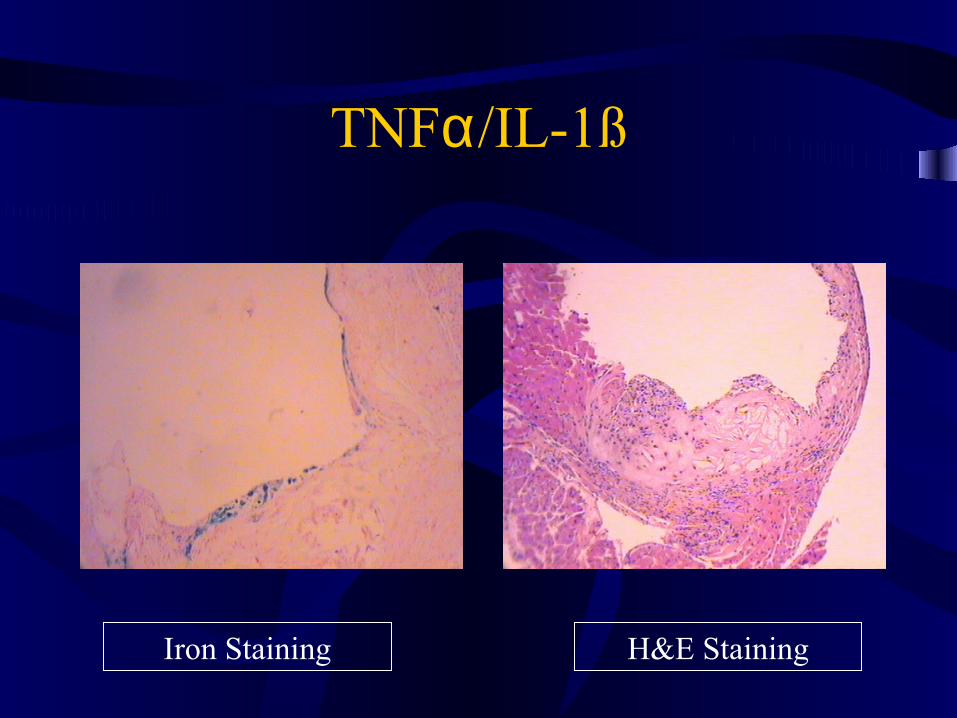
TNFα/IL-1ß
Iron Staining H&E Staining
TNFα/IL-1ß
Iron Staining H&E Staining
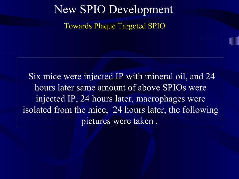
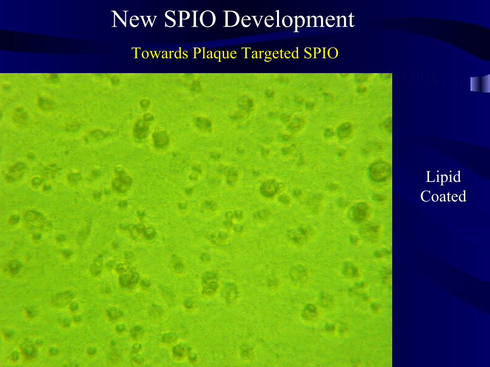
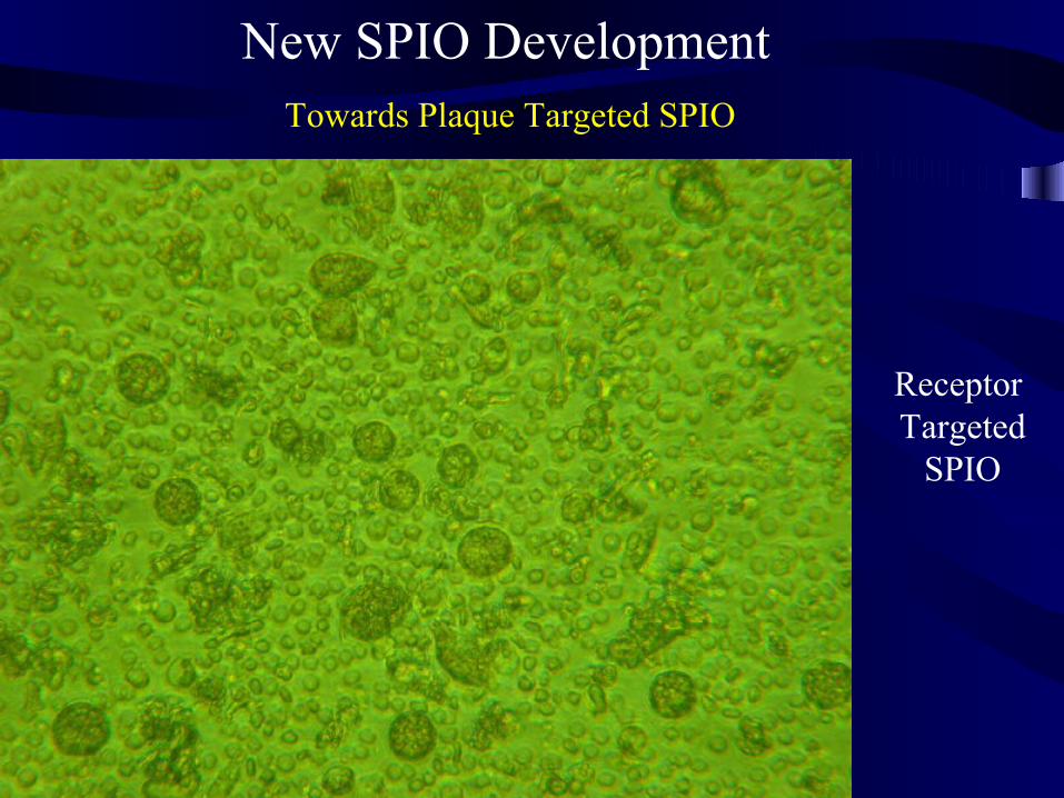
New SPIO Development Towards Plaque Targeted SPIO
Six mice were injected IP with mineral oil, and 24 hours later same amount of above SPIOs were injected IP, 24 hours later, macrophages were
isolated from the mice, 24 hours later, the following pictures were taken .
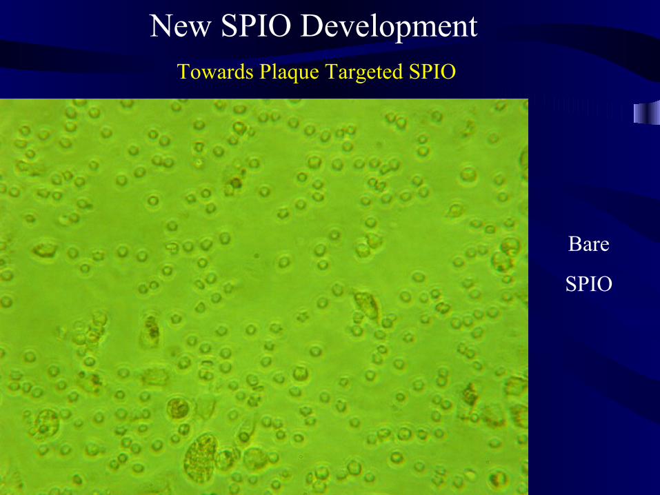
New SPIO Development Towards Plaque Targeted SPIO
Bare
SPIO
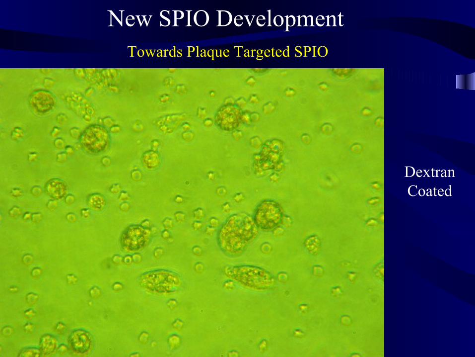
New SPIO Development Towards Plaque Targeted SPIO
Dextran Coated
New SPIO Development Towards Plaque Targeted SPIO
Lipid Coated
New SPIO Development Towards Plaque Targeted SPIO
Receptor Targeted
SPIO
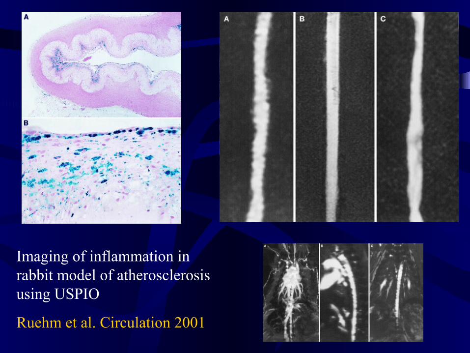
Imaging of inflammation in rabbit model of atherosclerosis using USPIO
Ruehm et al. Circulation 2001
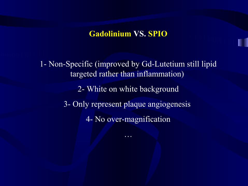
Gadolinium VS. SPIO
1- Non-Specific (improved by Gd-Lutetium still lipid targeted rather than inflammation)
2- White on white background
3- Only represent plaque angiogenesis
4- No over-magnification
…
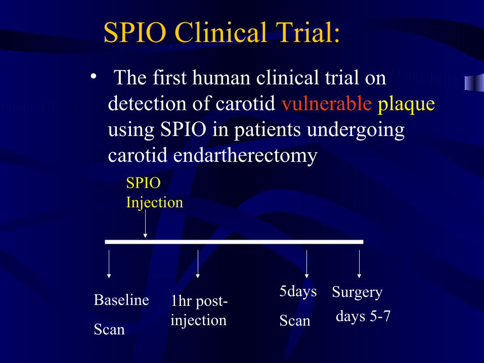
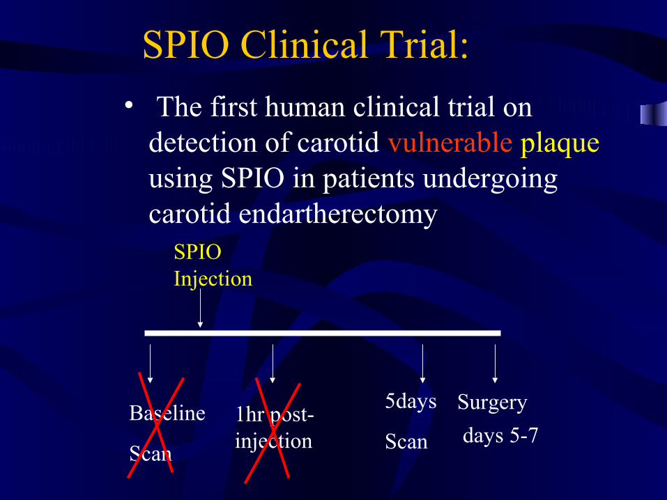
SPIO Clinical Trial:
• The first human clinical trial on detection of carotid vulnerable plaque using SPIO in patients undergoing carotid endartherectomy
Baseline
Scan
SPIO Injection
1hr post-injection
5days
Scan
Surgery
days 5-7
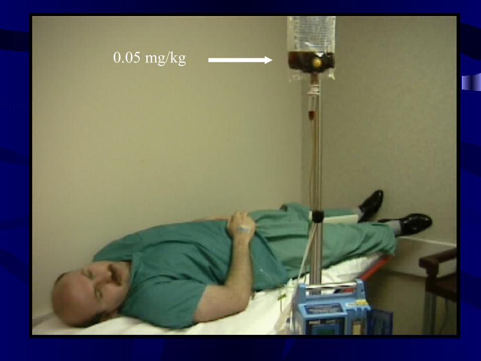
0.05 mg/kg
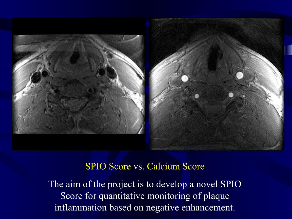
SPIO Score vs. Calcium Score
The aim of the project is to develop a novel SPIO Score for quantitative monitoring of plaque
inflammation based on negative enhancement.
SPIO Clinical Trial:
• The first human clinical trial on detection of carotid vulnerable plaque using SPIO in patients undergoing carotid endartherectomy
Baseline
Scan
SPIO Injection
1hr post-injection
5days
Scan
Surgery
days 5-7

… the question is not only vulnerable plaque
Stay Tuned!

HotPlaque.com 2000!
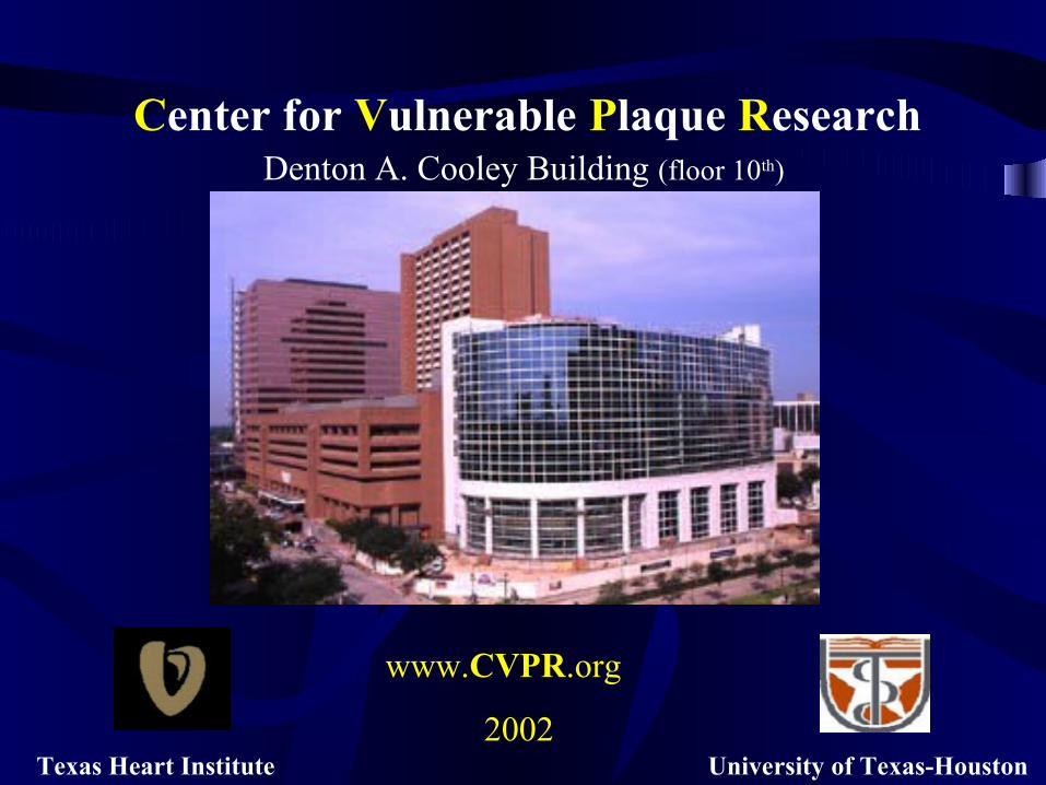
Texas Heart Institute University of Texas-Houston
Center for Vulnerable Plaque ResearchDenton A. Cooley Building (floor 10th)
www.CVPR.org
2002
Association for Eradication of Heart Attack
www.VP.org