Embed Size (px)
Citation preview
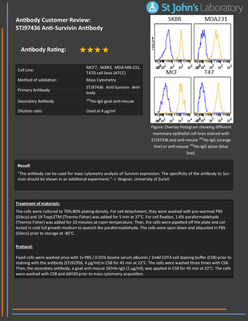
Cell Line: MCF7, SKBR3, MDA-MB-231, T47D cell lines (ATCC)
Method of validation: Mass Cytometry
Primary Antibody: STJ97436 Anti-Survivin Anti-body
Secondary Antibody 165Ho-IgG goat anti-mouse
Dilution ratio: Used at 4 µg/ml
Result
“The antibody can be used for mass cytometry analysis of Survivin expression. The specificity of the antibody to Sur-vivin should be shown in an additional experiment.”- J. Wagner, University of Zurich
Treatment of materials:
The cells were cultured to 70%-80% plating density. For cell detachment, they were washed with pre-warmed PBS (Gibco) and 1X TrypLETM (Thermo Fisher) was added for 5 min at 37°C. For cell fixation, 1.6% paraformaldehyde (Thermo Fisher) was added for 10 minutes at room temperature. Then, the cells were pipetted off the plate and col-lected in cold full growth medium to quench the paraformaldehyde. The cells were spun down and aliquoted in PBS (Gibco) prior to storage at -80°C.
Protocol: Fixed cells were washed once with 1x PBS / 0.05% bovine serum albumin / 2mM EDTA cell staining buffer (CSB) prior to staining with the antibody (STJ92356, 4 µg/ml) in CSB for 45 min at 22°C. The cells were washed three times with CSB. Then, the secondary antibody, a goat anti-mouse 165Ho-IgG (1 µg/ml), was applied in CSB for 45 min at 22°C. The cells were washed with CSB and ddH20 prior to mass cytometry acquisition.
Antibody Customer Review: STJ97436 Anti-Survivin Antibody
Antibody Rating:
Figure: Overlay histogram showing different
mammary epithelial cell lines stained with
STJ97436 and anti-mouse 165Ho-IgG (orange
line) or anti-mouse 165Ho-IgG alone (blue
line).