Embed Size (px)
Citation preview
Therapeutic Ultrasound
Salman Farooqi Lecturer
IPM&R
Sound
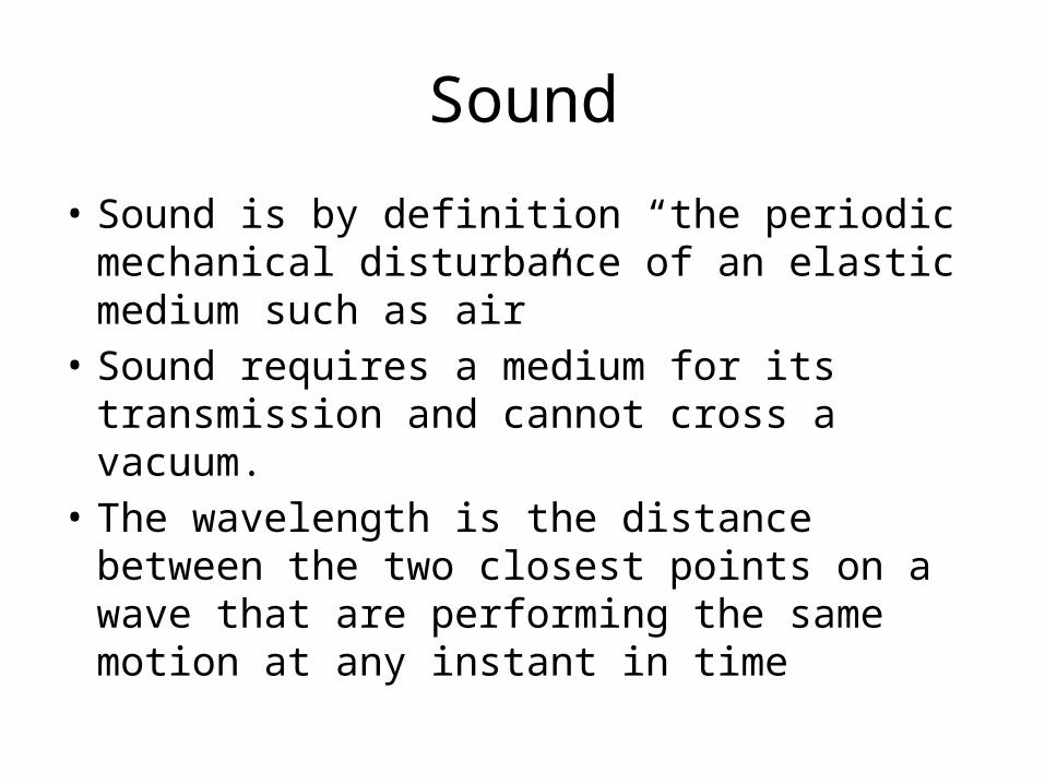
• Sound is by definition “the periodic mechanical disturbance of an elastic medium such as air”
• Sound requires a medium for its transmission and cannot cross a vacuum.
• The wavelength is the distance between the two closest points on a wave that are performing the same motion at any instant in time
Sound
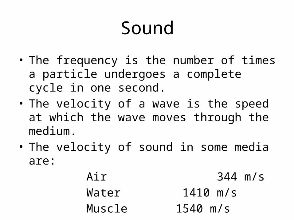
• The frequency is the number of times a particle undergoes a complete cycle in one second.
• The velocity of a wave is the speed at which the wave moves through the medium.
• The velocity of sound in some media are: Air 344 m/s Water 1410 m/s Muscle 1540 m/s
Ultrasound
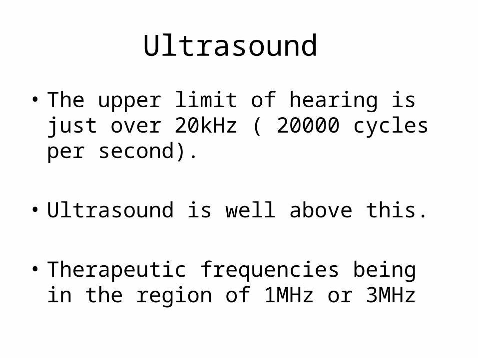
• The upper limit of hearing is just over 20kHz ( 20000 cycles per second).
• Ultrasound is well above this.
• Therapeutic frequencies being in the region of 1MHz or 3MHz
The production of Ultrasound
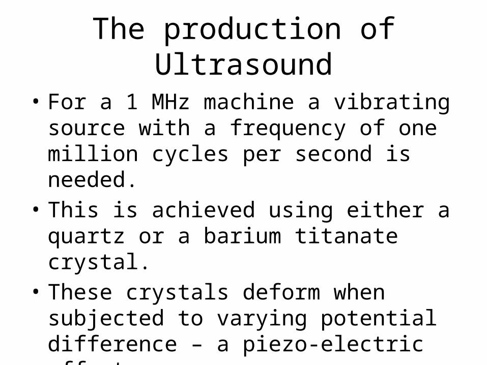
• For a 1 MHz machine a vibrating source with a frequency of one million cycles per second is needed.
• This is achieved using either a quartz or a barium titanate crystal.
• These crystals deform when subjected to varying potential difference – a piezo-electric effect

Piezoelectricity
• Piezoelectricity is a natural phenomenon found in certain mineral crystals, but may also be synthesized commercially.
• The crystal transforms mechanical energy into electrical energy and it reverse, electrical into mechanical.

Piezoelectricity• If a piezoelectric crystal were to
be compressed or deformed by mechanical means, a small electric charges would result within the crystal; conversely, if an electric charge were to be imposed on the crystal, a vibration of mechanical deformity of the molecular structure of crystal would ensure.
Transformation of electrical energy into sound
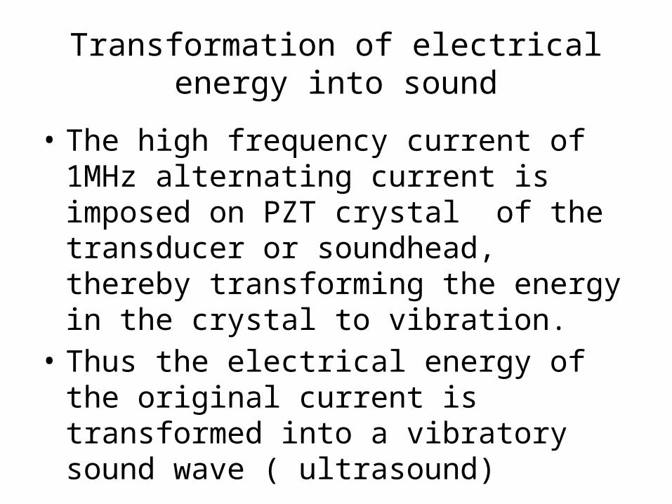
• The high frequency current of 1MHz alternating current is imposed on PZT crystal of the transducer or soundhead, thereby transforming the energy in the crystal to vibration.
• Thus the electrical energy of the original current is transformed into a vibratory sound wave ( ultrasound)
Reflection of Ultrasound
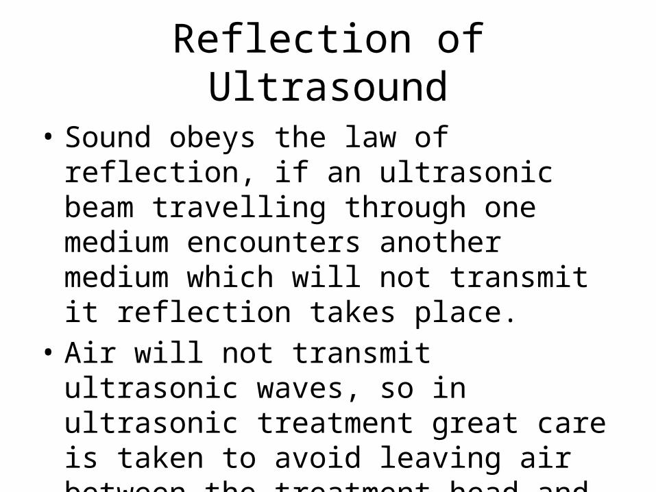
• Sound obeys the law of reflection, if an ultrasonic beam travelling through one medium encounters another medium which will not transmit it reflection takes place.
• Air will not transmit ultrasonic waves, so in ultrasonic treatment great care is taken to avoid leaving air between the treatment head and the patient.
Attenuation of ultrasound
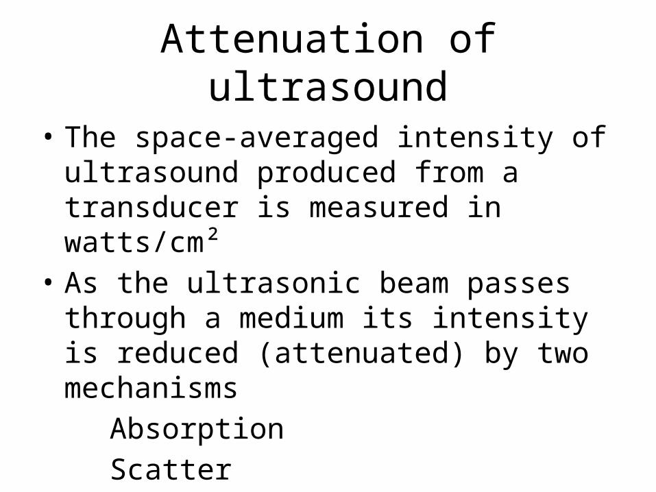
• The space-averaged intensity of ultrasound produced from a transducer is measured in watts/cm²
• As the ultrasonic beam passes through a medium its intensity is reduced (attenuated) by two mechanisms
Absorption Scatter
Attenuation of ultrasound
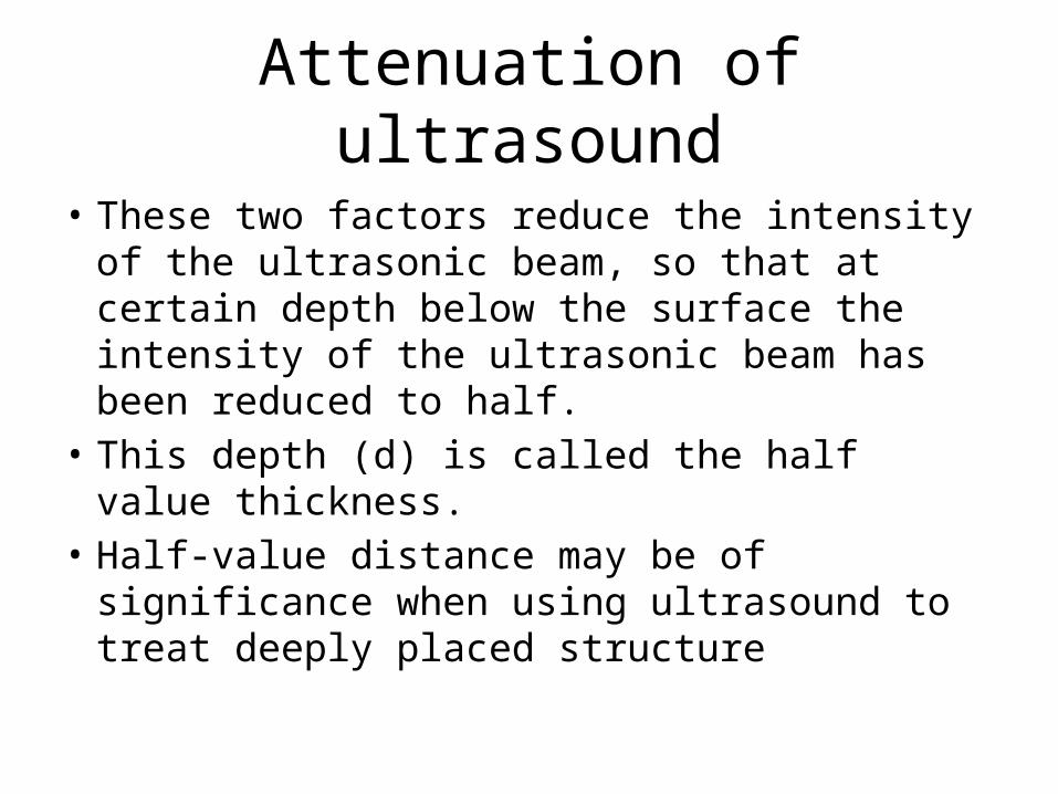
• These two factors reduce the intensity of the ultrasonic beam, so that at certain depth below the surface the intensity of the ultrasonic beam has been reduced to half.
• This depth (d) is called the half value thickness.
• Half-value distance may be of significance when using ultrasound to treat deeply placed structure
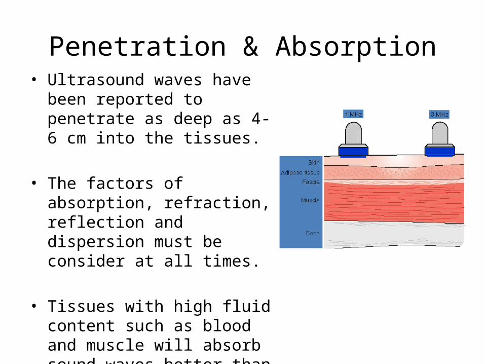
Penetration & Absorption• Ultrasound waves have been
reported to penetrate as deep as 4-6 cm into the tissues.
• The factors of absorption, refraction, reflection and dispersion must be consider at all times.
• Tissues with high fluid content such as blood and muscle will absorb sound waves better than will less hydrated tissues.
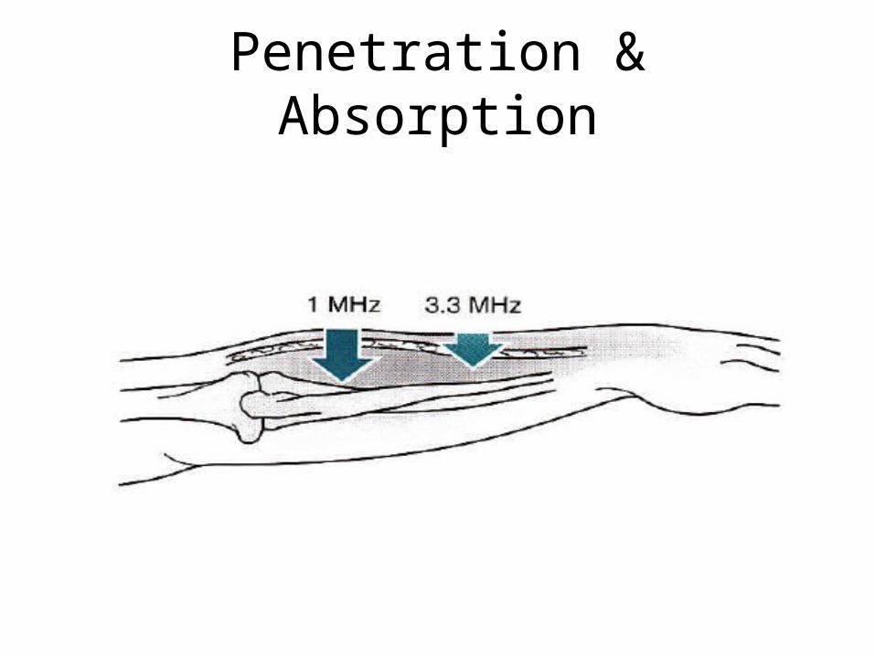
Penetration & Absorption
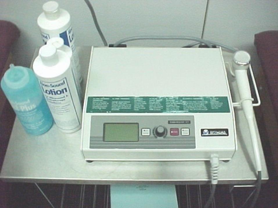
Coupling media
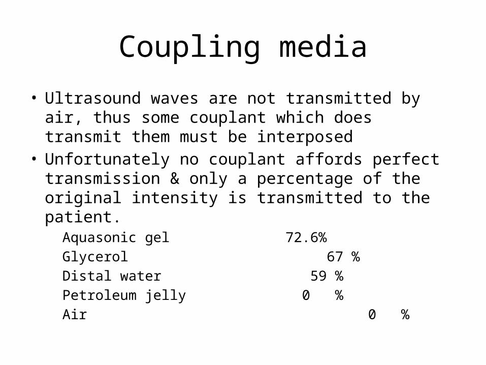
• Ultrasound waves are not transmitted by air, thus some couplant which does transmit them must be interposed
• Unfortunately no couplant affords perfect transmission & only a percentage of the original intensity is transmitted to the patient.
Aquasonic gel 72.6% Glycerol 67 % Distal water 59 % Petroleum jelly 0 % Air 0 %
Pulsed Ultrasound
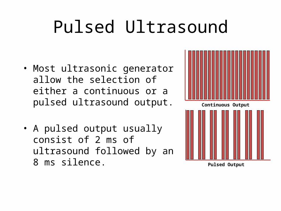
• Most ultrasonic generator allow the selection of either a continuous or a pulsed ultrasound output.
• A pulsed output usually consist of 2 ms of ultrasound followed by an 8 ms silence.
Continuous Output
Pulsed Output
Physiological Effects
• Ultrasound have four basic physiologic effects Chemical Reactions Biological Responses Mechanical Responses Thermal Effects
Chemical Reaction
• Just as test tube is shaken in the laboratory, to enhance chemical action.
• Ultrasound vibration stimulate tissue to enhance chemical reactions and processes therein and ensure circulation of necessary elements and radicals for recombination
Biological Responses
• The permeability of membranes is increased by ultrasound, which enhances transfer of fluid and nutrients to tissues.
• This quality is of importance in the process of Phonophoresis, where molecules are literally pushed through the skin by the sound wave front for therapeutic purposes.
Biological Responses
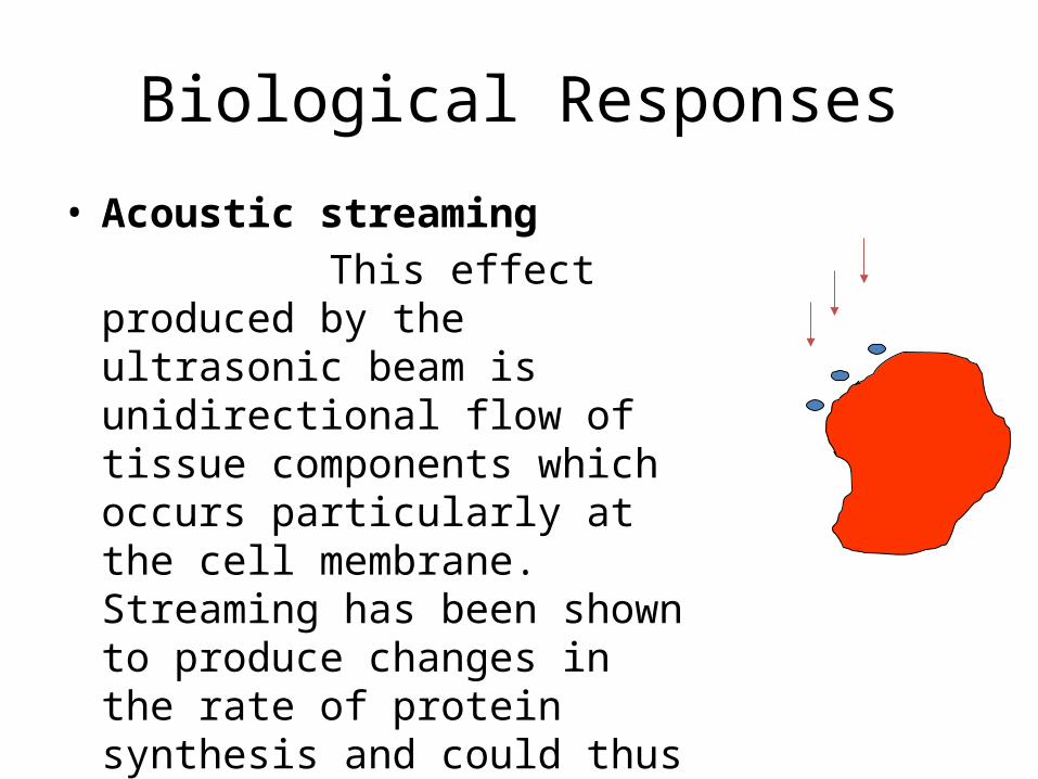
• Acoustic streaming This effect produced by the
ultrasonic beam is unidirectional flow of tissue components which occurs particularly at the cell membrane. Streaming has been shown to produce changes in the rate of protein synthesis and could thus have a role in the stimulation of repair.
Mechanical Responses
• Cavitation is a common condition in which a bubble gas is produced in the tissues as a result of insonation.
• Stable cavitation is not dangerous to the tissues, as the bubbles remain intact and oscillate harmlessly in the ultrasonic field.
• Transient cavitation is dangerous to the tissues, as the bubble grows and collapes rapidly in the ultrasonic beam and this is thought to cause very great increase in the tempreture
Mechanical Responses
• The micromassage effect of ultrasound occurs at a cellular level where the cells are alternately compressed and then pulled further apart. This effect on intracellular fluids and thus to reduce oedema.
• Ultrasound has been found to be effective at reducing both recent traumatic oedema and chronic induced oedema.
Mechanical Responses
• It has been shown that a standing wave produced in the tissues can produce inhibition of blood flow.
• Pulsing the ultrasonic beams or moving the treatment head should prevent this occurring.
Mechanical Responses
• Tendon Extensibility The sclerolytic action of ultrasound
apparently increases the extensibility of tendons, crucial to those that have been shortened by inflammation, strain, or disease, and providing the clinician with an excellent formula for management of spasm
Thermal effects
• As ultrasonic waves are absorbed they are converted to thermal energy (heat)
• The amount of heat produced depends upon factors such as the number of times the transducer passes over a part and on the space-average intensity (watt/cm²)
• Reflection of ultrasound may occur at tissue interfaces, producing a concentrated heating effect at that point.
Thermal effects
• As reflection from the bone occurs there is double the intensity of ultrasound in the periosteum region, which may cause localized over-heating and can manifest itself as a periosteal pain.
• It is best to avoid passing the ultrasonic head over subcuteneous bony points
Thermal effects
• Pulsed ultrasound has little thermal effects, as any heat produced by the 2ms of ultrasound is carried away by the circulation in the 8ms rest.
• Pulsed ultrasound may thus be indicated in the treatment of acute conditions where a thermal effect is not required
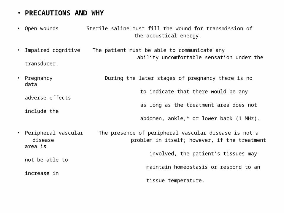
• PRECAUTIONS AND WHY
• Open wounds Sterile saline must fill the wound for transmission of the acoustical energy.
• Impaired cognitive The patient must be able to communicate any ability uncomfortable sensation under the transducer.
• Pregnancy During the later stages of pregnancy there is no data to indicate that there would be any adverse effects as long as the treatment area does not include the abdomen, ankle,* or lower back (1 MHz).
• Peripheral vascular The presence of peripheral vascular disease is not a disease problem in itself; however, if the treatment area is involved, the patient’s tissues may not be able to maintain homeostasis or respond to an increase in tissue temperature.
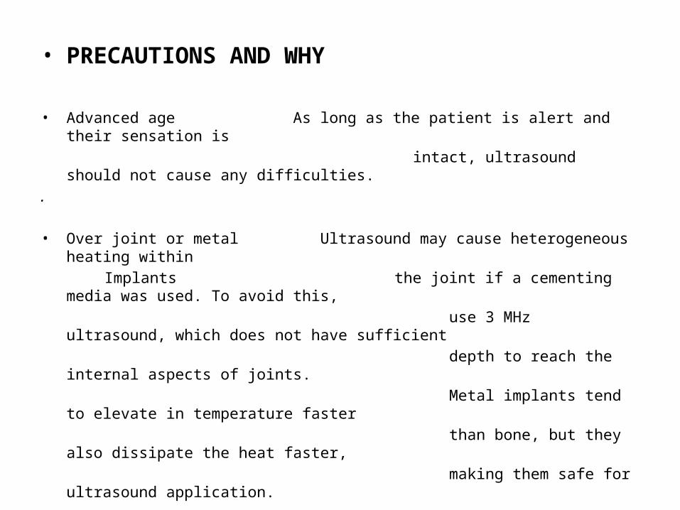
• PRECAUTIONS AND WHY
• Advanced age As long as the patient is alert and their sensation is intact, ultrasound should not cause any difficulties..
• Over joint or metal Ultrasound may cause heterogeneous heating within Implants the joint if a cementing media was used. To avoid this, use 3 MHz ultrasound, which does not have sufficient depth to reach the internal aspects of joints. Metal implants tend to elevate in temperature faster than bone, but they also dissipate the heat faster, making them safe for ultrasound application.
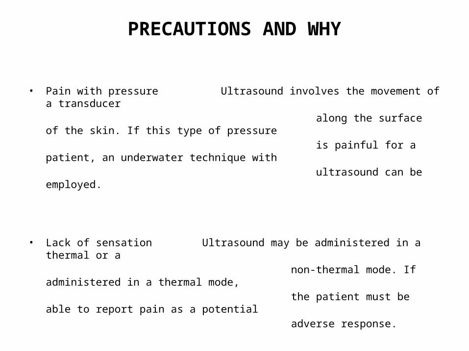
PRECAUTIONS AND WHY
• Pain with pressure Ultrasound involves the movement of a transducer along the surface of the skin. If this type of pressure is painful for a patient, an underwater technique with ultrasound can be employed.
• Lack of sensation Ultrasound may be administered in a thermal or a non-thermal mode. If administered in a thermal mode, the patient must be able to report pain as a potential adverse response.
Contraindications Why?
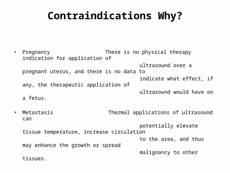
• Pregnancy There is no physical therapy indication for application of ultrasound over a pregnant uterus, and there is no data to indicate what effect, if any, the therapeutic application of ultrasound would have on a fetus.
• Metastasis Thermal applications of ultrasound can potentially elevate tissue temperature, increase circulation to the area, and thus may enhance the growth or spread malignancy to other tissues.
• Lack of sensation If the patient is unable to report pain, they can easily burn with thermal applications of ultrasound.
Contraindications Why?
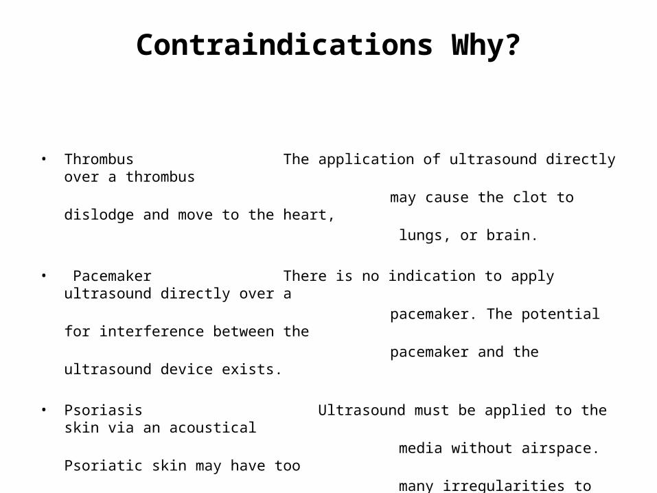
• Thrombus The application of ultrasound directly over a thrombus may cause the clot to dislodge and move to the heart, lungs, or brain.
• Pacemaker There is no indication to apply ultrasound directly over a pacemaker. The potential for interference between the pacemaker and the ultrasound device exists.
• Psoriasis Ultrasound must be applied to the skin via an acoustical media without airspace. Psoriatic skin may have too many irregularities to permit passage of the ultrasound into the patient.
Clinical application of US• Soft tissue shortening• Pain control• Surgical skin incisions• Tendon injuries• Resorption of calcium deposits• Bone fractures• Carpal tunnel syndrome
Soft tissue shortening
• Soft tissue shortening can be the result of immobilization, inactivity or scarring, and can cause
joint Range-of-motion (ROM) restrictions, pain, and functional limitations.
• Because ultrasound can penetrate to the depth of most joint capsules, tendons, and ligaments, since these tissues have high ultrasound absorption
coefficients, ultrasound can be an effective physical agent for heating these tissues prior to stretching
Soft tissue shortening
• The deep heating produced by 1 MHz continues ultrasound at 1.0 to 2.5W cm² has been shown to be more effective at increasing joint ROM than the superficial heating produced by infrared.
Pain control
• Ultrasound may control pain by altering its transmission or perception or by modifying the underlying condition causing the pain. These effects may be the result of stimulation of the cutaneous thermal receptors or increased soft tissue extensibility due to increased tissue temperature, the result of changes in nerve conduction due to increased tissue temperature
or the non-thermal effects of ultrasound, or the result of modulation of inflammation due to the non-thermal effects of ultrasound.
Surgical skin incisions
• The effect of ultrasound on the healing of surgical skin incisions has been studied in both animal and human subjects.
• Ultrasound has been shown to accelerate the evolution of angiogenesis a vital component of early wound healing.
• Angiogenesis is the development of new blood vessels at an injury site that serves to reestablish circulation and thus limits ischemic necrosis and facilitate repair.
Tendon injuries• Ultrasound has been reported to assist in the healing
of tendons after surgical incision and repair Although some studies with both animal and human subjects have reported treatment success others have failed to support these findings.
• It is recommended that ultrasound be applied in a Pulsed mode at a low intensity during the acute phase of tendon inflammation in order to minimize the risk of aggravating the condition and to accelerate recovery
Resorption of calcium deposits
• Ultrasound may facilitate the resorption of calcium deposits.
• Although the mechanism underlying calcific deposits resorption is not known, decrease in pain and improvements in function may be due to the reduction in inflammation produced by ultrasound.
Bone fractures
• Although prior reports have recommended that ultrasound not be applied over unhealed fracture because they have failed to provide data to support this recommendation and because recent studies have demonstrated that low-dose ultrasound can reduce the fracture healing time in animals and humans, the use of low-dose ultrasound to accelerate fracture healing is now recommended.
Carpal Tunnel Syndrome
• Continuous ultrasound has generally not been recommended for the treatment of carpal tunnel syndrome because of the risk of adversely impacting nerve conduction velocity by overheating. However a resent study found that pulsed ultrasound produced significantly greater improvement
Testing the apparatus
• Testing should always be carried out prior to treatment
• The simplest way of finding out whether ultrasound is in fact being produced is to use a water bath and to reflect an ultrasonic beam up to the surface where it should produce ripples
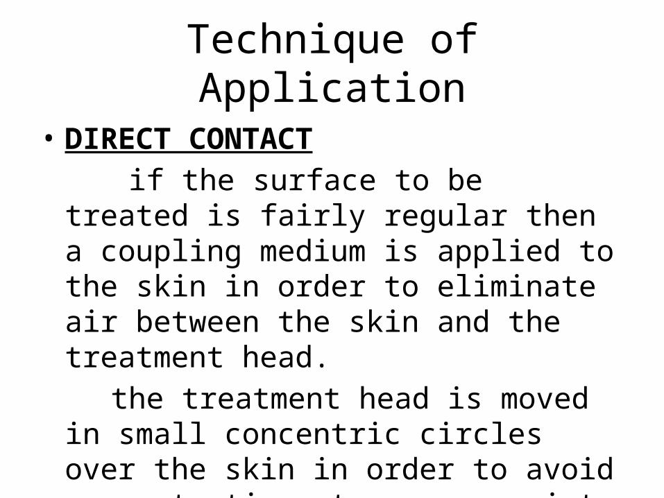
Technique of Application
• DIRECT CONTACT if the surface to be treated is fairly regular
then a coupling medium is applied to the skin in order to eliminate air between the skin and the treatment head.
the treatment head is moved in small concentric circles over the skin in order to avoid concentration at any one point
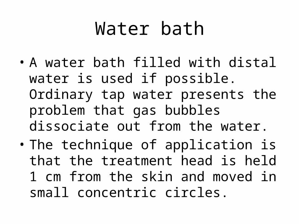
Water bath
• A water bath filled with distal water is used if possible. Ordinary tap water presents the problem that gas bubbles dissociate out from the water.
• The technique of application is that the treatment head is held 1 cm from the skin and moved in small concentric circles.
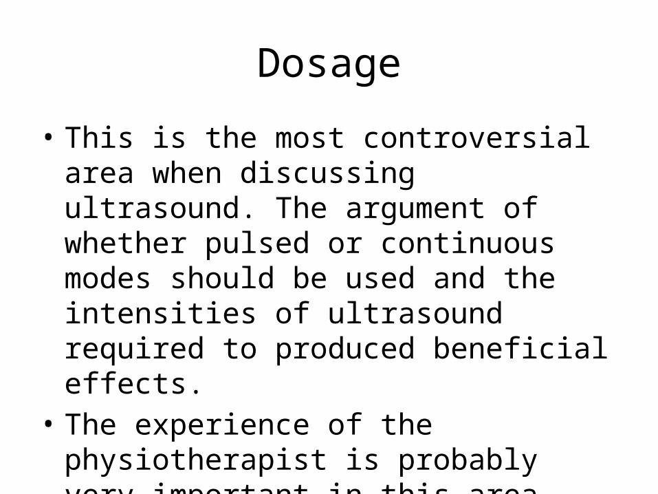
Dosage
• This is the most controversial area when discussing ultrasound. The argument of whether pulsed or continuous modes should be used and the intensities of ultrasound required to produced beneficial effects.
• The experience of the physiotherapist is probably very important in this area.
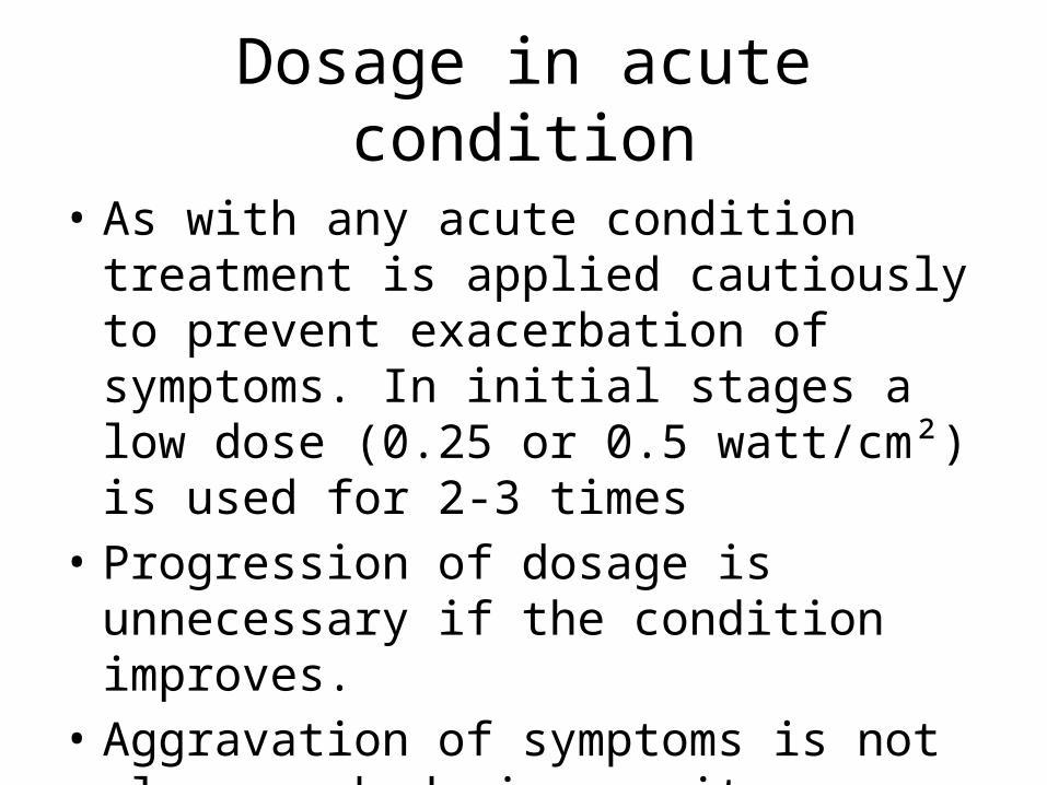
Dosage in acute condition
• As with any acute condition treatment is applied cautiously to prevent exacerbation of symptoms. In initial stages a low dose (0.25 or 0.5 watt/cm²) is used for 2-3 times
• Progression of dosage is unnecessary if the condition improves.
• Aggravation of symptoms is not always a bad sign as it may indicate that repair processes are taking place.
Dosage in chronic condition
• Chronic conditions can be treated with either a pulsed or a continuous beam.
• With a continuous beam, the maximum intensity which should be used is that which produced a mildly perceptible warmth. This usually occurs around 2 watt/cm²
• A dose of 2 watts/cm² for 8 minutes is usually considered
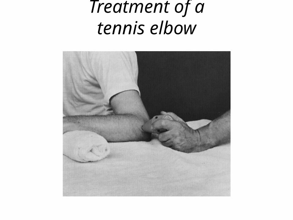
Treatment of atennis elbow
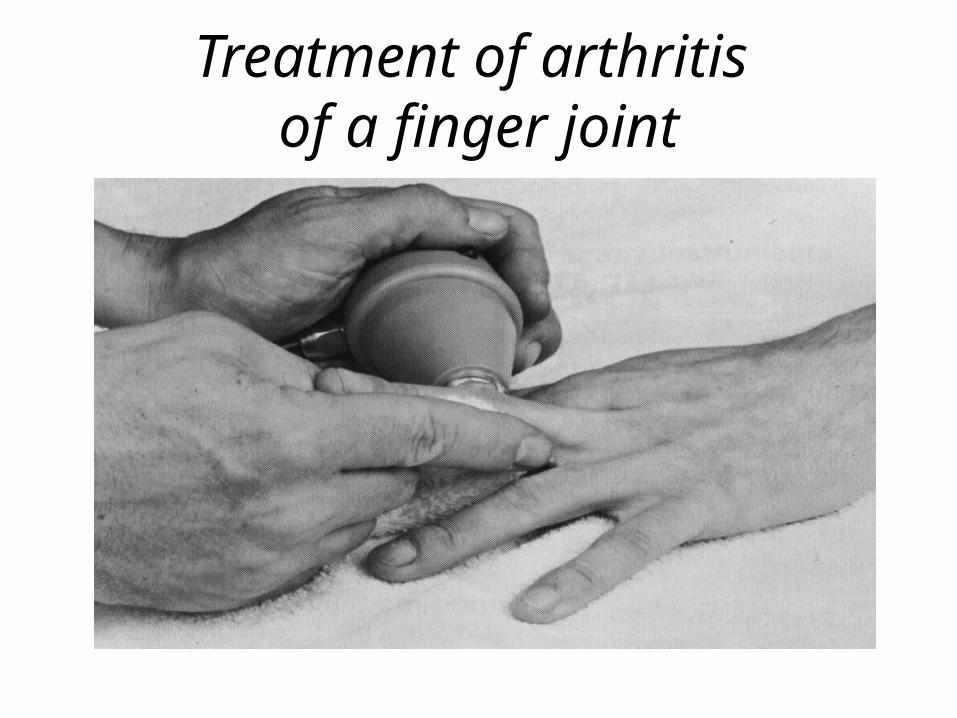
Treatment of arthritis of a finger joint
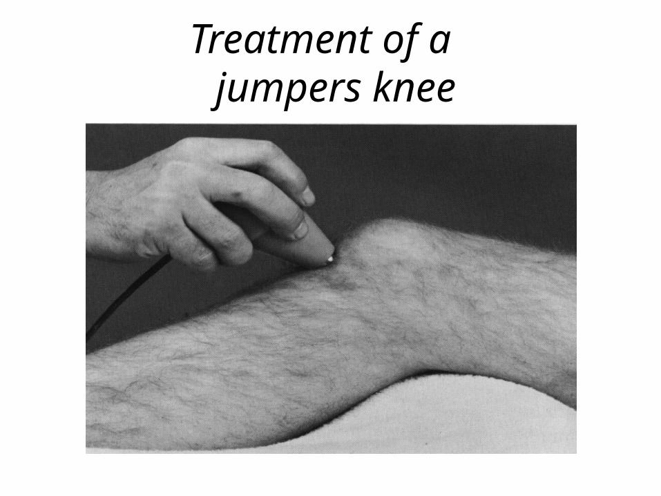
Treatment of a jumpers knee
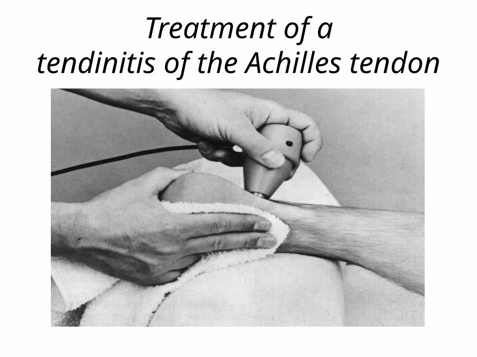
Treatment of atendinitis of the Achilles tendon
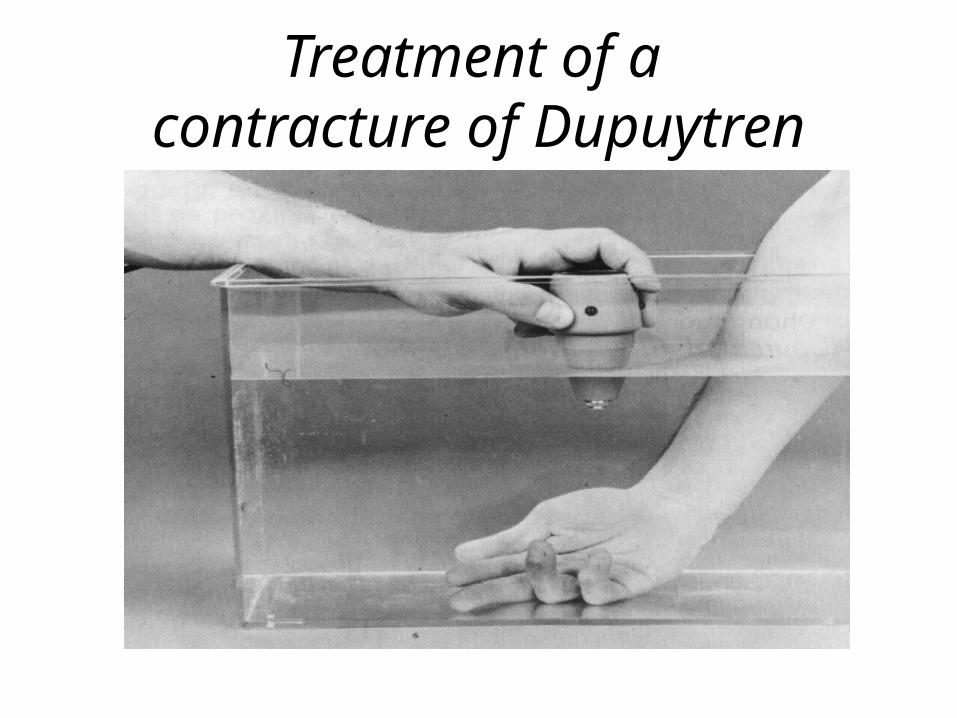
Treatment of a contracture of Dupuytren
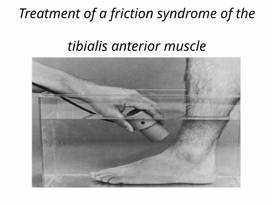
Treatment of a friction syndrome of the tibialis anterior muscle
Phonophoresis
Description
• Use of therapeutic ultrasound to assist in diffusion of medication through the skin
• Increases the diameter of skin to allow the medication to pass– Pores– Hair follicles– Sweat glands
Biophysical Effects
• Medication is introduced over a large area– Relative to an injection
• Noninvasive• Medication may not be filtered by the liver– Reducing metabolic elimination of the medicine
Transdermal Application
• Medication must diffuse through:– Enzymatic barrier of the epidermis– Stratum corneum• Rate-limiting barrier to absorption• Medications must be able to diffuse across this barrier
• Medication is stored in subcutaneous tissues for some time before being diffused deeper
Skin Influences
• Medication uptake is improved when the skin is:– Well hydrated– Highly vascularized– Relatively thin– “Younger” skin tends to have better diffusion
characteristics than “older” skin.
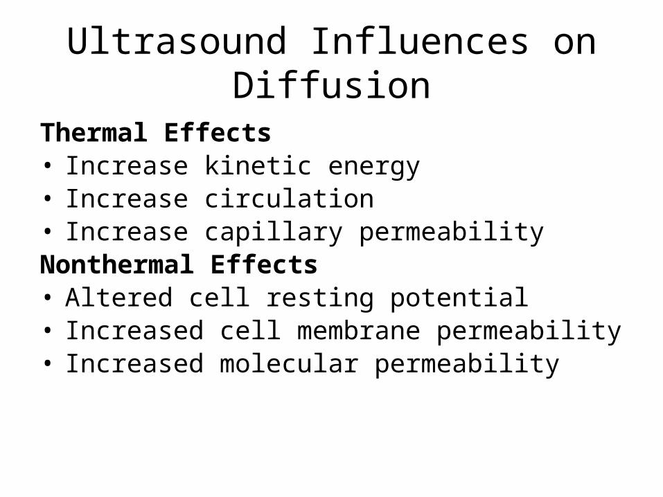
Ultrasound Influences on Diffusion
Thermal Effects• Increase kinetic energy• Increase circulation• Increase capillary permeabilityNonthermal Effects• Altered cell resting potential• Increased cell membrane permeability• Increased molecular permeability
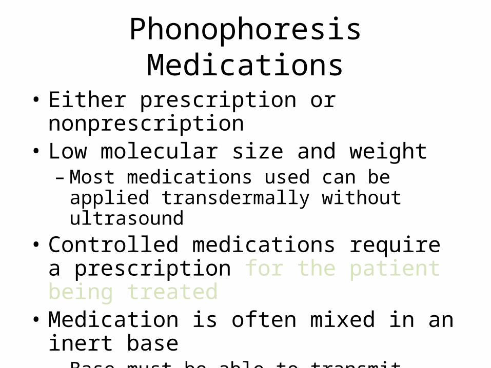
Phonophoresis Medications
• Either prescription or nonprescription• Low molecular size and weight– Most medications used can be applied
transdermally without ultrasound• Controlled medications require a prescription
for the patient being treated• Medication is often mixed in an inert base– Base must be able to transmit ultrasonic energy
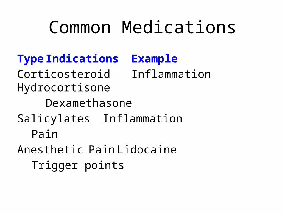
Common Medications
Type Indications ExampleCorticosteroid Inflammation Hydrocortisone
DexamethasoneSalicylates Inflammation
PainAnesthetic Pain Lidocaine
Trigger points
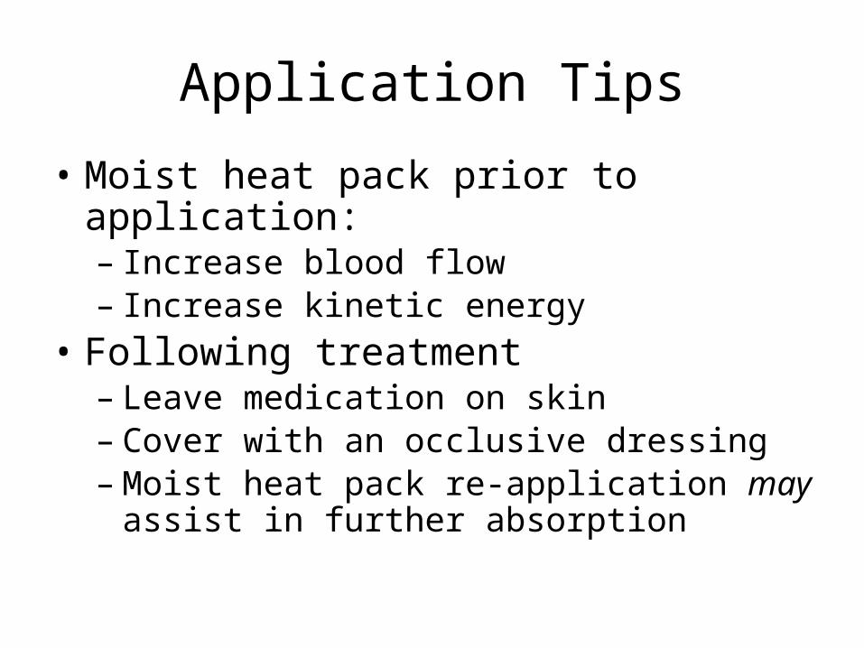
Application Tips
• Moist heat pack prior to application:– Increase blood flow– Increase kinetic energy
• Following treatment– Leave medication on skin– Cover with an occlusive dressing– Moist heat pack re-application may assist in
further absorption
Contraindications
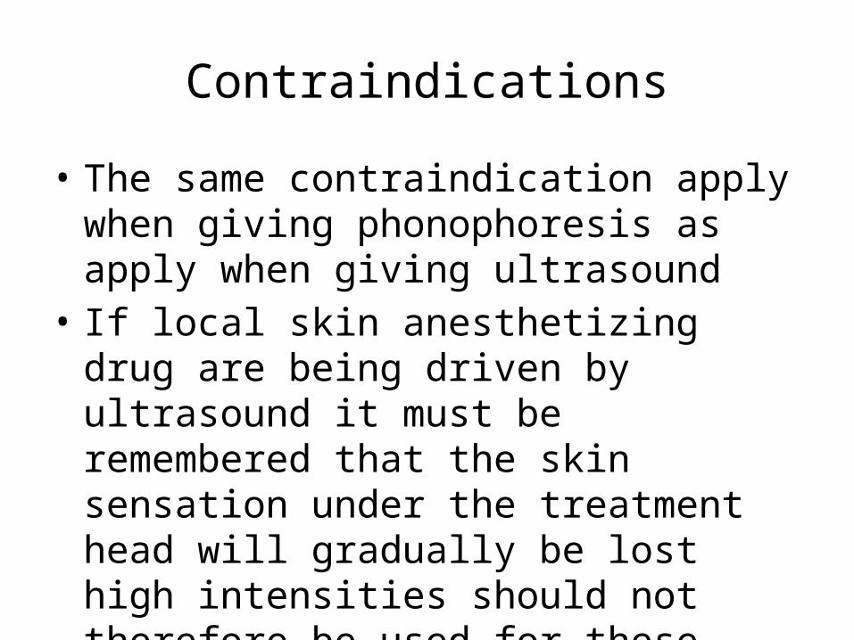
• The same contraindication apply when giving phonophoresis as apply when giving ultrasound
• If local skin anesthetizing drug are being driven by ultrasound it must be remembered that the skin sensation under the treatment head will gradually be lost high intensities should not therefore be used for these drugs
Some Conditions Treated With Phonophoresis
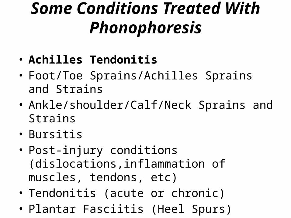
• Achilles Tendonitis• Foot/Toe Sprains/Achilles Sprains and Strains• Ankle/shoulder/Calf/Neck Sprains and Strains• Bursitis • Post-injury conditions (dislocations,inflammation of
muscles, tendons, etc)• Tendonitis (acute or chronic)• Plantar Fasciitis (Heel Spurs)
Some Conditions Treated With Phonophoresis
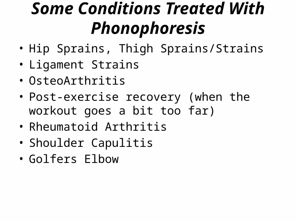
• Hip Sprains, Thigh Sprains/Strains• Ligament Strains• OsteoArthritis• Post-exercise recovery (when the workout goes a bit
too far)• Rheumatoid Arthritis• Shoulder Capulitis• Golfers Elbow
Some Conditions Treated With Phonophoresis
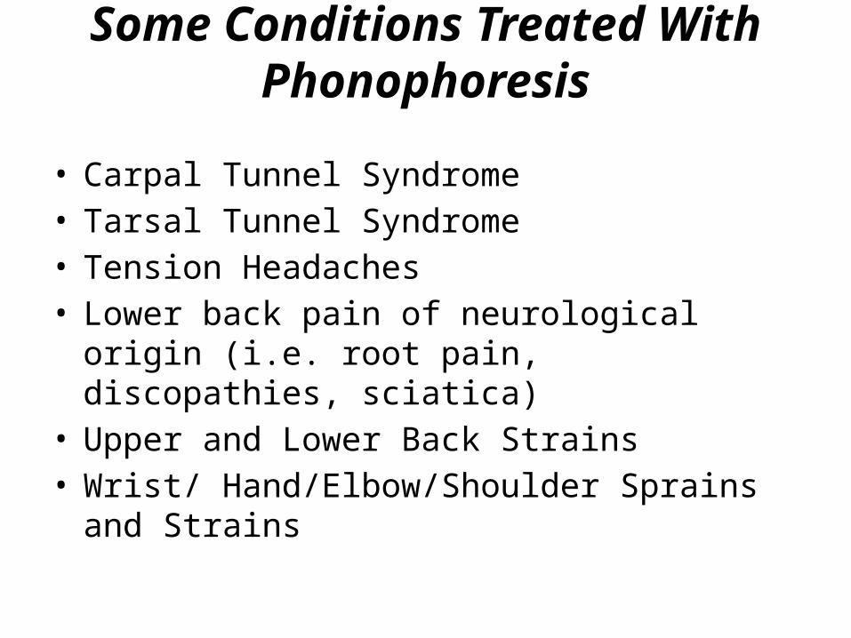
• Carpal Tunnel Syndrome• Tarsal Tunnel Syndrome• Tension Headaches• Lower back pain of neurological origin (i.e. root pain,
discopathies, sciatica)• Upper and Lower Back Strains• Wrist/ Hand/Elbow/Shoulder Sprains and Strains


![Ultrasound Contrast Agents: Fabrication, size distribution and …750462/FULLTEXT01.pdf · 2014. 9. 29. · Fig.2. Principle of Ultrasound Imaging [6] 1.1.2 Application Ultrasound](https://img.pdfslide.tips/doc/110x75/6080db269573ed4eb924dbf9/ultrasound-contrast-agents-fabrication-size-distribution-and-750462fulltext01pdf.jpg)


![[ ADDENDUM ] · 2019-02-11 · ultrasound principles, hemodynamics, and ultrasound instrumentation. Bioeffects, safety and the interactions between ultrasound and tissues will be](https://img.pdfslide.tips/doc/110x75/5f362916dbb6911dd53ea8a6/-addendum-2019-02-11-ultrasound-principles-hemodynamics-and-ultrasound-instrumentation.jpg)