Embed Size (px)
Citation preview

報告人:張聰舜報告日期: 2006-2-13

A male immigrant from Mexico was seen in the dermatology clinic at a California hospital for examination of a large, painless, cutaneous ulcer on his ear. There was evidence of cartilage destruction in the area. A biopsy specimen was taken from the skin lesion and was sent to the laboratory for examination. A section stained with hematoxylin and eosin showed small, oval, nonflagellated protozoan amastigotes. A diagnosis of a blood-borne parasitic infection was made, based on the morphological appearance of the Giemsa-stained smear.
Leishmania mexicana complex

Classification
原生動物界( Protista )-原生動物亞界( Protozoa )-變形鞭毛蟲門( Sarcomastigophora )-鞭毛蟲亞門( Mastigophra )-動鞭毛蟲綱( Zoomastigophorea )-動質蟲目( Kinetoplastida ) 利什曼原蟲( Leishmania ) 錐蟲( Trypanosoma )

Classification
L. donovani L. donovani donovani 、 L. donovani chagas 、 L. donovani infantum
L. tropica L. tropica aethiopica 、 L. tropica major 、 L. tropica minor 、 L. tropica
tropica L. maxicana
L. maxicana amazonensis 、 L. maxicana mexicana 、 L. maxicana pifanoi
L. viannia ( L. braziliensis ) L. viannia braziliensis 、 L. viannia guyanensis 、 L. viannia panamensis 、
L. viannia peruviana

Classification
臨床表徵 相關種類舊世界 新世界
VisceralL. donovani L. donovani
Muco-cutaneous
rare L. viannia
CutaneousL. tropica L. viannia
L. mexicana

Epidemiology
North-Eastern China, India, Middle-East, Southern Europe (Mediterranean bassin), Northern Africa, Central-East Africa and, in foci, Central and South America (especially Brazil and Honduras).

Epidemiology
O.W.L. is found especially in Asia (Middle East), Northern Africa and Southern Europe.
N.W.L. has a wide distribution in Central and South America (from Yucatan to Brazil).
The mucocutaneous form is prevalent in South America.

Life Cycle
無鞭毛體 amastigote (Leishman-Donovan body) found intracellularly in the vertebrate (i.e., human) host
前鞭毛體 promastigote (leptomonad) found in the digestive tract of the invertebrate host

Life Cycle
Vector
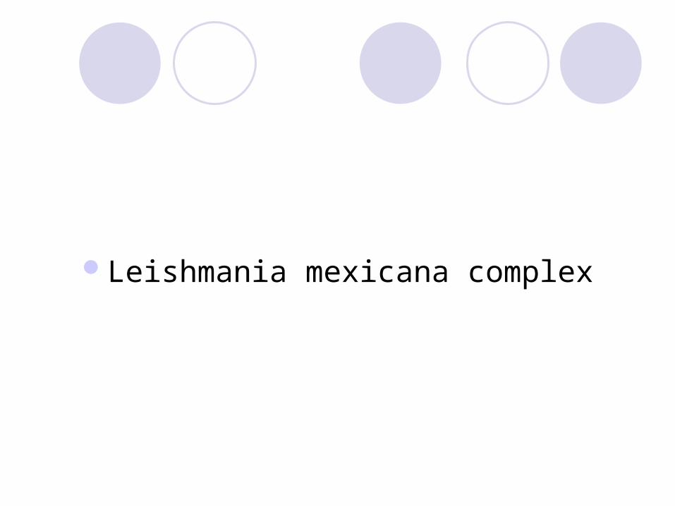
Sandfly (白蛉): order Diptera (雙翅目)- family Phlebotomidae (白蛉科) or Psychodidae (毛蠓科 ) . It includes the genera as follows : Phlebotomus genus (白蛉屬) Lutzomyia genus (沙蠅屬) Psychodopygus genus

Sandfly
small size, long legs, and abundant hair on both wings and body
flying, biting, blood-sucking insect Most sandflies will bite any warm-blooded animal and one spe
cies attacks penguins. Only female sandflies bite New world - Lutzomyia Old world - Phlebotomus
Animal reservoirs
人畜共通病( Zoonosis )齧齒目動物 , 樹懶 , 有袋動物 , 食肉動物印度的 Vsceral Leishmaniasis 幾乎侷限於
人 L. major 在亞洲鄉村是野生齧齒 ; 在亞洲
都市是犬 ; 在非洲市齧齒
Cutaneous leishmaniasis
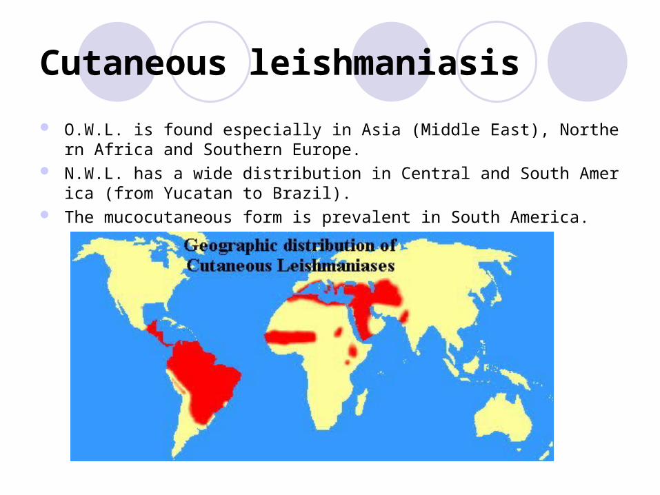
O.W.L. is found especially in Asia (Middle East), Northern Africa and Southern Europe.
N.W.L. has a wide distribution in Central and South America (from Yucatan to Brazil).
The mucocutaneous form is prevalent in South America.
Cutaneous leishmaniasis
incubation period is 2~8 weeks erythematous papule nodule nodule ulcerates and crusts typically large but painless unless there is secondary bacterial or fungal infection. Old World Cutaneous leishmaniasis
Leishmania, subgenus Leishmania, complexes major, tropica, donovani (infantum species), aethiopica (diffuse form).
dry or urban cutaneous leishmaniasis is caused by Leishmania tropica wet or rural cutaneous leishmaniasis is caused by L. major Ethiopian cutaneous leishmaniasis is caused by L. aethiopica
New World Cutaneous leishmaniasis Leishmania, subgenus Leishmania, complex mexicana (mexicana, amazonensis, pifanoi)
and subgenus Viannia, complexes brasiliensis and guyanensis develop and heal similarly to those of the Old World forms but tend to be less no
dular and more ulcerative and destructive some common forms are mucocutaneous leishmaniasis

Old World Cutaneous leishmaniasis
dry cutaneous large urban areas in the Middle East, the Mediterranean region, and the Indian subcontinent Leishmania tropica vectors Phlebotomus sergenti and P. papatasi reservoir may be either human or canine (犬) A slowly developing single lesion that persists for a year or more
wet cutaneous rural areas in parts of the Middle East, central Asia, and the Indian subcontinent Leishmania major reservoirs are desert rodents such as squirrels (松鼠) and gerbils (沙鼠) vector Phlebotomus papatasi Infection is acute, rapidly evolving, and characterized by multiple sores with inflammation, ulceration, an
d crusting Ethiopian cutaneous
the highlands of Kenya and Ethiopia Leishmania aethiopica hyraxes (蹄兔) vectors are Phlebotomus pedifer and P. longipes less inflamed and more chronic self-limited but may develop into diffuse cutaneous leishmaniasis

Old World Cutaneous leishmaniasis

New World Cutaneous leishmaniasis
Mucocutaneous chronic, progressive metastatic spread of the lesions of New World cutaneous leishmania
sis Leishmania viannia braziliensis nasal, pharyngeal, and buccal mucosa months to years after the appearance of the initial c
utaneous lesion,which has usually healed. mutilating destruction of the nasal septum, palate, lips, pharynx, and larynx
Diffuse cutaneous leishmaniasis
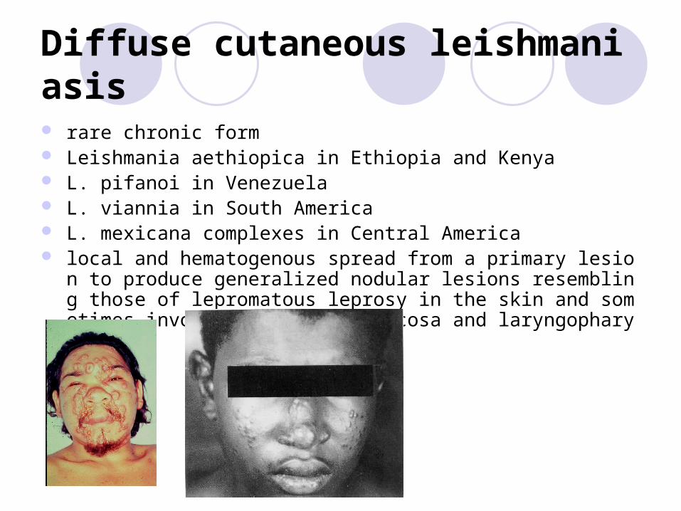
rare chronic form Leishmania aethiopica in Ethiopia and Kenya L. pifanoi in Venezuela L. viannia in South America L. mexicana complexes in Central America local and hematogenous spread from a primary lesion to produce generaliz
ed nodular lesions resembling those of lepromatous leprosy in the skin and sometimes involving the nasal mucosa and laryngopharynx.

Diagnosis
a history of exposure to sandfies Risk factors for HIV should besolicited, including sexual encounters, intra
venousdrug use, and blood transfusions obtained abroad. symptoms isolation of the organisms from the lesion aspirate or biopsy, by direct exa
mination or culture A skin test (delayed hypersensitivity: Montenegro test) and detection of ant
i-leishmanial antibodies by immuno-fluorescence are indicative of exposure.

Diagnosis

Diagnosis
Cutaneous scraping the simplest and most common test only 70 to 75 percent sensitive fixed with methanol, stained with Giemsa, and examined under oil immersion. Amastigotes are seen in monocytes or extracellularly It is important to see the nucleus and the rod-shaped kinetoplast, a mitochondri
al structure containing extranuclear DNA, to diagnose leishmaniasis.

Treatment
Sodium stibogluconate is the drug of choice. Machanism unknown Side effect ( reversible )
Gastrointestinal symptoms Fever Headache Myalgia , arthralgia Rash ECG changes, ex. T waves change and QT prolongatio
n

Treatment

Quiz 1
Which infection does this patient have? What is the name of the hemoflagellate causing his infection?
Quiz 2
Name the three main species belonging to
this complex.

Quiz 3
Which vector is responsible for the
transmission of this infection?

Quiz 4
Describe the life cycle of this parasite.

Quiz 5
What are the reservoirs for these parasites? Which populations are at particular risk of infection with these parasites?

Quiz 6
How is the diagnosis of this infection made?

Quiz 7
How is this infection treated?

Reference
醫用寄生蟲學 http://www.cdfound.to.it/html/atlas.htm#atlas http://www.biosci.ohio-state.edu/~parasite/home.html http://en.wikipedia.org/ http://www.dpd.cdc.gov/dpdx/Default.htm http://pathmicro.med.sc.edu/book/welcome.htm Basic and clinincally pharmacology
![Ⅳ 環境側面の報告 - Hitachi Metals · Ⅳ 環境側面の報告. 45. 日立金属グループcsr 活動報告2017[詳細活動報告] Ⅳ 環境側面の報告. 1. 環境マネジメント](https://img.pdfslide.tips/doc/110x75/5ec9a6db7c3455151d652bab/a-cfe-hitachi-metals-a-cfe-45-cefffcsr.jpg)


![[報告] 電影欣賞報告 - 奧圖瑪塔](https://img.pdfslide.tips/doc/110x75/58edf5eb1a28ab8c708b469b/-58edf5eb1a28ab8c708b469b.jpg)