Embed Size (px)
DESCRIPTION
流式细胞术在周期与凋亡 检测中的应用. 中山医学院药理学教研室 黄奕俊 2011.11. 本次培训讲座已录制视频,需要观看视频的各位老师和同学可带移动硬盘到科技楼北 810 房(原房号为北 806 房)拷贝视频 。 科研仪器管理中心. 流式细胞术的应用. 细胞 DNA 流式分析原理. 某些荧光染料( PI 、 EB 、 HOs 、 DAPI 、 7-AAD 、 AO 等)与细胞内 DNA 碱基结合,在特定波长激光激发下发射出荧光,荧光强度与 DNA 含量成正比。流式细胞仪通过测定细胞的荧光强度推算出细胞的 DNA 含量。. Our bullets - PowerPoint PPT Presentation
Citation preview
流式细胞术在周期与凋亡检测中的应用
中山医学院药理学教研室黄奕俊2011.11
本次培训讲座已录制视频,需要观看视频本次培训讲座已录制视频,需要观看视频的各位老师和同学可带移动硬盘到科技楼的各位老师和同学可带移动硬盘到科技楼北北 810810 房(原房号为北房(原房号为北 806806 房)拷贝视频 房)拷贝视频 。。
科研仪器管理中心科研仪器管理中心
0 5000 10000 15000
Quantitative cell fluorescence
Leukemia Lymphoma
Dendritic cells
Intracellular signal transduction
Intracellular receptors
Cell viability
Cell adhesion
Autoimmune
Stem cell
Organ transplantation
Cell signaling
Immune function
Apoptosis
HIV
Activation
Cytokines
Cell DNA
Cell surface receptors
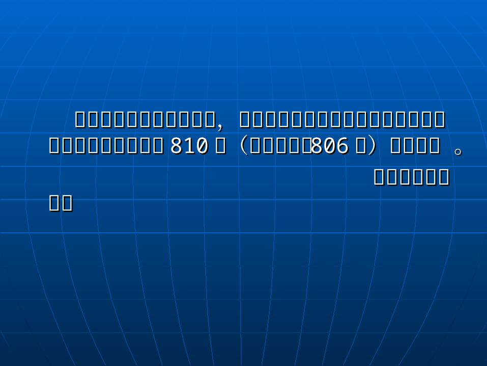
流式细胞术的应用
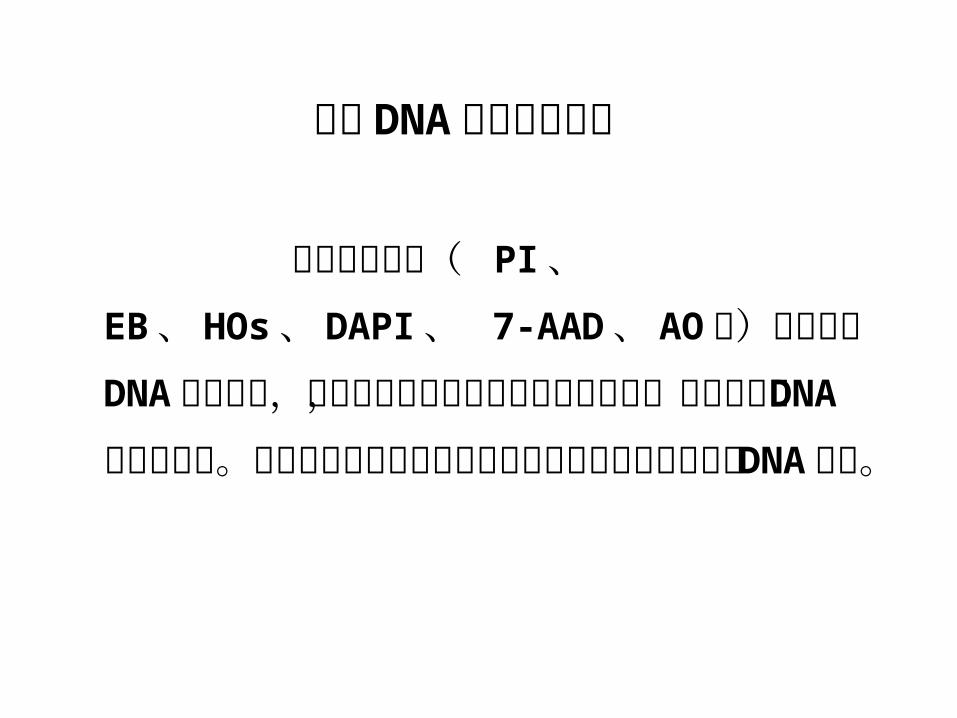
某些荧光染料( PI 、 EB 、 HOs 、 DAPI 、 7-AAD 、 AO 等)与细胞内 DNA 碱基结合,在特定波长激光激发下发射出荧光,荧光强度与 DNA 含量成正比。流式细胞仪通过测定细胞的荧光强度推算出细胞的 DNA 含量。
细胞 DNA 流式分析原理
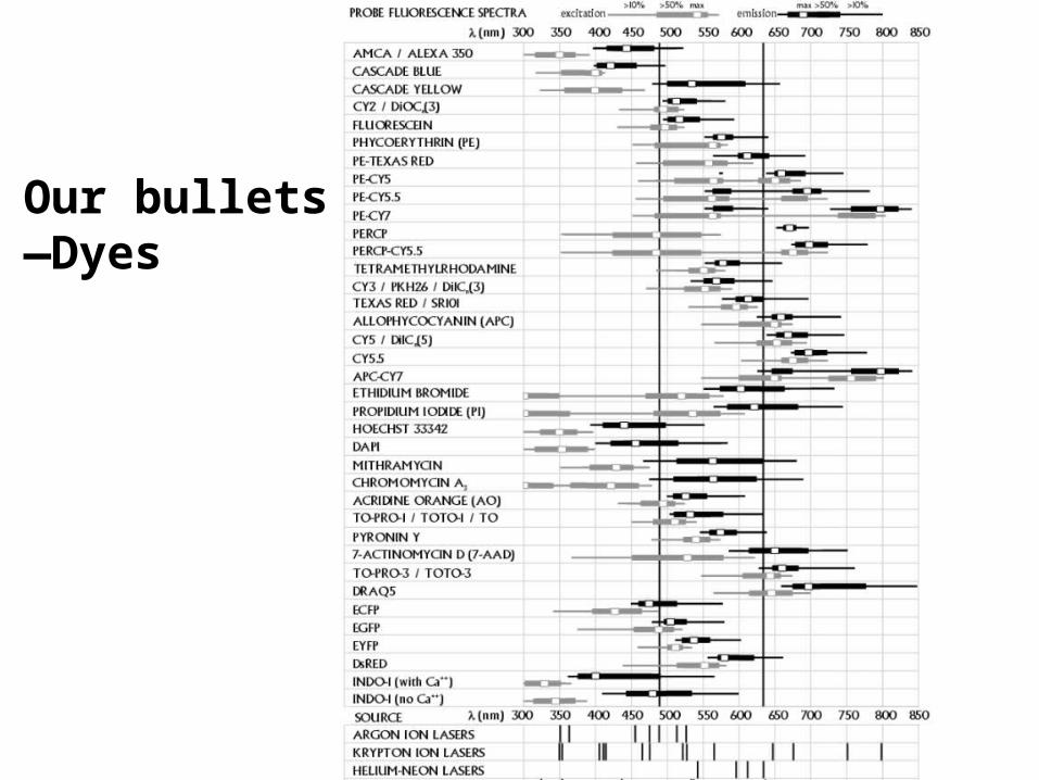
Our bullets—Dyes
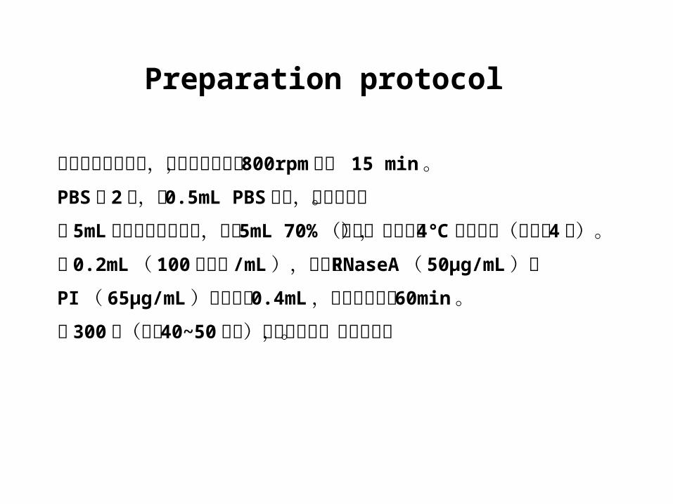
取对数生长期细胞,胰酶适度消化, 800rpm 离心 15 min 。PBS 洗 2 次,加 0.5mL PBS 吹匀,务必吹散。用 5mL 注射器将细胞吸起,打入 5mL 70% (预冷)乙醇中, 4℃
固定过夜(可长至 4 周)。取 0.2mL ( 100 万细胞 /mL ),加入含 RNaseA ( 50µg/mL )和PI ( 65µg/mL )的染色液 0.4mL ,室温避光染色 60min 。用 300 目(孔径 40~50 微米)尼龙网过滤,上机检测。
Preparation protocol
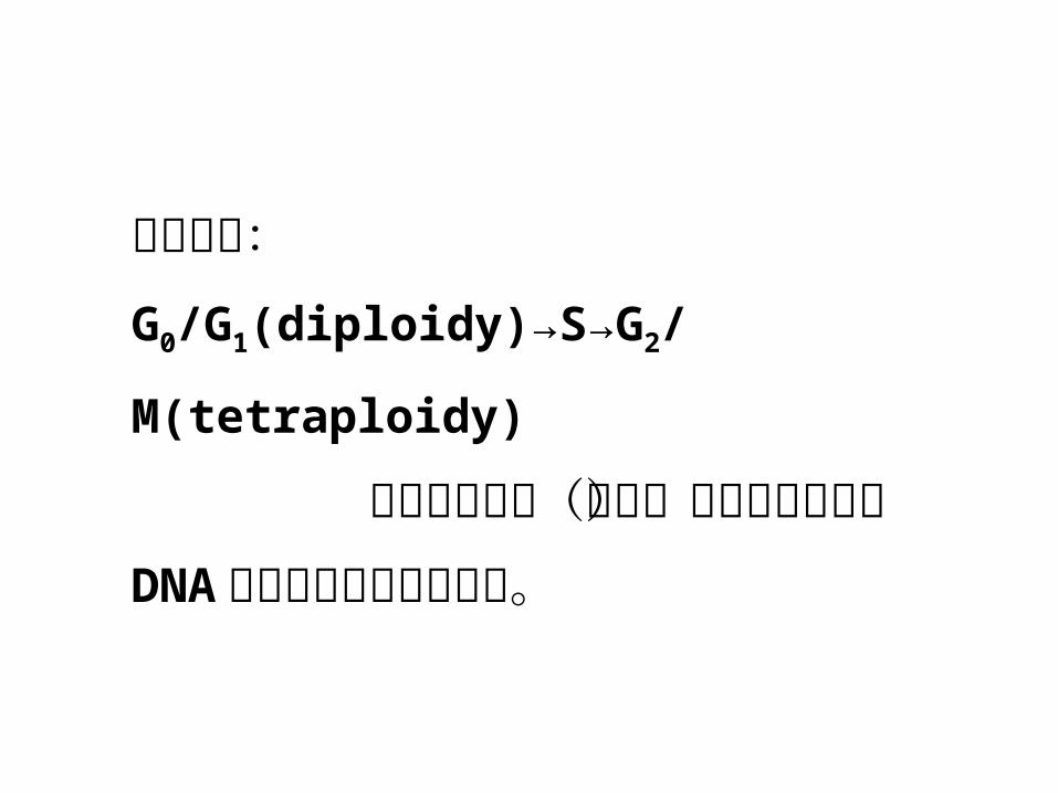
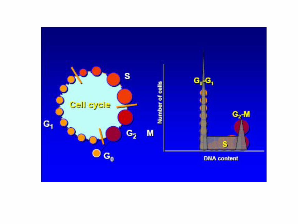
细胞周期:
G0/G1(diploidy)→S→G2/M(tetraploidy)
各期细胞分布(比例)的变化提示细胞 DNA 含量和增值特性的变化。
0 200 400 600 800 1000
PI Fluorescence
Coun
ts0
75
15
0 2
25
30
0
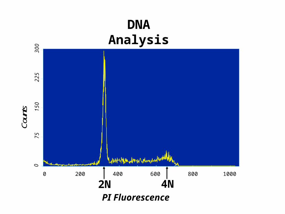
DNA Analysis
2N 4N
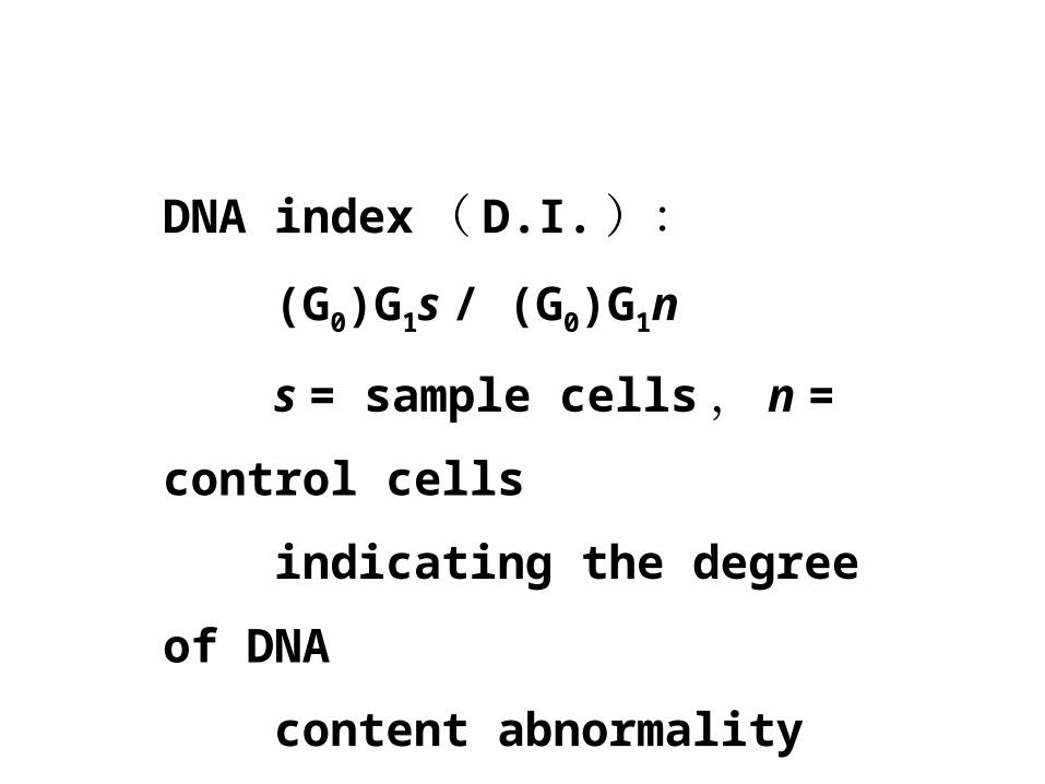
DNA index ( D.I. ):
(G0)G1s / (G0)G1n
s = sample cells , n = control
cells
indicating the degree of DNA
content abnormality
0 200 400 600 800 1000
PI Fluorescence
Coun
ts0
75
15
0 2
25
30
0
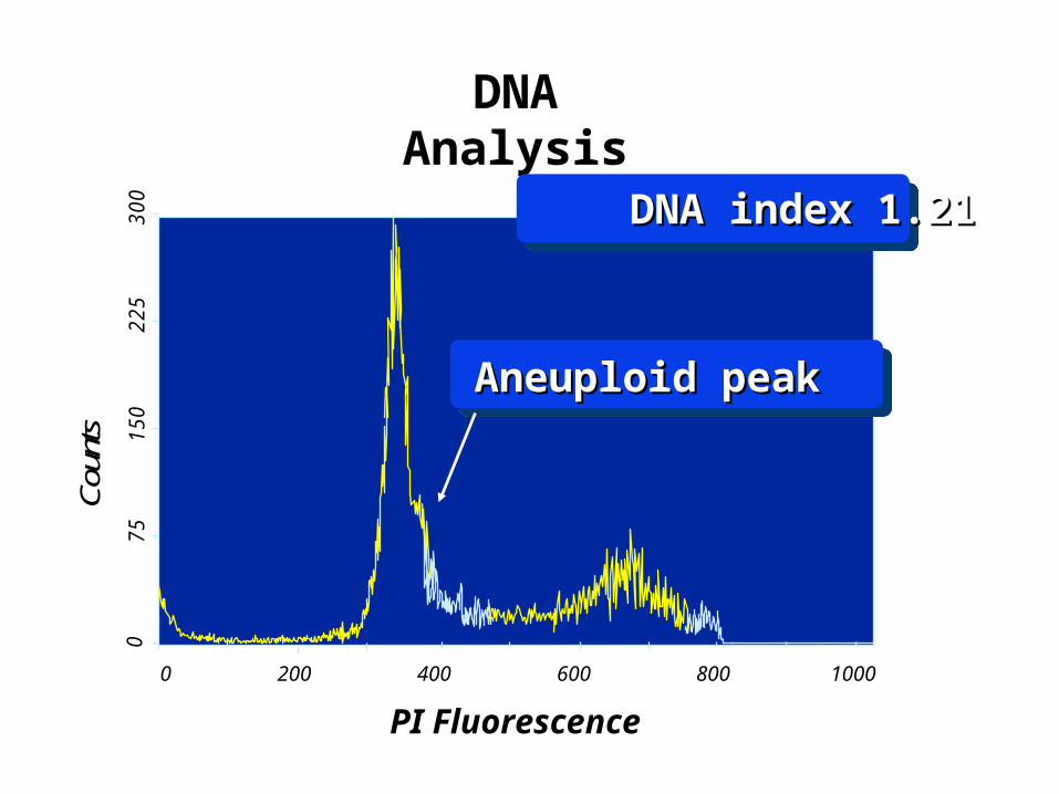
DNA Analysis
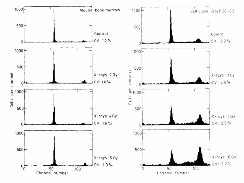
Aneuploid peakAneuploid peak
DNA index 1.21DNA index 1.21
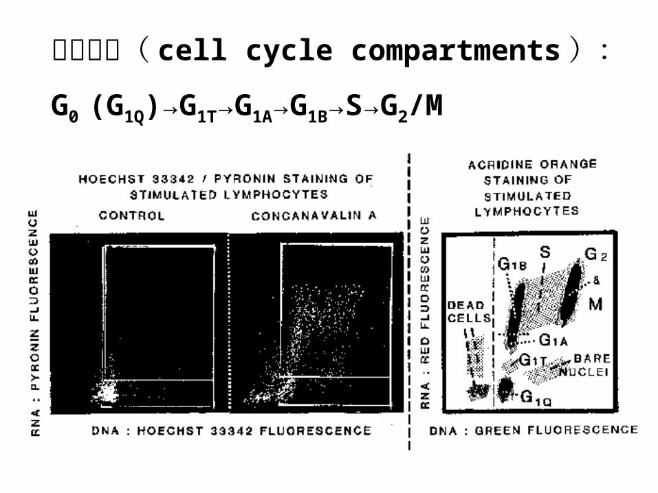
周期细分( cell cycle compartments ):
G0 (G1Q)→G1T→G1A→G1B→S→G2/M
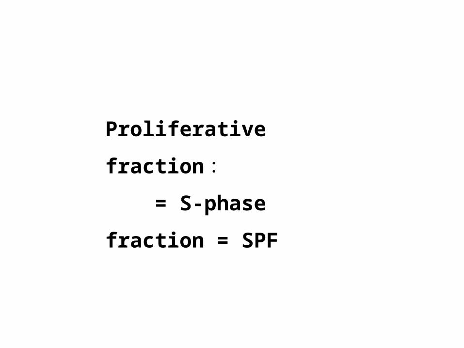
Proliferative fraction : = S-phase fraction = SPF
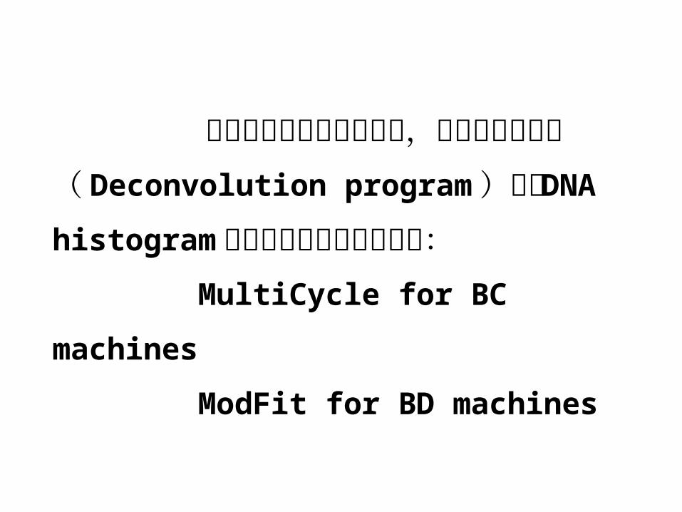
相比目测确定水平门方式,利用去叠合程序( Deconvolution program )拟合DNA histogram 是更为客观和准确的做法: MultiCycle for BC machines
ModFit for BD machines
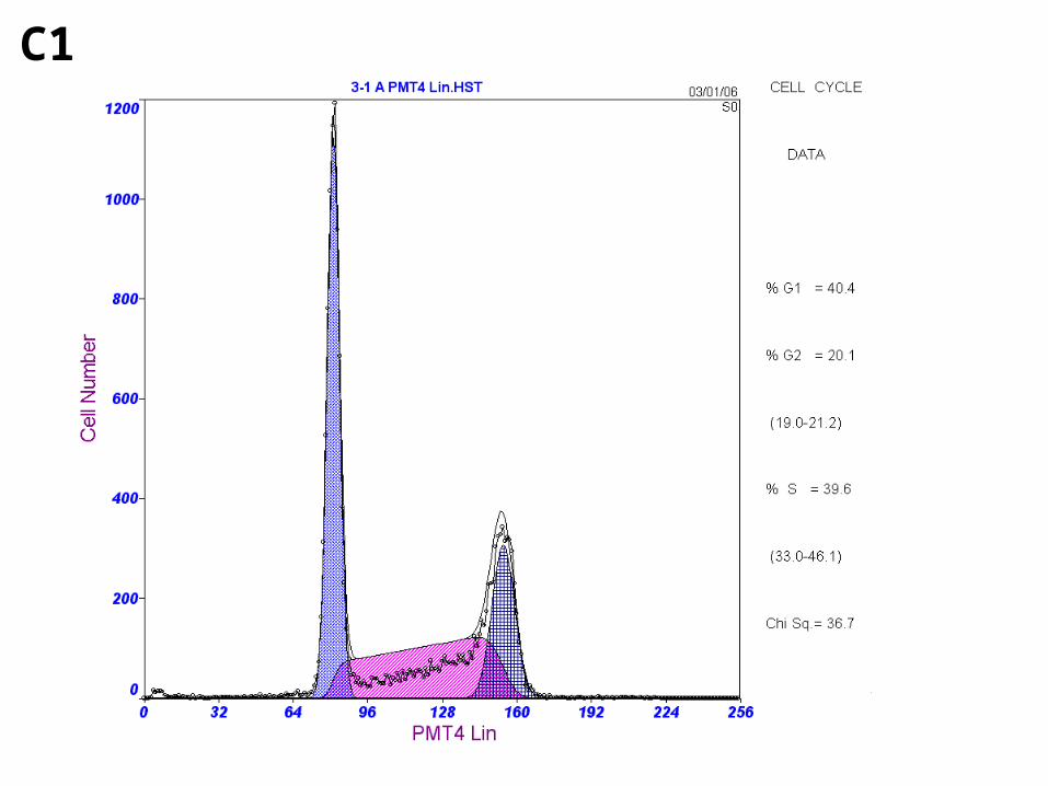
C1
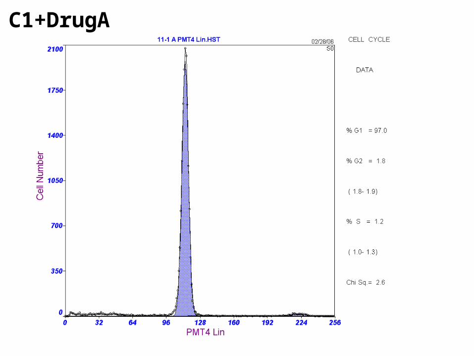
C1+DrugA
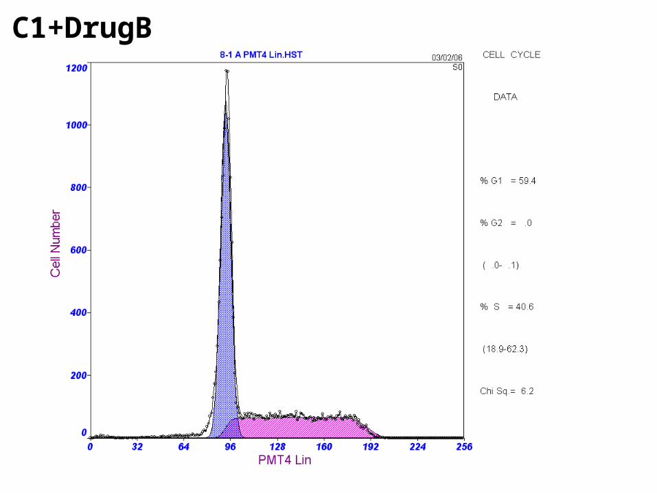
C1+DrugB
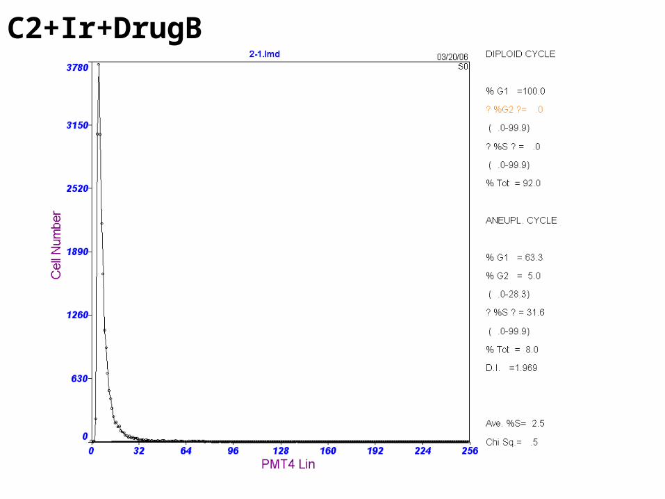
C2+Ir+DrugB
使用去叠合程序进行拟合计算,理想的计数低限为 200 个细胞 /
通道,一般计数不少于 5 万个细胞。
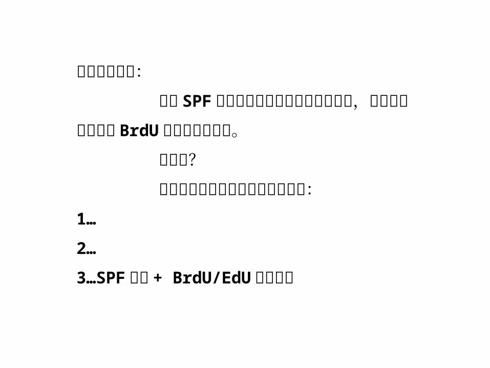
要注意的问题: 单独 SPF 数据不能完全正确反映增殖速率,应结合增值曲线或 BrdU 等数据联合分析。 为什么? 最可靠的细胞增殖测定方法依次是:1…
2…
3…SPF 测定 + BrdU/EdU 掺入测定
其它一些周期相关的 DNA 含量测定应用:mutation detected by the widening of G0/G1
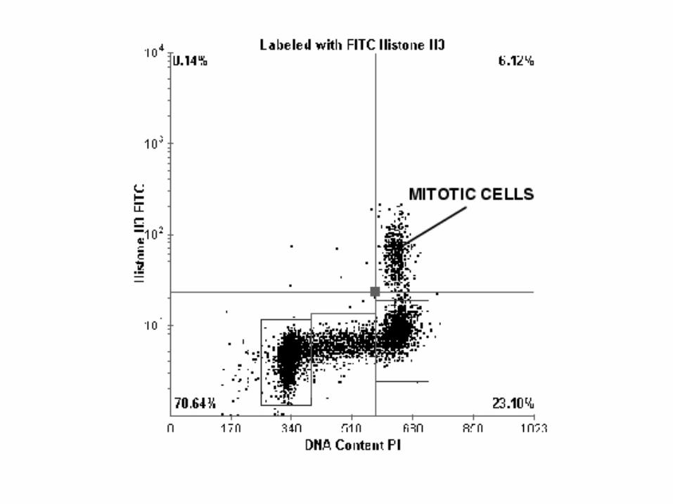
Detecting mitotic cells

细胞凋亡的检测
凋亡( apoptosis )是指在形态学上发生封闭性内部降解的一种主动的细胞死亡过程,由于其发生发展的严格有序性、受调性和精巧性,故在功能学上又被称为程序性细胞死亡( programmed cell deat
h ),是机体细胞在一定条件下激活某种自杀机制,按照一定的调控方式进行的自我清除。
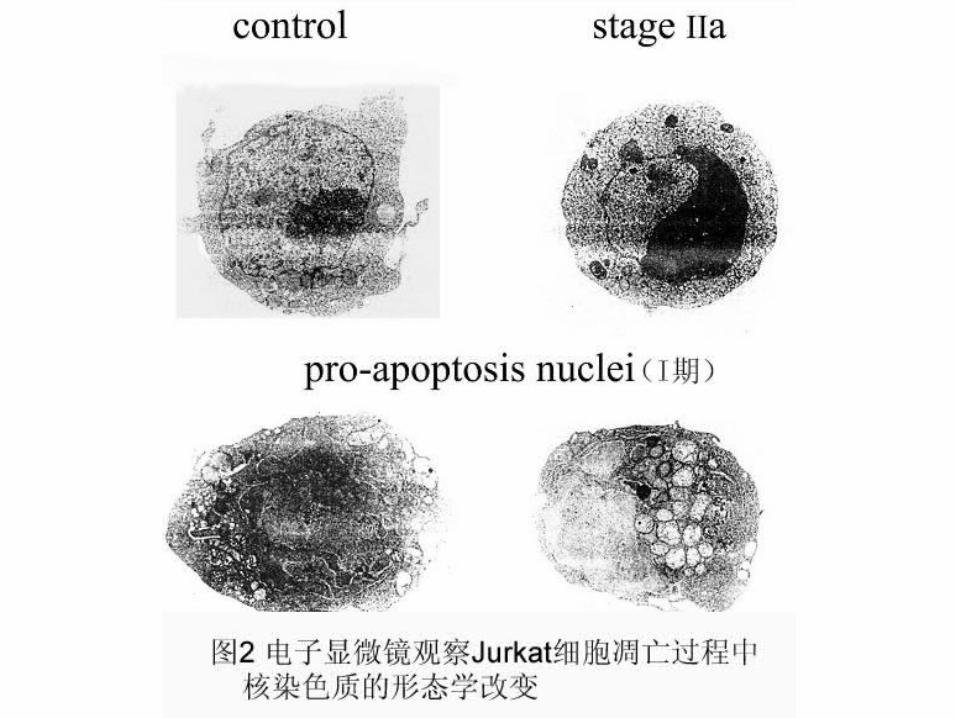
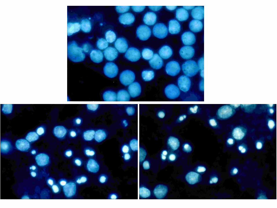
细胞凋亡提出的本质是要区别于细胞坏死,而且其概念最初是来自它们形态学上的差异,因此判断细胞死亡形式的首要标准是细胞的形态学( morphological )变化而非包括 FCM 指标在内的其它指标。
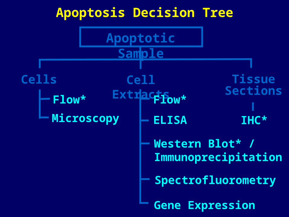
Apoptotic Sample
Cells Cell Extracts Tissue Sections
Flow*
Microscopy
Flow*
ELISA
Western Blot* / Immunoprecipitation
Spectrofluorometry
Gene Expression
IHC*
Apoptosis Decision Tree
MM bpbp
200200
100100
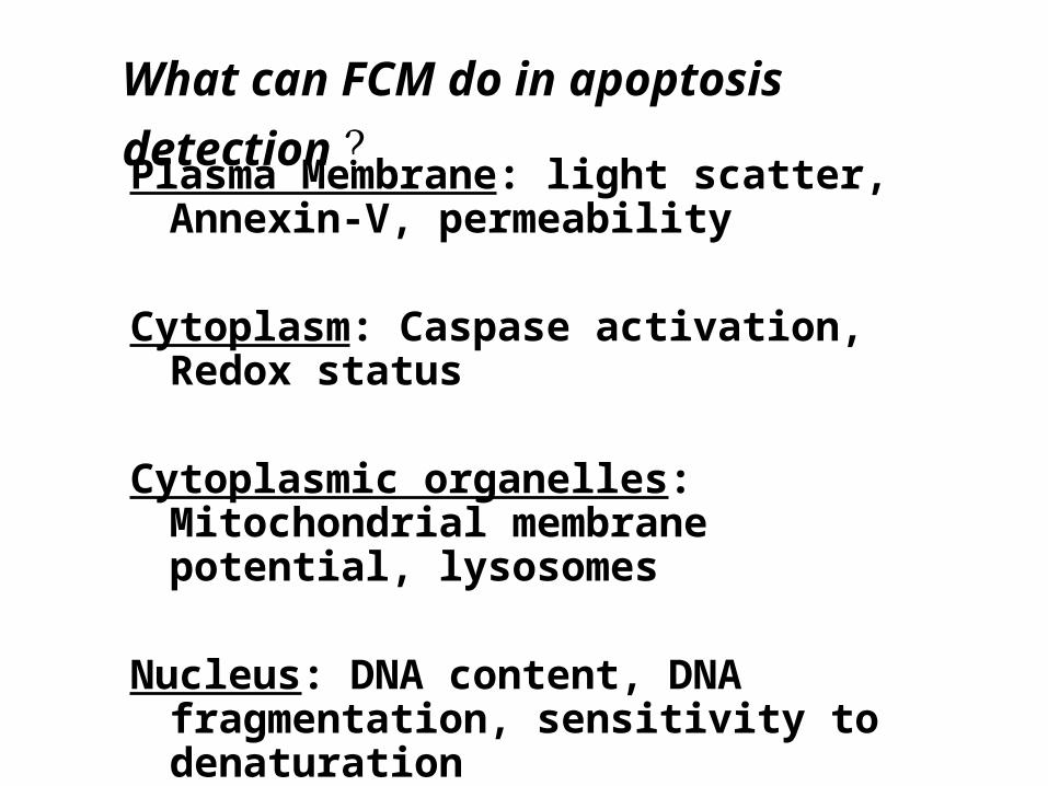
Plasma Membrane: light scatter, Annexin-V, permeability
Cytoplasm: Caspase activation, Redox status
Cytoplasmic organelles: Mitochondrial membrane potential, lysosomes
Nucleus: DNA content, DNA fragmentation, sensitivity to denaturation
What can FCM do in apoptosis detection ?
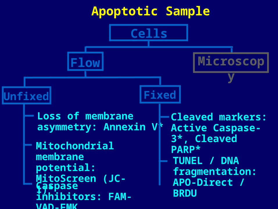
Cells
Flow Microscopy
Unfixed Fixed
Loss of membrane asymmetry: Annexin V*
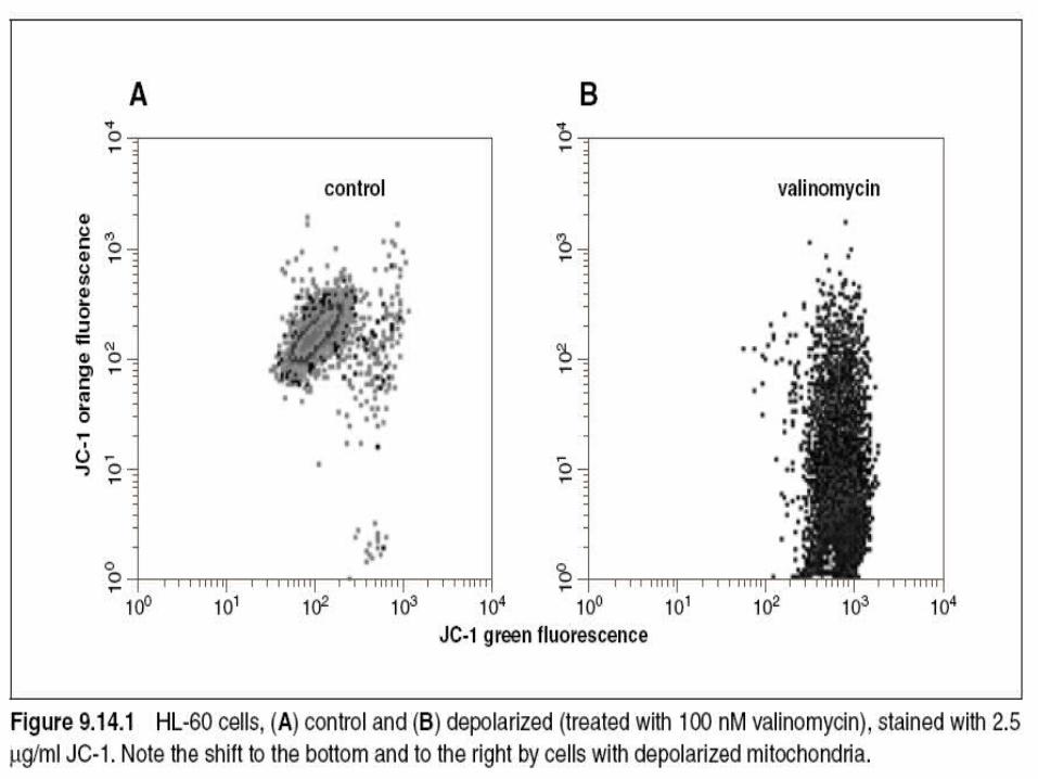
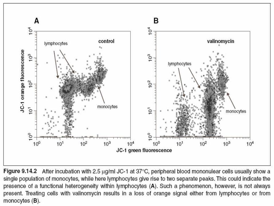
Mitochondrialmembrane potential: MitoScreen (JC-1)*
Cleaved markers:Active Caspase-3*, Cleaved PARP*
TUNEL / DNA fragmentation: APO-Direct / BRDU
Apoptotic Sample
Caspase inhibitors: FAM-VAD-FMK
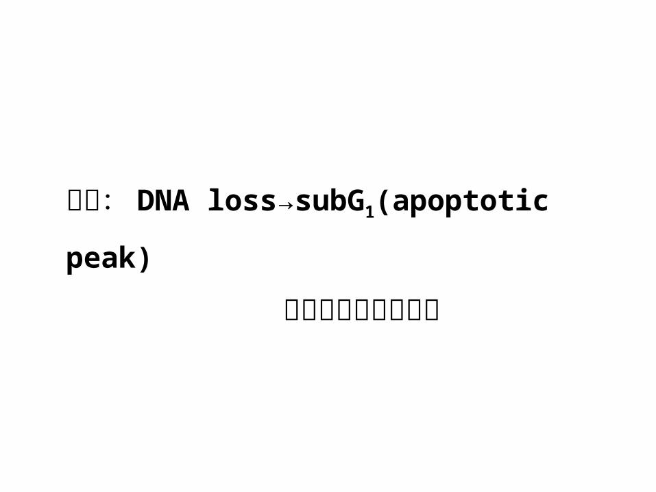
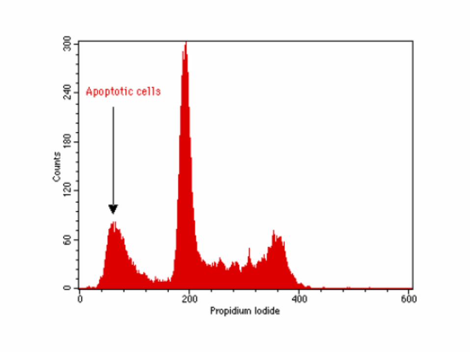
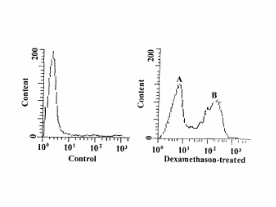
凋亡: DNA loss→subG1(apoptotic
peak)
提示凋亡细胞的出现
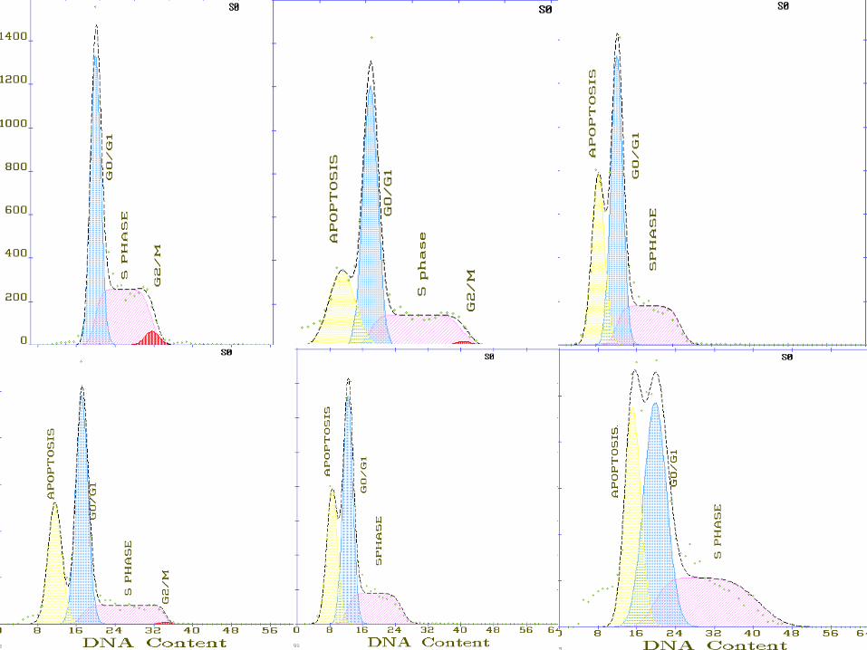
要注意的问题: 以 PI 单染法进行凋亡细胞定量是最经济的方法,但其漏检和错检率都很高。漏检主要发生在 S 期和 G2/M
期的凋亡细胞,这些细胞即使有 DNA 含量降低,但其存有的 DNA 含量仍不低于二倍体细胞,因而不会出现
subG1 。错检的原因主要是细胞碎片和小颗粒也表现出低
荧光。
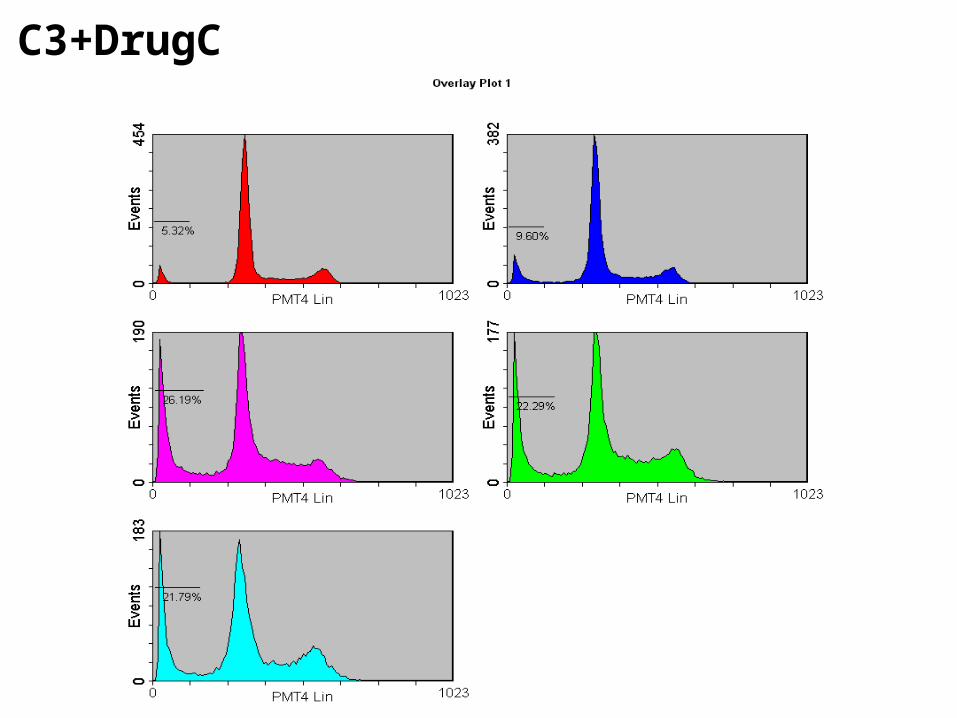
C3+DrugC
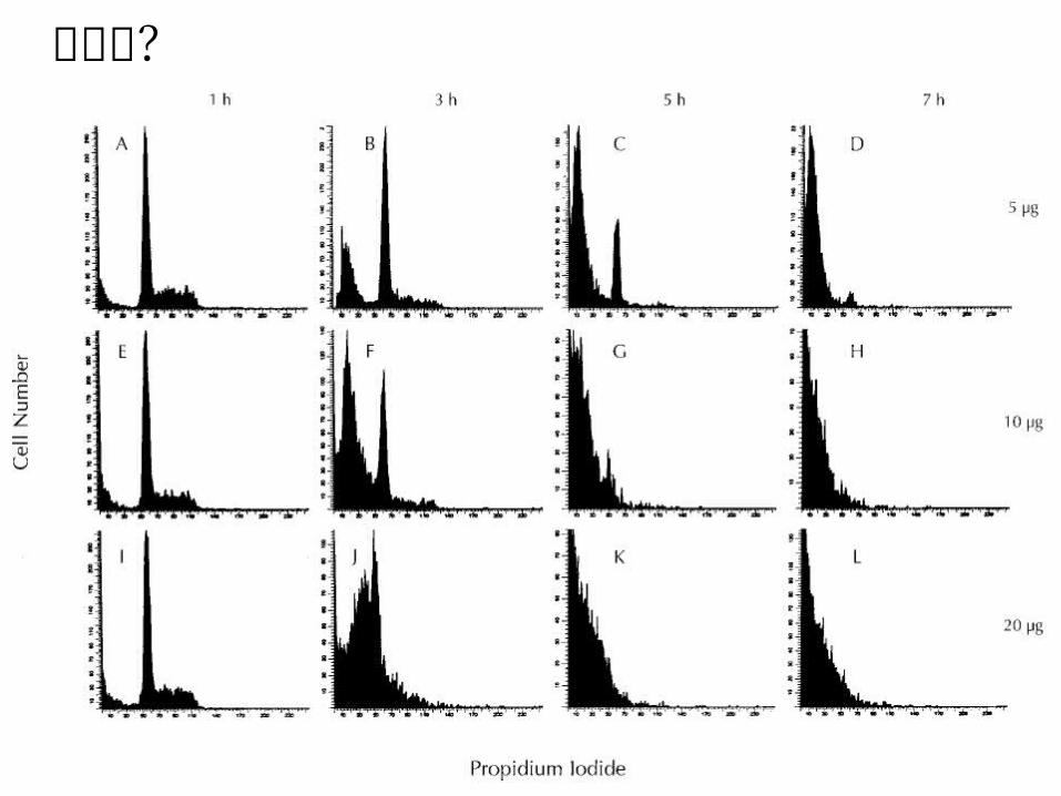
这是啥?

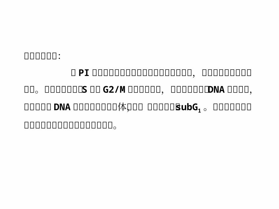
要注意的问题:
单细胞悬液的准备容易引起 subG1假
阳性和假阴性,容易引起细胞粘连。考虑降低胰酶的浓度,注意分散细胞时操作的柔和性,注意全程上清液的收集。
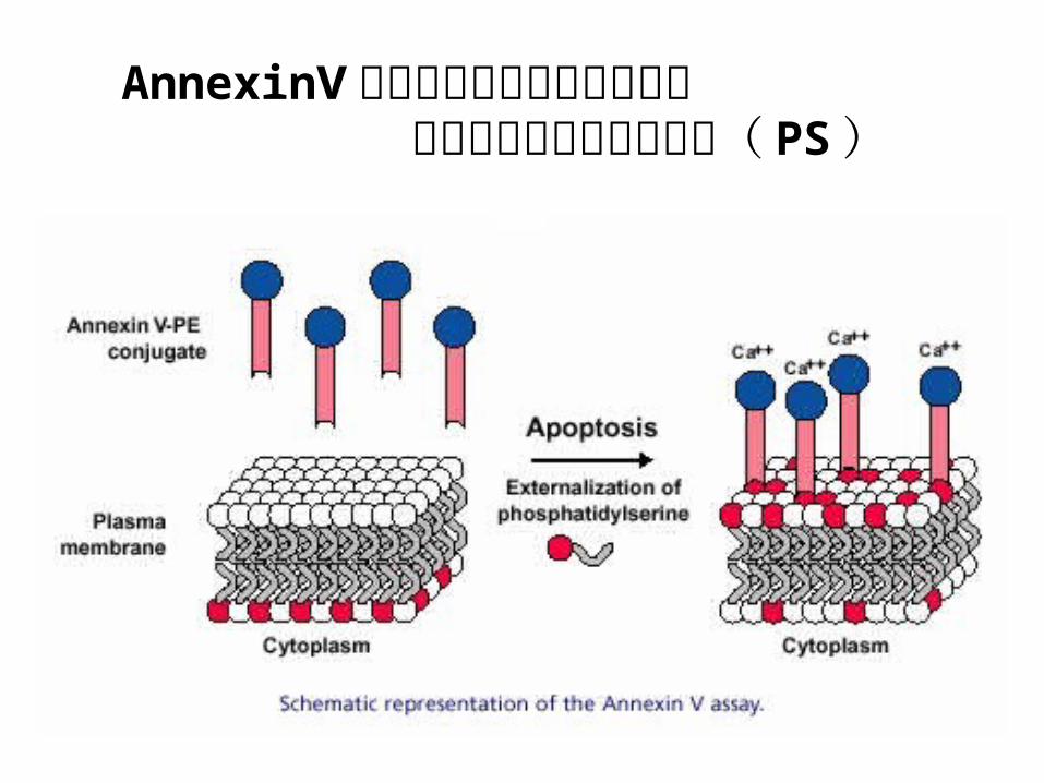
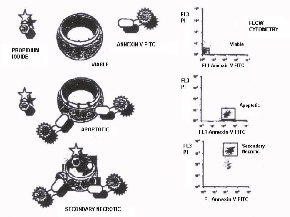
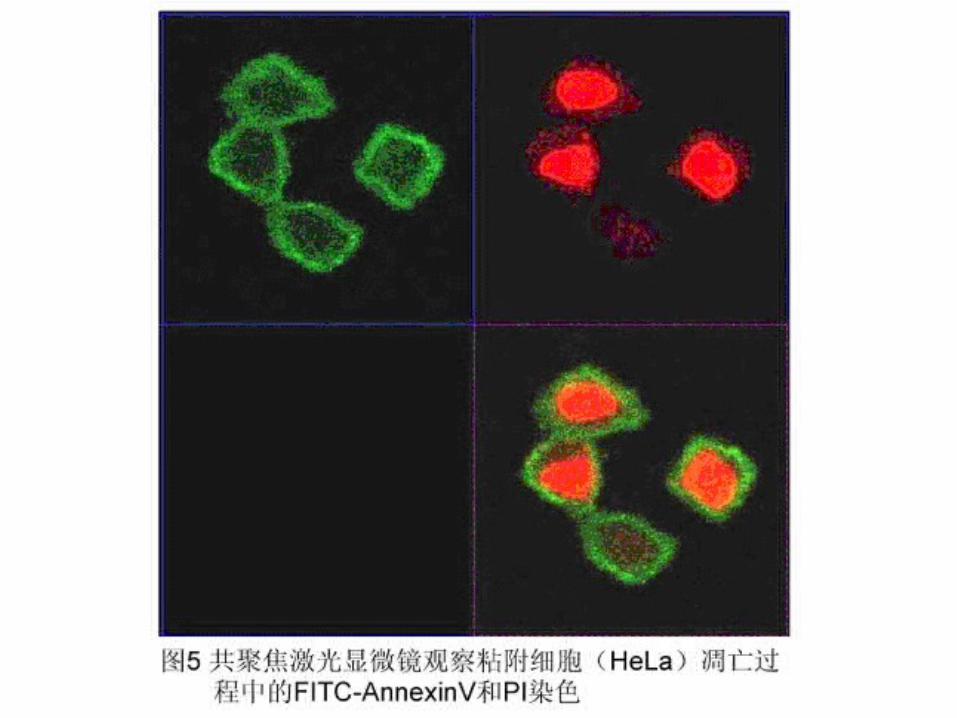
AnnexinⅤ磷脂结合蛋白作为探针识别 细胞膜表面磷脂酰丝氨酸( PS )
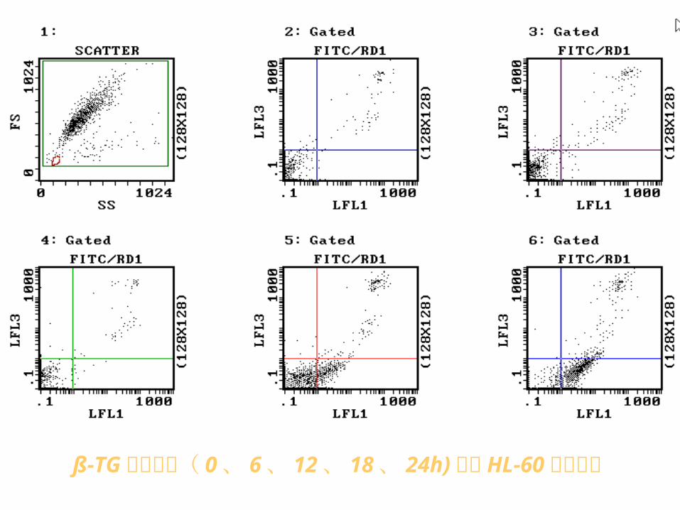
ß-TG 不同时间( 0 、 6 、 12 、 18 、 24h)诱导 HL-60 细胞凋亡
Q :同样是 PI 染色, subG1 ( AP )和
AV-PI 检测有什么不同?为什么?
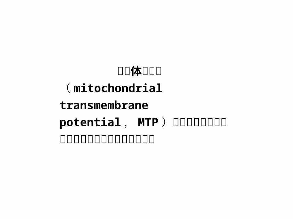
线粒体膜电位( mitochondrial transmembrane
potential , MTP )被认为是多数凋亡模型中胞内发生的早期重要事件
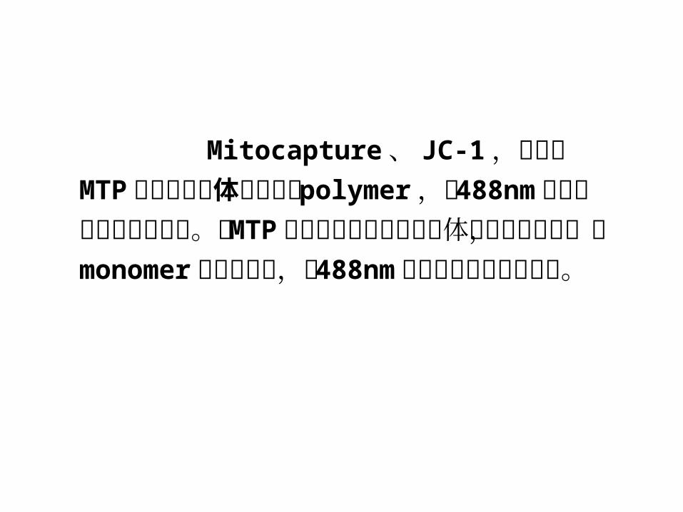
Mitocapture 、 JC-1 ,聚集在 MTP 正常的线粒体膜上形成 polymer ,被 488nm 光激发后发出橙色荧光。当MTP丢失时染料分子从线粒体膜上解离下来,以monomer游离于胞浆,被 488nm 光激发后发出绿色荧光。
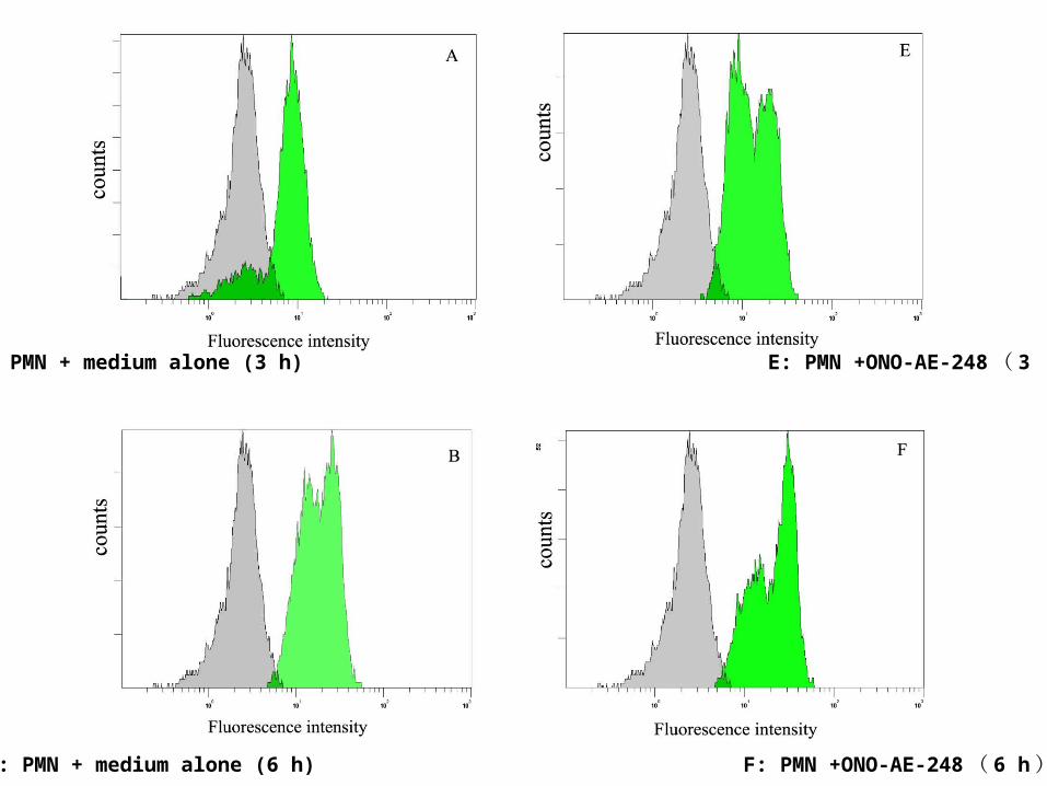
A: PMN + medium alone (3 h) E: PMN +ONO-AE-248 ( 3 h)
B: PMN + medium alone (6 h) F: PMN +ONO-AE-248 ( 6 h)
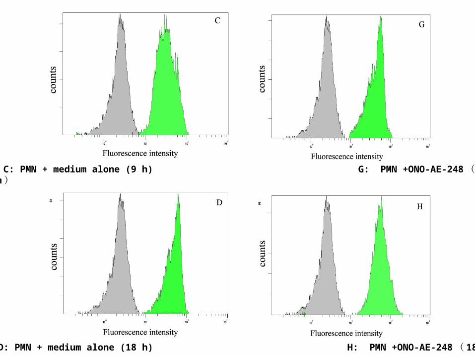
C: PMN + medium alone (9 h) G: PMN +ONO-AE-248 ( 9 h)
D: PMN + medium alone (18 h) H: PMN +ONO-AE-248 ( 18 h)
MTP 是电势能指标,必须是细胞在活性状态下被染色→不能固定细胞→表抗可同标,内抗不能同标。
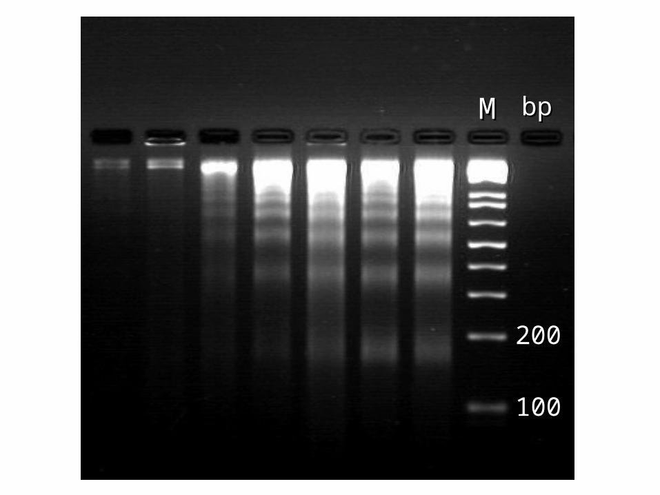
细胞核内 DNA 断裂标记分析( TUNEL 法, ISNT法) 细胞凋亡过程中细胞核内的 DNA 出现断裂,断裂点暴露出DNA 分子的残端,用末端转移酶( TdT )或者缺口连接酶都可以标记上可以识别的化学物质。标记 DAN 断裂的 3’—羧基末端,常采用由 DNA聚合酶 I催化的原位缺口转移( in situ nick translation ,ISNT ) 或 用 TdT 介导的 dUTP 原位缺口末端标记( terminal deoxynucleotidyl transferase mediated dUTP nick end labeling , TUNEL )技术。 ISNT 和 TUNEI 法均可进行间接或直接标记。
直接法:标记物常为异硫氰酸荧光素 FITC—dUTP 。
间接法:标记物通常是生物素化脱氧三磷酸尿苷 (biotin—dUTP) 或地高辛 Dig—dUTP 等;间接标记还需链霉亲合素 FITC 或抗 Dig—FITC ,使标记反应倍增,其灵敏度较高,但比直接标记复杂。
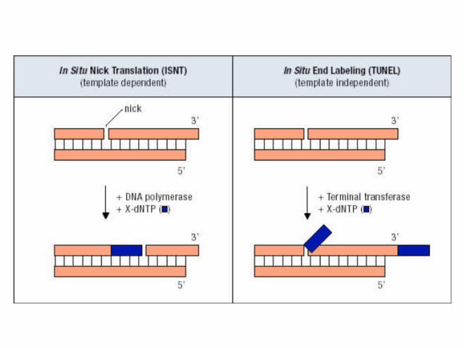
注意: TUNEL 法不能区分 DNA 断端的性质, 因此无法单独鉴别坏死和凋亡

APO2.7 是一种分子量为 38Kda 的线粒体膜蛋白,仅表达在凋亡细胞的线粒体膜表面, 2.7A6A3 单抗能与之识别,是细胞凋亡的早期信号。用经 Digitonin透膜的细胞与 APO2.7 的单抗反应后 FCM 检测,就可反应细胞凋亡的状态。
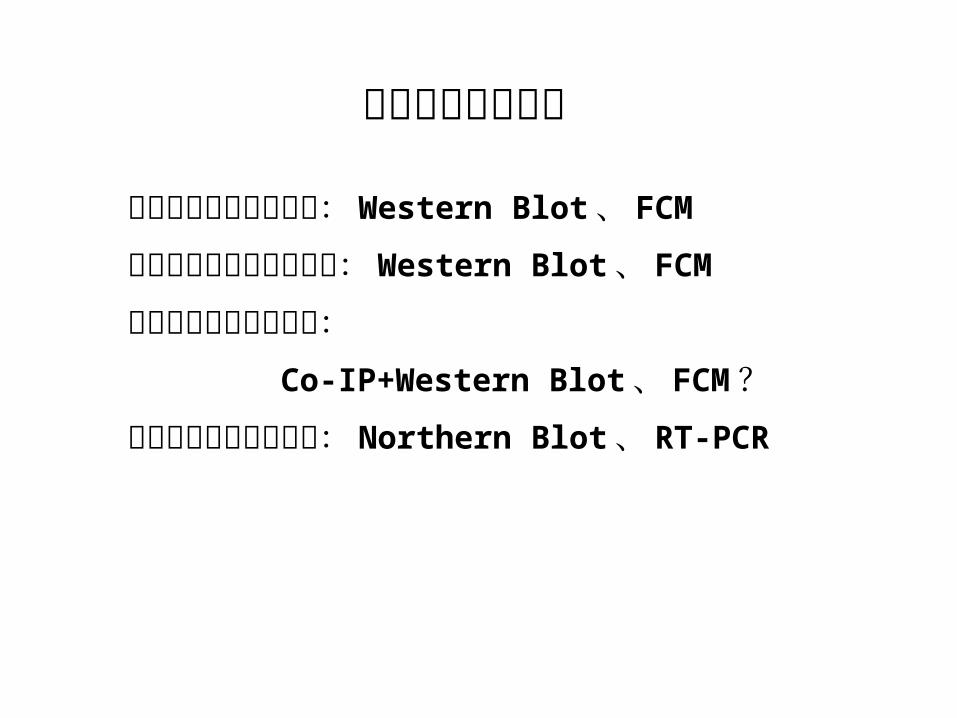
细胞凋亡分子检测
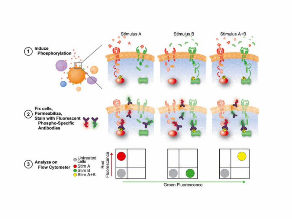
效应蛋白的活性和降解: Western Blot 、 FCM
信号通路蛋白磷酸化水平: Western Blot 、 FCM
信号通路蛋白激酶活性: Co-IP+Western Blot 、 FCM ?蛋白编码基因表达水平: Northern Blot 、 RT-PCR
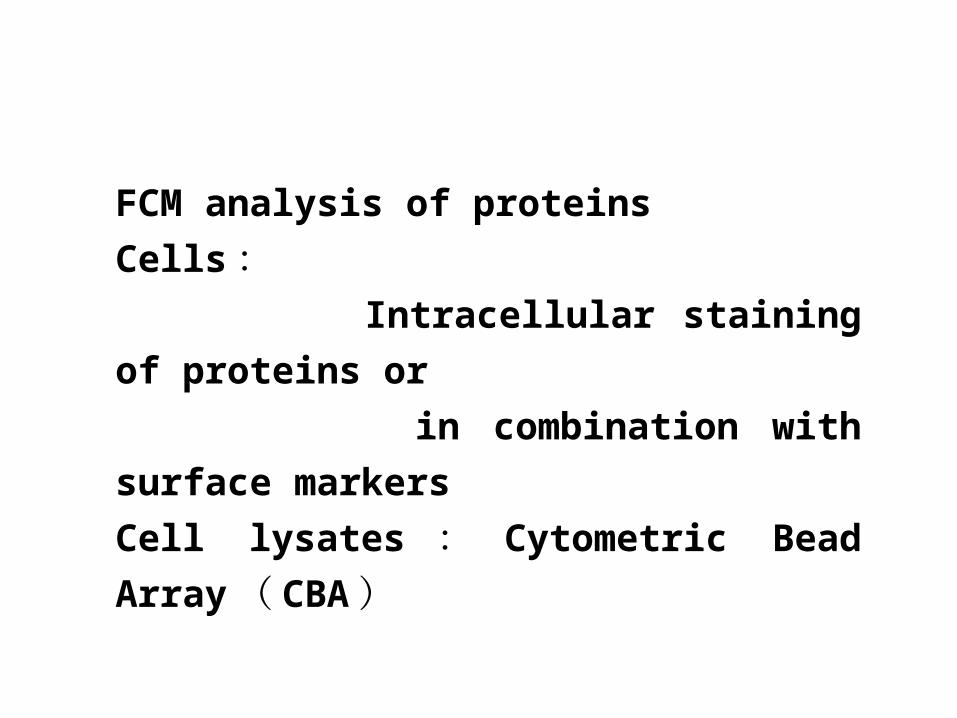
FCM analysis of proteins
Cells : Intracellular staining of proteins or
in combination with surface markers
Cell lysates : Cytometric Bead Array ( CB
A )
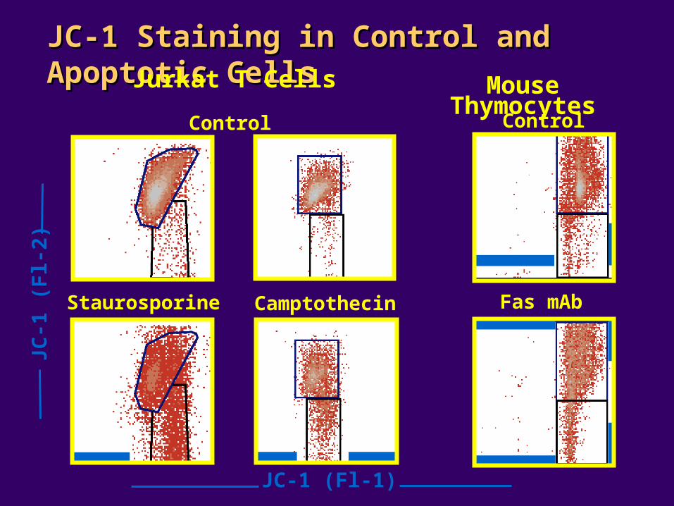
JC-1 Staining in Control and Apoptotic Cells JC-1 Staining in Control and Apoptotic Cells
JC-1 (Fl-1)
Jurkat T Cells Mouse Thymocytes
Control
Staurosporine Camptothecin Fas mAb
R1
R2
R1
R2
R1
R2
R1
R2
R1
R2
R1
R2
Control
JC-1
(F
l-2)
Co
ntr
ol
Cam
pto
thec
in4
µM
, 4
hActive Caspase-3
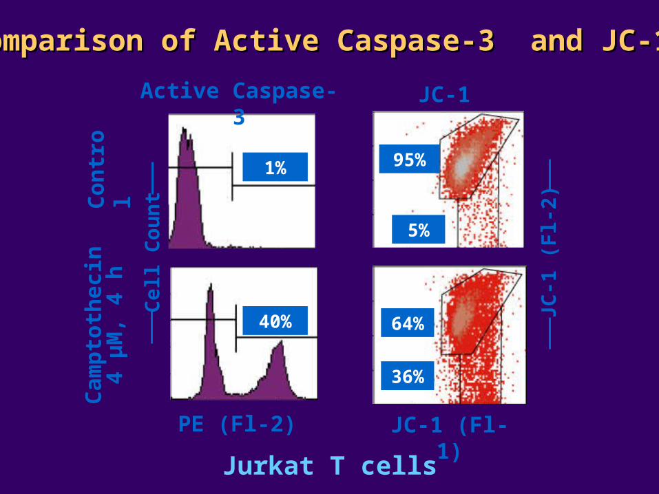
Comparison of Active Caspase-3 and JC-1Comparison of Active Caspase-3 and JC-1
JC-1 (Fl-1)
JC-1
(F
l-2)
1%
40%
95%
5%
36%
64%
JC-1
PE (Fl-2)
Jurkat T cells
Cel
l Co
un
t
Co
ntr
ol
Cam
pto
thec
in4
µM
, 4
h
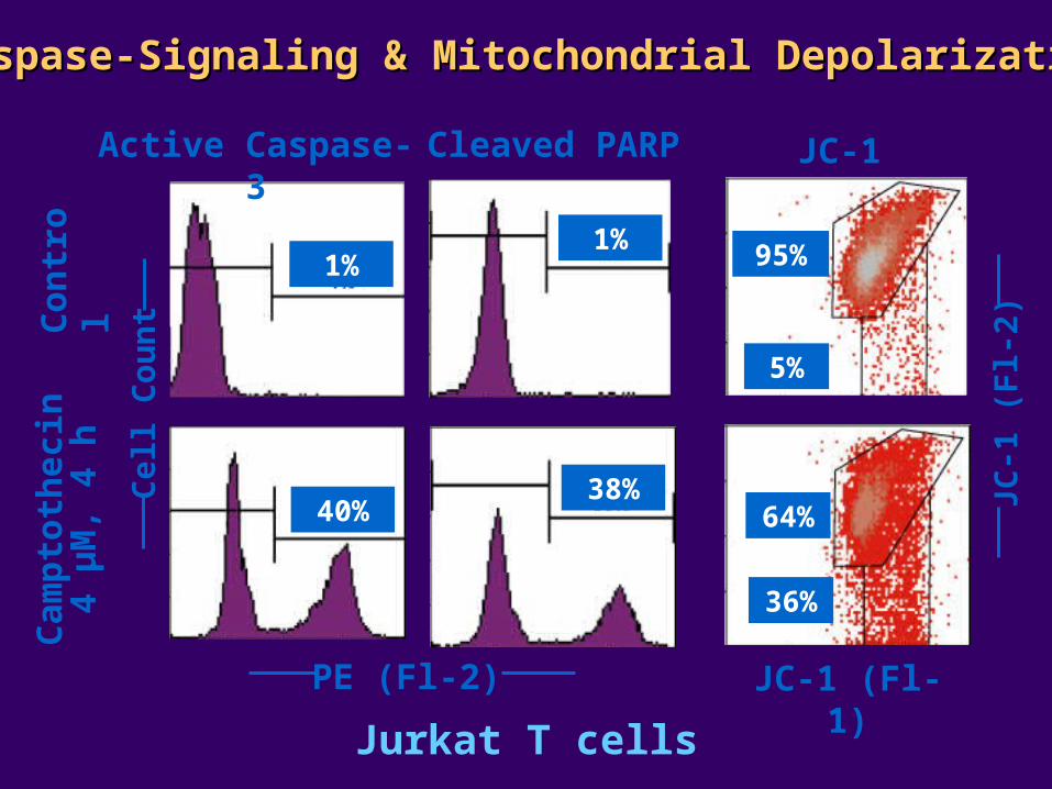
Active Caspase-3 Cleaved PARP
Caspase-Signaling & Mitochondrial DepolarizationCaspase-Signaling & Mitochondrial Depolarization
JC-1 (Fl-1)
JC-1
(F
l-2)
1%1%
40%38%
95%
5%
36%
64%
JC-1
PE (Fl-2)
Jurkat T cells
Cel
l Co
un
t
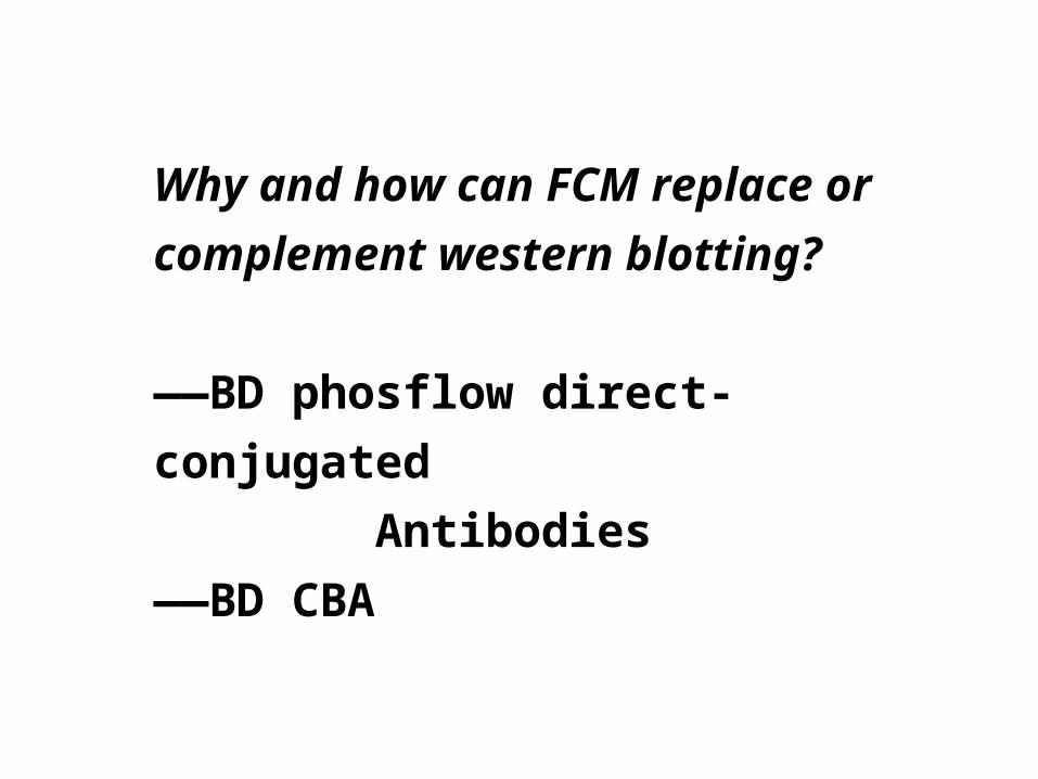
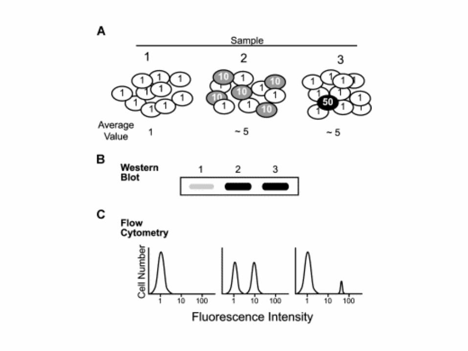
Why and how can FCM replace or
complement western blotting?
——BD phosflow direct-conjugated
Antibodies
——BD CBA
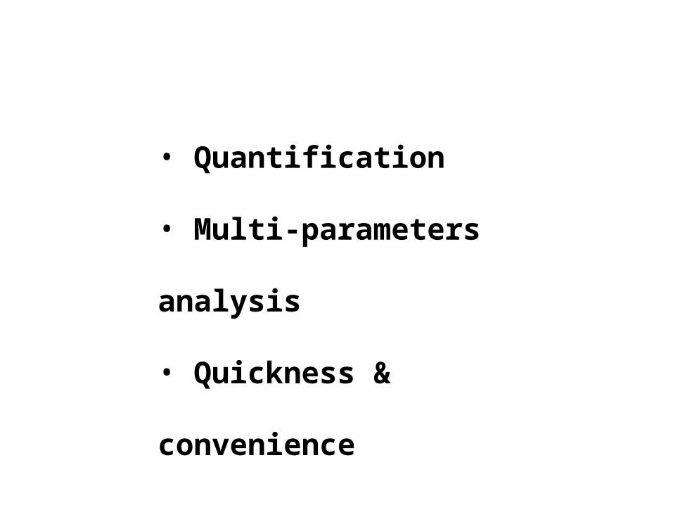
• Quantification
• Multi-parameters analysis
• Quickness & convenience
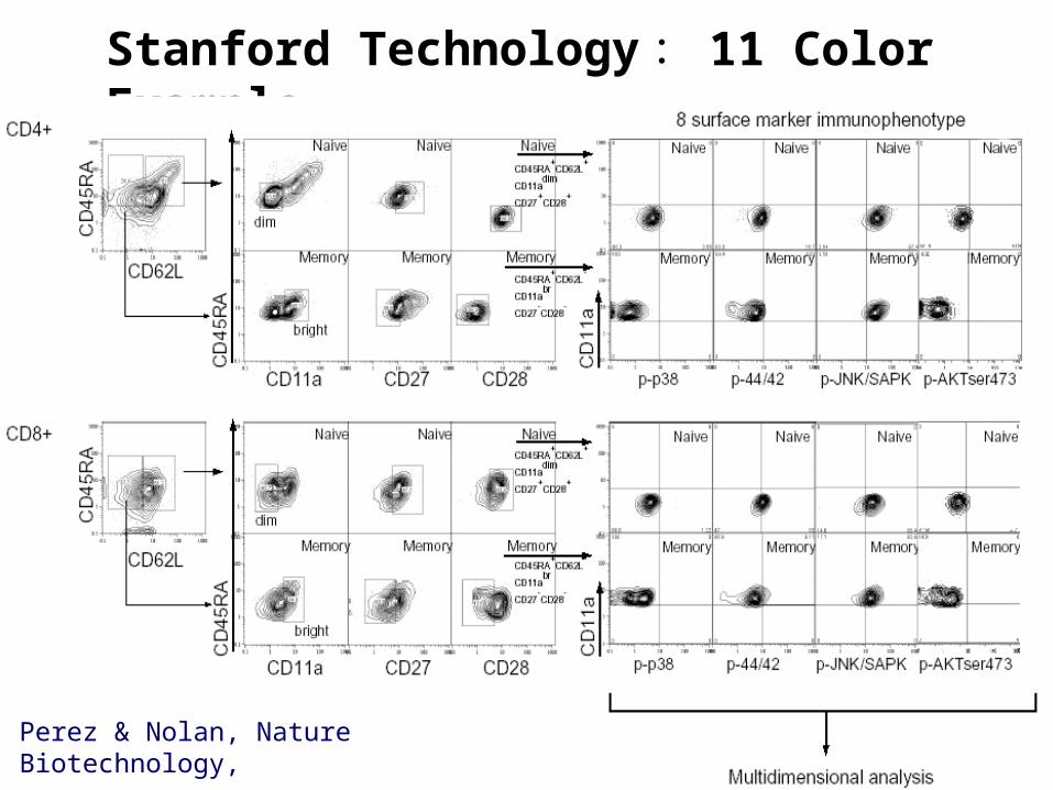
Stanford Technology : 11 Color Example
Perez & Nolan, Nature Biotechnology,
2002:20, 155-162
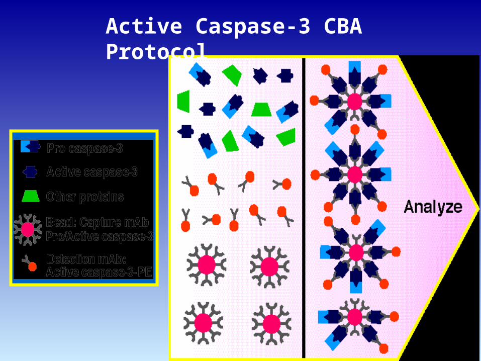
Active Caspase-3 CBA Protocol
CBA
Classical Flow Cytometry Western Blot
32 kDa
17 kDa
Active Caspase-3–PE Pro/Active Caspase-3 pAb
CamptothecinControl CamptothecinControl
Ce
ll C
ou
nt
ControlCamptothecin
Active Caspase-3Standard Curve
104
103
102
10
1104103102101
Protein (ng)
MF
I
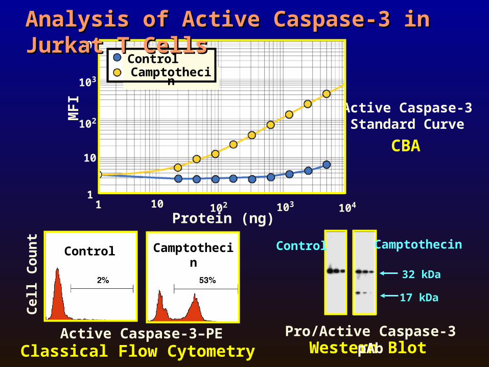
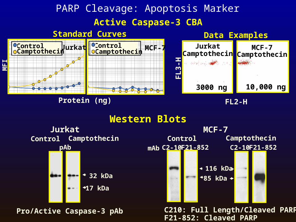
Analysis of Active Caspase-3 in Jurkat T CellsAnalysis of Active Caspase-3 in Jurkat T Cells
Western Blots
116 kDa
85 kDa
CamptothecinControlmAb F21-852C2-10 F21-852C2-10
Active Caspase-3 CBA
C210: Full Length/Cleaved PARPF21-852: Cleaved PARP
Protein (ng)
Data Examples
FL2-H
FL
3-H
MCF-7Camptothecin
JurkatCamptothecin
3000 ng 10,000 ng
MCF-7
Pro/Active Caspase-3 pAb
CamptothecinControl
32 kDa
17 kDa
Jurkat
Standard Curves
MF
I
JurkatControlCamptothecin MCF-7Control
Camptothecin
pAb
PARP Cleavage: Apoptosis Marker
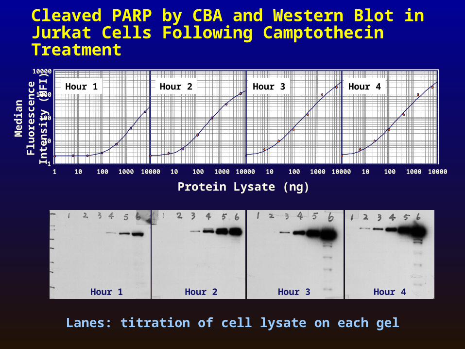
Cleaved PARP by CBA and Western Blot in Jurkat Cells Following Camptothecin Treatment
Hour 1 Hour 2 Hour 3 Hour 4
10
100
1000
1 100 1000 100001
10 100 1000 10000 10 100 1000 1000010 10 100 1000 10000
10000
Protein Lysate (ng)
Med
ian
Flu
ore
scen
ce
Inte
nsi
ty (
MF
I) Hour 1 Hour 2 Hour 3 Hour 4
Lanes: titration of cell lysate on each gel
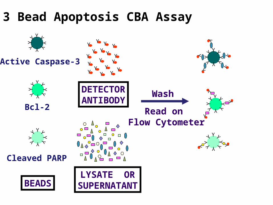
3 Bead Apoptosis CBA Assay Y
YYY
Y
YY
Y
Y
YYY
Y
YY
Y
Y
Y
YY
Y
YY
Y
DETECTORANTIBODY
LYSATE ORSUPERNATANTBEADS
YY
YY
Y
YY
YY
YY
Y
YY
YY
YY
Y
Y
YY
Y
YY
Y
Y Y
Y
YYY
YYY
Y
Y
YY
Y
YY
YY
Y
YY
Y
Y
Y
Y
Y
Wash
Read on Flow Cytometer
Active Caspase-3
Bcl-2
Cleaved PARP
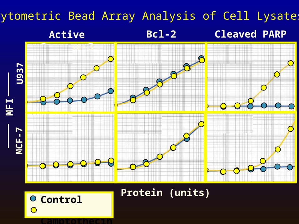
Active Caspase-3 Bcl-2 Cleaved PARP
U93
7M
CF
-7Cytometric Bead Array Analysis of Cell Lysates
MF
I
Protein (units)Control Camptothecin
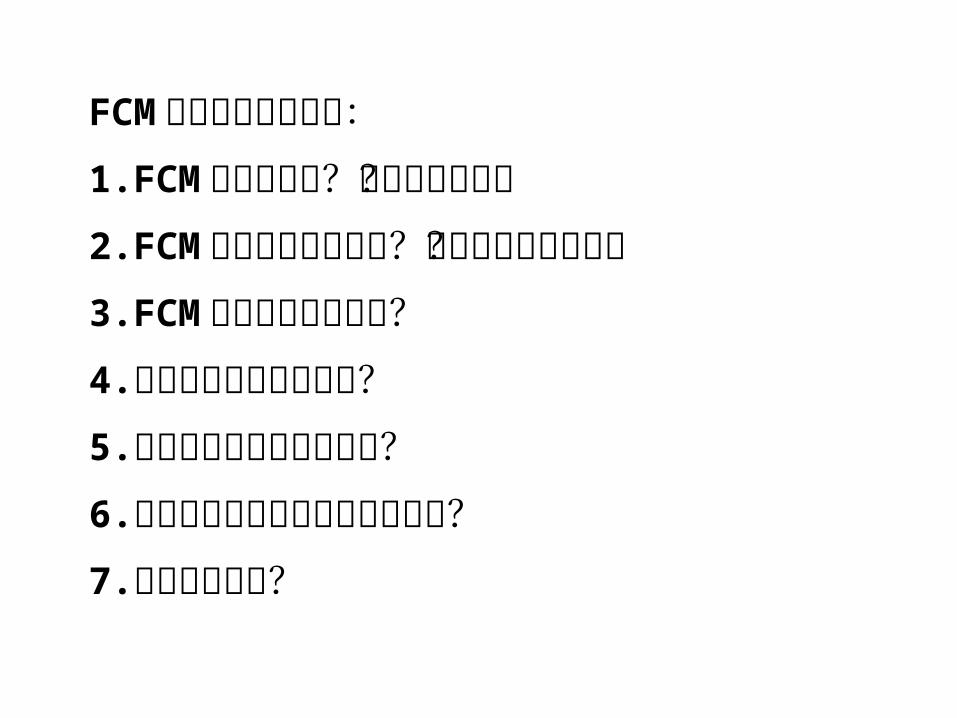
FCM 中需要注意的地方:1. FCM 可以做什么?不可以做什么?2. FCM 是否可以单独使用?需与什么设备共事?3. FCM需要进行什么准备?4.指标和荧光素如何选择?5.如何进行收集才是正确的?6.如何选择正确的分析方案和工具?7.如何判读数据?
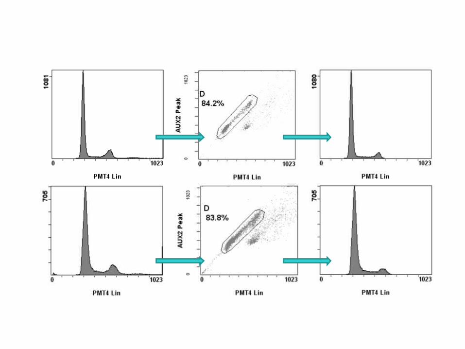
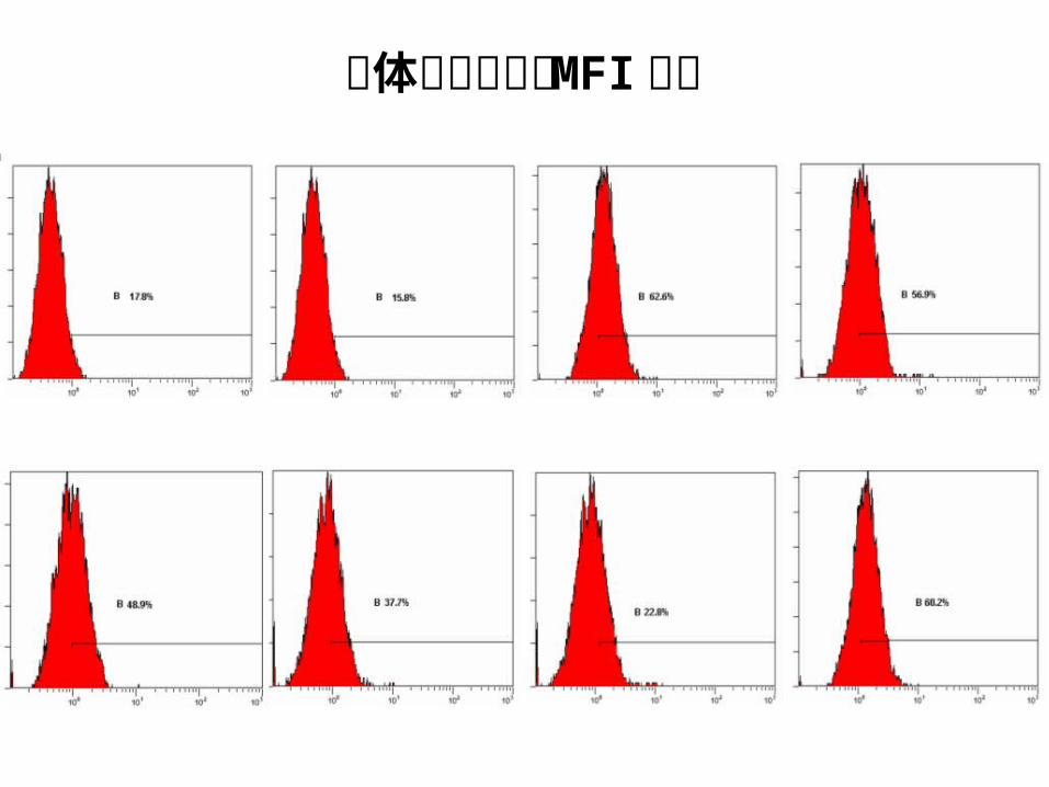
整体峰移只能用 MFI评价
背景群落应予跟踪疑似特异峰可以在斜率折点设门
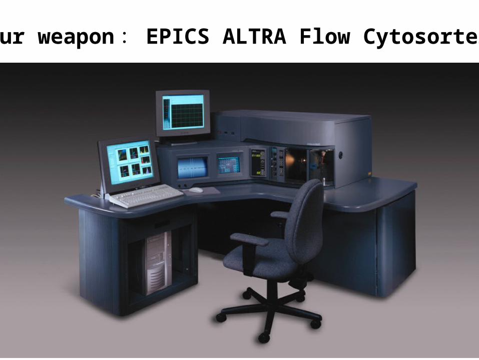
Our weapon : EPICS ALTRA Flow Cytosorter
References
Practical Flow Cytometry (4th ed.)Howard M. Shapiro, 2003
Flow Cytometry, A Practical Approach (2nd ed.)M. G. Ormerod, 1994
Flow Cytometry and Sorting (2nd ed.)M. R. Melamed, 1990

Acknowledgement
赵静、谭文军、薛向军、陈志忠BECKMAN COULTER公司流式应用工程师
Christopher “Kit” SnowPrincipal Staff Scientist Beckman Coulter Inc.


![L L亡t亡q亡『.]l亡TllnllT亡 亡エイズ 」 ..}tJJL ILlr.IL …...L L亡t亡q亡『.]l亡TllnllT亡 亡エイズ 」 ..}tJJL_ILlr.IL-.雷.JL- ,. LT 正しい理解のために](https://img.pdfslide.tips/doc/110x75/5f44ea357d417871ce4740ab/l-ltqltllnllt-i-tjjl-illril-l-ltqltllnllt.jpg)


![Tauroursodeoxycholic acid suppresses endoplasmic reticulum ...xbyxb.csu.edu.cn/xbwk/fileup/PDF/2015111165.pdf · 够激活下游通路,导致内质网应激介导的细胞凋 亡[8]](https://img.pdfslide.tips/doc/110x75/603588612683770b490efb3e/tauroursodeoxycholic-acid-suppresses-endoplasmic-reticulum-xbyxbcsueducnxbwkfileuppdf.jpg)