Embed Size (px)
Citation preview
Patologia esofagului
şi stomacului.
Болезни пищевода
и желудка.
Pathology of
esophagus
and stomach.
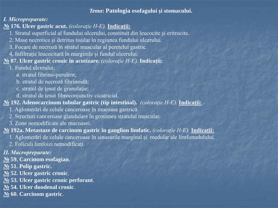
Tema: Patologia esofagului şi stomacului.
I. Micropreparate:
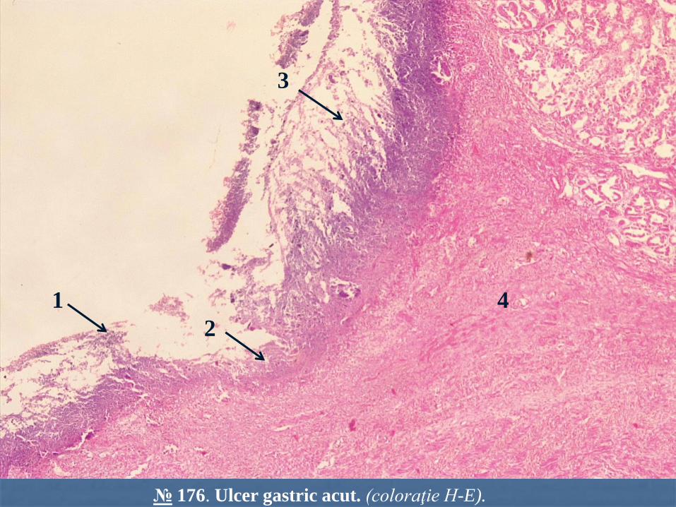
№ 176. Ulcer gastric acut. (coloraţie H-E). Indicaţii:
1. Stratul superficial al fundului ulcerului, constituit din leucocite şi eritrocite.
2. Mase necrotice şi detritus tisular în regiunea fundului ulcerului.
3. Focare de necroză în stratul muscular al peretelui gastric.
4. Infiltraţie leucocitară în marginile şi fundul ulcerului.
№ 87. Ulcer gastric cronic în acutizare. (coloraţie H-E). Indicaţii:
1. Fundul ulcerului:
a. stratul fibrino-purulent;
b. stratul de necroză fibrinoidă;
c. stratul de ţesut de granulaţie;
d. stratul de ţesut fibroconjunctiv cicatricial.
№ 192. Adenocarcinom tubular gastric (tip intestinal). (coloraţie H-E). Indicaţii:
1. Aglomerări de celule canceroase în mucoasa gastrică.
2. Structuri canceroase glandulare în grosimea stratului muscular.
3. Zone nemodificate ale mucoasei.
№ 192a. Metastaze de carcinom gastric în ganglion limfatic. (coloraţie H-E). Indicaţii:
1. Aglomerări de celule canceroase în sinusurile marginal şi medular ale limfonodulului.
2. Foliculi limfoizi nemodificaţi.
II. Macropreparate:
№ 59. Carcinom esofagian.
№ 51. Polip gastric.
№ 52. Ulcer gastric cronic.
№ 53. Ulcer gastric cronic perforant.
№ 54. Ulcer duodenal cronic.
№ 60. Carcinom gastric.
Тема: Патология пищевода и желудка.
I. Микропрепараты:
№ 176. Острая язва желудка. (Окраска Г-Э). Обозначения:
1. Поверхностный слой дна язвы, состоящий из лейкоцитов и эритроцитов.
2. Некротические массы и тканевой детрит в области дна язвы.
3. Очаги некроза мышечного слоя стенки желудка.
4. Лейкоцитарная инфильтрация в области краев и дня язвы.
№ 87. Хроническая язва желудка в стадии обострения. Окраска Г-Э. Обозначения:
1. Дно язвы:
а. фибринозно-гнойный экссудат;
б. некротический слой;
в. грануляционная ткань;
г. грубоволокнистая рубцовая ткань.
№ 192. Тубулярная аденокарцинома желудка (кишечный тип). Окраска Г-Э. Обозначения:
1. Скопления атипичных раковых клеток в слизистой оболочке.
2. Железистые раковые структуры в толще мышечного слоя.
3. Неизмененные зоны слизистой оболочки.
№ 192a. Метастаз рака желудка в лимфатический узел. Окраска Г-Э. Обозначения:
1. Скопления раковых клеток в краевом и мозговых синусах лимфоузла.
2. Неизмененные лимфоидные фолликулы.
II. Мaкропрепараты:
№ 59. Рак пищевода.
№ 51. Полип желудка.
№ 52. Хроническая язва желудка.
№ 53. Перфоративная хроническая язва желудка.
№ 54. Хроническая язва 12-перстной кишки.
№ 60. Рак желудка.
I. Microspecimens:
№ 176. Acute gastric ulcer. (H.E. stain). Indications:
1. The superficial layer of the ulcer, consisting of leukocytes and erythrocytes.
2. Necrotic masses and tissue debris in the area of the ulcer.
3. Foci of necrosis in the muscular layer of the gastric wall.
4. Leukocyte infiltration in the edges and bottom of the ulcer.
№ 87. Active chronic gastric ulcer. (H.E. stain). Indications:
1. The ulcer base:
a. zone of necrotic fibrinoid debris;
b. zone of infiltration with neutrophils;
c. zone of granulation tissue;
d. zone of fibrous, collagenous scar.
№ 192. Gastric tubular adenocarcinoma – intestinal type. (H.E. stain). Indications:
1. Agglomerations of cancer cells in the gastric mucosa.
2. Cancerous glandular structures in the thickness of the muscular layer.
3. Unmodified mucosal areas.
№ 192a. Metastasis of gastric carcinoma into lymph node. (H.E. stain). Indications:
1. Agglomerations of cancer cells in the marginal and medullary sinuses of lymph node.
2. Unmodified lymphoid follicles.
II. Macrospecimens:
№ 59. Esophageal carcinoma.
№ 51. Gastric polyp.
№ 52. Chronic gastric ulcer.
№ 53. Chronic gastric ulcer with perforation.
№ 54. Chronic duodenal ulcer.
№ 60. Gastric adenocarcinoma.
Pathology of esophagus and stomach.
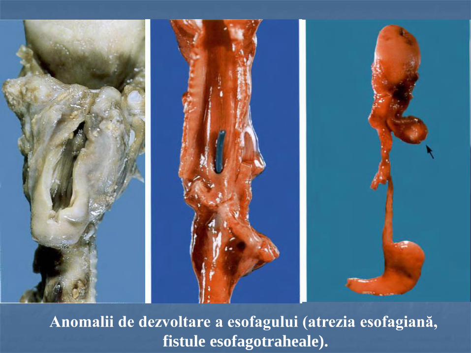
Anomalii de dezvoltare a esofagului (atrezia esofagiană,
fistule esofagotraheale).
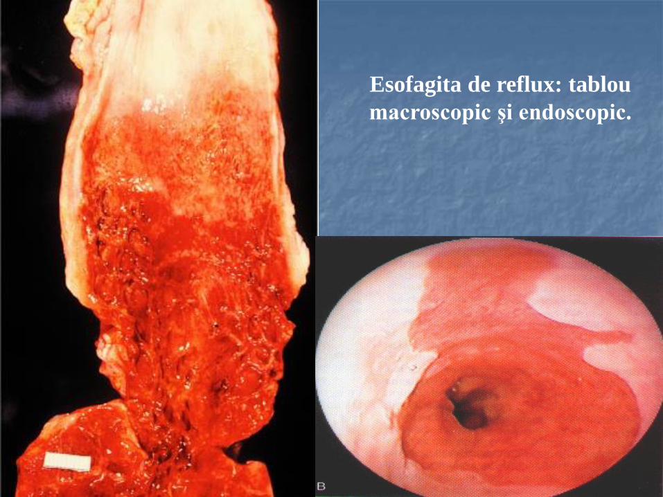
Esofagita de reflux: tablou
macroscopic şi endoscopic.
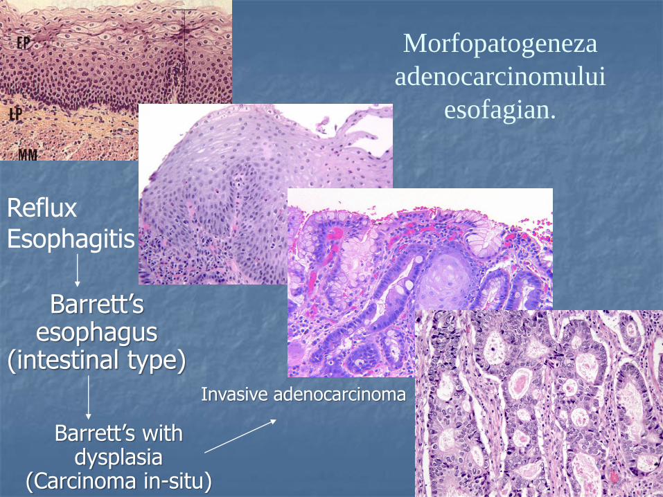
Morfopatogeneza
adenocarcinomului
esofagian.
Reflux Esophagitis
Barrett’s esophagus
(intestinal type)
Barrett’s with dysplasia
(Carcinoma in-situ)
Invasive adenocarcinoma
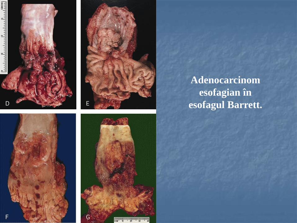
Adenocarcinom
esofagian în
esofagul Barrett.
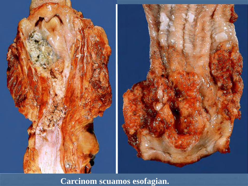
Carcinom scuamos esofagian.
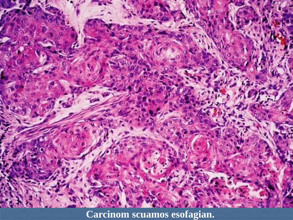
Carcinom scuamos esofagian.
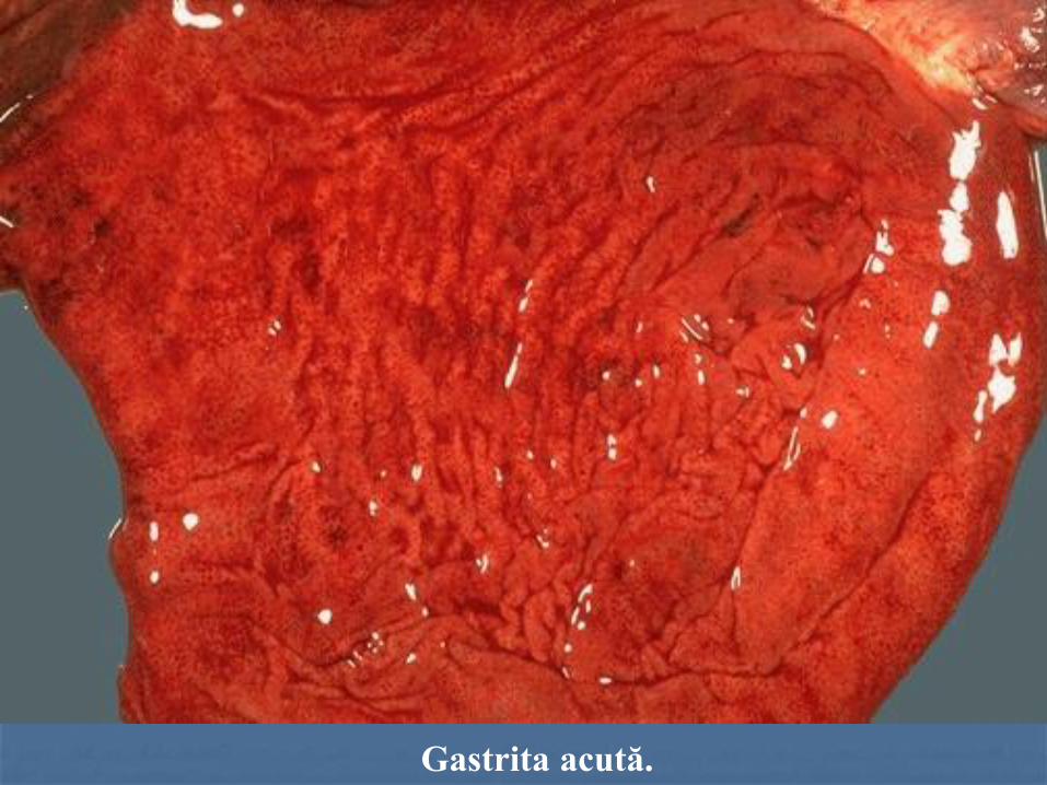
Gastrita acută.
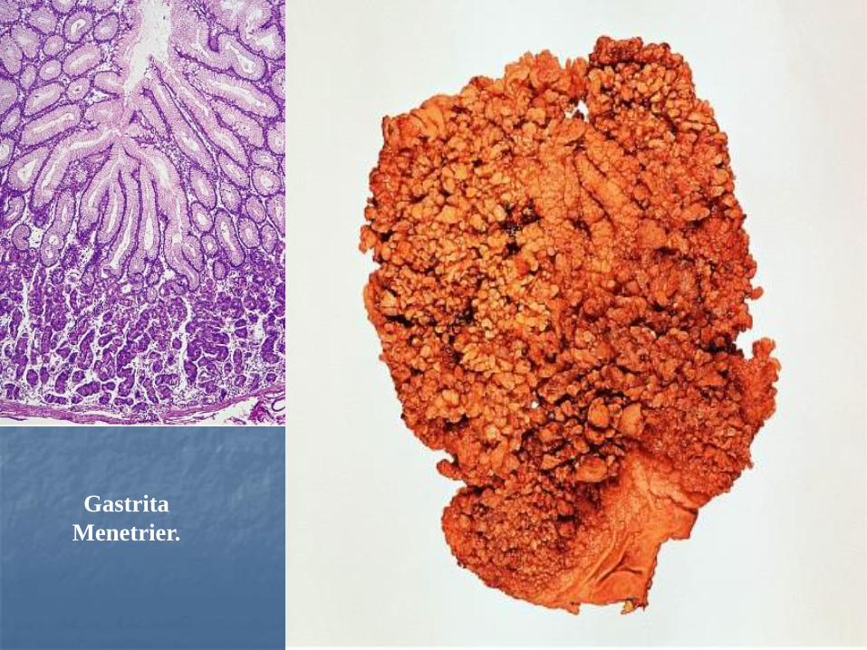
Gastrita
Menetrier.
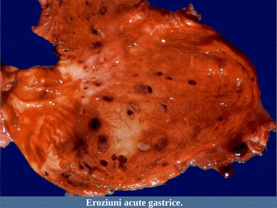
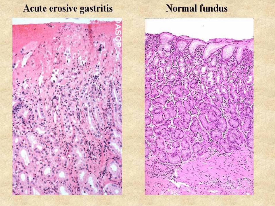
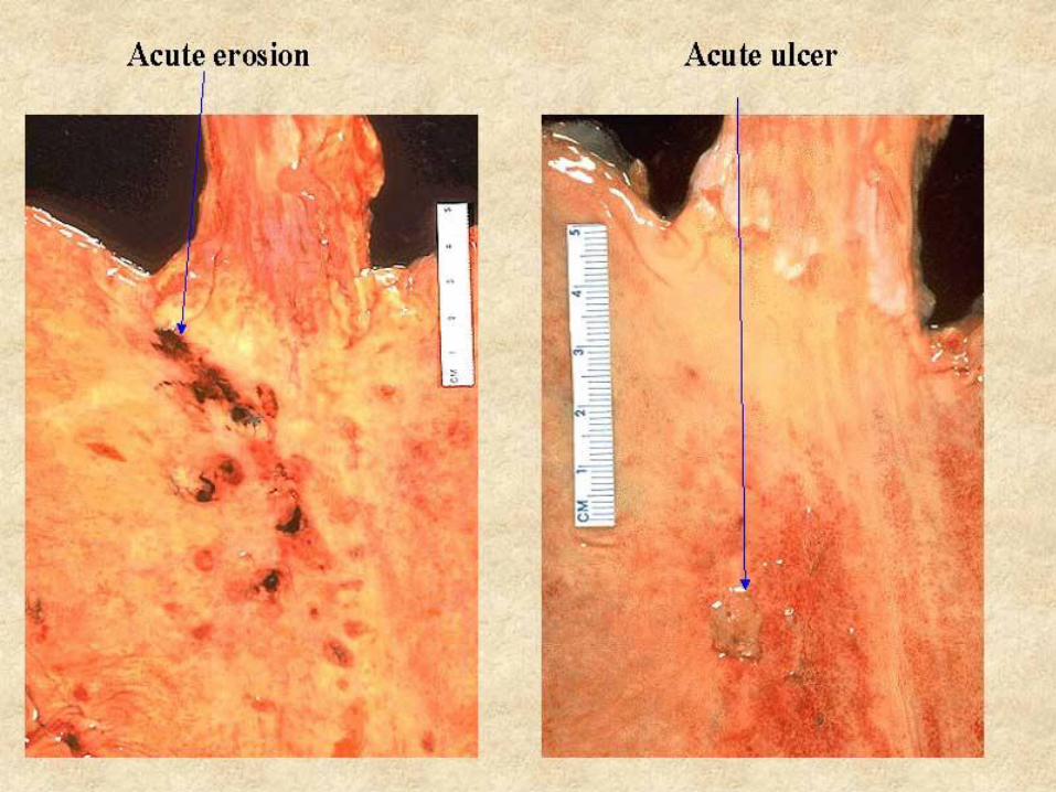
Eroziuni acute gastrice.
№ 176. Ulcer gastric acut. (coloraţie H-E).
3
4
2
1
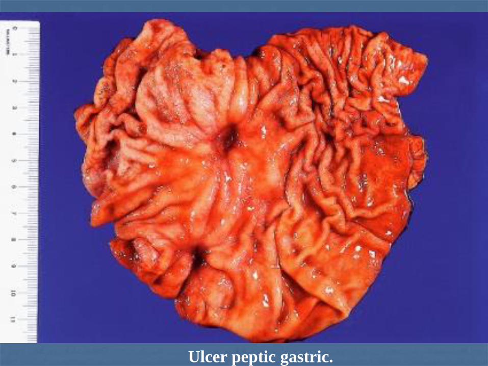
Ulcer peptic gastric.
1
2
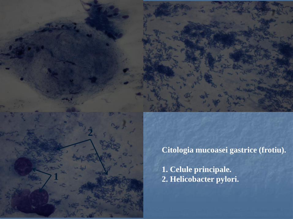
Citologia mucoasei gastrice (frotiu).
1. Celule principale.
2. Helicobacter pylori.

№ 87. Ulcer gastric cronic în acutizare. (coloraţie H-E).
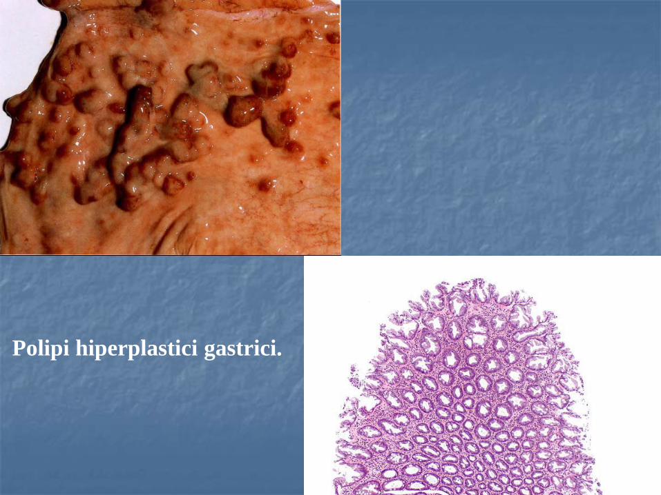
Polipi hiperplastici gastrici.
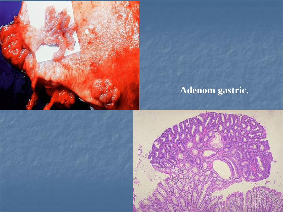
Adenom gastric.
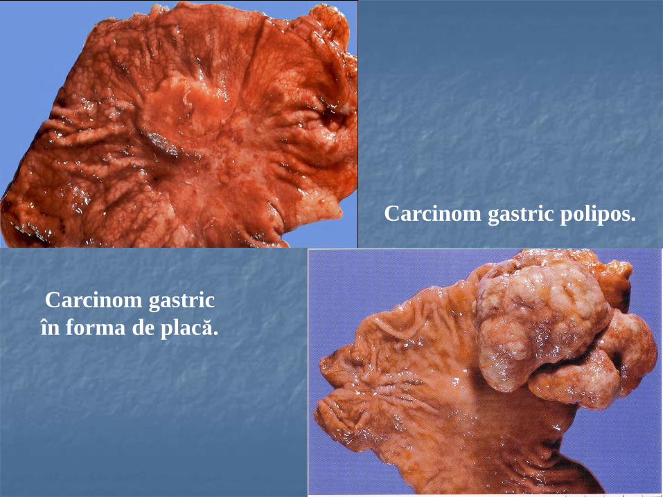
Carcinom gastric
în forma de placă.
Carcinom gastric polipos.
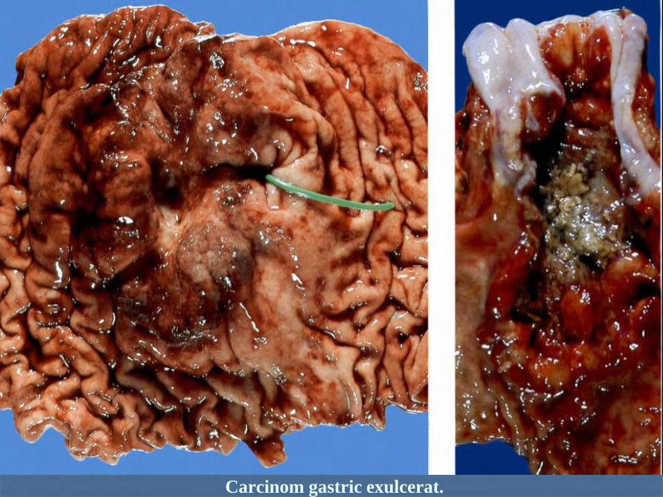
Carcinom gastric exulcerat.
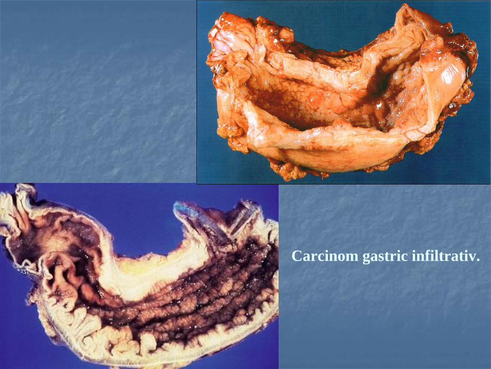
Carcinom gastric infiltrativ.
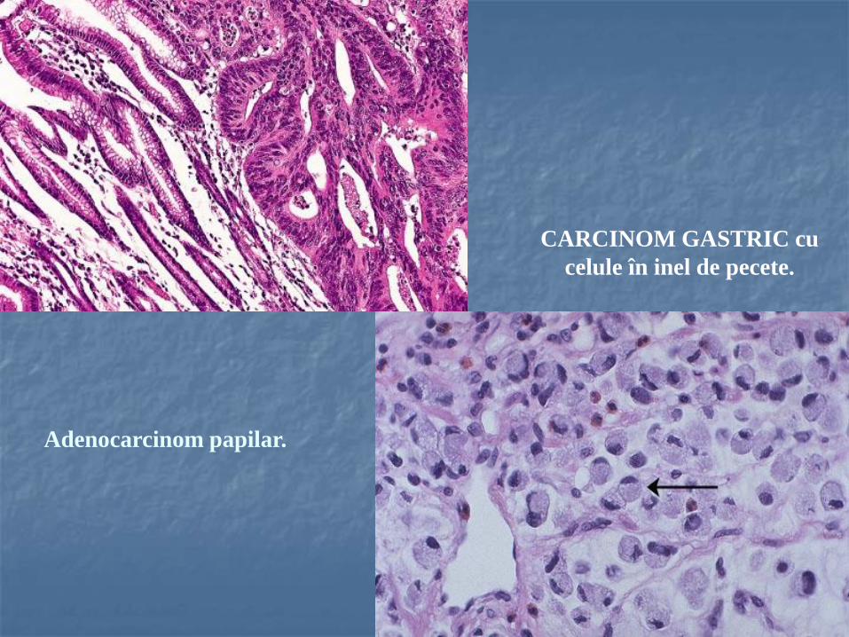
CARCINOM GASTRIC cu
celule în inel de pecete.
Adenocarcinom papilar.
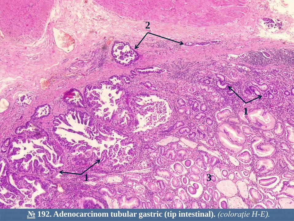
№ 192. Adenocarcinom tubular gastric (tip intestinal). (coloraţie H-E).
2
1 3
1
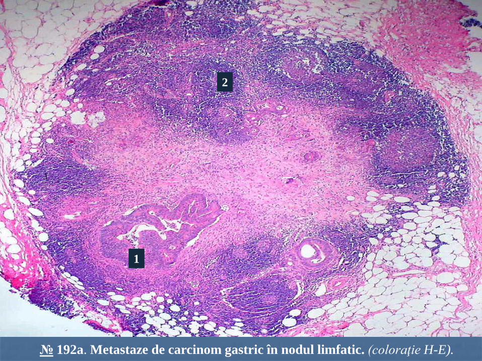
№ 192a. Metastaze de carcinom gastric în nodul limfatic. (coloraţie H-E).
1
2