Embed Size (px)
Citation preview
Tumor chemo-radiotherapy with rod-shaped and spherical gold
nano probes: shape and active targeting both matter
Lu Zhang, & Huilan Su,‡ Haolu Wang, Qian Li,& Xiao Li,⊥ Chuanqing Zhou,& Jia
Xu, & Yimin Chai, & Xiaowen Liang , * Liqin Xiong & and Chunfu Zhang &, †, *
Shanghai Jiao Tong University Affiliated 6th Hospital, School of Biomedical
Engineering, Shanghai Jiao Tong University, Shanghai 200030, China
‡ State Key Laboratory of Metal Matrix Composites, School of Materials Science and
Engineering, Shanghai Jiao Tong University, Shanghai 200240, China
The University of Queensland Diamantina Institute, The University of Queensland,
Woolloongabba, QLD 4102, Australia
⊥Department of Nuclear Medicine, Changhai Hospital, The Secondary Military
Medical University, Shanghai 200433, China
† Department of Nuclear Medicine, Rui Jin Hospital, School of Medicine, Shanghai
Jiao Tong University, Shanghai 200025, China
Corresponding to Chunfu Zhang and Xiaowen Liang:
E-mail: [email protected] Tel.: +86-21-62933323.
[email protected] Tel.: +61-07-34437488
KEYWORDS: Gold nanoparticles; Chemoradiotherapy; PBPK; SPECT/CT;
Angiogenesis targeting
AbstractThe morphologies of gold nanoparticles (NPs) affect their tumor accumulation
through enhanced permeability and retention effect. However, detailed information
and mechanisms of NPs’ characteristics affecting tumor accumulation are limited. The
aim of this study is to evaluate the effects of shape and active targeting ligands of
theranostic NPs on tumor accumulation and therapeutic efficacy, and to elucidate the
underlying mechanism.
Methods: v3 integrin-targeted, cisplatin-loaded and radioisotope iodine-125
labeled spherical and rod-shaped gold nano theranostic probes (RGD-125IPt-AuNPs
and RGD-125IPt-AuNRs) with similar sizes were fabricated and characterized. The in
vivo distribution and chemo-radio therapeutic efficacy against tumors of these newly
developed probes were subsequently evaluated. Moreover, a physiologically based
pharmacokinetic (PBPK) model was developed to characterize the in vivo kinetics of
these probes at the sub-organ level, and to reveal the mechanism of NPs’ shape and
active targeting ligands effects on tumor accumulation.
Result: Cisplatin and iodine-125 were loaded sequentially onto the NPs through a
thin polydopamine coating layer on the NPs. Both RGD-125IPt-AuNPs and RGD-
125IPt-AuNRs exhibited high specificity for v3 in vitro, with the rod-shaped probe
being more efficient. The PBPK model revealed that rod-shaped gold NPs diffused
more rapidly in tumor interstitial than the spherical ones. Tumor accumulations of
non-targeted and rod-shaped RAD-125IPt-AuNRs was higher in short term (1 h post
injection), but not pronounced and similar to that of non-targeted spherical RAD-
125IPt-AuNPs in 24 h after intravenous injection, revealing that the NPs’ shape did not
have a significant impact on tumor accumulations through enhanced permeability and
retention (EPR) effect in long-term. While for actively targeted NPs, in addition to a
higher distribution coefficient, RGD-125IPt-AuNRs also had a much higher tumor
maximum uptake rate constant than RGD-125IPt-AuNPs, indicating both the shape and
active targeting ligands affected the tumor uptake of rod-shaped NPs. As a result,
RGD-125IPt-AuNRs had a more effective inhibition of tumor growth than RGD-125IPt-
AuNPs by chemo-radiationtherapy.
Conclusion: Our study suggests that both the shape and active targeting ligands of
gold NPs play important roles on tumor accumulation and chemo-radio therapeutic
effect.
Introduction
Chemo-radiation combination therapy is a clinically well-established cancer
treatment [1–3]. However, due to the poor tumor targeting and side effects of chemo
drugs and the limited tolerance of radiation dose by normal tissues, chemo-
radiotherapy has not been widely applied in humans in some cases [4]. To
simultaneously enhance tumor accumulation of chemo drugs and radiation dose, the
metal-based nanoparticles (NPs) have been explored as both the chemo drug carriers
and radiosensitizers in cancer chemo-radiotherapy [5–8]. Gold NPs have been
considered as an ideal platform for chemo drugs loading and radiosensitizer to
improve tumor chemotherapy and radiotherapy simultaneously due to their
advantages such as diversity structures, easy modification, good biocompatibility [9–
11], high Z number, generation of photoelectrons and Auger electrons under
irradiation [12], [13-15].
The key factor dictating the therapeutic efficacy is tumor accumulation efficiency
of the NPs. In this context, many efforts have been made to optimize tumor uptake of
gold NPs using various NP sizes or shapes in variety of tumor types through enhanced
permeability and retention (EPR) effect [16–18]. For instances, gold NPs with
different sizes (10, 20, 30 and 50 nm) have been evaluated for tumor
radiosensitization therapy, where 10 nm particles had the highest tumor accumulation
and the best radiosensitization therapeutic efficacy [19]. Wang et al have compared
different shapes of gold NPs (rod, cube and star) for tumor thermotherapy and gold
nanorods showed more efficient for tumor accumulation than other shapes of NPs
through EPR effect [20]. However, tumor accumulation of NPs through EPR is
usually limited by physical or chemical barriers of biological systems [21–23], which
performed controversially due to the heterogeneity of EPR effect across tumor
locations and models [17]. NPs usually have a high accumulation in tumors of high
vasculature density and permeability, but not in tumors of low vasculature density and
permeability [17].
Therefore, targeting tumor angiogenesis may have advantages for chemo-
radiotherapy. By targeting tumor neovasculatures, the drug-loaded NPs can reach
tumor without crossing blood vascular barriers. However, previous studies comparing
different shapes of gold NPs for tumor accumulation and therapeutic effect in vivo
mainly focused either on non-targeting accumulation through EPR effect or active
targeting by addressing tumor cells [24, 25]. Few studies considered tumor
accumulation by simultaneously targeting tumor angiogenic vessels and cells along
with EPR effect. We have prepared RGD peptide-conjugated and v3 integrin-
targeted gold NPs in three different sizes (29, 50 and 80 nm) for tumor radiotherapy
by simultaneously targeting tumor cells and neovaculatures. Our results indicated that
larger particles (80 nm) were more effective for cell therapy in vitro, while the smaller
ones (29 nm) were more effective for tumor inhabitation in vivo due to much higher
tumor accumulation [26]. However, it is still unclear that how the shape effects on
gold NP accumulation efficiency in tumors, intra-tumor kinetics, and chemo-
radiotherapy efficacy when simultaneously targeting tumor cells and neovasculatures
along with EPR effect.
In this study, we developed v3-targeted, cisplatin-loaded, radioisotope iodine-
125 labeled spherical (RGD-125IPt-AuNPs) and rod-shaped (RGD-125IPt-AuNRs) gold
nano theranostic probes, and investigated their tumor accumulation and the efficacy of
chemo-radiotherapy. A physiologically based pharmacokinetic (PBPK) model was
also developed to elucidate the intra-tumor kinetics of developed probes and the
mechanism of shape and active targeting ligands of NPs effecting on their tumor
accumulation at the sub-organ level.
Results
Synthesis and Characterization of RGD-125IPt-AuNPs and RGD-125IPt-
AuNRs. Gold nanospheres (AuNPs) and nanorods (AuNRs) with uniform sizes were
prepared according to previous reports [26, 27]. AuNPs were 56.37 ± 3.04 nm in
diameter (Figure S1A) and AuNRs were 22.41 ± 1.01 nm in diameter and 56.12 ±
3.22 nm in length (aspect ratio of ∼2.5, Figure S1B). During the synthesis, a dense
polyvinyl pyrrolidone (PVP) coating on AuNPs and cetyltrimethyl ammonium
bromide (CTAB) on AuNRs were formed. As a result, zeta potentials of AuNPs and
AuNRs were -8.49 ± 2.58 mV and 31.01 ± 4.07 mV, respectively (Table S1). For
preparation of the theranostic probes, both AuNPs and AuNRs were firstly coated
with a thin layer of poly dopamine (PDA) (Figures 1A and B, Figures S1C and D)
[28]. The zeta potentials of PDA-coated AuNPs (PDA-AuNPs) and AuNRs (PDA-
AuNRs) were 24.93 ± 5.23 mV and 22.31 ± 2.68mV, respectively. Subsequently,
PDA-AuNPs and PDA-AuNRs were further modified by amino-poly (ethylene
glycol)-thiol (MW 5000, Creative PEGworks, Chapel Hill, NC) for peptide
conjugation and improving the in vivo behavior of the NPs [29]. After the
modification, zeta potentials of the PEGylated PDA-AuNPs and PDA-AuNRs were -
10.23 ± 3.33 mV and -13.67 ± 2.46 mV, respectively. There were 128.09 ± 14.01 and
103.51 ± 20.95 PEG molecules anchored on an individual AuNP and AuNR,
respectively, determined by Ellman method [30, 31].
By using the sulfosuccinimidyl-4-[N-maleimidomethyl]-cyclohexane-1-
carboxylate (sulfo-SMCC), the PEGylated PDA-AuNPs and PDA-AuNRs were
further activated and subsequently conjugated with c(RGDyC) peptides (RGD)
through thiol-maleimide linkages [32], designated as RGD-AuNPs and RGD-AuNRs.
The amounts of c(RGDyC) immobilized on each AuNP and AuNR were determined
to be 67.33 ± 9.04 and 61.62 ± 2.15, respectively. In addition, scramble peptide
c(RADyC) coupled PDA-AuNPs (RAD-AuNPs) and PDA-AuNRs (RAD-AuNRs)
were also prepared as the non-targeting NPs and compared with RGD NPs to evaluate
the active target efficiency using the same procedures.
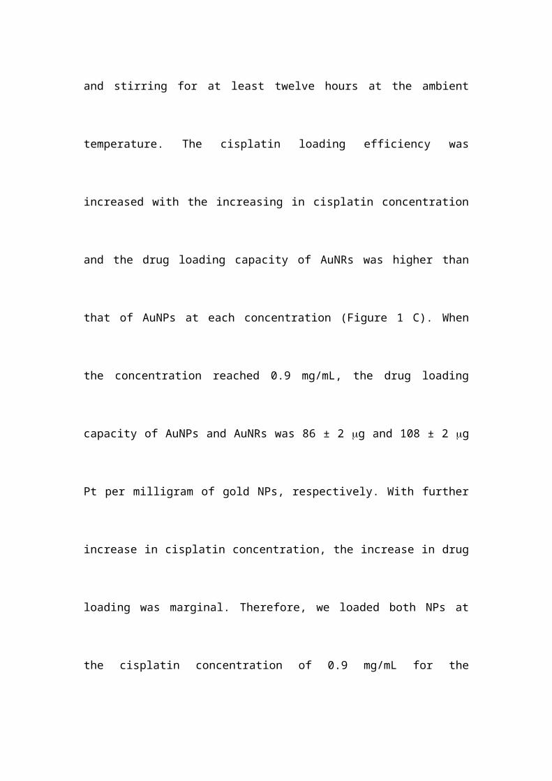
After the peptide conjugation, cisplatin was loaded by suspending RGD-AuNPs
or RGD-AuNRs (1 mg) into cisplatin aqueous solutions (0.3 mL) with various
concentrations and stirring for at least twelve hours at the ambient temperature. The
cisplatin loading efficiency was increased with the increasing in cisplatin
concentration and the drug loading capacity of AuNRs was higher than that of AuNPs
at each concentration (Figure 1 C). When the concentration reached 0.9 mg/mL, the
drug loading capacity of AuNPs and AuNRs was 86 ± 2 g and 108 ± 2 g Pt per
milligram of gold NPs, respectively. With further increase in cisplatin concentration,
the increase in drug loading was marginal. Therefore, we loaded both NPs at the
cisplatin concentration of 0.9 mg/mL for the following studies. To confirm the
presence of cisplatin on the NPs, an energy-dispersive X-ray spectroscopy element
mapping (EDS) was performed (Figures 1D – 1I). Both Au (red dots) and Pt elements
(green dots) located in the same region and could overlap with each other, indicating
successful cisplatin loading on both AuNPs and AuNRs.
Next, the drug-loaded RGD-AuNPs (RGD-Pt-AuNPs) and RGD-AuNRs (RGD-
Pt-AuNRs) were labeled with iodine-125 by mixing them (1mg in 0.3 mL) with Na125I
(300 Ci) and vortexing for 30 min [27]. The labeling were carrier-free and confirmed
by thin layer chromatography (TLC). The labeling efficiency of both types of NPs
were determined to be nearly 100%. After iodine-125 labeling, the final NPs
designated as RGD-125IPt-AuNPs and RGD-125IPt-AuNRs probes, were purified by a
Sephadex G-25 chromatography with the eluent of saline. The labeling stability of
radioactive 125I on the probes was examined by suspending the probes in fetal bovine
serum (FBS) at 37 °C for different time intervals. As shown in Figure 1J, detachment
of iodine-125 was unnoticeable after incubation for 24 h. In addition, RGD-Pt-AuNPs
and RGD-Pt-AuNRs were also labeled with non-radioactive iodine using as cold
probes. Meanwhile, non-targeting NPs (RAD-Pt-AuNPs and RAD-Pt-AuNRs) were
also labeled with radioactive iodine-125 (RAD-125IPt-AuNPs, RAD-125IPt-AuNRs)or
non-radioactive iodine (RAD-IPt-AuNPs, RAD-IPt-AuNRs), which were used as
control probes. The detailed probe preparation procedures were illustrated in Scheme
1.
Zeta potentials of RGD-IPt-AuNPs and RGD-IPt-AuNRs were -26.92 ± 6.17 mV
and -28.44 ± 4.36 mV, respectively (Table S1). Under the physiological condition
(RPMI-1640 culture medium within 10% FBS), pristine AuNPs and AuNRs began to
aggregate and settle in a few minutes, and the LSPR peaks exhibited red shift (Figures
S1I and J). Compared to the pristine NPs, both types of active targeting and non-
targeting probes were highly stable and exhibited no significant shift of LSPR peak
after incubation for 72 h (Figures S1 E - H).
To examine the cisplatin release profiles, RGD-IPt-AuNPs and RGD-IPt-AuNRs
were dispersed into PBS with different pH values and incubated for different time
intervals at 37 oC. As shown in Figure 1K and 1L, cisplatin release was pH-
responsive. At the alkaline condition (pH 8.5), it was nearly unreleased, while at the
neutral (pH 7.4) or acidic condition (pH 6), cisplatin released gradually. Consequently,
23.3 ± 1.7 % and 45.7 ± 2.3% of the loaded drug from RGD-IPt- AuNPs, and 24.5 ±
3.6 % and 54.2 ± 2.9 % from RGD-IPt-AuNRs were released after incubation for 24
hours in PBS of pH 7.4 and pH 6, respectively.
Specificity for v3 Integrin. To evaluate the specificity of probes, the human
derived v3 positive H1299 cells [33] were incubated with RGD probes (50 g/mL,
1h), using RAD probes as the control. In addition, competition studies were also
performed by incubating the cells with RGD probes in combination with free RGD
peptide (10 μM). Free RGD peptide could block v3 integrin receptor and reduce
RGD probes binding to cells if the probes had specificity for the receptor [34]. After
incubation, cell uptake profiles of the probes were examined by two-photon confocal
microscope and the intracellular gold contents were measured by atomic adsorption
spectrophotometer (AAS). Both the microscopic images and AAS measurements
indicated that cell ingested more RGD probes than RAD probes, and free RGD
peptide partially suppressed the ingestion (Figure 2A and 2D). However, for the RGD
probes, cell uptake of the rod-shaped probes was significantly more than that of the
spherical one (RGD-IPt-AuNRs vs RGD-IPt-AuNPs: 56.46 ± 6.44 pg/cell vs 48.86 ±
4.32 pg/cell, p < 0.05). These results suggested that RGD probes were able to
specifically target v3 integrin, the cell ingestion of the probes was induced partially
by v3 integrin [35] and could be influenced by the NP shape [36]. In addition, the
z-stack reconstructions of cell images from orthogonal projections using two-photon
confocal microscope revealed that both probes were internalized by the cells and
located quite close to the nuclei (Figure 2B and 2C).
Chemotherapy and Chemoradiotherapy of Tumor Cells. The chemotherapeutic
effect of the probes on tumor cells was assessed by treating H1299 cells with the RGD
probes at various concentrations for 6, 24 and 48 hours. Cell viability was examined
by cell counting kit-8 assay (CCK-8). No obvious cell death was observed after
treatment by both types of probes at lower concentrations ( 100 μg /mL). However,
dsDNA of the treated cells (50 g/mL for 48 h) was damaged remarkably (Figure S2
D), indicating the chemotherapeutic potency of the probes. Cell death was significant
when they were incubated with the probes at the concentration of 100 μg /mL for
more than 24 hours (Figure S2 A-C).
In order to evaluate the chemo-radio therapeutic effect of the probes, the cells
were first treated with both types of RGD probes at 50 μg /mL (in Au) for six hours,
and then irradiated by 320 kV X-ray at a dose of 4 Gy (Precision X-Ray, North
Branford, CT). Afterwards, the treated cells were further cultivated for different time
intervals and cell viability was assayed by CCK-8. Cell viability was inhibited after
radiotherapy (Figure 3). However, compared to radiation only group, pre-treated with
either drug-free or drug-loaded probes resulted in a significant cell death one day after
irradiation, indicating the probes have the radiosensitization effects. Four days after
the treatment, cell death was more pronounced for drug-loaded probes group, even
though the chemo therapeutic effect has not been observed at the same dose.
However, the rod-shaped probe RGD-IPt-AuNRs were more effective than the
spherical ones for both radiosensitization therapy (27.43 ± 5.72% vs 31.88 ± 4.79%, p
< 0.05) and the combination therapy (10.37 ± 3.32% vs 14.32 ± 6.6%, p < 0.05) four
days after the treatments.
To calculate the combination index (CI) of chemo-radiotherapy using both types
of probes, the cells were incubated with both types of probes at different
concentrations (10, 25, 50, 100 g Au /mL) for 6 hours and then irradiated by X-ray
for a dosage of 4 Gy. Cell viability was then examined (Figure S3). The CI values
using RGD-IPt-AuNPs and RGD-IPt-AuNRs were 0.70 and 0.85, respectively,
demonstrating the synergetic therapeutic effect of both types of probes.
Specificity of RGD Probes for Tumor. To evaluate tumor specificity of the
probes, SPECT/CT imaging was performed. Radioactive RGD or RAD probes were
intravenously administered into H1299 tumor-bearing mice (300 μCi). The
competition studies were conducted by administering the RGD probes in combination
with free RGD peptide. As indicated in Figure 4A and 4B, SPECT/CT imaging
demonstrated that tumor uptake of both RGD-125IPt-AuNPs and RGD-125IPt-AuNRs
increased with the circulation time of the probes, which was noticeable one hour after
probe injection and reached the maximum at 6 h after injection. However, the uptakes
were significantly inhibited when competing with free RGD peptide. The tumor
uptake was marginal after administrating non-targeting RAD-125IPt-AuNPs and RAD-
125IPt-AuNRs probes in mice.
In vivo bio-distributions of the probes were also evaluated and quantified based on
the percentage of injected dose per gram of tissue (%ID/g) (Figure 4C-D and Figure
S4). In consistent with SPECT/CT imaging, both spherical and rod-shaped probes had
high radiation dose in the liver and spleen, indicating they were mainly eliminated
from the body through the reticuloendothelial system (RES) (Figure 4C and 4D)
[37,38]. However, the RGD probes were accumulated more in tumors than RAD ones
(AuNPs probe: 5.33 ± 1.71% vs 2.5 ± 0.3%, p < 0.01; AuNRs probe: 6.93 ± 0.51% vs
2.67 ± 0.11%, p < 0.01), and the tumor accumulation was markedly decreased after
competing with free RGD peptide. These results suggested that the RGD probes
specifically targeted tumor v3 integrin receptor [39,40]. However, the rod-shaped
probes exhibited a much higher targeting efficiency than the spherical one (6.93 ±
0.51 vs 5.33 ± 1.71% ID/g, p < 0.05). The blood elimination half-lives were
determined to be 227.36 ± 28.91 min and 79.99 ± 4.39 min (Figure S5 and Table S3)
for RGD-125IPt-AuNRs and RGD-125IPt- AuNPs, respectively, using two compartment
model [18]. The half-lives of RAD probes were 213.39 ± 22.23 min (RAD-125IPt-
AuNRs) and 94.73 ± 3.39 min (RAD-125IPt-AuNPs), while PEG modified plain NPs
were 258.22 ± 11.03 min (125IPt-AuNRs) and 124.60 ± 12.71 min (125IPt-AuNPs),
respectively.
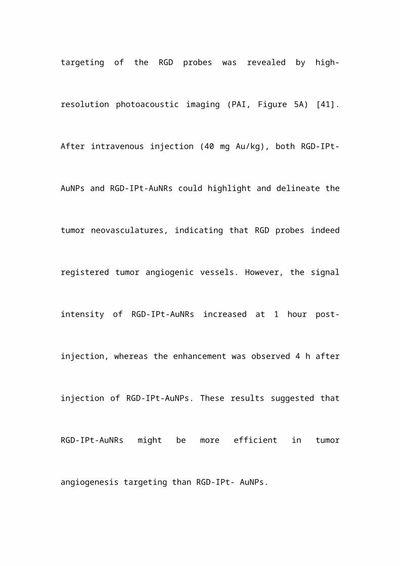
Tumor Angiogenesis Targeting. Tumor angiogenesis targeting of the RGD probes
was revealed by high-resolution photoacoustic imaging (PAI, Figure 5A) [41]. After
intravenous injection (40 mg Au/kg), both RGD-IPt-AuNPs and RGD-IPt-AuNRs
could highlight and delineate the tumor neovasculatures, indicating that RGD probes
indeed registered tumor angiogenic vessels. However, the signal intensity of RGD-
IPt-AuNRs increased at 1 hour post-injection, whereas the enhancement was observed
4 h after injection of RGD-IPt-AuNPs. These results suggested that RGD-IPt-AuNRs
might be more efficient in tumor angiogenesis targeting than RGD-IPt- AuNPs.
To verify these observations, silver staining of tumor tissues was performed. Both
tumor angiogenic vessels and cells were v3 positive (Figure S6). Both the spherical
and the rod-shaped RGD probes could target tumor neoveculatures and cells (Figure
5B). However, RGD-IPt-AuNRs penetrated much deeper into tumor interstitial than
RGD-IPt-AuNPs.
In vivo Chemo-radiotherapy and Histological Studies. Since RGD probes had
the high tumor accumulation efficiency, the potential of the RGD probes for tumor
chemo-radiotherapy was evaluated. Tumor-bearing mice were divided into six groups
(PBS, drug-free probe, drug-loaded probe, PBS + X-ray, drug-free probe + X-ray and
drug-loaded probe + X-ray) and intravenously administered with RGD-IPt-AuNPs or
RGD-IPt-AuNRs (40 mg Au /kg in body weight). Tumor radiotherapy was conducted
six hours after probe injection for a dose of 6 Gy (320 kV, Precision X-Ray, North
Branford, CT). After the treatment, the tumor volumes were monitored (Figure 6).
Compared to the PBS group, chemotherapy, radiotherapy and chemo-radiotherapy
with both types of probes were able to inhibit tumor growth significantly 21 days after
treatment. As expected, chemo-radiotherapy was more effective than chemo or
radiation therapy alone possibly due to the synergetic therapeutic effect, which is
consistent with previous reports [42, 43]. However, for each therapeutic modality,
RGD-IPt-AuNRs outperformed RGD-IPt-AuNPs in the therapeutic efficacy (v/v0,
chemotherapy: 25.47 ± 1.6 vs 28.63 ± 2.3, p < 0.05; radiation therapy: 19.74 ± 2.7 vs
24.8 ± 3.4 p < 0.05; chemoradiotherapy: 4.73 ± 0.5 vs 9.8 ± 2.2, p < 0.05).
To verify these observations, three mice from each treatment groups were sacrificed
two days post the treatment and tumors were surgically removed. Immunostaining of
tumor tissues against γ-H2AX was conducted and the γ-H2AX positive areas were
quantified (Figure 7 and S7) [44]. It was found that both chemotherapy with drug-
loaded probes and radiotherapy with drug-free probes could damage dsDNA
obviously. Compared to radiotherapy without probes (PBS + radiation, γ-H2AX
positive areas: 0.07 ± 0.02), radiotherapy with RGD-I-AuNPs (0.12 ± 0.04, p < 0.01)
or RGD-I-AuNRs (0.14 ± 0.02, p < 0.01) significantly induced dsDNA damage,
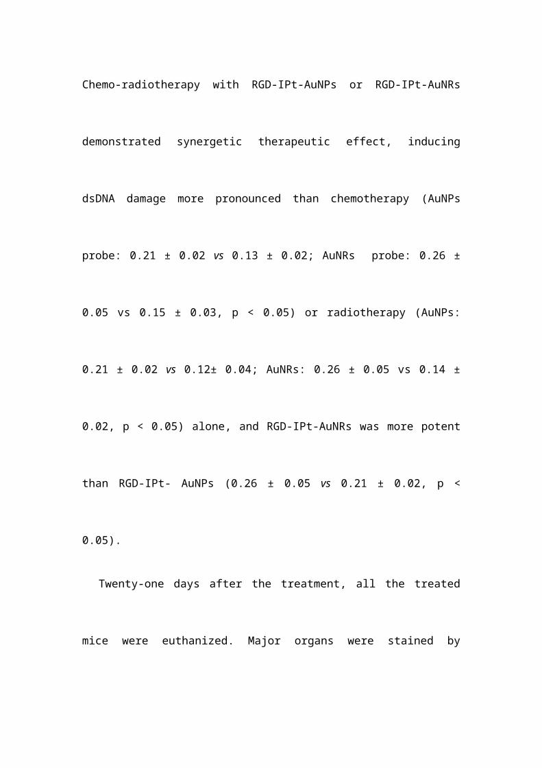
indicating that radiation dose was increased by gold NPs. Chemo-radiotherapy with
RGD-IPt-AuNPs or RGD-IPt-AuNRs demonstrated synergetic therapeutic effect,
inducing dsDNA damage more pronounced than chemotherapy (AuNPs probe: 0.21 ±
0.02 vs 0.13 ± 0.02; AuNRs probe: 0.26 ± 0.05 vs 0.15 ± 0.03, p < 0.05) or
radiotherapy (AuNPs: 0.21 ± 0.02 vs 0.12± 0.04; AuNRs: 0.26 ± 0.05 vs 0.14 ± 0.02,
p < 0.05) alone, and RGD-IPt-AuNRs was more potent than RGD-IPt- AuNPs (0.26 ±
0.05 vs 0.21 ± 0.02, p < 0.05).
Twenty-one days after the treatment, all the treated mice were euthanized. Major
organs were stained by hematoxylin and eosin staining (H&E) for histological
examinations (Figure S8). The H&E images did not reveal noticeable damage in
organs, indicating these treatments did not have obvious toxic effects to normal
tissues in vivo.
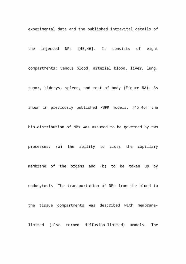
Suborgan Kinetics of AuNPs and AuNRs in the Tumor. To elucidate the
underlying mechanism that RGD-IPt-AuNRs had a better performance for tumor
chemo-radiotherapy than RGD-IPt-AuNPs, we developed a PBPK model to
characterize the intra-tumor kinetics of the two types of probes. The model structure
was based on all of the above experimental data and the published intravital details of
the injected NPs [45,46]. It consists of eight compartments: venous blood, arterial
blood, liver, lung, tumor, kidneys, spleen, and rest of body (Figure 8A). As shown in
previously published PBPK models, [45,46] the bio-distribution of NPs was assumed
to be governed by two processes: (a) the ability to cross the capillary membrane of the
organs and (b) to be taken up by endocytosis. The transportation of NPs from the
blood to the tissue compartments was described with membrane-limited (also termed
diffusion-limited) models. The uptake rate constant was time-dependent and described
by a Hill equation to adequately describe a fraction of NPs sequestered by tumor cells
with time.
As shown in Figure S9, the time profiles of probe concentration observed in
major organs of mice were adequately described by the developed mathematical
model with an overall regression coefficient (R2) of 0.92. Non-targeting AuNRs
(GNR NT) displayed a much higher tumor accumulation than non-targeting AuNPs
(GNP NT) at 1 h post-injection (2.43 vs 1.71 ID% mL, p < 0.05, Figure 8B).
However, similar tumor accumulations were observed for both types of non-targeting
probes at 24 h post-injection (2.52 vs 2.67 ID%/mL), indicating that the shapes of NPs
did not have a high influence on their tumor accumulations through EPR effect in
long term. For RGD-conjugated target NPs, the distribution coefficients (P) and the
maximum uptake rate constant (Kmax) were determined to be higher for RGD-125IPt-
AuNRs than RGD-125IPt-AuNPs (0.25 vs 0.15; 20 h–1 vs 10 h–1), indicating the more
rapid distribution of RGD-125IPt-AuNRs into tumor interstitial fluid and the higher
ingestion of RGD-125IPt-AuNRs by tumor cells, while other parameters of these two
probes were the same (Table S5).
Discussion
Gold NPs have been extensively explored for tumor theranostics [47] and the key
factor governing the performances is the tumor accumulation efficiency of NPs.
Therefore, in vivo behaviors of gold NPs with different configurations and
formulations have been intensively studied to obtain the maximal tumor accumulation
[48]. In this study, RGD peptide-conjugated, cisplatin-loaded, 125I-labeled, spherical
(RGD-125IPt-AuNPs) and rod-shaped (RGD-125IPt-AuNRs) gold nano theranostic
probes have been developed. By using a H1299 tumor model, we were able to
evaluate the shape-effect of active targeting NPs on tumor accumulation efficiency
and thus the tumor therapeutic efficacy by chemo-radiotherapy when NPs
simultaneously targeting tumor neovesculatures and cells along with EPR effect,
which was different from previous reports considering the shape effect on tumor
accumulation through either EPR effect or active targeting by addressing tumor cells.
Tumor accumulation of NPs was influenced by extravasation, intra-tumoral
diffusion and interaction of NPs with tumor cells. The particle size, morphology and
surface properties are key parameters affecting their extravasation, intra-tumoral
diffusion and retention [16, 49]. In our study, both real-time observations and
simulation by the PBPK model revealed that tumor accumulations of the non-
targeting, rod-shaped RAD-125IPt- AuNRs through EPR effect were more efficient
than those of the spherical RAD-125IPt-AuNPs in short-term (1h post-injection), being
consistent with previous reports [16, 20]. However, the tumor accumulations of both
particles were similar in long-term (24 h post-injection). For RGD probes, however,
tumor accumulation of the rod-shaped RGD-125IPt-AuNRs was significantly more
than the spherical RGD-125IPt-AuNPs at each time point. PBPK simulation-derived
estimates of the distribution coefficients (P), a parameter reflecting distribution of
NPs into interstitial fluid, was much higher for rod-shaped RGD-125IPt-AuNRs than
spherical RGD-125IPt-AuNPs. The simulation results could be validated by previous
observations. Chauhan et al have tested tumor penetration efficiency of fluorescent
nanorods and nanospheres using multiphoton microscopy within the same tumor, and
found that the nanorods penetrated into tumors 4.1 times as rapidly as the nanospheres
[50]. Moreover, the rod-shaped RGD-125IPt-AuNRs penetrated deeper into tumor
interstitials than the spherical one, as revealed by silver staining of tumor tissues. Our
findings were consistent with previous studies on intra-tumoral distributions of
radioactive 198Au nanostructures by autoradiographic imaging, which revealed that the
AuNPs were only distributed on the surfaces of tumors, while the AuNRs were
observed throughout tumors [51]. In addition, the maximum uptake rate constant
(Kmax) of RGD-125IPt-AuNRs, a parameter indicating tumor cell uptake of NPs, was 2
times higher than RGD-125IPt-AuNPs. The differential effects of NP shape on tumor
accumulations through EPR effect and active targeting might be explained by the fact
that although AuNRs diffuses faster and deeper than AuNPs in tumor interstitials,
high intra tumor pressure induced by impaired lymphatic drainage would induce
intravasation of NPs into blood vessels if they could not anchor the tumor cells [52].
As revealed by in vitro studies, the RGD probes were able to specifically target to
v3 positive tumor cells, with the rod-shaped RGD-125IPt-AuNRs had more efficient
targeting than RAD probes. Moreover, compared to its spherical counterpart, RGD-
125IPt-AuNRs had a longer blood half-life because the rod-shaped materials tend to
evade phagocytosis and clearance by macrophages in blood and reticuloendothelial
system [53,54]. Longer blood half-life means NPs have the more chance to permeate
into tumor interstitials and address tumor cells. These rod shape-related, favorable
factors could contribute to higher tumor accumulation of RGD-125IPt-AuNRs. These
results indicated that the morphology affected both in vivo and intra-tumor kinetics of
gold nanoprobes, and NP shape and active targeting ligands are important for efficient
tumor accumulation. However, we did not find the significant difference of
distribution patterns of these two types of probes in the other major organs such as the
liver and spleen.
In addition to high tumor accumulation, the rod-shaped NPs were also found to be
more efficient for cisplatin loading. Under alkaline conditions, dopamine is able to
self-polymerize and spontaneously deposit on the surface of almost any material [55].
Lots of functional groups, such as carboxyl , quinone functions and catechol, might be
included in the PDA layer, which may provide a versatile platform for drug loading
and surface modifications with functionalities.[56] We loaded cisplatin onto PDA-
coated gold NPs by hydrating cisplatin into [Pt(H2O)2(NH2)2]2+, which could chelate
the catechol groups in PDA [32]. As demonstrated previously, the PDA coating layer
was thin (around 12 nm in thickness) and uniform, and the drug loading efficiency
was determined by the PDA coating area. Because the surface area of AuNRs is much
larger than that of AuNPs in the same mass (5.52 × 10¹⁷ vs 2.85 × 10¹⁷ nm2 per
milligram), it is conceivable that RGD-125IPt-AuNRs had a much higher loading
efficiency for cisplatin than RGD-125IPt-AuNPs.
Angiogenesis is very important for tumor growth and metastasis [57]. Targeting
tumor angiogenesis through the highly expressed v3 integrin has been approved as
an effective way for tumor therapy [58]. In our study, as revealed by high resolution
PAI, both RGD-IPt-AuNPs and RGD-IPt-AuNRs were able to localize tumor
neovasculatures, with the rod-like probes being more effective. By releasing the
chemo drug and localizing radiation damage directly to the tumor-associated
endothelial cells, the tumor neovesculatures would be damaged and unable to
transport nutrients to tumor tissues, inducing ischemic necrosis of tumor. Although
both types of RGD probes could also address tumor cells as indicated by silver
staining, the nutrients supply inhibited mechanism for tumor suppression may be
more effective than tumor clonogenic cells death itself [59].
Conclusions
In conclusion, both RGD-125IPt-AuNPs and RGD-125IPt-AuNRs were able to
target tumor cells and angiogenic vessels. Compared to the spherical RGD-125IPt-
AuNPs, the rod-shaped RGD-125IPt-AuNRs have a higher drug loading efficiency,
more rapid diffusion in and deep penetration into tumor interstitials, higher specificity
for v3 integrin and thus more efficient tumor accumulation. Therefore, RGD-125IPt-
AuNRs have more antitumor efficacy for chemotherapy and chemo-radiotherapy. Our
study suggests that both gold NPs’ shapes and targeting ligands are both important in
tumor accumulation and chemo-radiotherapy.
Experimental
Materials. All the chemical reagents were purchased from Sinopharm Chemical
Regent Col Ltd (Shanghai, China) unless indicated otherwise. All the glass containers
used in experiments were cleaned by aqua regia, washed extensively with ultrapure
water (Milli Q Integral 5 system).
Synthesis of Gold Nanospheres (AuNPs) and Nanorods (AuNRs). AuNPs
were synthesized according to the previous report [60]. In brief, gold nano seeds were
first synthesized by direct reduction of HAuCl4 by NaBH4 in the presence of trisodium
citrate dihydrate. Then, 200 μL of seeds solution was injected into a growth solution
quickly and allowed for growth for 10 min. Afterwards, the particles were retrieved,
rinsed and then kept in water for next procedure.
AuNRs were synthesized according to our previous report [61]. The as-
synthesized AuNRs were coated by CTAB in situ and dispersed in deionized water.
The numbers of AuNPs and AuNRs per milligram were calculated. Herein,
AuNPs are assumed as a perfect sphere, and the volume of one AuNP (VAuNP) is
calculated by
V AuNP=16
π R3 (1)
Where R is the diameter of AuNPs.
AuNRs are assumed as a cylinder with two hemispherical caps [62], and the
volume of an individual AuNR (VAuNR) can be calculated by
V AuNR=π D3
8 [43+2 (Y−1 )] (2)
Here, Y is the aspect ratio of AuNRs and equals to L/D, D and L are the diameter
and length of the AuNRs, respectively.
The weight of an individual AuNP and AuNR can be determined by M = V, in
which and V are density of gold and volume of a gold NP, respectively.
The amount of AuNPs (mAuNPs) per milligram is calculated by
mAuNPs=V
V AuNPs= 6 M
π R3 (3)
and the amount of AuNRs (mAuNRs) in one milligram is calculated by the
following formula:
mAuNRs=V
V AuNRs= 8 M
π D3[ 43+2 (Y −1 )] (4)
Preparation of RGD-125IPt-AuNPs and RGD-125IPt-AuNRs Probes. Both the
spherical and rod-shaped probes were prepared by five steps: poly dopamine (PDA)
surface coating, PEG modification, RGD peptide conjugation, cisplatin loading and
iodine-125 labeling. The detailed synthesis procedure referred to our previous report
[27].
Characterization. Zeta-potentials of AuNPs and AuNRs during the probe
preparation were measured by Malvern Zetasizer Nano ZSP instrument (Malvern,
Britain). The morphology, EDS element mapping images were obtained by using
TEM (JEOL, Japan) operating at 200 kV acceleration voltage. Probe size distributions
were analyzed using ImageJ software by measuring the diameters of ≥ 200 particles.
The optical properties and the stability of both types of NPs and probes in
physiological conditions (RPMI-1640 culture medium plus 10% FBS) were evaluated
by examining their UV–VIS spectra (Cary 50 Bio, Varian, USA) at different time
point (30 min, 3, 6, 24, 72 h). The Ellman method was used to evaluate the efficiency
of PEG modification and peptide conjugation [30]. Accordingly, the amount of PEG
molecules and RGD peptides anchored on an individual particle were able to be
calculated.
Drug Release. The cisplatin release profile was studied by suspending RGD-IPt-
AuNPs or RGD-IPt-AuNRs (10 mg) into PBS (20 mL) in dialysis tubes (800 Da
MWCO) and dialyzed against plain PBS at 37 °C. One milliliter of dialysis solution
was removed at different time intervals and replenished by fresh corresponding buffer.
The detached cisplatin was measured by atomic absorption spectroscopy (AAS,
Hitachi, Japan).
Cell Culture. A human lung cancer H1299 cells line was obtained from cell bank
of Chinese Academy of Sciences (Shanghai, China) and cultured in RPMI-1640
culture medium (Invitrogen, California, USA) containing glucose 2.5 g/L, NaHCO3
1.5g/L, and Sodium Pyruvate 0.11g/L, and supplemented with 10% FBS (Invitrogen,
California, USA) under 5% CO2 at 37 °C. The H1299 cells were re-seeded in 6-well
plates at a population of 5×10⁵ cells per well 24 h before experiments.
Specificity of RGD-probes for v3 Integrin. H1299 cells were disseminated
on cover slices and cultured with media containing RGD, RAD- probes (50 μg Au
/mL) or RGD probes + free RGD peptide (10 μM, defined as competition groups) for
1 h in 6-well plates. Subsequently, the culture media were removed. The cells were
then washed with PBS, fixed with 4% paraformaldehyde and stained by DAPI
sequentially. The cells were imaged by two-photo fluorescence microscopy (A1plus,
Nikon, Japan) and the horizontal z-stack images were captured at a distance of 0.25
μm between each two slices along the z-axis. Moreover, the intracellular contents of
gold element were also quantified by AAS. Triplet experiments were conducted and
the intracellular gold content was indicated as mean ± SD in pictograms of gold
element per cell.
Chemotherapy on Tumor Cells. Chemotherapeutic effect of the probes on
tumor cells was examined by treating H1299 cells with RGD-IPt-AuNPs or RGD-IPt-
AuNRs at different concentrations (10, 25, 50, and 100 g Au /mL) for 6, 24 or 48 h,
using drug-free probes as controls. The cell viability was assayed by CCK-8 assay
(Beyotime Biotechnology, Shanghai, China) [63]. For immunofluorescence staining,
the cells were treated with 50 g Au/mL of probes for 48 h, fixed by
paraformaldehyde, and then incubated with anti-γ-H2AX primary antibody (1:250,
Invitrogen) and Alexa-488 conjugated secondary antibody (1: 400, Invitrogen)
sequentially. After co-staining with DAPI, the immunofluorescence images of the
treated cells were obtained by inverted fluorescence microscope (Leica, Germany).
Chemo-radiotherapy on Tumor Cells. For chemo-radiotherapy of the probes
on tumor cells, H1299 cells were first treated with RGD-IPt-AuNPs or RGD-IPt-
AuNRs at 50 g Au /mL for 6 h, and then irradiated by X-ray (320 kV, Precision X-
Ray, North Branford, CT) for a dosage of 4 Gy. The viability of the treated cells was
examined by using CCK-8 assay during the next 4 days [5].
To calculate the combination index (CI) of chemo-radiotherapy by using the
probes, the cells were incubated with both types of probes at different concentrations
(10, 25, 50, 100 g Au /mL) for 6 h and then irradiated by X-ray for a dosage of 4 Gy.
Cell viability was then examined. The combination indexes of chemo-radiotherapy
were calculated by using the formula as below:
CI=C1
C x 1+
C2
C x 2
(5)
Where C1 and C2 are the concentration of RGD-IPt-AuNPs or RGD-IPt-AuNRs
to achieve a certain effect in chemo-radiotherapy, and Cx1 and Cx2 are the
concentrations of RGD-IPt-AuNPs or RGD-IPt-AuNRs and the drug free probes
(RGD-I-AuNPs or RGD-I-AuNRs) with radiotherapy that obtain equal effect alone
[64]. CI < 1, = 1, or > 1, chemotherapy and radiotherapy are indicated to have
synergistic, additive, or antagonistic effects, respectively. Here, we chose the 75 % of
cell viability as the equal therapeutic effect and using IBM SPSS statistics 19.0 (IBM,
New York, USA) to calculate the certain relative concentration of all types of probes.
SPECT/CT Imaging and Bio-distribution Studies. All the animal experiments
were carried out complying with the guidelines of Animal Protection and Care
Committee of Shanghai Jiao Tong University. H1299 tumor xenograft models were
first prepared. Briefly, H1299 cells (3 × 10⁶ cell/site) suspended in 200 L of serum-
free RPMI-1640were subcutaneously inoculated into the right flank of nude mice
(Balb/c, 20 g, Slaccas, China). After 2-3 weeks of inoculation, the size of the tumors
reached around 5 mm, and the mice were then used for follow-up experiments. For
SPECT/CT imaging, the tumor-bearing mice were administered with RGD or RAD
probes (0.15 mL, 300 Ci) intravenously. Competition studies were conducted by
injection of RGD peptide conjugated probes plus free RGD peptides (10 mM, 0.1mL).
SPECT/CT scans were performed by using a small-animal SPECT/CT scanner (Nano
SPECT/CT® PLUS, Bioscan, USA). For bio-distribution study, the mice (three mice
in each group) were intravenously injected with the probes at the conditions exactly
same as that for SEPCT/CT scanning. The mice were euthanized at different time
points after probe injection, and the samples of main organs (heart, lungs, kidney,
spleen, liver, intestine, stomach, and muscle), blood and tumors were obtained,
weighed, and countered by well-type scintillation detector. The counters were
transferred into radioactivity. Accordingly, the percentage of injected dose per gram of
tissue (%ID/g) were obtained [65].
Evaluation of Probe Blood Half-life. Mice with H1299 tumor (five mice in
each group) were administered intravenously with 200 L, 100 μCi of PEG-125IPt-
AuNRs, PEG-125IPt-AuNPs, RAD-125IPt-AuNRs, RAD-125IPt-AuNPs, RGD-125IPt-
AuNRs or RGD-125IPt-AuNPs. The blood samples were collected at 10, 15, 20, 30, 60,
120, 240 and 360 min post-injection from the tail vein, and countered in a well-type
scintillation detector. By using Berkeley Madonna software (version 8.3.18, Berkeley,
CA, USA), blood half-lives of probes in %ID/g were fitted with the two-compartment
bolus intravenous injection model [66, 67].
Photoacoustic Imaging. In photoacoustic imaging process, a confocal optical
high-resolution photoacoustic microscopy (OR-PAM) was utilized [41]. Briefly, the
animal was anesthetized by isoflurane and fixed in a custom-built holder. A two-
dimensional translation stage drove the OR-PAM imaging head to realize imaging by
implementing a raster scan. To guarantee minimal interference from animal
displacement, the animal was maintained in its original position until all PA MAP
images were acquired. The light source for the system is a pulsed dye laser (DL-10-5,
Elforlight). The repetition rate was 5 kHz in imaging. The imaging wavelength was
532 nm, at which the hemoglobin, AuNPs and AuNRs have optical absorption. After
imaging, maximum amplitude projection (MAP) image was obtained by projecting
the maximum signal amplitude in each depth-resolved one-dimensional PA signal
onto the transverse plane. MAP images were acquired before injection and at 1, 4, 24
h after injection.
The mice were euthanatized after PA imaging. The tumors were obtained and the
tumor tissues were processed for immunohistochemical (IHC) staining against tumor
vessels (CD31) and v3 integrin (CD 61) and silver staining against the probes
(gold) according to our previous reports [26].
Chemo-radiotherapy in vivo and Histological Studies. To evaluate the chemo-
radiotherapy effect of the probes, mice with H1299 tumors (around 50 mm3 in
volume) were treated with both types of RGD probes for chemotherapy only (40 mg
Au/kg in body weight) or the probes plus 6 Gy of X-ray irradiation (320 kV, Precision
X-Ray, North Branford, CT) for chemo-radiotherapy 6 h post probe injection. All the
treatments were done only once. After the treatments, the tumor sizes were measured
and the tumor volumes (V, mm3) were calculated according to formula V = 3πlw2/4
[68], in which w and l were the width and length of the tumor. Tumor growth was
indicated by V/V0, whereV0 is the tumor volume before treatment.
To assess the therapeutic effect histologically, three mice from each group were
sacrificed two days after the treatments, and tumors were removed and processed for
IHC studies against -H2AX following the same procedure for cell staining.
Moreover, 21 days after the treatments, all the treated mice were sacrificed. The major
organs (heart, spleen, liver, kidney and lung) were obtained, and processed for
hematoxylin/eosin staining to evaluate the biocompatibility of probes [26,27].
Statistics. All experiment data expressed are the calculated mean ± standard
deviation (SD). A student’s t test was used for differences of statistically significant
evaluation, and p < 0.05 was regarded as significant differences between groups.
Compartment Modeling of Suborgan Kinetics of AuNPs and AuNRs. The
mass balance equation for each organ compartment is described as below:
dAblood
dt=Q ×Ca−Q ×
Ablood
V vas−kup× Ablood+kℜ× A tissue
(6)
dA tissue
dt=kup× Ablood−kℜ× A tissue (7)
C k=Ablood+ A tissue
V k
(8)
Where Ablood is the amount of NPs in the organ blood sub-compartment, Atissue is
amount of NPs in the organ tissue sub-compartment, Q is blood flow to the organ, Vvas
is blood volume of the organ, Vk is total organ volume, Ca is concentration of NP in
the artery, kup is the uptake rate constant, kre is the release rate constant, and Ck is the
concentration of NPs in organ. The physiological parameters (Vvas,Q, Vk) were
according to the mouse bodyweight [69]. As NPs were gradually taken up by tumor
cells, kup was assumed to be time-dependant and can be described by a Hill equation
as below:
kup=Kmax × T n
T 50n× Tn
(9)
Where Kmax is the maximum uptake rate constant, T is the time, T50 is the time to
reach half of Kmax, and n is the Hill coefficient. These NP dependent parameters were
determined by curve fitting of the time profile of NP concentration in organs.
Compartment modeling was carried out using Berkeley Madonna version 8.3.18
(Berkeley, CA, USA).
Acknowledgements:
This work was supported by grants from NSFC (81571729 and 81772338, to C.
Zhang), SJTU (YG2017ZD05, to C. Zhang), Innovation Research Plan supported by
Shanghai Municipal Education Commission (ZXWF082101, to C. Zhang), and
Australian National Health and Medical Research Council (APP 1141121, to H.
Wang).
References
1. Seiwert TY, Salama JK, Vokes EE. The concurrent chemoradiation paradigm-
general principles. Nat Clin Pract Oncol. 2007; 4: 86–100.
2. Begg AC, Stewart FA, Vens C. Strategies to improve radiotherapy with
targeted drugs. Nat Rev Cancer. 2011; 11: 239–53.
3. Liu H, Xie Y, Zhang Y, Cai Y, Li B, Mao H, et al. Development of a hypoxia-
triggered and hypoxic radiosensitized liposome as a doxorubicin carrier to
promote synergetic chemo-/radio-therapy for glioma. Biomaterials. 2017; 121:
130–43.
4. Chen S, Lin L, Kuo Y, Liang JA, Kuo CC, Chiou JF. Phase 2 study of
combined sorafenib and radiation therapy in patients with advanced
hepatocellular carcinoma. Int J Radiat Oncol Biol Phys. 2014; 88: 1041–7.
5. Fan W, Shen B, Bu W, Chen F, Zhao K, Zhang S, et al. Rattle-structured
multifunctional nanotheranostics for synergetic chemo-radiotherapy and
simultaneous magnetic/luminescent dual-mode imaging. J Am Chem Soc.
2013; 135: 6494–503.
6. Zhang H, Li L, Liu X, Jiao J, Ng C, Yi J, et al. Ultrasmall ferrite nanoparticles
synthesized via dynamic simultaneous thermal decomposition for high-
performance and multifunctional T1 magnetic resonance imaging contrast
agent. ACS Nano. 2017; 11: 3614–31.
7. Song G, Cheng L, Chao Y, Yang K, Liu Z. Emerging nanotechnology and
advanced materials for cancer radiation therapy. Adv Mater. 2017; 29: 1700996
8. Fan W, Yung B, Huang P, Chen X. Nanotechnology for multimodal synergistic
cancer therapy. Chem Rev. 2017; 117: 13566–638.
9. Yang X, Yang M, Pang B, Vara M, Xia Y. Gold Nanomaterials at work in
biomedicine. Chem Rev. 2015; 115: 10410–88.
10. Chithrani BD, Jelveh S, Jalali F, van Prooijen M, Allen C, Bristow RG, et al.
Gold nanoparticles as radiation sensitizers in cancer therapy. Radiat Res. 2010;
173: 719–28.
11. Retif P, Pinel S, Toussaint M, Frochot C, Chouikrat R, Bastogne T, et al.
Nanoparticles for radiation therapy enhancement: the key parameters.
Theranostics. 2015; 5: 1030–44.
12. Cui L, Her S, Borst GR, Bristow RG, Jaffray DA, Allen C. Radiosensitization
by gold nanoparticles: will they ever make it to the clinic?. Radiother Oncol.
2017; 124: 344–56.
13. Her S, Jaffray DA, Allen C. Gold nanoparticles for applications in cancer
radiotherapy: mechanisms and recent advancements. Adv Drug Deliv Rev.
2017; 109: 84–101.
14. Ma N, Jiang Y, Zhang X, Wu H, Myers JN, Liu P, et al. Enhanced
radiosensitization of gold nanospikes via hyperthermia in combined cancer
radiation and photothermal therapy. ACS Appl Mater Interfaces. 2016; 8:
28480-94
15. Ma N, Liu P, He N, Gu N, Wu F, Chen Z, et al. Action of gold nanospikes-
based nanoradiosensitizers: cellular internalization, radiotherapy, and
autophagy. ACS Appl Mater Interfaces. 2017; 9 : 31526-42
16. Arnida, Janát-Amsbury MM, Ray A, Peterson CM, Ghandehari H. Geometry
and surface characteristics of gold nanoparticles influence their biodistribution
and uptake by macrophages. Eur J Pharm Biopharm. 2011; 77: 417–23.
17. Perry JL, Reuter KG, Luft JC, Pecot CV, Zamboni W, DeSimone JM.
Mediating passive tumor accumulation through particle size, tumor type, and
location. Nano Lett. 2017; 17: 2879–86.
18. Tong X, Wang Z, Sun X, Song J, Jacobson O, Niu G, et al. Size dependent
kinetics of gold nanorods in EPR mediated tumor delivery. Theranostics. 2016;
6: 2039–51.
19. Zhang X, Wu D, Shen X, Chen J, Sun Y, Liu P, et al. Size-dependent
radiosensitization of PEG-coated gold nanoparticles for cancer radiation
therapy. Biomaterials. 2012; 33: 6408–19.
20. Wang Y, Black KCL, Luehmann H, Li W, Zhang Y, Cai X, et al. Comparison
study of gold nanohexapods, nanorods, and nanocages for photothermal cancer
treatment. ACS Nano. 2013; 7: 2068–77.
21. Wilhelm S, Tavares AJ, Dai Q, Ohta S, Audet J, Dvorak HF, et al. Analysis of
nanoparticle delivery to tumours. Nat Rev Mater. 2016; 1: 16014–26.
22. England CG, Gobin AM, Frieboes HB. Evaluation of uptake and distribution of
gold nanoparticles in solid tumors. Eur Phys J Plus. 2015; 130: 231–47.
23. Jain RK, Stylianopoulos T. Delivering nanomedicine to solid tumors. Nat Rev
Clin Oncol. 2010; 7: 652–64.
24. Li R, Zheng K, Yuan C, Chen Z, Huang M. Be active or not : the relative
contribution of active and passive tumor targeting of nanomaterials.
Nanotheranostics. 2017; 1: 346–57.
25. Bertrand N, Wu J, Xu X, Kamaly N, Farokhzad OC. Cancer nanotechnology:
the impact of passive and active targeting in the era of modern cancer biology.
Adv Drug Deliv Rev. 2014; 66: 2–25.
26. Yang Y, Zhang L, Cai J, Li X, Cheng D, Su H, et al. Tumor angiogenesis
targeted radiosensitization therapy using gold nanoprobes guided by
MRI/SPECT imaging. ACS Appl Mater Interfaces. 2016; 8: 1718–32.
27. Zhang L, Su H, Cai J, Cheng D, Ma Y, Zhang J, et al. A multifunctional
platform for tumor angiogenesis-targeted chemo-thermal therapy using
polydopamine-coated gold nanorods. ACS Nano. 2016; 10: 10404–17.
28. Lynge ME, Schattling P, Städler B. Recent developments in poly ( dopamine )
- based coatings for biomedical applications. Nanomedicine (Lond). 2015; 10:
2725–42.
29. Perrault SD, Walkey C, Jennings T, Fischer HC, Chan W. Mediating tumor
targeting efficiency of nanoparticles through design. Nano Lett. 2009; 9: 1909–
15.
30. Ellman GL. Tissue sulfhydryl groups. Arch Biochem Biophys. 1959; 82: 70–7.
31. Hu M. Measurement of protein thiol groups and glutathione in plasma.
Methods Enzymol. 1994; 233: 380–5.
32. Fan Q, Cheng K, Hu X, Ma X, Zhang R, Yang M, et al. Transferring biomarker
into molecular probe: melanin nanoparticle as a naturally active platform for
multimodality imaging. J Am Chem Soc. 2014; 136: 15185–94.
33. Irigoyen M, Pajares MJ, Agorreta J, Ponz-Sarvisé M, Salvo E, Lozano MD, et
al. TGFBI expression is associated with a better response to chemotherapy in
NSCLC. Mol Cancer. 2010; 9: 130–42.
34. Kiessling F, Huppert J, Zhang C, Jayapaul J, Zwick S, Woenne EC et al. RGD-
labeled USPIO inhibits adhesion and endocytotic activity of v3-integrin–
expressing glioma cells and only accumulates in the vascular tumor
compartment. Radiology. 2009; 253: 462–9.
35. Zhang C, Xie X, Liang S, Li M, Liu Y, Gu H. Mono-dispersed high magnetic
resonance sensitive magnetite nanocluster probe for detection of nascent
tumors by magnetic resonance molecular imaging. Nanomedicine: NBM. 2012;
8: 996–1006.
36. Ma N, Wu F, Zhang X, Jiang Y, Jia H, Wang H, et al. Shape-dependent
radiosensitization effect of gold nanostructures in cancer radiotherapy:
comparison of gold nanoparticles, nanospikes, and nanorods. ACS Appl Mater
Interfaces. 2017; 9: 13037–48.
37. Yu M, Zheng J. Clearance pathways and tumor targeting of imaging
nanoparticles. ACS Nano. 2015; 9: 6655–74.
38. Wen L, Chen L, Zheng S, Zeng J, Duan G, Wang Y, et al. Ultrasmall
biocompatible WO3-x nanodots for multi-modality imaging and combined
therapy of cancers. Adv Mater. 2016; 28: 5072–9.
39. Mizejewski GJ. Role of integrins in cancer: survey of expression patterns. Proc
Soc Exp Biol Med. 1999; 22: 124–38.
40. Brooks P, Clarck R, Cheresh D. Requirement of vascular integrin
alpha(V)beta(3) for angiogenesis. Science. 1994; 264: 569–71.
41. Li Q, Li L, Yu T, Zhao Q, Zhou C, Chai X. Vascular tree extraction for
photoacoustic microscopy and imaging of cat primary visual cortex. J
Biophotonics. 2016; 12: 780–91.
42. Wang Z, Shao D, Chang Z, Lu M, Wang Y, Yue J, et al. Janus gold
nanoplatform for synergetic chemoradiotherapy and computed tomography
imaging of hepatocellular carcinoma. ACS Nano. 2017; 11: 12732–41.
43. Chen Y, Song G, Dong Z, Yi X, Chao Y, Liang C, et al. Drug-loaded
mesoporous tantalum oxide nanoparticles for enhanced synergetic
chemoradiotherapy with reduced systemic toxicity. Small. 2017; 13: 1602869.
44. Setua S, Ouberai M, Piccirillo SG, Watts C, Welland M. Cisplatin-tethered
gold nanospheres for multimodal chemo-radiotherapy of glioblastoma.
Nanoscale. 2014; 6: 10865–73.
45. Liang X, Wang H, Grice JE, Li L, Liu X, Xu ZP, et al. Physiologically based
pharmacokinetic model for long-circulating inorganic nanoparticles. Nano Lett.
2016; 16: 939–45.
46. Lin Z, Monteiro-Riviere NA, Riviere JE. A physiologically based
pharmacokinetic model for polyethylene glycol-coated gold nanoparticles of
different sizes in adult mice. Nanotoxicology. 2016; 10: 162–72.
47. Cabral RM, Baptista PV. Anti-cancer precision theranostics: a focus on
multifunctional gold nanoparticles. Expert Rev Mol Diagn. 2014; 14: 1041–52.
48. Sykes EA, Chen J, Zheng G, Chan W. Investigating the impact of nanoparticle
size on active and passive tumor targeting efficiency. ACS Nano. 2014; 8:
5696–706.
49. Frellsen AF, Hansen AE, Jølck RI, Kempen PJ, Severin GW, Rasmussen PH,
et al. Mouse positron emission tomography study of the biodistribution of gold
nanoparticles with different surface coatings using embedded copper-64. ACS
Nano. 2016;10: 9887–98.
50. Chauhan VP, Popovic Z, Chen O, Cui J, Fukumura D, Bawendi MG, et al.
Fluorescent nanorods and nanospheres for real-time in vivo probing of
nanoparticle shape-dependent tumor penetration. Angew Chem Int Ed Engl.
2011; 50: 11417–20.
51. Black KCL, Wang Y, Luehmann HP, Cai X, Xing W, Pang B, et al. Radioactive
198Au-Doped Nanostructures with Different Shapes for in Vivo Analyses of
Their Biodistribution, Tumor Uptake, and Intratumoral Distribution. ACS
Nano. 2014; 8: 4385–94.
52. Adams GP, Schier R, McCall AM, Simmons HH, Horak EM, Alpaugh RK, et
al. High affinity restricts the localization and tumor penetration of single-chain
Fv antibody molecules. Cancer Res. 2001; 61: 4750–5.
53. Champion JA, Mitragotri S. Role of target geometry in phagocytosis. Proc Natl
Acad Sci U S A. 2006; 103: 4930−34.
54. Geng Y, Dalhaimer P, Cai SS, Tsai R, Tewari M, Minko T, et al. Shape effects
of filaments versus spherical particles in flow and drug deliver. Nat
Nanotechnol. 2007; 2: 249–55.
55. Liu Y, Ai K, Lu L. Polydopamine and its derivative materials: synthesis and
promising applications in energy, environmental, and biomedical fields. Chem
Rev. 2014; 114: 5057–115.
56. Lee H, Dellatore SM, Miller WM, Messersmith PB. Mussel-ispired surface
chemistry for multifunctional coating. Science. 2007; 318: 426–30.
57. Carmeliet P, Jain RK. Angiogenesis in cancer and other diseases. Nature. 2000;
407: 249–57.
58. Kunjachan S, Pola R, Gremse F, Theek B, Ehling J, Moeckel D, et al. Passive
versus active tumor targeting using RGD- and NGR-modified polymeric
nanomedicines. Nano Lett. 2014; 14: 972–81.
59. Garcia-Barros M, Paris F, Cordon-Cardo C, Lyden D, Rafii S, Haimovitz-
Friedman A, et al. Tumor response to radiotherapy regulated by endothelial cell
apoptosis. Science. 2003; 300: 1155−9.
60. Gao C, Vuong J, Zhang Q, Liu Y, Yin Y. One-step seeded growth of Au
nanoparticles with widely tunable sizes. Nanoscale. 2012; 4: 2875–8.
61. Ye X, Zheng C, Chen J, Gao Y, Murray CB. Using binary surfactant mixtures
to simultaneously improve the dimensional tunability and monodispersity in
the seeded growth of gold nanorods. Nano Lett. 2013; 13: 765–71.
62. Edgar JA, McDonagh AM, Cortie MB. Formation of gold nanorods by a
stochastic “popcorn” mechanism. ACS Nano. 2012; 6: 1116–25.
63. Zhang L, Wang Y, Tang Y, Jiao Z, Xie C, Zhang H, et al. High MRI
performance fluorescent mesoporous silica-coated magnetic nanoparticles for
tracking neural progenitor cells in an ischemic mouse model. Nanoscale. 2013;
5: 4506–16.
64. Yang J, Su H, Sun W, Cai J, Liu S, Chai Y et al. Dual chemodrug-loaded
single-walled carbon nanohorns for multimodal imaging-guided chemo-
photothermal therapy of tumors and lung metastases. Theranostics. 2018; 8:
1966–84.
65. Xue S, Wang Y, Wang M, Zhang L, Du X, Gu H, et al. Iodinated oil-loaded,
fluorescent mesoporous silica-coated iron oxide nanoparticles for magnetic
resonance imaging/computed tomography/fluorescence trimodal imaging. Int J
Nanomedicine. 2014; 9: 2527–38.
66. Yang RSH, Chang LW, Wu J, Tsai M, Wang H, Kuo Y, et al. Persistent tissue
kinetics and redistribution of nanoparticles, quantum dot 705, in mice: ICP-MS
quantitative assessment. Environ Health Perspect. 2007; 115: 1339–43.
67. Longmire M, Choyke PL, Kobayashi H. Clearance properties of nano-sized
particles and molecules as imaging agents: considerations and caveats.
Nanomedicine (Lond). 2008; 3: 703–17.
68. Zhao L, Peng J, Huang Q, Li C, Chen M, Sun Y, et al. Near-infrared
photoregulated drug release in living tumor tissue via yolk-shell upconversion
nanocages. Adv Funct Mater. 2014; 24: 363–71.
69. Brown RP, Delp MD, Lindstedt SL, Rhomberg LR, Beliles RP. Physiological
parameter values for physiologically based pharmacokinetic models. Toxicol
Ind Health. 1997; 13: 407–84.
Figures:
Graphical Table of Contents entry
Figure 1. Characterization of NPs and probes. A, B: TEM images of poly dopamine
coated AuNPs (A) and AuNRs (B). C: Cisplatin loading on the probes at different Pt
concentrations. D-I: TEM images and EDS mapping images of gold (E, H) and
platinum elements (F, I) of RGD-IPt-AuNPs (D-F) and RGD-IPt-AuNRs (G-I). J:
Labeling stability of the radioisotope on surface of the probes in fetal bovine serum.
K, L: The release profiles of cisplatin from RGD-IPt- AuNPs (K) and RGD-IPt- AuNRs
(L) in PBS at different pH values.
Scheme 1. Schematic process for synthesis of RGD probe.
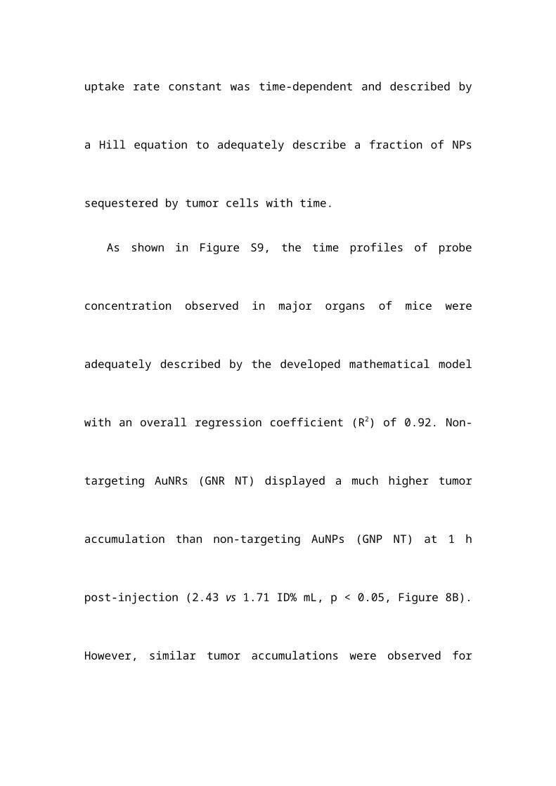
Figure 2. RGD-probe specificity for v3 positive cells. A: Two-photon
microscopy images of H1299 cells after incubated with 50 μg Au/mL of probes for 1
h. B, C: The z-stack reconstructions for orthogonal projections showing the relative
position of RGD-IPt- AuNPs (B) and RGD-IPt-AuNRs (C) (green) to cell body (blue,
cell nuclei stained by DAPI). D: Intracellular gold contents measured by AAS. RAD:
RAD probes; RGD: RGD probes; COM: RGD probes plus free peptide. **, p < 0.01.
Scale bar: 20 μm.
Figure 3. Cell viability of H1299 tumor cells after chemoradiotherapy. The cells were
incubated with RGD-IPt- AuNPs (A) or RGD-IPt- AuNRs (B) at the concentration of
50 μg Au/mL for 6 h. Radiotherapy was performed by irradiating the treated cells to 4
Gy of 320 kV X-ray.
Figure 4. SPECT/CT imaging and bio-distributions at the organ level. A, B:
SPECT/CT imaging of H1299 tumor bearing mice after intravenous administration of
RGD-125IPt- AuNPs (A) or RGD-125IPt- AuNRs (B) (300 μCi). C, D: Bio-distributions of
RGD-125IPt- AuNPs (C) and RGD-125IPt- AuNRs (D) 6 h after injection. **, p < 0.01.
Figure 5. Distributions of the NPs in tumors at the sub-organ and cellular levels. A:
Photoacoustic imaging of tumor regions before and after the mice were intravenously
administered with both types of RGD probes using RAD probes as controls. Scale
bar = 10 μm. B: Silver and hematoxylin co-staining images of the tumor tissues after
PAI. Scale bar = 100 μm.
Figure 6. Tumor size after chemoradiotherapy. A, B: Growth curves of tumors after
tumor-bearing mice were treated with both AuNPs and AuNRs probes (40 mg Au /kg
b. w) (A) or with the probes plus X-ray radiations (320 kV, 6Gy) (B). *, p < 0.05; **, p
< 0.01.
Figure 7. γ-H2AX immunostaining images of tumor tissues two days after the
treatments. The mice were treated with either RGD-IPt-AuNPs or RGD-IPt- AuNRs in
combination with 6 Gy of X-ray radiation.
Figure 8. In vivo kinetics of AuNPs and AuNRs probes. (A) Schematic diagram of the
mathematical compartment model. Gray boxes indicate the NPs uptake by
phagocytic cells isolated from blood circulation or tissues. (B) Model calibration
results with experimental data. The line represents the concentration-time profile of
probes simulated by the model.