Embed Size (px)
Citation preview

S1
Supplementary data
7-Deacetyl-10-alkylthiocolchicine derivatives – new compounds with potent anticancer and fungicidal activity
Joanna Kurek*a, Patrycja Kwaśniewska-Sipb, Krzysztof Myszkowskic, Grzegorz Coftad
Marek Muriasb, Piotr Barczyński a, Beata Jasiewicz a, Rafał Kurczabe
a Laboratory for Chemistry of Heterocyclic Compounds, Faculty of Chemistry, Adam Mickiewicz University, Umultowska 89b, 61-614 Poznań, Poland
b Wood Technology Institute, Environmental Protection and Wood Chemistry Department, Winiarska 1, 60-654 Poznan, Poland
c Department of Toxicology, Poznan University of Medical Sciences, Dojazd 30, 60- 631 Poznań, d Poland Institute of Chemical Wood Technology, University of Life Science, Wojska Polskiego 38/42,
60-037 Poznań, Polande Department of Medicinal Chemistry, Institute of Pharmacology, Polish Academy of Sciences, Smętna
12, 31-343 Kraków, Poland
TABLE OF CONTENTS
1. Chemistry
1.1. Materials
.................................................................................................................... S2
1.2. Experimental
............................................................................................................. S2
1.3. Synthesis of 7-deacetyl-10-alkylthiocolchicines 7-11
………..…………………......... S3
1.4. EI MS mass spectra for 7-deacetyl-10-alkylthiocolchicines 7-11
............................ S8
1.5. FT IR spectra of 7-deacetyl-10-alkylthiocolchicines 7-11
…………..…...………. S10
1.6. 13C NMR spectra (CDCl3) of 7-deacetyl-10-i-propylthiocolchicine
(10)…….....… S13
Electronic Supplementary Material (ESI) for MedChemComm.This journal is © The Royal Society of Chemistry 2018

S2
1.7. 1H and 13C NMR spectra (CDCl3) of 7-deacetyl-10-n-butylthiocolchicine
(11)…. S14
1.8. 1H and 13C NMR spectra (DMSO) of 7-deacetyl-10-n-butylthiocolchicine
(11)…. S16
2. Lipophilicity of the molecules 1-11 ......................................................................... S18
3. DFT calculations ……………………………………………………………….….. S19
4. Docking studies …………………………………………………………………..... S20
5. Cytotoxic activity ………………………………………………………………….. S22
6. Fungicidal activity ………………………………………………………………... S24
1. Chemistry
1.1. Materials
Colchicine 1 is commercially available on ApplyChem. For all reactions a natural isomer
(-)-(aR,7S) was used. 10-Alkylthiocolchicine 2-6 were obtained according literature procedure
from colchicine and respective sodium alkylthiolate RSNa1. Sodium alkylthiolates are
commercially available of Fluka.
(1) Kurek, J.; Boczoń, Wł.; Murias, M.; Myszkowski, K.; Borowiak, T.; Wolska, I., Synthesis of sulfur containing colchicine derivatives and their biological evaluation as cytotoxic agents, Let. Drug Des. Disc. 2014, 11, 279–289.
1.2. Experimental Measurements
The NMR spectra of 7-dacetyl-10-alkilthiocolchicines 7-11 (0.07 mol L-1) were recorded
in DMSO-d6 and CDCl3 solutions using a Varian Gemini 300 MHz spectrometer. All spectra
were locked to deuterium resonance of DMSO. The 1H NMR measurements in DMSO-d6 and
CDCl3 were carried out at the operating frequency 300.075 MHz; flip angle, pw = 450;
spectral width 4500 Hz; acquisition time 2.0 s; relaxation delay, d1=1.0 s; T = 293.0 K and
using TMS as the internal standard. No window function or zero filling was used. Digital
resolution was 0.2 Hz per point. The error of chemical shift value was 0.01 ppm. 13C NMR
spectra were recorded at the operating frequency 75.454 MHz; pw = 600; sw = 19000 Hz; at =
1.8 s; d1=1.0 s; T = 293.0 K and TMS as the internal standard. Line broadening parameters
were 0.5 or 1 Hz. The error of chemical shift value was 0.01 ppm. The 1H and 13C NMR
signals were assigned for each species using one or two-dimensional (COSY, HETCOR,
HMBC) spectra. The FT IR spectra (0.07 mol dm-3) were recorded in the mid infrared region
in KBr pellets. The spectra were taken with an IFS 113v FT IR spectrophotometer (Bruker,

S3
Karlsruhe) equipped with a DTGS detector; resolution 2 cm–1, NSS = 125. A cell with Si
windows and wedge-shaped layers was used to avoid interferences (mean layer thickness 170
μm). The Happ-Genzel apodization function was used. All manipulations with the substances
were performed in a carefully dried and CO2-free glove box. The EI mass spectra were
recorded on a Waters/Micromass (Manchester, UK) ZQ mass spectrometer equipped with a
Harvard Apparatus syringe pump. Elemental analysis (% C, N, S, H) was carried out by
means of a Elementar Analyser Vario EL III. Melting point was determined on BUCHI SMP-
20. Melt-Temp II apparatus (Laboratory Devices Inc.).
The EI mass spectra were recorded on an AMD-402 two-sector mass spectrometer (AMD
Intectra GmbH Co. Harpstedt, Germany) with an acceleration voltage of 8 kV, electron
energy 70 eV, mass resolution 6000, and an ion source temperature of ∼150 °C. The samples
were introduced using a direct insertion probe. The UV-Vis spectra were recorded in
methanol by JASCO V-550 spectrophotometer at 200-600 nm range.
1.3. Synthesis of 7-deacetyl-10-alkylthiocolchicines 7-11
100 mg of 2, 3, 4, 5 and 6 was dissolved in 5 mL of methanol. To this solution 100-150 mL of
1M hydrochloric acid in MeOH was added, then reaction mixture was refluxed for 6-16h
(temperature ~70oC). The reaction progress was checked by TCL analysis (chloroform :
aceton, 3:2, v/v). New derivatives 7-11 were obtained with very good yields. Carbon atoms
numbering of colchicine 1 and derivatives 2-11 are given below in Figure 1.
O
R
OCH3
CH3O
CH3O
NH R'7
10
-S-CH2-CH2-CH2-CH3R:16171819
-O-CH3
1
34
4a5 6
7a2 1a
8
811
12
12a
13
14
15 R R’
1 OCH3 COCH32 SCH3 COCH33 SCH2CH3 COCH34 SCH2CH2CH3 COCH35 SCH(CH3)2 COCH36 SCH2CH2CH2CH3 COCH37 SCH3 H8 SCH2CH3 H9 SCH2CH2CH3 H10 SCH(CH3)2 H11 SCH2CH2CH2CH3 H
Figure S1. Carbon atom numbering of Colchicine 1 and derivatives 2-11.

S4
7-deacetyl-10-methylthiocolchicine (7)
The title compound was prepared from 10-methylthiocolchicine 2 and methanolic solution of
hydrochloric acid. M.p. 183-186oC, yield 86%, 1H NMR (300 Hz, DMSO-d6, TMS, ppm):
6.84 (HC-4, s), 2.21, 2.69 (HC-5, m), 1.98, 2.51 (HC-6, m), 2.32 (HC-7, m), 7.07 (HC-8, s),
7.35 (HC-11, d), 7.20 (HC-12, d), 3.62 (H3C-13, s), 3.85 (H3C-14, s), 3.79 (H3C-15, s), 8.79
(NH), 2.57 (H3C-16, s); 13C NMR (75 MHz, DMSO-d6, TMS, ppm): 144.88 (C-1), 124.52 (C-
1a) 140.84 (C-2), 153.51 (C-3), 107.82 (C-4), 134.90 (C-4a), 28.58 (C-5), 34.75 (C-6), 52.57
(C-7), 150.38 (C-7a), 128.20 (C-8), 180.78 (C-9), 157.93 (C-10), 126.98 (C-11), 133.64 (C-
12), 136.48 (C-12a), 61.02 (C-13) 60.60 (C-14) 55.4 (C-15) 14.42 (C-16),
13C NMR (75 MHz, CDCl3, TMS, ppm): 145.84 (C-1), 124.49 (C-1a), 141. 48 (C-2), 154.08
(C-3), 107.63 (C-4), 137.98 (C-4a), 29.51 (C-5), 35.59 (C-6), 54.02 (C-7), 150.90 (C-7a),
129.24 (C-8), 181.71 (C-9), 159.05 (C-10), 127.39 (C-11), 135.82 (C-12), 133.69 (C-12a),
61.67 (C-13), 61.17 (C-14) 56.02 (C-15) 15.05 (C-16), 1H NMR (300 Hz, CDCl3, TMS, ppm):
6.56 (HC-4, s), 2.36, 2.56 (HC-5, m), 1.96, 2.45 (HC-6, m), 4.70 (HC-7, m), 7.63 (HC-8, s),
7.08 (HC-11, d), 7.34 (HC-12, d), 3.71 (H3C-13, s), 3.93 (H3C-14, s), 3.87 (H3C-15, s), 9.19
(NH), 2.36 (H3C-16, s); Anal. elem. calc. for C20H23NO4S∙3.5H2O C 55.04, H 6.88, N 3.21, S
7.33 %; found: C 54.88, H 6.84, N 2.89, S 5.53 %. UV (CH3OH) [nm]: max1 360, max2 245;
FT IR (KBr): 3377 (NH), 2933, 2858, 1598 (C=O), 1535, 1487, 1232, 1138, 1092, 844 (C-S).
7-deacetyl-10-ethylthiocolchicine (8)
The title compound was prepared from 10-ethylthiocolchicine 3 and methanolic solution of
hydrochloric acid. M.p. 174-176oC, yield 80%, 1H NMR (300 Hz, DMSO- d6, TMS, ppm):
6.85 (HC-4, s), 2.23, 2.70 (HC-5, m), 1.97, 2.51 (HC-6, m), 2.32 (HC-7, m), 7.08 (HC-8, s),
7.41 (HC-11, d), 7.18 (HC-12, d), 3.69 (H3C-13, s), 3.86 (H3C-14, s), 3.79 (H3C-15, s), 8.89

S5
(NH), 2.57 (H3C-16, s); 13C NMR (75 MHz, DMSO-d6, TMS, ppm): 144.79 (C-1), 124.51 (C-
1a), 140.82 (C-2), 153.48 (C-3), 107.81 (C-4), 134.87 (C-4a), 28.59 (C-5) 34.70 (C-6) 52.53
(C-7) 150.37 (C-7a), 128.48 (C-8), 180.79 (C-9), 156.66 (C-10), 127.21 (C-11), 133.64 (C-
12), 136.60 (C-12a), 61.00 (C-15) 60.58 (C-15) 55.92 (C-16), 24.48 (C-17), 12.51 (C-16),
13C NMR (75 MHz, CDCl3, TMS, ppm): 145.62 (C-1), 124.52 (C-1a), 141.49 (C-2), 154.01
(C-3), 107.60 (C-4), 138. 10 (C-4a), 29.51 (C-5), 35.44 (C-6), 53.98 (C-7), 150.92 (C-7a),
129.41 (C-8), 181.62 (C-9), 158.16 (C-10), 127.67 (C-11), 135.74 (C-12), 133.70 (C-12a),
61.74 (C-13) 61.17 (C-14), 56.00 (C-15), 25.43 (C-17), 12.35 (C-16), 1H NMR (300 Hz,
CDCl3, TMS, ppm): 6.55 (HC-4, s), 2.47, 2.69 (HC-5, m), 1.99, 2.55 (HC-6, m), 4.27 (HC-7,
m), 7.62 (HC-8, s), 7.29 (HC-11, d), 7.14 (HC-12, d), 3.74 (H3C-13, s), 3.94 (H3C-14, s), 3.92
(H3C-15, s), 9.52 (NH), 1.40 (H3C-16, s), 2.83 (H2C-17);
Anal. elem. calc. for C21H25NO4S∙3H2O C 57.14, H 7.02, N 3.17, S 7.25 %, found C 57.60, H
7.58, N 2.90, S 6.53 %; UV (CH3OH) [nm]: max1 375, max2 245; FT IR (KBr): 3375 (NH),
2931, 2869, 1596 (C=O), 1538, 1487, 1234, 1136, 1092, 844 (C-S).
7-deacetyl-10-n-propylthiocolchicine (9)
The title compound was prepared from 10-n-propylthiocolchicine and methanolic solution of
hydrochloric acid. m.p. 133-135oC, yield 73%, 1H NMR (300 Hz, DMSO- d6, TMS, ppm):
6.84 (HC-4, s), 2.23, 2.68 (HC-5, m), 1.99, 2.51 (HC-6, m), 2.34 (HC-7, m), 7.08 (HC-8, s),
7.41 (HC-11, d), 7.18 (HC-12, d), 3.62 (H3C-13, s), 3.86 (H3C-14, s), 3.79 (H3C-15, s), 8.88
(NH), 2.92 (H2C-18),1.69 (H2C-17), 1.04 (H3C-16); 13C NMR (75 MHz, DMSO-d6, TMS,
ppm): 144.77 (C-1), 124.53 (C-1a), 140.83 (C-2), 153.49 (C-3), 107.82 (C-4), 134.87 (C-4a),
28.59 (C-5), 34.71 (C-6), 52.53 (C-7), 150.38 (C-7a), 128.41 (C-8), 180.81 (C-9), 156.83 (C-
10), 127.25 (C-11), 133.65 (C-12), 136.58 (C-12a), 61.01 (C-13), 60.59 (C-14), 55.92 (C-15),
32.28 (C-18), 20.65 (C-17), 13.55 (C-16),

S6
13C NMR (75 MHz, CDCl3, TMS, ppm): 145.71 (C-1), 124.56 (C-1a), 141.46 (C-2), 153.95
(C-3), 107.59 (C-4), 137.94 (C-4a), 29.56 (C-5), 35.47 (C-6), 54.00 (C-7), 150.93 (C-7a),
129.36 (C-8), 181.61 (C-9), 158.29 (C-10), 127.60 (C-11), 135.62 (C-12), 133.76 (C-12a),
61.77 (C-13), 61.19 (C-14), 55.99 (C-15), 13.85 (C-16), 20.88 (C-17), 33.43 (C-18), 1H NMR
(300 Hz, CDCl3, TMS, ppm): 6.56 (HC-4, s), 2.21, 2.55 (HC-5, m), 1.98, 2.49 (HC-6, m),
4.26 (HC-7, m), 7.64 (HC-8, s), 7.27 (HC-11, d), 7.12 (HC-12, d), 3.94 (H3C-13, s), 3.92
(H3C-14, s), 3.71 (H3C-15, s), 9.62 (NH), 2.80 (H3C-16, s), 1.76 (H2C-17, m), 1.10 (H2C-18,
t); Anal. elem. calc. for C22H27NO4S∙3H2O C 58.02, H 7.25, N 3.07, S 7.03 %, found C 58.53,
H 7.64, N 2.69, S 5.89 %. UV (CH3OH) [nm]: max1 370, max2 250; IR (KBr): 3386 (NH),
2959, 2929, 2869, 1597 (C=O), 1538, 1487, 1233, 1137, 1093, 841 (S-C).
7-deacetyl-10-i-propylthiocolchicine (10)
The title compound was prepared from 10-i-propylthiocolchicine and methanolic solution of
hydrochloric acid. m.p. 165-167oC, yield 75%, 1H NMR (300 Hz, DMSO- d6, TMS, ppm):
6.84 (HC-4, s), 2.23, 2.68 (HC-5, m), 1.98, 2.51 (HC-6, m), 2.92 (HC-7, m), 7.06 (HC-8, s),
7.47 (HC-11, d), 7.19 (HC-12, d), 3.62 (H3C-13, s), 3.85 (H3C-14, s), 3.78 (H3C-15, s), 8.79
(NH), 2.32 (HC-17), 1.30, 1.36 (H3C-16, s); 13C NMR (75 MHz, DMSO-d6, TMS, ppm):
144.72 (C-1), 124.48 (C-1a), 140.83 (C-2), 153.50 (C-3), 107.82 (C-4), 134.90 (C-4a), 28.60
(C-5), 34.73 (C-6), 52.51 (C-7), 150.40 (C-7a), 128.66 (C-8), 180.87 (C-9), 155.89 (C-10),
127.93 (C-11), 133.67 (C-12), 136.68 (C-12a), 61.01 (C-13), 60.60 (C-14), 55.93 (C-15),
33.70 (C-17), 21.93, 21.93 (C-16). 13C NMR (75 MHz, CDCl3, TMS, ppm): 145.47 (C-1),
124.51 (C-1a), 141.48 (C-2), 154.03 (C-3), 107.61 (C-4), 138.08 (C-4a), 29.57 (C-5), 35.47
(C-6), 54.00 (C-7), 150.96 (C-7a), 129.70 (C-8), 181.77 (C-9), 157.44 (C-10), 128.38 (C-11),
135.67 (C-12), 133.75 (C-12a), 61.85 (C-13) 61.22 (C-14), 56.03 (C-15), 34.48 (C-17), 22.02
(C-16x2), 1H NMR (300 Hz, CDCl3, TMS, ppm): 6.56 (HC-4, s), 2.21, 2.58 (HC-5, m), 1.98,
S7
2.55 (HC-6, m), 4.25 (HC-7, m), 7.63 (HC-8, s), 7.27 (HC-11, d), 7.18 (HC-12, d), 3.94 (H3C-
13, s), 3.92 (H3C-14, s), 3.71 (H3C-15, s), 9.52 (NH), 1.24 (H3C-16, s); 3.36(H2C-17, m),
Anal. elem. calc. for C22H27NO4S∙4H2O C 55.81, H 8.0, N 2.95, S 6.76 %, found C 55.64, H
7.41, N 2.66, S 6.55 %; UV (CH3OH) [nm]: max1 375, max2 250; FT IR (KBr): 3381 (NH),
2963, 2930, 2866, 1597 (C=O), 1536, 1488, 1237, 1137, 1093, 844 (C-S).
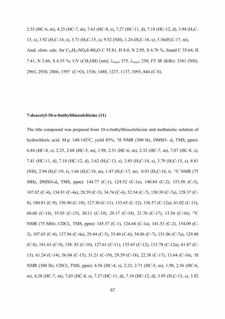
7-deacetyl-10-n-buthylthiocolchicine (11)
The title compound was prepared from 10-n-buthylthiocolchicine and methanolic solution of
hydrochloric acid. M.p. 140-142oC, yield 85%, 1H NMR (300 Hz, DMSO- d6, TMS, ppm):
6.84 (HC-4, s), 2.25, 2.68 (HC-5, m), 1.98, 2.51 (HC-6, m), 2.32 (HC-7, m), 7,07 (HC-8, s),
7.41 (HC-11, d), 7.18 (HC-12, d), 3.62 (H3C-13, s), 3.85 (H3C-14, s), 3.79 (H3C-15, s), 8.81
(NH), 2.94 (H2C-19, t), 1.66 (H2C-18, m), 1.47 (H2C-17, m), 0.93 (H3C-16, t). 13C NMR (75
MHz, DMSO-d6, TMS, ppm): 144.77 (C-1), 124.52 (C-1a), 140.84 (C-2), 153.50 (C-3),
107.82 (C-4), 134.91 (C-4a), 28.59 (C-5), 34.74 (C-6), 52.54 (C-7), 150.39 (C-7a), 128.37 (C-
8), 180.81 (C-9), 156.90 (C-10), 127.30 (C-11), 133.65 (C-12), 136.57 (C-12a), 61.02 (C-13),
60.60 (C-14), 55.93 (C-15), 30.11 (C-19), 29.17 (C-18), 21.70 (C-17), 13.54 (C-16), 13C
NMR (75 MHz, CDCl3, TMS, ppm): 145.57 (C-1), 124.64 (C-1a), 141.53 (C-2), 154.09 (C-
3), 107.65 (C-4), 137.94 (C-4a), 29.44 (C-5), 35.40 (C-6), 54.06 (C-7), 151.06 (C-7a), 129.48
(C-8), 181.61 (C-9), 158. 83 (C-10), 127.61 (C-11), 135.65 (C-12), 133.78 (C-12a), 61.87 (C-
13), 61.24 (C-14), 56.04 (C-15), 31.21 (C-19), 29.59 (C-18), 22.38 (C-17), 13.64 (C-16), 1H
NMR (300 Hz, CDCl3, TMS, ppm): 6.56 (HC-4, s), 2.23, 2.71 (HC-5, m), 1.98, 2.56 (HC-6,
m), 4.28 (HC-7, m), 7.65 (HC-8, s), 7.27 (HC-11, d), 7.10 (HC-12, d), 3.95 (H3C-13, s), 3.92

S8
(H3C-14, s), 3.71 (H3C-15, s), 9.60 (NH), 0.98 (H3C-16, t), 1.51 (H2C-17, m), 1.69 (H2C-18,
m), 2.66 (H2C-19, t); Anal. elem. calc. for C23H29NO4S∙4.5H2O C 55.6, H 7.66, 2.82 N, 6.45
S%, found C 55.32, H 7.23, N 2.73, S 6.03 %. UV (CH3OH) [nm]: max1 375, max2 250; FT
IR (KBr): 3387 (NH), 2953, 2930, 2866, 1596 (C=O), 1538, 1487, 1232, 1136, 1092, 844 (C-
S).
1.4. EI MS mass spectra for 7-deacetyl-10-alkylthiocolchicines 7-11
Figure S2. EI MS mass spectra of 7-deacetyl-10-methylthiocolchicine 7
S9
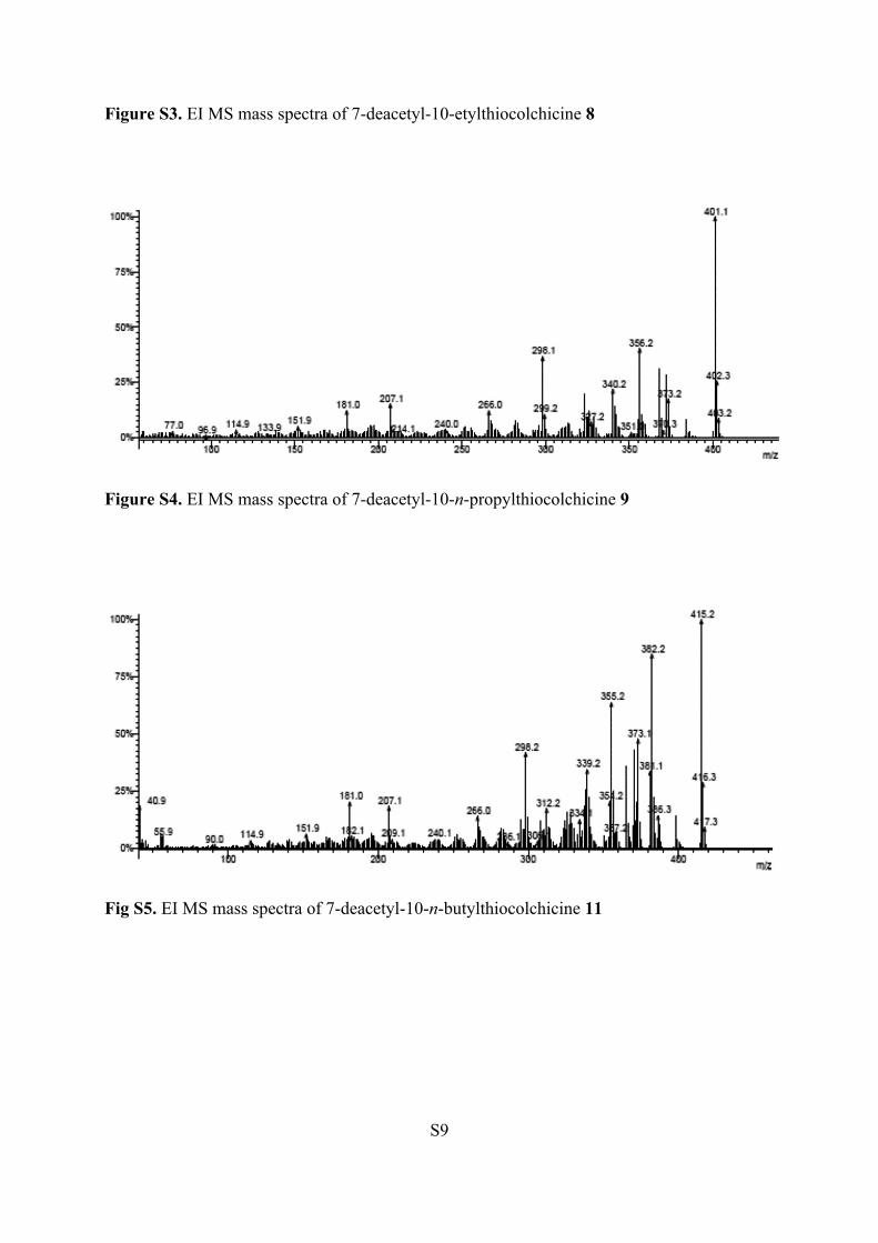
Figure S3. EI MS mass spectra of 7-deacetyl-10-etylthiocolchicine 8
Figure S4. EI MS mass spectra of 7-deacetyl-10-n-propylthiocolchicine 9
Fig S5. EI MS mass spectra of 7-deacetyl-10-n-butylthiocolchicine 11

S10
1.5. FT IR spectra of 7-deacetyl-10-alkylthiocolchicines 7-11
1598
Trams
mitan
ce [%
]
Wavenumbers cm-1
2
0
20
40
60
80
100
1800 16001800 1750 1700 1650 1600 1550 1500 1450
16031681
7
Fig. S6. FT IR spectra for 2 and 7 in the region of carbonyl group
Tran
smita
nce
[%]
Wavenumbers [cm-1]
0
25
50
75
100
1750 1700 1650 1600 1550 1500 1450 14001750 1700 1650 1600 1550 1500 1450 1400
3
15971603
1680
8
Fig. S7. FT IR spectra for 3 and 8 in the region of carbonyl group
S11
Tran
smita
nce
[%]
Wavenumbers [cm-1]
4
1800 1700 1600 1500 14000
20
40
60
80
100
16061678
1597
9
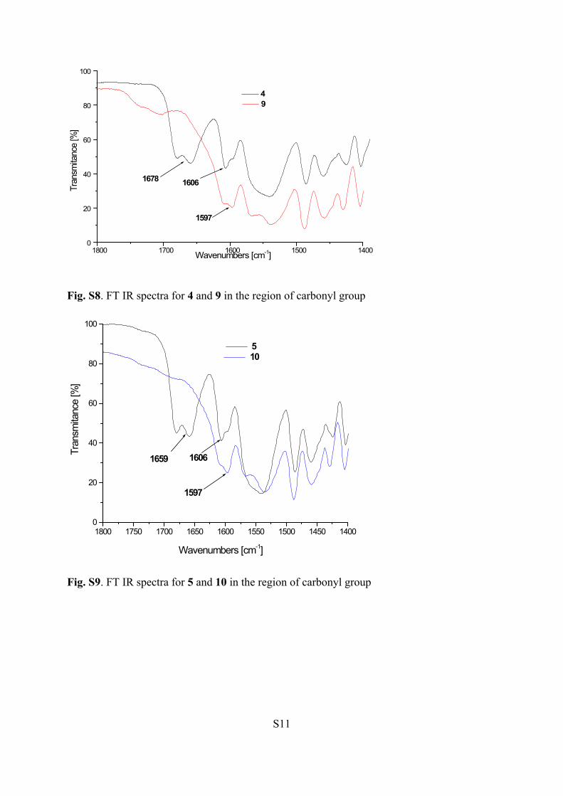
Fig. S8. FT IR spectra for 4 and 9 in the region of carbonyl group
1597
5
1659 1606Tran
smita
nce
[%]
Wavenumbers [cm-1]
1800 1750 1700 1650 1600 1550 1500 1450 1400
10
0
20
40
60
80
100
Fig. S9. FT IR spectra for 5 and 10 in the region of carbonyl group

S12
1750 1700 1650 1600 1550 1500 1450 1400
11
15961678
1604
Tran
smita
nce
[%]
Wavenumbers [cm-1]1750 1700 1650 1600 1550 1500 1450 1400
6
0
20
40
60
80
100
Fig. S10. FT IR spectra for 6 and 11 in the region of carbonyl group
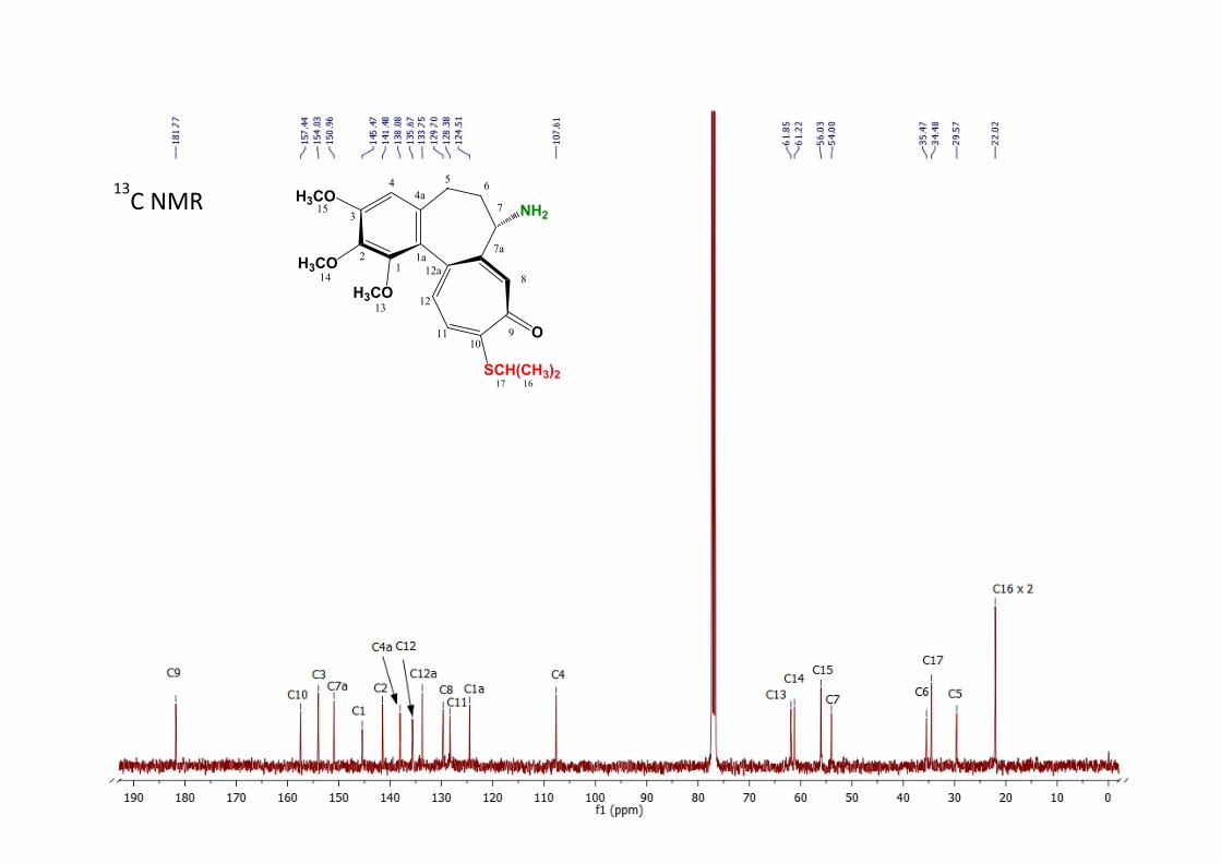
S13
SCH(CH3)2
H3CO
H3CO
H3CO
O
NH2
12
3
44a
5
7
6
7a
8
910
11
12
12a1a
13
14
15
1617
13C NMR (CD3Cl)

S14
SCH2CH2CH2CH3
H3CO
H3CO
H3CO
O
NH2
12
3
44a
5
7
6
7a
8
910
11
12
12a1a
13
14
15
16171819
1H NMR (CDCl3)

S15
SCH2CH2CH2CH3
H3CO
H3CO
H3CO
O
NH2
12
3
44a
5
7
6
7a
8
910
11
12
12a1a
13
14
15
16171819
13C NMR (CDCl3)

S16
SCH2CH2CH2CH3
H3CO
H3CO
H3CO
O
NH2
12
3
44a
5
7
6
7a
8
910
11
12
12a1a
13
14
15
16171819
1H NMR (DMSO)

S17
SCH2CH2CH2CH3
H3CO
H3CO
H3CO
O
NH2
12
3
44a
5
7
6
7a
8
910
11
12
12a1a
13
14
15
16171819
13C NMR (DMSO)

S18
2. Lipophilicity of the molecules
1
26
7 11Fig S11. Hydrophobic (blue color) and hydrophilic (orange and red colors) parts of molecules of derivatives 1, 2, 6, 7 and 11 (space-filling CPK models).

S19
4 5
9 10Fig S12. Comparison of hydrophobic (blue color) and hydrophilic (orange and red colors) parts of molecules of derivatives with propylthio chain 4, 5, 9 and 10 (space-filling CPK models).
Differences between 4, 9 and 5, 10 derivatives with unbranched CH3CH2CH2S- and branched (CH3)2CHS- propylthio chain were visualized in Fig. 5.
3. DFT calculation
Information on geometry of the new compounds was obtained using quantum-chemical calculations. The calculations were carried out by the density functional theory method (DFT) at the B3LYP/6-311G level implemented in the Gaussian 03 program package.2
(2) Frisch, M.J.; Trucks, G.W.; Schlegel, H.B.; Scuseria, G.E.; Robb, M.A.; Cheeseman, J.R.; Montgomery, J.A.; Vreven, Jr. T.; Kudin, K.N.; Burant, J.C.; Millam, J.M.; Iyengar, S.S.; Tomasi, J.; Barone, V.; Mennucci, B.; Cossi, M.; Scalmani, G.; Rega, N.; Petersson, G.A.; Nakatsuji, H.; Hada, M.; Ehara, M.; Toyota, K.; Fukuda, R.; Hasegawa, J.; Ishida, M.; Nakajima, T.; Honda, Y.; Kitao, O.; Nakai, H.; Klene, M.; Li, X.; Knox, J.E.; Hratchian, H.P.; Cross, J.B.; Adamo, C.; Jaramillo, J.; Gomperts, R.; Stratmann, R.E.; Yazyev, O.; Austin, A.J.; Cammi, R..; Pomelli, C.; Ochterski, J.W.; Ayala, P.Y.; K. Morokuma, G.A.; Voth, P.; Salvador, J.J.; Dannenberg, V.G.; Zakrzewski, S.; Dapprich, A.D.; Daniels, M.C.; Strain, O.; Farkas, D.K.; Malick, A.D.; Rabuck, K.; Raghavachari, J.B.; Foresman, J.V.; Ortiz, Q;. Cui, A.G.; Baboul, S.; Clifford, J.; Cioslowski, B.B.; Stefanov, G.; Liu, A.; Liashenko, P.; Piskorz, I.; Komaromi, R.L.; Martin, D.J;. Fox, T.; Keith, M.A.; Al-Laham, C.Y.; Peng, A.; Nanayakkara, M.; Challacombe, P.M.; Gill, W.; Johnson, B. Chen, W.; Wong, M.W., Gonzalez, C.; Pople. J.A.; Gaussian 03, Revision B.04, Gaussian, Inc., Pittsburgh PA, 2003.

S20
4. Molecular docking
The structures of all synthesized molecules were prepared using LigPrep v3.6 3, and the
appropriate ionization states at pH = 7.4 were assigned using Epik v3.4.4 The crystal
structures of human tubulin in complex with colchicine (PDB ID: 1SA0)5 and the
mitochondrial cytochrome bc1 enzyme complex (CYTBC1) with azoxystrobin (PDB ID:
1SQB)6 were retrieved from Brookhaven Protein Data Bank.7 The Protein Preparation
Wizard8 was used to assign the bond orders, check the steric clashes, and assign appropriate
amino acid ionization states. The receptor grids were generated (the OPLS_2005 force field)
by set up the grid box on the center of co-crystalized ligand. Automated docking of all
synthetized compounds was performed by using Glide v6.9 9 at SP level with the flexible
docking option turned on. The ligand-receptor complexes were visualized by means of the
PyMOL Molecular Graphics System.
4.2. Sequence alignment and construction of fungal tubulin models
The sequences of fungal β-tubulins were obtained from the UniProtKB/Swiss-Prot
database:
Aspergillus niger van Tiegen ID: A2QQP0,
Aspergillus versicolor ID: A0A1L9P7L4,
Paecilomyces variotii ID: V5G9H5,
Penicillium funiculosum ID: not available,
Chaetomium globosum ID: Q2GSL5,
Aureobasidium pullulans ID: A0A074XVP4,
Penicillium cyclopium ID: G5CIU9,
Trichoderma viride Pers ID: P31863.
A crystal structure of human β-tubulin (PDB ID: 1SA0, co-crystalized with colchicine) was
used as the 3-dimensional template for the homology modeling. The multiple sequence
alignment (Figure S11) and identity matrix (Table S1) were obtained using Schrödinger Suit.
The homology models for all fungal β-tubulin were generated using Prime module in

S21
Schrödinger. Next, the models were energetically optimized using the steepest descent
algorithm and OPLS3 force field using MacroModel. The minimization was completed when
the RMS gradient convergence reached a 0.05 kJ/(Å∙mol).
Figure S13. Multiple sequence alignment between human and fungal β-tubulin units. The key
amino acids in the colchicine binding site are indicated by a red frame.
Table S1. Identity matrix calculated between sequences of human and fungal β-tubulins.
Hum
an sa
pien
s
Cha
etom
ium
glo
bosu
m
Peni
cilli
um c
yclo
pium
Aure
obas
idiu
m p
ullu
lans
Paec
ilom
yces
var
iotii
Aspe
rgill
us v
ersi
colo
r
Aspe
rgill
us n
iger
Tric
hode
rma
viri
de P
ers
Human sapiens 100.00 84.41 83.13 40.91 80.73 81.53 81.31 80.36Chaetomium globosum 84.41 100.00 93.75 41.85 94.93 93.33 93.60 93.05Penicillium cyclopium 83.13 93.75 100.00 41.57 94.28 94.58 94.58 89.46Aureobasidium pullulans 40.91 41.85 41.57 100.00 39.35 39.55 39.77 40.55Paecilomyces variotii 80.73 94.93 94.28 39.35 100.00 96.13 96.13 90.41Aspergillus versicolor 81.53 93.33 94.58 39.55 96.13 100.00 98.88 90.36Aspergillus niger 81.31 93.60 94.58 39.77 96.13 98.88 100.00 91.26Trichoderma viride Pers 80.36 93.05 89.46 40.55 90.41 90.36 91.26 100.00
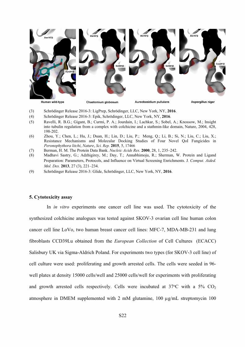
Figure S14. Comparison of the colchicine binding site shapes between human wt and several
fungal β-tubulin homology models. The human wild-type tubulin was visualized with co-
crystalized ligand (colchicine, cyan).
S22
(3) Schrödinger Release 2016-3: LigPrep, Schrödinger, LLC, New York, NY, 2016.(4) Schrödinger Release 2016-3: Epik, Schrödinger, LLC, New York, NY, 2016.(5) Ravelli, R. B.G.; Gigant, B.; Curmi, P. A.; Jourdain, I.; Lachkar, S.; Sobel, A.; Knossow, M.; Insight
into tubulin regulation from a complex with colchicine and a stathmin-like domain, Nature, 2004, 428, 198-202.
(6) Zhou, Y.; Chen, L.; Hu, J.; Duan, H.; Lin, D.; Liu, P.; Meng, Q.; Li, B.; Si, N.; Liu, C.; Liu, X.; Resistance Mechanisms and Molecular Docking Studies of Four Novel QoI Fungicides in Peronophythora litchi, Nature, Sci. Rep. 2015, 5, 17466
(7) Berman, H. M. The Protein Data Bank. Nucleic Acids Res. 2000, 28, 1, 235–242.(8) Madhavi Sastry, G.; Adzhigirey, M.; Day, T.; Annabhimoju, R.; Sherman, W. Protein and Ligand
Preparation: Parameters, Protocols, and Influence on Virtual Screening Enrichments. J. Comput. Aided. Mol. Des. 2013, 27 (3), 221–234.
(9) Schrödinger Release 2016-3: Glide, Schrödinger, LLC, New York, NY, 2016.
5. Cytotoxicity assay
In in vitro experiments one cancer cell line was used. The cytotoxicity of the
synthesized colchicine analogues was tested against SKOV-3 ovarian cell line human colon
cancer cell line LoVo, two human breast cancer cell lines: MFC-7, MDA-MB-231 and lung
fibroblasts CCD39Lu obtained from the European Collection of Cell Cultures (ECACC)
Salisbury UK via Sigma-Aldrich Poland. For experiments two types (for SKOV-3 cell line) of
cell culture were used: proliferating and growth arrested cells. The cells were seeded in 96-
well plates at density 15000 cells/well and 25000 cells/well for experiments with proliferating
and growth arrested cells respectively. Cells were incubated at 37oC with a 5% CO2
atmosphere in DMEM supplemented with 2 mM glutamine, 100 g/mL streptomycin 100

S23
U/mL penicillin and 10% foetal bovine serum (FBS). For proliferating cells regular FBS
while for growth arrested cells charcoal treated FBS was used. The cells were allowed to
attach and after 24 h, the compounds (0.1–100 µM) dissolved in DMSO were added to each
well and incubated for 72 h. Control cells were treated with DMSO alone. The final DMSO
concentration in both treated and control samples was 0.1%. The growth of tumour cells was
quantified by the ability of the living cells to reduce the yellow dye 3-(4,5- dimethyl-2-
thiazolyl)-2,5-diphenyl-2H-tetrazolium bromide (MTT) to a purple formazan product.10 The
formazan product is formed and accumulates only in healthy cells, therefore colorimetric
signal generated from the assay is proportional to the number of living cells in the sample.10
At the end of the incubation, the plates were centrifuged and the medium was replaced by
fresh medium (200 μL) containing 0.5 mg/mL MTT. Three hours later, the MTT formazan
product was dissolved in 150 μL DMSO, and the absorbance was measured using a multiplate
reader (BioTek Elx-800, BioTek Instruments, Inc. Winooski, Vermont, USA). The drug effect
was quantified as the percentage of the absorbance of reduced dye at 550 nm in relation to
control wells. Statistical analyses were carried out using one-way ANOVA with Dunnett's
multiple comparison tests. The results presented as the mean ± SD from three independent
experiments. The values indicated cytotoxicity concentration (EC50) were calculated
according to the Hill’s equation (sigmoidal model of concentration-response curve) and
expressed as a mean ± SEM (standard error of mean) using GraphPad Prism version 5.00 for
Windows, GraphPad Software, San Diego California USA.
(10) Berridge, M.V.; Herst, P.M.; Tan, A.S. Biotechnol Annu Rev. 2005, 11, 127-133.
S24
6. Fungicidal activity
Fungi strains. The antifungal activity of tested compounds was evaluated against
microfungi commonly known as mold: Aspergillus niger van Tiegen BAM 4 (ATCC 6275),
Aspergillus versicolor BAM 8 (ATCC 11730), Paecilomyces variotii BAM 19 (ATCC
18502), Penicillium funiculosum BAM 22 (ATCC 11797) Chaetomium globosum BAM 12
(ATCC 6205), Aureobasidium pullulans BAM 10 (ATCC 9348), Penicillium cyclopium
Westling, Trichoderma viride Pers.
Antimicrobial assay
The cultures were prepared by single-spore isolation technique on PDA (potato dextrose
agar) slants and maintained by periodic transfer on the same medium for further experiments.
The fungal spores suspensions were obtained from two-week agar slants. The species were
provided by the BAM Federal Institute for Materials Research and Testing collection or by
the Institute of Chemical Wood Technology (Poznan University of Life Science).
96-Well fungal bioassay. The 96-well microtiter assay was used to determine the
sensitivity of eight strains of fungi A. niger, A. versicolor, P. variotii, P. funiculosum, Ch.
globosum, A. pullulans, P. cyclopium and T. viride to the new obtained derivatives of
colchicine. Tested compounds (10 mg) were dissolved in 200 µL methanol to obtain a high
concentration of the solution. After complete dissolving 10 µL volumes of tested solutions
were added using micropipette to 100 µL PDA as a culture medium into the wells. Before
that, PDA powder was dissolved in distilled water to a final concentration of 39 g/L a water
bath to lower the temperature. To each wells was added 10 µL of freshly made fungal spores
suspension (10-5 to 10-6 CFU/mL). The plates were incubated aerobically for 7 days in a moist
chamber with relative humidity (RH) above 95% at 28±1°C in the dark. Differences in
mycelial growth in each of the wells in the 96-well plates demonstrate sensitivity of pure
compounds and indicated fungistatic or fungicidal effects. Fungal growth was evaluated
macroscopically according to the three point scale of intensity mycelium growth: ¸¸+“ - no
visible growth under the microscope; ¸¸±“ - growth visible with the naked eye, growth of
hyphae without spores; ¸¸-“ - growth visible with the naked eye, sporulation mycelium. For
reproducibility and accuracy evaluation of the microtiter plate screening method experiments

S25
were done in triplicates and the results for each compounds were compared to the control
wells (without any additives, with 10 µL (5 mg/L) of commercial fungicide such as 3-iodo-2-
propynylbutylcarbamate (IPBC) as Preventol® MP100 from Lanxess. IPBC (Iodopropynyl
butylcarbamate) is a water-soluble preservative used globally in the paints & coatings, wood
preservatives, personal care, and cosmetics industries. It is used as an active substance for
formulation of antimicrobial products. It is effective against a wide range of fungal species,
such as Aspergillus niger and Trichoderma virens. The results of bioassay tests against
microfungi for compounds 7-11 are given in Table 1S below. Concentration of IPBC was
chosen based on previous studies. 11
(11) Viitanen, H.; Ritschkoff, A.C.; Coating and surface treatment of wood. In: Adan and Samson (Ed.) Fundamentals of mold growth in indoor environments and strategies for healthy living, Wageningen Academic Publishers, 2011, 463-488
Table S2. The results of bioassay tests against microfungi for compounds 7-11
IPBC (3-Iodo-2-propynyl butyl carbamate) Antimicrobial Preventol® MP100 from Lanxes
¸¸+“ - no visible growth under the microscope; ¸¸±“ - growth of hyphae without spores; ¸¸-“ - sporulation mycelium.
Fungal speciesCompound
A. niger
A. versicolor
P.variotti
P. funiculosum
T. viride
P. cyclopium
A. pullulans
Ch. globosum
7 - - - - - - ± +
8 - - - - - - ± +
9 - - + + + + ± ±
10 - - + + ± ± ± +
11 - - + + + + + +
chalkone + + + + + + + +
IPBC + + + + + + + +
control - - - - - - - -

S26
Table S3. Antifungal activity of compounds 1 and 8-11 the results of MFC [g/mL] and [mMol/mL]Fungi
Compounds A.niger
A. versicolor
P.variotti
P.funiculosum
T.viride
P. cyclopium
A.pullulans
Ch. globosum
1 >4000 >4000 >4000 >4000 >4000 >4000 1∙10-3
[2.5∙10-7] >4000
2 >4000 >4000 >4000 >4000 >4000 >4000 >4000 >40003 >4000 >4000 >4000 >4000 >4000 >4000 >4000 >40004 >4000 >4000 >4000 >4000 >4000 >4000 >4000 >40005 >4000 >4000 >4000 >4000 >4000 >4000 >4000 >40006 >4000 >4000 >4000 >4000 >4000 >4000 >4000 >40007 >4000 >4000 >4000 >4000 >4000 >4000 >4000 >40008 >4000 >4000 >4000 >4000 >4000 >4000 >4000 >4000
9 >4000 >4000 1000±0.0[2.5∙10-10]
2000±600[4.9∙10-10]
2000±0.0[4.9∙10-10]
2000±0.0[4.9∙10-10] >4000 >4000
10 >4000 >4000 1000±0.0[2.5∙10-10]
1000±0.0[2.5∙10-10]
2000±0.0[4.9∙10-10]
500±0.0[1.2∙10-10]
260±0.0[6.5∙10-11]
130±0.0[3.2∙10-11]
11 >4000 >4000 1000±0.0[2.4∙10-10]
1000±0.0[2.4∙10-10]
2000±0.0[4.8∙10-10]
260±0.0[6.3∙10-11]
260±0.0[6.3∙10-11]
130±0.0[3.1∙10-11]
chalcone (fungicide)*
65±0.0[3.1∙10-11]
2000±0.0[9.6∙10-10]
1000±0.0[4.8∙10-10]
130±0.0[6.2∙10-11]
500±0.0[2.4∙10-10]
260±0.0[1.2∙10-10]
500±0.0[2.4∙10-10]
130±0.0[6.2∙10-11]
IPBC (fungicide)*
2±0.0[7∙10-12]
2±0.0[7∙10-12]
2±0.0[7∙10-11]
2±0.0[7∙10-12]
100±0.0[3.5∙10-8]
2±0.0[7∙10-12]
1±0.0[3.5∙10-12]
5±0.0[1.7∙10-11]
![LGDNWLVFKHV 0DWHULDO ]XU $XVVWHOOXQJ XQG ]XP … fff.pdf · pica 0xvhxpvslgdjrjln 1dwxuklvwrulvfkhv 0xvhxp ghu %xujhujhphlqgh %huq du )heuxdu (yroxwlrqvprelol:hofkh *hvfklfkwh hu]lkohq](https://img.pdfslide.tips/doc/110x75/5ecd6d23b4aa2f226f0d0775/lgdnwlvfkhv-0dwhuldo-xu-xvvwhooxqj-xqg-xp-fffpdf-pica-0xvhxpvslgdjrjln-1dwxuklvwrulvfkhv.jpg)


![Acupuntura Pulso Tornozelo 21.2.ppt - portalunisaude.com.br tornozelo - regis.pdf · 0dwhuldo surgx]lgr shor surihvvru º g ² 5hjlqdogr /rughor0dwhuldo surgx]lgr shor surihvvru $](https://img.pdfslide.tips/doc/110x75/5e112701a85f816445318770/acupuntura-pulso-tornozelo-212ppt-tornozelo-regispdf-0dwhuldo-surgxlgr.jpg)
![% 9HQWLOSRVDXQH :HULO · 2020. 7. 16. · $owhu • fd -dkuh jhvfk¦w]w 0dwhuldo • 0hvvlqj $xvvwdwwxqj • :dvvhunodssh • 6wlpp]¾jh =xvwdqg • odfnlhuw • whfkqlvfk ¾ehukrow](https://img.pdfslide.tips/doc/110x75/61123643776918279f5ec1eb/-9hqwlosrvdxqh-hulo-2020-7-16-owhu-a-fd-dkuh-jhvfkww-0dwhuldo-a.jpg)


![0DWHULDO SURGX]LGR SHOR SURIHVVRU GIJGIJ 2019.pdf · 0dwhuldo surgx]lgr shor surihvvru d e/^dk^ e hzk&/^/k>m'/ k^ yh ^h^d ed d hwhedhz / ed1&/ $ djxokd gh dfxsxqwxud phwiolfd shor](https://img.pdfslide.tips/doc/110x75/5ea7eb2bc73c4f3d7c1ccbd3/0dwhuldo-surgxlgr-shor-surihvvru-2019pdf-0dwhuldo-surgxlgr-shor-surihvvru-d.jpg)