Embed Size (px)
Citation preview
I ,
Illl'l',
.1 11.' I I'
I' ,
,! 1\,,\
~ L I
f l , '
, \I I .. ~
~ ..
II l ' ,lll ,d
1/.1 j "
III ,:! \ ,~~ . .
III:.: .J '
ACia Chir S(anJ 141: 557-56.1.1975
FIVE CASES AND FIVE UNUSUAL INDICATIONS
FOR AUTOGENIC RENAL TRANSPLANTATION
B . S. Husberg . K. BakshanJeh. J. Lilly. R . Pfister.
D. p, Stables anJ T. E. Starzl
Frol/l Ih l' Dt'pUrtllIl'lIl ,' oIS/lrgen 01/(/ RCldi"/ogv , Ullil'er,fitv oIC%","o ,\It'diclll Cl'lIler,
{/l/(llhl' DI'IlI 'C'r Vell''''''.\' AdlllillislfUlioll Hospilol , D I'III '", , C%ra do 802l0, USA
(Submitted for publication July 27,1974)
Five cases of renal autotransplantation repres · difTerent indications for the procedure are pre· discussed.
nt to the development of techniques for
kidney transplantation and ex vil'o kid ney
. indic a tions have arisen for autogenic
transplantation with or without ill I'ilro re
surgery, Several cases have been de
earlier (Belzer et al.. 1970; Gelin et al ..
et al.. 1973; Caine. 1973; Corman et
CASE REPORTS
male had a history of severe hypertension (210/130 mmHg). He had markedly elevated
renin in peripheral venous blood . A proximal stenosis in the left renal artery was diagnosed.
hospital. he received a saphenous vein by-pass the aorta to the left renal artery in February, blood pressure improved postoperatively but ensuing months it again rose to approximately
and excretory urography showed a de. ,hrol11r"nhic phase of the left kidney . Repeat
demonstrated a severely stenosed by-pass
. 1973. he was referred to Colorado Genand underwent an autotransplantation of the
to the left iliac fossa . After completion of the the kidney was connected to a Waters perfusion
approximately one hour. using long extension . the operating field . Since the ureter was not
interrupted, it was occluded with a sponge-rubber-shod clamp duri ng the perfusion to prevent blood from the ureteral vessels from entering the perfusate . Postoperatively he became and remained normotensive (140/80 mmHg), Postoperative arteriography and urography show satisfactory arterial and ureteral anatomy (Fig. 2), The kidney function is normal and unchanged.
Case 2
A 52-year-old female had the left kidney removed in 1951 because of renal stones. In December, 1973, a renal cell carcinoma was diagnosed after she had noticed a lower right quadrant mass. No metastases were detectable . Her kidney function was normal with a creatinine clearance of 80 cc/min. Renal arteriography demonstrated a double renal artery blood supply with apparent blood supply to the tumor in the mid-region from both arteries (Figs . 3 and 4). In January. 1974. the kidney was approached through a right para media n extraperitoneal incision. There were no palpable retroperitoneal lymph nodes . The kidney was removed with a maximal length of renal vessels, The ureter was left uninterrupted. The kidney was perfused with Perfadex® solution and was then connected to a Waters perfusion machine using long extension tubes to the operating field . A soft. foam-rubber-coated clamp was placed on the ureter.
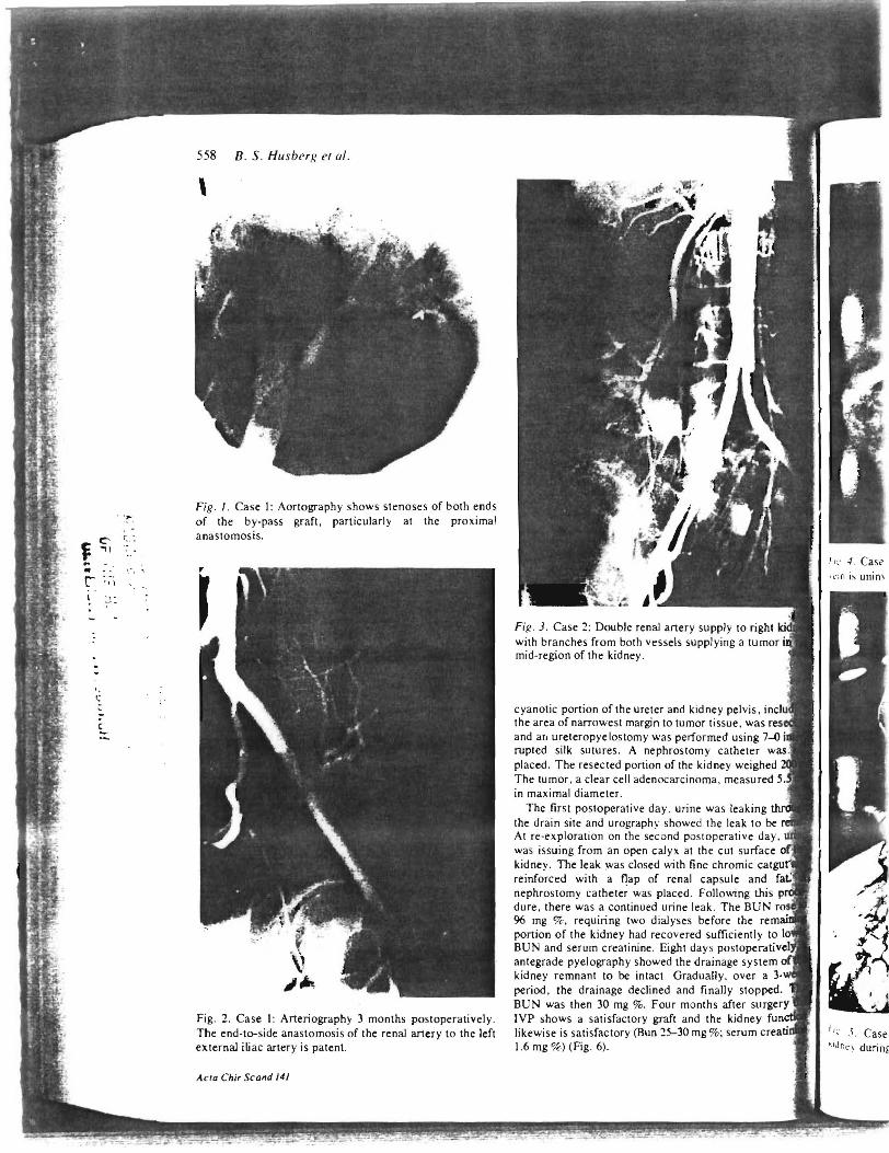
The lower artery and the branches from the upper artery supplying the tumor with blood were identified. tied off and injected with methylene blue solution distal to the ligatures. The area of the kidney thus demarcated was then resected . Thae was no extension of tumor beyond the lines of resection , as determined by frozen section. Approximately one-third of the kidney was left after the resection. With the perfusion machine working intermittently, numerous small blood vessels on the cut surface of the kidney were suture ligated . The calyceal openings were closed with fine chromic catgut (Fig. 5),
The kidney remnant was then autotransplanted to the right iliac fossa. The vessel and ureter clamps were removed after 4 hours and 25 min cold ischemic time . A
Acta Chir Scand 141
. ';p-
558 B. S. Hu sberR ef al .
\
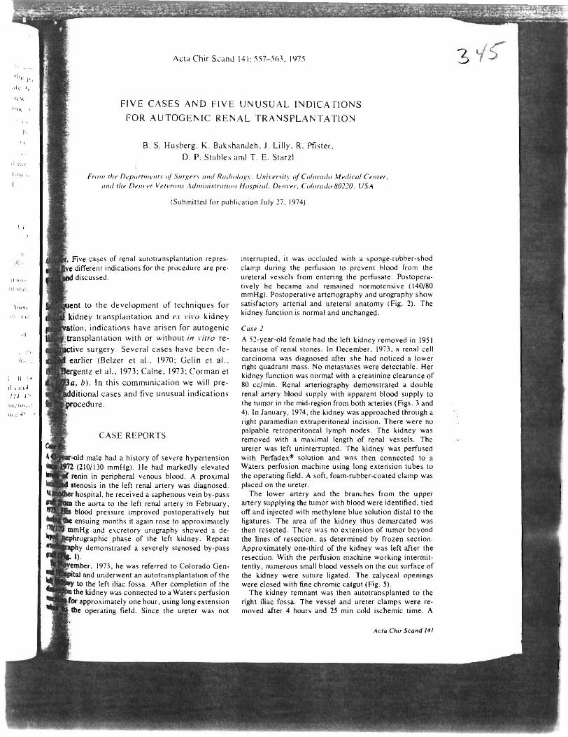
Fig. I . Case I: Aortography shows stenoses of both ends of the by-pass graft, particularly at the proximal ana stomosis.
Fig. 2. Case 1: Arteriography 3 months postoperatively . The end -to-side anastomosis of the renal artery to the left external i~ac artery is patent.
ACla Chir Scand J4J
FiR · J. Case 2: Double renal artery supply to right with branches from both vessels supplying a tumor mid-region of the kidney .
cyanotic portion of the ureter and kidney pelvis, . the area of narrowest margin to tumor tissue, was and a[1 ureteropyelostomy was performed using 7-0 rupted silk sutures . A nephrostomy catheter pl aced . The resected portion of the kidney weighed The tumor, a clear cell adenocarcinoma , measured in maximal diameter.
The first postoperative day. urine was leaking the drain site and urography showed the leak to be At re-exploration on the second postoperative day, was issu ing from an open calyx at the cut surface kidney . The leak was closed with fine chromic ca reinforced with a flap of renal capsule and nephrostomy cathete~ was placed. Following this dure , there was a continued urine lea k. The BUN 96 mg % , requiring two dialyses before the portion of the kidney had recovered sufficiently BUN and serum creatinine . Eight day s postn""p"'I"~'''I' antegrade pyelography showed the drainage system kidney remnant to be intact. Gradually , over a 3-period, the drainage declined and finally stopped. BUN was then 30 mg %. Four months after surgery IVP shows a satisfactory graft and the kid ney fu likewise is satisfactory (Bun 25-30 mg % ; serum creati 1.6 mg %) (Fig. 6).
J I,' .I , Case ',IJ r,n durin!,
n.d
559
Fig . 6. Case 2: Excretory urography 4 months postoperatively. The ureteropyelostomy (ARROW) is patent without residual urine leak.
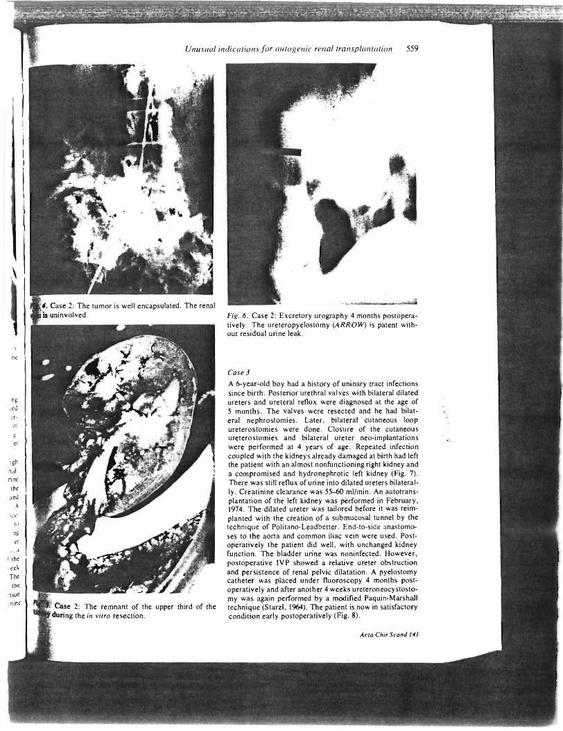
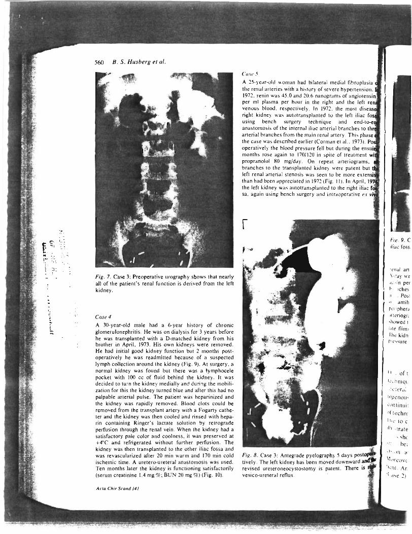
Case J A 6-year-old boy had a history of uninary tract infections since birth . Posterior urethral valves with bilateral dilated ureters and ureteral reflux were diagnosed at the age of 5 months. The valves were resected and he had bilateral nephrostomies . Later. bilateral cutaneous loop ureterostomies were done. Closure of the cutaneous ureterostomies and bilateral ureter neo-implantations were performed at 4 years of age . Repeated infection coupled with the kidneys already damaged at binh had left the patient with an almost nonfunctioning right kidney and a compromised and hydronephrotic left kidney (Fig. 7) . There was still reflux of urine into dilated ureters bilaterally . Creatinine clearance was 55-60 ml/min . An autotransplantation of the left kidney was performed in February. 1974. The dilated ureter was tailored before it was reimplanted with the creation of a submucosal tunnel by the technique of Politano-Leadbetter. End-to-side anastomoses to the aona and common iliac vein were used. Post· operatively the patient did well . with unchanged kidney function. The bladder urine was noninfected . However. postoperative IVP showed a relative ureter obstruction and persistence of renal pelvic dilatation. A pyelostomy catheter was placed under fluoroscopy 4 months postoperatively and after another 4 weeks ureteroneocystostomy was again performed by a modified Paquin-Marshall technique (Starzl . 1964). The patient is now in satisfactory condition early postoperatively (Fig. 8) .
Acla Chi, Scafld 141
, ',
C' ( .. f ~\ " '.' , '" ,... : ~:, ..
J:
560 B. S. Husberg et al.
Fig. 7. Case 3; Preoperative urography sbows that nearly all of the patient's renal function is derived from the left kidney.
Case 4
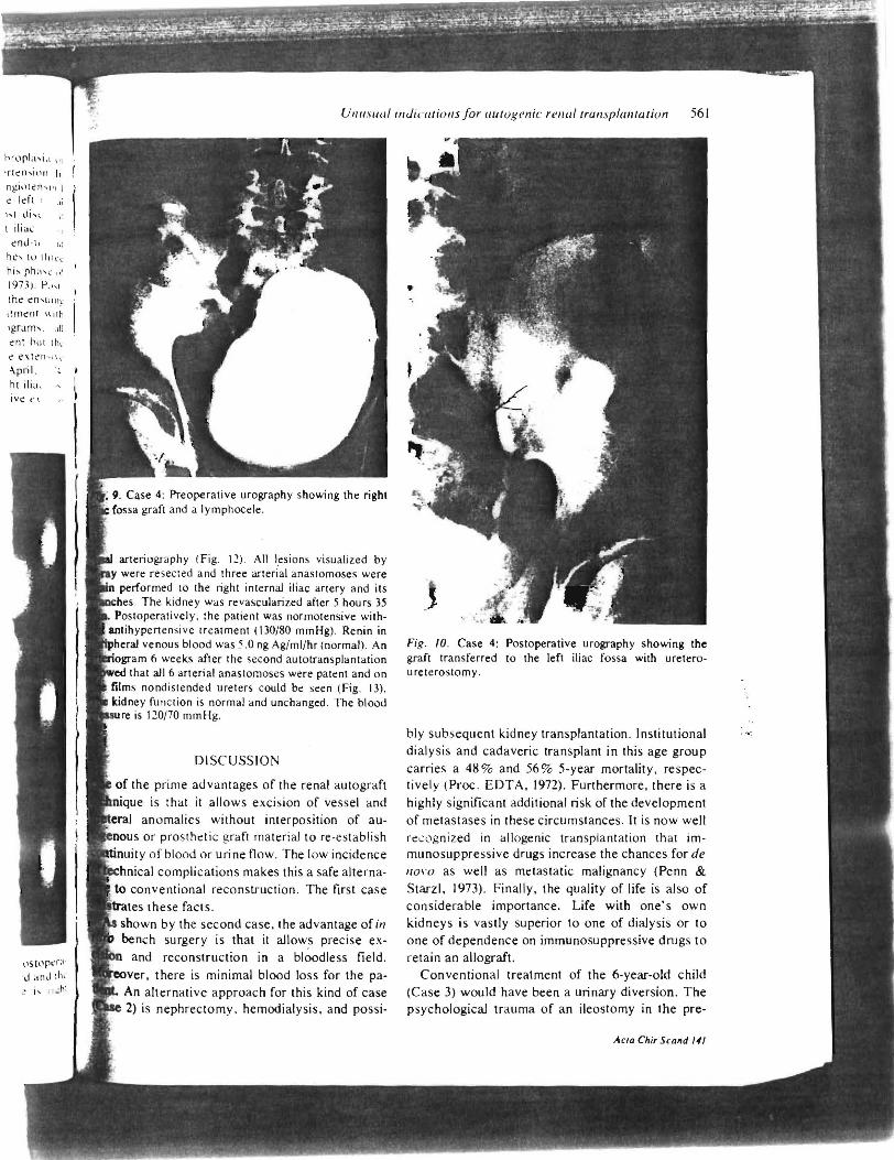
A 30-year-old male had a 6-year history of chronic glomerulonephritis . He was on dialysi s for 3 years before he was transplanted with a D-matched kidney from his brother in April , 1973. His own kidneys were removed. He had initial good kidney function but 2 months postoperatively he was readmitted because of a suspected lymph collection around the kidney (Fig. 9). At surgery, a normal kidney was found but there was a Iymphocele pocket with 100 cc of Ouid behind the kidney. It was decided to turn the kidney medially and during the mobili· zatiun for this the kidney turned blue and after this had no palpable arterial pulse. The patient was heparinized and the kidney was rapidly removed. Blood clots could be removed from the transplant artery with a Fogany catheter and the kidney was then cooled and rinsed with heparin containing Ringer's lactate solution by retrograde perfusion through the renal vein . When the kidney had a satisfaclOry pale color and coolness, it was preserved at +4°C and refrigerated without funher perfusion. The kidney was then transplanted to the other iliac fossa and was revascularized after 20 min warm and 170 min cold ischemic time. A uretero-ureteral anastomosis was used. Ten months later the kidney is functioning satisfactorily (serum creatinine 1.4 mg 0/[; BUN 20 mg '7c) (Fig . 10) .
ACla ChiT Scand 141
lllSI' 5
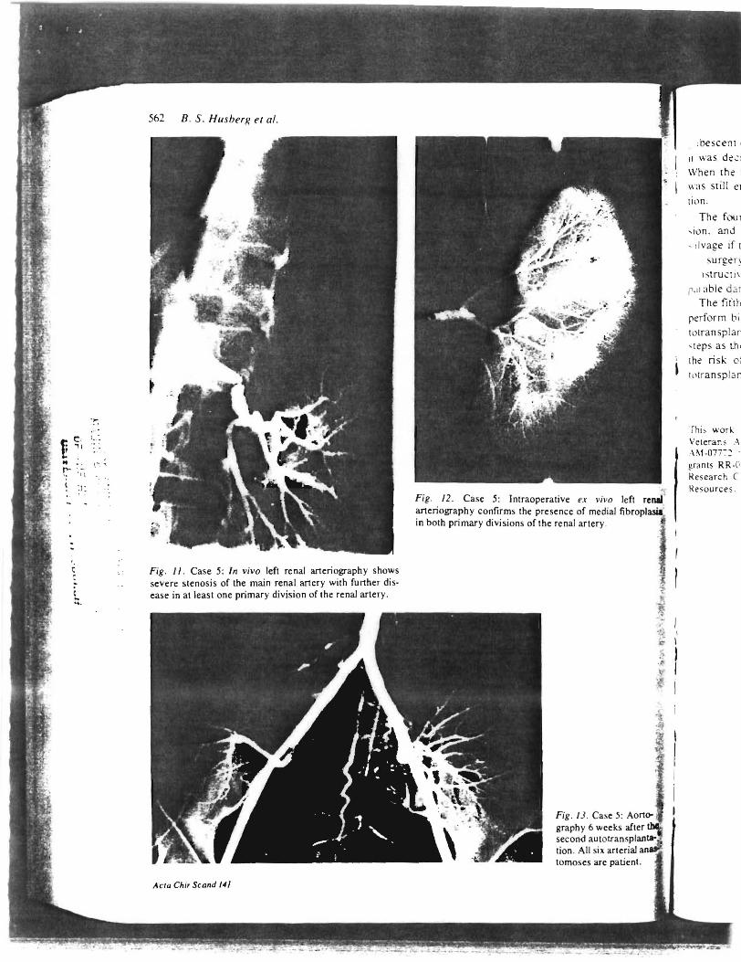
A 25·year-uld "oman had bilaleral medi:.tl fibroplasia the renal arteries with a hislury of severe hypertension. 1972. renin was 45 .0 and 20 .6 nanugrams of angiole per ml plasma per huur in the right and the left venuus blood. respeclively . In 1972 . the most U("east'!l.' right kidney was aututransplanted to the left iliac using bench surgery technique and end-t anastomosis of the internal iliac arterial branches to anerial branches frum the main renal artery . This phase the case was described earlier (Corman el al. . 1973). operalivel) the blood pressure fell but during the ens months rose again to 170/120 in spite of treatment propranolol 80 mg/day . On repeat arteriograms, branches 10 the transplanted kidney were patent but lefl renal arterial stenosis was seen tu be more ex than had been appreciated in 1972 (Fig . I I) . In April, I the left kidney was aUlOtransplanted 10 the right iliac sa, again using bench surgery and intraoperative ex
Fig . 8. Case 3; Antegrade pyelography 5 days pos tively . The left kidney has been moved duwnward revised ureteroneocystostomy is patent. There is vesico-ureteral reOux .
rliac foss .
' I;"n,,1 an ' ·ray we .,.,' , In per rIches
. POS I
antih P'Tiphera '(f1criogr; ,h"wed I
';'Ic film , 1 he kidn-
ofl
I,· , hniql'
l ' {c: rd :
Itl~cnou •
'tlntinuit
,,' te(;hn i
. trale
, Shl he;
,I~ . , In d
\1..rcllV(
' (e il i. Ar
.( '''e ~ )
hr\lpla,i .. d ·rten .... i~Hl 11
ngil)t~n'lll I I ~ Idt, .,i , hi Ji'l t ili,,(
cnJ-11 hI
he, tll I h,t'o
hi, ph"", ,'I 197:\1 . P,,,, the:!' C'n"ullI~' dm('nr \ \lIh
19ram, . ,r/I
ent hUI Ih, r: ~ .\ tcon -1\ r \pril, .~
hi iii", ivc l ' \
llStllp~r;r
'J anJ :h. ~ j, :..: h:
, 9. Case 4: Preoperative urography showing the right fossa graft and a Iymphocele.
arteriography (Fig. 12), All lesions visualized by were resected and three arterial anaSlomoses were performed to the right internal iliac artery and its
The kidney was revascularized after 5 hours 35 ratively. the patient was normotensive with
pertensive treatment (130/80 mmHg). Renin in venous blood was 5.0 ng Ag/ml/hr (normal) . An
m 6 weeks after the second autotransplantation that all 6 arterial anastomoses were patent and on
films nondistended ureters could be seen (Fig. 13) . . kidney function is normal and unchanged. The blood
re is 120170 mm Hg,
DISCUSSION
of the prime advantages of the renal autograft
is that it allows excision of vessel and
anomalies without interposition of aus or prosthetic graft material to re-establish
ty of blood or urine flow . The low incidence
I complications makes this a safe alterna-
shown by the second case, the advantage of in bench surgery is that it allows precise ex
and reconstruction in a bloodless field.
, there is minimal blood loss for the paAn alternative approach for this kind of case 2) is nephrectomy. hemodialysis, and possi-
561
Fig . 10. Case 4: Postoperative urography showing the graft transferred to the left iliac fossa with ureteroureterostomy.
bly subsequent kidney transplantation. Institutional
dialysis and cadaveric transplant in this age group
carries a 48 % and 56 % 5-year mortality, respectively (Proc, EDTA, 1972). Furthermore, there is a
highly significant additional risk of the development
of metastases in these circumstances. It is now well
rec:ognized in allogenic transplantation that immunosuppressive drugs increase the chances for de 1/0\'0 as well as metastatic malignancy (Penn & Starzl. 1973). Finally, the quality of life is also of considerable importance. Life with one's own kidneys is vastly superior to one of dialysis or to
one of dependence on immunosuppressive drugs to
retain an allograft. Conventional treatment of the 6-year-old child
(Case 3) would have been a urinary diversion. The psychological trauma of an ileostomy in the pre-
ACla ChiT Scafld 141
.. ,,',
. ~.
~ ~; ' . ...
M " ~ .
r.- '. ~ i
t .•.
562 B. S. HusberR el al.
Fig. ll. Case 5: In vivo left renal arteriography shows severe stenosis of the main renal artery with further disease in at least one primary division of the renal artery.
ACla Chir Scand 141
.. Fig. 12. Case 5: Intraoperative ex vil'o left refill' arteriography confirms the presence of medial fibroplasia' in both primary divisions of the renal artery .
Fig . /J. Case 5: graphy 6 weeks second "'r.n.<nlonl"~
tion. All six. arterial tomoses are patient.
:beseem c II was de'::l
When the f was stil l er til)n .
The fe.ur , i,ln . and
, dvage if t surgen
htru'::li\
1'. 11 able d;,.r The fiflh
rerform bi: totransplar "teps as th e
the risk 0;
I, 'transp la r .
Thi , wor~ Veteran:; A
.\,,'-07T: grants RR-C' Research C Resources .
Aorto· .lfter th' iplanta· ial ana" en!.
VI/llstlill ill£iiC({TiOllsjor allTogenic rellul Trullspl({IITaTioll 563
scent child may be devastating. Consequently. was decided to perform an autotransplantation .
the first ureteroneocystostomy failed . there still enough length to do another reimpl anta-
e fourth case illustrates that removal. perfuand reimplantation is a method of kidney
if the organ is inadvertently damaged dursurgery in the area. If necessary. ill \'iTro re
ructive work can also be performed if redamage has occurred.
firth case points out that indications exist to " iof'nrm bilateral autotransplantation. Bilateral au
antation should always be done in two
of an acute tubular necrosis in ted kidney is always ·present.
ACKNOWLEDGEMENTS
work was supported by research grants from the Administration; by grants AI-AM-08898 and
from the Na tional Institutes of Health : by -000:;1 and RR·00069 from the General Clinical
"~rch Centers Program of the Division of Research ....,""JIn.:<". National Institutes of Health .
REFERENCES Belzer. F. 0 .. Keaveny. T . V .. Reed. T . W. & Pryor. 1. P.
1970. A ne" method for renal artery reconstruction . SlIrgl'n·68.619.
Bergentz. S . E .. Faarup . P .. Hegedus. V .. Lindholm. T . & Lindstedt . E . 1973 . Diagnosis of hypertension due to occlusion of a supplemental renal arter~' ; its localization. treatment by removal from the body. microsurgi· cal repair ~nd reimplantation . AI/I/ SlIr~ 178. 643-647 .
Caine . R. Y. 19n. Treatment of bilateral hypernephromas by nephrectomy. excision of tumor and autotransplantation. Lanc e!. 1973 .
Combined report on regular dialysis and transplantation in Europe. 197~ . Proc EDTA 9. 3-34 .
Corman. 1. L .. Anderson . 1. T .. Taubman. 1.. Stables. D. P .. Halgrimson. C. G. & Popovtzer. M. 1973a. Ex "i\ '(J perfusion. arteriography . and autotran splantation procedures for kidney salvage . 511r): CI'IIN' Ohste! 137. 659--665 .
Corman . 1. L .. Girard. R .. Fiala . M .. Gallot. D .. Stonington. 0 .. Stables. D. P . . Taubman . 1. & Starz!. T . E. 1973h . Arterography duringex \'i,'O renal perfusion. A complication . Urologl' II. 222-226.
Gelin. L. E .. Claes. G .. Gustafsson . A. & Storm. B . 1971 . Total bloodlessness for extracorporeal organ repair. ReI' Surg 28 . 305-312.
Penn. I. & Starzl. T. E . 1973. Immunosuppression and cancer. Transplant Proc 5.943-947.
Starzl. T . E. 1964 . Experiel/ce in rel/al !ral/splal//(/rion. p. 100. W. B . Saunders Co .. Philadelphia . Pa .
ACla Chir Scand 141