Embed Size (px)
Citation preview
PF NEWS Vol. 29 No. 4 FEB, 2012 22 最近の研究から
最近の研究から
1. Introduction The progress of time-resolved X-ray diffraction (TRXD) techniques has strongly impacted the investigation on structural dynamics, especially in the field of shock-wave physics [1]. By using TRXD techniques, in situ study of dynamic behaviors of materials under shock compression has been extended to microscopic scale from traditionally macroscopic one, providing significant information of transformation mechanism [2, 3]. Recently, Ichiyanagi et al. [4] employed a single-shot time-resolved Laue technique to study phase transitions of single-crystal CdS under laser shock, and observed the overshoot of the wurtzite structure which is different from Gupta et al.’s results [5, 6]. In gas gun shock experiments Gupta et al. observed not only a transition to rocksalt structure but also an intermediate phase of a faced centered tetragonal structure at lower shock pressure using time-resolved absorption spectroscopy. This disagreement is explained by different time scales of shock compression. In laser-shock experiments the shock duration (~10 ns) in single crystal CdS is not sufficient for transformation, comparing to the order of μs in gas gun experiments. In this sense, the structural dynamics of laser-shocked materials has its particularity. The TRXD observation will improve remarkably our understanding on dynamics response of materials under laser shock. In this report, we use single-shot nanosecond time-resolved X-ray power diffraction technique to study structural dynamics of laser-shocked Y2O3 (3 mol%) stabilized tetragonal zirconia polycrystalline (3Y-TZP) ceramics. 3Y-TZP ceramics, consisting of almost 100 % tetragonal phase, has been extensively studied due to its excellent mechanical properties, such as strength and toughness [7].
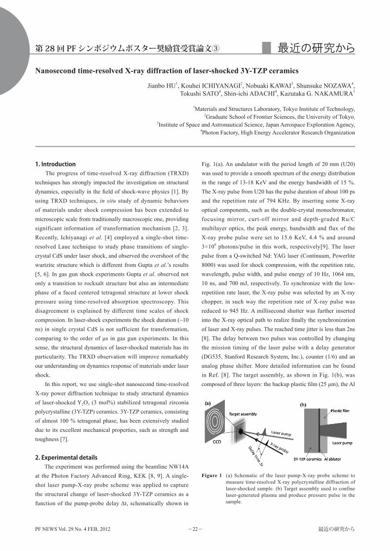
2. Experimental details The experiment was performed using the beamline NW14A at the Photon Factory Advanced Ring, KEK [8, 9]. A single-shot laser pump-X-ray probe scheme was applied to capture the structural change of laser-shocked 3Y-TZP ceramics as a function of the pump-probe delay Δt, schematically shown in
Fig. 1(a). An undulator with the period length of 20 mm (U20) was used to provide a smooth spectrum of the energy distribution in the range of 13-18 KeV and the energy bandwidth of 15 %. The X-ray pulse from U20 has the pulse duration of about 100 ps and the repetition rate of 794 KHz. By inserting some X-ray optical components, such as the double-crystal monochromator, focusing mirror, curt-off mirror and depth-graded Ru/C multilayer optics, the peak energy, bandwidth and flux of the X-ray probe pulse were set to 15.6 KeV, 4.4 % and around 3×10⁸ photons/pulse in this work, respectively[9]. The laser pulse from a Q-switched Nd: YAG laser (Continuum, Powerlite 8000) was used for shock compression, with the repetition rate, wavelength, pulse width, and pulse energy of 10 Hz, 1064 nm, 10 ns, and 700 mJ, respectively. To synchronize with the low-repetition rate laser, the X-ray pulse was selected by an X-ray chopper, in such way the repetition rate of X-ray pulse was reduced to 945 Hz. A millisecond shutter was further inserted into the X-ray optical path to realize finally the synchronization of laser and X-ray pulses. The reached time jitter is less than 2ns [8]. The delay between two pulses was controlled by changing the mission timing of the laser pulse with a delay generator (DG535, Stanford Research System, Inc.), counter (1/6) and an analog phase shifter. More detailed information can be found in Ref. [8]. The target assembly, as shown in Fig. 1(b), was composed of three layers: the backup plastic film (25 μm), the Al
Nanosecond time-resolved X-ray diffraction of laser-shocked 3Y-TZP ceramics
Jianbo HU1, Kouhei ICHIYANAGI2, Nobuaki KAWAI3, Shunsuke NOZAWA4, Tokushi SATO4, Shin-ichi ADACHI4, Kazutaka G. NAKAMURA1
1Materials and Structures Laboratory, Tokyo Institute of Technology, 2Graduate School of Frontier Sciences, the University of Tokyo,
3Institute of Space and Astronautical Science, Japan Aerospace Exploration Agency, 4Photon Factory, High Energy Accelerator Research Organization
Figure 1 (a) Schematic of the laser pump-X-ray probe scheme to measure time-resolved X-ray polycrystalline diffraction of laser-shocked sample. (b) Target assembly used to confine laser-generated plasma and produce pressure pulse in the sample.
第 28 回 PF シンポジウムポスター奨励賞受賞論文③
PF NEWS Vol. 29 No. 4 FEB, 2012 23 最近の研究から
ablation film (1 μm) and the 3Y-TZP plate (50 μm, Tosoh Co.). Such geometry confined laser-generated plasma at the interface of Al and plastic films, driving the pressure pulse through the Al film into the sample. The X-ray probe was normally incident upon the target assembly, focusing on a spot of 0.49×0.24 mm2, and the laser pump was slightly deviated from the probe in angle with a focal spot of 0.4×0.4 mm2. Debye-Scherrer diffraction patterns were recorded on an integrating charge-coupled device detector (MarCCD165, MarUSA) of diameter 165 mm with the pixel size of 79×79 µm2, which provides a 2θ-angle resolution of ~0.047 degree in this experiment. Due to shock-induced damage in the target assembly, we have to use a fresh sample for each delay, so-called single-shot technique.
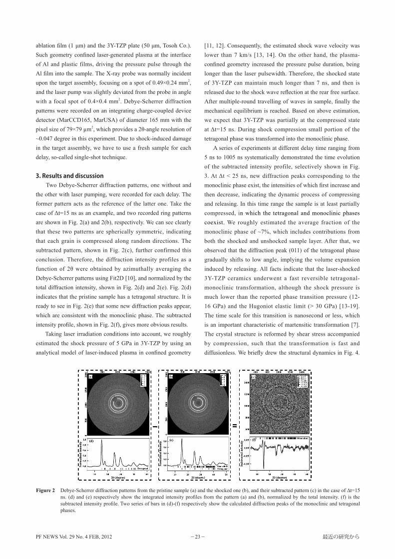
3. Results and discussion Two Debye-Scherrer diffraction patterns, one without and the other with laser pumping, were recorded for each delay. The former pattern acts as the reference of the latter one. Take the case of Δt=15 ns as an example, and two recorded ring patterns are shown in Fig. 2(a) and 2(b), respectively. We can see clearly that these two patterns are spherically symmetric, indicating that each grain is compressed along random directions. The subtracted pattern, shown in Fig. 2(c), further confirmed this conclusion. Therefore, the diffraction intensity profiles as a function of 2θ were obtained by azimuthally averaging the Debye-Scherrer patterns using Fit2D [10], and normalized by the total diffraction intensity, shown in Fig. 2(d) and 2(e). Fig. 2(d) indicates that the pristine sample has a tetragonal structure. It is ready to see in Fig. 2(e) that some new diffraction peaks appear, which are consistent with the monoclinic phase. The subtracted intensity profile, shown in Fig. 2(f), gives more obvious results. Taking laser irradiation conditions into account, we roughly estimated the shock pressure of 5 GPa in 3Y-TZP by using an analytical model of laser-induced plasma in confined geometry
[11, 12]. Consequently, the estimated shock wave velocity was lower than 7 km/s [13, 14]. On the other hand, the plasma-confined geometry increased the pressure pulse duration, being longer than the laser pulsewidth. Therefore, the shocked state of 3Y-TZP can maintain much longer than 7 ns, and then is released due to the shock wave reflection at the rear free surface. After multiple-round travelling of waves in sample, finally the mechanical equilibrium is reached. Based on above estimation, we expect that 3Y-TZP was partially at the compressed state at Δt=15 ns. During shock compression small portion of the tetragonal phase was transformed into the monoclinic phase. A series of experiments at different delay time ranging from 5 ns to 1005 ns systematically demonstrated the time evolution of the subtracted intensity profile, selectively shown in Fig. 3. At Δt < 25 ns, new diffraction peaks corresponding to the monoclinic phase exist, the intensities of which first increase and then decrease, indicating the dynamic process of compressing and releasing. In this time range the sample is at least partially compressed, in which the tetragonal and monoclinic phases coexist. We roughly estimated the average fraction of the monoclinic phase of ~7%, which includes contributions from both the shocked and unshocked sample layer. After that, we observed that the diffraction peak (011) of the tetragonal phase gradually shifts to low angle, implying the volume expansion induced by releasing. All facts indicate that the laser-shocked 3Y-TZP ceramics underwent a fast reversible tetragonal-monoclinic transformation, although the shock pressure is much lower than the reported phase transition pressure (12-16 GPa) and the Hugoniot elastic limit (> 30 GPa) [13-19]. The time scale for this transition is nanosecond or less, which is an important characteristic of martensitic transformation [7]. The crystal structure is reformed by shear stress accompanied by compression, such that the transformation is fast and diffusionless. We briefly drew the structural dynamics in Fig. 4.
Figure 2 Debye-Scherrer diffraction patterns from the pristine sample (a) and the shocked one (b), and their subtracted pattern (c) in the case of ∆t=15 ns. (d) and (e) respectively show the integrated intensity profiles from the pattern (a) and (b), normalized by the total intensity. (f) is the subtracted intensity profile. Two series of bars in (d)-(f) respectively show the calculated diffraction peaks of the monoclinic and tetragonal phases.

PF NEWS Vol. 29 No. 4 FEB, 2012 24 最近の研究から
To clearly show the structure change, the crystal structures are displayed in two different views.
4. Summary In summary, we demonstrated the ability of single-shot time-resolved X-ray power diffraction technique to study structural dynamics of laser-shocked polycrystalline materials. A reversible martensitic transformation has been observed in the laser-shocked 3Y-TZP ceramics. This work gives new insights into the phase transition dynamics of 3Y-TZP ceramics, such as transformation conditions and pathway. Application of TRXD techniques to study shock-induced phase transitions in more complex materials is now underway.
References[1] B. A. Remington, G. Bazan, J. Belak, E. Bringa, M. Caturla, J. D.
Colvin, M. J. Edwards, S. G. Glendinning, D. S. Ivanov, B. Kad, D. H. Kalantar, M. Kumar, B. F. Lasinski, K. T. Lorenz, J. M. McNaney, D. D. Meyerhofer, M. A. Meyers, S. M. Pollaine, D. Rowley, M. Schneider, J. S. Stolken, J. S. Wark, S. V. Weber, W.
G. Wolfer, B. Yaakobi, and L. V. Zhigilei, Metal. Mater. Trans. A 35A, 2587 (2004).
[2] T. d'Almeida, and Y.M. Gupta, Phys. Rev. Lett. 85, 330 (2000).[3] D. H. Kalantar, J. F. Belak, G. W. Collins, J. D. Colvin, H. M.
Davies, J. H. Eggert, T.C. Germann, J. Hawreliak, B. L. Holian, K. Kadau, P. S. Lomdahl, H. E. Lorenzana, M. A. Meyers, K. Rosolankova, M. S. Schneider, J. Sheppard, J. S. Stolken, and J. S. Wark, Phys. Rev. Lett. 95, 075502 (2005).
[4] K. Ichiyanagi, S. Adachi, S. Nozawa, Y. Hironaka, K. G. Nakamura, T. Sato, A. Tomita, and S. Yoshihara, Appl. Phys. Lett. 91, 231918 (2007).
[5] M. D. Knudson, Y. M. Gupta, and A. B. Kunz, Phys. Rev. B 59, 11704 (1999).
[6] M. D. Knudson and Y. M. Gupta, J. Appl. Phys. 91, 9561 (2002).[7] R. C. Garvie, R. H. Hannink, and R. T. Pascoe, Nature 258,
703 (1975).[8] S. Nozawa, S. Adachi, J. Takahashi, R. Tazaki, L. Guerin, M.
Daimon, A. Tomita, T. Sato, M. Chollet, E. Collet, H. Cailleau, S. Yamamoto, K. Tsuchiya, T. Shioya, H. Sasaki, T. Mori, K. Ichiyanagi, H. Sawa, H. Kawata, and S. Koshihara, J. Synch. Rad. 14, 313 (2007).
[9] K. Ichiyanagi, T. Sato, S. Nozawa, K.H. Kim, J.H. Lee, J. Choi, A. Tomita, H. Ichikawa, S. Adachi, and S. Koshihara, J. Synch. Rad. 16, 391 (2009).
[10] http://www.esrf.eu/computing/scientific/FIT2D/.[11] R. Fabbro, J. Fournier, P. Ballard, D. Devaux, and J. Virmont, J.
Appl. Phys. 68, 775 (1990).[12] D. Devaux, R. Fabbro, L. Tollier, and E. Bartnicki, J. Appl. Phys.
74, 2268 (1993).[13] D. E. Grady and T. Mashimo, J. Appl. Phys. 71, 4868 (1992). [14] T. Mashimo, A. Nakamura, M. Nishida, S. Matsuzaki, K. Kusaba,
K. Fukuoka, and Y. Syono, J. Appl. Phys. 77, 5069 (1995). [15] O. Ohtaka, S. Kume, T. Iwami, and K. Urabe, J. Am. Ceram. Soc.
71, C164 (1988).O. Ohtaka, D. Andrault, P. Bouvier, E. Schultz and M. Mezouar, J. Appl. Cryst. 38, 727 (2005).
[16] O. Ohtaka, S. Kume, and E. Ito, J. Am. Ceram. Soc. 71, C448 (1988).B. Alzyab, C. H. Perry, and R. P. Ingel, J. Am. Ceram. Soc. 70, 760 (1987).
[17] T. Mashimo, J. App. Phys. 63, 4747 (1988).[18] A. Matsuda, T. Hongo, H. Nagao, Y. Igarashi, K. G. Nakamura,
and K. Kondo, Sci. Tech. Adv. Mater. 5, 511 (2004).[19] Y. Igarashi, A. Matsuda, A. Akiyoshi, K. Kondo, K. G. Nakamura,
and K. Niwase, J. Mater. Sci. Lett. 39, 4371 (2004).(原稿受付日:2012 年 1 月 13 日)
著者紹介胡建波 Jianbo HU東京工業大学応用セラミックス研究所 博士課程 3 年〒 226-8503 神奈川県横浜市緑区長津田町 4259TEL: 045-924-5382 FAX: 045-924-5339e-mail: [email protected]略歴:2005 年中国工程物理研究院大学院(北京)修士課程修了,2005 年 - 現在:中国工程物理研究院流体物理研究所研究員,2008 年 -2009 年:エコール • ノルマル • シュペリウール(フランス),2009 年—現在:東京工業大学大学院総合理工学研究科物質科学創造専攻/応用セラミックス研究所。
Figure 4 Structural dynamics of 3Y-TZP ceramics under shock compression along two different views.
Figure 3 Selected normalized intensity change as a function of the delay time Δt, demonstrating the time evolution of structural dynamics of the laser-shocked 3Y-TZP.


![Envolventes para Centros de Transformación · 16 Centros de Transformación ... pf hasta 36 kV pf.301 pf.302 pf.303 pf.304 pf.3015 pf.3030 Longitud [mm] ... Combinaciones Posibilidad](https://img.pdfslide.tips/doc/110x75/5bb1a96c09d3f2f1188b9734/envolventes-para-centros-de-transformacion-16-centros-de-transformacion-.jpg)