Embed Size (px)
Citation preview
UNIVERSIDADE ESTADUAL DO OESTE DO PARANÁ – CAMPUS CASCAVEL
CENTRO DE CIÊNCIAS BIOLÓGICAS E DA SAÚDE
PROGRAMA DE PÓS-GRADUAÇÃO STRICTO SENSU EM BIOCIÊNCIAS E
SAÚDE – NÍVEL MESTRADO
CAROLYNE DONEDA SILVA SANTOS
A EXPOSIÇÃO MATERNA E DA PROLE À DIETA DE CAFETERIA
PROMOVE ALTERAÇÕES MORFOLÓGICAS TECIDO
ESPECÍFICAS
CASCAVEL-PR
Janeiro/2017
CAROLYNE DONEDA SILVA SANTOS
A EXPOSIÇÃO MATERNA E DA PROLE À DIETA DE CAFETERIA
PROMOVE ALTERAÇÕES MORFOLÓGICAS TECIDO
ESPECÍFICAS
Dissertação apresentada ao Programa de Pós-graduação Stricto Sensu em Biociências e Saúde – Nível Mestrado, do Centro de Ciências Biológicas e da Saúde, da Universidade Estadual do Oeste do Paraná, como requisito parcial para a obtenção do título de Mestre em Biociências e Saúde. Linha de Pesquisa: Fatores que influenciam a morfofisiologia orgânica. Orientadora: Profª. Drª. Sandra L. Balbo
Co-orientadora: Profª. Drª Sabrina Grassiolli
CASCAVEL-PR
Janeiro/2017
FOLHA DE APROVAÇÃO
CAROLYNE DONEDA SILVA SANTOS
A EXPOSIÇÃO MATERNA E DA PROLE À DIETA DE CAFETERIA
PROMOVE ALTERAÇÕES MORFOLÓGICAS TECIDO
ESPECÍFICAS
Esta dissertação foi julgada adequada para obtenção do título de Mestre em
Biociências e Saúde e aprovada em sua forma final pelo Orientador e pela Banca
Examinadora.
Orientador: Profª. Drª. Sandra L. Balbo _____________________________
UNIOESTE
Profª. Drª. Patrícia Cristina Lisbôa da Silva __________________________
UERJ
Profª. Drª. Elaine Manoela Porto Amorim _____________________________
UNIOESTE
AGRADECIMENTOS
Recebi tanto carinho e apoio durante esta jornada, que nenhuma palavra
escrita aqui poderá descrever o quanto sou grata. Mas inicio agradecendo àquele
que me deu o dom de ser, toda minha gratidão à Deus por estar à minha frente e
me permitir realizar todos os sonhos que Ele mesmo sonhou para mim, obrigada
por ser meu sustento. Ao meu esposo, Elion, meu amor e admiração, eu sempre
soube que você é especial, mas após estes anos e tudo que enfrentamos juntos,
eu tenho ainda mais certeza que Deus sabia exatamente o que eu precisava
quando me deu você, neste tempo eu não fui tudo o que você merecia, mas você
foi exatamente tudo o que eu precisava, amo você . Aos meus pais e minha família,
que não perdem nenhuma oportunidade de demonstrar seu amor e fé em tudo que
me proponho a fazer, suas orações e presença são essenciais para que eu consiga
realizar o que desejo, sei que tenho para quem voltar sempre, mas o mais
importante é que sei que irão comigo para onde eu for. Aos meus amigos que foram
pacientes e entederam minha ausência, torceram por mim e vibraram comigo à
cada vitória, obrigada por estarem ao meu lado e por me aceitarem sempre. Aos
meus colegas de laboratório, que aprendi a admirar e que me ensinaram um novo
sentido de doação e companheirismo, vocês merecem todo o sucesso e alegria,
vocês são muito especiais. Aos meus colegas de trabalho da Gastroclínica
Cascavel, que me apoiaram em todas as etapas deste Mestrado, aceitando minha
ausência e torcendo por mim em cada novo projeto. Finalmente, aos meus
professores amados, que me formaram e me fizeram enxergar o quanto é bonita a
forma que vocês têm, dentro desta Universidade, de dividir o conhecimento sem
nenhum egoísmo, somente pela alegria de ver as pessoas crescerem. Sandra,
tenho orgulho de ter sido escolhida por você e por contar com sua confiança, sua
amizade é um presente. Angélica, sua delicadeza e dedicação, são dons especiais,
te agradeço por todo tempo e paciência. Ana, encontrei uma amiga, pois só
consideramos amigos aqueles que realmente admiramos muito, obrigada por tudo.
Sabrina, sua determinação e inteligência me estimularam à ir além, sem seu apoio
este trabalho não teria o mesmo brilho. Sara e Fábio, vocês iniciaram um projeto
que conseguimos juntos finalizar, vocês me aceitaram, apoiaram e permitiram que
através da idéia de vocês eu realizasse um sonho, muito obrigada.
RESUMO
O objetivo do presente trabalho foi avaliar se a dieta de cafeteria durante o período
pré e pós desmame altera as características histológicas do tecido adiposo branco
(TAB), tecido adiposo marron (TAM), e do fígado em filhotes machos adultos. Para
isso, ratas Wistar com 21 dias de vida foram separadas em dois grupos: controle
(CTL; ratas alimentadas com ração padrão para roedores) e cafeteria (CAF; ratas
alimentadas com dieta de cafeteria durante todo seu período de vida). Aos 70 dias
de idade as ratas foram acasaladas e mantiveram o mesmo padrão alimentar
durante a gestação e amamentação. Após o nascimento, somente os filhotes
machos (F1) foram separados em 4 grupos (8 filhotes/ninhada) e receberam dieta
controle (CTL F1) ou dieta cafeteria (CAF F1) ao longo de suas vidas, conforme os
seguintes grupos: CTL-CTL F1, filhotes alimentados com dieta controle, nascidos
de mães que receberam dieta controle; CTL-CAF F1, filhotes alimentados com dieta
cafeteria, nascidos de mães que receberam dieta controle; CAF-CTL F1, filhotes
alimentados com dieta controle, nascidos de mães que receberam dieta cafeteria;
CAF-CAF F1, filhotes alimentados com dieta cafeteria, nascidos de mães que
receberam dieta cafeteria. Aos 100 dias de vida os animais foram eutanasiados e
os parâmetros biométricos e metabólicos, bem como a histologia do fígado, TAB e
TAM foram avaliados. Para avaliação dos dados obtidos foi realizada uma Análise
de Componentes Principais, a qual demonstrou que a dieta de cafeteria maternal
protegeu os filhotes dos efeitos deletérios provocados pela exposição à dieta
obesogênica durante suas vidas, como demonstrada pela ausência de alterações
no peso corporal e acúmulo de gordura, porém falhou em proteger o fígado e o
TAM, sugerindo que o impacto da dieta de cafeteria maternal é tecido específico.
Palavras-chave: obesidade, programação metabólica, prole e histologia
ABSTRACT
We, herein, evaluated whether the exposure of rats to a cafeteria diet pre- and/or
post-weaning, alters histological characteristics in the White Adipose Tissue (WAT),
Brown Adipose Tissue (BAT), and liver of young adult male offspring. Female Wistar
rats were divided into Control (CTL; rats fed on standard rodent chow) and Cafeteria
(CAF; fed on cafeteria diet during their entire life). After birth, male offspring only
(F1) were divided into four groups (8 pups/dams) and received the CTL or CAF diet
during their entire lives: CTL-CTLF1, control offspring born from dams that were fed
on control diet; CTL-CAF F1, cafeteria offspring born from dams that were fed on
control diet; CAF-CTL F1, control offspring born from dams that were fed on a
cafeteria diet; CAF-CAF F1, cafeteria offspring born from dams that were fed on a
cafeteria diet. Biometrics, metabolic parameters, and liver, BAT and WAT histology
were assessed. Data obtained were integrated using the Principal Component
Analysis (PCA). PCA showed that maternal CAF diet protects offspring from the
deleterious effects provoked by the exposure to an obesogenic diet during adult life,
as demonstrated by the absence of alteration in body weight and fat accumulation,
but failed to protect BAT and liver, suggesting that the impact of maternal CAF diet
is tissue-specific.
Key words: obesity, dams, offspring and morphology
SUMÁRIO
LISTA DE ABREVIATURA...........................................................................…........7
1. INTRODUÇÃO.....................................................................................................8
2. REVISÃO DE LITERATURA....................................................................…......12
2.1 Obesidade..............................................................................................…......12
2.2 Programação Metabólica........................................................................…......13
2.3 Alterações morfofuncionais induzidas pela obesidade e programação
metabólica.... ................................................................................................ ….....16
2.3.1 Obesidade e sua ação no fígado …....................................................…......17
2.3.2 Obesidade e sua ação no tecido adiposo ….......................................…......19
2.4 Modelos experimentais de obesidade …...............................................…......21
3. REFERÊNCIAS ........................................................................................ ….....30
4. ARTIGO.................................................................................................... ….....28
5. ANEXOS .................................................................................................. ….....48
5.1 Anexo A – Certificado de aprovação do projeto pelo CEEAAP ............... ….....48
5.2 Anexo B – Normas da revista científica ….......................................................49
LISTA DE ABREVIATURAS
AGL – Ácidos graxos livres
CAF- Cafeteria
CCBS – Centro de Ciências
Biológicas e da Saúde
CTL- Controle
CEEAAP – Comitê de Ética em
Experimentação Animal e Aulas
Práticas
DHGNA – Doença hepática
gordurosa não alcoólica
DM – Diabetes melitus
DM1 – Diabetes melitus tipo 1
DM2 – Diabetes melitus tipo 2
F1 – Primeira geração
HE – Hematoxilina Eosina
IBGE – Instituto Brasileiro de
Geografia e Estatística
IL4 – Interleucina 4
IL5 – Interleucina 5
IL13 – Interleucina 13
IMC – Índice de massa corporal
LAFEM – Laboratório de Fisiologia
Endócrina e Metabolismo
MSG – Glutamato monossódico
NK – Natural killers
SM – Síndrome metabólica
SNS – Sistema nervoso simpático
TA – Tecido adiposo
TAB – Tecido adiposo branco
TAM – Tecido adiposo marrom
TAR – Tecido adiposo retroperitoneal
TG – Triglicerídeos
TNF- – Fator de necrose tumoral
UNICAMP – Universidade Estadual
de Campinas
UNIOESTE – Universidade Estadual
do Oeste do Paraná
VIGITEL – Vigilância e fatores de
risco e proteção para doenças
crônicas por inquérito telefônico
VMH – Hipotálamo ventromedial
1. INTRODUÇÃO
A obesidade é definida como um acúmulo excessivo de gordura no tecido
adiposo, condição frequentemente acompanhada de alterações metabólicas,
hormonais e complicações no estado de saúde (WHO, 2000).
No boletim divulgado pela Organização Mundial da Saúde (OMS), em Julho
de 2015, o crescente aumento da obesidade no mundo foi classificado como
epidemia. Segundo estes dados, entre os anos de 1980 e 2013, a proporção de
adultos com excesso de peso e obesidade aumentou aproximadamente 8,5% em
ambos os sexos (NG et al., 2013). O rápido avanço da obesidade também tem sido
observado na população brasileira, conforme demonstram dados recentes obtidos
pela Vigilância de fatores de risco e proteção para doenças crônicas por inquérito
telefônico (VIGITEL) (MS, 2015). De acordo com estes dados a frequência do
excesso de peso na população adulta brasileira no ano de 2014 foi de 52,5%, sendo
maior entre homens (56,5%) que em mulheres (49,1%). A obesidade por sua vez,
incide sobre 17,9% da população avaliada, sendo minimamente maior no sexo
feminino.
A obesidade é uma doença de origem multifatorial relacionado a fatores
sociais, ambientais, culturais, genéticos e hormonais. Todavia, os estudos
epidemiológicos claramente demonstram que os hábitos alimentares, atrelados ao
sedentarismo são os principais determinantes para a atual epidemia de obesidade
(BRAUN et al., 2014; WHO, 2000; MS, 2015). Dentro deste contexto, pesquisas
recentes indicam que a condição metabólica materna, em especial, o estado
nutricional materno pré e pós gestacional podem afetar o feto em desenvolvimento
influenciando o estado de saúde deste novo indivíduo ao nascimento e com
repercussões para a homeostase energética na vida adulta (DESAI; JELLYMAN;
ROSS, 2015).
Estudos epidemiológicos demonstram que a incidência de obesidade entre
as mulheres grávidas acompanha o crescimento da obesidade na população em
geral, chegando a 25% em alguns países (GHELINCKX et al., 2008). O estado
nutricional e o consumo alimentar materno, em particular durante a gestação e
lactação, podem influenciar o peso corporal do bebê ao nascimento tendo
repercussões para a instalação de doenças crônicas na vida adulta, sendo
denominado de programação metabólica (DESAI, JELLYMAN; ROSS, 2015).
Estudo demonstrou que crianças expostas a programação metabólica, estarão
mais vulneráveis aos efeitos deletérios do atual estilo de vida, onde há predomínio
na oferta de alimentos de alto teor calórico associados à redução da prática de
atividade física (OLIVEIRA; FISBERG, 2003).
A super nutrição durante a gestação, associada à obesidade, promove
resistência à insulina, intolerância à glicose, acúmulo de gordura visceral e
dislipidemias, condições metabólicas que exercem impacto direto sobre o
desenvolvimento do bebê e modulam o estado de saúde futuro deste indivíduo,
favorecendo a instalação de doenças crônicas no adulto, em especial a Síndrome
Metabólica (SM) (SMITH; RYCKMAN, 2015; ECKEL; GRUNDY; ZIMMET, 2005).
Diferentes modelos experimentais que manipulam o estado nutricional
materno na gestação e/ou lactação tem sido usados para avaliar alterações
metabólicas e histomorfológicas relacionadas à programação metabólica e seus
efeitos deletérios sobre a prole, dentre os quais destacam-se o consumo de dieta
hipercalórica. A dieta de cafeteria (CAF) apresenta composição semelhante à dieta
hipercalórica consumida pelos humanos. Animais que consomem dieta CAF
apresentam aumento de peso, alto teor de tecido adiposo branco, hiperglicemia e
resistência à insulina, associada à inflamação severa do fígado, esteatose hepática,
dislipidemia e disfunção das ilhotas pancreáticas. Deste modo, a dieta CAF é
efetiva em reproduzir em modelos animais, as características da SM observada em
humanos (SAMPEY et al., 2011).
O consumo de dieta CAF durante os períodos pré-natal também é capaz de
promover programação metabólica na prole, provocando alterações como:
aumento de peso, desordens metabólicas e hormonais, esteatose hepática,
inflamação do tecido adiposo branco e do fígado (HOWIE et al, 2009; WHITE;
PURPERA; MORRISON, 2009; LI; SLOBADE; VICKERS, 2011; ZOE et al., 2014;
JACOBS et al., 2014).
Por outro lado, existem estudos contraditórios quanto aos efeitos da dieta
CAF nestes períodos do desenvolvimento e seus efeitos à longo prazo. Em estudo
realizado por Tamashiro et al., 2009 conclui-se que a dieta CAF ofertada à prole
após o desmame pode ter papel mais significativo no desenvolvimento da
obesidade na vida adulta que a dieta CAF consumida durante a gestação e lactação
pela mãe.
Considerando que a instalação da obesidade e o desenvolvimento da SM na
vida adulta podem estar associados a alterações nutricionais que modulam
períodos críticos do desenvolvimento, o presente estudo avaliou as adaptações

histomorfológicas no fígado, tecido adiposo unilocular e tecido adiposo multilocular,
induzidas pelo consumo de dieta CAF na primeira geração (F1) oriunda de mães
que consumiram dieta CAF na gestação e lactação.
2. REVISÃO DE LITERATURA
2.1 Obesidade
A obesidade já é considerada a quinta causa de morte no mundo, em média,
5% da população mundial morre a cada ano em decorrência do sobrepeso e
obesidade. Além deste número expressivo que pode caracterizar esta alteração do
estado nutricional como uma pandemia, podemos relacionar o excesso de peso a
diversas outras doenças, em 44% dos diabéticos, 23% dos indivíduos com
cardiopatia isquêmica e 7% a 41% dos pacientes com câncer, a obesidade foi causa
secundária para o desenvolvimento da patologia (WHO, 2011).
Estima-se que 500 milhões de pessoas no mundo são obesas e este
aumento de peso é crescente, bem como a incidência de Diabetes mellitus (DM).
Em 2011, a Federação Internacional de DM revelou que 366 milhões de pessoas
tinham DM e 4,6 milhões morreram em consequência da doença no mundo todo. A
previsão para 2030 é de um aumento de 43% na incidência desta doença.
Adicionalmente estes dados também mostram que em 2011 existiam 280 milhões
de indivíduos pré diabéticos e a previsão para 2030 é que 400 milhões de pessoas
sejam portadoras desta doença (IDF, 2012) e ainda entre as complicações da
obesidade, a DM aparece entre as 20 causas que podem diminuir a expectativa de
vida (WHO, 2014)
A associação entre a DM e o excesso de tecido adiposo é decorrente do
rompimento da homeostase glicêmica, promovida pela reunião de diferentes
elementos sendo denominada atualmente de Síndrome Metabólica (SM). A SM
caracteriza-se pela associação da obesidade, em particular o acúmulo de gordura
visceral, intolerância à glicose, resistência à insulina, dislipidemia e complicações
cardiovasculares. A SM por sua vez eleva a chance de desenvolver DM, em
especial, Diabetes Melitus tipo 2 (DM2), o qual representa cerca de 95% dos cados
da doença no mundo todo (FILHO et al., 2006;GUARNANI; BIRKEN; HAMILTON,
2015; KAUR, 2014).
Segundo o projeto Diretrizes, da Associação Médica Brasileira e Conselho
Federal de Medicina (DITEN, 2011) a obesidade pode ser definida como uma
doença crônica, inflamatória, endócrino-metabólica, heterogênea e multifatorial,
caracterizada pelo excesso de gordura corporal. Dentre as diferentes formas de
classificar a obesidade, a relação entre o peso e altura avaliada pelo Índice de
Massa Corporal (IMC)1 tem sido a ferramenta mais usada, em especial em grandes
estudos epidemiológicos (WHO, 1995).
No Brasil, o acompanhamento do estado nutricional da população, realizado
pelo Ministério da Saúde (MS, 2015), demonstrou que entre 2006 e 2012 houve um
crescimento contínuo do excesso de peso e obesidade na população adulta.
Eventos que permaneceram estáveis entre os anos de 2012 e 2014. O aumento do
peso corporal na população brasileira foi acompanhada de uma maior incidência
de DM2, embora a prática de atividade física, melhora do consumo de frutas e
verduras e redução do tabagismo também tenham sido observadas neste período.
Preocupantemente o excesso de peso e a obesidade são fenômenos que
afetam a população mais jovem, incluindo crianças e adolescentes no mundo todo,
tendo relação direta com hábito alimentar e sedentarismo. Por exemplo, nos EUA
em média 1/3 das crianças americanas está acima do peso, eventos que parecem
ser decorrente de fatores ambientais (GURNANI; BIRKEN; HAMILTON, 2015), tais
como: aumento da ingestão de bebidas adoçadas com açúcar, consumo de fast-
foods e lanches processados, tempo reduzido de sono, redução da prática de
atividade física e estresse familiar (BROWN et al., 2015).
O avanço da obesidade infantil também é observada em nosso país, sendo
reflexo de mudanças nutricionais e sócio econômicas observadas nas últimas
décadas conforme demonstram dados da Pesquisa e Orçamento Familiar (POF
2008-2009), realizada pelo Instituto Brasileiro de Geografia e Estatística (IBGE) e
Ministério da Saúde. Nesta pesquisa foi verificado que o aumento do consumo de
carnes, produtos panificados, derivados de leite e bebidas pela população urbana,
além de ressaltar o aumento do consumo de alimentos fora do domicílio. Ainda de
acordo com esta pesquisa, a transição nutricional teve impacto direto nos índices
de peso corporal, em particular na infância. De acordo com estes dados, os índices
de obesidade apresentam aumento significativo, principalmente, em crianças com
idade entre 5 a 9 anos. Entre os anos de 1989 e 2009, o sobrepeso entre meninos
e meninas dobrou e a obesidade aumentou mais de 300% (MS, 2010)
Em estudo recente, Smith; Rickman, 2015 apresentam evidências de que a
dieta e a saúde materna promovem modificações no feto em desenvolvimento,
1IMC: Índice que é calculado pelo peso do indivíduo em quilogramas, dividido por sua altura em metros ao quadrado (kg/m²). Classifica o estado nutricional do adulto com valores de IMC abaixo de 18,5kg/m², em magreza graus I, II e III., eutrofia; com valores até 25kg/m²; pré-obesidade ou sobrepeso, com valores entre 25 e 30kg/m² e obesidade graus I, II e III, com valores acima de 30kg/m² (WHO, 1995).
afetando a condição ao nascimento e tendo repercussões tardias na saúde dos
filhos, resultando em mecanismos que contribuem para o desenvolvimento da SM,
DM2 e obesidade na vida adulta. Neste sentido, a manutenção de um ambiente
intra-uterino saudável modula o metabolismo do organismo em desenvolvimento e
previne ou atenua a possibilidade de instalação de doenças crônicas no futuro. A
relação entre estados nutricionais materno e a condição de saúde futura do
indivíduo tem sido explorada no conceito de Programação Metabólica e diferentes
modelos animais têm sido amplamente utilizados para esclarecer as adaptações
moleculares, fisiológicas e morfológicas que alicerçam esta hipótese.
2.2 Programação Metabólica
O interesse na origem da obesidade e de suas comorbidades têm crescido
nos últimos anos. Evidências atuais demonstram que alterações nutricionais e/ou
hormonais ocorridas em janelas críticas do desenvolvimento, em particular
gestação e/ou lactação podem programar o metabolismo do feto em
desenvolvimento ou recém-nascido, repercutindo para seu estado de saúde na vida
adulta (SEDAGHAT; ZAHEDIASL; GHASEMIL, 2015; SMITH; RYCKMAN, 2015).
O conceito essencial da “programação metabólica” significa que a nutrição, os
fatores hormonais, ambientais e metabólicos podem alterar permanentemente a
estrutura dos órgãos, respostas celulares, expressão gênica, metabolismo e
fisiologia da prole. Essas alterações podem acontecer no feto, recém-nascido ou
no adulto (ROSS; DESAI, 2013).
Dentro deste contexto, importante atenção ao metabolismo materno tem sido
empregada, uma vez que a condição nutricional e metabólica materna, durante os
períodos gestacional e lactacional pode acarretar alterações morfo-funcionais na
prole, pois durante estes períodos, um oragnismo em desnvolvimento passa por
fases de intensa proliferação e crescimento celular, acompanhando marcantes
alterações funcionais (PEREIRA et al., 2014).
Existem diferentes modelos experimentais de programação metabólica,
dentre os quais se destacam a desnutrição e hiperalimentação gestacional e/ou
lactacional e suas repercussões para a prole. Interessantemente similares
impactos sobre o metabolismo tem sido observado nos descendentes ou primeira
geração (F1) nestas duas condições opostas, incluindo alterações no peso
corporal, acúmulo excessivo de gordura corporal, complicações na homeostase
glicêmica e lipídica e maior incidência de co-morbidades como DM2 e doenças
cardiovasculares (SEGOVIA et al., 2014; JI et al., 2015)
Modelos experimentais com restrição de diversos nutrientes específicos,
oxigênio e hormônios procuram avaliar seus efeitos no desenvolvimento da prole e
suas consequências na vida adulta (SEDAGHAT; ZAHEDIASL ; GHASEMI, 2015).
Neste sentido, a má nutrição é uma preocupação quando estudamos programação
metabólica, a condição de restrição alimentar ou deficiência nutricional pode causar
atraso no crescimento intra-uterino, distúrbios metabólicos, dislipidemias, risco de
arterosclerose, enquanto a deficiência de micronutrientes como ácido fólico,
vitamina B12, vitamina A, ferro, magnésio, zinco e cálcio podem afetar a saúde
cardiovascular (SZOSTAK-WEGIEREK, 2014).
Porém, considerando a atual epidemia de obesidade, dados recentes têm
ressaltado as complicações decorrentes da programação induzida pelo consumo
de dieta rica em calorias pelas mães e seu impacto na prole F1, sugerindo que a
obesidade materna durante a gravidez está associada ao risco de macrossomia e
aumento do risco de obesidade e DM2 na vida adulta. As alterações no padrão de
crescimento intra- uterino podem levar ao risco de desenvolvimento da SM na vida
adulta da prole (ROSS; DESAI, 2013).
O estado metabólico e hormonal materno tanto no período gestacional
quanto no lactacional tem repercussões para o organismo em desenvolvimento.
Por exemplo, a obesidade materna e suas comorbidades desencadeiam um
processo inflamatório e os mediadores inflamatórios liberados nesse processo,
podem atingir o feto. As citocinas pró-inflamatórias afetam a função placentária
alterarando o fluxo de nutrientes e, indiretamente, podem prejudicar o
desenvolvimento fetal. Dentro deste contexto foi demonstrado que a concentração
de citocinas inflamatórias placentárias podem estar relacionados ao aumento do
tecido adiposo visceral, do conteúdo lipídico hepatocelular e fluxo alterado de
triglicerídeos da prole (INGVORSEN et al., 2015; TARRADE et al.; 2015).
Quando aspectos relacionados a homeostase energética materna são
avaliados, também são observados efeitos sobre a prole. Por exemplo, a
hiperglicemia e hiperinsulinemia materna provocam uma sequência de eventos que
conduzem à compensação das células β-pancreáticas do feto, provocando
proliferação e hiperplasia das mesmas, podendo levar à exaustão, morte e
disfunção destas células (CERF, 2014).
Em recente estudo, Sullivan et al., 2015, também relacionaram a
programação metabólica materna, induzida por dietas hiperlipídicas e
hipercalóricas, com distúrbios de comportamento, como: déficit de atenção e
hiperatividade, depressão, autismo, esquizofrenia e aumento da ansiedade na
prole. Todas essas alterações seriam provenientes de alterações na formação
neuroendócrina do feto.
Vários estudos em humanos correlacionam a mudança epigenética2 ao risco
de doenças metabólicas, porém seus resultados são limitados. A maioria dos
estudos disponíveis sobre programação e epigenética baseia-se em modelos
animais (VICKERS, 2014).
2.3 Alterações morfofuncionais induzidas pela obesidade e programação
metabólica
Associado ao excesso de tecido adiposo e ao rompimento da homeostase
glicêmica, a obesidade também é acompanhada de dislipidemia, caracterizada
primariamente por aumento de ácidos graxos livres, triglicerídeos e colesterol total,
resultando em processo inflamatório em diversos tecidos envolvidos na
homeostase energética, incluindo tecido adiposo, músculo e fígado (DONATH et
al., 2013; PETRUZELLI; MOSCHETTA, 2010; ECKEL; GRUNDY; ZIMMET 2005),
Somado à presença do processo inflamatório, os tecidos periféricos também
podem sofrer outras alterações importantes, em especial a instalação de resistência
à insulina em diversos órgãos. A resistência insulínica hepática, por exemplo, altera
a liberação de triglicerídeos e glicose pelo fígado, provocando um acúmulo de
lipídeos neste órgão, caracterizando a esteatose hepática. Adicionalmente a
resistência a insulina no próprio tecido adiposo reduz sua capacidade de extrair
substratos da circulação, favorecendo acúmulo da gordura visceral, hipertrofia dos
adipócitos e remodelação do tecido (IOZZO, 2015; GREGORIO et al., 2013).
Deste modo, frequentemente as alterações metabólicas e endócrinas
características da obesidade são acompanhadas de ajustes morfológicos nos três
principais setores da homeostase glicêmica e lipídica, o tecido adiposo, o fígado e
o pâncreas endócrino. É relevante notar que, estes setores estão entre os principais
tecidos alvos da programação metabólica gestacional e lactacional com
2Epigenética se refere a mudanças hereditárias na função de um gene sem alterações na sequência de nucleotídeos. Modificações epigenéticas podem ser transmitidas entre células ou entre gerações de indivíduos (CERF, 2015)
repercussões na vida adulta (SAMPEY et al., 2011; DESAI; JELLYMAN; ROSS,
2015).
2.3.1 Obesidade e sua ação no fígado
O fígado é um órgão que executa diversas funções essenciais para a
homeostase corporal, tendo papel central no metabolismo, síntese,
armazenamento e distribuição de nutrientes. Este órgão é composto por vários tipos
celulares, sendo os hepatócitos responsáveis por 78% do volume do fígado e 70%
de todas as células do fígado, sendo assim, a função deste órgão é dependente
particularmente da atividade dos hepatócitos (GENTRIC; DESDOUETS, 2014).
Os hepatócitos em roedores e seres humanos têm uma vida útil longa e
raramente se dividem em condições normais na vida adulta. Contudo, estas células
tem uma notável capacidade de proliferação e regeneração em resposta a
situações de estresse, como ressecção cirúrgica, exposição à substâncias tóxicas
e infecção viral, além de hepatites crônicas B e C, exposição à sobrecarga de ferro
e cobre e processos inflamatórios, como esteatose hepática (GUIDOTTI et al.,
2003).
O fígado contribui para a homeostase glicêmica, tanto no estado alimentado
como em situações de estresse ou jejum. Em situações pós-prandiais o excesso
de substratos, particularmente glicose, são armazenados no fígado sob a forma de
glicogênio hepático. Adicionalmente o fígado também processa os lipídeos da dieta,
armazenando ácido graxos hepáticos, na forma de triglicerídeos. A glicose
armazenada no fígado provém essencialmente da dieta, enquanto a fonte hepática
de lipídeos é oriunda da diet(15%), da lipólise do tecido adiposo (59%) e também
do processo de lipogênese (26%) (ONYEDWERE et al., 2015). Durante o jejum, as
concentrações glicêmicas são mantidos pela glicogenólise hepática e em situações
de jejum prolongado também mantido pela síntese de glicose (gliconeogênese).
O controle das vias glicolíticas e lipogênicas hepática é diretamente
modulado pela ação do hormônio insulina. A obesidade provoca a resistência
insulínica hepática, promovendo liberação irregular de glicose e triglicerídeos.
Adicionalmente, a hiperinsulinemia decorrente da resistência a insulina induz
acúmulo de triglicerídeos no fígado (esteatose hepática), fibrose hepática,
inflamação hepática e cirrose hepática (IOZZO, 2015).
A esteatose hepática que é caracterizada por um acúmulo de gordura nos
hepatócitos, comprometendo mais de 5% no peso ou volume total do fígado, na
ausência do consumo de álcool e exposição à toxina ou doença viral é nomeada
como Doença Hepática Gordurosa Não Alcoólica (DHGNA). Esta condição
acomete 30% dos adultos de países desenvolvidos, possivelmente pelo aumento
da obesidade e expectativa de vida na população (CALVO et al., 2015). Esta
associação entre obesidade, SM e função hepática pode ser observada pelo
aumento do fator de crescimento do hepatócito, que acontece pelo acúmulo de
gordura no fígado, tendo relação com o aumento do IMC e aumento dos adipócitos,
paralelamente a um quadro de resistência à insulina (LINNEMANN; BAAN; DAVIS,
2014).
Existem algumas hipóteses para o aumento da concentração dos
triglicerídeos nos hepatócitos e o acontecimento da esteatose hepática, são
chamados de 3 “hits” 3 . No “primeiro hit” acontece um acúmulo metabólico de
lipídeos, que leva à lipotoxicidade, estresse oxidativo e inflamação. No “segundo
hit” acontece uma lesão tecidual e ativação de células estreladas, este processo é
provocado por ativação de citocinas inflamatórias e fatores de crescimento. O
aumento da sinalização de TNF- associado à síntese de colágeno hepático resulta
em alterações da matriz extracelular e fibrose, desequilíbrio entre a taxa de morte
e regeneração dos hepatócitos que promovem distorção na arquitetura do fígado
caracterizando o “terceiro hit”. (ONYEKWERE et al., 2015; WILLIAMS; KANG;
WASSERMAN, 2015).
A extensão da esteatose hepática pode ser classificada pela presença de
gordura nas seguintes proporções: leve (0 – 33%), moderada (34 – 66%) e grave
(>66%) (EBERTZ et al., 2014), esta avaliação pode ser feita através de exames de
imagem, como tomografias e ecografias. As principais características histológicas
da DHGNA são semelhantes àquelas da doença hepática induzida pelo álcool e
incluem esteato-hepatite (inflamação do fígado gorduroso, do parênquima, com ou
sem o acompanhamento de necrose focal) e diferentes graus de fibrose, incluindo
cirrose. A esteatose é predominantemente macrovesicular e, geralmente, é
distribuída por todo o fígado, embora possam existir proeminências de esteatose
microvesicular. Neutrofilia leve, alterações linfocitárias ou infiltrados inflamatórios
mistos também podem ser observados (ONYEKWERE et al., 2015; WILLIAMS;
KANG; WASSERMAN, 2015; EL KADER; EL DEN ASHMAWY, 2015).
3“hits”: passos
O consumo de dieta CAF promove esteatose hepática, principalmente
macrovesicular, associada à presença de diversas células inflamatórias,
especialmente macrófagos (SAMPEY et al., 2011), um proceso inflamatório
também presente em animais programados metabolicamente.
2.3.2 Obesidade e sua ação no tecido adiposo
O tecido adiposo (TA) é dividido no organismo em tecido adiposo
unilocular/branco e tecido adiposo multilocular/marrom, os quais apresentam
funções e histologia diferentes. O tecido adiposo unilocular exerce um papel
importante no metabolismo energético, o qual ocorre pelos processos bioquímicos
de lipogênese e lipólise associados a eventos secretórios que modulam ingestão
alimentar e gasto energético. Durante a lipogênese os adipócitos convertem o
excesso de substratos energéticos (glicose e lipídeos) em triglicerídeos (TG), os
quais são clivados em ácidos graxos livres (AGL) e glicerol durante a lipólise
(SANCHEZ-DELGADO, et al., 2015). Morfologicamente os adipócitos são
caracterizados pelo grande vacúolo lipídico central, com núcleo achatado em
direção à periferia celular (BLOOR ; SYMONDS, 2014).
De modo geral o tecido adiposo unilocular pode ser subdividido em:
subcutâneo e visceral. A gordura subcutânea está localizada na hipoderme,
enquanto a gordura visceral conecta órgãos internos e acumula-se no interior da
cavidade abdominal (GHAZARIAN et al., 2015). A gordura é armazenada em forma
de retroperitoneal e perigonadal, também pode ser armazenada na região intra-
abdominal, que forma o tecido adiposo visceral. Ainda pode cercar alguns órgãos
como coração e rins. Os roedores possuem ainda depósitos nas regiões: gonadal,
epididimal e inguinal (BLOOR ; SYMONDS, 2014).
O tecido adiposo multilocular tem características funcionais e morfológicas
distintas do unilocular. Metabolicamente os adipócitos do multilocular são
especializados em dissipar energia sob a forma de calor, pela oxidação de glicose
e lipídeos, um processo denominado termogênese. O controle deste processo é
diretamente feito pela atividade do Sistema Nervoso Simpático (SNS), via
norepinefrina. Morfologicamente este tecido é caracterizado pela coloração rosa
claro à vermelho escuro, apresentando alta vascularização, presença de gotículas
de gordura dispersas no citoplasma e grande número de mitocôndrias (SANCHEZ-
DELGADOt al., 2015; LEE; JUNG; CHOI, 2015; WHITTLE; LÓPEZ ; VIDAL-POING,
2011).
Em recente revisão Tupone, Maddem & Morris, 2014, afirmaram que a maior
quantidade de tecido adiposo multilocular encontrada em indivíduos magros estava
relacionada a maior gasto energético. Adicionalmente estes autores demonstratam
que o multilocular diminui com o aumento da idade, com a elevação do IMC bem
como, com adiposidade visceral. Finalmente o maior teor de multilocular, pelo
menos em humanos, é inversamente proporcional à presença de esteatose
hepática e obesidade (PORTER; CHONDRONIKOLA; SIDOSSIS, 2015).
Com a instalação da obesidade e resistência insulínica, que são
características da SM, os adipócitos sofrem uma remodelação, resultado do
acúmulo de TG, que provoca disfunção secretória e associado à hiperinsulinemia
promove proliferação e expansão do tecido adiposo. Assim, o número e área dos
adipócitos são aumentados. O processo de hipertrofia do adipócito está
diretamente relacionado ao aumento na produção de citocinas pró-inflamatórias,
tais como TNF- diferenciação e apoptose (SANCHES-DELGADO, 2015; BLOOR;
SYMONDS, 2014; LEE; JUNG; CHOI, 2015).
O sistema imunológico tem papel importante no controle da estrutura do
tecido adiposo e sua homeostase, inibindo a inflamação e remodelação do tecido.
Esses processos tem relação direta com o acometimento da função endócrina e
obesidade. Sob condições normais, sem excesso de peso, o tecido adiposo é
povoado por várias células do sistema imunológico, principalmente macrófagos
(WENSVEEN et al., 2015). Os eosinófilos também tem papel importante no controle
inflamatório e são residentes no tecido adiposo, controlando a ação de Interleucina
4 (IL-4) e Interleucina 5 (IL-5), citocinas anti-inflamatórias, sua deficiência provoca
aumento de citocinas pró-inflamatórias, aumento da sensibilidade ao
desenvolvimento da obesidade e resistência à insulina, tornando esse eixo,
dominante no mecanismo de homeostase do tecido adiposo (GHAZARIAN et al.,
2015).
As células Natural killer (NK) representam uma segunda população de
células imunes no TA, a sua falta conduz a concentrações reduzidas de IL-4 e IL-
13 e aumento de citocinas pró-inflamatórias, na obesidade, sua resposta pode
mudar, podendo até promover resistência à insulina (WENSVEEN et al., 2015;
COUSIN, et al., 2015). As células T-reguladores, principalmente as células T CD4+,
também estão envolvidas na inibição da inflamação no TA (GHAZARIAN et al.,
2015; WILLIAMS; KANG; WASSERMAN, 2015).
Uma dieta rica em calorias provoca no tecido adiposo visceral um aumento
de gordura nos adipócitos, acúmulo de células imunológicas (neutrófilos,
macrófagos e células NK), aumento da massa do adipócito, hipertrofia e hiperplasia
do adipócito, redução da produção de adiponectina, menor captação de glicose.
Todos estes elementos diminuem a sensibilidade do unilocular à insulina, bem
como, também favorecem a instalação de resistência a insulina em outros tecidos
(WENSVEEN et al., 2015).
Estudos realizados em ratos Wistar mostraram um aumento de 3 vezes do
unilocular nos animais que consumiram dieta CAF em relação à dieta controle; além
disso, apresentou resultados de infiltração de macrófagos 14 vezes maior neste
tecido em relação ao grupo controle (SAMPEY et al., 2011)
No tecido adiposo, a adipogênese acontece no final da gestação e no início
da vida pós-natal sendo sensível às condições uterinas, principalmente ao
fornecimento deficientes ou excessivo de nutrientes (SEGOVIA et al., 2014).
Quando exposto a uma grande oferta de gorduras, açúcar e sal pela alimentação
materna (ZELTSER, 2015), ocorrem alterações no unilocular do feto, podendo
aumentar sua capacidade de armazenar lipídeos e redução na sua capacidade de
termogênese, na fase adulta a produção de adipócitos é estabilizada, quando
produzidos em excesso na primeira infância, podem levar à obesidade tardia
(SEGOVIA et al., 2014).
2.4 Modelos animais de obesidade experimental
Existem diversos modelos experimentais de obesidade utilizados para
estudar a fisiopatologia desta doença e suas comorbidades. Dentre eles destacam-
se os animais submetidos a lesão eletrolítica no hipotálamo ventromedial (VMH);
ratos e camundongos tratados com glutamato monossódico (MSG); animais
geneticamente obesos como ratos Zucker e camundongos ob/ob (DIEMEN;
TRINDADE; TRINDADE, 2006; CESARETTI; JUNIOR, 2006).
Um modelo de dieta rica em gordura bastante estudado, baseado no
aumento da proporção de lipídeos, é a dieta hiperlipídica (HIGA, et al.; 2014).
Estudos demonstram a rapidez no ganho do peso, acúmulo de gordura, alterações
inflamatórias, hipertrigliceridemia e hiperglicemia em animais submetidos ao
consumo de dieta hiperlipídica (PODRINI, et al., 2013; HAIRI; THIBAULT, 2010).
As dietas hipercalóricas que são caracterizadas por proporção de
carboidratos e gordura inadequados, também são amplamente utilizadas como
ferramenta para promover o ganho excessivo de peso nos animais. Um dos
modelos utilizados para este fim é a dieta de CAF, pois reflete a realidade da dieta
ocidental, fornecendo grandes quantidades de sal, gordura e açúcar. Este tipo de
dieta promove hiperfagia, ganho rápido de peso, aumento da massa de gordura,
alterações no metabolismo da glicose, níveis elevados de insulina e hipertrofia dos
adipócitos (CASTELL-AUVÍ, 2011; HIGA et al., 2014).
Estudos afirmam que este é um dos modelos mais confiáveis para promover
efeitos semelhantes à SM em humanos, bem como, reproduzir a inflamação do TAB
(SAMPEY et al., 2011). Esta dieta também tem sido utilizada como modelo para
programação metabólica, sendo ofertada às mães durante gestação e lactação, e
promovendo alterações sobre parâmetros corporais, bioquímicos, ho rmonais e
reprodutivos da prole (JACOBS et al., 2014).
Durante o período de desenvolvimento do feto e até no crescimento inicial
do recém nascido, estes são muito vulneráveis a estímulos nutricionais da
alimentação materna, relacionadas à má ou super nutrição, resultando na mudança
das estrutura dos órgãos, repostas celulares e expressão gênica que provoca
impacto no metabolismo e fisiologia da prole. Esta programação pode ter efeitos
imediatos ou tardios na vida do indivíduo (DESAI; JELLYMAN; ROSS, 2015).
O resultado desta programação é controverso. Estudo que avaliou os efeitos
do consumo da dieta materna CAF e a oferta da mesma dieta à prole demonstrou
aumento de peso e gordura retroperitoneal, colesterol, leptina e insulinemia em
todos os grupos que consumiram dieta CAF, especialmente no grupo CTL-CAF, em
que somente a prole consumiu dieta CAF, comprovando que nesta situação a dieta
materna não teve efeito de programação metabólica. Este estudo ainda mostrou
que estes efeitos foram mais expressivos em animais com 120 dias de vida que em
animais com 30 dias de vida, esclarecendo que o maior efeito da dieta CAF ocorre
na vida adulta (MUCELLINI et al., 2014).
Considerando que alterações nutricionais durante períodos críticos do
desenvolvimento, tais como, a gestação e lactação podem programar o
metabolismo da prole favorecendo a instalação de obesidade na vida adulta, é
necessário avaliar a interação de efeitos entre a dieta materna e a da prole; sua
repercussão nos aspectos histomorfológicos da prole na vida adulta o que permitirá
caracterizar melhor o processo de programação metabólica, com o intuito de
atenuar ou mesmo reverter suas consequências a longo prazo.
REFERÊNCIAS
BLOOR, T.D.; SYMONDS, M.E. Sexual dimorphism in White and brown adipose tissue with obesity and inflammation. Hormones and Behavior, 2014.. 2015.
BRASIL. Ministério da Saúde e IBGE – Pesquisa de Orçamentos Familiares 2008-2009: Despesas, rendimentos e condições de vida. Rio de Janeiro, 2010. BRASIL. Ministério da Saúde. VIGITEL -. Monitoramento de fatores de risco para as doenças crônicas por entrevistas telefônicas, 2015. BRAUN, M.; SCHELL, J.; SIEGFRIED, W.; MULLER, M.J.; RIED, J. Re-entering obesity prevention: a qualitative-empirical inquiry into the subjective aetiology of extreme obese adolescents. BMC Public Health, 2014.
BROWN, C.L.; HALVORSON, E.E.; CHOHEN, G.M.; LAZORICK, S.; SKELTON,J. Adressing childhood obesity: opportunities for prevention. Pediartric Clin N Am, pag.1241-1261, 2015.
CALVO,M.; BELTRÁN-DELSÓN,R.; RODRÍGUEZ-GALLEGO,E.; HERNÁNDEZ-AGUILERA, A.; GUIRRO, M.; MARINÉ-CASADÓ, R.; MILLÁ, L.; ALEGRET, J.M.; SABENCH, F.; CASTILHO, D.; VINAISCA, M.; RODRÍGUEZ, M.A.; CORREIG, X.; GARCÍA-ÁLVAREZ, R.; MENENDEZ, J.A.; CAMPS, J.; FORIN, J. Liver fat deposition and mitochondrial dysfunction in morbid obesity: Na approach combining metabolomics with liver imaging and histology. World Journal of Gastroenterology, pag. 7529-7544, June, 2015.
CASTELL-AUVÍ, A.; CEDÓ, L.; PALLARÉS, V.; BLAY, M.; ARVÉDOL, A.; PINENT, M. The effects of a cafeteria diet on insulin production and clearance in rats. British Journal of Nutrition, pag. 1155-1162, 2012.
CERF, M.E. Hight fat programming of beta cell compensation, exhaustion, death and dysfunction. Pediatric Diabetes, 2015. CESARETTI, M.L.R.; JUNIOR, O.R. Modelos experimentais de resistência à insulina e obesidade: lições aprendidas. Arq Bras Endocrinol Metabol. Vol. 50, n.
2. Abril, 2006. COUSIN, B.; CASTEILLA, L.; LAHARRAGUE, P.; LUCHI, E.; LORSIGNOL, A.; CUMINETTI, V.; PAUPERT, J. Immuno-metabolism and adipose tissue the key role of hematopoietic stem cells. Biochimie, 2015. DESAI, M.; JELLYMAN, J.K.; ROSS, M.G. Epigenomics, gestacional programming and risk of metabolic syndrome. International Journal of obesity, pag. 622-641,
2015.
DIEMEN, V.V.; TRINDADE, E.N.; TRINDADE, M.R.M. Experimental model to induce obesity in rats. Acta cirúrgica brasileira. vol. 21, 2006.
DONATH, M.Y.; DALMAS, E.; SAUETR, N.S.; SONI-SCHNETZLER, M. Inflammation in obesity and diabetes: islet dysfunction and therapeutic opportunity. Cell Metabolism, 2013.
EBERTZ, C.E.; BONFELUR, M.L.; BERTAM, I.M.; MENDES, M.C.; LUBACZEUSKI, C.; ARAÚJO, A.C.F.; PAES, A.M.; AMORIM, E.M.P.; BALBO, S.L. Duodenal jejunal bypass attenuates non-alcoholic fatty liver disease in western diet-obese rats. Acta Cir. Bras. Vol 29. n. 9. SP, September, 2014. ECKEL, R.H.; GRUNDY, S.M.; ZIMMET, P.Z. The metabolic syndrome. Lancet, vol.
365, April 2005. EL-KADER, S.M.A.; ASHMAWEY, E.M.S.E.D. Non-alcoholic fatty liver disease: The diagnosis and management. World Journal of Hepatology, pag. 846-858, Abril, 2015.
FILHO, F.F.R; MARIOSA, L.S.; FERREIRA, S.R.G.; ZANELLA, MT. Gordura visceral e síndrome metabólica: mais que uma simples associação. Arq. Bras. Endocrinol. Metabol. v. 50 n.2. Abril, 2006. GENTRIC, G.; DESDOUETS, C. Polyploidization in liver tissue. The American Journal of Pathology. Vol. 184 n.2 Frebruary, 2014.
GHAZARIAN, M.; LUCK, H.; REVELO, X.S.; WINERS, S.; WINER, D.A. Immunopathology of adipose tissue during metabolic syndrome. Turk Patoloji Der, 2015.
GUELINKX, I.; DEVLIEGER, R.; BECKERS, K.; VANSAT, G. Maternal obesity: pregnancy complication, gestacional weight gain and nutrition. Obesity reviews,
2008.
GUIDOTTI, J.E.; BRÉGERIE, O., ROBERT, A.; DEBERY, P.; BRECHOT, C.; DESDOUETS, C. Liver cell polyploidization: A pivotal role for binuclear hepatocytes. The Journal of Biological Chemistry. Pag. 278 n.21. May, 2003.
GUARNANI, M.; BIRKEN, C.; HAMILTON, J. Childhood obesity: causes, consquences, and management. Pediatr Clin N Am, pag. 821-840, 2015.
HAIRI, N.; THIBAULT, L.; Hight fat diet induced obesity in animal models. Nutrition Researcch Reviews, pag. 270-299, 2010.
HIGA, T.S.; SPINDOLA, A.V.; ALANIZ, M.H.F.; EVANGELISTA, F.A. Comparison
between cafeteria and high-fat diets in the induction of metabolic dysfunction in
mice. Int Physiol Pathophysiol Pharmacol, pag. 47-54, 2014.
HOWIE, G.J.; SLOBODA, D.M.; DAMAL,T.; VICKERS, M.H. Maternal nutrition history predicts obesity in adult aoffspring independet of posnatal diet. J. Physiol.
Pag. 905-915, 2009. IDF. Diabetes Atlas. 5th. ed. International Diabetes Federation, 2012.
INGVORSEN, C.; BRIX, S.; OZANNE, S.E.; HELLGREN, L.I. Inffect of maternal inflammation on foetal programming of metabolic disease. Acta Physiol, pag. 440-
449, 2015. IOZZO, P. Metabolic imaging in obesity: underlying mechanisms and consequences in the whole body. Ann. N. Y. Acad. Sci., 2015.
JACOBS, S.; TEIXEIRA, D.S.; GUILHERME, C.; ROCHA, C.F.K.; ARANDA, B.C.C.; REIS, A.R.; SOUZA, M.A.; FRANCI, C.R.; SANVITTO, G.L. The impact of maternal consuption of cafeteria diet on reproductive function in the offspring. Physiology & Behavior, pag. 280-286, 2014.
JI, Y.; WU, Z.; DAI, Z.; SOL, K.; WANG, J.; WU, L. Nutritional epigenetics with a focus on amino acids: implications for the development and treatment of metabolic syndrome. J Nutr Biochem. Aug, 2015. KAUR, J.A. Comprehensive review on metabolic syndrome. Cardiology Research and Practice, 2014. LEE, Y.; JUNG, Y.S.; CHOI, D. Recent advance in brown adipose physiology and its therapeutic potential. Experimental & Molecular Medicine, 2014.
LI, M.; SLOBODA, D.M.; VICKERS, M.H. Maternal obesity and developmental programming of disordes in offspring: Evidence from animal models. Experimental Diabetes Research, 2011.
LINNEMANN, A.K.; BAAN, M.; DAVIS, D.B. Pancreatic β- cell proliferation in obesity. American society for nutrition. Adv. Nutr., pag. 278-288, 2014. MUCELINI, A.B.; GOULARTE, J.F.; CUNHA, A.C.A.; CACERES, R.C.; NOSCHANG, C.; BENETTI, C.S; SILVEIRA, P.P.; SANVITTO, G.L. Effects of exposure to a cafeteria diet during gestation and after weaning on the metabolism and body weight of adult male offspring in rats. British Journal of Nutrition. pag. 1499-1506, 2014. NG, M. The GDB 2013 Obesity Collaboration Global, regional and nationa prevalence of overweight and obesity in children and adults 1980-2013: a systematic analysis. Lancet. Pag. 766-781, 2014.
OLIVEIRA, C.L.; FISBERG, M. Obesidade na infância e adolescência – uma verdadeira epidemia. Arq. Bras. Endocrinol. Metabol. Vol.47 n.2. Abril, 2003. ONYEKWERE, C.A.; OGBERA, A.O.; SAMAILA, A.A.; BALOGUN, B.O.; ABDULKAREEN, F.F. Nonalcoholic fatty liver desease: synopsis of current developments. Nigerian Journal of Clinical Practice. Vol.18 n.6, 2015.
PEREIRA, T.J.; MOYCE, B.L.; KERELUIK, S.M.; DOLINSKY, V.W. Influence of maternal overnutrition and gestacional diabetes on the programming of metabolic health outcomes in the offspring: experimental evidence. Biochem. Cell Biol. vol.
93 pag. 438-451, 2015.
PETRUZELLI, M.; MOSCHETTA, A. Intestinal ecology in the metabolic syndrome. Cell Metabolism. Elsevier. May, 2010.
PODRINI, C.; CAMBRIDGE, E.L.; LELLIOTT, C.J.; CARRAGHER, D.M.; ESTABEL, J.; GERDIN, A.K.; KARP, N.A.; SCUDAMORE, C.L.; PROJECT, S.M.G.; SOLIS, R.R.; WHITE, J.K. High-fat feeding rapidly induces obesity and lipid derangements in C57BL/6N mice. Mamm Genome, pag. 240-251, 2013.
PORTER, C.; CHONDRONIKOLA, M.; SIDOSSILL, L.S. The therapeutic potential of brown adipocytes in human. Frontiers in Endocrinology. vol. 6. October, 2015.
PROJETO DIRETRIZES, volume IX. São Paulo: Associação Médica Brasileira;
Distrito Federal: Conselho Federal de Medicina, 2011.
ROSS, M.G.; DESAI, M. Developmental programming of offspring obesity, adipogenesis, and appetite. Clinical obstetrics and gynecology. Vol. 56 n.3 pag.
529-536. September, 2013.
SAMPEY, B.P.; VANHOOSE, A.M.; WINFIELD, H.M.; FREEMEXMAN, A.J.; MUEHLBAUER, M.J.; FUEGER, P.T.; NEWGARD, C.B.; MOKOWSKI, L. Cafeteria diet is a robust model of human metabolic syndrome with liver and adipose inflammation: comparison to high-fat diet. Obesity. Vol. 19 n.6 June, 2011.
SANCHEZ-DELGADO, G.; MARTINEZ-TELLEZ, B.; OLGA, J.; AGUILERA, C.M.; GIL, A.; RUIZ, J.R. Role of exercise in the activation of brown adipose tissue. Ann Nutr Metab, pag. 21-32, 2015.
SEDAGHAT, K.; ZAHEDIASL, S.; GASEMI, A. Intrauterine programming. Iranian Journal of Basic medical sciences. vol. 18. pag. 212-220, 2015. SEGOVIA, A.S.; VIKERS, M.H.; GRAY, C.; REYNOLDS, C.M. Maternal obesity, inflammation, and development programming. Bio Med Research International,
2014. SMITH, C.J.; RYCKMAN, K.K. Epigenetic and developmental influence on the risk of obesity, diabetes, and metabolic syndrome. Diabetes, metacolic syndrome and obesity: targets and therapy. pag. 295-302. June, 2015.
SULLIVAN, E.L.; KIPER, K.M.; LOCKARD, K.; VALLEARU, J.C. Maternal hight-fat diet programming of the neuroendocrine system and behavior. Hormones and Behavior, 2015.
SZOSTAK-WEGIEREK, D. Intrauterine nutrition: long-term consequences for vascular health. International Journal of Women’s health. pag. 647-656. July, 2014.
TAMASHIRO, K.L.K.; TERRILON, C.E.; HYUN, J.; KOENIG, J.I.; MORAN, T.H. Prenatal stress or high-fat diet increases susceptibility to diet-induceobesity in rats offspring. Diabetes. vol. 58. May, 2009.
TARRADE, A.; PANCHENKO, P.; JUNIEN, C.; GABORY, A. Placental contribution to nutritional programming of health and deseases: epigenetics and sexual dimorphism. The Journal of experimental Biology, 2015.
TUPONE, D.; MADDEN, C.J.; MORRISON, S. Autonomic regulation of brown adipose tissue thermogenesis in health and disease: potential clinical apllications for altering BAT thermogenesis. Frontiers in Neuroscience. Frebruary, 2014.
VICKERS, M.H. Early life nutrition, epigenetics and programming of later life disease. Nutrientes. Vol.6 pag. 2165-2178, 2014.
WENSVEEN, F.M.; VALENTIC, S.; SISTAN, M.; WENSVEEN, T.T.; POLIC, B. The “Big Bang” in obese fat: Events initiating obesity-induced adipose tissue inflammation. Eur. J. Imunol. Vol.45, pag. 2446-2456, 2015. WHITE, C.L.; PURPERA, M.N.; MORRISON, C.D. Maternal obesity for programming effect on high fat diet offspring. Am J Physiol Regul Integr Comp Physiol. pag. 1464-1472. May, 2009. WHITTLE, A.J.; LÓPEZ, M.; VIDAL-PUIG, A. Using brown adipose tissue to treat obesity the central issue. Trends in Molecular Medicine, vol.17 n.8. August, 2011.
WILLIAMS, A.S.; KANG, L.; WASSERMAN, D.H. The extracellular matrix and insulin resistance. Trends in Endocrinology and Metabolism. Vol.58 n.7. July, 22015. World Health Organization – WHO. Physical status: the use and interpretation of anthropometry, 1995. World Health Organization – WHO. Consultation on obesity. Obesity: preventing and managing the global epidemic: report of a WHO consultation. Genova,
2000. World Health Organization – WHO. Noncommunicable diseases country profiles, 2011.
World Health Organization – WHO. World Health Statistics, 2014.
World Health Organization – WHO. Global status report on noncommuniclable diseases, 2014.
ZELTSER, L.M. Developmental influences on circuits programming susceptibility to obesity. Fronties in neuroendocrinology, 2015.
ZOE, C.D.; AKYOL, A.; MCMULLER, SL.; EVAM. S.C.L. Exposure of neonatal rats to maternal cafeteria feeding during suckling alters hepatic gene expression and DNA methylation in the insulin signalling pathaway. Genes Nutr., 2014.
4. ARTIGO CIENTÍFICO
THE MATERNAL AND OFFSPRING EXPOSURE TO CAFETERIA
DIET PROMOTES TISSUE-SPECIFICS MORPHOLOGICAL
CHANGES
Life-long Maternal Cafeteria Diet Promotes Tissue-Specific Morphological Changes
in Male Offspring Adult Rats.
Carolyne Doneda Silva Santos1, Sandra Lucinei Balbo1, Ana Tereza Bittencourt
Guimarães1, Sara Cristina Sagae2, Fábio Negretti3, Sabrina Grassiolli1,
1 Program of Biosciences and Health, Universidade Estadual do Oeste do Paraná,
Cascavel, Paraná, Brazil
2 Center of Biological Sciences and Health, Universidade Estadual do Oeste do
Paraná,
Cascavel, Paraná, Brazil
3 Pathologist, Cascavel, Paraná, Brazil
Laboratório de Fisiologia Endócrina e Metabolismo, Centro de Ciências Biológicas
e da Saúde, Universidade Estadual do Oeste do Paraná (UNIOESTE), Cascavel,
PR, Brazil CEP: 858119-110
Correspondent author: [email protected]
Abstract
We, herein, evaluated whether the exposure of rats to a cafeteria diet pre- and/or
post-weaning, alters histological characteristics in the White Adipose Tissue (WAT),
Brown Adipose Tissue (BAT), and liver of young adult male offspring. Female Wistar
rats were divided into Control (CTL; rats fed on standard rodent chow) and Cafeteria
(CAF; fed on cafeteria diet during their entire life). After birth, male offspring only
(F1) were divided into four groups (8 pups/dams) and received the CTL or CAF diet
during their entire lives: CTL-CTLF1, control offspring born from dams that were fed
on control diet; CTL-CAF F1, cafeteria offspring born from dams that were fed on
control diet; CAF-CTL F1, control offspring born from dams that were fed on a
cafeteria diet; CAF-CAF F1, cafeteria offspring born from dams that were fed on a
cafeteria diet. Biometrics, metabolic parameters, and liver, BAT and WAT histology
were assessed. Data obtained were integrated using the Principal Component
Analysis (PCA). PCA showed that maternal CAF diet protects offspring from the
deleterious effects provoked by the exposure to an obesogenic diet during adult life,
as demonstrated by the absence of alteration in body weight and fat accumulation,
but failed to protect BAT and liver, suggesting that the impact of maternal CAF diet
is tissue-specific.
Key words: obesity, dams, offspring and histology
Introduction
Maternal over nutrition during pregnancy and lactation increases the risk of obesity,
Metabolic Syndrome (MS) and Type 2 Diabetes (T2D) in the offspring during
adulthood [1]. These early effects of the nutritional maternal environment on the
growth and metabolism of offspring and their long-term impact on health are defined
as metabolic programming [1,2,3]; a concept previously established by Barker and
colleagues. These authors showed that an adverse fetal environment, followed by
an obesogenic diet in postnatal life, may lead to chronic disease in adulthood [4].
Moreover, post-weaning exposure to hypercaloric diet induces the development of
obesity, disruption in glucose-insulin homeostasis, dyslipidemia, liver steatosis, and
cardiovascular diseases [5,6,7]. As such, experimental obesity can be produced by
maternal or post-weaning dietary manipulations. The cafeteria diet (CAF) is a
reliable model of dietary obesity in humans, promoting voluntary hyperphagia, body
weight gain, exacerbated adipose tissue expansion, hyperglycemia and
hyperinsulinemia, and inflammatory processes in the liver and adipose tissue [6,8].
It is well known that maternal life-time exposure to CAF diet induces body weight
gain, higher adipose tissue content and metabolic abnormalities, such as
hypercholesterolemia, hyperinsulinemia and hyperleptinemia [6]. This maternal
state can induce metabolic programming in adult offspring, culminating in obesity
and its associated metabolic disorders [9,10,11,12,13]. As such, the programmed
metabolic phenotype, found in the offspring, could be exacerbated during growth, in
particular when offspring are also exposed to a life-long obesogenic diet [6].
However, while the effects of maternal over nutrition on weight gain and metabolism
in offspring, before weaning, are well characterized [1,3], their persistent effects on
adipose tissue content, glucose tolerance, insulin resistance and liver abnormalities
in adulthood are contradictory [5,6,14,15]. Thus, an understanding as to how pre-
and post-natal environment interactions affect the growth and development of
offspring is fundamental, since the timing of an insult determines which organ or
systems will be altered, when obesity occurs and the severity of diseases, later in
life [16,17,18]. As such, some tissues appear to be more vulnerable to nutritional
insults during development. Thus, marked programming effects have been
observed in White Adipose Tissue (WAT), Brown Adipose Tissue (BAT) and liver
[19,20]. Surprisingly, it was recently shown that maternal over nutrition could protect
against the deleterious effects of the obesogenic diet during adulthood [6]. In the
present study, we evaluated whether the exposure to a CAF diet, pre- and post-
weaning (alone or combined), modifies the histological characteristics of the WAT,
BAT and liver of young adult male offspring.
Materials and Methods
Experimental methods
The Committee on Ethics in Animal Experimentation of the State University of
Western Parana approved all experiments (CEUA-10/12/2013). All animals used in
this study were housed under controlled room temperature (23±3◦C), light (12 h
light/dark cycle), and had free access to food and water. At 21 days of age, 16 Wistar
female rats were randomly divided into two dietary experimental groups: (1) Control
group (CTL), fed on standard rodent chow (12.39 kJ/g — NuvilabTM, Colombo,
Brazil) and water ad libitum; and (2) Cafeteria group (CAF), fed on a cafeteria diet.
The CAF diet used in this study was adapted from previous studies [21,22]. Detailed
information about the nutritional value and ingredients of all foods used in this
model, demonstrating that the CAF diet, when introduced at weaning, is able to
induce obesity in adult female rats, has been previously published [6].
At 70 d of age, CTL and CAF female rats (n=16) were mated with a control male
(n=8) in a harem system (ratio of 2 females to 1 male) during approximately two
weeks. The pregnant females were housed in individual cages until delivery. After
birth, the litter size was adjusted to eight pups/dam, to maximize lactation
performance. After weaning, these offspring (F1) were fed with CTL or CAF diets for
11 weeks and were allocated to the groups:
CTL-CTLF1, control offspring born from dams that were fed on control diet
CTL-CAF F1, cafeteria offspring born from dams that were fed on control diet
CAF-CTL F1, control offspring born from dams that were fed on a cafeteria diet
CAF-CAF F1, cafeteria offspring born from dams that were fed on a cafeteria diet.
Importantly, in all experimental groups the number of animals analyzed was 5-7 per
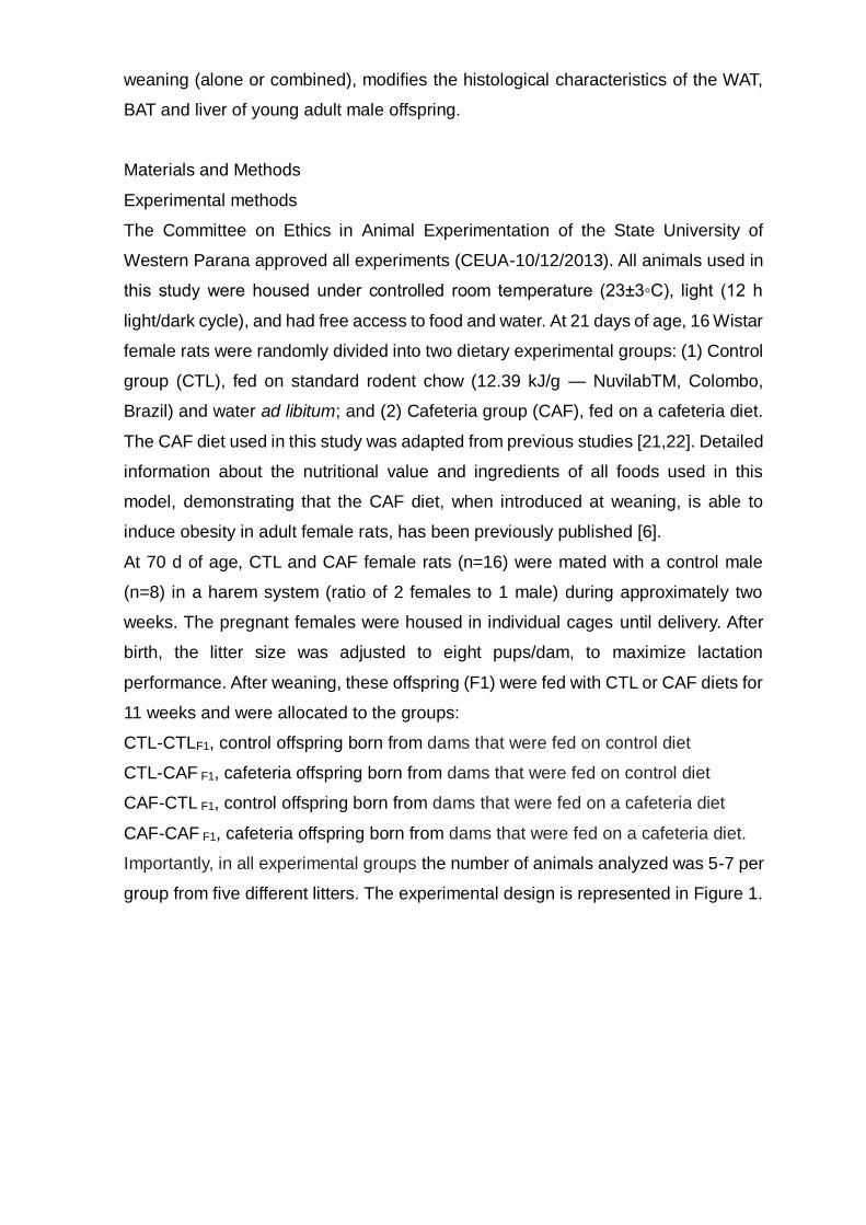
group from five different litters. The experimental design is represented in Figure 1.
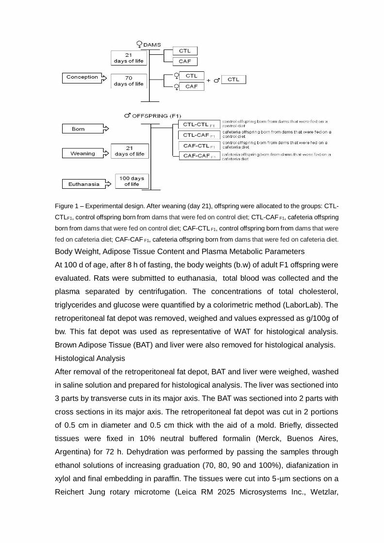
Figure 1 – Experimental design. After weaning (day 21), offspring were allocated to the groups: CTL-
CTLF1, control offspring born from dams that were fed on control diet; CTL-CAF F1, cafeteria offspring
born from dams that were fed on control diet; CAF-CTL F1, control offspring born from dams that were
fed on cafeteria diet; CAF-CAF F1, cafeteria offspring born from dams that were fed on cafeteria diet.
Body Weight, Adipose Tissue Content and Plasma Metabolic Parameters
At 100 d of age, after 8 h of fasting, the body weights (b.w) of adult F1 offspring were
evaluated. Rats were submitted to euthanasia, total blood was collected and the
plasma separated by centrifugation. The concentrations of total cholesterol,
triglycerides and glucose were quantified by a colorimetric method (LaborLab). The
retroperitoneal fat depot was removed, weighed and values expressed as g/100g of
bw. This fat depot was used as representative of WAT for histological analysis.
Brown Adipose Tissue (BAT) and liver were also removed for histological analysis.
Histological Analysis
After removal of the retroperitoneal fat depot, BAT and liver were weighed, washed
in saline solution and prepared for histological analysis. The liver was sectioned into
3 parts by transverse cuts in its major axis. The BAT was sectioned into 2 parts with
cross sections in its major axis. The retroperitoneal fat depot was cut in 2 portions
of 0.5 cm in diameter and 0.5 cm thick with the aid of a mold. Briefly, dissected
tissues were fixed in 10% neutral buffered formalin (Merck, Buenos Aires,
Argentina) for 72 h. Dehydration was performed by passing the samples through
ethanol solutions of increasing graduation (70, 80, 90 and 100%), diafanization in
xylol and final embedding in paraffin. The tissues were cut into 5-µm sections on a
Reichert Jung rotary microtome (Leica RM 2025 Microsystems Inc., Wetzlar,
Germany) and Hematoxilin and Eosin (H&E) were used for staining. The slides were
photographed using a light microscope (Olympus BX 50), coupled to a digital
camera (SAMSUNG SHC-410NAD) using photo Micro 5.6 software. Sections were
photographed along their entire length, field to field and the images captured were
analyzed using the Image J 1.48v program, which was previously calibrated to 100x
and 200x and standardized for the analysis of each image (Corel Draw X7 program).
All analyzes were performed by a single observer.
Histological measurements were carried out for each tissue evaluated. The
retroperitoneal fat depot was photographed at a magnification 100x and an
adipocyte nuclei count performed. Additionally, all adipocytes in all the sections were
circled and the adipocyte area (mm2) was measured. For the BAT images were
photographed at 200x magnification and cell proliferation evaluated by the adipocyte
nuclei count (3 sections/slide). Qualitative analysis of the fat depot was also
performed to assess fat droplets in the adipocyte cytoplasm. Finally, the diameters
of the areas evaluated were delimited in the hepatic tissue (0.5 cm) using a default
template. Ares were photographed at a magnification 100x and images were used
for qualitative analysis of the presence of infiltrated fat in the hepatocyte cytosol. For
this analysis, at least 20 photos were assessed by a single observer.
Statistical analysis
The body weight, weight of the retroperitoneal fat depot, triglycerides, cholesterol
and glucose were assessed for normality by Shapiro-Wilk test and homoscedasticity
by the Levene test. The variables were compared by two-way ANOVA, maternal diet
(CTL and CAF) and offspring diet (CTLF1 and CAFF1) as factors to evaluate the
effects of isolated factors and their interaction (F values) on the metabolic state of
offspring. The Tukey-HSD post-hoc test used. Associations of biometric, metabolic
plasma parameters and histological variables with maternal and offspring diet was
made by Principal Components Analysis (PCA) [23]. Analyses were performed
using the Prism 6.0 software (GraphPad Prism version 6.05 for Windows; GraphPad
Software, La Jolla, CA, USA) and Past [24]. Differences were considered as
significant when p<0.05.
Results
Effect of maternal and offspring CAF diets on body weight, Liver, BAT,
retroperitoneal adipose tissue content, triglycerides, glucose, and total plasma
cholesterol in male adult F1 offspring, at 100d of age.
As shown in Table 1, at 100d of age, body weight was affected by the interaction
between offspring and maternal diets (F1,18=7.423; p= 0.014), as well as by the
isolated effects of exposure to maternal (F1,18=5.86; p= 0.026) or offspring diet
(F1,18=61.11; p<0.0001). Neither maternal nor offspring diets affect significantly the
weight of liver and BAT in adult offspring at 100d of age. Thus, adult F1 offspring in
the CTL-CAFF1 and CAF-CAF F1 groups presented higher body weights than the
animals of the CTL-CTLF1 and CAF-CTLF1 groups. Retroperitoneal fat depot weight
was influenced only by the offspring diet (F1,18=15.77; p= 0.0009). Thus, adult F1
offspring in CTL-CAFF1 group presented higher retroperitoneal content compared
with those of the CTL-CTLF1 and CAF-CTL F1 groups.
Table 1. Body weight, retroperitoneal adipose tissue content, liver weight, BAT weight, triglycerides,
total cholesterol and plasma glucose concentrations in male adult F1 offspring aged 100 d.
CTL-CTL CTL-CAF CAF-CTL CAF-CAF p-value
mother
p-value
offspring
p-valor
interaction
Body weight (g) 211.2±4.0
b,d
256.4±4.99a,c,
d
212.5±3.76b,
d
234.3±4.32a,
b,c
0.026 <0.0001 0.014
Retroperitoneal fat
weight (g/100g)
1.0±0.29
b
1.9±0.11
a,c
0.9±0.10
b
1.6±0.21 0.299 0.0009 0.580
Liver weight (g/100g) 3,25±0,19
b
3.87±0,05
a
3.59±0,10
3.58±0,12
0.860 0.033 0.030
BAT weight (g/100g) 0.25±0,08 0.19±0,02 0.21±0,02 0.29±0,03
0.574 0.806 0.151
Triglycerides (mg/dL) 181.0±25.9 b
450.0±106.9 a,c,d
116.0±7.6 b
219.1±28.1 b
0.041 0.009 0.353
Cholesterol (mg/dL) 51.1±4.4 b,d
75.3±5.9 a
62.9±1.7 d
91.0±8.2 a,c
0,027 0.0002 0.735
Glucose (mg/dL) 131.9±3.2b,c 207.6±29.4a 91.3±3.8c 164.5±8.4a,b 0.0052 <0.0001 0.926
Data are mean ± SEM of each group. The letters above numbers represent statistical differences in
Two-way ANOVA with Tukey post- test (p<0. 05). a CTL-CTLF1, control offspring born from dams that
were fed on control diet; b CTL-CAF F1, cafeteria offspring born from dams that were fed on control
diet; c CAF-CTL F1, control offspring born from dams that were fed on a cafeteria diet; d CAF-CAF F1,
cafeteria offspring born from dams that were fed on a cafeteria diet.
The exposure of the mothers to the CAF diet promoted isolated effects on plasma
triglyceride levels, total cholesterol, and glucose (F1,17=10.66; p=0.004; F1,18=5.84;
p=0.02; F1,18=4.94, p=0.039; respectively). Similarly, the exposure of offspring to the
diets also promoted isolated effects on the plasma triglyceride levels, total
cholesterol, and glucose (F1,17=16.87; p=0.0007; F1,18=21.11; p=0.0002;
F1,18=19.99, p=0.0003, respectively). No interactions between maternal and
offspring diets were observed, as shown in Table 1. Thus, the concentration of
triglycerides in plasma was significantly higher in CTL-CAFF1, compared with other
groups (p<0.05). In addition, at 100 d of age, the adult CTL-CAFF1 and CAF-CAFF1
groups displayed hypercholesterolaemia and hyperglycemia, compared with CTL-
CTLF1.
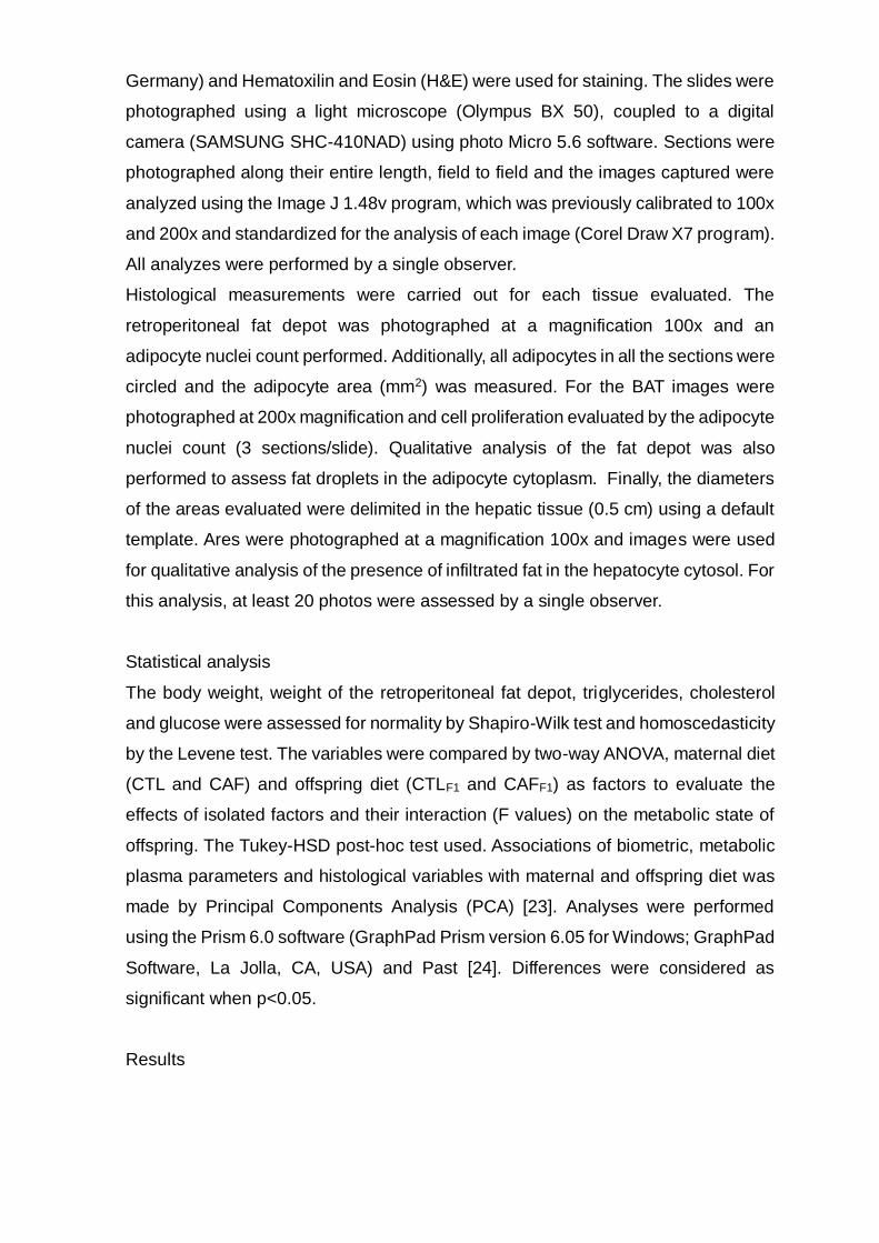
Some histological aspects of the retroperitoneal fat depot were altered only by post-
weaning exposure to CAF diet. Adult F1 offspring in the CTL-CAFF1 group
presented lower numbers of adipocytes and larger individual adipocyte sizes, in the
retroperitoneal fat depot, when compared with adipocytes of the CTL-CTLF1 rats
(p<0.05). Both adipocyte numbers (F1,17=7.76; p=0.01; Figure 2E) and adipocyte
size (F1,17=6.59; p=0.02; Figure 2F) were affected only by post weaning exposure
to CAF diet. Neither maternal nor post weaning exposure to the CAF diet altered
inflammatory processes in the retroperitoneal fat depot (data not shown). The
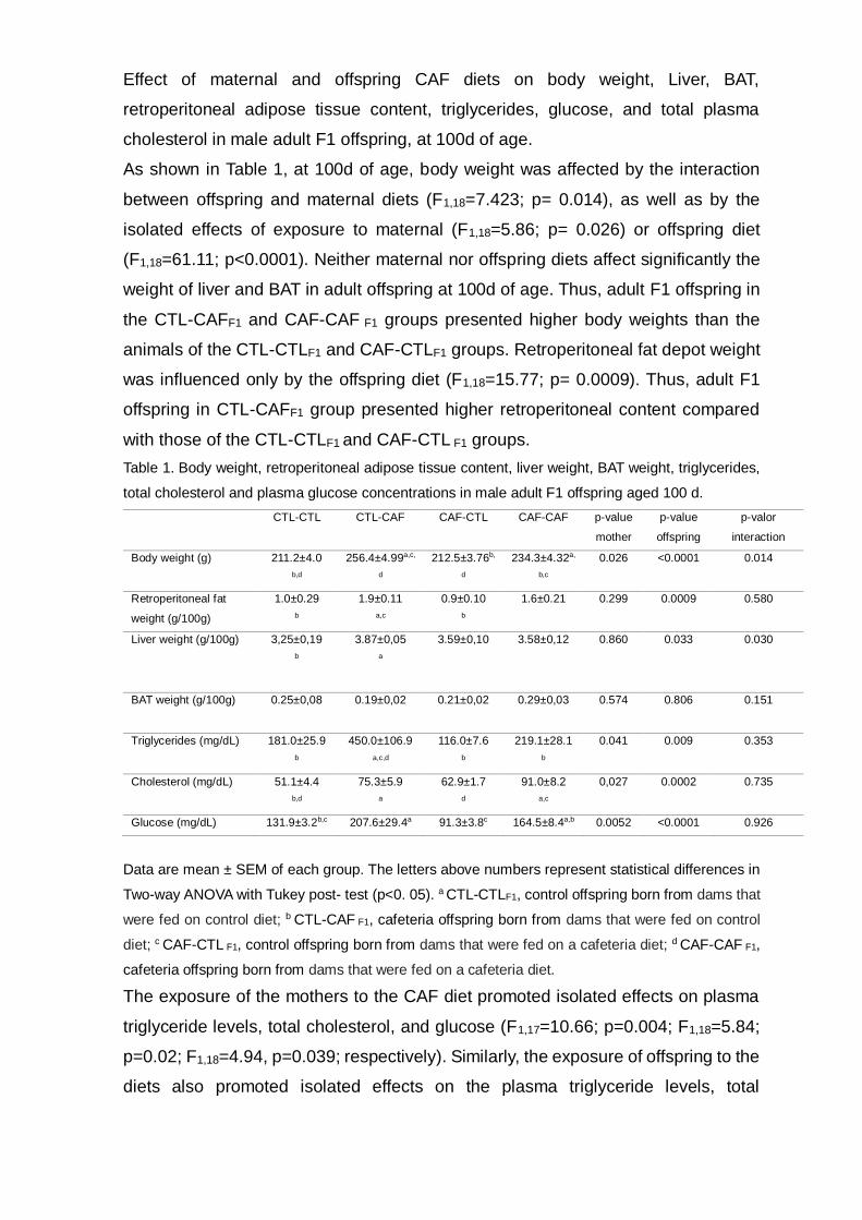
histological analyses of BAT are shown in Figure 3A-E. Cell proliferation in BAT was
analyzed quantitatively by counting nuclei, revealing no significant difference
between groups. Isolated effects of maternal or offspring diet, and no interactions
were observed (F1,18=0.67; p=0.42) (Figure 3E). Qualitative analysis demonstrated
that only adult F1 offspring exposed to the CAF diet post weaning induced lipid
accumulation in BAT. Thus, increased lipid droplet counts were found in BAT from
adult F1 offspring of the CTL-CAFF1 and CAF-CAF F1 groups, compared with those
of the CTL-CTLF1 and CAF-CTLF1 groups. However, the exposure of rats to the CAF
diet, during the pre- or post-natal phases, resulted in fat deposition in the liver. Thus,
in the CTL-CAFF1, CAF-CTLF1 and CAF-CAF F1 groups lipid accumulation was found
in the liver (Figure 4A-D).
Figure 2. Effects of exposure to CAF diet during pre and post weaning, alone or in combination, on
histological aspects of the retroperitoneal adipose tissue depot of male adult F1 offspring at 100d of
age.

WAT adipocyte count/area
Representative histology of retroperitoneal adipose tissue depot with H&E staining (100X
magnification); Figure 2A – D; Scale bars: 200 μm. Histological data for adipocyte area (Figure 2E),
measured for all cells, and adipocyte number (Figure 2F), expressed as mean ±SEM. The letters
above the bars represent statistical differences by Two-way ANOVA with the Tukey post- test (p<0.
05). a CTL-CTLF1, control offspring born from dams that were fed on a control diet; b CTL-CAF F1,
cafeteria offspring born from dams that were fed on a control diet; c CAF-CTL F1, control offspring
born from dams that were fed on a cafeteria diet; d CAF-CAF F1, cafeteria offspring born from dams
that were fed on a cafeteria diet.
Figure 3. Effect of exposure to CAF diet pre and post weaning, alone or in combination, on
histological aspects of BAT from male adult F1 offspring at 100d of age.

BAT nuclei count
Representative histology of BAT with H&E staining (200X magnification); Figure 3A – D; Scale bars:
150 μm. Qualitative histological analysis to evaluate the profile of lipid droplets in the cytosol of
adipocytes. Quantitative analysis was performed of by nuclei counts and expressed as mean±SEM
(Figure 3E). Nuclei are indicated by red arrows and lipid droplets are indicated by yellow arrows. The
letters above bars represent statistical differences; Two-way ANOVA with Tukey post- test (p<0. 05).
a CTL-CTLF1, control offspring born from dams that were fed on a control diet; b CTL-CAF F1,
cafeteria offspring born from dams that were fed on a control diet; c CAF-CTL F1, control offspring
born from dams that were fed on a cafeteria diet; d CAF-CAF F1, cafeteria offspring born from dams
that were fed on a cafeteria diet.
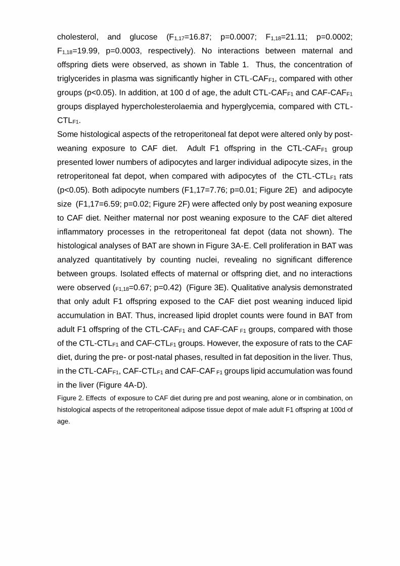
Figure 4. Effect of exposure to CAF diet pre and post weaning, alone or in combination, on
histological aspects of the liver pf male adult F1 offspring at 100d of age.
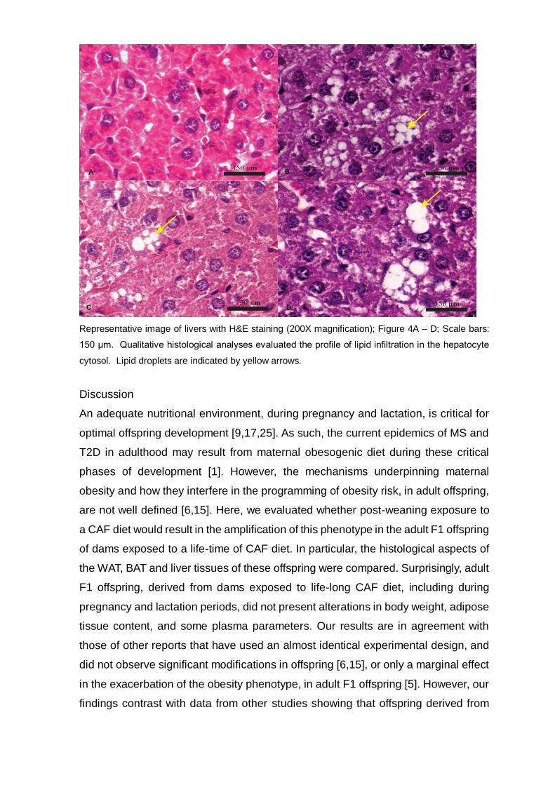
Representative image of livers with H&E staining (200X magnification); Figure 4A – D; Scale bars:
150 μm. Qualitative histological analyses evaluated the profile of lipid infiltration in the hepatocyte
cytosol. Lipid droplets are indicated by yellow arrows.
Discussion
An adequate nutritional environment, during pregnancy and lactation, is critical for
optimal offspring development [9,17,25]. As such, the current epidemics of MS and
T2D in adulthood may result from maternal obesogenic diet during these critical
phases of development [1]. However, the mechanisms underpinning maternal
obesity and how they interfere in the programming of obesity risk, in adult offspring,
are not well defined [6,15]. Here, we evaluated whether post-weaning exposure to
a CAF diet would result in the amplification of this phenotype in the adult F1 offspring
of dams exposed to a life-time of CAF diet. In particular, the histological aspects of
the WAT, BAT and liver tissues of these offspring were compared. Surprisingly, adult
F1 offspring, derived from dams exposed to life-long CAF diet, including during
pregnancy and lactation periods, did not present alterations in body weight, adipose
tissue content, and some plasma parameters. Our results are in agreement with
those of other reports that have used an almost identical experimental design, and
did not observe significant modifications in offspring [6,15], or only a marginal effect
in the exacerbation of the obesity phenotype, in adult F1 offspring [5]. However, our
findings contrast with data from other studies showing that offspring derived from
dams submitted to maternal hypercaloric diets display obesity and metabolic
abnormalities in adult life [2,7,9]. In this sense, to evaluate the impact of CAF diet
on maternal metabolism is important, once that, this state will determine the degree
of metabolic programming on adult offspring. Using same CAF maternal diet in the
present study, Sagae et al. 2015 and Mucellini et al. 2014 showed that the consume
of CAF diet by female at long of life promotes rises in body weight associated with
greater white adipose tissue accumulation; confirming the effectiveness of this diet
to induce obesity in mothers. Moreover, Mucellini et al. 2014 also showed that
maternal cafeteria diet does not alter glycemia or triglycerides levels, although
induces hyperinsulinemia and the increase of total cholesterol. Therefore, it is
important to keep in mind that the degree of mismatch between the pre- and
postnatal environments may be crucial to metabolic programming. Thus, the initial
adaptive physiological changes in fetal and pre-natal periods, necessary to
guarantee survival, may be maladaptive in later life [26,27]. Nevertheless, our
findings show that, independently of maternal diet, post-weaning exposure to the
CAF diet promotes obesity, hyperglycemia, and dyslipidemia in adult F1 offspring.
Mucellini et al. 2014, using the same experimental design that we used, including
the same cafeteria diet, analyzed the offspring immediately after weaning (21 days
of age) and evidenced no difference in the body weight of animals whose mothers
were fed with cafeteria diet or standard diet; an effect also observed at 30 days of
age. However, at 30 days of age, offspring fed with cafeteria diet showed higher
visceral fat, without maternal diet influence. In rats, intense adipogenesis occurs
during the last week of pregnancy and also during lactation; showing that these
periods are particularly sensitive to the developmental programming of adiposity
[16]. The expansion of WAT is morphologically characterized by increases in the
sizes of individual adipose cells (hypertrophy) or augmented adipocyte numbers
(hyperplasia) [16,28,29]. Any imbalance in this mechanism favors chronic
inflammatory processes in this tissue, which are the hallmark of obesity and
metabolic disorders [30]. Interestingly, we found the worst morphological profile in
the WAT of adult F1 offspring exposed to the CAF diet just during post weaning;
these alterations were characterized by adipocyte hypertrophy and a reduction in
adipocyte numbers. Furthermore, the combined effect of maternal CAF diet with the
post-weaning CAF diet (CAF-CAFF1 group) prevented morphological changes in the
adipocytes in the adult F1 offspring, showing that maternal CAF diet did not
exacerbate the obesity phenotype (induced by exposure to the CAF diet) during post
weaning. In fact, the maternal obesogenic diet appears to protect the WAT in adult
F1 offspring. This protective effect of maternal overnutrition on adult F1 offspring
has been previously reported [26,31], where authors suggested that the maternal
obesogenic diet alters hypothalamic leptin signaling, programming the metabolism
of adult offspring to minimize the degree of diet-induced obesity [25,30]. However,
as described below, the impact of early life exposure to maternal CAF diet on
morphological tissue aspects, in adult F1 offspring, appears be tissue-specific.
The development of BAT starts during pregnancy, with intense recruitment occurring
during the lactation and post-weaning phases [31,32]. Thus, nutritional insults during
these critical periods can reduce thermogenic activity in the BAT, thereby
suppressing energy expenditure and, ultimately, promoting obesity in adulthood
[33]. BAT adipocytes contain numerous smaller lipid vesicles, dispersed throughout
the cytosol, giving a multilocular morphological aspect to this depot [28]. The profile
of lipid droplets in the cytosol of BAT adipocytes reflects the degree of thermogenic
activity and, indirectly, represents increased sympathetic flux driven to the tissue
[34]. Maternal overnutrition induces functional and morphologic changes in the BAT
of adult offspring, leading to fat deposition, inflammation, and alterations in the
activity of sympathetic nerves [16]. However, our results show that the maternal
exposure to CAF diet did not affect morphologic aspects in the BAT of adult F1
offspring that consumed a normal diet at post weaning. These results contrast with
those obtained by others, where maternal CAF dietary was found to promote
significant alterations in BAT in adult offspring that were not reversed by the
exposure to a normal post-weaning diet [35,36]. As expected, we observed that
post-weaning exposure to a CAF diet induces lipid overload in the BAT of adult F1
offspring, an effect that is independent of the maternal diet profile. Thus, in contrast
to observations in the retroperitoneal depot, the maternal CAF diet did not protect
adult offspring that were also exposed to a life-long CAF diet. In a recent review,
BAT was reported to be vulnerable to nutritional insults, especially those occurring
during the pre- and post-natal periods of life, as important autonomic sympathetic
input and gene expression profiles are established during these periods [36,37].
Considering that WAT and BAT depots have different embryological origins, and
employ different pathways of proliferation and differentiation during pregnancy and
lactation, it is probable that the windows of vulnerability to nutritional insults are
depot-specific, resulting in different morphological adjustments when animals are
fed on a CAF diet, later on in life.
Taken together, we can draw two important conclusions from these results. Firstly,
the maternal CAF diet does not modulate the metabolism, adipose tissue content or
histological aspects of the retroperitoneal fat and BAT in adult offspring, at 100d of
age, suggesting that maternal CAF diet, alone, does not alter the programming in
these tissues. Secondly, while the exposure to maternal CAF diet, associated with
post-weaning CAF exposure, protects WAT in adult F1 offspring, CAF diet exposure
during the post-weaning period has a deleterious effect on the BAT, independently
of the maternal diet. Considering that, at 100d of age, the rats are young adults, it
is possible that the deleterious effects of programming in these tissues may occur
at a more advanced age. This hypothesis is supported reports that the effects of
maternal programs are found in adult offspring at 140-155d of age [9,38]. In addition,
the impact of maternal diet on adult offspring is sex dependent, as females appear
to be more sensitive to the effect of maternal diet programming, when compared
with males [39].
In contrast to the observations in WAT and BAT, maternal exposure to a CAF diet
exerts programming effects on the liver in adult F1 offspring, at 100d of age. Glucose
and lipid homeostasis are dependent on liver metabolism and these events are
intimately coupled to hepatic tissue function [40]. As such, abnormal lipid deposition
in hepatocytes results in hepatic steatosis, a pathological state related to liver
abnormalities, such as the nonalcoholic fatty liver disease (NAFLD). NAFLD is the
most common chronic liver disease present in obese subjects, and is closely
associated with manifestations of MS [41,42]. Although hepatic steatosis is a key
histological feature in this process, variations in morphological aspects have been
observed; thus hepatocellular steatosis is usually classified as either macrovesicular
or microvesicular [43]. Our data confirm the impact of the maternal CAF diet on the
liver programming effect in adult F1 offspring, as previously demonstrated by others
[44,45]. For example, it has been demonstrated that the administration of a
hypercaloric diet to dams, during pregnancy and lactation, induces hyperlipidemia,
increasing hepatic triglycerides and resulting in fat liver accumulation and,
consequently, hepatic steatosis in offspring [19,43]. The maternal obesogenic diet
can induce lipid deposition in the fetal liver before birth, a crucial event in metabolic
disease during adult life [46]. Recently, using an identical experimental design to
that of our study, a programming effect of maternal high fat diet was demonstrated
on the liver of adult offspring [47]. Interestingly, in this study, the plasma metabolic
parameters, body weight, and adipose tissue content were not programmed by
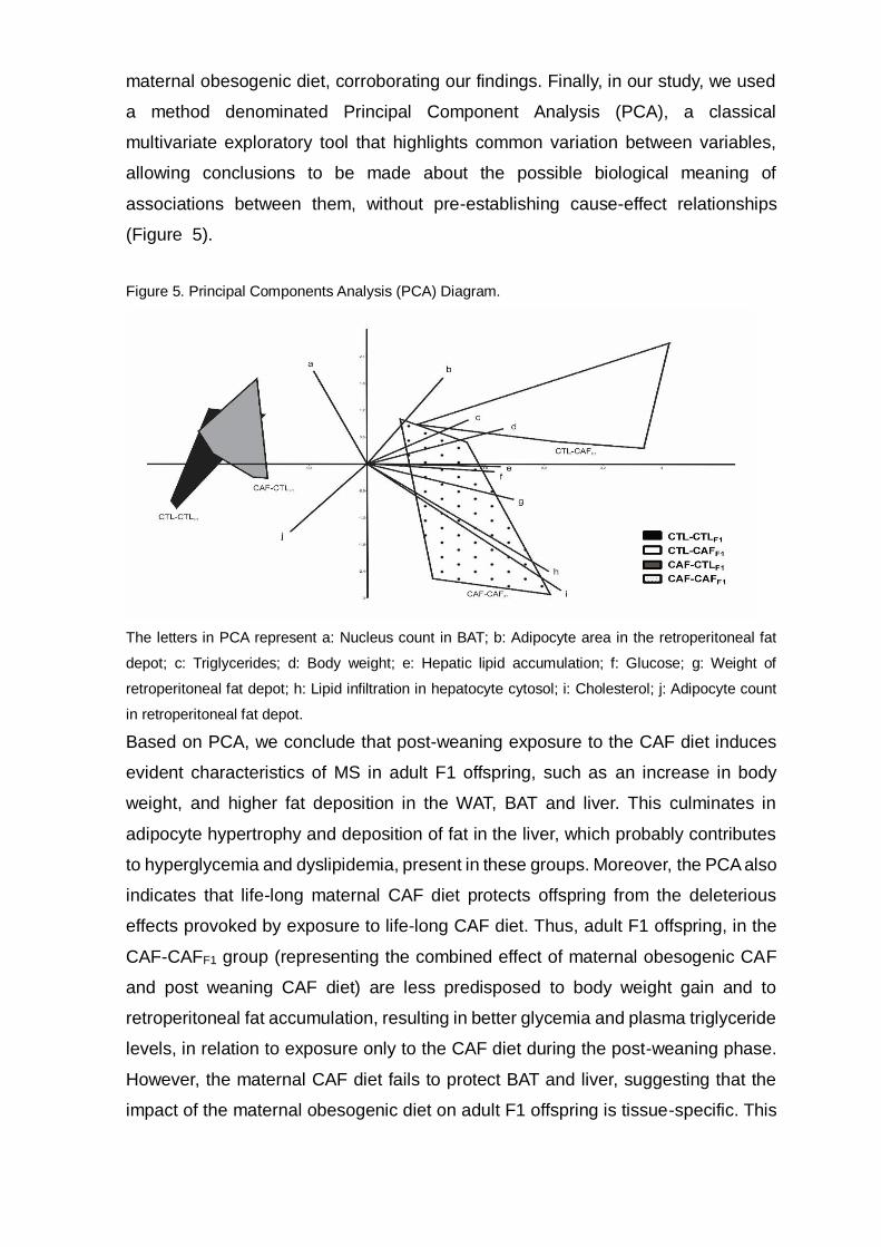
maternal obesogenic diet, corroborating our findings. Finally, in our study, we used
a method denominated Principal Component Analysis (PCA), a classical
multivariate exploratory tool that highlights common variation between variables,
allowing conclusions to be made about the possible biological meaning of
associations between them, without pre-establishing cause-effect relationships
(Figure 5).
Figure 5. Principal Components Analysis (PCA) Diagram.
The letters in PCA represent a: Nucleus count in BAT; b: Adipocyte area in the retroperitoneal fat
depot; c: Triglycerides; d: Body weight; e: Hepatic lipid accumulation; f: Glucose; g: Weight of
retroperitoneal fat depot; h: Lipid infiltration in hepatocyte cytosol; i: Cholesterol; j: Adipocyte count
in retroperitoneal fat depot.
Based on PCA, we conclude that post-weaning exposure to the CAF diet induces
evident characteristics of MS in adult F1 offspring, such as an increase in body
weight, and higher fat deposition in the WAT, BAT and liver. This culminates in
adipocyte hypertrophy and deposition of fat in the liver, which probably contributes
to hyperglycemia and dyslipidemia, present in these groups. Moreover, the PCA also
indicates that life-long maternal CAF diet protects offspring from the deleterious
effects provoked by exposure to life-long CAF diet. Thus, adult F1 offspring, in the
CAF-CAFF1 group (representing the combined effect of maternal obesogenic CAF
and post weaning CAF diet) are less predisposed to body weight gain and to
retroperitoneal fat accumulation, resulting in better glycemia and plasma triglyceride
levels, in relation to exposure only to the CAF diet during the post-weaning phase.
However, the maternal CAF diet fails to protect BAT and liver, suggesting that the
impact of the maternal obesogenic diet on adult F1 offspring is tissue-specific. This
study emphasizes the importance of the maternal diet during animals’ entire life for
establishing tissue-specific effects in the offspring in response to an obesogenic diet
during adulthood.
AUTHOR CONTRIBUTIONS
Balbo; S. L. and Grassiolli; S. designed the study. Santos; C.D. S.; Negretti, F. and
Sagae; S. C. collected the data. Guimarães, A.T.B. analysed the data. Balbo; S. L.;
Grassiolli; S.; Santos; C.D. S. wrote the paper and performed corrections. All authors
read and approved the final manuscript.
COMPETING INTERESTS
This manuscript describes original work and is not under consideration by any other
journal. All authors approved the manuscript and this submission. None of the authors
declared a conflict of interest.
FUNDING
This research received no specific grant from any funding agency in the public,
commercial, or not-for-profit sectors.
ACKNOWLEDGMENTS
We are grateful to Doctor Antonio Carlos Boschero for performed corrections
and Nicola Conran for editing English
References
1. Smith CJ, Ryckman KK. Epigenetic and developmental influences on the risk of
obesity , diabetes , and metabolic syndrome. Diabetes, Metabolic Syndrome and
Obesity: Targets and Therapy. 2015;8:295–302.
2. Desai M, Jellyman JK, Ross MG. Epigenomics , gestational programming and
risk of metabolic syndrome. International Journal of Obesity. 2015;39:633–641.
doi:10.1038/ijo.2015.13.
3. Sedaghat K, Zahediasl S, Ghasemi A. Intrauterine programming. Iranian Journal
of Basic Medical Sciences. 2015;18:212–220.
4. Barker, DJ. The developmental origins of adult disease. J Am Coll Nutr
23, Suppl 6: 588S–595S, 2004
5. King V, Norman JE, Seckl JR, Drake AJ. Post-weaning diet determines
metabolic risk in mice exposed to overnutrition in early life. Reproductive
Biology and Endocrinology. 2014;12(73):1–7. doi:10.1186/1477-7827-12-73.
6. Mucellini AB Goularte F, Carla A, Araujo D, Noschang C, Benetti S, et al.
Effects of exposure to a cafeteria diet during gestation and after weaning on the
metabolism and body weight of adult male offspring in rats British Journal of
Nutrition. British Journal of Nutrition. 2014;111:1499–1506.
doi:10.1017/S0007114513003838.PLOS 9/12
7. Li M, Reynolds CM, Segovia SA, Gray C, Vickers MH. Developmental
Programming of Nonalcoholic Fatty Liver Disease : The Effect of Early Life
Nutrition on Susceptibility and Disease Severity in Later Life. BioMed Research
International. 2015;2015:1–12. doi:10.1155/2015/437107.
8. Sampey BP, Vanhoose AM, Winfield HM, Freemerman AJ, Muehlbauer MJ,
Fueger PT, et al. Cafeteria Diet Is a Robust Model of Human Metabolic
Syndrome With Liver and Adipose Inflammation : Comparison to High-Fat Diet.
9. Howie GJ, Sloboda DM, Kamal T, Vickers MH. Maternal nutritional history
predicts obesity in adult offspring independent of postnatal diet. J Physiol.
2009;4:905–915. doi:10.1113/jphysiol.2008.163477.
10. Li M, Sloboda DM, Vickers MH. Maternal Obesity and Developmental
Programming of Metabolic Disorders in Offspring : Evidence from Animal Models.
Experimental Diabetes Research. 2011;2011:1–9. doi:10.1155/2011/592408.
Obesity. 2011;19(6):1109–1117. doi:10.1038/oby.2011.18.
11. White CL, Purpera MN, Morrison CD. Maternal obesity is necessary for
programming effect of high-fat diet on offspring. Am J Physiol Regul Integr
Comp Physiol. 2009;296:1464–1472. doi:10.1152/ajpregu.91015.2008.
12. Daniel ZC, Akyol A, Mcmullen S, Langley-evans SC. Exposure of neonatal rats
to maternal cafeteria feeding during suckling alters hepatic gene expression and
DNA methylation in the insulin signalling pathway. Genes Nutr. 2014;9(365):1–10.
doi:10.1007/s12263-013-0365-3.
13. Jacobs S, Teixeira DS, Guilherme C, Claudio FK, Aranda BCC, Reis AR, et al.
Physiology & Behavior The impact of maternal consumption of cafeteria diet on
reproductive function in the offspring. Physiology & Behavior. 2014;129:280–286.
doi:10.1016/j.physbeh.2014.03.003.
14. Tamashiro KLK, Terrillion CE, Hyun J, Koenig JI, Moran TH. Prenatal Stress
or High-Fat Diet Increases Susceptibility to Diet-Induced Obesity in Rat
Offspring. Diabetes. 2009;58(May):1116–1125. doi:10.2337/db08-1129.
15. Akyol A, Mcmullen S, Langley-evans SC. Glucose intolerance associated with
early-life exposure to maternal cafeteria feeding is dependent upon post-weaning
diet. British Journal of Nutrition2. 2012;107:964–978.
doi:10.1017/S0007114511003916.
16. Lukaszewski Ma, Eberl´e D, Vieau D, Breton C. Nutritional manipulations in the
perinatal period program adipose tissue in offspring. Am J Physiol Endocrinol
Metab. 2013;305(4):E1195–E1207. doi:10.1152/ajpendo.00231.2013.
17. Lee Hs. Impact of Maternal Diet on the Epigenome during In Utero Life and the
Developmental Programming of Diseases in Childhood and Adulthood. Nutrients.
2015;7:9492–9507. doi:10.3390/nu7115467.
18. Ramírez-Lopez MT, Berrios MV, Gonzalez RA, Noemı R, Velilla B, Decara J,et
al. El papel de la dieta materna en la programacion metabolica y conductual:
revision de los mecanismos biologicos implicados. Nutricion Hospitalaria.
2015;32(6):2433–2445. doi:10.3305/nh.2015.32.6.9716.
19. Bringhenti I, Ornellas F, Martins MA, Mandarim-de lacerda CA, Aguila MB.
Early hepatic insult in the offspring of obese maternal mice. Nutrition Research.
2015;35(2):136–145. doi:10.1016/j.nutres.2014.11.006.
20. Kayser BD, Goran MI, Bouret SG. Perinatal Overnutrition Exacerbates Adipose
Tissue Inflammation Caused by High-Fat Feeding in C57BL / 6J Mice. PlosOne.
2015;10(4):1–15. doi:10.1371/journal.pone.0121954.
21. Reeves PG. Components of the AIN-93 Diets as Improvements in the AIN-76A
Diet. In: Simposium: Animal Diets for Nutritional and Toxicological Reserarch; 1997.
p. 838–841.PLOS 10/12
22. Goularte F, Ferreira MBC, Sanvitto GL. Effects of food pattern change and
physical exercise on cafeteria diet-induced obesity in female rats British Journal
of Nutrition. British Journal of Nutrition. 2012;108:1511–1518.
doi:10.1017/S0007114511006933.
23. Hair JF, Black WC, Babin BJ, Anderson RE. Multivariate Data Analysis. vol. 7ed;
1998. Available from: http://www.pearsonhighered.com/educator/
product/Multivariate-Data-Analysis/9780138132637.page.
24. Hammer Ø, Harper DAT, Ryan PD. Paleontological statistics software package
for education and data analysis. Palaeontologia Electronica. 2001;4(1):9–18.
doi:10.1016/j.bcp.2008.05.025.
25. Couvreur O, Ferezou J, Gripois D, Serougne C, Cr´epin D, Aubourg A, et al.
Unexpected long-term protection of adult offspring born to high-fat fed dams
against obesity induced by a sucrose-rich diet. PLoS ONE. 2011;6(3).
doi:10.1371/journal.pone.0018043.
26. Fernandez-Twinn DS, Ozanne SE. Mechanisms by which poor early growth
programs type-2 diabetes, obesity and the metabolic syndrome. Physiology and
Behavior. 2006;88(3):234–243. doi:10.1016/j.physbeh.2006.05.039.
27. Gluckman PD, Hanson MA, Beedle AS, Spencer HG. Predictive adaptive
responses in perspective. Trends in Endocrinology and Metabolism.
2008;19(4):109–110. doi:10.1016/j.tem.2008.02.002.
28. Cinti S. The adipose organ. Disease Models & Mechanisms. 2012;73:9–15.
doi:10.1016/j.plefa.2005.04.010.
29. Bezpalko L, Gavrilyuk O, Zayachkivska O. Inflammatory response in visceral fat
tissue and liver is prenatally programmed: experimental research. Journal of
Physiology and Pharmacology. 2015;66(1):57–64.
30. Rosen ED, Spiegelman BM. NIH Public Access. Nature. 2006;444(7121):847–
853. doi:10.1126/scisignal.2001449.Engineering.
31. Ferezou-Viala J, Roy AF, Serougne C, Gripois D, Parquet M, Bailleux V, et al.
Long-term consequences of maternal high-fat feeding on hypothalamic leptin
sensitivity and diet-induced obesity in the offspring. Am J Physiol Regul Integr
Comp Physiol. 2007;293:R1056–R1062. doi:10.1152/ajpregu.00117.2007.
32. Giralt M, Martin I, Iglesias R, Vi˜nas O, Villarroya F, Mampel T. Ontogeny and
perinatal modulation of gene expression in rat brown adipose tissue to
environmental temperature at birth. Eur J Biochem. 1990;193:297–302.
33. Cannon B, Nedergaard JAN. Brown Adipose Tissue : Function and Physiological
Significance. Physiol Rev. 2004;84:277–359. doi:10.1152/physrev.00015.2003.
34. Tupone D, Madden CJ, Morrison SF. Autonomic regulation of brown adipose
tissue thermogenesis in health and disease: Potential clinical applications for
altering BAT thermogenesis. Frontiers in Neuroscience. 2014;8(8 FEB):1–14.
doi:10.3389/fnins.2014.00014.
35. Dinh CHL, Szabo A, Yu Y, Camer D, Zhang Q, Wang H, et al. Bardoxolone
Methyl Prevents Fat Deposition and Inflammation in Brown Adipose Tissue and
Enhances Sympathetic Activity in Mice Fed a High-Fat Diet. Nutrients.
2015;7:4705–4723. doi:10.3390/nu7064705. PLOS 11/12
36. Barbato DL, Tatulli G, Vegliante R, Cannata SM, Bernardini S, Ciriolo MR,
et al. Dietary fat overload reprograms brown fat mitochondria. Frontiers in
Physiology. 2015;6(SEP):1–12. doi:10.3389/fphys.2015.00272.
37. Bayol AS, Farrington SJ, Stickland NC. A maternal ‘junk food’ in pregnancy and
lactation promotes na exacerbated taste for ‘junk food’ and greater propensity for
obesity in rat offspring. British Journal of Nutrition. 2007. Doi
10.1017/50007114507812037.
38. Shankar, K, Harrell, A, Liu, X, Gilchrist, JM, Ronis, MJJ, Badger, TM. Maternal
obesity at conception programs obesity in the offspring
Am J Physiol Regul Integr Comp Physiol 294: R528–R538, 2008.
39. Onyekwere CA, Ogbera AO, Samaila AA, Bologum BO. Nonalcoholic fatty liver
disease: synopsis of current developments. Nigerian Journal of Clinical practice.
Nov-Dez 2015. Vol 18. Issue 6.
40. Zeltser LM. Developmental influences on circuits programming susceptibility to
obesity. Frontiers in Endocrinology. 2015;39(Oct):17–27.
41. Calvo N, Beltran-debon R, Rodrıguez-gallego E, Hernandez-aguilera A, Guirro
M, Marine-casado R, et al. Liver fat deposition and mitochondrial dysfunction in
morbid obesity : An approach combining metabolomics with liver imaging and
histology. World Journal of Gastroenterology. 2015;21(24):7529–7544.
doi:10.3748/wjg.v21.i24.7529.
42. Linnemann AK, Baan M, Davis DB. Pancreatic b -Cell Proliferation in Obesity 1,
2. Advances in nutrition. 2014;5:278–288. doi:10.3945/an.113.005488.278.
43. Kleiner DE, Brunt EM, Van Natta M, Behling C, Contos MJ, Cummings OW,
et al. Design and validation of a histological scoring system for nonalcoholic fatty
liver disease. Hepatology. 2005;41(6):1313–1321. doi:10.1002/hep.20701.
44. Podrini C, Cambridge EL, Lelliot CJ, Carragher DM, Estabel J, Gerdin AK,
et al. High-fat feeding rapidly induces obesity and lipid derangements in C57BL /
6N mice. Mamm Genome. 2013;24:240–251. doi:10.1007/s00335-013-9456-0.
45.Bouane,S, Merzouk, H, Benkalfat, NB, Souliman, N, Merzouk, SA, Gusti, J,
Tessier, C, Narci, M. Hepatic and very low-density lipoprotein fatty acids in obese
offspring of overfed dams. Metabolism clinical and experimental. 2010 (59):1701-
1709.
46. Ingvorsen C, Brix S, Ozanne SE, Hellgren LI. The effect of maternal
Inflammation on foetal programming of metabolic disease. Acta Physiologica.
2015;2:440–449. doi:10.1111/apha.12533.
47. Ito J, Nakagawa K, Kato S, Miyazawa T, Kimura F, Miyazawa T. The
combination of maternal and offspring high-fat diets causes marked oxidative

stress and development of metabolic syndrome in mouse offspring. Life Sciences.
2016;151:70–75. doi:10.1016/j.lfs.2016.02.089.
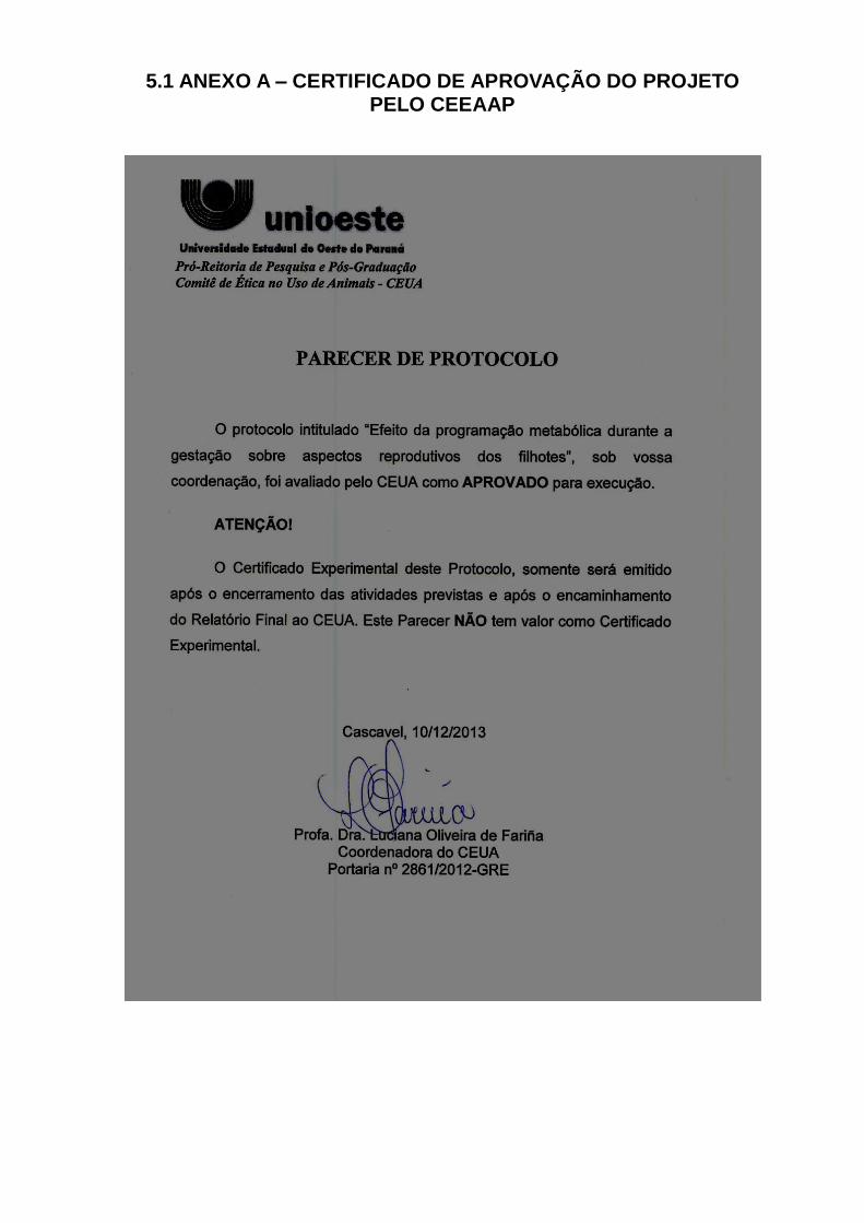
5.1 ANEXO A – CERTIFICADO DE APROVAÇÃO DO PROJETO PELO CEEAAP
5.1 ANEXO B – NORMAS DA REVISTA CIENTÍFICA
Language editing
Hindawi has partnered with Editage to provide an English-language editing service
to authors prior to submission. Authors that wish to use this service will receive a
10% discount on all editing services provided by Editage. To find out more
information or get a quote.
Submission
Manuscripts should be submitted by one of the authors of the manuscript through
the online Manuscript Tracking System. Regardless of the source of the word-
processing tool, only electronic PDF (.pdf) or Word (.doc, .docx, .rtf) files can be
submitted through the MTS. There is no page limit. Only online submissions are
accepted to facilitate rapid publication and minimize administrative costs.
Submissions by anyone other than one of the authors will not be accepted. The
submitting author takes responsibility for the paper during submission and peer
review. If for some technical reason submission through the MTS is not possible,
the author can contact [email protected] for support.
Terms of Submission
Papers must be submitted on the understanding that they have not been published
elsewhere and are not currently under consideration by another journal published
by Hindawi or any other publisher. The submitting author is responsible for ensuring
that the article’s publication has been approved by all the other coauthors. It is also
the authors’ responsibility to ensure that the articles emanating from a particular
institution are submitted with the approval of the necessary institution. Only an
acknowledgment from the editorial office officially establishes the date of receipt.
Further correspondence and proofs will be sent to the author(s) before publication
unless otherwise indicated. It is a condition of submission of a paper that the authors
permit editing of the paper for readability. All inquiries concerning the publication of
accepted papers should be addressed to [email protected].
Peer Review
All manuscripts are subject to peer review and are expected to meet standards of
academic excellence. If approved by the editor, submissions will be considered by
peer-reviewers, whose identities will remain anonymous to the authors.
Microarray Data Submission
For any article that includes microarray data, this data should be deposited in an
appropriate database such as Gene Expression Omnibus (GEO) or Array Express,
and an entry name or accession number must be included in the manuscript prior
to its publication. Microarray data should be MIAME compliant. During the reviewing
process, submitting authors are committed to provide the editor and the reviewers
handling his/her manuscript with the login information by which they can access this
information in the database.
Concurrent Submissions
In order to ensure sufficient diversity within the authorship of the journal, authors will
be limited to having two manuscripts under review at any point in time. If an author
already has two manuscripts under review in the journal, he or she will need to wait
until the review process of at least one of these manuscripts is complete before
submitting another manuscript for consideration. This policy does not apply to
Editorials or other non-peer reviewed manuscript types.
Article Processing Charges
The Scientific World Journal is an open access journal. Open access charges allow
publishers to make the published material available for free to all interested online
visitors. For more details about the article processing charges of The Scientific
World Journal, please visit the Article Processing Charges information page.
Units of Measurement
Units of measurement should be presented simply and concisely using System
International (SI) units.
Title and Authorship Information
The following information should be included
• Paper title • Full author names • Full institutional mailing addresses • Email addresses
Abstract
The manuscript should contain an abstract. The abstract should be self-contained
and citation-free and should not exceed 200 words.
Introduction
This section should be succinct, with no subheadings.
Materials and Methods
This part should contain sufficient detail so that all procedures can be repeated. It
can be divided into subsections if several methods are described.
Results and Discussion
This section may each be divided by subheadings or may be combined.
Conclusions
This should clearly explain the main conclusions of the work highlighting its
importance and relevance.
Acknowledgments
All acknowledgments (if any) should be included at the very end of the paper before
the references and may include supporting grants, presentations, and so forth.
References
Authors are responsible for ensuring that the information in each reference is
complete and accurate. All references must be numbered consecutively and
citations of references in text should be identified using numbers in square brackets
(e.g., “as discussed by Smith [9]”; “as discussed elsewhere [9, 10]”). All references
should be cited within the text; otherwise, these references will be automatically
removed.
Preparation of Figures
Upon submission of an article, authors are supposed to include all figures and tables
in the PDF file of the manuscript. Figures and tables should not be submitted in
separate files. If the article is accepted, authors will be asked to provide the source
files of the figures. Each figure should be supplied in a separate electronic file. All
figures should be cited in the paper in a consecutive order. Figures should be
supplied in either vector art formats (Illustrator, EPS, WMF, FreeHand, CorelDraw,
PowerPoint, Excel, etc.) or bitmap formats (Photoshop, TIFF, GIF, JPEG, etc.).
Bitmap images should be of 300 dpi resolution at least unless the resolution is
intentionally set to a lower level for scientific reasons. If a bitmap image has labels,
the image and labels should be embedded in separate layers.
Preparation of Tables
Tables should be cited consecutively in the text. Every table must have a descriptive
title and if numerical measurements are given, the units should be included in the
column heading. Vertical rules should not be used.
Proofs
Corrected proofs must be returned to the publisher within 2-3 days of receipt. The
publisher will do everything possible to ensure prompt publication. It will therefore
be appreciated if the manuscripts and figures conform from the outset to the style
of the journal.
Copyright
Open Access authors retain the copyrights of their papers, and all open access
articles are distributed under the terms of the Creative Commons Attribution
License, which permits unrestricted use, distribution and reproduction in any
medium, provided that the original work is properly cited.
The use of general descriptive names, trade names, trademarks, and so forth in this
publication, even if not specifically identified, does not imply that these names are
not protected by the relevant laws and regulations.
While the advice and information in this journal are believed to be true and accurate
on the date of its going to press, neither the authors, the editors, nor the publisher
can accept any legal responsibility for any errors or omissions that may be made.
The publisher makes no warranty, express or implied, with respect to the material
contained herein.
Disclosure Policy
A competing interest exists when professional judgment concerning the validity of
research is influenced by a secondary interest, such as financial gain. We require
that our authors reveal any possible conflict of interest in their submitted
manuscripts.
If there is no conflict of interest, authors should state that “The author(s) declare(s)
that there is no conflict of interest regarding the publication of this paper.”
Clinical Study
When publishing clinical studies, Hindawi aims to comply with the recommendations
of the International Committee of Medical Journal Editors (ICMJE) on trials
registration. Therefore, authors are requested to register the clinical trial presented
in the manuscript in a public trials registry and include the trial registration number
at the end of the abstract. Trials initiated after July 1, 2005 must be registered
prospectively before patient recruitment has begun. For trials initiated before July 1,
2005, the trial must be registered before submission.
International Commission on Zoological Nomenclature
When publishing papers which describe a new zoological taxon name, Hindawi aims
to comply with the requirements of the International Commission on Zoological
Nomenclature (ICZN). Therefore, for all papers that include the naming of a new
zoological taxon, authors are requested to contact Zoobank, the online registration
system for the International Commission on Zoological Nomenclature, to obtain a
Life Science Identifier (LSID). Moreover, authors are requested to insert the
following text in the “Materials and Methods” section, in a subsection to be called
“Nomenclatural Acts”:
The new names contained in this article are available under the International Code
of Zoological Nomenclature. This work and the nomenclatural acts it contains have
been registered in ZooBank. Zoobank Life Science Identifier (LSID) for this
publication is: urn:lsid:zoobank.org:pub: XXXXXXX. The LSID registration and any
associated information can be viewed in a web browser by adding the LSID to the
prefix “http://zoobank.org/.”
Ethical Guidelines
In any studies that involve experiments on human or animal subjects, the following
ethical guidelines must be observed. For any experiments on humans, all work must
be conducted in accordance with the Declaration of Helsinki (1964). Papers
describing experimental work which carries a risk of harm to human subjects must
include a statement that the experiment was conducted with the human subjects’
understanding and consent, as well as a statement that the responsible Ethical
Committee has approved the experiments. In the case of any animal experiments,
the authors must provide a full description of any anesthetic or surgical procedure
used, as well as evidence that all possible steps were taken to avoid animal suffering
at each stage of the experiment.