Embed Size (px)
Citation preview

AMEBIASIS & GIARDIOSIS

AMEBIASIS

AMEBIAS PATHOGENS
Intestinals Entamoeba histolytica
Tisulares (Amebas of Libra) AcanthamoebaNaegleria

AMEBIAS INTESTINALS
EntamoebaE.histolytica (pathogen)E. coliE. hartmaniE. gingivalis (oral)
Endolimax nanaIodamoeba butschlii
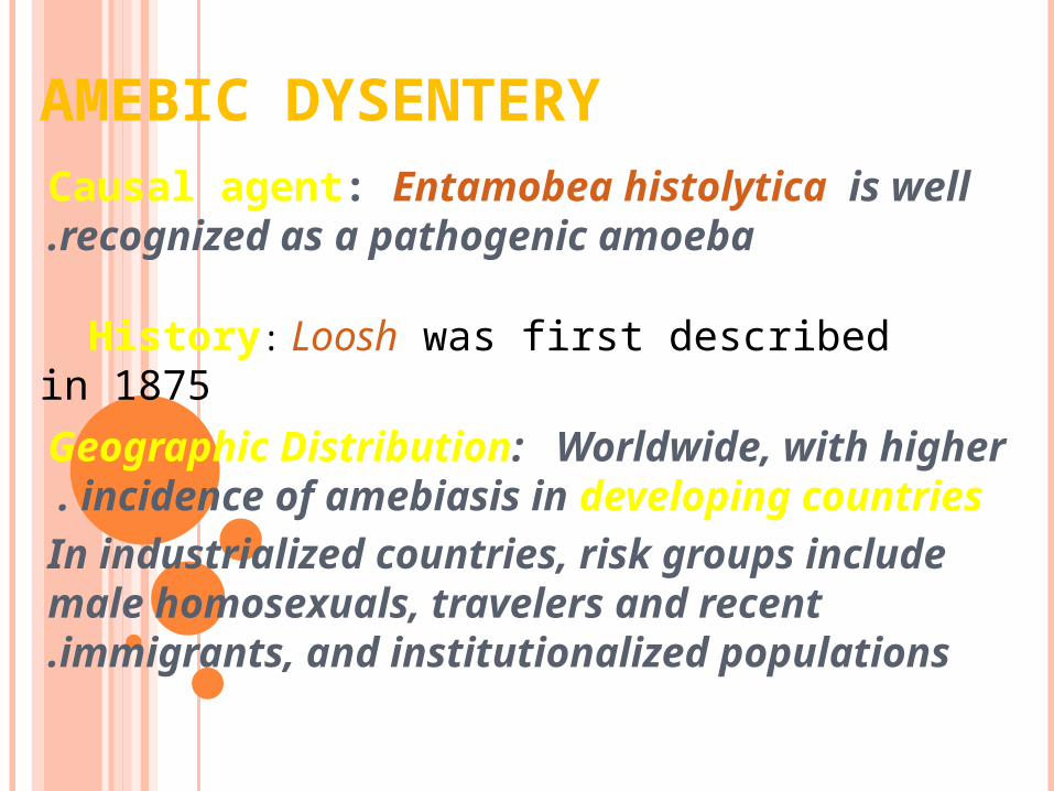
AMEBIC DYSENTERYCausal agent: Entamobea histolytica is well recognized as a pathogenic amoeba.
Geographic Distribution: Worldwide, with higher incidence of amebiasis in developing countries .
In industrialized countries, risk groups include male homosexuals, travelers and recent immigrants, and institutionalized populations.
History: Loosh was first described in 1875

MORPHOLOGYDifferent form of E. histolytica ;
1 -Trophozoite
2 -Precyst
3 -Cyst(1, 2, 4 nuclei)

TROPHOZOITE CHARACTERSize: 12-60μm in diameter ;
Non-invasive form ( minuta) / E. dispare
Invasive form (magna) contain RBC, E. histolyticaPseudopodiaMotility
EctoplasmEndoplasm: may be contain ingested RBC
Nucleoplasm
Non-invasive forminvasive form

DIFFERENT FORM OF E.HISTOLYTICA CYST

LIFE CYCLE
Life cycle

EPIDEMIOLOGY
Prevalence of amebic infection varies with level of sanitation and generally higher in tropics and
subtropics than in tempearate climates .
*Worldwide prevalence is about 10% to 50%*Cyst passers are important source of infection
The true estimated prevalence of E. histolytica is close to 1% worldwide.
Entamoeba histolytica is the second leading cause of mortality due to parasitic disease in
humans. (The first being malaria). Amebiasis is the cause of an estimated 50,000-100,000 deaths
each year.

TRANSMISSION
1-Direct contact of person to person( fecal-oral).
2 -Veneral transmission among homosexual males( oral-anal) . 3- Food or drink
contaminated with feces containing the E.his. Cyst.
4 -Use of human feces (night soil) for soil fertilizer.
5 -Contamination of foodstuffs by flies, and possibly cockroaches.

PATHOGENESIS
Effective factores:
1 -strain virulence
2 -susceptibility of the host; nutrition status, immune-sys.
3 -breakdown of immunologic barrier (tissue invasion)

PATHOGENICITY MECHANISMS
1 -secreting proteolytic enzymes( histolysine ) and cytotoxic substances.
2 - contact-dependent cell killing
3 – cytophagocytosis
Amebic killing target cell :
1 -receptore-mediated adherence of amebae to target cell ( adherence lectin)
2 -amebic cytolysis of target cell 3 -amebic phagocytosis of killed target cell


CLINICAL SYMPTOMS(2-6W AFTER INGESTION)
Asymptomatic infection Symptomatic infection
Intestinal Amebiasis Extraintestinal Amebiasis
Dysenteric(40% Fever) Non-Dysenteric colitis Hepatic Pulmonary(R) The extra foci(p,p,b)
( Fulminant,perfuration
Toxic megacolon) Liver abscces Acute nonsupprative
Intestinal Amebiasis symptoms: Diarrhea or dysentery,Lower abdominal pain, cramping , anorexia, weight loss, chronic fatigue


PATHOLOGY OF AMEBIASIS

FLASK-LIKE ULCER

EXTRA-INTESTINALAMEBIASIS


This is an amebic abscess of liver. Abscesses may arise in liver when there is seeding of infection from the bowel, because the infectious agents are carried to the liver from the portal venous circulation.

DIAGNOSIS
Paraclinical Diagnosis:Sigmoidoscopic examination:
precence of a grossly normal mucosa between the ulcers serves to differentiate amebic from bacillary
dysentery,( the entire mucosa being involvoed in bacillary dysentery).
HepatomegallyC.B.C. : leukocytosis in Amebic dys. rises above
12000 per microliter, but counts may reach 16000 to 20000 per microliter.

LABORATORY DIAGNOSIS
Entamoeba histolytica must be differentiated from other intestinal protozoa including: E. coli, E. hartmanni, E.
dispare……,
Differentiation is possible, but not always easy, based on morphologic characteristics of the cysts and trophozoites.
The nonpathogenic Entamoeba dispar, however, is
morphologically identical to E. histolytica, and differentiation must be based on isoenzymatic or
immunologic analysis .
Molecular methods are also useful in distinguishing between E. histolytica and E. dispar and can also be
used to identify E. polecki .

MICROSCOPY
Microscopic identification
This can be accomplished using:
Fresh stool: wet mounts and permanently stained preparations (e.g., trichrome) .
Concentrates from fresh stool: wet mounts, with or without iodine stain, and permanently
stained preparations (e.g., trichrome) .

TROPHOZOITES OF ENTAMOEBA HISTOLYTICA /E. DISPAR ( TRICHROME STAIN )
Microscopy
AB
In the absence of erythrophagocytosis, the pathogenic E. histolytica is morphologically indistinguishable from the nonpathogenic E. dispar!
Each trophozoite has a single nucleus, which has a centrally placed karyosome and uniformly distributed peripheral chromatin.

TROPHOZOITES OF ENTAMOEBA HISTOLYTICA WITH
INGESTED ERYTHROCYTES (TRICHROME STAIN)
The ingested erythrocytes appear as dark inclusions . Erythrophagocytosis is the only morphologic
characteristic that can be used to differentiate E. histolytica from the nonpathogenic E. dispar .
EF

CYSTS OF ENTAMOEBA HISTOLYTICA /E. DISPAR
GHI
GHICysts of Entamoeba histolytica/E. dispar, permanent preparations stained with trichrome.

IMMUNODIAGNOSIS1 -Antibody detection :
The indirect hemagglutination (IHA)
The EIZA test detects antibody specific for E. histolytica in approximately 95% of patients with
extraintestinal amebiasis, 70% of patients with active intestinal infection, and 10% of asymptomatic persons
who are passing cysts of E. histolytica .

2 -Antigen detection may be useful as an adjunct to microscopic diagnosis in detecting
parasites and to distinguish between pathogenic and nonpathogenic infections .
Recent studies indicate improved sensitivity and specificity of fecal antigen assays with
the use of monoclonal antibodies which can distinguish between E. histolytica and E.
dispar infections .

MOLECULAR DIAGNOSIS
In reference diagnosis laboratories, stool PCR is the method of choice for discriminating
between the pathogenic species (E. histolytica) from the (nonpathogenic species
(E. dispar.

TREATMENT
Amebic Colitis or Liver Abscess:Tinidazole: Better tolerate & more effective for: colitis
and liver abscess(2 gr.3d)Metronidazol: Parenteral available (750 mg tid po or
IV 5-10d)
Entamoeba histolytica Luminal Infection:
Paromomycin: 30mg/kg tid po 5-7 dIdoquinol: 650 mg tid po 20d

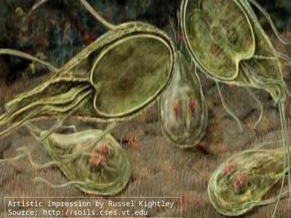
Giardia lamblia
Source: http://soils.cses.vt.edu

The genus Giardia belongs to the class Zoomastigophorea, the order Diplomonadida, and the family Hexamitidae .
It is one of the most primitive eukaryotes: it has a small subunit ribosomal RNA sequence and no mitochondria and Golgi apparatus
Now Giardia can be classified according to antigen, isoenzyme, and genetic analysis in addition to their morphology and host range.

Species identified
Giardia lamblia (intestinalis, duodenalis) - humans, mammals
Giardia muris - mammals
Giardia ardeae - birds
Giardia psittaci - birds
Giardia agilis - amphibians

Of the Giardia species, only G. lamblia has been successfully cultured in vitro.
The trophozoite divides by longitudinal binary fission
Two morphological forms: Trophozoite and cyst ( infective form)

Tear drop shaped2 adhesive discs ,
2 median bodies,2 nuclei
4 pairs of flagella
Source: www.sd01.k12.id.us

Tear drop shaped2 adhesive discs ,
2 median bodies,2 nuclei
4 pairs of flagella
Source: www.sd01.k12.id.us

intracytoplasmic projections axonemes
Source: http://medlib.med.utah.edu

Haematoxilyn staining
two nuclei, each with a prominent central karyosome (characteristic
facelike image(
Source: Gallery of histology Woods and Ellis2000

8 to 12 mm long and 7 to 10 mm wide
convex dorsal surface
a flat ventral surface sucking or adhesive disk
four pairs flagellae
Source: http://soils.cses.vt.edu

Epidemiology
Host can be humans, primates, cats, dogs, calves ,beavers, rabbits, etc .
World wide distribution
Highest incidence in children, young adults in late summer .

Transmission
1 -Person to person transmission
2 -Water sports, surface contamination. Watershed contamination
3 -sexually active male homosexuals and persons in custodial institutions.

Pathogenesis and Immune response (1)
•The production of diarrhea, and occasionally malabsorption, is the result of a complex interaction of Giardia with the host ,
•Infection occurs after oral ingestion of as few as 10 to 25 cysts.
•After excystation, trophozoites colonize and multiply in the upper small bowel
•Adherence of G. lamblia in the human gut may be via the disk,
but may also involve specific receptor-ligand interactions

Pathogenesis and Immune response (2)
Several pathogenic mechanisms have been postulated
Disruption of the brush border
Mucosal invasion
Elaboration of an enterotoxin
Stimulation of an inflammatory infiltration leading to fluid and electrolyte secretion and occasionally to villous changes

TEM micrograph showing the method of attachment to the duodenal wall.
Ventral sucking discSource: Gallery of histology Woods and Ellis2000

Immune Response
Partially protective immunity may develop to Giardia
Immune response involves both cellular and humoral immunity
-Ig A, serum Ig G and Ig M are detected in patients: role of Ig A is not completely understood, probably inhibits trophozoite attachment
-IgA deficiency lead to chronic giardiasis
-Cell mediated immune response may also play a role
Human milk may also play a role in protection of the host against Giardia : Free fatty acids and IgA antibodies

Infection with G. lamblia includes1 -Asymptomatic cyst passage (5 to 15% ) •acute self-limited diarrhea (25 to 50% )
•and a chronic syndrome of diarrhea •malabsorption, and weight loss
2 -Symptomatic giardiasis is characterized by •acute onset of diarrhea ,
•abdominal cramps, bloating, and flatulence •feelings of malaise, nausea, and anorexia
•may complain of sulfuric belching •Vomiting, fever, and tenesmus occur less commonly .
•stools may be profuse and watery, but later they are commonly greasy, and foul-smelling and may float

The role that chronic infection with Giardia plays in the growth and development of children in the developing world has been controversial

Life Cycle
Trophozoites : Lives in duodenum, jejenum and upper ileum
They come in close contact to the mucosal, but do not invade the host. Adhesive disc fits over surface of epithelial cell
The flagella act as a pump to move nutrients away from the microvilla and hold the adhesive disc near the mucosa.
Rapid division to produce large numbers quickly


Source: Doug Allington

14 billion parasites in diarrheic stool (trophozoites only) Moderate infection: 300 million cysts.
As the organism traverses the colon it is stimulated to encyst .
Produce an oval cyst with thick walls, with 2-4 nuclei .
Dividing within cyst (4 nuclei is older cyst)
Complete division in duodenum of host after ingestion
Cyst is approximately 8-10m and ellipsoid in shape .
The cyst is the infective state and is transferred by the fecal-oral route.

Diagnosis
• Giardia should be identified 50 to 70% of the time after one stool, and 90% identification after three stools
• Wet, saline mounts: falling leaf motion, fibrils present, and nucleic characteristics.
• Biopsy tissue/duodenal aspirate stained by trichrome or Giemsa stain .
• Enzyme immunoassay and fluorescent-anitbody monoclonal antigen detection systems
• Sensitivity & specificity: 90-100% (ProSpec T, GiardEIA, MeriFluor, Color Vue, and DD
System (

Source: http://soils.cses.vt.edu

Source: Doug Allington

Source: Doug Allington
Source: http://www. cbc.ca

DrugsDose
Metronidazole250mgtidX 5-7 d
Nitazoxanide500mg bdX3d
Paromomycin 25–30 mg/kg/d in 3 doses × 5–10 d
Tinidazole 2 g × 1 dose
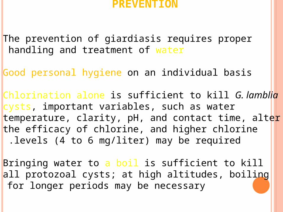
PREVENTION
The prevention of giardiasis requires proper handling and treatment of water
Good personal hygiene on an individual basis
Chlorination alone is sufficient to kill G. lamblia cysts, important variables, such as water temperature, clarity, pH, and contact time, alter the efficacy of chlorine, and higher chlorine levels (4 to 6 mg/liter) may be required .
Bringing water to a boil is sufficient to kill all protozoal cysts; at high altitudes, boiling for longer periods may be necessary
Artistic impression by Russel KightleySource: http://soils.cses.vt.edu


![[PPT]Conceptos generales de parasitología · Web viewCurso de Microbiología y parasitología Amebas de vida libre Naegleria fowleri: Meningoencefalitis amebiana primaria (M AP)](https://img.pdfslide.tips/doc/110x75/5ab627a27f8b9a2f438d5ef1/pptconceptos-generales-de-parasitologa-viewcurso-de-microbiologa-y-parasitologa.jpg)