Embed Size (px)
Citation preview

Gaziantep Medical Journal 2016;22(1):14-21 • DOI: 10.5578/GMJ.27960
A new dressing material in diabetic wounds: Wound healing activity of oleuropein-rich olive leaf extract in
diabetic ratsDiyabetik yaralar için yeni bir pansuman malzemesi: Diyabetik olan ve olmayan ratlarda
oleuropeince zenginleştirilmiş zeytin yaprağı ekstresinin yara iyileştirici aktivitesi
RESEARCH ARTICLE
Sevgin SAMANCIOĞLU1, Aynur ESEN2, Gülinnaz ERCAN3, Navid Hosseini MANSOUB3,
Seda VATANSEVER4, İskender İNCE5
1 Department of Nursing, Faculty of Health Sciences, Gaziantep University, Gaziantep, Turkey2 Department of Internal Nursing, Faculty of Nursing, Ege University, Izmir, Turkey
3 Department of Medical Biochemistry, Faculty of Medicine, Ege University, Izmir, Turkey4 Department of Histology and Embryology, Faculty of Medicine, Celal Bayar University, Manisa, Turkey
5 Center for Drug Reseacrh and Development and Pharmacokinetic Applications, Ege University, Izmir, Turkey
ABSTRACT
Introduction: The objective of this study was to evaluate a dressing material on ischemic wound model in diabetic rats. Study was conducted during the months of June 2012-March 2013 at Ege University in Izmir, Turkey.
Materials and Methods: Spraque Dawley rats weighing 200-250 g obtained from an experimental animal production center (Saki Yenili-Ankara,Turkey) were used in this study. Animals were randomly assigned to diabetic (n= 20) and nondiabetic (n= 20) groups. After diabetes induction and wound creation, animals within each group were assigned to two wound dressing groups by a second randomization. The study was carried out on these four groups. After diabetes induction and wound creation, animals within each group (n= 10) were assigned to olive leaf extract (OLE) wound dressing and normal saline (sodium chloride, 0.9% NaCl) wound dressing groups by a second randomization. 0.9% NaCl and OLE dressing was applied to wounds once a day by the researcher. The areas of wounds were measured by Walker Formula. OLE wound dressing healed wounds faster and earlier than classic wound dressing (p< 0.05).
Results: In the diabetic group; wounds closure time was found to be 24.80 ± 1.48 in OLE wound dressing and 28.00 ± 2.31 days in classical wound dressing.
Conclusion: As a result in terms of success ratios, OLE wound dressing for wound healing in diabetic and non-diabetic wounds has been determined to be more effective in comparison with classic wound dressing method.
Keywords: Diabetes, olive leaf extract, diabetic wound, wound dressing
ÖZ
Giriş: Bu çalışmada; iskemik yara modeli geliştirilen diyabetik sı-çanlarda, bir pansuman materyelinin etkisinin karşılaştırılması amaçlanmıştır. Çalışma Ege Üniversitesi Tıp Fakültesi Hastanesi Deneysel Cerrahi Hayvan Araştırmaları Laboratuvarı’nda Haziran 2012-Mart 2013 tarihleri arasında gerçekleştirilmiştir.
Materyal ve Metod: Çalışmada 200-250 g ağırlığında Spraque Daw-ley türü sıçanlar kullanılmıştır (Saki Yenili-Ankara, Turkey). Hayvanlar randomize olarak diyabetik (n= 20) ve non-diyabetik (n= 20) gruba ayrılmıştır. Diyabet ve yara oluşturduktan sonra gruplardaki hayvan-lar pansuman işlemi için tekrar ikinci bir randomizasyona tabi tutul-du. Çalışma bu dört grupla devam etti. Araştırmacı tarafından günde bir kez; birinci yaraya %0.9’luk NaCl, ikincisine ise 12.39 mg/100 mL oleuropein içeren zeytin yaprağı ekstresi (olive leaf extract, OLE) yara pansumanı uygulanmıştır. Yara alanı Walker formülü ölçümleri ile hesaplandı. Çalışmanın her 2 grubunda da OLE yara pansumanı yapılan yaralar klasik yara pansumanı yapılan yaralara göre daha hız-lı iyileşmiş ve daha erken kapanmıştır (p< 0.05).
Bulgular: Diyabet grubunda; klasik yara pansumanı yapılan yara-lar 28.00 ± 2.31 günde kapanırken, OLE yara pansumanı yapılan yaralarda yara kapanma zamanı 24.80 ± 1.48 olarak bulunmuştur.
Sonuç: Yara iyileşmesinde OLE yara pansumanının diyabetik ve diyabetik olmayan yaralarda klasik yara pansumanına göre daha etkili olduğu bulunmuştur.
Anahtar Kelimeler: Diyabet, zeytin yaprağı ekstresi, diyabetik yara, yara pansumanı
Yazışma Adresi/Correspondence: Sevgin SAMANCIOĞLUGaziantep Üniversitesi Sağlık Bilimleri Fakültesi, Hemşirelik Bölümü, Gaziantep, Türkiye Telefon/Tel: +90 342 360 60-76766 • E-posta/E-mail: [email protected]
Geliş Tarihi/Received: 27.03.2015 • Kabul Ediliş Tarihi/Accepted: 04.07.2015
* Sevgin Samancioglu, Aynur Esen, Gülinnaz Ercan, Navid Hosseini Mansoub, Seda Vatansever, Eff ect of Olive Leaf Extract on Diabetic Wound Healing: In Vivo Preclinical Study, 15 National Congress of Internal Medicine Nursing Program
2-6 October 2013 Antalya (Oral Presentation - First Degree)
Gaziantep Med J 2016;22(1):14-21
15
Samancıoğlu et al.
INTRODUCTION
Diabetes is a group of metabolic diseases characterized by hyperglycemia which leads to long-term damage, dysfunction, and even failure of many tissues and organs such as eyes, kidneys, nerves, heart, and blood vessels (1).
Studies show that the number of individuals affected from diabetes mellitus (DM) will rise from 171 million in 2000 to 366 million worldwide by 2030 (2). With its increasing prevalance day by day and complications with high mortality and morbidity rates, diabetes mellitus is an expensive disease that brings a burden to the individual, his family and the healthcare system all over the world. Impaired cutaneous wound healing is one of these serious complications in diabetes causing wound healing problems and infection leading even to amputations of diabetic foot (3,4). This is mainly due to hyperglycemia and its-related microvasculer complications, since high blood glucose not only leads to neuropathy and microangiopathy but also prevents cell reproduction and collagen generation leading to a decrease in wound tensile strength and hydroxyproline production (5,6).
In patients with diabetic foot ulcers can be applied debridement of necrosis, made antibiotic treatment for the infection, drainage and also blood sugar control and edema. Wound dressing treatment of diabetic foot ulcers should be absorbent, adhesive and non-occlusive (maceration is not desirable). Film dressings, hydrocolloids, hydrogels dressings, foams, hidrofi ber, composite and alginate dressings are used to treat ulcers. In addition, hyperbaric oxygen therapy, PRP (Platelet Rich Plasma), growth factors and tissue engineered products are also playing an active role in treatment. Choice of the cost / benefi t ratio is important in wound dressing, is considering making balanced with appropriate dressing change frequency (7-10).
In the treatment of diabetes mellitus, plants began to be used from ancient times play an important role worldwide. Olive is put forward in many ethnopharmacological studies carried around the world as a plant source used in wound treatment and diabetes (11-14). It is observed in these ethnopharmacological studies that substances such as soap made out of the oil, leaf, seed and olive itself are used (15-17). Besides their ethnopharmacological usage, olive leaf and its extract are of great interest because of their antioxidant and antimicrobial activities and, in vivo, by their blood pressure-lowering, hypocholesterolemic, antidiabetic, and anti-infl ammatory effects (18).
In this study, olive leaf extract (OLE) enriched in oleuropein has been used. Oleuropein is the main phenolic compound of olive tree and also is non-toxic (19-21). In this study an aqueous extract of olive leaf which was prepared from the leaves of the olive plant was analyzed for antimicrobial activity by using MIC and then, tested in
vivo by applying on the wounds of rats with STZ-induced diabetes.
It is thought by examining the effect of the oleuropein found in olive leaf extract which is known as having a healing property on wounds created on diabetic animals, it will be possible to develope a cost effective, easily accessible and easily applicable wound care product that can be used for the treatment of diabetic wounds.
MATERIALS and METHODS
Plant Material
Olive leaves used in this study were collected in winter 2011 from Balikesir province in Turkey. They were collected and properly prepared for drying process on the day of their collection.
Preparation Aqueous Extract of Oleuropein-Rich Olive Leaf
After collecting, the wet leaves were dried under vacuum. The dried leaves were soaked in 1/6 ratio to 78% ethyl alcohol for 48 hours. The heterogeneous mixture obtained was fi ltered through thin fi lters. The part with the fl uid solution was placed in a spray dry machine. The alcohol in the solution was evaporated inside this machine that operates at high rpm and olive leaf extract in dust form was obtained. The 20 g extract was combined with the 100 g apolar part of the olive oil to prepare medical dressing material with 24 g oleuropein in 120 g solution.
Preparation of Medical Dressing Material with Oleuropein
1 liter olive oil and 1 liter 96% ethyl alcohol was mixed by stirring for 6 hours and left for phase separation in a vertical column. The apolar part separated on the top phase while the polar part separated in the bottom phase. The apolar part was placed in a glass bell jar. Here, 1 liter 10 % apolar material was obtained. The apolar substance is the part with thin molecules and the absorption of this part by the skin is high. 100 g apolar molecule was left behind when the alcohol of the 10% apolar substance was evaporated. 20 g. Olive leaf extract (with 20% oleuropein) was added. Thus, 120 g solution, 24 g oleuropein was obtained. The resulting solution was applied to the gauze and dressing material was prepared. This material was applied to 2 x 2 cm in diameter gauze to wound area 2 times by dropped in. Each instillation is a drop of 0.05 mL. 2 drops of 0.1 mL.
Analyses of Olive Leaf Extract
Determination of phenolic compounds in olive leaf extract: There are various methods in literature regarding the determination of oleuropein (22). In our study, The HPLC (High-performance liquid chromatography) analysis was used for the determination of phenolic

16
Gaziantep Med J 2016;22(1):14-21 Samancıoğlu et al.
compounds and especially for the quantifi cation of oleuropein. For this purpose, Schimadzu UFLC (Ultra Fast Liquid Chromatograph) device and as detector Diode Array Detector (DAD) was used. The HPLC equipment used was a Hewlett-Packard Series HP 1100 equipped with a diode array detector. C18 column with a diameter of 4.6 mm diameter and a length of 250 mm with a particle size of 5 μm was used.
In our study, 5 mM ammonium acetate (pH: 5, concentrated acetic acid- Mobile phase A) and methanol (mobile phase B) were used as mobile phase. Flow rate was 1 mL/min and isocritical phase mobile phase A/B (60/40) was used. Injection volume was 20 μL. The temperature of the automatic sampler was set to 22°C, column temperature to 40°C and detector temperature to 40°C. Analyses were carried out at 200 nm wavelength. 10 injections were made to determine retention time (RT), Limit of Detection (LOD) and Limit of Quantifi cation (LOQ). Calibration Curve was drawn. The oleuropein amount in the sample was determined via an analysis.
Determination of the relative antimicrobial activities of olive leaf extracts: MIC (minimum inhibition concentration) is the lowest concentration of an antimicrobial that will inhibit the visible growth of a microorganism. Minimum
inhibition concentrations are important for monitoring the activity of new antimicrobials (23).
MIC values were determined for bacteria strains sensitive to extracts via the microdilution method that was developed according to procedures developed by the National Committee of Clinical Laboratory Standards (24). The strains were obtained from American Type Culture Collection (ATCC) and Ege University, Faculty of Science, Biology Department. The standard strains used in this study were Escherichia coli ATCC 8739, Staphylococcus aureus ATCC 6538p, Staphylococcus epidermidis ATCC 12228, Enterecoccus faecalis ATCC 29212, Enterobacter cloacae ATCC 13047, Klebsiealla pneumoniae ATCC 13883, Bacillus cereus ATCC 7064 ve Pseudomonas aeruginosa ATCC 9027. In this study, a serial 2-fold micro-broth dilution method was performed to determine the MICs of olive leaf extracts (25).
Animals
Healthy adult male Spraque Dawley rats weighing 200-250 g obtained from an experimental animal production center (Saki Yenili-Ankara, Turkey) were used in this study. The rights of animals are conserved in accordance with “Guide for the Care and Use of
Figure 1. Flow chart of the study.
Gaziantep Med J 2016;22(1):14-21
17
Samancıoğlu et al.
Laboratory Animals”. Animals were randomly assigned to diabetic and nondiabetic groups. After diabetes induction and wound creation, as described below, animals within each group were assigned to olive leaf extract wound dressing and normal saline (sodium chloride, 0.9% NaCl) wound dressing groups by a second randomization (Figure 1). The study was carried out on these four groups. Group 1: nondiabetic animals that received topical olive leaf extract as dressing material OLE (Kale Natural, Balikesir, Turkey) (C-OLE; n= 10); group 2: nondiabetic control animals that received topical 0.9% NaCl (normal saline) as dressing material (C-0.9% NaCl; n= 10); group 3: diabetic animals that received topical olive leaf extract as dressing material (DM-OLE; n= 10); and group 4: diabetic control animals that received topical 0.9% NaCl as dressing material (DM-0.9% NaCl; n= 10).
Experimentally Induced Diabetes
After a 12 h fast, rats randomized to the diabetic group were injected with a single dose of intraperitoneal (ip.) streptozotocin (STZ, Sigma) 60 mg⁄ kg body weight. STZ freshly dissolved in 0.1 M sodium citrate buffer (pH 4.5) was used to induce diabetes. The solutions were prepared freshly and kept on ice protected from light and injected to rats without any delay. The rats were fed a standard diet and maintained in the controlled environment of the animal centre at 25 ± 1°C under a 12 h light-dark cycle. Symptoms are controlled. A blood glucose measurement was done 48 h after STZ injection. Blood was collected from the tail vein and decided the glucose level using a glucometer (Accu-Chek; Roche Diagnostics, Mannheim, Germany). Rats with blood glucose levels > 250 mg⁄dL (13.9 mmol/L) were considered as diabetic (26-29). Rats in the nondiabetic group were injected i.p. with a single dose of 0.1 M sodium citrate buffer (pH 4.5). The procedures in this experimental study were performed in accordance with the National Guidelines for the Use and Care of Laboratory Animals and approved by the Animal Ethics Committee of Ege University.
Experimentally Induced Wound
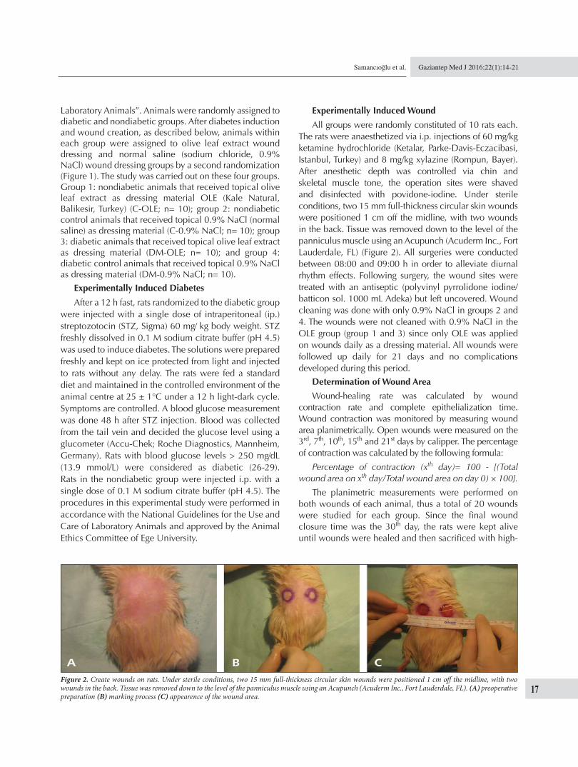
All groups were randomly constituted of 10 rats each. The rats were anaesthetized via i.p. injections of 60 mg/kg ketamine hydrochloride (Ketalar, Parke-Davis-Eczacibasi, Istanbul, Turkey) and 8 mg/kg xylazine (Rompun, Bayer). After anesthetic depth was controlled via chin and skeletal muscle tone, the operation sites were shaved and disinfected with povidone-iodine. Under sterile conditions, two 15 mm full-thickness circular skin wounds were positioned 1 cm off the midline, with two wounds in the back. Tissue was removed down to the level of the panniculus muscle using an Acupunch (Acuderm Inc., Fort Lauderdale, FL) (Figure 2). All surgeries were conducted between 08:00 and 09:00 h in order to alleviate diurnal rhythm effects. Following surgery, the wound sites were treated with an antiseptic (polyvinyl pyrrolidone iodine/batticon sol. 1000 mL Adeka) but left uncovered. Wound cleaning was done with only 0.9% NaCl in groups 2 and 4. The wounds were not cleaned with 0.9% NaCl in the OLE group (group 1 and 3) since only OLE was applied on wounds daily as a dressing material. All wounds were followed up daily for 21 days and no complications developed during this period.
Determination of Wound Area
Wound-healing rate was calculated by wound contraction rate and complete epithelialization time. Wound contraction was monitored by measuring wound area planimetrically. Open wounds were measured on the 3rd, 7th, 10th, 15th and 21st days by calipper. The percentage of contraction was calculated by the following formula:
Percentage of contraction (xth day)= 100 - [(Total wound area on xth day/Total wound area on day 0) × 100].
The planimetric measurements were performed on both wounds of each animal, thus a total of 20 wounds were studied for each group. Since the fi nal wound closure time was the 30th day, the rats were kept alive until wounds were healed and then sacrifi ced with high-
Figure 2. Create wounds on rats. Under sterile conditions, two 15 mm full-thickness circular skin wounds were positioned 1 cm off the midline, with two wounds in the back. Tissue was removed down to the level of the panniculus muscle using an Acupunch (Acuderm Inc., Fort Lauderdale, FL). (A) preoperative preparation (B) marking process (C) appearence of the wound area.
18
Gaziantep Med J 2016;22(1):14-21 Samancıoğlu et al.
dose ketamine hydrochloride at postoperative 21-30th days.
Statistical Analysis
The data obtained in this study were evaluated using SPSS 20.0 package software. Whereas differences between groups were examined via normality test, Mann-Whitney U test was applied on variables with abnormal distribution in double groups. Wilcoxon Sign test was used to examine the difference between the measurement times of variables. p< 0.05 was considered as statistically signifi cant.
RESULTS
Analyses of Oleuropein in Olive Leaf Extract
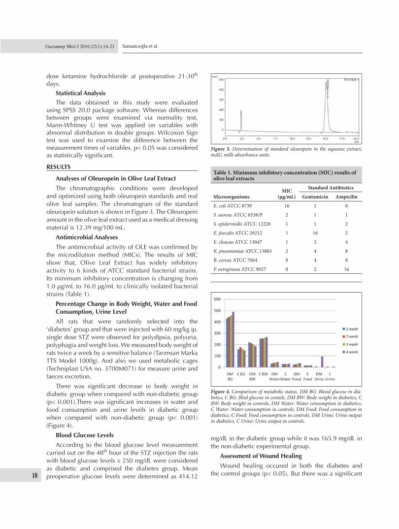
The chromatographic conditions were developed and optimized using both oleuropein standards and real olive leaf samples. The chromatogram of the standard oleuropein solution is shown in Figure 3. The Oleuropein amount in the olive leaf extract used as a medical dressing material is 12.39 mg/100 mL.
Antimicrobial Analyses
The antimicrobial activity of OLE was confi rmed by the microdilution method (MICs). The results of MIC show that, Olive Leaf Extract has widely inhibitory activity to 6 kinds of ATCC standard bacterial strains. Its minimum inhibitory concentration is changing from 1.0 μg/mL to 16.0 μg/mL to clinically isolated bacterial strains (Table 1).
Percentage Change in Body Weight, Water and Food Consumption, Urine Level
All rats that were randomly selected into the ‘diabetes’ group and that were injected with 60 mg/kg ip. single dose STZ were observed for polydipsia, polyuria, polyphagia and weight loss. We measured body weight of rats twice a week by a sensitive balance (Tarımsan Marka TTS Model 1000g). And also we used metabolic cages (Techniplast USA no. 3700M071) for measure urine and faeces excretion.
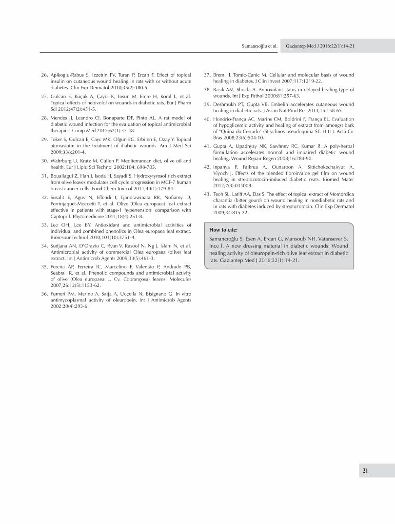
There was signifi cant decrease in body weight in diabetic group when compared with non-diabetic group (p< 0.001).There was signifi cant increases in water and food consumption and urine levels in diabetic group when compared with non-diabetic group (p< 0.001) (Figure 4).
Blood Glucose Levels
According to the blood glucose level measurement carried out on the 48th hour of the STZ injection the rats with blood glucose levels ≥ 250 mg/dL were considered as diabetic and comprised the diabetes group. Mean preoperative glucose levels were determined as 414.12
mg/dL in the diabetic group while it was 165.9 mg/dL in the non-diabetic experimental group.
Assessment of Wound Healing
Wound healing occured in both the diabetes and the control groups (p< 0.05). But there was a signifi cant
Figure 3. Determination of standard oleuropein in the aqueous extract, mAU, milli-absorbance units.
Figure 4. Comparison of metabolic status. DM BG: Blood glucose in dia-betics, C BG: Blod glucose in contols, DM BW: Body weight in diabetics, C BW: Body weight in controls, DM Water: Water consumption in diabetics, C Water: Water consumption in controls, DM Food: Food consumption in diabetics; C Food: Food consumption in controls, DM Urine: Urine output in diabetics, C Urine: Urine output in controls.
Table 1. Minimum inhibitory concentration (MIC) results of olive leaf extracts
Microorganisms MIC
(μg/mL)
Standard Antibiotics
Gentamicin Ampicilin
E. coli ATCC 8739 16 1 8
S. aureus ATCC 6538/P 2 1 1
S. epidermidis ATCC 12228 1 1 2
E. faecalis ATCC 29212 1 16 2
E. cloacae ATCC 13047 1 2 4
K. pneumoniae ATCC 13883 2 4 8
B. cereus ATCC 7064 8 4 8
P. aeroginosa ATCC 9027 8 2 16

Gaziantep Med J 2016;22(1):14-21
19
Samancıoğlu et al.
difference in the size of wound area in diabetic group when compared with control group (p< 0.001). Healing of the wounds was faster in both the diabetes and the control groups for wounds on which OLE wound dressing was applied in comparison with the normal saline wound dressing applications (p> 0.05) (Figure 5).
Assessment of Wound Closure
Wounds dressed with OLE as a wound dressing material closed earlier in both the diabetes and the control groups in comparison to the wounds of the normal saline wound dressing groups (respectively u= 13, p< 0.05; u= 22.5, p< 0.05). While wounds with normal saline wound dressing in the diabetes group closed in 28.00 ± 2.31 days, the wound closure time for wounds of diabetic wounds on which OLE wound dressing was applied was determined as 24.80 ± 1.48 days. Whereas wound closure time on which normal saline wound dressing was applied in the control was 22.50 ± 0.53 days, the wound closure time for wounds on which OLE wound dressing was applied was determined as 21.60 ± 0.97 days in the control group (Table 2).
DISCUSSION
Many researchers have conducted studies with the aim of accelerating wound healing in diabetes. OLE has been reported to possess wound-healing properties (18). However, the enhancing effects of OLE on wound healing in diabetic patients have been not studied. There are many studies supporting the potentially benefi cial effects of olive leaves on human health (21,30-32).
In this study, we demonstrated that wound closure was signifi cantly shortened when the wounds of diabetic rats and non diabetic rats were treated with OLE wound dressing.
According to the minimum inhibitor concentration results obtained in our study, OLE at various concentrations was determined to be more effective against various
microorganisms in comparison with the positive control group. Oleuropein is a natural antimicrobial and it has been shown in various studies that it delays and inhibits the growth rate of microorganisms (33-36).
Delay in wound healing is a signifi cant clinical problem in diabetes mellitus. Problems of wound healing in diabetes can be summarized as the increase of infectious complications due to the decrease of cellular infi ltration, angiogenesis, granulation tissue, collagen amount and organization (6,37). This is tried to be explained by the increase of free radicals due to the auto-oxidation of glucose and glycosylated proteins in the existence of hyperglycemia that persists in diabetes along with the decrease of defense mechanisms (38).
It is of signifi cance that no other study investigating the effects of olive leaf extract on the diabetic wounds has been observed indicates the necessity of such a research. In this study, we demonstrated that wound closure was signifi cantly shortened when the wounds of diabetic rats and non diabetic rats were treated with OLE wound dressing in comparison with saline wound dressing application. In the studies reported by Deshmukh and Gupta (2013), the effect of embelin from Myrssinaceae family on wound healing process of diabetic wound was evaluated, and Deshmukh and Gupta (2013) had determined that topically applied 5% embelin increased wound contraction and speeded up the process of wound healing starting from the 12th day onwards (39). In another study, Honório-França et al. (2008) had determined no difference in the healing process of the wounds of the plant extract obtained from the Loganiaceae family applied group in comparison to the control group (40). In their studies, Gupta et al. (2008) used a plant based formula and put forth that there was a statistically signifi cant increase in wound contraction on the 4th and 7th days of the surgical wounds in diabetic group in comparison with the control wounds (41). It has been stated by Inpanya et al. (2012) that diabetic wounds which can be treated using an extract obtained from aloevera leaves recovers faster in comparison with
Figure 5. Comparison of wound healing. It shows the percent of wound closure for the four groups on each day. Complete wound closure was 21th day on 3 groups.
Table 2. Comparison of wound closure
Variables
DMMean ± SS (Min-max)
Non-DMMean ± SS (Min-max) U p
OLE 24.80 ± 1.48 21.60 ± 0.97 4.500 0.000
23.00 - 27.00 21.00 - 23.00Normal saline 28.00 ± 2.31 22.50 ± 0.53 2.500 0.000
23.00 - 30.00 22.00 - 23.00

20
Gaziantep Med J 2016;22(1):14-21 Samancıoğlu et al.
diabetic wounds that cannot be treated using this extract (42). In our study, diabetic wounds on which olive leaf extract wound dressing was applied recovered faster starting from the 3rd day on wards in comparison with the wounds on which only 0.9% NaCl wound dressing was applied.
In their study examining the effect of embelin on diabetic wounds, Deshmukh and Gupta (2013) have stated that 5% embelin applied topically decreases wound closing time from 25.84 days to 21.45 days while Teoh et al. (2009) have stated that diabetic wounds that were treated with Momordica charantia extract closed earlier in comparison with diabetic wounds that were not treated (39,43). In our study, wounds of both the diabetes and control groups on which OLE wound dressing was applied closed earlier in comparison with the wounds on which normal saline wound dressing was applied.
Impaired wound healing is a well-known and major complication of diabetes. In conclusion, results of this study indicate that an OLE wound dressing can be a suitable option for diabetic wounds. These fi ndings suggest for the fi rst time the possibility of the novel application of aqueous extract of oleuropein-rich olive leaf as a therapeutic agent for wound healing.
ACKNOWLEDGMENTS
This study was supported by a grant (05-DPT-0003/37) from the Republic of Turkey, Prime Ministry, State - DPT. Kale Natural’s Laboratory (Balikesir, Turkey) were used in the preparation of plant extracts.
REFERENCES
1. American Diabetes Assocation. Diagnosis and classifi cation of diabetes mellitus. Diabetes Care 2013(Supple 36):S67-S74.
2. Wild S, Roglic G, Green A, Sicree R, King H. Global prevalence of diabetes: estimates for the year 2000 and projections for 2030. Diabetes Care 2004;27(5):1047-53.
3. Falanga V. Wound healing and its impairment in the diabetic foot. Lancet 2005;366:1736-43.
4. Sugimoto T, Huang L, Minematsu T, Yamamoto Y, Asada M, Nakagami, et al. Impaired aquaporin 3 expression in reepithelialization of cutaneous wound healing in the diabetic rat. Biol Res Nurs 2013:15(3):347-55.
5. Altay P, Basal G. Wound dressings. Electronic Journal of Textile Technologies 2010;4(1):109-21.
6. Blakytny R, Jude E. The molecular biology of chronic wounds and delayed healing in diabetes. Diabet Med 2006;23:594-608.
7. Türsen Ü. Ülser tedavisinde yara örtüleri. Turk J Dermatol 2013;7:61-71.
8. Cobos R, Aizpuru F, Parraza N, Anitua E, Orive G. Effectiveness and effi ciency of platelet rich plasma in the treatment of diabetic ulcers. Curr Pharm Biotechnol 2015:16(7):630-4.
9. Rhee SM, Valle MF, Wilson LM, Lazarus G, Zenilman JM, Robinson KA. Negative Pressure Wound Therapy Technologies for Chronic Wound Care in the Home Setting [Internet]. Agency for Healthcare Research and Quality (US) 2014:15.
10. Hafeez K, Haroon-Ur Rashid, Kaim Khani GM, Kumar D, Kumar, S. Vacuum Assisted Closure- utilization as home based therapy in the management of complex diabetic extremity wounds. Pak J Med Sci 2015;1(1):95-9.
11. Wiwanitkit V. Thai ethnopharmacological herbs for diabetes treatment: data collection and informatics tracing for therapeutic property. Diabetes Metab Syndr 2011;5:103-4.
12. Alzweiri M, Al Sahran A, Mansi K, Hudaib M, Aburja T. Ethnopharmacological survey of medicinal herbs in Jordan, the Northern Badia region. J Ethnopharmacol 2011;137:27-35.
13. Cakilcioglu U, Khatun S, Turkoglu I, Hayta S. Ethnopharmacological survey of medicinal plants in Maden (Elazig-Turkey). J Ethnopharmacol 2011:137(1):469-86.
14. Boudjelal A, Henchiri C, Sari M, Sarri D, Hendel N, Benkhaled A, et al. Herbalists and wild medicinal plants in M’Sila (North Algeria): an ethnopharmacology survey. J Ethnopharmacol 2013: 148(2):395-402.
15. Sargin SA, Akçicek E, Selvi S. An ethnobotanical study of medicinal plants used by the local people of Alasehir (Manisa) in Turkey. J Ethnopharmacol 2013:12;150(3):860-74.
16. Polat R, Satil, F. An ethnobotanical survey of medicinal plants in Edremit Gulf (Balikesir-Turkey). J Ethnopharmacol 2012;139(2):626-41.
17. Saric-Kundalic B, Dobes C, Klatte-Asselmeyer V, Saukel J. Ethnobotanical study on medicinal use of wild and cultivated plants in middle, south and west Bosnia and Herzegovina. J Ethnopharmacol 2010;131(1):33-55.
18. Koca U, Suntar I, Kupeli Akkol E, Yilmazer D, Alper M. Wound repair potential of Olea europaea L. leaf extracts revealed by in vivo experimental models and comparative evaluation of the extracts’ antioxidant activity. J Med Food 2011;14(1/2):140-6.
19. Perrinjaquet-Moccetti T, Busjahn A, Schmidlin C, Schmidt A, Bradl B, Aydogan C. Food supplementation with an olive (Olea europaea L.) leaf extract reduces blood pressure in borderline hypertensive monozygotic twins. Phytother Res 2008;22:1239-42.
20. Hamdi HK, Castellon R. Oleuropein, leuropein, a non-toxic olive iridoid, is an anti-tumor agent and cytoskeleton disruptor. Biochem Biophys Res Commun 2005;334;769-78.
21. El SN, Karakaya S. Olive tree (Olea europaea) leaves: potential benefi cial effects on human health. Nutr Rev 2009:67(11):632-8.
22. Tsarbopoulos A, Gikas E, Papadopoulos N, Aligiannis N, Kafatos A. Simultaneous determination of oleuropein and its metabolites in plasma by high-performance liquid chromatography. J Chromatogr B Analyt Technol Biomed Life Sci 2003;785:157-64.
23. Andrews JM. Determination of minimum inhibitory concentrations. J Antimicrob Chemother 2001;48(Supple 1):S5-S16.
24. Kiehlbauch JA, Hannett GE, Salfi nger M, Archinal W, Monserrat C, Carlyn C. Use of the National Committee for Clinical Laboratory Standards Guidelines for Disk Diffusion Susceptibility Testing in New York State Laboratories. J Clin Microbiol 2000;38(9):3341-8.
25. Kuete V, Ngameni B, Tsafack AM, Ambassa P, Konga Simo I, Bezabih M, et al. Antimicrobial activity of the extract from the twigs of Dorstenða Ellðptðca (Moraceae). Pharmacology Online 2007;1:573-580.
Gaziantep Med J 2016;22(1):14-21
21
Samancıoğlu et al.
26. Apikoglu-Rabus S, Izzettin FV, Turan P, Ercan F. Effect of topical insulin on cutaneous wound healing in rats with or without acute diabetes. Clin Exp Dermatol 2010;35(2):180-5.
27. Gulcan E, Kuçuk A, Çayci K, Tosun M, Emre H, Koral L, et al. Topical effects of nebivolol on wounds in diabetic rats. Eur J Pharm Sci 2012;47(2):451-5.
28. Mendes JJ, Leandro CI, Bonaparte DP, Pinto AL. A rat model of diabetic wound infection for the evaluation of topical antimicrobial therapies. Comp Med 2012;62(1):37-48.
29. Toker S, Gulcan E, Cayc MK, Olgun EG, Erbilen E, Ozay Y. Topical atorvastatin in the treatment of diabetic wounds. Am J Med Sci 2009;338:201-4.
30. Wahrburg U, Kratz M, Cullen P. Mediterranean diet, olive oil and health. Eur J Lipid Sci Technol 2002;104: 698-705.
31. Bouallagui Z, Han J, Isoda H, Sayadi S. Hydroxytyrosol rich extract from olive leaves modulates cell cycle progression in MCF-7 human breast cancer cells. Food Chem Toxicol 2011;49(1):179-84.
32. Susalit E, Agus N, Effendi I, Tjandrawinata RR, Nofi arny D, Perrinjaquet-Moccetti T, et al. Olive (Olea europaea) leaf extract effective in patients with stage-1 hypertension: comparison with Captopril. Phytomedicine 2011;18(4):251-8.
33. Lee OH, Lee BY. Antioxidant and antimicrobial activities of individual and combined phenolics in Olea europaea leaf extract. Bioresour Technol 2010;101(10):3751-4.
34. Sudjana AN, D’Orazio C, Ryan V, Rasool N, Ng J, Islam N, et al. Antimicrobial activity of commercial Olea europaea (olive) leaf extract. Int J Antimicrob Agents 2009;33(5):461-3.
35. Pereira AP, Ferreira IC, Marcelino F, Valentão P, Andrade PB, Seabra R, et al. Phenolic compounds and antimicrobial activity of olive (Olea europaea L. Cv. Cobrançosa) leaves. Molecules 2007;26:12(5):1153-62.
36. Furneri PM, Marino A, Saija A, Uccella N, Bisignano G. In vitro antimycoplasmal activity of oleuropein. Int J Antimicrob Agents 2002:20(4):293-6.
37. Brem H, Tomic-Canic M. Cellular and molecular basis of wound healing in diabetes. J Clin Invest 2007;117:1219-22.
38. Rasik AM, Shukla A. Antioxidant status in delayed healing type of wounds. Int J Exp Pathol 2000:81:257-63.
39. Deshmukh PT, Gupta VB. Embelin accelerates cutaneous wound healing in diabetic rats. J Asian Nat Prod Res 2013;15:158-65.
40. Honório-França AC, Marins CM, Boldrini F, França EL. Evaluation of hypoglicemic activity and healing of extract from amongst bark of “Quina do Cerrado” (Strychnos pseudoquina ST. HILL). Acta Cir Bras 2008;23(6):504-10.
41. Gupta A, Upadhyay NK, Sawhney RC, Kumar R. A poly-herbal formulation accelerates normal and impaired diabetic wound healing. Wound Repair Regen 2008;16:784-90.
42. Inpanya P, Faikrua A, Ounaroon A, Sittichokechaiwut A, Viyoch J. Effects of the blended fi broin/aloe gel fi lm on wound healing in streptozotocin-induced diabetic rvats. Biomed Mater 2012;7(3):035008.
43. Teoh SL, Latiff AA, Das S. The effect of topical extract of Momordica charantia (bitter gourd) on wound healing in nondiabetic rats and in rats with diabetes induced by streptozotocin. Clin Exp Dermatol 2009;34:815-22.
How to cite:
Samancıoğlu S, Esen A, Ercan G, Mansoub NH, Vatansever S, İnce İ. A new dressing material in diabetic wounds: Wound healing activity of oleuropein-rich olive leaf extract in diabetic rats. Gaziantep Med J 2016;22(1):14-21.