Embed Size (px)
Citation preview
Biomedical Research (Tokyo) 39 (1) 1–11, 2018
Absorption and metabolism of orally administered collagen hydrolysates evaluated by the vascularly perfused rat intestine and liver in situ
Yoshihiro OSAWA1, Takafumi MIZUSHIGE
2, Sayuri JINNO2, Fumihito SUGIHARA
3, Naoki INOUE3, Hideyuki TANAKA
2, and Yukihito KABUYAMA
2
1 United Graduate School of Agricultural Science, Tokyo University of Agriculture and Technology, Tokyo, Japan; 2 Faculty of Agricul-ture, Utsunomiya University, Utsunomiya, Japan; 3 Peptide Division, Nitta Gelatin Inc., Osaka Japan
(Received 24 October 2017; and accepted 30 October 2017)
ABSTRACTA number of studies have shown that oral administration of collagen hydrolysate (CH) results in the absorption of di- and tri-peptides. In order to understand the dynamics of CH absorption and metabolism, molecular profiles of hydroxyproline (Hyp) and Hyp-containing peptides (HCPs) were analyzed by in situ perfusion of rat intestine and liver. The total amount of absorbed HCPs during 1 h of perfusion was 16.6 μmol, which was significantly higher than that of free Hyp (6.6 μmol). In addition, HCPs were also reliably detected in hepatic perfusate at the level higher than free Hyp. Thus, the results demonstrated that CH is absorbed predominantly as peptides, which subse-quently enter systemic circulation. Size exclusion chromatography showed that perfusates include significant amount of HCPs larger than tripeptides, leading us to analyze these peptides in detail. Mass spectrometric analysis of intestinal perfusate finally identified three CH-derived peptides, which are surprisingly large as food-derived circulating peptides. Peptide quantitation by liquid chromatography-tandem mass spectrometry (LC-MS/MS) revealed that di- and tri-peptides, which are previously identified as major peptides in circulating blood, comprise only a part of HCPs in intestinal and liver perfusate. Finally, analysis of portal vein blood revealed that the larger peptides, such as pentadecapeptide identified in this study, could be absorbed in vivo. Taken all together, this study showed that peptides which are larger than tripeptide could reach to the circulation sys-tem after administration of CH, revealing previously unknown dynamics of absorption of CH.
Collagen is one of the most abundant protein in ver-tebrate, accounting for one-third of total protein (21). Gelatin, a heat denatured product of collagen, has been widely used as an gelling agent (16). Exam-ples include food processing, pharmaceutical appli-cation as capsule, and photographic materials (3). Recently, collagen hydrolysate (CH), a product of enzymatic digestion of gelatin, has received atten-tion as dietary supplement due to its solubility in water (5). A number of studies have reported benefi-
cial effect of CH on physiological regulation related to joint diseases (2), skin wound healing (35) and bone biology (33). Accumulating evidence has re-vealed an active ingredient in CH. Prolyl-hydroxypro-line (PO), a circulating dipeptide after oral ingestion of CH, is shown to have chemotactic activities on cultured fibroblasts (23) and leukocytes (14). Ohara et al. has reported that PO stimulates proliferation and hyaluronic acid synthesis in cultured dermal fi-broblasts (18). Shimizu et al. has reported that PO ameliorates skin barrier dysfunction (24). It is also reported that PO stimulates differentiation of osteo-blastic cells (12). In addition, PO is circulating in the blood at least for a few hours after oral inges-tion of CH (10). Together, it is reasonable to think that PO plays an important role in the physiological
Address correspondence to: Dr. Yukihito Kabuyama,Department of Applied Biological Chemistry, Faculty of Agriculture, Utsunomiya University, Tochigi, JapanTel: +81-28-649-5465, Fax: +81-28-649-5401E-mail: [email protected]
Y. Osawa et al.2
were reliably detected in hepatic perfusate, suggest-ing that larger peptides could pass through the liver and reach the systemic circulation system at least once. Together, these results have revealed previous-ly unknown aspect of absorption and metabolism of HCPs after oral administration of CH.
MATERIALS AND METHODS
Chemicals. The synthetic peptides which have ami-no acid sequences in type I collagen, such as Pro-Hyp (PO), Pro-Hyp-Gly (POG), Hyp-Gly-Pro (OGP), Gly-Glu-Thr-Gly-Pro-Ala-Gly-Arg-Hyp-Gly (GETGPAGROG), Asp-Gly-Glu-Ala-Gly-Lys-Pro-Gly-Arg-Hyp-Gly-Glu (DGEAGKPGROGE), and Gly-Glu-Ala-Gly-Pro-Ala-Gly-Pro-Ala-Gly-Pro-Ala-Gly-Pro-Arg (GEAGPAGPAGPAGPR), were pur-chased from PH Japan (Hiroshima, Japan). Gly-Gly (GG) was purchased from Kanto Chemical (Tokyo, Japan). Gly-Pro (GP), Leu-Gly-Gly (LGG), Pro-Hyp-Gly-Pro (POGP), Hyp-Gly-Pro-Hyp (OGPO), (Pro-Pro-Gly)5, (Pro-Hyp-Gly)5, and α-Cyano-4-hy-droxycinnamic acid (CHCA) were purchased from Peptide Institutes (Osaka, Japan). Hyp-Gly (OG), Ala-Hyp (AO), Ser-Hyp (SO), and Gly-Pro-Ala (GPA) were synthesized in our laboratory, and used after crystallization. Enzymatic hydrolysate of por-cine skin gelatin (SCP-5000: average molecular weight is 5000 Da) was provided by Nitta Gelatin (Osaka, Japan) (10). L-amino acids, including L-hy-droxyproline (Hyp), and the other analytical grade chemicals were purchased from Wako Pure Chemi-cal Industries (Osaka, Japan). The HPLC solvents were obtained from Sigma Chemical (St. Louis, MO, USA).
Animals and diets. Six-week-old male Wistar rats (approximately 200 g) were purchased from Charles River Japan (Kanagawa, Japan) and were individu-ally housed in stainless-steel wire cages in a room maintained at 22 ± 2°C with a 12-h light-dark cycle. The rats were fed with a solid feed (MF; Oriental Yeast, Tokyo, Japan) for more than three days be-fore the experiments. The rats were fasted overnight prior to the experiments. The animal care protocol was approved by the Utsunomiya University Animal Research Committee under the Guidelines for Ani-mal Experiments of Utsunomiya University (autho-rization number: AP16-0017). CH was solubilized by water at the concentration of 0.5 g/mL, then 2 mL of CH solution was intragastrically administered to rats by using teflon sonde.
regulation by CH. However, several studies have identified at least 29 di- and tri-peptides other than PO in plasma after oral administration of CH (9, 19, 22, 25, 26, 31, 34). Recently, some of these peptides have been shown to play a regulatory role in skin barrier function (13, 20, 22, 24). Thus, in order to understand molecular mechanisms of beneficial ef-fect of CH administration, it is important to clarify the detailed molecular profiles of CH-derived metab-olites circulating in the blood. Recent studies have shown the delivery of CH-derived peptides to target tissues. Watanabe-Kamiyama et al. have analyzed the intestinal absorption and tissue distribution of radio-labeled collagen-derived tripeptide (glycyl-pro-lyl-hydroxyproline: GPO) (32). They reported that radioactivity was predominantly detected in specific tissues, such as the liver, kidney, cartilage and skin. Kusubata et al. has shown that food-derived PO is delivered abundantly to the area of damaged skin (13). These results indicate that circulating HCPs in-cluding PO have some functions in the regulation of target tissues. Although these studies provide important informa-tion about dynamics of absorption and metabolism of orally administrated CH, the profile of molecules just after absorption remains largely elusive. In many studies, evaluation of these circulating peptides has been carried out after 30 min–several hours post oral administration of CH (9, 19, 22, 25, 26, 31, 34). Therefore, the analytes were the mixture of peptides circulating just after absorption and their metabolites stably circulating for a while. In addition, endoge-nous peptides, residual materials and peptidases in blood sample could interfere the identification of CH-derived peptides with short half-lives or lower content, which could have some functions to regu-late target tissues. In this study, we used in situ vascular perfusion technique to analyze the dynamics of CH-derived peptides. We prepared perfusates from portal vein and inferior vena cava, in order to understand intes-tinal absorption and hepatic metabolism of CH-de-rived peptides, respectively. Interestingly, size exclusion chromatography revealed that intestinal perfusates include significant amount of HCPs which are larger than tripeptides, suggesting existence of unidentified HCPs in perfusate. Mass-spectrometric analysis identified novel HCPs, including pentadeca-peptides which are surprisingly large as food-de-rived circulating peptides. Importantly, these peptides were detected in portal vein blood after oral admin-istration of CH, indicating intestinal absorption of larger peptides in vivo. Interestingly, these peptides
Absorption of collagen peptide 3
collected directly from the portal vein by using 21-gauge needles. In order to suppress the serum peptidase activity toward CH-derived peptides, blood samples were transferred immediately into ice-cold ethanol solution. This step was carried out within 2 min after collection of blood samples. Plasma samples were prepared after centrifugation of etha-nol-treated blood at 3000 × g for 10 min and used for the analyses.
Quantitation of hydroxyproline and HCPs by bio-chemical method. The amount of free Hyp and HCPs in perfusate was determined before and after hydro-lysis by hydrochloric acid. Therefore, the amount of HCPs was evaluated by the amount of Hyp generat-ed by the hydrolysis of HCP. The deproteinized samples were analyzed for the quantitation of free Hyp and HCPs. An aliquot of the deproteinized su-pernatant was hydrolyzed in 6N HCl at 110°C for 24 h and subsequently dried under vacuum. Finally, the amount of peptide form of Hyp was estimated by subtracting free Hyp from total Hyp.
Size exclusion chromatography analysis. Size exclu-sion chromatography was used for the analysis of molecular size distribution of absorbed peptides. The ethanol treated sample was dried under vacuum and dissolved in 30% acetonitrile solution in the presence of 0.1% trifluoroacetic acid (TFA). A spin column (0.22 μm, Ultrafree-MC GV; Merck Milli-pore, Billerica, MA, USA) was used for the clarifi-cation. The filtrated sample was applied to Superdex Peptide HR 10/30 column (GE Healthcare, Chicago, IL, USA). Elution was carried out with 30% aceto-nitrile solution in the presence of 0.1% TFA for 60 min at flow rate of 0.5 mL/min. Fractions were collected every 1 min and used for the quantitation of Hyp.
Analysis of perfusate and plasma by LC-MS/MS. The quantitation of the peptides was carried out for three times and the results were expressed as the mean values ± standard error. For the analysis of di- or tri-peptides, one sample was prepared from each rat. For the analysis of large peptides identified in this study, one sample was prepared by pooling sam-ples from five rats. The samples were analyzed by LC-MS/MS. Inert-sustain peptide C18 column (20 × 150 cm, 3 μm; GL Sciences, Tokyo, Japan) was used for the separation of peptides. For the quantitation of CH-derived pep-tides, elution was performed with 0.1% TFA for 5 min followed by a linear gradient to 15% acetoni-
Vascular perfusion of the rat small intestine and liv-er. Intestinal perfusion was performed by the report-ed method of Liu et al. (15). Briefly, rats were anesthetized with sodium thiopental (12.5 mg/200 g BW, i.p.) at 40 min after injection of CH, and then fixed to a surgical platform in perfusion apparatus maintained at 37°C using a thermoregulatory-con-trolled heating unit. The thoracic cavity was dis-closed by cutting upwards and bilaterally along the rib wall, leading to the expose of the heart. An anti-coagulant was injected into the heart, and then a small incision was made through the apex of the left ventricle. A catheter was inserted into the aorta, which was clamped in order to fix the position of the catheter. Then, the portal vein was exposed, and another catheter was inserted into the vessel. A sin-gle-pass perfusion was established by securing the in-flow catheter in the aorta, and the perfusate was collected through an out-flow catheter of the portal vein. The exposed surface of the small intestine was covered with wet gauze and plastic film to avoid surface drying. Portal venous perfusion was per-formed with a Krebs Ringer bicarbonate solution (pH 7.4) containing 2% bovine serum albumin and 15 mM glucose. The perfusate solution was supple-mented with dexamethasone and noradrenaline to counteract the effects of denervation and continu-ously bubbled with 95% O2–5% CO2. This perfusion was started at 1 h after injection of CH. For first 5 min, the flow rate was adjusted to approximately 5 mL/min to prevent vascular occlusion. Then, the flow rate was re-adjusted to approximately 1 mL/min, finally achieving a blood pressure of 110–115 mm of Hg. Hepatic perfusion was performed basically by the same method. For collecting the liver perfusate, an out-flow catheter was inserted into the inferior vena cava which connect to the right atrium of the heart. In order to prevent the leakage of perfusate, the in-ferior vena cava which connect to the lower trunk and abdomen, as well as the hepatic artery, were oc-cluded tightly by a klemme.
Preparation of plasma samples. The sampling was carried out by decapitating the rats after 2 h post ad-ministration of CH. Plasma samples were collected by the centrifugation at 1000 × g for 20 min, fol-lowed by the sedimentation of proteins by adding 3 volumes of ethanol. The supernatants were used for the analyses. For collecting blood samples from por-tal vein, rats were anesthetized by intraperitoneal injection of thiopental sodium after 2 h post admin-istration of CH. After surgical operation, blood was
Y. Osawa et al.4
of the background standard errors.
Stability of CH-derived peptides against the diges-tion by plasma peptidase. The stability of the pep-tides was analyzed by using rat plasma. The plasma was prepared and incubated for 10 min at 37°C. Five μL of each peptide dissolved in PBS was add-ed to 45 μL of pre-warmed rat plasma at the con-centration of 1 μg/mL. After 30 min, the samples were deproteinized by adding 3 volumes of ethanol. The supernatants were used for the LC-MS/MS analyses.
Statistical analysis. One way ANOVA with Tukey’s post hoc test was used for the statistical analysis. Values of P < 0.05 were considered significant. All data was expressed as the mean ± SEM.
RESULTS
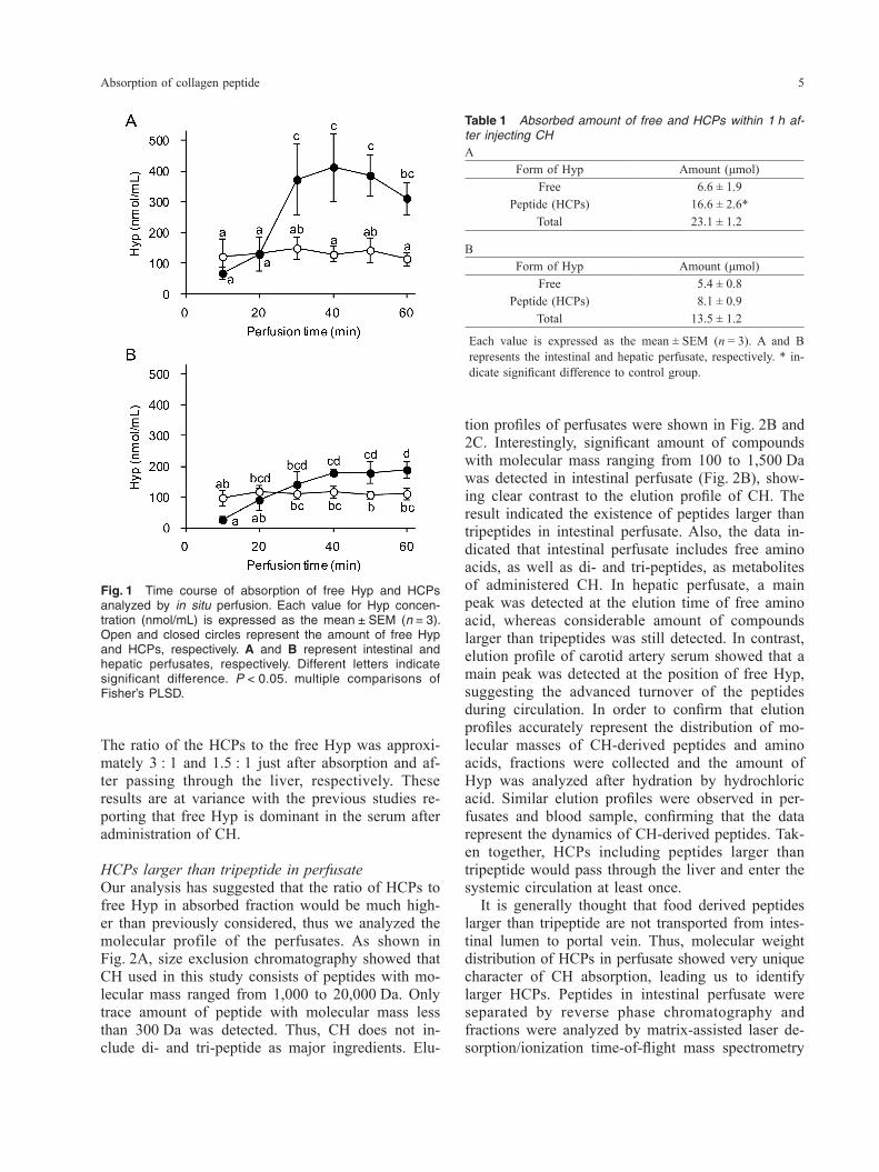
Amount of free Hyp and HCPs in intestinal and he-patic perfusatePreviously, we used in situ vascular perfusion tech-nique to analyze the transport of synthetic HCPs across the rat small intestine (15). In this study, we applied this method for the evaluation of intestinal absorption and hepatic metabolism of CH which is orally administrated. After intragastric injection of CH, perfusates were collected for 60 min from the catheter which is inserted into portal vein or inferior vena cava. First, we assessed the amount of CH-de-rived free Hyp and HCPs in perfusates. As shown in Fig. 1, free Hyp was observed constantly in the in-testinal perfusate throughout the perfusion period. The average level of free Hyp was 109 nmol/mL, which was significantly higher than the basal levels of free Hyp in perfusate (around 20 nmol/mL, Liu et al.) (15), suggesting the transport of CH-derived Hyp into portal vein. Interestingly, the concentration of HCPs significantly increased approximately 20 min after the start of perfusion, finally reaching a maximum level of 400 nmol/mL. Thus, molecular mechanism for the absorption of HCPs diverges from that for Hyp. Absorbed amount of HCPs during 1 h after oral administration of CH was 16.6 μmol, which was significantly higher than that of free Hyp (6.6 μmol). Together, these results have indicated that CH is absorbed predominantly as peptides in the small intestine. Similar results were observed in hepatic perfusates (Fig. 1, Table 1). Although the concentration of HCPs decreased to one-half after passing through the liver, significant amount of HCPs appeared to enter the systemic circulation.
trile in the presence of 0.1% TFA over 15 min. The flow rate was 0.4 mL/min. For the analysis of free Hyp, isocratic elution was performed with 0.1% TFA for 3 min. Mass spectrometry was performed with a Triple TOF 5600 mass spectrometer (AB Sciex, Framing-ham, MA, USA) equipped with an electro-spray ionization source. The pressure values of nebulizer gas, the heater gas, and Curtain Gas were adjusted to 50, 50, and 25 psi, respectively. The source tem-perature was adjusted to 500°C. The ion spray volt-age was adjusted to 5.5 kV in the positive ionization mode. MS/MS experiments were carried out using nitrogen as the collision gas. The collision energy was optimized for each peptide. Data acquisition and analyses were performed by using Analyst soft-ware (ver. 1.6.; AB Sciex, Framingham, MA, USA). Quantification of the corresponding CH-derived pep-tides in the sample by an LC-MS/MS analysis was carried out by using MRM (Multiple Reaction Mon-itoring), in which the transition of m/z 133.061 > 76.039 (CE: 15) for GG, m/z 173.092 > 116.071 (CE: 20) for GP, m/z 189.087 > 86.061 (CE: 25) for OG, m/z 203.103 > 132.066 (CE: 20) for AO, m/z 219.098 > (CE: 20) for SO, m/z 229.118 > 132.066 (CE: 20) for PO, m/z 244.129 > 155.082 (CE: 15) for GPA, m/z 246.146 > 171.113 (CE: 15) for LGG, m/z 260.124 > 189.087 (CE: 20) for AOG, m/z 276.119 > 189.087 (CE: 20) for SOG, m/z 286.140 > 189.087 (CE: 25) for POG, m/z 286.140 > 155.082 (CE: 15) for GPO, m/z 286.140 > 173.092 (CE: 25) for OGP, m/z 302.171 > 189.087 (CE: 25) for LOG, m/z 318.130 > 225.087 (CE: 20) for EOG, m/z 399.187 > 186.140 (CE: 30) for POGP, m/z 399.187 > 229.118 (CE: 25) for OGPO, m/z 457.720 (diva-lent cation) > 570.299 (CE: 25) for GETGPAGROG, m/z 395.855 (trivalent cation) > 628.305 (CE: 25) for DGEAGKPGROGE, m/z 631.318 (divalent cat-ion) > 779.416 (CE: 35) for GEAGPAGPAGPAGPR, m/z 637.830 (divalent cation) > 1005.515 (CE: 30) for (PPG)5, m/z 677.817 (divalent cation) > 1069.495 (CE: 35) for (POG)5, and m/z 132.066 > 86.061 (CE: 25) for Hyp, was selected. For the quantitation of trace amount of peptides, the height of the MS/MS peak was carefully ana-lyzed with respect to the recommended criteria re-ported by the Japan Society for Analytical Chemistry (29). Briefly, a specific MS/MS peak was judged as reliable when the peak intensity exceeds the detec-tion limit value calculated by using background peak value detected in blank sample. According to the recommended method, we configured the detec-tion limit as background peak value plus 10 times
Absorption of collagen peptide 5
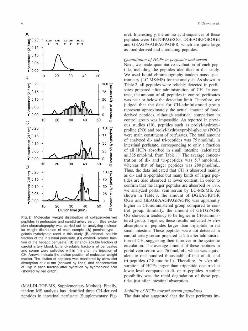
tion profiles of perfusates were shown in Fig. 2B and 2C. Interestingly, significant amount of compounds with molecular mass ranging from 100 to 1,500 Da was detected in intestinal perfusate (Fig. 2B), show-ing clear contrast to the elution profile of CH. The result indicated the existence of peptides larger than tripeptides in intestinal perfusate. Also, the data in-dicated that intestinal perfusate includes free amino acids, as well as di- and tri-peptides, as metabolites of administered CH. In hepatic perfusate, a main peak was detected at the elution time of free amino acid, whereas considerable amount of compounds larger than tripeptides was still detected. In contrast, elution profile of carotid artery serum showed that a main peak was detected at the position of free Hyp, suggesting the advanced turnover of the peptides during circulation. In order to confirm that elution profiles accurately represent the distribution of mo-lecular masses of CH-derived peptides and amino acids, fractions were collected and the amount of Hyp was analyzed after hydration by hydrochloric acid. Similar elution profiles were observed in per-fusates and blood sample, confirming that the data represent the dynamics of CH-derived peptides. Tak-en together, HCPs including peptides larger than tripeptide would pass through the liver and enter the systemic circulation at least once. It is generally thought that food derived peptides larger than tripeptide are not transported from intes-tinal lumen to portal vein. Thus, molecular weight distribution of HCPs in perfusate showed very unique character of CH absorption, leading us to identify larger HCPs. Peptides in intestinal perfusate were separated by reverse phase chromatography and fractions were analyzed by matrix-assisted laser de-sorption/ionization time-of-flight mass spectrometry
The ratio of the HCPs to the free Hyp was approxi-mately 3 : 1 and 1.5 : 1 just after absorption and af-ter passing through the liver, respectively. These results are at variance with the previous studies re-porting that free Hyp is dominant in the serum after administration of CH.
HCPs larger than tripeptide in perfusateOur analysis has suggested that the ratio of HCPs to free Hyp in absorbed fraction would be much high-er than previously considered, thus we analyzed the molecular profile of the perfusates. As shown in Fig. 2A, size exclusion chromatography showed that CH used in this study consists of peptides with mo-lecular mass ranged from 1,000 to 20,000 Da. Only trace amount of peptide with molecular mass less than 300 Da was detected. Thus, CH does not in-clude di- and tri-peptide as major ingredients. Elu-
Fig. 1 Time course of absorption of free Hyp and HCPs analyzed by in situ perfusion. Each value for Hyp concen-tration (nmol/mL) is expressed as the mean ± SEM (n = 3). Open and closed circles represent the amount of free Hyp and HCPs, respectively. A and B represent intestinal and hepatic perfusates, respectively. Different letters indicate significant difference. P < 0.05. multiple comparisons of Fisher’s PLSD.
Table 1 Absorbed amount of free and HCPs within 1 h af-ter injecting CHA
Form of Hyp Amount (μmol)Free 6.6 ± 1.9
Peptide (HCPs) 16.6 ± 2.6*Total 23.1 ± 1.2
BForm of Hyp Amount (μmol)
Free 5.4 ± 0.8Peptide (HCPs) 8.1 ± 0.9
Total 13.5 ± 1.2
Each value is expressed as the mean ± SEM (n = 3). A and B represents the intestinal and hepatic perfusate, respectively. * in-dicate significant difference to control group.
Y. Osawa et al.6
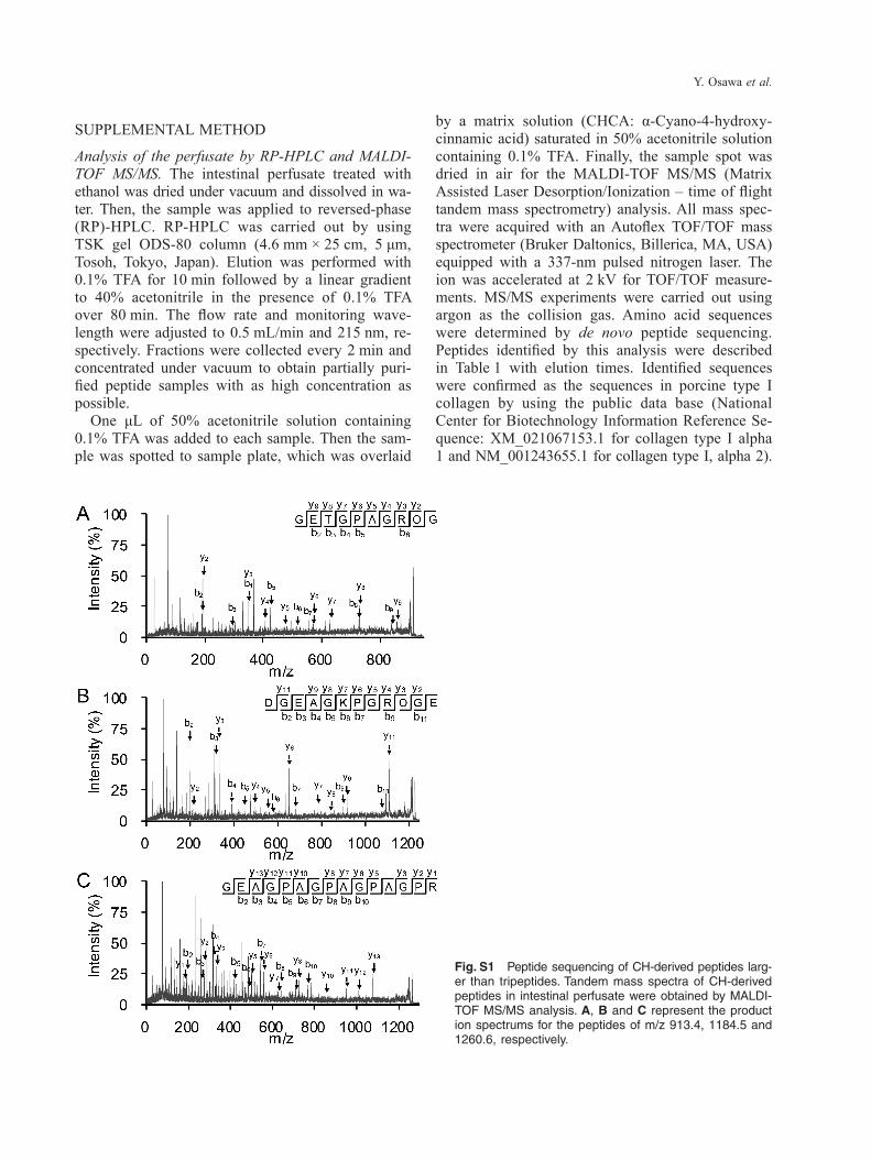
ure). Interestingly, the amino acid sequences of these peptides were GETGPAGROG, DGEAGKPGROGE and GEAGPAAGPAGPAGPR, which are quite large as food-derived and circulating peptides.
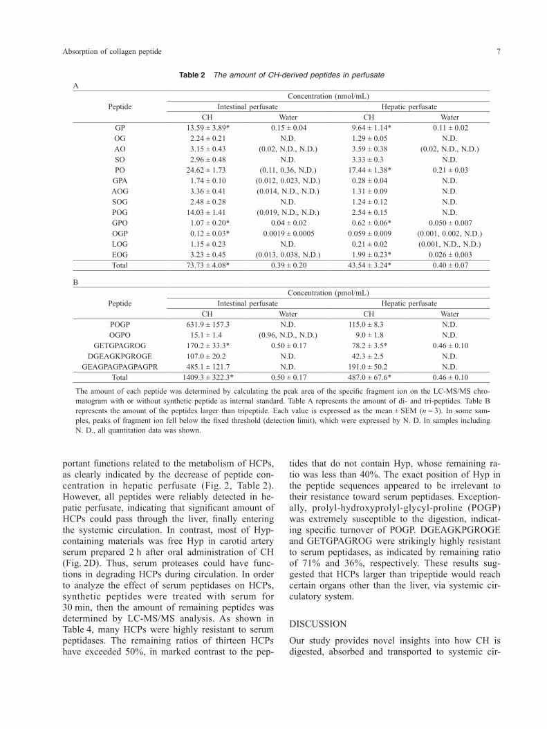
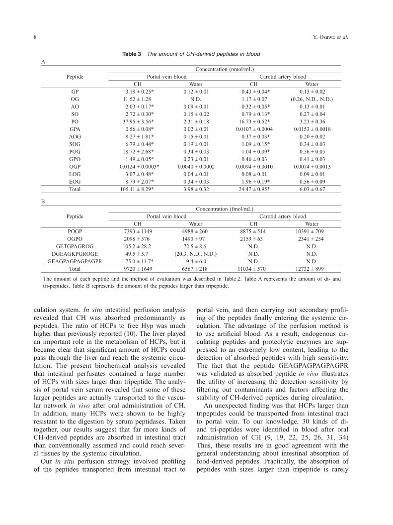
Quantitation of HCPs in perfusate and serumNext, we made quantitative evaluation of each pep-tide, including the peptides identified in this study. We used liquid chromatography-tandem mass spec-trometry (LC-MS/MS) for the analysis. As shown in Table 2, all peptides were reliably detected in perfu-sates prepared after administration of CH. In con-trast, the amount of all peptides in control perfusates was near or below the detection limit. Therefore, we judged that the data for CH-administrated group represent approximately the actual amount of food- derived peptides, although statistical comparison to control group was impossible. As reported in previ-ous studies (10), peptides such as prolyl-hydroxy-proline (PO) and prolyl-hydroxyprolyl-glycine (POG) were main constituent of perfusates. The total amount of analyzed di- and tri-peptides was 75 nmol/mL in intestinal perfusate, corresponding to only a fraction of all HCPs absorbed in small intestine (calculated as 385 nmol/mL from Table 1). The average concen-tration of di- and tri-peptides was 5.7 nmol/mL, whereas that of larger peptides was 280 pmol/mL. Thus, the data indicated that CH is absorbed mainly as di- and tri-peptides but many kinds of larger pep-tides are also absorbed at lower content. In order to confirm that the larger peptides are absorbed in vivo, we analyzed portal vein serum by LC-MS/MS. As shown in Table 3, the amount of DGEAGKPGR OGE and GEAGPAAGPAGPAGPR was apparently higher in CH-administered group compared to con-trol group. Similarly, the amount of GETGPAGR OG showed a tendency to be higher in CH-adminis-tered group. Together, these results indicated in vivo absorption of peptides larger than tripeptide in rat small intestine. These peptides were not detected in carotid artery serum prepared at 2 h after administra-tion of CH, suggesting their turnover in the systemic circulation. The average amount of these peptides in portal vein serum was 76 fmol/mL, which was equiv-alent to one hundred thousandth of that of di- and tri-peptides (7.8 nmol/mL). Therefore, in vivo ab-sorption of HCPs larger than tripeptide occurred at lower level compared to di- or tri-peptides. Another possibility was the rapid degradation of these pep-tides just after intestinal absorption.
Stability of HCPs toward serum peptidasesThe data also suggested that the liver performs im-
(MALDI-TOF-MS, Supplementary Method). Finally, tandem MS analysis has identified three CH-derived peptides in intestinal perfusate (Supplementary Fig-
Fig. 2 Molecular weight distribution of collagen-derived peptides in perfusates and carotid artery serum. Size exclu-sion chromatography was carried out for analyzing molecu-lar weight distribution of each sample. (A) porcine type 1 gelatin hydrolysate used in this study. (B) ethanol- soluble fraction of the intestinal perfusate. (C) ethanol- soluble frac-tion of the hepatic perfusate. (D) ethanol- soluble fraction of carotid artery blood. Ethanol-soluble fractions of perfusates and serum were collected within 1 h after the injection of CH. Arrows indicate the elution position of molecular weight marker. The elution of peptides was monitored by ultraviolet absorption at 215 nm (showed by lines) and concentration of Hyp in each fraction after hydration by hydrochloric acid (showed by bar graph).
Absorption of collagen peptide 7
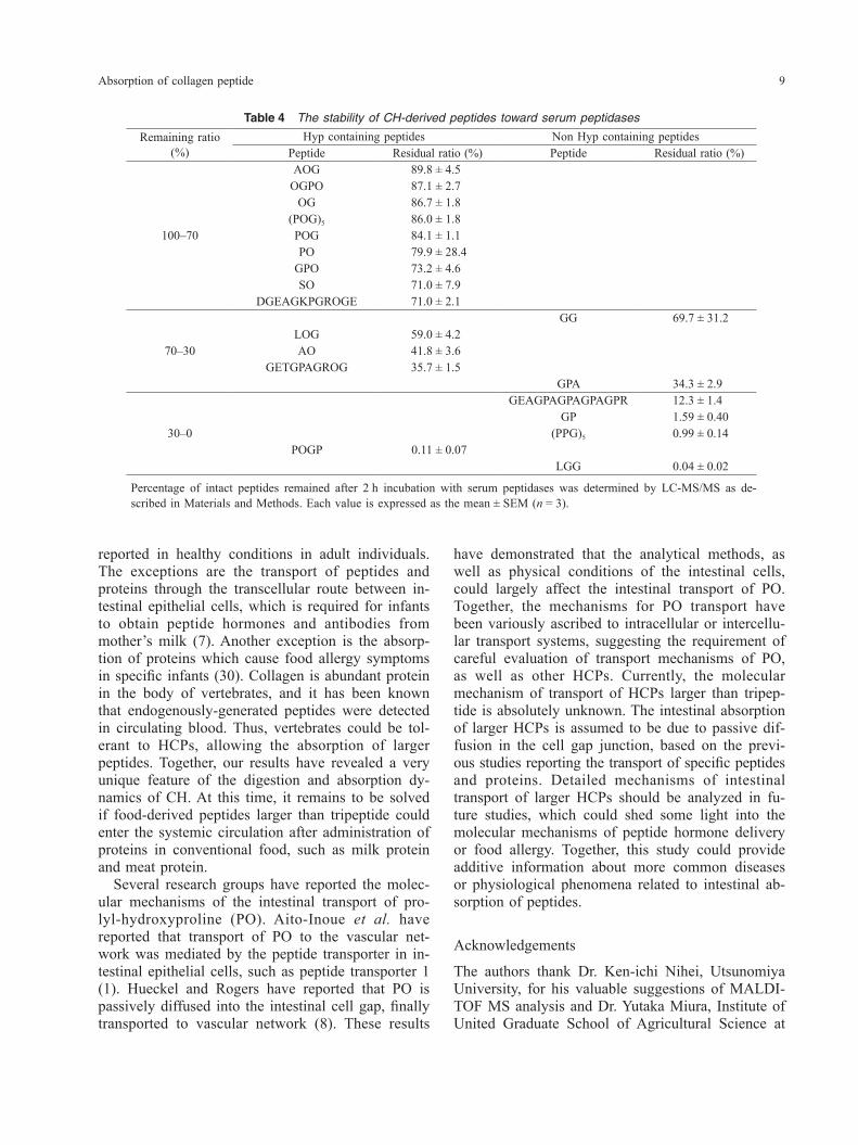
tides that do not contain Hyp, whose remaining ra-tio was less than 40%. The exact position of Hyp in the peptide sequences appeared to be irrelevant to their resistance toward serum peptidases. Exception-ally, prolyl-hydroxyprolyl-glycyl-proline (POGP) was extremely susceptible to the digestion, indicat-ing specific turnover of POGP. DGEAGKPGROGE and GETGPAGROG were strikingly highly resistant to serum peptidases, as indicated by remaining ratio of 71% and 36%, respectively. These results sug-gested that HCPs larger than tripeptide would reach certain organs other than the liver, via systemic cir-culatory system.
DISCUSSION
Our study provides novel insights into how CH is digested, absorbed and transported to systemic cir-
portant functions related to the metabolism of HCPs, as clearly indicated by the decrease of peptide con-centration in hepatic perfusate (Fig. 2, Table 2). However, all peptides were reliably detected in he-patic perfusate, indicating that significant amount of HCPs could pass through the liver, finally entering the systemic circulation. In contrast, most of Hyp- containing materials was free Hyp in carotid artery serum prepared 2 h after oral administration of CH (Fig. 2D). Thus, serum proteases could have func-tions in degrading HCPs during circulation. In order to analyze the effect of serum peptidases on HCPs, synthetic peptides were treated with serum for 30 min, then the amount of remaining peptides was determined by LC-MS/MS analysis. As shown in Table 4, many HCPs were highly resistant to serum peptidases. The remaining ratios of thirteen HCPs have exceeded 50%, in marked contrast to the pep-
Table 2 The amount of CH-derived peptides in perfusateA
PeptideConcentration (nmol/mL)
Intestinal perfusate Hepatic perfusateCH Water CH Water
GP 13.59 ± 3.89* 0.15 ± 0.04 9.64 ± 1.14* 0.11 ± 0.02OG 2.24 ± 0.21 N.D. 1.29 ± 0.05 N.D.AO 3.15 ± 0.43 (0.02, N.D., N.D.) 3.59 ± 0.38 (0.02, N.D., N.D.)SO 2.96 ± 0.48 N.D. 3.33 ± 0.3 N.D.PO 24.62 ± 1.73 (0.11, 0.36, N.D.) 17.44 ± 1.38* 0.21 ± 0.03
GPA 1.74 ± 0.10 (0.012, 0.023, N.D.) 0.28 ± 0.04 N.D.AOG 3.36 ± 0.41 (0.014, N.D., N.D.) 1.31 ± 0.09 N.D.SOG 2.48 ± 0.28 N.D. 1.24 ± 0.12 N.D.POG 14.03 ± 1.41 (0.019, N.D., N.D.) 2.54 ± 0.15 N.D.GPO 1.07 ± 0.20* 0.04 ± 0.02 0.62 ± 0.06* 0.050 ± 0.007OGP 0.12 ± 0.03* 0.0019 ± 0.0005 0.059 ± 0.009 (0.001, 0.002, N.D.)LOG 1.15 ± 0.23 N.D. 0.21 ± 0.02 (0.001, N.D., N.D.)EOG 3.23 ± 0.45 (0.013, 0.038, N.D.) 1.99 ± 0.23* 0.026 ± 0.003Total 73.73 ± 4.08* 0.39 ± 0.20 43.54 ± 3.24* 0.40 ± 0.07
B
PeptideConcentration (pmol/mL)
Intestinal perfusate Hepatic perfusateCH Water CH Water
POGP 631.9 ± 157.3 N.D. 115.0 ± 8.3 N.D.OGPO 15.1 ± 1.4 (0.96, N.D., N.D.) 9.0 ± 1.8 N.D.
GETGPAGROG 170.2 ± 33.3* 0.50 ± 0.17 78.2 ± 3.5* 0.46 ± 0.10DGEAGKPGROGE 107.0 ± 20.2 N.D. 42.3 ± 2.5 N.D.
GEAGPAGPAGPAGPR 485.1 ± 121.7 N.D. 191.0 ± 50.2 N.D.Total 1409.3 ± 322.3* 0.50 ± 0.17 487.0 ± 67.6* 0.46 ± 0.10
The amount of each peptide was determined by calculating the peak area of the specific fragment ion on the LC-MS/MS chro-matogram with or without synthetic peptide as internal standard. Table A represents the amount of di- and tri-peptides. Table B represents the amount of the peptides larger than tripeptide. Each value is expressed as the mean ± SEM (n = 3). In some sam-ples, peaks of fragment ion fell below the fixed threshold (detection limit), which were expressed by N. D. In samples including N. D., all quantitation data was shown.
Y. Osawa et al.8
portal vein, and then carrying out secondary profil-ing of the peptides finally entering the systemic cir-culation. The advantage of the perfusion method is to use artificial blood. As a result, endogenous cir-culating peptides and proteolytic enzymes are sup-pressed to an extremely low content, leading to the detection of absorbed peptides with high sensitivity. The fact that the peptide GEAGPAGPAGPAGPR was validated as absorbed peptide in vivo illustrates the utility of increasing the detection sensitivity by filtering out contaminants and factors affecting the stability of CH-derived peptides during circulation. An unexpected finding was that HCPs larger than tripeptides could be transported from intestinal tract to portal vein. To our knowledge, 30 kinds of di- and tri-peptides were identified in blood after oral administration of CH (9, 19, 22, 25, 26, 31, 34) Thus, these results are in good agreement with the general understanding about intestinal absorption of food-derived peptides. Practically, the absorption of peptides with sizes larger than tripeptide is rarely
culation system. In situ intestinal perfusion analysis revealed that CH was absorbed predominantly as peptides. The ratio of HCPs to free Hyp was much higher than previously reported (10). The liver played an important role in the metabolism of HCPs, but it became clear that significant amount of HCPs could pass through the liver and reach the systemic circu-lation. The present biochemical analysis revealed that intestinal perfusates contained a large number of HCPs with sizes larger than tripeptide. The analy-sis of portal vein serum revealed that some of these larger peptides are actually transported to the vascu-lar network in vivo after oral administration of CH. In addition, many HCPs were shown to be highly resistant to the digestion by serum peptidases. Taken together, our results suggest that far more kinds of CH-derived peptides are absorbed in intestinal tract than conventionally assumed and could reach sever-al tissues by the systemic circulation. Our in situ perfusion strategy involved profiling of the peptides transported from intestinal tract to
Table 3 The amount of CH-derived peptides in bloodA
PeptideConcentration (nmol/mL)
Portal vein blood Carotid artery bloodCH Water CH Water
GP 3.19 ± 0.25* 0.12 ± 0.01 0.43 ± 0.04* 0.13 ± 0.02OG 11.52 ± 1.28 N.D. 1.17 ± 0.07 (0.26, N.D., N.D.)AO 2.03 ± 0.17* 0.09 ± 0.01 0.32 ± 0.05* 0.13 ± 0.01SO 2.72 ± 0.30* 0.15 ± 0.02 0.79 ± 0.13* 0.27 ± 0.04PO 37.95 ± 3.56* 2.31 ± 0.18 16.73 ± 0.52* 3.23 ± 0.36
GPA 0.56 ± 0.08* 0.02 ± 0.01 0.0107 ± 0.0004 0.0153 ± 0.0018AOG 8.27 ± 1.81* 0.15 ± 0.01 0.37 ± 0.03* 0.20 ± 0.02SOG 6.79 ± 0.44* 0.19 ± 0.01 1.09 ± 0.15* 0.34 ± 0.03POG 18.72 ± 2.68* 0.34 ± 0.03 1.04 ± 0.09* 0.56 ± 0.05GPO 1.49 ± 0.05* 0.23 ± 0.01 0.46 ± 0.03 0.41 ± 0.03OGP 0.0124 ± 0.0003* 0.0040 ± 0.0002 0.0094 ± 0.0010 0.0074 ± 0.0013LOG 3.07 ± 0.48* 0.04 ± 0.01 0.08 ± 0.01 0.09 ± 0.01EOG 8.79 ± 2.07* 0.34 ± 0.03 1.96 ± 0.19* 0.56 ± 0.09Total 105.11 ± 8.29* 3.98 ± 0.32 24.47 ± 0.95* 6.03 ± 0.67
B
PeptideConcentration (fmol/mL)
Portal vein blood Carotid artery bloodCH Water CH Water
POGP 7393 ± 1149 4988 ± 260 8875 ± 514 10391 ± 709OGPO 2098 ± 576 1490 ± 97 2159 ± 63 2341 ± 254
GETGPAGROG 105.2 ± 28.2 72.5 ± 8.6 N.D. N.D.DGEAGKPGROGE 49.5 ± 5.7 (20.3, N.D., N.D.) N.D. N.D.
GEAGPAGPAGPAGPR 75.0 ± 11.7* 9.4 ± 6.0 N.D. N.D.Total 9720 ± 1649 6567 ± 218 11034 ± 570 12732 ± 899
The amount of each peptide and the method of evaluation was described in Table 2. Table A represents the amount of di- and tri-peptides. Table B represents the amount of the peptides larger than tripeptide.
Absorption of collagen peptide 9
have demonstrated that the analytical methods, as well as physical conditions of the intestinal cells, could largely affect the intestinal transport of PO. Together, the mechanisms for PO transport have been variously ascribed to intracellular or intercellu-lar transport systems, suggesting the requirement of careful evaluation of transport mechanisms of PO, as well as other HCPs. Currently, the molecular mechanism of transport of HCPs larger than tripep-tide is absolutely unknown. The intestinal absorption of larger HCPs is assumed to be due to passive dif-fusion in the cell gap junction, based on the previ-ous studies reporting the transport of specific peptides and proteins. Detailed mechanisms of intestinal transport of larger HCPs should be analyzed in fu-ture studies, which could shed some light into the molecular mechanisms of peptide hormone delivery or food allergy. Together, this study could provide additive information about more common diseases or physiological phenomena related to intestinal ab-sorption of peptides.
Acknowledgements
The authors thank Dr. Ken-ichi Nihei, Utsunomiya University, for his valuable suggestions of MALDI- TOF MS analysis and Dr. Yutaka Miura, Institute of United Graduate School of Agricultural Science at
reported in healthy conditions in adult individuals. The exceptions are the transport of peptides and proteins through the transcellular route between in-testinal epithelial cells, which is required for infants to obtain peptide hormones and antibodies from mother’s milk (7). Another exception is the absorp-tion of proteins which cause food allergy symptoms in specific infants (30). Collagen is abundant protein in the body of vertebrates, and it has been known that endogenously-generated peptides were detected in circulating blood. Thus, vertebrates could be tol-erant to HCPs, allowing the absorption of larger peptides. Together, our results have revealed a very unique feature of the digestion and absorption dy-namics of CH. At this time, it remains to be solved if food-derived peptides larger than tripeptide could enter the systemic circulation after administration of proteins in conventional food, such as milk protein and meat protein. Several research groups have reported the molec-ular mechanisms of the intestinal transport of pro-lyl-hydroxyproline (PO). Aito-Inoue et al. have reported that transport of PO to the vascular net-work was mediated by the peptide transporter in in-testinal epithelial cells, such as peptide transporter 1 (1). Hueckel and Rogers have reported that PO is passively diffused into the intestinal cell gap, finally transported to vascular network (8). These results
Table 4 The stability of CH-derived peptides toward serum peptidases
Remaining ratio(%)
Hyp containing peptides Non Hyp containing peptidesPeptide Residual ratio (%) Peptide Residual ratio (%)
100–70
AOG 89.8 ± 4.5OGPO 87.1 ± 2.7
OG 86.7 ± 1.8(POG)5 86.0 ± 1.8POG 84.1 ± 1.1PO 79.9 ± 28.4
GPO 73.2 ± 4.6SO 71.0 ± 7.9
DGEAGKPGROGE 71.0 ± 2.1
70–30
GG 69.7 ± 31.2LOG 59.0 ± 4.2AO 41.8 ± 3.6
GETGPAGROG 35.7 ± 1.5GPA 34.3 ± 2.9
30–0
GEAGPAGPAGPAGPR 12.3 ± 1.4GP 1.59 ± 0.40
(PPG)5 0.99 ± 0.14POGP 0.11 ± 0.07
LGG 0.04 ± 0.02
Percentage of intact peptides remained after 2 h incubation with serum peptidases was determined by LC-MS/MS as de-scribed in Materials and Methods. Each value is expressed as the mean ± SEM (n = 3).
Y. Osawa et al.10
Tokyo University of Agriculture and Technology, for his valuable suggestions on this study. We also thank Dr. Norihiro Azuma, Utsunomiya University, for their valuable comments on our research plan. The authors have no conflict of interest.
REFERENCES 1. Aito-Inoue M, Lackeyram D, Fan MZ, Sato K and Mine Y
(2007) Transport of a tripeptide, Gly-Pro-Hyp, across the porcine intestinal brush-border membrane. J Pept Sci 13, 468–474.
2. Arborelius M Jr, Konttinen YT, Nordstróm DC and Solovieva SA (1999) Gly-X-Y repeat sequences in the treatment of ac-tive rheumatoid arthritis. Rheumatol Int 18, 129–135.
3. Boran G and Regenstein JM (2010) Fish gelatin. Adv Food Nutr Res 60, 119–143.
4. Firshein HE and Shill JP (1966) The determination of total hydroxyproline in urine and bone extracts. Anal Biochem 14, 296–304.
5. Gómez-Guillén MC, Giménez B, López-Caballero ME and Montero MP (2011) Functional and bioactive properties of collagen and gelatin from alternative sources: A review. Food Hydrocolloid 25, 1813–1827.
6. Guo L, Harnedy PA, Zhang L, Li B, Zhang Z, Hou H, Zhao X and FitzGerald RJ (2015) In vitro assessment of the multi-functional bioactive potential of Alaska pollock skin collagen following simulated gastrointestinal digestion. J Sci Food Agric 95, 1514–1520.
7. Higashida H, Furuhara K, Yamauchi AM, Deguchi K, Harashima A, Munesue S, Lopatina O, Gerasimenko M, Salmina AB, Zhang JS, Kodama H, Kuroda H, Tsuji C, Sato S, Yamamoto H and Yamamoto Y (2017) Intestinal transepi-thelial permeability of oxytocin into the blood is dependent on the receptor for advanced glycation end products in mice. Sci Rep 7, 7883.
8. Hueckel HJ and Rogers QR (1972) Prolylhydroxyproline ab-sorption in hamsters. Can J Biochem 50, 782–790.
9. Ichikawa S, Morifuji M, Ohara H, Matsumoto H, Takeuchi Y and Sato K (2010) Hydroxyproline-containing dipeptides and tripeptides quantified at high concentration in human blood after oral administration of gelatin hydrolysate. Int J Food Sci Nutr 61, 52–60.
10. Iwai K, Hasegawa T, Taguchi Y, Morimatsu F, Sato K, Nakamura Y, Higashi A, Kido Y, Nakabo Y and Ohtsuki K (2005) Identification of food-derived collagen peptides in hu-man blood after oral ingestion of gelatin hydrolysates. J Ag-ric Food Chem 53, 6531–6536.
11. Kawaguchi T, Nanbu PN and Kurokawa M (2012) Distribu-tion of prolylhydroxyproline and its metabolites after oral ad-ministration in rats. Biol Pharm Bull 35, 422–427.
12. Kimira Y, Ogura K, Taniuchi Y, Kataoka A, Inoue N, Sugihara F, Nakatani S, Shimizu J, Wada M and Mano H (2014) Collagen-derived dipeptide prolyl-hydroxyproline pro-motes differentiation of MC3T3-E1 osteoblastic cells. Bio-chem Biophys Res Commun 453, 498–501.
13. Kusubata M, Koyama Y, Tometsuka C, Shigemura Y and Sato K (2015) Detection of endogenous and food-derived collagen dipeptide prolylhydroxyproline (Pro-Hyp) in allergic contact dermatitis-affected mouse ear. Biosci Biotechnol Bio-chem 79, 1356–1361.
14. Laskin DL, Kimura T, Sakakibara S, Riley DJ and Berg RA (1986) Chemotactic activity of collagen-like polypeptides for
human peripheral blood neutrophils. J Leukoc Biol 39, 255–266.
15. Liu C, Sugita K, Nihei K, Yoneyama K and Tanaka H (2009) Absorption of hydroxyproline-containing peptides in vascu-larly perfused rat small intestine in situ. Biosci Biotechnol Biochem 73, 1741–1747.
16. Morimura S, Nagata H, Uemura A, Shigematsu T and Kida K (2002) Development of an effective process for utilization of collagen from livestock and fish waste. Process Biochem 37, 1403–1412.
17. Oesser S, Adam M, Babel W and Seifert J (1999) Oral ad-ministration of 14C labeled gelatin hydrolysate leads to an ac-cumulation of radioactivity in cartilage of mice (C57/BL). J Nutr 129, 1891–1895.
18. Ohara H, Ichikawa S, Matsumoto H, Akiyama M, Fujimoto N, Kobayashi T and Tajima S (2010) Collagen-derived dipep-tide, proline-hydroxyproline, stimulates cell proliferation and hyaluronic acid synthesis in cultured human dermal fibro-blasts. J Dermatol 37, 330–338.
19. Ohara H, Matsumoto H, Ito K, Iwai K and Sato K (2007) Comparison of quantity and structures of hydroxyproline- containing peptides in human blood after oral ingestion of gelatin hydrolysates from different sources. J Agric Food Chem 55, 1532–1535.
20. Pickart L, Vasquez-Soltero JM and Margolina A (2015) GHK peptide as a natural modulator multiple cellular pathways in skin regeneration. Biomed Res Int 2015, 648108.
21. Ricard-Blum S (2011) The collagen family. Cold Spring Harb Perspect Biol 3, a004978.
22. Shigemura Y, Akaba S, Kawashima E, Park EY, Nakamura Y and Sato K (2011) Identification of a novel food-derived col-lagen peptide, hydroxyprolyl-glycine, in human peripheral blood by pre-column derivatisation with phenyl isothiocya-nate. Food Chem 129, 1019–1024.
23. Shigemura Y, Iwai K, Morimatsu F, Iwamoto T, Mori T, Oda C, Taira T, Park EY, Nakamura Y and Sato K (2009) Effect of Prolyl-hydroxyproline (Pro-Hyp), a food-derived collagen peptide in human blood, on growth of fibroblasts from mouse skin. J Agric Food Chem 57, 444–449.
24. Shimizu J, Asami N, Kataoka A, Sugihara F, Inoue N, Kimira Y, Wada M and Mano H (2015) Oral collagen-derived dipep-tides, prolyl-hydroxyproline and hydroxyprolyl-glycine, ame-liorate skin barrier dysfunction and alter gene expression profiles in the skin. Biochem Biophys Res Commun 456, 626–630.
25. Sugihara F, Inoue N, Kuwamori M and Taniguchi M (2012) Quantification of hydroxyprolyl-glycine (Hyp-Gly) in human blood after ingestion of collagen hydrolysate. J Biosci Bioeng 113, 202–203.
26. Taga Y, Kusubata M, Ogawa-Goto K and Hattori S (2014) Highly accurate quantification of hydroxyproline-containing peptides in blood using a protease digest of stable isotope- labeled collagen. J Agric Food Chem 62, 12096–12102.
27. Tanaka H, Arai M and Nishi H (1985) Metabolic fate of Gly-cyl-14C-prolylhydroxyproline in young Rats. Agric Biol Chem 49, 2949–2954.
28. Tsai WC, Hsu CC, Chung CY, Lin MS, Li SL and Pang JH (2007) The pentapeptide KTTKS promoting the expressions of type I collagen and transforming growth factor-beta of tendon cells. J Orthop Res 25, 1629–34.
29. Uemoto M (2010) Concepts and definitions of the limit of detection and the limit of quantitation. Bunseki 425, 216–221.
30. Untersmayr E and Jense-Jarolim E (2008) The role of protein digestibility and antacids on food allergy outcomes. J Allergy
Absorption of collagen peptide 11
Clin Immunol 121, 1301–1308.31. Wang L, Wang Q, Qian J, Liang Q, Wang Z, Xu J, He S and
Ma H (2015) Bioavailability and bioavailable forms of colla-gen after oral administration to rats. J Agric Food Chem 63, 3752–3756.
32. Watanabe-Kamiyama M, Shimizu M, Kamiyama S, Taguchi Y, Sone H, Morimatsu F, Shirakawa H, Furukawa Y and Komai M (2010) Absorption and effectiveness of orally administered low molecular weight collagen hydrolysate in rats. J Agric Food Chem 58, 835–841.
33. Wu J, Fujioka M, Sugimoto K, Mu G and Ishimi Y (2004)
Assessment of effectiveness of oral administration of colla-gen peptide on bone metabolism in growing and mature rats. J Bone Miner Metab 22, 547–553.
34. Yamamoto S, Hayasaka F, Deguchi K, Okudera T, Furusawa T and Sakai Y (2015) Absorption and plasma kinetics of col-lagen tripeptide after peroral or intraperitoneal administration in rats. Biosci Biotechnol Biochem 79, 2026–2033.
35. Zhang Z, Wang J, Ding Y, Dai X and Li Y (2011) Oral ad-ministration of marine collagen peptides from Chum Salmon skin enhances cutaneous wound healing and angiogenesis in rats. J Sci Food Agric 91, 2173–2179.
Y. Osawa et al.
Fig. S1 Peptide sequencing of CH-derived peptides larg-er than tripeptides. Tandem mass spectra of CH-derived peptides in intestinal perfusate were obtained by MALDI- TOF MS/MS analysis. A, B and C represent the product ion spectrums for the peptides of m/z 913.4, 1184.5 and 1260.6, respectively.
by a matrix solution (CHCA: α-Cyano-4-hydroxy-cinnamic acid) saturated in 50% acetonitrile solution containing 0.1% TFA. Finally, the sample spot was dried in air for the MALDI-TOF MS/MS (Matrix Assisted Laser Desorption/Ionization – time of flight tandem mass spectrometry) analysis. All mass spec-tra were acquired with an Autoflex TOF/TOF mass spectrometer (Bruker Daltonics, Billerica, MA, USA) equipped with a 337-nm pulsed nitrogen laser. The ion was accelerated at 2 kV for TOF/TOF measure-ments. MS/MS experiments were carried out using argon as the collision gas. Amino acid sequences were determined by de novo peptide sequencing. Peptides identified by this analysis were described in Table 1 with elution times. Identified sequences were confirmed as the sequences in porcine type I collagen by using the public data base (National Center for Biotechnology Information Reference Se-quence: XM_021067153.1 for collagen type I alpha 1 and NM_001243655.1 for collagen type I, alpha 2).
SUPPLEMENTAL METHOD
Analysis of the perfusate by RP-HPLC and MALDI- TOF MS/MS. The intestinal perfusate treated with ethanol was dried under vacuum and dissolved in wa-ter. Then, the sample was applied to reversed-phase (RP)-HPLC. RP-HPLC was carried out by using TSK gel ODS-80 column (4.6 mm × 25 cm, 5 μm, Tosoh, Tokyo, Japan). Elution was performed with 0.1% TFA for 10 min followed by a linear gradient to 40% acetonitrile in the presence of 0.1% TFA over 80 min. The flow rate and monitoring wave-length were adjusted to 0.5 mL/min and 215 nm, re-spectively. Fractions were collected every 2 min and concentrated under vacuum to obtain partially puri-fied peptide samples with as high concentration as possible. One μL of 50% acetonitrile solution containing 0.1% TFA was added to each sample. Then the sam-ple was spotted to sample plate, which was overlaid