Embed Size (px)
Citation preview
Hindawi Publishing CorporationJournal of NanomaterialsVolume 2010, Article ID 818717, 7 pagesdoi:10.1155/2010/818717
Research Article
An Insight into the Interactions between α-Tocopheroland Chitosan in Ultrasound-Prepared Nanoparticles
Majid Naghibzadeh,1 Amir Amani,1 Mohsen Amini,2 Elina Esmaeilzadeh,1
Negar Mottaghi-Dastjerdi,2 and Mohammad Ali Faramarzi2, 3
1 Department of Medical Nanotechnology, Faculty of Advanced Medical Technologies, Tehran University of Medical Sciences,Tehran 14174, Iran
2 Department of Pharmaceutical Biotechnology, Biotechnology Research Center, Faculty of Pharmacy,Tehran University of Medical Sciences, P.O. Box 14155-6451, Tehran 14174, Iran
3 Biotechnology Research Center, Tehran University of Medical Sciences, Tehran 14174, Iran
Correspondence should be addressed to Mohammad Ali Faramarzi, [email protected]
Received 31 January 2010; Accepted 6 June 2010
Academic Editor: Doron Yadlovker
Copyright © 2010 Majid Naghibzadeh et al. This is an open access article distributed under the Creative Commons AttributionLicense, which permits unrestricted use, distribution, and reproduction in any medium, provided the original work is properlycited.
The aim of this study was to investigate the interactions between α-tocopherol and chitosan molecules prepared subsequent topreparation of α-tocopherol-loaded chitosan nanoparticles using ultrasonication. Dynamic light scattering (DLS) and scanningelectron microscopy (SEM) analyses showed semispherical particles with an average size of approximately 350 nm. Also fromreconstitution test, α-tocopherol was suggested as stabilizing agent during lyophilization/reconstitution process. The zeta potentialsof chitosan and α-tocopherol nanoparticles were larger than ±30 mV, representing suitable stability. Data obtained from FTIRshowed possibility of chemical interaction between chitosan and α-tocopherol. Furthermore, the results from FTIR, NMR, andXRD spectroscopy confirmed electrostatic interactions between the two molecules. Overall, this procedure could be considered asa facile method to prepare α-tocopherol-loaded nanoparticles.
1. Introduction
Chitosan, the second plentiful polysaccharied after cellulosein nature, is a biopolymer derived by alkaline deacetylationof chitin [1]. This polymer, exhibiting unique propertiessuch as biocompatibility, biodegradability, low cytotoxicity,bioadhesivity, and the ability to increase the drugs absorptionthrough polyanionic membranes, has attracted attention ofmany researchers in drug delivery systems [2, 3]. Amongdifferent types of chitosans, medium and high molecularweight chitosans are not fully dispersible in neutral aqueousmedia. This in turn limits the application of chitosansin delivery of pH-sensitive active agents such as proteins,enzymes, and genes [4]. During recent years, among differenttypes of chitosans, low molecular weight chitosan has gainedattention as a drug conjugate according to its water solubilitywithout the need for low pH values [5, 6].
α-Tocopherol, one of the lipid soluble vitamins, hasproved useful in anticancer, anti-inflammatory, and antiox-idant activities [7]. However, conventional formulations oftocopherol often fail to provide satisfactory bioavailabilitydue to its hydrophobic characteristics [8]. To overcome thisproblem, based on Bruner and Tolloczko equation [9], itcould be hypothesized that increasing the particles surfacearea leads to an increase in the dissolution velocity [9]. Thishypothesis had been confirmed by Kotyla and coworkersthat had compared nanosized tocopherol with microsizedparticles of this drug. The results showed an increase inbioavailability of tocopherol [10].
The principal of ultrasonication is based on formation,growth, and implosion of acoustic cavity. Ultrasonication is agreen, simple, fast, popular, and reproducible method. In thisstudy, we used low frequency ultrasonication for preparationof α-tocopherol-loaded chitosan nanoparticles. The study
2 Journal of Nanomaterials
aimed to investigate the possibility of loading α-tocopherolon the chitosan nanoparticles followed by determininginteractions between the drug and the nanocarrier.
2. Experiments
2.1. Chemicals. Water soluble chitosan (MW<18 KD, degreeof deacetylation = 86%) was gifted from Easter HoldingGroup (China). α-Tocopherol, deuterium oxide (D2O), anddimethyl sulfoxide (DMSO) were of analytical grades andpurchased from Merck (Darmstadt, Germany).
2.2. Preparation of α-Tocopherol-Loaded Chitosan Nanoparti-cles. Fifty mg of low molecular weight chitosan was dispersedin 10 mL of deionized water. A specified amount of α-tocopherol was dispersed in 1 mL of ethanol and droppedinto chitosan solution. The mixtures were placed in icebath and sonicated using the probe ultrasonication (LabSonic, B. Braun, Germany) at 20 KHz to form α-tocopherol-loaded chitosan nanoparticles. Subsequently, ethanol wasevaporated using rotary evaporator, and the prepared sam-ples were lyophilized for future analyses. All procedureswere performed in darkened condition. Four weight ratio(mg/mg) of chitosan to α-tocopherol, namely, 50/2 (i.e.,Ch50/2), 50/5 (i.e., Ch50/5), 50/10 (i.e., Ch50/10), and 50/20(i.e., Ch50/20) were prepared using ultrasonication.
2.3. Particle Size and Zeta Potential Measurement. Dynamiclight scattering (DLS) studies were performed using Zetasizer3000 (Malvern Instruments, UK) at 25◦C to obtain the parti-cle size and zeta potential of the particles. Each measurementwas repeated three times. No dilution was made to thesamples, and the viscosity of water was taken as the viscosityof dispersant.
2.4. Scanning Electron Microscopy. To study the morphologyof α-tocopherol-loaded nanoparticles, scanning electronmicroscopy (SEM) was employed. The suspension was goldcoated and placed on a sputter and a DSM, 960 A; Zeiss(Germany) operating at 10 kV was used to visualize thenanoparticles.
2.5. Reconstitution Test. To investigate the possible changesin the particles properties before lyophilization and afterreconstitution, for each chitosan/α-tocopherol weight ratio(i.e., Ch50/2, Ch50/5, Ch50/10, and Ch50/20), 10 mg offinal lyophilized powder was dissolved in 10 mL of deionizedwater and vortexed for 10 s. The zeta potential and particlesize of the samples were then measured using the methoddescribed in Section 2.3.
2.6. Fourier-Transform-Infrared Spectroscopy. Chemicalinteractions and structure of chitosan and α-tocopherol innanoparticles were studied using Fourier-transform-infrared(FT-IR) spectroscopy employing KBr discs on an FT-IRspectrometer (Shimadzu, FT-IR 8700, Japan).
2.7. 1H Nuclear Magnetic Resonance Spectra Studies. Theinteractions between chitosan and α-tocopherol were fur-
ther investigated using 1H nuclear magnetic resonance(1H NMR) spectroscopy. The chitosan and α-tocopherolnanoparticles were dissolved in dimethyl sulfoxide (DMSO)and deuterium oxide (D2O). 1H NMR spectra were recordedon a Bruker DRX 400 spectrometer, (500 MHz, USA)operating at a probe temperature of 298◦K.
2.8. X-Ray Diffraction. The powder X-ray diffraction (XRD)was performed by an X’Pert MPD Powder X-ray DiffractionSystem (Philips, Holland) using Cu Kα radiation (40 kV,40 mA) to study chemical structure of nanoparticles.
3. Results and Discussion
3.1. Characterization of α-Tocopherol and Chitosan Nanopar-ticles. α-Tocopherol is a hydrophobic compound which ispractically insoluble in water (<0.1 mg/mL) [11]. Thus,conventional formulation of this active agent will lead tolow bioavailability when taken orally. Water soluble chitosan,with high contents of free-amine groups, has been reportedsuitable for solubilization of hydrophobic compounds likeall-transretinoic acid through establishing ionic complexesbetween the two molecules [5]. Dissolution velocity as thesecond factor that affects bioavailability is also affected bythe particle size. According to Noyes and Whitney as wellas Bruner and Tolloczko equations, reducing particle sizedirectly affects the dissolution rate and bioavailability ofthe active agent [10]. Therefore, a proper nanoparticlesformulation of chitosan that is able to solubilize α-tocopherolwill be able to increase its bioavailability.
In this paper, we used low molecular weight chitosanwhich takes the advantage of being water soluble. In contrastto other chitosans which require acidic conditions to bedissolved, low molecular weight chitosan can be easilydissolved in neutral aqueous media and thus be used fordelivery of wider range of drugs including pH-sensitive ones.
The different formulations were subjected to DLS particlesize analysis. The particle size and zeta potential valuesobtained for α-tocopherol-loaded nanoparticles 30 minutesafter preparation are shown in Table 1. The data shows thatby increasing the α-tocopherol content in the formulation,a sharp increase in the particle size can be observed whenincreasing the α-tocopherol content from 2 to 5, followedby a slight decrease in the observed particle size in furtherincreases of the α-tocopherol content. One explanation forthis observation is that when in low value of α-tocopherolamount in the formulation (i.e., Ch50/5), the excess α-tocopherol molecules will mainly deposit on the surface ofthe chitosan particles leading to an increase in the observedhydrodynamic sizes. However, by further increase in theα-tocopherol content, the α-tocopherol molecules migrateto the internal parts of the nanoparticles, establishingproper interactions between the two molecules which leadto molecules rearrangements in the molecular structure ofnanoparticles to make more stable particles. This rearrange-ment will be the main cause for the observed decrease in theparticle size. This hypothesis appears to be applicable due tothe fact that ultrasonication can render the rearrangementin the molecular structure of particles [12] and has been
Journal of Nanomaterials 3
10 kV12 mm
×20000
500 nm
XYZR
331
331
802
0130
3590
506·
In
# 1095
FOS-TV
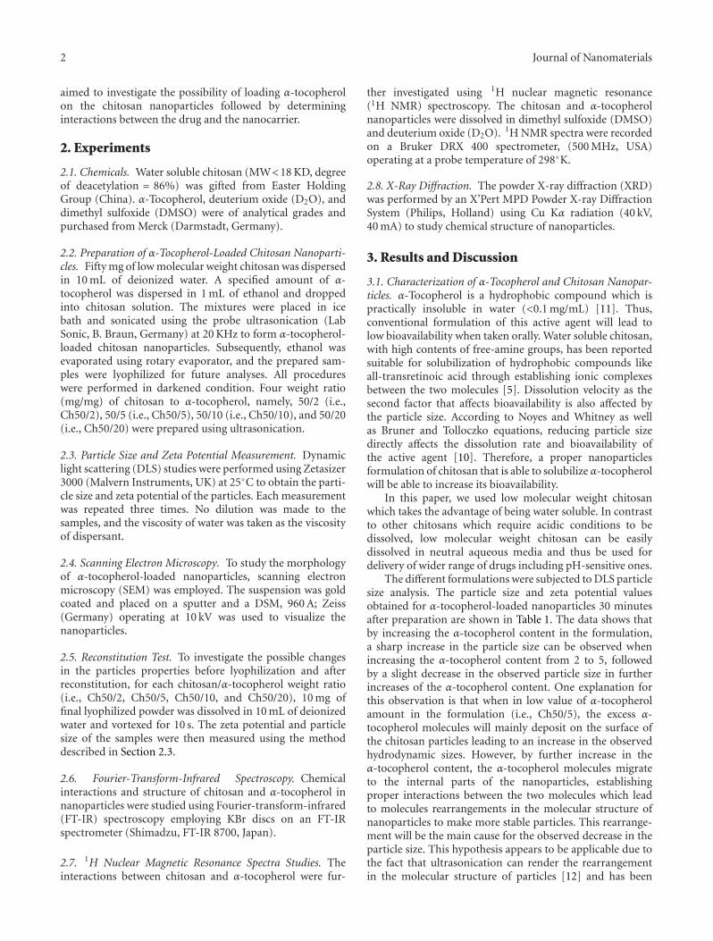
ZEISSFigure 1: SEM images of chitosan and α-tocopherol nanoparticles.
Table 1: Characterization of freshly prepared α-tocopherol and chitosan nanoparticles.
Chitosan/α-tocopherol Weight ratio(mg/mg)
Averageparticle size(nm, n = 3)
Zeta potential (mV) Polydispersity Index
Ch50/2 50/2 277 −76.8± 4.8 0.276
Ch50/5 50/5 378 −74.0± 5.4 0.197
Ch50/10 50/10 358 −61.7± 8.7 0.253
Ch50/20 50/20 348 −61.5± 4.1 0.245
verified by the zeta potential and reconstitution tests as willbe discussed later. The polydispersity index is another factorthat represents the dispersion homogeneity. The preparednanoparticles exhibited relatively narrow particle size distri-bution, as indicated by a relatively low polydispersity index(PDI) values with all values being less than 0.3 (Table 1).
Tables 1 and 2 indicate zeta potential changes beforeand after reconstitution of the four samples. In our study,the zeta potentials of all studied samples were larger than±30 mV (see Table 1), and thus acceptable stability can bepredicted for the samples. Also, as can be observed fromTable 1, the zeta potential has decreased by increasing theα-tocopherol content in the formulation. As α-tocopherolis probably the major cause for the change in the zetapotential of the nanoparticles, it could be suggested thatthe considerable “jump” observed in the zeta potential ofCh50/5 compared to Ch50/10 indicates the insertion of theα-tocopherol molecules into the chitosan particles, causingthe zeta potential to decrease from −74.0 to −61.7 mV.Also, as shown in Table 2, zeta potential and particle sizeof reconstituted α-tocopherol-loaded nanoparticles weresimilar to original preparation when having more than10 mg of α-tocopherol in the formulation which also suggeststhat α-tocopherol plays an important role in stabilizing thechitosan nanoparticles.
Morphology of the chitosan and α-tocopherol nanopar-ticles was characterized by scanning electron microscope
(SEM) (see Figure 1). The SEM images indicated thatnanoparticles were nearly monodisperse with semisphericalshapes. The observed particles size are also almost similar tothe particle size results from DLS.
3.2. Reconstitution of α-Tocopherol and Chitosan Nanopar-ticles. One of the factors that can potentially affect thenanoparticles properties is the freeze-drying procedure. Ithas been reported that additives such as saccharides areusually essential for cryoprotection of the nanoparticlesin the lyophilization procedure [13]. Based on our resultsin reconstitution test, when lyophilized nanoparticles werereconstituted into water (Table 2), the particle size anddistribution of the particle size were practically similar tooriginal aqueous solution (Table 1) when the α-tocopherolto chitosan ratio was more than 10/50 (i.e., Ch50/20 andCh50/10). This observation can confirm the hypothesis ofα-tocopherol molecules inserting the chitosan nanoparticlesand stabilizing them. As a result, it could be arguedthat the α-tocopherol-loaded chitosan nanoparticles can besuccessively lyophilized and reconstituted without the needto a cryoprotectant.
Stability of solution is an essential factor in marketing.Klaypradit and Huang indicated that chitosan alone isunable to produce stable emulsions, showing the need fora surfactant when designing chitosan preparations [14];
4 Journal of Nanomaterials
Table 2: Characterization of α-tocopherol and chitosan nanoparticles after reconstitution.
Chitosan/α-tocopherol Weight ratio(mg/mg)
Averageparticle size(nm, n = 3)
Zeta potential (mV) Polydispersity
Ch50/2 50/2 383 −48.7± 7.6 0.360
Ch50/5 50/5 325 −57.3± 4.9 0.192
Ch50/10 50/10 330 −61.5± 9.6 0.258
Ch50/20 50/20 351 −61.2± 5.3 0.237
2873.4
3446.94
1424.18
1659.74
1588.05
3472.55
2924.612863.16
2847.24
1465.15
1388.34
1255.19
1163.02
2917.081639.95 1580.09
2852.92
1649.51562.452924.61
2852.92
2919.49
1654.621562.45
3431.58
(a)
(b)
(c)
(d)
(e)
3000 2000 1000
Wavenumber (cm−1)
Tran
smit
tan
ce(%
)
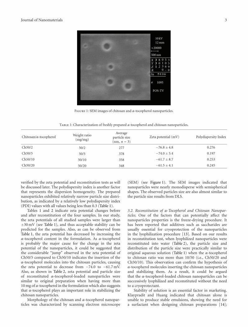
Figure 2: FTIR spectra of (a) chitosan, (b) α-tocopherol, (c) Ch50/2, (d) Ch50/5, and (e) Ch50/20.
however based on our results in reconstitution test, α-tocopherol-loaded chitosan nanoparticles were completelyreconstituted into neutral aqueous solution without addingcryoprotectants, indicating economical benefits in scaleup.
3.3. Analysis of α-Tocopherol and Chitosan Nanoparticles.Figure 2 shows the FTIR spectra obtained for α-tocopherol,chitosan and α-tocopherol-loaded chitosan. In Figure 2(a)the broadband at around 3446 cm−1 attributes to –NH and–OH stretching vibration. The weak band at 2873 cm−1 may
Journal of Nanomaterials 5
7 6 5 4 3 2 1
(ppm)
1 1.15
NanoparticlesCh 50/20
(a)
7 6 5 4 3 2 1 0 −1
(ppm)
0.39 1
NanoparticlesCh 50/10
(b)
7 6 5 4 3 2 1
(ppm)
NanoparticlesCh 50/2
(c)
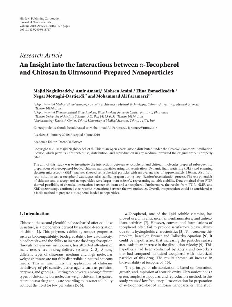
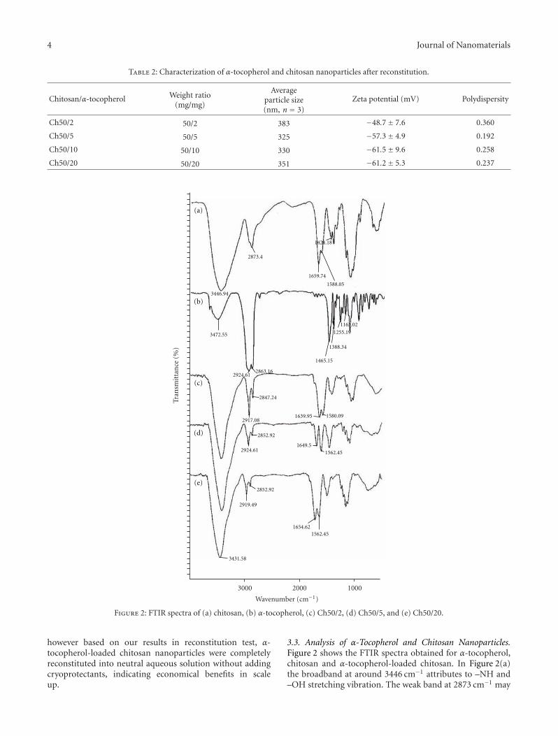
Figure 3: 1H NMR spectra of α-tocopherol-loaded chitosan nanoparticles.
be due to –CH– stretching in chitosan. The bands at 1659 and1590 cm−1 represent amide and amine groups of chitosan,respectively, and the bands at 1070 cm−1 are assigned to theskeletal vibration of C–O stretching [15].
Figure 2(b) shows the spectrum of α-tocopherol. Theband at 3472 cm−1 is associated with –OH as has beenreported previously in [16]. Also, bands at 2924 and2863 cm−1 represent the asymmetric and symmetric stretch-ing vibrations of the –CH2– and –CH3, respectively,1465 cm−1 is for phenyl, skeletal and methyl asymmetricbending, 1388 cm−1 is associated with methyl symmetricbending, 1081 cm−1 is attributed with plane bending ofphenyl and 922 cm−1 stands for trans =CH2 stretching.
Comparing Figures 2(a) and 2(c)–2(e), the peak at1590 cm−1 for primary amine bending has been shifted tonew positions, 1580 cm−1 and 1562, most probably due tothe electrostatic interactions occurring for –NH2 group ofchitosan with α-tocopherol. On the other side, the peak of3472 cm−1 from α-tocopherol (i.e., Figure 2(b)) has beendisappeared in Figures 2(c)–2(e). This may be due to –OHbeing engaged in an interaction with chitosan, thus shiftingthe associated peak value to lower values and being locatedbehind the large peak of 3431 cm−1 observed in Figures 2(c)–2(e). In general, from FTIR results, it can be suggested that achemical interaction between –NH2 group of chitosan and –OH group of α-tocopherol has been occurred. Considering
the molecular structure of α-tocopherol and chitosan, anelectrostatic interaction bond between –OH and –NH2
seems reasonable here.To further investigate the properties of formed nanopar-
ticles, 1H NMR spectroscopy was employed (see Figure 3).In the 1H NMR spectra, the broad multiplet peaks from 2.2to 2.6 ppm are attributed to α-tocopherol [17]. Also peaksfrom 3.3 to 3.9 ppm correspond to protons of glucose aminein chitosan [15].
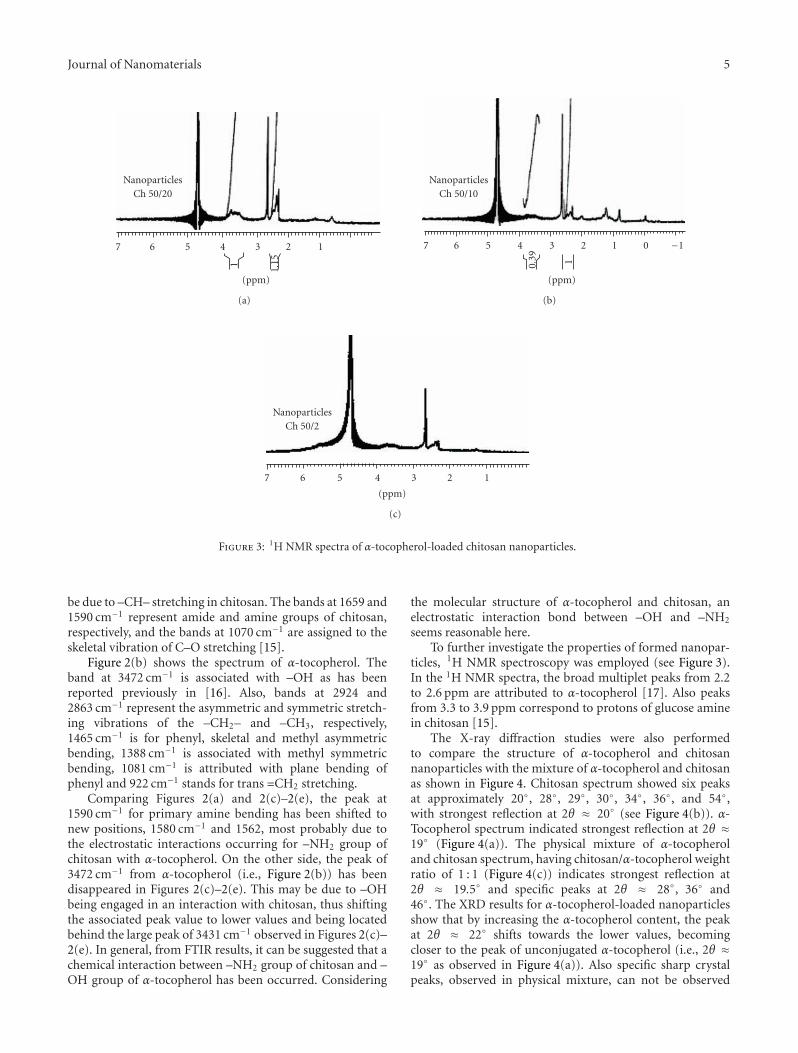
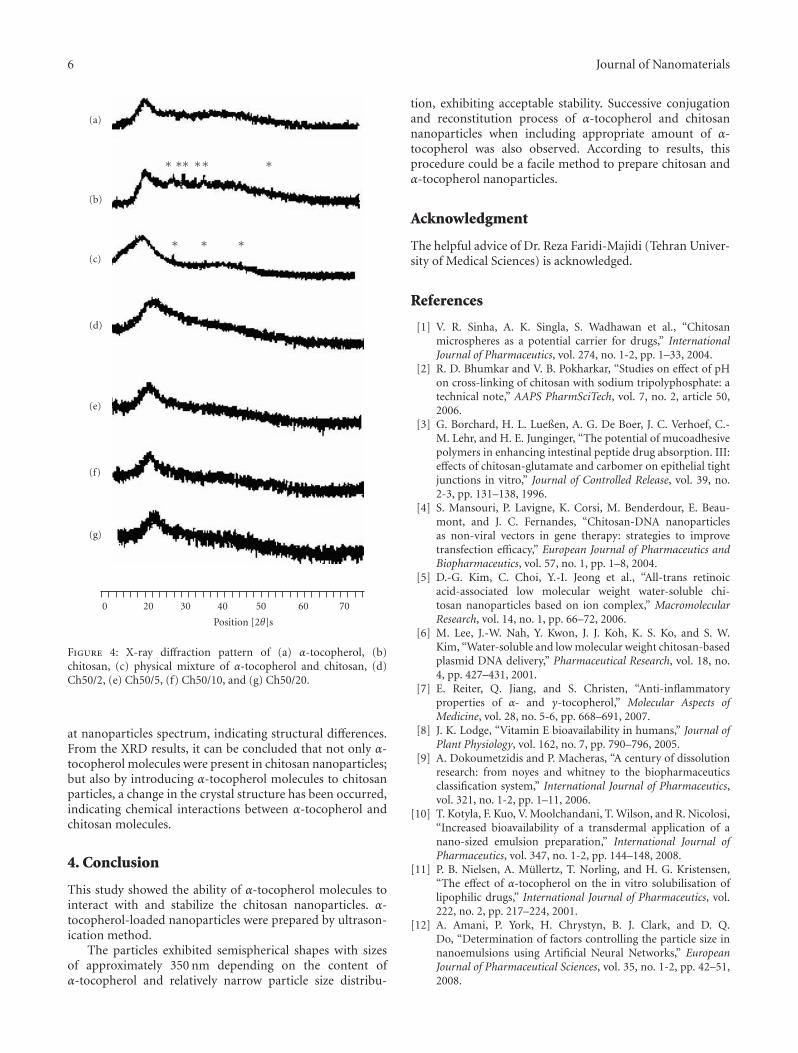
The X-ray diffraction studies were also performedto compare the structure of α-tocopherol and chitosannanoparticles with the mixture of α-tocopherol and chitosanas shown in Figure 4. Chitosan spectrum showed six peaksat approximately 20◦, 28◦, 29◦, 30◦, 34◦, 36◦, and 54◦,with strongest reflection at 2θ ≈ 20◦ (see Figure 4(b)). α-Tocopherol spectrum indicated strongest reflection at 2θ ≈19◦ (Figure 4(a)). The physical mixture of α-tocopheroland chitosan spectrum, having chitosan/α-tocopherol weightratio of 1 : 1 (Figure 4(c)) indicates strongest reflection at2θ ≈ 19.5◦ and specific peaks at 2θ ≈ 28◦, 36◦ and46◦. The XRD results for α-tocopherol-loaded nanoparticlesshow that by increasing the α-tocopherol content, the peakat 2θ ≈ 22◦ shifts towards the lower values, becomingcloser to the peak of unconjugated α-tocopherol (i.e., 2θ ≈19◦ as observed in Figure 4(a)). Also specific sharp crystalpeaks, observed in physical mixture, can not be observed
6 Journal of Nanomaterials
0 20 30 40 50 60 70
(a)
(b)
(c)
(d)
(e)
(f)
(g)
∗ ∗∗ ∗∗ ∗
∗ ∗ ∗
Position [2θ]s
Figure 4: X-ray diffraction pattern of (a) α-tocopherol, (b)chitosan, (c) physical mixture of α-tocopherol and chitosan, (d)Ch50/2, (e) Ch50/5, (f) Ch50/10, and (g) Ch50/20.
at nanoparticles spectrum, indicating structural differences.From the XRD results, it can be concluded that not only α-tocopherol molecules were present in chitosan nanoparticles;but also by introducing α-tocopherol molecules to chitosanparticles, a change in the crystal structure has been occurred,indicating chemical interactions between α-tocopherol andchitosan molecules.
4. Conclusion
This study showed the ability of α-tocopherol molecules tointeract with and stabilize the chitosan nanoparticles. α-tocopherol-loaded nanoparticles were prepared by ultrason-ication method.
The particles exhibited semispherical shapes with sizesof approximately 350 nm depending on the content ofα-tocopherol and relatively narrow particle size distribu-
tion, exhibiting acceptable stability. Successive conjugationand reconstitution process of α-tocopherol and chitosannanoparticles when including appropriate amount of α-tocopherol was also observed. According to results, thisprocedure could be a facile method to prepare chitosan andα-tocopherol nanoparticles.
Acknowledgment
The helpful advice of Dr. Reza Faridi-Majidi (Tehran Univer-sity of Medical Sciences) is acknowledged.
References
[1] V. R. Sinha, A. K. Singla, S. Wadhawan et al., “Chitosanmicrospheres as a potential carrier for drugs,” InternationalJournal of Pharmaceutics, vol. 274, no. 1-2, pp. 1–33, 2004.
[2] R. D. Bhumkar and V. B. Pokharkar, “Studies on effect of pHon cross-linking of chitosan with sodium tripolyphosphate: atechnical note,” AAPS PharmSciTech, vol. 7, no. 2, article 50,2006.
[3] G. Borchard, H. L. Lueßen, A. G. De Boer, J. C. Verhoef, C.-M. Lehr, and H. E. Junginger, “The potential of mucoadhesivepolymers in enhancing intestinal peptide drug absorption. III:effects of chitosan-glutamate and carbomer on epithelial tightjunctions in vitro,” Journal of Controlled Release, vol. 39, no.2-3, pp. 131–138, 1996.
[4] S. Mansouri, P. Lavigne, K. Corsi, M. Benderdour, E. Beau-mont, and J. C. Fernandes, “Chitosan-DNA nanoparticlesas non-viral vectors in gene therapy: strategies to improvetransfection efficacy,” European Journal of Pharmaceutics andBiopharmaceutics, vol. 57, no. 1, pp. 1–8, 2004.
[5] D.-G. Kim, C. Choi, Y.-I. Jeong et al., “All-trans retinoicacid-associated low molecular weight water-soluble chi-tosan nanoparticles based on ion complex,” MacromolecularResearch, vol. 14, no. 1, pp. 66–72, 2006.
[6] M. Lee, J.-W. Nah, Y. Kwon, J. J. Koh, K. S. Ko, and S. W.Kim, “Water-soluble and low molecular weight chitosan-basedplasmid DNA delivery,” Pharmaceutical Research, vol. 18, no.4, pp. 427–431, 2001.
[7] E. Reiter, Q. Jiang, and S. Christen, “Anti-inflammatoryproperties of α- and γ-tocopherol,” Molecular Aspects ofMedicine, vol. 28, no. 5-6, pp. 668–691, 2007.
[8] J. K. Lodge, “Vitamin E bioavailability in humans,” Journal ofPlant Physiology, vol. 162, no. 7, pp. 790–796, 2005.
[9] A. Dokoumetzidis and P. Macheras, “A century of dissolutionresearch: from noyes and whitney to the biopharmaceuticsclassification system,” International Journal of Pharmaceutics,vol. 321, no. 1-2, pp. 1–11, 2006.
[10] T. Kotyla, F. Kuo, V. Moolchandani, T. Wilson, and R. Nicolosi,“Increased bioavailability of a transdermal application of anano-sized emulsion preparation,” International Journal ofPharmaceutics, vol. 347, no. 1-2, pp. 144–148, 2008.
[11] P. B. Nielsen, A. Mullertz, T. Norling, and H. G. Kristensen,“The effect of α-tocopherol on the in vitro solubilisation oflipophilic drugs,” International Journal of Pharmaceutics, vol.222, no. 2, pp. 217–224, 2001.
[12] A. Amani, P. York, H. Chrystyn, B. J. Clark, and D. Q.Do, “Determination of factors controlling the particle size innanoemulsions using Artificial Neural Networks,” EuropeanJournal of Pharmaceutical Sciences, vol. 35, no. 1-2, pp. 42–51,2008.
Journal of Nanomaterials 7
[13] M. L. Hans and A. M. Lowman, “Biodegradable nanoparticlesfor drug delivery and targeting,” Current Opinion in Solid Stateand Materials Science, vol. 6, no. 4, pp. 319–327, 2002.
[14] W. Klaypradit and Y.-W. Huang, “Fish oil encapsulation withchitosan using ultrasonic atomizer,” LWT—Food Science andTechnology, vol. 41, no. 6, pp. 1133–1139, 2008.
[15] G. Ma, D. Yang, J. F. Kennedy, and J. Nie, “Synthesizeand characterization of organic-soluble acylated chitosan,”Carbohydrate Polymers, vol. 75, no. 3, pp. 390–394, 2009.
[16] Y. B. Che Man, W. Ammawath, and M. E. S. Mirghani,“Determining α-tocopherol in refined bleached and deodor-ized palm olein by Fourier transform infrared spectroscopy,”Food Chemistry, vol. 90, no. 1-2, pp. 323–327, 2005.
[17] A. Lienau, T. Glaser, M. Krucker et al., “Qualitative andquantitative analysis of tocopherols in toothpastes and gingivaltissue employing HPLC NMR and HPLC MS coupling,”Analytical Chemistry, vol. 74, no. 20, pp. 5192–5198, 2002.