Embed Size (px)
Citation preview

8/20/2019 Anatomy Ch. 5
http://slidepdf.com/reader/full/anatomy-ch-5 1/62
C h a p t e r
5
The
IntegumentarySystem
Copyright © 2010 Pearson Education, Inc.

8/20/2019 Anatomy Ch. 5
http://slidepdf.com/reader/full/anatomy-ch-5 2/62
Introduction to the Integumentary System• The integumentary system consists of skin, hair, nails, and various glands
– The integument is the largest system of the body• 16% of body weight• 1.5 to 2 m 2 in area
– The integument is made up of two parts:• Cutaneous membrane (skin)
• Superficial epithelium called epidermis
• Underlying connective tissue called dermis• Subcutaneous layer ( hypodermis ) made of loose connectivetissue attaches integument to deeper structures (muscle, bone)
• Hypodermis is notconsidered part ofintegumentary system
• Location of“hypodermic” injections
• Accessory structures – originate in dermis and extendthrough epidermis to skin
surface• Include hair, nails, and glands

8/20/2019 Anatomy Ch. 5
http://slidepdf.com/reader/full/anatomy-ch-5 3/62
Functions of Integument• The integument has 5 major functions:
– Pro tec t ion : cov ers and pro tec t s underlying tissues and organs from
impacts, chemicals, and infections• Also prevents loss of body fluids
– Temp erature Maintenanc e: skin maintains normal body temperatureby regulating heat exchange with environment (insulation andevaporation)
– Synth esis and Storage of Nutr ients : epidermis makes vitamin D 3 (aids in calcium uptake)
• Dermis also stores large reserves of lipids in adipose tissue – Senso ry Recept ion : receptors detect touch, pressure, pain, and
temperature• This information is then relayed to nervous system
– Excret ion and Secret ion: integumentary glands excrete salts, water,and organic wastes
• Specialized glands of breasts secrete milk

8/20/2019 Anatomy Ch. 5
http://slidepdf.com/reader/full/anatomy-ch-5 4/62
5-1 The epidermis is
composed of strata (layers)with various functions

8/20/2019 Anatomy Ch. 5
http://slidepdf.com/reader/full/anatomy-ch-5 5/62
Epidermis• Epidermis consists of stratified squamous epithelium
containing several different cell layers
– Thick skin: contains 5 layers of cells (keratinocytes)
• Found on palms of hands and soles of feet
• Thickness of ~ 0.5mm (paper towel)
– Thin skin: contains 4 layers of keratinocytes
• Covers the rest of the body
• Thickness of ~0.08mm (wall of plastic sandwich bag)
• Epidermis is avascular
– Nutrients and oxygen diffuse from capillaries in the dermis

8/20/2019 Anatomy Ch. 5
http://slidepdf.com/reader/full/anatomy-ch-5 6/62
Epidermis• Structures of the Epidermis
– The five strata of keratinocytes in thick skin
– From basal lamina to free surface:
• Stratum germinativum
• Stratum spinosum
• Stratum granulosum
• Stratum lucidum
• Stratum corneum

8/20/2019 Anatomy Ch. 5
http://slidepdf.com/reader/full/anatomy-ch-5 7/62
Stratum Germinativum• Deepest epidermal layer is called the stratum germinativum (or stratum
basale ) – Hemidesmosomes attach this layer to basement membrane
• Basement membrane separates epidermis from loose connectivetissue of dermis
– Form epidermal ridges that extend into dermis• Dermal projections called dermal papillae extend upward between
adjacent ridges – Increase surface area for diffusion of nutrients between dermis
and epidermis• Contours of skin follow ridge patterns
– Ex) Form complex whorlsin thick skin that make up
fingerprints» Increases surfaces
area of skin» Increases friction to
ensure secure grip

8/20/2019 Anatomy Ch. 5
http://slidepdf.com/reader/full/anatomy-ch-5 8/62
Stratum Germinativum• Stratum germinativum is the layer where new cells are generated and
begin to grow (“germinative layer”)
– Has many germinative (stem) cells
• Division of these cells replace cells that are lost or shed at
epithelial surface – Also contains melanocytes
• Cells that have cytoplasmic processes extend betweenepithelial cells
– Synthesize melanin , the brown, yellow-brown, or blackpigment that colors skin
– Receptors in this layer also provide information about objects
touching the skin

8/20/2019 Anatomy Ch. 5
http://slidepdf.com/reader/full/anatomy-ch-5 9/62
Intermediate Strata• Intermediate strata include 3 layers
– 2nd layer of epidermis is called the stratum spinosum (the “spiny layer”): – Produced by division stem cells in stratum germinativum – Contains 8-10 layers of keratinocytes bound by desmosomes
» Accumulation of desmosomes causes cells to shrink untilcytoskeletons stick out (spiny)
» Cells also begin to synthesize keratin in this layer – 3 rd layer of epidermis is called the stratum granulosum (the “grainy
layer”) • Consists of cells displaced from stratum spinosum• Cells in this layer have stopped dividing
– Begin making large amounts of the protein keratin» Keratin is extremely durable and water-resistant» Coats surface of skin and forms basic structure of hair,
calluses, and nails» Also forms horns, hooves, and feathers
– 4 th layer of epidermis is called stratum lucidum (the “clear layer”) • Found only in thick skin of palms and soles• Contains flattened, densely-packed cells filled with keratin

8/20/2019 Anatomy Ch. 5
http://slidepdf.com/reader/full/anatomy-ch-5 10/62
Stratum Corneum• The most superficial layer of epidermis is called the stratum
corneum (the “horn layer”) – Consists of 15-30 layers of flattened and dead cells with large
amounts of keratin ( keratinized or cornified cells)
• These cells remain tightly connected by desmosomes – Cells are therefore shed
in large groups or sheets
rather than individually
– Surface of stratum corneum is
dry and waterproof
• Unsuitable for growth of many
microorganisms

8/20/2019 Anatomy Ch. 5
http://slidepdf.com/reader/full/anatomy-ch-5 11/62
Skin Life Cycle
• It takes 2-4 weeks for cells to move from stratumgerminativum to stratum corneum
– Cells die as they are displaced from oxygen and nutrients
• Also become increasingly packed with keratin as they move – Dead cells usually remain in stratum corneum for 2
additional weeks before they are shed• Replaced by new layers arriving from underlying strata as these
superficial layers are lost
– Occurs on all exposed skin surfaces except eyes

8/20/2019 Anatomy Ch. 5
http://slidepdf.com/reader/full/anatomy-ch-5 12/62
5-2 Factors influencing skin color
are epidermal pigmentation anddermal circulation

8/20/2019 Anatomy Ch. 5
http://slidepdf.com/reader/full/anatomy-ch-5 13/62
The Role of Pigmentation• Skin color is the result of interaction between pigments in epidermis and
blood flow in dermis
– Epidermis contains varying amounts of 2 pigments:• Carotene: orange-yellow pigment – Present in orange vegetables (carrots, squashes)
» Eating large amounts of these vegetables can cause light-skinned individuals to appear orange
– Accumulates in epidermal cells and fatty tissues of the dermis – Can be converted to vitamin A» Vitamin A is required for normal maintenance of epithelial
tissue and for synthesis of photoreceptor pigments in eyes• Melanin: yellow-brown or black pigment produced by melanocytes in
stratum germinativum – Stored in transport vesicles
called melanosomes – Melanocyte activity
increases in response tosunlight exposure
– Freckles represent areas ofgreater melanin production

8/20/2019 Anatomy Ch. 5
http://slidepdf.com/reader/full/anatomy-ch-5 14/62
Melanin and UV• Melanin helps prevent skin damage by absorbing UV radiation before
it reaches deep layers of epidermis and dermis
– Ultraviolet (UV) radiation causes DNA mutations and burns thatlead to cancer and wrinkles
• Melanin concentrates around nuclear envelope and absorbsUV radiation before it can damage nuclear DNA
• Long-term damage can still result from repeated exposure tosunlight – Global depletion of ozone in upper atmosphere has caused
a sharp increase in rate of skin cancers like malignantmelanoma
– Skin color depends on melaninproduction, not on the numberof melanocytes
• Albinism results from lack of
melanin production bymelanoc tes

8/20/2019 Anatomy Ch. 5
http://slidepdf.com/reader/full/anatomy-ch-5 15/62
Skin Color and Dermal Circulation• Dermal circulation also contributes to skin color
– Oxygenated red blood contributes to skin color:
• Blood vessels in dermis normally give skin a reddish tint that ismost apparent in lightly-pigmented individuals
• Skin reddens even further when blood vessels dilate from heat
• Skin pales if blood flow decreases as a result of temporaryblood vessel constriction
– Cyanosis: bluish skin tint caused by sustained reduction incirculatory supply
» Apparent in areas of thin skin (lips, ears, beneath nails)
» Can be caused by extreme cold or circulatory (heartfailure) and respiratory (asthma) disorders

8/20/2019 Anatomy Ch. 5
http://slidepdf.com/reader/full/anatomy-ch-5 16/62
5-3 Sunlight has detrimental
and beneficial effectson the skin

8/20/2019 Anatomy Ch. 5
http://slidepdf.com/reader/full/anatomy-ch-5 17/62
The Epidermis and Vitamin D 3
• Limited exposure to sunlight is very beneficial – In presence of UV radiation, epidermal cells in stratum
spinosum and stratum germinativum convert a cholesterol-related steroid into vitamin D 3
• Liver and kidneys convert vitamin D 3 into calcitriol:
– Calcitriol is a hormone that aids absorption of calciumand phosphorus by small intestine
– Insufficient vitamin D 3 can lead to abnormally weak andflexible bones (rickets)

8/20/2019 Anatomy Ch. 5
http://slidepdf.com/reader/full/anatomy-ch-5 18/62
Types of Skin Cancer• Interaction between sunlight and skin cells can sometimes result in
different forms of skin cancer
– Some skin cancers rarely metastasize and can usually beremoved surgically:• Basal cell carcinoma : most common form of skin cancer that
originates in stratum germinativum (basal) layer• Squamous cell carcinoma : involves more superficial layers of
epidermal cells – Malignant melanomas are extremely dangerous in comparison• Usually begin as moles that can appear anywhere on the body• Cancerous melanocytes then grow rapidly and metastasize
through the lymphoidsystem
• Outlook for long-termsurvival depends onwhen condition isdetected and treated

8/20/2019 Anatomy Ch. 5
http://slidepdf.com/reader/full/anatomy-ch-5 19/62
5-4 The dermis is thetissue layer that
supports the epidermis

8/20/2019 Anatomy Ch. 5
http://slidepdf.com/reader/full/anatomy-ch-5 20/62
The Dermis• The Dermis is located between epidermis and subcutaneous layer
– Anchors epidermal accessory structures (hair follicles, sweatglands):
– Has two major components:• Outer papillary layer : consists of loose connective (areolar)
tissue that supports and nourishes the epidermis – Contains capillaries and nerves supplying the skin surface
– Has dermal papillae projecting between epidermal ridges• Deep reticular layer: consists of interwoven meshwork of
dense irregular connective tissue containing both collagen andelastic fibers
– Elastic fibers provide flexibility – Collagen fibers limit flexibility to prevent tissue damage
» Collagen fibers also extend into subcutaneous layerbelow
– Contains larger blood vessels, lymph vessels, and nervefibers

8/20/2019 Anatomy Ch. 5
http://slidepdf.com/reader/full/anatomy-ch-5 21/62
Other Components of the Dermis• Dermis also contains mixed cell populations of connective tissue
proper
• Epidermal accessory organs also extend into the dermis – Include hair follicles and sweat glands
• Other organ systems communicate with skin through theirconnections to the dermis – Both the papillary and reticular layer contain blood vessels
(cardiovascular system), lymphatic vessels (lymphoid system) andnerve fibers (nervous system)• Blood vessels provide nutrients and oxygen and remove
carbon dioxide and waste products• Lymphatic vessels help local
tissues defend and repairthemselves after injury orinfection
• Nerve fibers control bloodflow, adjust gland secretionrates, and monitor sensoryreceptors

8/20/2019 Anatomy Ch. 5
http://slidepdf.com/reader/full/anatomy-ch-5 22/62
5-5 The hypodermis is tissuethat connects the dermis to
underlying tissues
h d

8/20/2019 Anatomy Ch. 5
http://slidepdf.com/reader/full/anatomy-ch-5 23/62
The Hypodermis• The subcutaneous layer or hypodermis is located below the dermis
– Not actually part of integumentary system• Important in stabilizing position of skin relative to underlying tissues
while allowing independent movement• Is connected to the reticular layer of integument by connective tissue
fibers – Has few capillaries and no vital organs
• Make it useful as the site of subcutaneous injections usinghypodermic needles
– Consists of loose connective (areolar) tissue with many fat cells• Adipose tissue provides infants with layer of “baby fat” that helps
reduce heat loss
• Fat cells also serves as energy reserve and shock absorber tosupport active lifestyles of younger children
• Distribution of subcutaneous fat changes after puberty – Men accumulate fat at neck, upper arms, along lower back, and
over buttocks – Women accumulate fat in breasts, buttocks, hips, and thighs

8/20/2019 Anatomy Ch. 5
http://slidepdf.com/reader/full/anatomy-ch-5 24/62
5-6 Hair is composed ofkeratinized dead cells that
have been pushedto the surface

8/20/2019 Anatomy Ch. 5
http://slidepdf.com/reader/full/anatomy-ch-5 25/62
Hair• Hair, hair follicles, sebaceous glands, sweat glands , and nails
are integumentary accessory structures – Located in dermis
– Project through skin surface
• Hair: nonliving structure produced in organs called hair follicles – Project above skin surface almost everywhere on human body
• Exceptions include sides and
soles of feet, palms, sides of
fingers and toes, lips, and
portions of external genital
organs
ll l d

8/20/2019 Anatomy Ch. 5
http://slidepdf.com/reader/full/anatomy-ch-5 26/62
Hair Follicles and Hairs• Hair follicles project deep into dermis and often extend into subcutaneous
layer – Walls of each follicle contain all cell layers found in epidermis – Base of follicle consists of a peg of connective tissue containing
capillaries and nerves called the hair papilla• Epithelium at base of follicle forms a cap over this hair papilla
– Hair is formed by repeated division of epithelial stem cells
surrounding the hair papilla – Hair lengthens as daughter cells are pushed toward the surface – About halfway to skin surface,
these cell also undergokeratinization and die
» Marks the boundary betweenthe hair root (portion thatanchors hair into skin) andhair shaft (the portion of hairwe see)

8/20/2019 Anatomy Ch. 5
http://slidepdf.com/reader/full/anatomy-ch-5 27/62
Structure of Hair Follicles• Each hair shaft is made of 3 layers of dead, keratinized
cells
– Cuticle : surface layer made of an overlapping, shingle-like layer of cells
– Cortex : middle layer of hair shaft• Both the cuticle and cortex contain thick layers of
hard keratin that gives hair its stiffness – Medulla : deepest layer of hair shaft
making up the core of hair• Contains a
flexiblesoft keratin
h h l

8/20/2019 Anatomy Ch. 5
http://slidepdf.com/reader/full/anatomy-ch-5 28/62
The Hair Growth Cycle• Hairs grow and are shed according to the hair growth cycle
– Hairs in scalp grow for 2-5 years at a rate of ~0.3mm/day
– The follicle of these hairs may then become inactive for acomparable amount of time
– When another growth cycle begins, the follicle produces anew hair
• Causes old hair to be pushed to the surface and shed – Variations in growth rate and length of hair growth cycle
account for differences in uncut hair length amongindividuals
• Other differences in hair appearance result from the sizeof follicles and shapes of hairs – Ex) Straight hairs are round when viewed in cross
section while curly ones are rather flat

8/20/2019 Anatomy Ch. 5
http://slidepdf.com/reader/full/anatomy-ch-5 29/62
H i C l

8/20/2019 Anatomy Ch. 5
http://slidepdf.com/reader/full/anatomy-ch-5 30/62
Hair Color• Hair color reflects differences in type and amount of
pigment produced by melanocytes at hair papilla – Different forms of melanin produce hair colors that
range from black to red• These pigment difference are genetically determined
– Hormonal and environmental factors also influence hair• Hair color lightens as pigment production decreases
with age – White hair results from both lack of pigment and
air bubbles within hair shaft – Hair color is described as gray as proportion of
white hairs increases

8/20/2019 Anatomy Ch. 5
http://slidepdf.com/reader/full/anatomy-ch-5 31/62
5-7 Sebaceous glands and
sweat glands areexocrine glands found in the skin
S b Gl d

8/20/2019 Anatomy Ch. 5
http://slidepdf.com/reader/full/anatomy-ch-5 32/62
Sebaceous Glands• Sebaceous Glands (oil Glands): holocrine glands that discharge an oily
lipid secretion into hair follicles or onto skin surface – Caused by contraction of arrector pili muscle
• Squeezes the gland and forcesthe oily secretion into the hairfollicle and onto surroundingskin
– Secretion is called sebum• Lubricates hair and skin and inhibits bacterial growth – Sensitive to changes in concentrations of sex hormones
• Secretions accelerate at puberty, causing individuals with largeglands to be prone to acne during adolescence
– Acne occurs when sebaceous ducts become blocked andsecretions accumulate, causing inflammation and a raised“pimple”
– Include sebaceous follicles : large sebaceous glands that dischargesebum directly onto the skin
• Located on face, back, chest, nipples, and external genitalia
A i S t Gl d

8/20/2019 Anatomy Ch. 5
http://slidepdf.com/reader/full/anatomy-ch-5 33/62
Apocrine Sweat Glands• The skin also contains 2 types of sudoriferous (sweat) glands:
apocr ine and meroc r ine sweat glands
– Apocrine sweat glands : secrete their products into fair follicle inarmpits, around nipples, and in the groin• Originally thought to use apocrine method of secretion
– We now know that they rely on merocrine secretion• These glands begin producing sweat at puberty
• Sweat becomes odorous when bacteria break it downas a food source
– In most mammals, this odor is animportant form of communication
– In humans, this odor is masked bydeodorants and antiperspirants
» Antiperspirants contain astringentcompounds that contract skin andsweat gland openings, decreasingthe quantity of secretion
M i S t Gl d

8/20/2019 Anatomy Ch. 5
http://slidepdf.com/reader/full/anatomy-ch-5 34/62
Merocrine Sweat Glands• Merocrine (eccrine) sweat glands: coiled tubular glands that
discharge their secretions directly onto skin surface
– Far more numerous and widely distributed than apocrineglands
• Ex) Adult skin contains 2-5 millions of these glands – Palms and soles have the highest numbers of
merocrine glands (500/square centimeter) – Sweat (perspiration) produced by
merocrine glands is 99% water• Also contains a mixture of
electrolytes (NaCl), organicnutrients, and waste products(urea) – Sodium chloride gives sweat its
salty taste
M i S t Gl d F ti

8/20/2019 Anatomy Ch. 5
http://slidepdf.com/reader/full/anatomy-ch-5 35/62
Merocrine Sweat Gland Function• Primary function of merocrine secretions is to cool skin surface
and lower body temperature
– Excessive perspiration, however, can lead to problems• Perspiration results in excretion of water and electrolytes
from the body• Perspiration may exceed 1 gallon/hour when all
merocrine sweat glands are working at maximum rate – Can cause dangerous fluid and electrolyte loss
• Sweat also provides protection from environmental hazards – Dilutes harmful chemicals in contact with skin – Flushes microorganisms from skin surface – Also contains molecule with antibiotic properties called
dermicidin• Provides additional protection from microorganisms

8/20/2019 Anatomy Ch. 5
http://slidepdf.com/reader/full/anatomy-ch-5 36/62
Modified Sweat Glands
• The skin also contains other types of modifiedsweat glands with specialized secretions – Mammary glands in breasts are similar in structure
to apocrine sweat glands• These glands secrete milk
– Ceruminous glands in passageway of external ear
have specialized secretions• Combine with those of nearby sebaceous glands to formearwax

8/20/2019 Anatomy Ch. 5
http://slidepdf.com/reader/full/anatomy-ch-5 37/62
5-8 Nails are keratinizedepidermal cells that protectthe tips of fingers and toes
N il

8/20/2019 Anatomy Ch. 5
http://slidepdf.com/reader/full/anatomy-ch-5 38/62
Nails• Nails : form on dorsal surfaces of fingers and toes
– Protect exposed tips and limit distortion when subjected tomechanical stress
– Nail body is made of dead cells packed with keratin• Covers a recessed level of epidermis called the nail bed
• Nail production occurs in a deep epidermal fold not visible from the
surface called the nail root – Portion of the stratum corneum of thisfold extends over exposed nail nearroot, forming the cuticle (eponychium )
– Underlying blood vessels give nails
their pink color• These vessels may be obscured
near the root, leaving a palecrescent called a lunula

8/20/2019 Anatomy Ch. 5
http://slidepdf.com/reader/full/anatomy-ch-5 39/62
R i f Ski I j i

8/20/2019 Anatomy Ch. 5
http://slidepdf.com/reader/full/anatomy-ch-5 40/62
Repair of Skin Injuries• Skin can regenerate even after considerable damage
because stem cells are present in both its epithelial and
connective tissue components – Division of these stem cells produces epidermal and
dermal cells – Process can be slow and complicated
• Infection and fluid loss can result when large surfaceareas are involved
– Relative speed and effectiveness of repair variesdepending on type of wound
• Slender, straight cuts called incisions may healrelatively quickly
• Scrapes that involve larger areas, known asabrasions , need more time to heal
R i f Ski I j i

8/20/2019 Anatomy Ch. 5
http://slidepdf.com/reader/full/anatomy-ch-5 41/62
Repair of Skin Injuries• There are 4 stages in the regeneration of skin following an
injury: – Step 1: Bleeding occurs at site of injury immediately
after being incurred• Mast cells in this region trigger the inflammatory
response – Results in enhancedblood flow tosurrounding regions
and attraction ofphagocytes
R i f Ski I j i

8/20/2019 Anatomy Ch. 5
http://slidepdf.com/reader/full/anatomy-ch-5 42/62
Repair of Skin Injuries• There are 4 stages in the regeneration of skin following an injury:
– Step 2: A blood clot (scab) forms temporarily at the surface to restoreintegrity of epidermis and restrict additional entry of microorganisms
• Clot consists mainly of a network of fiber proteins called fibrins thatform from blood proteins during the clotting response
• Cells of stratum germinativum rapidly divide and begin to migratealong sides of wound to replace missing epidermal cells
• Macrophages and newly-arrived phagocytes patrol damaged area ofdermis and clear away debris and pathogens
• If the wound covers an extensive area orinvolves a region of thin skin, dermal repairsmust be underway before epithelial cells cancover the surface
– Fiber-producing cells (fibroblasts) andconnective tissue stem cells divide toproduce mobile cells that invade deeperareas of injury
– Epithelial cells lining damaged bloodvessels follow these fibroblasts as theybegin to divide to form capillaries
• Combination of blood clot, fibroblasts, and an extensive capillarynetwork is called granulation tissue

8/20/2019 Anatomy Ch. 5
http://slidepdf.com/reader/full/anatomy-ch-5 43/62
Repair of Skin Injuries• There are 4 stages in the regeneration of skin
following an injury: – Step 3 : One week after injury:
• The clot dissolves
• Number of capillariesdeclines
• Fibroblast activity has
formed an extensivemeshwork of collagenfibers in the dermis
R i f Ski I j i

8/20/2019 Anatomy Ch. 5
http://slidepdf.com/reader/full/anatomy-ch-5 44/62
Repair of Skin Injuries• There are 4 stages in the regeneration of skin following an
injury:
– Step 4: After several weeks, the scab is shed and theepidermis is complete• These repairs do not restore integument to its original
condition: – Dermis now contains an abnormally large number of
collagen fibers and relativelyfew blood vessels – Severely damaged hair follicles,
sebaceous and sweat glands,muscle cells, and nerves are
seldom repaired and insteadreplaced by fibrous tissue» Leads to the formation of an
inflexible, fibrous,noncellular scar tissue thatwill gradually elevate theoverlying epidermis
8/20/2019 Anatomy Ch. 5
http://slidepdf.com/reader/full/anatomy-ch-5 45/62
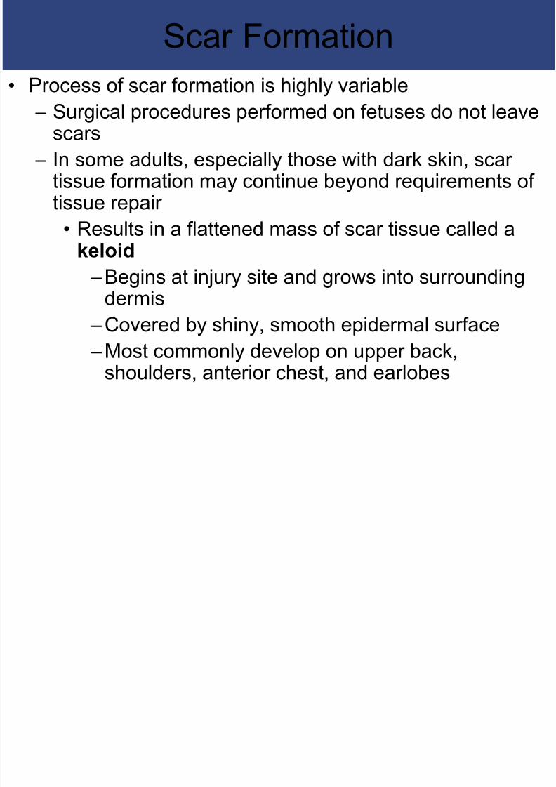
Scar Formation• Process of scar formation is highly variable
– Surgical procedures performed on fetuses do not leavescars
– In some adults, especially those with dark skin, scartissue formation may continue beyond requirements oftissue repair
• Results in a flattened mass of scar tissue called akeloid – Begins at injury site and grows into surrounding
dermis – Covered by shiny, smooth epidermal surface – Most commonly develop on upper back,
shoulders, anterior chest, and earlobes
Effects of B rns

8/20/2019 Anatomy Ch. 5
http://slidepdf.com/reader/full/anatomy-ch-5 46/62
Effects of Burns• Burns are a type of skin injury that can have many causes
– Include exposure of skin to heat, radiation, electricalshock, or strong chemical agents
• Severity of injury depends on depth of penetration and totalarea affected – Larger affected areas correlate with greater impact on
integumentary function – The most common classification of burns is based on the
depth of penetration (see below)

8/20/2019 Anatomy Ch. 5
http://slidepdf.com/reader/full/anatomy-ch-5 47/62
5-10 Effects of aging includedermal thinning, wrinkling, and
reduced melanocyte activity
Effects of Aging

8/20/2019 Anatomy Ch. 5
http://slidepdf.com/reader/full/anatomy-ch-5 48/62
Effects of Aging• Aging affects all components of the integument and causes several
major changes: – Skin injuries and infections become more common
• Due to thinning of epidermis and decline in stem cell activity – The sensitivity of the immune system is reduced
• Number of macrophages and other immune system cells
residing in the skin decreases by ~50% – Muscles become weaker, and bone strength decreases• Related to reduced calcium and phosphate absorption caused
by a 75% decline in vitamin D3 production – Sensitivity to sun exposure increases
• Less melanin is produced as melanocyte activity declines – The skin becomes dry and often scaly
• Due to declines in glandular activity, sebum production, andperspiration
Effects of Aging

8/20/2019 Anatomy Ch. 5
http://slidepdf.com/reader/full/anatomy-ch-5 49/62
Effects of Aging• Aging affects all components of the integument and causes
several major changes (continued): – Hair thins and changes color
• Follicles stop functioning or produce thinner, finer hairs• These hairs are gray or white due to decreased melanocyte
activity – Sagging and wrinkling of the skin occur
• Dermis becomes thinner and elastic fiber network decreasesin size• Effects are most pronounced in areas exposed to sun
– The ability to lose heat decreases• Blood supply to dermis is reduced• Sweat glands become less active
– Skin repairs proceed relatively slowly• Repairs can take twice as long as those of a young adult• Recurrent infection may occurs because these repairs are
so slow

8/20/2019 Anatomy Ch. 5
http://slidepdf.com/reader/full/anatomy-ch-5 50/62
5-11 The integumentarysystem provides protection for
all other body systems
Importance of the Integumentary

8/20/2019 Anatomy Ch. 5
http://slidepdf.com/reader/full/anatomy-ch-5 51/62
Importance of the IntegumentarySystem
• Protects and interacts with all organ
systems
• Changes in skin appearance are used to
diagnose disorders in other systems

8/20/2019 Anatomy Ch. 5
http://slidepdf.com/reader/full/anatomy-ch-5 52/62
The Integumentary System
in Perspective
Functional Relationships Betweenthe Integumentary System and Other Systems
Th Sk l t l S t

8/20/2019 Anatomy Ch. 5
http://slidepdf.com/reader/full/anatomy-ch-5 53/62
• The Skeletal System
provides structural support• The Integumentary System
synthesizes vitamin D 3, essentialfor calcium and phosphorusabsorption (bone maintenanceand growth)
The Skeletal System
Th M l S t

8/20/2019 Anatomy Ch. 5
http://slidepdf.com/reader/full/anatomy-ch-5 54/62
The Muscular System
• The Muscular System’s facialmuscles pull against skin of face,producing expressions importantin communication
• The Integumentary System synthesizes vitamin D 3 , essentialfor normal calcium absorption(calcium ions play an essential
role in muscle contraction)
Th N S t

8/20/2019 Anatomy Ch. 5
http://slidepdf.com/reader/full/anatomy-ch-5 55/62
The Nervous System
• The Nervous System controlsblood flow and sweat glandactivity for thermoregulation;
stimulates contraction ofarrector pili muscles to elevatehairs
• The Intergumentary System’sreceptors in dermis and deepepidermis provide sensations oftouch, pressure, vibration,temperature, and pain
The Endocrine System

8/20/2019 Anatomy Ch. 5
http://slidepdf.com/reader/full/anatomy-ch-5 56/62
The Endocrine System
• The Endocrine System includesthe sex hormones that stimulatesebaceous and apocrine glandactivity, and develop secondarysexual characteristics; suprarenalhormones alter blood flow to skinand mobilize lipids from fat cells
• The Integumentary Systemsynthesizes vitamin D 3, precursorof calcitriol, a hormone producedby the kidneys
The Cardiovascular System

8/20/2019 Anatomy Ch. 5
http://slidepdf.com/reader/full/anatomy-ch-5 57/62
The Cardiovascular System
• The Cardiovascular Systemprovides oxygen and nutrients;delivers hormones and cells ofimmune system; carries awaycarbon dioxide, waste products,and toxins; provides heat tomaintain normal skintemperature
•
The Integumentary System’smast cells produce localizedchanges in blood flow andcapillary permeability
The Lymphatic System

8/20/2019 Anatomy Ch. 5
http://slidepdf.com/reader/full/anatomy-ch-5 58/62
The Lymphatic System
• The Lymphoid System assistsin defending the integument byproviding additionalmacrophages and mobilizinglymphocytes
• The Integumentary Systemprovides physical barriers that
prevent pathogen entry;macrophages resist infection;mast cells trigger inflammationand initiate the immune reponse
The Respiratory System

8/20/2019 Anatomy Ch. 5
http://slidepdf.com/reader/full/anatomy-ch-5 59/62
The Respiratory System
• The Respiratory Systemprovides oxygen and eliminatescarbon dioxide
• The Integumentary System’shairs guard entrance to nasal
cavity
The Digestive System

8/20/2019 Anatomy Ch. 5
http://slidepdf.com/reader/full/anatomy-ch-5 60/62
The Digestive System
• The Digestive System Providesnutrients for all cells and lipidsfor storage by adipocytes
• The Integumentary Systemsynthesizes vitamin D 3, neededfor absorption of calcium and
phosphorus
The Urinary System

8/20/2019 Anatomy Ch. 5
http://slidepdf.com/reader/full/anatomy-ch-5 61/62
The Urinary System
• The Urinary System excreteswaste products, maintainsnormal body fluid pH and ioncomposition
• The Integumentary Systemassists in elimination of water
and solutes; keratinizedepidermis limits fluid lossthrough skin
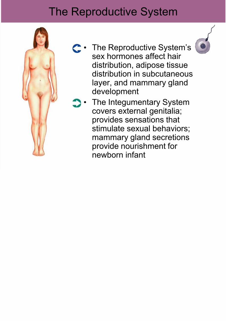
The Reproductive System
8/20/2019 Anatomy Ch. 5
http://slidepdf.com/reader/full/anatomy-ch-5 62/62
The Reproductive System
• The Reproductive System’ssex hormones affect hairdistribution, adipose tissuedistribution in subcutaneouslayer, and mammary glanddevelopment
• The Integumentary Systemcovers external genitalia;
provides sensations thatstimulate sexual behaviors;mammary gland secretionsprovide nourishment fornewborn infant