Embed Size (px)
Citation preview
RESEARCH Open Access
Assessment of cardiac remodeling inasymptomatic mitral regurgitation for surgerytiming: a comparative study of echocardiographyand magnetic resonance imagingOner Ozdogan1*†, Alper Yuksel2†, Cemil Gurgun3, Meral Kayikcioglu3, Oguz Yavuzgil3, Cahide S Cinar3
Abstract
Background: Early surgery is recommended for asymptomatic severe mitral regurgitation (MR), because ofincreased postoperative left ventricular (LV) dysfunction in patients with late surgery. On the other hand, recentreports emphasized a “watchful waiting” process for the determination of the proper time of mitral valvesurgery. In our study, we compared magnetic resonance imaging (MRI) and transthoracic echocardiography toevaluate the LV and left atrial (LA) remodeling; for better definitions of patients that may benefit from earlyvalve surgery.
Methods: Twenty-one patients with moderate to severe asymptomatic MR were evaluated by echocardiographyand MRI. LA and LV ejection fractions (EFs) were calculated by echocardiography and MRI. Pulmonary veins (PVs)were measured from vein orifices in diastole and systole from the tangential of an imaginary circle that completedLA wall. Right upper PV indices were calculated with the formula; (Right upper PV diastolic diameter- Right upperPV systolic diameter)/Right upper PV diastolic diameter.
Results: In 9 patients there were mismatches between echocardiography and MRI measurements of LV EF. LV EFswere calculated ≥60% by echocardiography, meanwhile < 60% by MRI in these 9 patients. Severity of MRevaluated by effective regurgitant orifice area (EROA) didn’t differ with preserved and depressed EFs by MRI (p >0.05). However, both right upper PV indices (0.16 ± 0.06 vs. 0.24 ± 0.08, p: 0.024) and LA EFs (0.19 ± 0.09 vs. 0.33 ±0.14, p: 0.025) were significantly decreased in patients with depressed EFs when compared to patients with normalEFs.
Conclusions: MRI might be preferred when small changes in functional parameters like LV EF, LA EF, and PV indexare of clinical importance to disease management like asymptomatic MR patients that we follow up for appropriatesurgery timing.
IntroductionIn chronic mitral regurgitation (MR), symptoms do notdevelop until decompensation of the left ventricle (LV).Patients with MR, who are asymptomatic with normalventricular performance, will need valve surgery at anannual rate of 10.3% [1] and early surgery is recom-mended for asymptomatic severe MR because of
increased postoperative LV dysfunction [2] in patientswith delayed surgery. However, recent reports haveemphasized another process called “watchful waiting”[3] for relatively low-risk patients.Mortality after delayed valve surgery is significantly
increased with severe MR (Effective Regurgitant OrificeArea (EROA) ≥0.4 cm2) [4] and depressed LV ejectionfraction (EF < 60%). Therefore, timing of the valve surgeryfor asymptomatic MR should be based on quantitativegrading of the regurgitation severity and assessment of LVsystolic dysfunction by transthoracic echocardiography [5].
* Correspondence: [email protected]† Contributed equally1Tepecik Training and Research Hospital, Cardiology Department, Izmir,TurkeyFull list of author information is available at the end of the article
Ozdogan et al. Cardiovascular Ultrasound 2010, 8:32http://www.cardiovascularultrasound.com/content/8/1/32
CARDIOVASCULAR ULTRASOUND
© 2010 Ozdogan et al; licensee BioMed Central Ltd. This is an Open Access article distributed under the terms of the CreativeCommons Attribution License (http://creativecommons.org/licenses/by/2.0), which permits unrestricted use, distribution, andreproduction in any medium, provided the original work is properly cited.

Cardiac magnetic resonance imaging (MRI) is a super-ior method in evaluating EFs, LA volumes, and pulmon-ary veins (PVs) [6,7]. Enlarged LA volumes and PVs areimportant components of clinical deterioration inpatients with MR; including atrial fibrillation and thesecould be subtle markers of early deterioration in asymp-tomatic patients with moderate to severe MR. In thepresent study, we compared cardiac MRI and transthor-acic echocardiography to evaluate the concealed remo-deling in LV, LA, and PV; for better definitions ofpatients that may benefit from early valve surgery.
MethodsStudy populationForty-six patients who had been under medical follow-up for moderate to severe MR in our outpatient clinicwere evaluated by transthoracic echocardiography.Patients were excluded if they had MR due to ischemicheart disease and if they had an LV EF < 60% by echo-cardiography. Associated valve diseases such as mitralstenosis, aortic or tricuspid valve disease and arrhythmia(including sinus tachycardia) were also accepted asexclusion criteria. After these exclusion criteria, remain-ing 38 patients were assessed with respect to New YorkHeart Association (NYHA) functional class. Asympto-matic MR was defined as having functional capacity ofclass I or I-II according to patient’s history (NYHAClass ≤I-II) and physical examination. Finally, 21patients with asymptomatic MR were included to thestudy analysis. These 21 patients underwent transthor-acic echocardiography and MRI for further cardiac eva-luation. Before MRI was performed, all patients werereevaluated for their NYHA functional Class and physi-cal examination findings like hypertension, tachycardia,crepitan ralles and edema; to minimize the hemody-namic dependent differences. Study protocol wasapproved by the local ethics committee and writteninformed consent was obtained from all participants.
Echocardiographic MeasurementsAll patients were evaluated by Sonos 7500 ultrasoundmachine (Philips) equipped with 2.5 MHz transducer.Two-dimensional and Doppler flow parameters weremeasured according to American Society of Echocardio-graphy recommendations [8]. LA volume was calculatedusing the area-length technique [9,10] from 4-chamberand 2-chamber views. LV diameters, volumes, and EFswere measured as recommended [11]. All images wereanalyzed on two occasions by two independent cardiolo-gists; inter-observer correlation (rho) for maximumLVEF was 0.95 (p < 0.001) and for LA volume was 0.94(p < 0.001).The etiology of MR was determined according to sub-
valvular apparate and mitral valve morphology. The
proximal isovelocity surface area was determined bymeasuring proximal-flow convergence [12]. Patient withan EROA >0.4 cm2 was accepted as having severe MR,and >0.2 cm2 as moderate MR [13]. Standard LV diasto-lic inflow was obtained in apical 4-chamber view by pla-cing the sample volume at the valve tips level. Peakearly inflow velocity (E) (m/s), peak atrial inflow velocity(A) (m/s), and their ratio were determined. Pulmonaryvein (PV) flow velocities were obtained by positioningthe sample volume into the right upper PV approxi-mately 1 cm above its entry into the LA. Peak systolic(S) and peak diastolic (D) PV flow velocities were mea-sured and S/D ratio was calculated. PV flow reversalduration and the velocity during atrial contraction weredetermined [14].
Cardiac Magnetic Resonance ImagingAll MRI studies were acquired with a 1.5-T MRI scan-ner (Siemens Magnetom, Shymphony, Erlangen) andevaluated in ARGUS cardiac software (Siemens). For theevaluation of cardiac functions; images were acquired instandard planes, which were positioned either parallel to(horizontal and vertical long-axis planes) or perpendicu-lar to the long axis of the heart (short-axis planes) [15].We evaluated MR with standard cardiac planes obtainedwith ECG triggering cine gradient echo (GRE) sequences(FLASH, TrueFISP). Phase images were obtained bymitral FLASH technique. LV was scanned by consecu-tive sections at four chambers, long-axis, and short-axisby cine MRI. LA volumes, LA EFs, and LV EFs weremeasured with ARGUS software from the acquiredimages. LA EFs were calculated in 20 patients by MRIwith the following formula: (LA end-diastolic volume-LA end-systolic volume)/LA end-diastolic volume. Axialand coronal planes were scanned at 6 mm sections byECG-gated TrueFISP sequence to identify anatomicstructure of PVs because of the connection and size var-iations. After determining PV connections at theseimages, all PVs [right upper PV, right lower PV, leftupper PV, left lower PV] were displayed separately bybreathold ECG-gated TrueFISP sequence with 5 mmsection thickness (Figure 1). Because of breath dispari-ties; when these sections did not pass through PVsproperly, we used gap function (shifting the sections for-wards and backwards only as thick as the interslice gap)to observe better images of PVs. When crosswise rightupper-left lower PVs or left upper-right lower PVs weredisplayed at the same plane; if we considered the imagewas adequate, we didn’t obtain additional images. Tostandardize these measurements, an imaginary circlethat completed LA wall was drawn and right upper PVswere measured from the tangential of this circle (Figure2). These measurements were repeated at the end ofboth systole and diastole. Right upper PV indices were
Ozdogan et al. Cardiovascular Ultrasound 2010, 8:32http://www.cardiovascularultrasound.com/content/8/1/32
Page 2 of 8

calculated in 19 patients with the following formula:(Right upper PV diastolic diameter- Right upper PV sys-tolic diameter)/Right upper PV diastolic diameter as asign of atrial compliance. Right and LV volumes wereassessed by ECG-triggered cine sequence and breathholdtechnique. The contours were drawn manually. Therelationship between the severity of MR and PV indexwas assessed. LA EFs were also evaluated in patientswith both depressed and normal LV EFs. Variabilitybetween the measurements of MRI was evaluated in 12randomly selected subjects twice by the same observerand by two independent observers for interobserver andintraobserver variabilities. The measurements by twoindependent observers and by the same observer at dif-ferent times did not differ in statistically significantterms (p > 0.05).
Statistical AnalysisStatistical analysis was performed with SPSS 13.0 soft-ware. All values were expressed as mean ± standarddeviation. Association between PV indices and echocar-diographic parameters were evaluated with Pearson andSpearman correlation analysis. P value less than 0.05was considered as statistically significant. Independentsample t test was performed to compare means of vari-ables of groups to determine the statistical significanceof difference. The relationship between echocardiograhicmeasurements and MRI measurements was analyzed bylinear regression and Bland Altman analysis (Figure 3).
ResultsBaseline characteristics of the patients are shown inTable 1. The etiology of MR was rheumatic valve dis-ease in 13 patients and mitral valve prolapse in 6
Figure 1 Demonstration of four pulmonary veins. 1a *Left lowerpulmonary vein. 1b *Right upper pulmonary vein. +Left upperpulmonary vein. 1c *Right lower pulmonary vein.
Figure 2 Measurement of right upper pulmonary vein index.Right upper pulmonary vein index: PV diastolic diameter-PV systolicdiameter. PV diastolic diameter.
Ozdogan et al. Cardiovascular Ultrasound 2010, 8:32http://www.cardiovascularultrasound.com/content/8/1/32
Page 3 of 8
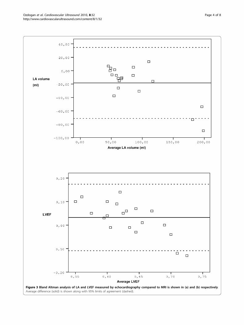
Figure 3 Bland Altman analysis of LA and LVEF measured by echocardiography compared to MRI is shown in (a) and (b) respectively.Average difference (solid) is shown along with 95% limits of agreement (dashed).
Ozdogan et al. Cardiovascular Ultrasound 2010, 8:32http://www.cardiovascularultrasound.com/content/8/1/32
Page 4 of 8

patients, whereas both etiologies were observed only in2 patients. None of the patients had accompanying valvedisease except for mild tricuspid valve regurgitation in10 patients. Qualitatively assessed MR severity was thirdgrade in twelve patients. All of the patients were in reg-ular sinus rhythm with heart rate of 60-90/min. Themean systolic and diastolic blood pressure values mea-sured before echocardiography and MRI were not statis-tically different (122.3 ± 8.9, 77.9 ± 5.1 and 127.7 ± 8.8,78.0 ± 5.0, respectively, p > 0.05).Table 2 demonstrates the echocardiographic findings
of the study population. None of the patients had systo-lic pulmonary artery pressure over 40 mmHg. Mean LAEF calculated by MRI was 0.27 ± 0.13. Figure 3 showsthe agreement between echocardiography and MRI. Thedifferences between the two methods were not statisti-cally significant for LA volume or LV EF. LA volumescalculated by cardiac MRI were larger than LA volumescalculated by echocardiography; however the differenceswere not statistically significant (p > 0.05) (Figure 4).There were positive correlations between LA volumescalculated by echocardiography and cardiac MRI (r:0.940, p < 0.0001). There was a negative relationshipbetween EROA and LA EF, but did not reach statisti-cally significant level (r:-0.412, p: 0.071).The mean LV EF values measured by echocardiogra-
phy and MRI were not statistically different (0.64 ± 0.03and 0.61 ± 0.08, respectively, p > 0.05). However, in 9patients there were mismatches between LV EFs calcu-lated by these two techniques. In these 9 patients, LVEFs were determined moderately decreased (<60%) bycardiac MRI. Besides, there were no significant differ-ences in EROAs between patients with normal and
depressed EFs determined by MRI (0.27 ± 0.13 cm2 vs.0.28 ± 0.18 cm2, p > 0.05). LA EFs were also signifi-cantly reduced in patients with depressed LV EFs byMRI (0.19 ± 0.09 vs. 0.33 ± 0.14, p: 0.025) (Table 3).There was no correlation between right upper PV
indices and the severity of MR. According to LV EF,right upper PV indices were found lower in ninepatients who were supposed to be late for mitral valvesurgery (EF < 60%), in comparison with patients withpreserved EFs (0.16 ± 0.06 vs. 0.24 ± 0.08, p: 0.024).There was no significant correlation between the veloci-ties measured with pulse wave Doppler from the rightupper PV (including systolic, diastolic velocities andthe proportion of systolic-diastolic flow velocities: S/D)and right upper PV index and the other PV indices(p > 0.05).
DiscussionDespite the increasing tendency for early surgery in themanagement of asymptomatic severe MR, watchful wait-ing is a notable option for some selected patients [3].Therefore, it is important to differentiate the subgroupsof asymptomatic patients with MR that early surgeryshould be considered. This study was designed to findout if cine MRI could give us more information to eval-uate this subgroup, i.e. to define patients in this group
Table 1 Characteristics of Patients with AsymptomaticMitral Regurgitation (n:21)*
Characteristics
Age (y) 44 ± 16*
Female gender n (%) 15 (71)
Functional capacity, n (%)
NYHA Functional Class I 13 (61)
NYHA Functional Class I-II 8 (38)
Drugs, n (%)
ACE inh. 3 (14)
Beta-Blocking agent 3 (14)
Mitral regurgitation follow-up n (%)
0-6 month 3 (14)
6 months-1 year 3 (14)
1 year< x < 3 years 7 (33)
3 year< x < 6 years 2 (10)
>6 years 6 (29)
* Plus-minus values are means ± SD
† NYHA: New York Heart Association, ACE inh.: Angiotensin ConvertingEnzyme Inhibitors
Table 2 Echocardiographic data of patients with mitralregurgitation (n:21)
Echocardiographic data Mean (min-max)
Left atrium (cm) 4.2 ± 0.7 (3.0-6.0)
Right ventricle (cm) 2.3 ± 0.3 (1.7-2.8)
Left ventricle end-diastole (cm) 4.9 ± 0.4 (4.4-5.9)
Left ventricle end-systole (cm) 3.0 ± 0.4 (2.4-3.8)
Interventricular septum (cm) 1.0 ± 0.1 (0.8-1.3)
Posterior wall (cm) 1.0 ± 0.1 (0.9-1.3)
Mitral E wave velocity (cm/s) 108 ± 28 (67.3-190.0)
Mitral A wave velocity (cm/s) 91.2 ± 30.2 (48.0-156.0)
E/A ratio 1.2 ± 0.3 (0.7-1.7)
Deceleration time (ms) 212 ± 51 (125-300)
Isovolumic relaxation time (ms) 87 ± 32 (55-150)
Left atrial area 1 (cm2) 21.6 ± 6.3 (13-33)
Left atrial area 2 (cm2) 20.7 ± 6.8 (12-36)
Left atrial volume (cm3) 77.3 ± 38.7 (36-169)
Pulmonary vein S wave velocity (cm/s) 58.5 ± 11.0 (45-84)
Pulmonary vein D wave velocity (cm/s) 52.8 ± 13.0 (27 -82)
Pulmonary vein A wave velocity (cm/s) 37.2 ± 9.5 (26.4-64.7)
Pulmonary vein A wave duration (ms) 107.5 ± 18.4 (85-150)
Mitral regurgitation velocity (cm/s) 524.1 ± 98.9 (266-662)
Left ventricle ejection fraction (%) 60 ± 3 (60-70)
SPAP (mmHg) 24.8 ± 5.5 (18-33)
A: Atrial, D: Diastolic, S: Systolic, Left atrial area 1: From four chamber view,Left atrial area 2: From two chamber view, SPAP: Systolic pulmonary arterypressure
Ozdogan et al. Cardiovascular Ultrasound 2010, 8:32http://www.cardiovascularultrasound.com/content/8/1/32
Page 5 of 8

who might have irreversible changes in LV and LA thatcouldn’t be detected by echocardiography at the time ofevaluation for surgery decision.According to the published guidelines; moderately
reduced LV EF (<60%) is an indicator of mitral valvesurgery decision in MR [5]. Echocardiography couldsometimes fail in calculating the LV EFs, because of the3-dimentional enlargement of the ventricle. Similarly; in9 patients (42%); LV EFs were moderately depressed(<60%) by MRI, while all LV EFs were preserved (≥60%)by echocardiographic evaluation. The values of LV EFs(<60%) published on guidelines [5] are based on echo-cardiographic results and not on MRI results and forthat reason maybe specific LV EF threshold values byMRI should be determined as indicators for mitral valvesurgery timing. Lower LA EFs in these 9 patients could
also indicate the ongoing remodeling including both LVand LA, despite watchful waiting. Additionally, reducedright upper PV indices might be the indicators of clini-cal deterioration including atrial fibrillation. Therefore,MRI should be preferred when small changes in func-tional parameters are of clinical importance to diseasemanagement like asymptomatic MR that we follow upfor appropriate surgery timing [7,16].The advantages of MRI over echocardiography are
emphasized by Bellenger at al. as better image qualityand quantification possibilities with high reproducibilitythat requires smaller sample sizes to prove statistical sig-nificance [17]. MRI was suggested to be an accurate andreproducible method in patients with heart failure thatyields serial assessment of LV remodeling [18]. Westen-berg et al. searched restrictive annuloplasty results by
Figure 4 Comparison of left atrial volumes calculated by echocardiography and cardiac magnetic resonance imaging. LA V: Left atrialvolume. TTE: Transthoracic echocardiography. MRI: Magnetic resonance imaging.
Table 3 Comparison of echocardiography and magnetic resonance imaging (MRI) data according to left ventricleejection fractions (LV EFs)
Patients with preserved LV EF (≥60%) in TTE anddepressed EF(<60%) in MRI
Patients with preserved LV EF (≥60%) both inTTE and MRI
p
n 9 12
Age (yrs) 50 ± 15 40 ± 16 0.17
LA volume (cm3) 86.1 ± 47.36 70.36 ± 31.42 0.67
EROA (cm2) 0.27 ± 0.13 0.28 ± 0.18 0.8
CFA (cm2) 7.38 ± 2.62 7.20 ± 4.17 0.65
LA EF 0.19 ± 0.09 0.33 ± 0.14 0.025
RUPV index 0.16 ± 0.06 0.24 ± 0.08 0.024
CFA: color flow area, EF: ejection fraction, EROA: effective regurgitant orifice area, LA: left atrium, LV: left ventricle, MRI: magnetic resonance imaging, RUPV: rightupper pulmonary vein, TTE: transthoracic echocardiography.
Ozdogan et al. Cardiovascular Ultrasound 2010, 8:32http://www.cardiovascularultrasound.com/content/8/1/32
Page 6 of 8
MRI. They demonstrated significant LA and LV reverseremodeling over time using MRI [19]. Since follow-upstudies performed by echocardiography are not optimalfor precise assessment of LA and LV volumes, MRI wasconsidered to be the gold standard method for assess-ment of LV function and volumes [20]. On the otherhand, LA volume measurements in MR images werealso shown to be reproducible in clinical practiceaccording to previous studies [21] and MRI has becomethe gold standard for LA volume determination [22-25].History of mitral valve disease duration sometimes
does not reflect the exact period, because of the longasymptomatic clinical course of chronic MR. Hence wedefined a new index; PV index that could help to defineboth mitral valve disease duration and the severity. Allfour PVs could be displayed by MRI and their indicescould be calculated as the components of LA remodel-ing. In our study, right upper PV diameters were mea-sured both in diastole and systole by MRI and rightupper PV indices for each patient were calculated. Innine patients who were supposed to be late for mitralvalve surgery according to LV EFs (LV EF < 60%), rightupper PV indices were found lower by MRI. AlthoughPV index does not give information about MR severity;it might reflect the remodeling of LA. LA remodeling isa consequence of the duration of MR and its severity,therefore it is an important determinant of hemody-namic and clinical picture in patients with MR. In apublished study, the comparison of 3 D magnetic reso-nance angiography with 2 D cine MRI for characterizinganatomy and size of PV revealed that these two methodswere similar with regard to the evaluation of PVs. Inthat study, reduced difference between the PV diameterat systole and diastole was observed in patients withatrial fibrillation [26]. Accordingly, reduced right upperPV index may alert us for a new atrial fibrillation thatoriginates from PVs and points out a relatively high-risksubgroup of asymptomatic MR. It is important forwatchful waiting process with MR. After atrial fibrilla-tion, patients become symptomatic and a new stagebegins with a rapid deterioration of the disease. As thisindex is defined for the first time, its thresholds are notclear yet, but we believe that, randomized clinical trialswith large study populations can determine the thresh-olds for PV index.
LimitationsThe most important limitation of our study is the smallsample size because of using too many exclusion criteriathat could affect our study results. Age was not consid-ered as eligibility criteria; however age could be animportant factor for PV indices and LA remodeling. PVindex is a novel parameter and a preliminary validationstudy could be helpful for this index. Our study could
be improved by following these patients for long-termmortality and morbidity, but it was not ethical toobserve patients with severe MR and reduced EF deter-mined by MRI. Therefore, mitral valve surgery wasrecommended for these 9 patients with depressed ejec-tion fractions by MRI.
ConclusionIn asymptomatic patients with MR, the most importantissue is the decision of valve surgery. Despite the newapproaches like watchful waiting process, early surgeryis recommended when valve repair is easy and feasible.Evaluating asymptomatic MR by MRI is important formore favorable calculation of LV EF. Reduced LA EFsand right upper PV indices by MRI, as signs of LAremodeling might be the predictors of worsening clinicaloutcome of asymptomatic MR; including atrial fibrilla-tion. We believe that, MRI should be preferred whensmall changes in functional parameters like LV EF, LAEF, and PV index are of clinical importance to diseasemanagement like asymptomatic MR that we follow upfor appropriate surgery timing [7,16].
ConsentAll patients provided written informed consent to parti-cipate in the research.
AcknowledgementsThis study was performed at the Cardiology Department of Ege UniversityMedical School and the Radiology Department of Kent Hospital, Izmir.This manuscript was presented as an abstract “Determining the High-RiskSubgroup of Asymptomatic Mitral Regurgitation with Cine MagneticResonance Imaging: Left Atrial Compliance and Pulmonary Vein Index”in December, 2007 in EUROECHO (Lisbon, Portugal).
Author details1Tepecik Training and Research Hospital, Cardiology Department, Izmir,Turkey. 2Kent Hospital, Radiology Department, Izmir, Turkey. 3Ege University,Cardiology Department, Izmir, Turkey.
Authors’ contributionsOO carried out the study, made echocardiographic examinations anddrafted the manuscript. AY performed magnetic resonance imaging studies.First two authors were equally contributed to the study. MK performed thestatistical analysis. CG and OY participated in the design of the study. CSCparticipated in study design and coordination. All the authors read andapproved the manuscript. None of the authors have conflict of interest inany of the material mentioned in the manuscript.
Competing interestsThe authors declare that they have no competing interests.
Received: 10 July 2010 Accepted: 13 August 2010Published: 13 August 2010
References1. Rosen SE, Borer JS, Hochreiter C, Supino P, Roman MJ, Devereux RB,
Kligfield P, Bucek J: Natural history of the asymptomatic/minimallysymptomatic patient with severe mitral regurgitation secondary tomitral valve prolapse and normal right and left ventricular performance.Am J Cardiol 1994, 74(4):374-380.
Ozdogan et al. Cardiovascular Ultrasound 2010, 8:32http://www.cardiovascularultrasound.com/content/8/1/32
Page 7 of 8

2. Enriquez-Sarano M, Tribouilloy C: Quantitation of mitral regurgitation:rationale, approach, and interpretation in clinical practice. Heart 2002,88(Suppl 4):iv1-3.
3. Rosenhek R, Rader F, Klaar U, Gabriel H, Krejc M, Kalbeck D, Schemper M,Maurer G, Baumgartner H: Outcome of watchful waiting in asymptomaticsevere mitral regurgitation. Circulation 2006, 113(18):2238-2244.
4. Enriquez-Sarano M, Avierinos JF, Messika-Zeitoun D, Detaint D, Capps M,Nkomo V, Scott C, Schaff HV, Tajik AJ: Quantitative determinants of theoutcome of asymptomatic mitral regurgitation. N Engl J Med 2005,352(9):875-883.
5. Bonow RO, Carabello BA, Kanu C, de Leon AC Jr, Faxon DP, Freed MD,Gaasch WH, Lytle BW, Nishimura RA, O’Gara PT, O’Rourke RA, Otto CM,Shah PM, Shanewise JS, Smith SC Jr, Jacobs AK, Adams CD, Anderson JL,Antman EM, Faxon DP, Fuster V, Halperin JL, Hiratzka LF, Hunt SA, Lytle BW,Nishimura R, Page RL, Riegel B: ACC/AHA 2006 guidelines for themanagement of patients with valvular heart disease: a report of theAmerican College of Cardiology/American Heart Association Task Forceon Practice Guidelines (writing committee to revise the 1998 Guidelinesfor the Management of Patients With Valvular Heart Disease): developedin collaboration with the Society of Cardiovascular Anesthesiologists:endorsed by the Society for Cardiovascular Angiography andInterventions and the Society of Thoracic Surgeons. Circulation 2006,114:84-231, Review.
6. Epstein FH: MRI of left ventricular function. J Nucl Cardiol 2007,14:729-744.
7. Ichikawa Y, Sakuma H, Kitagawa K, Ishida N, Takeda K, Uemura S,Motoyasu M, Nakano T, Nozaki A: Evaluation of left ventricular volumesand ejection fraction using fast steady state cine MR imaging:comparison with left ventricular angiography. J Cardiovasc Magn Reson2003, 5:333-342.
8. Gardin JM, Adams DB, Douglas PS, Feigenbaum H, Forst DH, Fraser AG,Grayburn PA, Katz AS, Keller AM, Kerber RE, Khandheria BK, Klein AL,Lang RM, Pierard LA, Quinones MA, Schnittger I: American Society ofEchocardiography. Recommendations for a standardized report for adulttransthoracic echocardiography: a report from the American Society ofEchocardiography’s Nomenclature and Standards Committee and TaskForce for a Standardized Echocardiography Report. J Am SocEchocardiogr 2002, 15(3):275-290.
9. Feigenbaum H, Armstrong WF, Ryan T: Feigenbaum’s echocardiography.Lippincott Williams & Wilkins, 6 2005, 182-184.
10. Wang Y, Gutman JM, Heilbron D, Wahr D, Schiller NB: Atrial volume innormal adultpopulation by two dimensional echocardiography. Chest1984, 86:595-601.
11. Schiller NB, Shah PM, Crawford M, DeMaria A, Devereux R, Feigenbaum H,Gutgesell H, Reichek N, Sahn D, Schnittger I: Recommendations forquantitation of the left ventricle by two-dimensional echocardiography.J Am Soc Echocardiogr 1989, 2(5):358-367.
12. Enriquez-Sarano M, Miller FA Jr, Hayes SN, Bailey KR, Tajik AJ, Seward JB:Effective mitral regurgitant orifice area: clinical use and pitfalls of theproximal isovelocity surface area method. J Am Coll Cardiol 1995,25(3):703-709.
13. Zoghbi WA, Enriquez-Sarano M, Foster E, Grayburn PA, Kraft CD, Levine RA,Nihoyannopoulos P, Otto CM, Quinones MA, Rakowski H, Stewart WJ,Waggoner A, Weissman NJ: American Society of Echocardiography:Recommendations for evaluation of the severity of native valvularregurgitation with two-dimensional and Doppler echocardiography.J Am Soc Echocardiogr 2003, 16(7):777-802.
14. Rossvoll O, Hatle LK: Pulmonary venous flow velocities recorded bytransthoracic Doppler ultrasound: relation to left ventrular diastolicpressures. J Am Coll Cardiol 1993, 21:1687-1696.
15. Ratib O, Didier D, Chatelain P, Righetti A, Lerch R, Townsend D: Standardviews incardiac multimodality tomographic imaging. Am J Card Imaging1995, 9:67-76.
16. Gardner BI, Bingham SE, Allen MR, Blatter DD, Anderson JL: Cardiacmagnetic resonance versus transthoracic echocardiography for theassessment of cardiac volumes and regional function after myocardialinfarction: an intrasubject comparison using simultaneous intrasubjectrecordings. Cardiovasc Ultrasound 2009, 7:38.
17. Bellenger NG, Davies LC, Francis JM, Coats AJ, Pennell DJ: Reduction insample size for studies of remodeling in heart failure by the use of
cardiovascular magnetic resonance. J Cardiovasc Magn Reson 2000,2:271-278.
18. Rajappan K, Bellenger NG, Anderson L, Pennell DJ: The role ofcardiovascular magnetic resonance in heart failure. Eur J Heart Fail 2000,2:241-252.
19. Westenberg JJ, van der Geest RJ, Lamb HJ, Versteegh MI, Braun J,Doornbos J, de Roos A, van der Wall EE, Dion RA, Reiber JH, Bax JJ: MRI toevaluate left atrial and ventricular reverse remodeling after restrictivemitral annuloplasty in dilated cardiomyopathy. Circulation 2005, 112(9Suppl):I437-1442.
20. Van der Geest RJ, Reiber JHC: Quantification in cardiac MRI. J Magn ResonImaging 1999, 10:602-608.
21. Sievers B, Kirchberg S, Addo M, Bakan A, Brandts B, Trappe H: Assessmentof left atrial volumes in sinus rhythm and atrial fibrillation using thebiplane area-length method and cardiovascular magnetic resonanceimaging with TrueFISP. J Cardiovasc Magn Reson 2004, 6:855-863.
22. Bowman AW, Kova’cs SJ: Assessment and consequences of the constant-volume attribute of the four-chambered heart. Am J Physiol Heart CircPhysiol 2003, 285:H2027-H2033.
23. Bowman AW, Kova’cs SJ: Left atrial conduit volume is generated bydeviation from the constant-volume state of the left heart: a combinedMRI-echocardiographic study. Am J Physiol Heart Circ Physiol 2004, 286:H2416-H2424.
24. Jarvinen VM, Kupari MM, Poutanen VP, Hekali PE: A simplified method forthe determination of left atrial size and function using cine magneticresonance imaging. Magn Reson Imaging 1996, 14:215-226.
25. Rodevand O, Bjornerheim R, Ljosland M, Maehle J, Smith HJ, Ihlen H: Leftatrial volumes assessed by three- and two-dimensionalechocardiography compared to MRI estimates. Int J Card Imaging 1999,15:397-410.
26. Syed MA, Peters DC, Rashid H, Arai AE: Pulmonary vein imaging:comparison of 3 D magnetic resonance angiography with 2 D cine MRIfor characterizing anatomy and size. J Cardiovasc Magn Reson 2005,7:355-360.
doi:10.1186/1476-7120-8-32Cite this article as: Ozdogan et al.: Assessment of cardiac remodeling inasymptomatic mitral regurgitation for surgery timing: a comparativestudy of echocardiography and magnetic resonance imaging.Cardiovascular Ultrasound 2010 8:32.
Submit your next manuscript to BioMed Centraland take full advantage of:
• Convenient online submission
• Thorough peer review
• No space constraints or color figure charges
• Immediate publication on acceptance
• Inclusion in PubMed, CAS, Scopus and Google Scholar
• Research which is freely available for redistribution
Submit your manuscript at www.biomedcentral.com/submit
Ozdogan et al. Cardiovascular Ultrasound 2010, 8:32http://www.cardiovascularultrasound.com/content/8/1/32
Page 8 of 8