Embed Size (px)
Citation preview

B Cells Are Critical toT-cell–Mediated AntitumorImmunity Induced by aCombined Immune-Stimulatory/Conditionally CytotoxicTherapy for Glioblastoma1,2
Marianela Candolfi*,†,‡,§,3, James F. Curtin*,†,‡,§,Kader Yagiz*,†,‡,§, Hikmat Assi*,†,‡,§,Mia K. Wibowo*,†,‡,§, Gabrielle E. Alzadeh*,†,‡,§,David Foulad*,†,‡,§, AKM G. Muhammad*,†,‡,§,Sofia Salehi†,‡,¶, Naomi Keech†,‡,¶,Mariana Puntel*,†,‡,§, Chunyan Liu*,†,‡,§,Nicholas R. Sanderson*,†,‡,§, Kurt M. Kroeger*,†,‡,§,Robert Dunn#, Gislaine Martins†,‡,¶,Pedro R. Lowenstein*,†,‡,§,**,4
and Maria G. Castro*,†,‡,§,**,4
*Gene Therapeutics Research Institute, Cedars-SinaiMedical Center, Los Angeles, CA, USA; †Department ofBiomedical Sciences, Cedars-Sinai Medical Center, LosAngeles, CA, USA; ‡Department of Medicine, David GeffenSchool of Medicine, UCLA, Los Angeles, CA, USA;§Department of Molecular and Medical Pharmacology,David Geffen School of Medicine, UCLA, Los Angeles, CA,USA; ¶Inflammatory Bowel and Immunobiology ResearchInstitute, Cedars-Sinai Medical Center, Los Angeles, CA,USA; #Biogen Idec, Immunology/Allergy, San Diego, CA,USA; **The Brain Research Institute, and JonssonComprehensive Cancer Center, David Geffen School ofMedicine, UCLA, Los Angeles, CA, USA
AbstractWe have demonstrated that modifying the tumor microenvironment through intratumoral administration of adeno-viral vectors (Ad) encoding the conditional cytotoxic molecule, i.e., HSV1-TK and the immune-stimulatory cytokine,i.e., fms-like tyrosine kinase 3 ligand (Flt3L) leads to T-cell–dependent tumor regression in rodent models of glio-blastoma. We investigated the role of B cells during immune-mediated glioblastomamultiforme regression. Althoughtreatment with Ad-TK+Ad-Flt3L induced tumor regression in 60% of wild-type (WT) mice, it completely failed inB-cell–deficient Igh6−/− mice. Tumor-specific T-cell precursors were detected in Ad-TK+Ad-Flt3L–treated WT micebut not in Igh6−/− mice. The treatment also failed in WT mice depleted of total B cells or marginal zone B cells.Because we could not detect circulating antibodies against tumor cells and the treatment was equally efficient in
Abbreviations: GBM, glioblastoma multiforme; TK, thymidine kinase; Flt3L, fms-like tyrosine kinase 3; WT, wild-type; APC, antigen-presenting cell; Ad, adenoviral vector;MZB, marginal zone B cell; LN, lymph node; MLR, mixed leukocyte reaction; GFP, green fluorescent protein; DC, dendritic cellAddress all correspondence to: Dr. Maria G. Castro, Department of Neurosurgery, Department of Cell and Developmental Biology, University of Michigan School of Medicine, 4570MSRB II, 1150 W Medical Center Dr, Ann Arbor, MI 48109-0650. E-mail: [email protected] work was supported by National Institutes of Health/National Institute of Neurological Disorders & Stroke (NIH/NINDS) grants 1R21-NS054143, 1UO1 NS052465,and 1RO1-NS057711 to M.G.C.; NIH/NINDS grants 1RO1-NS 054193 and 1RO1-NS061107 to P.R.L.; and NIH grant 1R21-AI83948-01 IBIRI-CSMC to G.M. Thiswork was also supported by the Bram and Elaine Goldsmith and the Medallions Group Endowed Chairs in Gene Therapeutics to P.R.L. and M.G.C., respectively, and by theDrown Foundation, the Linda Tallen & David Paul Kane Foundation Annual Fellowship, and the Board of Governors at Cedars-Sinai Medical Center. M.C. was supported byan NIH/NINDS 1F32 NS058156 fellowship.2This article refers to supplementary materials, which are designated by Table W1 and Figures W1 to W4 and are available online at www.neoplasia.com.3Present address: Instituto de Investigaciones Biomédicas (INBIOMED), Facultad de Medicina, Universidad de Buenos Aires, Buenos Aires, Argentina.4Present address: Department of Neurosurgery, Department of Cell and Developmental Biology, University of Michigan School of Medicine, Ann Arbor, MI 48109.Received 22 July 2011; Revised 19 August 2011; Accepted 23 August 2011
Copyright © 2011 Neoplasia Press, Inc. All rights reserved 1522-8002/11/$25.00DOI 10.1593/neo.11024
www.neoplasia.com
Volume 13 Number 10 October 2011 pp. 947–960 947
WT mice and in mice with B-cell–specific deletion of Prdm 1 (encoding Blimp-1), in which B cells are present butunable to fully differentiate into antibody-secreting plasma cells, tumor regression in this model is not dependenton B cells’ production of tumor antigen–specific immunoglobulins. Instead, B cells seem to play a role as antigen-presenting cells (APCs). Treatment with Ad-TK+Ad-Flt3L led to an increase in the number of B cells in the cervicallymph nodes, which stimulated the proliferation of syngeneic T cells and induced clonal expansion of antitumorT cells. Our data show that B cells act as APCs, playing a critical role in clonal expansion of tumor antigen–specificT cells and brain tumor regression.
Neoplasia (2011) 13, 947–960
IntroductionGlioblastoma multiforme (GBM) is a malignant brain cancer, account-ing for approximately 50% of newly diagnosed primary brain tumorsin the United States. GBM has a dismal prognosis owing to the localinfiltrative tumor growth that makes complete surgical resection virtu-ally impossible, the intrinsic radiotherapy and chemotherapy resistanceof glioma cells, and their high rate of mutation. Novel therapeuticstrategies such as vaccination/immunotherapies have been developedto target GBM cells disseminated throughout the brain [1]. We devel-oped an anti-GBM immunotherapeutic approach based on engineeringthe tumor microenvironment, which uses a combined conditionalcytotoxic/immune–stimulatory gene therapeutic modality. It consistsof an adenoviral vector (Ad) encoding herpes simplex virus type I–thymidine kinase (Ad-TK), which, in the presence of ganciclovir, killsproliferating cells, and a second Ad encoding fms-like tyrosine kinase 3ligand (Ad-Flt3L), which recruits antigen-presenting cells (APCs) tothe brain tumor microenvironment [2]. We have shown that thiscombination therapy induces an antitumor immune response andimmunologic memory [2–4]. While the role of cytotoxic T cells inthe clearance of peripheral and brain tumors has been well documented,the role of B cells in antitumor immunity has remained debatable. Thisis due to several reasons: 1) increased numbers of tumor-infiltrating Blymphocytes can correlate with poor prognosis of patients harboringmetastatic carcinomas [5] or with improved survival of breast carci-noma patients [6,7]; 2) resting B cells have been implicated in pro-moting carcinogenesis by exacerbating inflammation [8]; 3) althoughB cells are relatively abundant, the frequency of B cells bearing a BcRspecific for a particular antigen is very low (between 10−4 and 10−5),potentially limiting the effectiveness of B cells as APCs during initialpriming of immune responses [9]; and 4) studies in B-cell knockoutmice revealed that B cells actually suppress the development of immuneresponses in vivo against lymphoma, colon cancer, and melanoma(but not sarcomas) [10,11] and depletion of B lymphocytes enhancesmelanoma vaccination efficacy [12], whereas in separate studies,B lymphocytes were implicated in promoting fibrosarcoma tumorregression [13].
Bone marrow–derived B cells develop into either follicular B cells ormarginal zone B cells (MZB) in the spleen. Follicular B cells (B220+/CD23high/CD21low), which account for most peripheral mature B cells,are found in the circulation, the germinal center of peripheral lymphnodes (LNs), and the white pulp of the spleen. They participate inT-cell–dependent immune responses and immunologic memory[14]. MZB cells (B220+/CD23low/CD21high) are derived from circu-lating progenitors, but when they arrive to the spleen, they locate in
the marginal zone and do not recirculate; they have been shown tocapture blood-borne antigens and deliver them to dendritic cells(DCs) of the follicular areas [15]. Also, activated MZB cells can migrateto the T-B border and directly induce the expansion of antigen-specificT cells [16].
Prompted by the central role of B cells in autoimmune diseases[17–19] and by the successful induction of in vitro T-cell responsesusing tumor antigen–pulsed, CD40-activated B cells [20,21], weinvestigated the role of B cells in brain tumor regression induced byintratumoral treatment with Ad-TK+Ad-Flt3L. Using KO mice thatlack B cells and specific antibodies that deplete total B cells or MZBcells, we found that, in the absence of B cells, Ad-TK+Ad-Flt3L failsto induce the regression of intracranial GBM. Tumor antigen–specificT-cell clonal expansion was also abolished in B-cell–deficient mice(Igh6−/−), indicating that functional, mature B cells were requiredfor mounting a systemic immune response against brain tumor anti-gens. The role of B cells in this antitumor immune response does not,however, seem to be mediated by the production of antitumor-specificantibodies because we could not detect evidence of humoral antitumorimmunity and the treatment was still efficacious in mice deficient inplasma cells formation, Prdmflox/floxCD19Cre/+ mice. Although themost evident function of B cells in adaptive immune responses is theclonal differentiation of antigen-specific B cells into plasma cells andthe subsequent secretion of antigen-specific immunoglobulin (Ig),B cells can also function as efficient APCs [9,17,20–22]. Ad-Flt3L/Ad-TK treatment induced an increase in the levels of B cells in the cer-vical LNs of WT mice. These B cells contained brain tumor remnants,increased expression of coactivation markers, and induced the clonalexpansion of syngeneic tumor antigen–specific T lymphocytes. Takentogether, our results imply that B cells may act as APCs to enhanceclonal expansion of tumor antigen–specific T lymphocytes andT cell–dependent tumor regression within the central nervous system.
Materials and Methods
AdsFirst-generation, E1/E3–deleted replication-deficient recombinant
adenovirus serotype 5 was used in this study. We used Ad-Flt3L [3]and Ad-TK [3]; both transgenes are under the control of humanCMV promoter. An Ad without a transgene was used as a control(Ad-0). All viral preparations were confirmed to be replication com-petent adenovirus and lipopolysaccharide (LPS) free. Viral titers weredetermined by an end-point dilution cytotoxic-effect assay. Themethodsfor Ad generation, purification, characterization, and scale-up have been
948 B Cell Dependent Brain Tumor Regression Candolfi et al. Neoplasia Vol. 13, No. 10, 2011
previously described by our laboratory [3]. Ads were administeredwithin the intracranial tumors as described below using the followingdoses: Ad-TK, 108 infectious units (iu); Ad-Flt3L, 2 × 10⁸ iu; andAd.0, 3 × 10⁸ iu (to deliver equivalent total iu).
Mouse Glioma ModelsFemale C57BL/6 wild-type mice, green fluorescent protein (GFP+/+)
mice, and Igh6−/− on C57BL/6 background were purchased fromJackson Laboratories (Bar Harbor, ME). Inbred VM/Dk mice weredonated by the McLaughlin Research Institute (Great Falls, MT)and bred in the CSMC vivarium. Prdm1flox/flox CD19Cre/+ mice thatlack Blimp-1 in B cells were generated as previously described [23] bycrossing mice expressing Cre-recombinase under the control of theCD19 promoter (CD19Cre/+) [23] with Prdm1flox/flox mice, which havethe prdm-1 locus encoding Blimp-1 flanked by loxP sites [23]. Ex-perimental mice used in this study were of Prdm1flox/floxCD19Cre/+
genotype. Both the CD19Cre/+ and the Prdm1flox/flox mice were back-crossed to C57BL/6J 10 times before mating to generate Prdm1flox/flox
CD19Cre/+. Intracranial GBM models were generated as previouslydescribed [2,24]. Mice were stereotactically injected with GBM cells(either 20,000 GL26 cells, 20,000 GL26-OVA cells [24] [stably ex-pressing cytoplasmic chicken ovalbumin], or 5000 SMA-560 cells[25], a gift from Darell Bigner [Duke University]), in a volume of1 μl into the right striatum (+0.5 mm anterior-posterior, +2.1 mmmedial-lateral, and −3.2 mm dorsoventral from bregma). Fourteen dayslater, vectors were administered intratumorally using the same burr hole(at −2.9, −3.2, and −3.5 mm DV from bregma: 0.5 μl per site). Ganci-clovir (GCV, 25 mg/kg per day) was administered intraperitoneally (i.p.)for 7 days after vector administration to all animals receiving Ad-TK. Toassess whether B cells are involved in the memory response elicited by thecombined conditional cytotoxic/immune gene therapy, mice were rechal-lenged by injection of 20,000 tumor cells in the contralateral brain hemi-sphere [2]. Mice were killed at specific time points or when their healthstatus reached criteria established by the guidelines of the InstitutionalAnimal Care and Use Committee at Cedars-Sinai Medical Center (ap-proved protocol 2259). Mice were killed under deep anesthesia by termi-nal perfusion with oxygenated, heparinized Tyrode solution. Blood,cervical LNs, spleen, and tumors were collected immediately after perfu-sion and processed for flow cytometric analysis or ELISA. When brainswere analyzed by immunofluorescence, mice were also perfused with 4%paraformaldehyde (PFA) in phosphate-buffered saline (PBS), and brainswere postfixed in 4% PFA for three additional days.
Generation of Bone Marrow Chimeric MiceBone marrow chimeric mice were generated using GFP+/+ and
wild-type C57BL/6 mice as described by us previously [2]. Briefly,irradiated recipient mice (7 Gy) were adoptively transferred with 107
bone marrow cells from donor mice through tail vein injection. Bonemarrow in the recipients was allowed to reconstitute for 10 weeksbefore intracranial implantation of GL26 tumor cells. Tumor-infiltratingimmune cells were purified using Percoll gradient centrifugation asdescribed below. The origin of tumor-infiltrating B cells (CD19+/CD45+) was determined by analyzing their relative expression of GFPby flow cytometry.
B-Cell Depletion Experiments
B-cell depletion. The IgG2a 18B12 monoclonal antimouse CD20antibody was generated as previously described [26]. The IgG2a
mouse antihuman 2B8 CD20 monoclonal antibody was used as iso-type control [26]. The duration of B-cell depletion was assessed innaive mice injected i.p. with 300 μl of 1 mg/ml antimouse CD20or isotype control. B-cell content and integrity of T-cell populationswere determined by flow cytometry in spleen 7 or 12 days later. Theintegrity of T-cell populations in B-cell–depleted mice was deter-mined 7 days after depletion using the following antibodies: hamsterantimouse CD3ɛ, rat antimouse CD8α, and rat antimouse CD4(Table W1). GL26 tumor-bearing mice were injected i.p. with anti-mouse CD20 on the day of the treatment with Ad vectors and again2 weeks later to maintain depletion. Animals undergoing rechallengeexperiments received i.p. injections of depleting antibodies on the dayof the rechallenge (day 90) and 2 weeks later (day 105).
MZ B-cell depletion. MZB cells were depleted with 100 μg of anti–LFA-1 and 100 μg of anti-CD49d [27]. To assess the duration of MZBcell depletion, naive mice were injected with these antibodies or isotypecontrols (TableW1). The content of MZB cells was determined by flowcytometry in the spleen 3, 7, and 10 days later. The integrity of T-cellpopulations in MZB cell–depleted mice was determined 7 days afterdepletion using the antibodies indicated above. GL26 tumor-bearingmice were injected i.p. with anti–LFA-1 and CD49d or isotype controlson the day of the treatment with Ad vectors and 10 days later.
Isolation and Characterization of Immune Cells inBrain Tumor, Spleen, and Cervical LNs
Brains were carefully separated from the meninges and removed fromthe skull. The tumor was dissected, taking care to avoid the ventricles.Tumors were homogenized in RPMI medium using a glass Tenbroeckhomogenizer (Kontas Glass Company). To purify mononuclear cellsfrom the tumor, homogenized tumor tissue was centrifuged through aPercoll (GEHealthcare, Piscataway, NJ) step gradient (70%-30% Percollin PBS) for 20 minutes at 2200 RPM. Immune cells were extractedfrom draining cervical LNs by homogenization using a Tenbroeckhomogenizer. Splenocytes were collected by homogenization of wholespleen followed by incubation in red blood cell lysis buffer ACK(0.15 mM NH4Cl, 10 mM KHCO3, and 0.1 mM sodium EDTA atpH 7.2) for 3 minutes on ice. Immune cells were counted and labeledwith the rat IgGs (BD Pharmingen, San Diego, CA) depicted inTable W1 and dissolved in 1% FBS–0.1 M PBS. Samples were analyzedusing a FACScan Flow cytometer (Beckton Dickinson, Franklin Lakes,NJ) and Summit software (Cytomation, Inc, Fort Collins, CO).
ImmunofluorescenceCoronal sections of PFA-fixed brains (50 μm) were obtained using a
Leica vibratome (Leica Microsystems Heidelberg, Mannheim, Germany).Sections were incubated in 10mM citrate (60°C) for antigen retrieval, andimmunofluorescence was performed using the antibodies described inTable W1. Nuclei were stained with 4′, 6-diamidino-2-phenylindole(DAPI, 5 μg/ml; Invitrogen, Carlsbad, CA), and tissues were mountedwith ProLong Antifade (Invitrogen). Confocal micrographs were obtainedusing a Leica confocal microscope TCS SP2 with AOBS equipped with405-nm violet-diodeUV laser, 488-nm argon laser, and 594-helium-neonlasers and using a HCX PL APO 63× 1.4 numerical aperture oil objective(Leica Microsystems Heidelberg, Mannheim, Germany) [28].
Detection of Total IgGHomogenized splenocytes were collected followed by incubation
in red blood cell lysis buffer ACK and then incubated with RPMI
Neoplasia Vol. 13, No. 10, 2011 B Cell Dependent Brain Tumor Regression Candolfi et al. 949

alone or in the presence of LPS (10 μg/ml) for 72 hours. After acti-vation, total IgG levels were determined in 20 μl of cell culturesupernatants using the Easy-Titer Mouse IgG Kit (Pierce, Rockford,IL) following the manufacturer’s instructions.
Detection of Circulating Antitumor AntibodiesDetection of circulating antitumor antibodies was performed in
mouse serum collected 7 days after Ad-TK+Ad-Flt3L treatment aspreviously described [4]. Briefly, cultured GL26, GL26-OVA, andSMA-560 cells were stained with rat antimouse IgM-fluoresceinisothiocyanate (FITC, 1:50, 553437; BD Pharmingen) or antimouseIgG-FITC (1:1000, A-11017; Invitrogen). Samples were analyzedusing a FACScan flow cytometer (Beckton Dickinson).
Flt3L ELISADetection of Ad-derived human Flt3L and endogenous mouse
Flt3L was performed in serum 7 days after vector injection. Serumwas diluted 1:10 inDulbecco PBS. The expression of human andmouseFlt3L was determined using species-specific ELISA kits (DFK00 andMFK00; R&D Systems).
Trafficking of CellTracker Green–Labeled Tumor AntigenCells were labeled with CellTracker Green as described previously
[2]. Briefly, 107 GL26 cells were labeled with CellTracker Green(4 μM). A total of 250,000 cells in 1.5 μl were injected in the brainstriatum of C57BL/6 mice. Mice were treated 2 days later by intra-tumoral injection of saline or Ad-Flt3L and Ad-TK, and tumor-infiltrating immune cells and cervical-draining LNs were harvested2 and 7 days later and processed for flow cytometry.
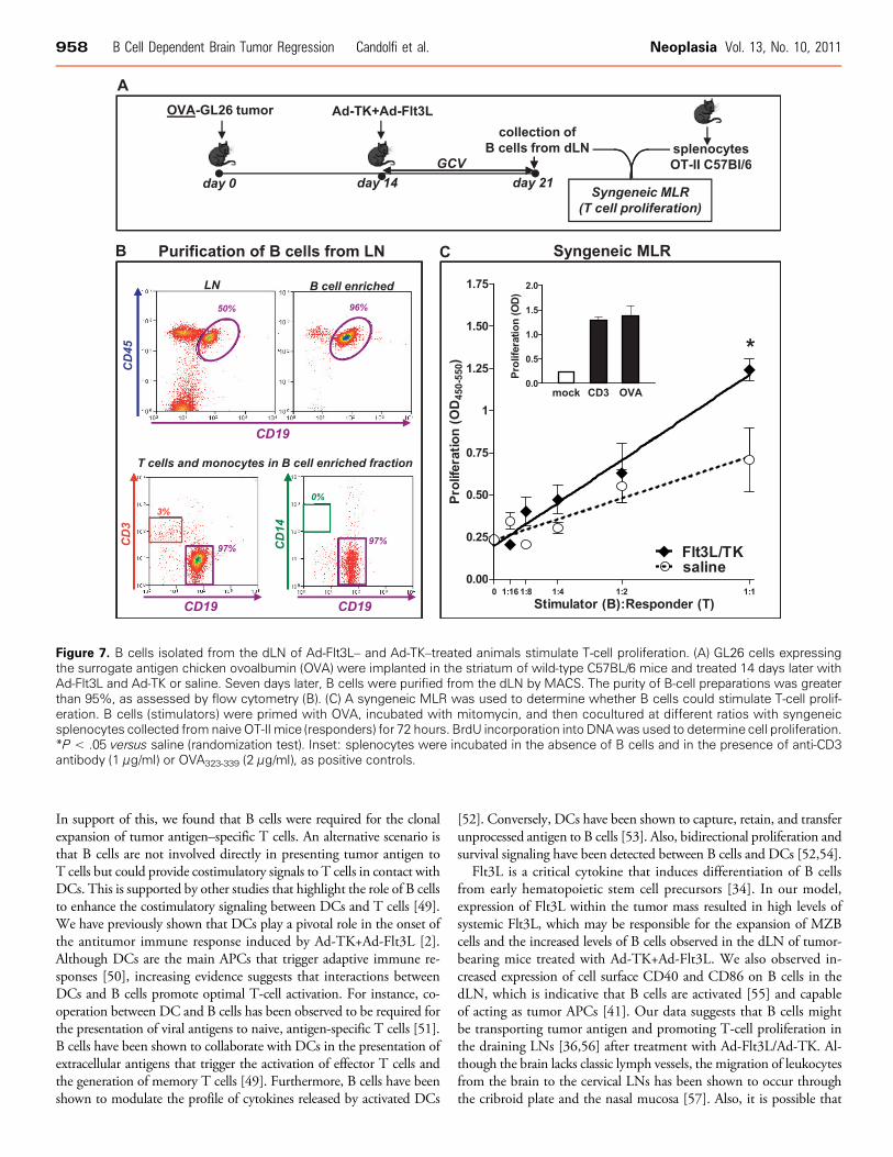
Syngeneic Mixed Leukocyte ReactionB cells were purified from the cervical LNs of GL26-OVA tumor-
bearing mice 7 days after the treatment with Ad-TK+Ad-Flt3L or salineusing a pan B-cell isolation kit (130-095-813; Miltenyi Biotec, BergischGladbach, Germany) in anOctoMACS Separator (Miltenyi Biotec). Thepurity of B-cell preparations was more than 95%, as determined by flowcytometry using antibodies against mouse CD45, mouse CD19, mouseCD14, andmouse CD3ɛ (TableW1). Enriched B-cell preparations wereincubated with 2 μg/ml OVA323-339, a known H-2b–restricted OVAclass II epitope [29] for 3 hours at 37°C, followed by incubation with50 μg/ml mitomycin C for 30 minutes at 37°C to inhibit the proliferationof B cells in the syngeneic mixed leukocyte reaction (MLR). Splenocyteswere collected from naive OT-II mice, donated by Dr Jonathan Kaye(Gene Therapeutics Research Institute; Cedars-Sinai Medical Center).The syngeneicMLRwas performed by incubating 200,000OT-II spleno-cytes in the presence of increasing ratios of B cells (1:16, 1:8, 1:4, 1:2, and1:1) for 90 hours. Cell proliferation was determined by the incorporation ofBrdU into nascent DNA strands and assessed by ELISA (BrdU Cell Pro-liferation Assay, catalog no. 11647229001; Roche, Mannheim, Germany).
Interferon γ ELISPOTC57BL/6 mice and Igh6−/− mice were implanted with GL26 tumor
cells and treated 14 days later with Ads expressing Ad-Flt3L and Ad-TKor saline. Mice were killed 7 days later, and splenocytes were collectedas described previously. A total of 1 × 10⁵ splenocytes were plated intriplicate onto a capture anti–interferon γ (IFNγ) antibody–coated(1:60; R&D Systems, Minneapolis, MN) 96-well ELISPOT plate(polyvinylidene fluoride; Millipore, Billerica, MA), and IFNγ secretionwas quantified according to the manufacturer’s protocol. Briefly, 1 × 10⁵
splenocytes were stimulated for 24 hours at 37°C in RPMI 1640 me-dium with or without 1 μg/ml Trp2180-188 peptide (Sigma, Milwaukee,WI). ConA (1 μg/ml, 4 hours) stimulation of splenocytes served as apositive control. Plates were vigorously washed with PBS before incuba-tion with IFNγ detection antibody (1:60; R&D Systems). Twenty-fourhours later, plates were developed after incubation with streptavidin–horseradish peroxidase (R&D Systems) and visualized by amino ethylcarbazole (Sigma-Aldrich). Spots were detected and counted using theKS ELISPOT (version 4.7; Zeiss, Göttingen, Germany).
Statistical AnalysisSample sizes were calculated to detect differences between groups
with a power of 80% at a 0.05 significance level using PASS 2008(Power and sample size software; NCSS, Kaysville, UT). Kaplan-Meiersurvival curves were analyzed using the Mantel log-rank (GraphPadPrism version 3.00; GraphPad Software, San Diego, CA). One- ortwo-way analysis of variance (ANOVA) followed by the Tukey testor unpaired two-tailed Student’s t test (NCSS) were used to analyzeELISPOT, ELISA, and flow cytometry data, as indicated in the figurelegends. When data failed normality test or Levene equal-variancetest, they were square root transformed. Curve inequality in MLRexperiments was assessed using the randomization test (NCSS). P <.05 was considered the cutoff for significance. All experiments wereperformed at least twice to confirm the findings.
Results
Bone Marrow–Derived B Cells Infiltrate IntracranialBrain Tumors and Are Required for AntitumorImmunity and Tumor Regression In Vivo
Our previous results show that intratumoral administration ofAd-Flt3L/Ad-TK leads to a T-cell–dependent antitumor immuneresponse that induces tumor regression in rodent models of glioblas-toma [3,4]. Considering that the role of B cells in anti-brain tumorimmunity remains elusive, we aimed to investigate whether B cells playa role in the generation of adaptive immune responses against braintumor antigens (Figure 1A).
Using immunofluorescence techniques followed by confocal micro-scopy, we detected CD19+ cells within the brain tumor mass inC57BL/6 bearing syngeneic GL26 tumors (Figure 1B). These findingsare in agreement with the fact that B cells have been found to infiltrateGBM in humans [30]. To investigate whether the CD19+ cells thatinfiltrate the tumor originate from the bone marrow or from the cen-tral nervous system, we generated bone marrow chimeric mice con-sisting of bone marrow from wild-type C57BL/6 mice adaptivelytransferred into irradiated GFP+/+ mice. We found that only 17.5%of tumor-infiltrating B cells in chimeric mice were GFP+, which wasonly slightly higher than the background observed in WT mice (8%GFP+ B cells). Therefore, our data suggest that most of the CD19+
infiltrating immune cells in Ad-TK+Ad-Flt3L–treated brain tumorsare bone marrow derived (Figure 1C).
We next evaluated whether B cells are required to induce the anti-tumor immune response triggered by the treatment with Ad-TK+Ad-Flt3L. We implanted GL26 tumor in the brain of Igh6−/− mice, whichare deficient in mature B cells, and treated them 14 days later withAd-TK+Ad-Flt3L, or saline as a control (Figure 1, D and E). Whereasin wild-type mice, treatment with Ad-TK+Ad-Flt3L induced tumorregression and long-term survival in 60% of the animals, the treatmentfailed in Igh6−/− mice (Figure 1D). Tallying with these findings, we
950 B Cell Dependent Brain Tumor Regression Candolfi et al. Neoplasia Vol. 13, No. 10, 2011
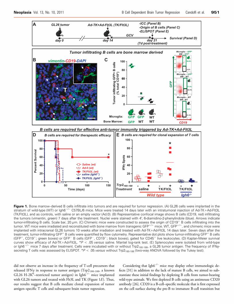
did not observe an increase in the frequency of T-cell precursors thatreleased IFNγ in response to tumor antigen (Trp2180-188, a knownGL26 H-2Kb–restricted tumor antigen) in Igh6−/− mice implantedwith GL26 tumors and treated with Flt3L and TK (Figure 1E). Thus,our results suggest that B cells mediate clonal expansion of tumorantigen–specific T cells and subsequent brain tumor regression.
Considering that Igh6−/− mice may display other immunologic de-fects [31] in addition to the lack of mature B cells, we aimed to sub-stantiate these initial findings by depleting B cells from tumor-bearingwild-type animals. We first depleted B cells using an antimouse CD20antibody [26]. CD20 is a B-cell–specific molecule that is first expressedon the cell surface during the pre-B to immature B-cell transition but
Figure 1. Bone marrow–derived B cells infiltrate into tumors and are required for tumor regression. (A) GL26 cells were implanted in thestriatum of wild-type (WT) or Igh6−/− C57BL/6 mice. Mice were treated 14 days later with an intratumoral injection of Ad-TK+Ad-Flt3L(TK/Flt3L), and as controls, with saline or an empty vector (Ad.0). (B) Representative confocal image shows B cells (CD19, red) infiltratingthe tumors (vimentin, green) 7 days after the treatment. Nuclei were stained with 4′, 6-diamidino-2-phenylindole (blue). Arrows indicatetumor-infiltrating B cells. Scale bar, 20 μm. (C) Chimeric mice were constructed to assess the origin of CD19+ B cells infiltrating into thetumor. WT mice were irradiated and reconstituted with bone marrow from transgenic GFP+/+ mice. WT, GFP+/+, and chimeric mice wereimplanted with intracranial GL26 tumors 10 weeks after irradiation and treated with Ad-TK+Ad-Flt3L 14 days later. Seven days after thetreatment, tumor-infiltrating GFP+ B cells were quantified by flow cytometry. Representative dot plots show tumor-infiltrating GFP+ B cells(GFP+, CD19+; green boxes) or GFP− B cells (GFP−, CD19+; black boxes), gated for CD45+ live leukocytes. (D) Kaplan-Meier survivalcurves show efficacy of Ad-TK+Ad-Flt3L. *P < .05 versus saline. Mantel log-rank test. (E) Splenocytes were isolated from wild-typeor Igh6−/− mice 7 days after treatment. Cells were incubated with or without Trp2180-188, a GL26 tumor antigen. The frequency of IFNγ-secreting T cells was assessed by ELISPOT. *P < .05 versus without Trp2180-188 (two-way ANOVA followed by the Tukey test).
Neoplasia Vol. 13, No. 10, 2011 B Cell Dependent Brain Tumor Regression Candolfi et al. 951
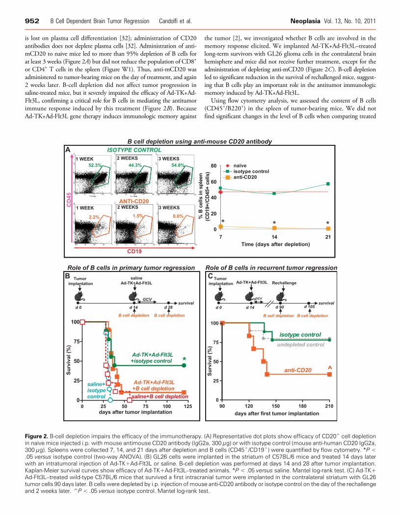
is lost on plasma cell differentiation [32]; administration of CD20antibodies does not deplete plasma cells [32]. Administration of anti-mCD20 to naive mice led to more than 95% depletion of B cells forat least 3 weeks (Figure 2A) but did not reduce the population of CD8+
or CD4+ T cells in the spleen (Figure W1). Thus, anti-mCD20 wasadministered to tumor-bearing mice on the day of treatment, and again2 weeks later. B-cell depletion did not affect tumor progression insaline-treated mice, but it severely impaired the efficacy of Ad-TK+Ad-Flt3L, confirming a critical role for B cells in mediating the antitumorimmune response induced by this treatment (Figure 2B). BecauseAd-TK+Ad-Flt3L gene therapy induces immunologic memory against
the tumor [2], we investigated whether B cells are involved in thememory response elicited. We implanted Ad-TK+Ad-Flt3L–treatedlong-term survivors with GL26 glioma cells in the contralateral brainhemisphere and mice did not receive further treatment, except for theadministration of depleting anti-mCD20 (Figure 2C). B-cell depletionled to significant reduction in the survival of rechallenged mice, suggest-ing that B cells play an important role in the antitumor immunologicmemory induced by Ad-TK+Ad-Flt3L.
Using flow cytometry analysis, we assessed the content of B cells(CD45+/B220+) in the spleen of tumor-bearing mice. We did notfind significant changes in the level of B cells when comparing treated
Figure 2. B-cell depletion impairs the efficacy of the immunotherapy. (A) Representative dot plots show efficacy of CD20+ cell depletionin naive mice injected i.p. with mouse antimouse CD20 antibody (IgG2a, 300 μg) or with isotype control (mouse anti-human CD20 IgG2a,300 μg). Spleens were collected 7, 14, and 21 days after depletion and B cells (CD45+/CD19+) were quantified by flow cytometry. *P <.05 versus isotype control (two-way ANOVA). (B) GL26 cells were implanted in the striatum of C57BL/6 mice and treated 14 days laterwith an intratumoral injection of Ad-TK+Ad-Flt3L or saline. B-cell depletion was performed at days 14 and 28 after tumor implantation.Kaplan-Meier survival curves show efficacy of Ad-TK+Ad-Flt3L–treated animals. *P < .05 versus saline. Mantel log-rank test. (C) Ad-TK+Ad-Flt3L–treated wild-type C57BL/6 mice that survived a first intracranial tumor were implanted in the contralateral striatum with GL26tumor cells 90 days later. B cells were depleted by i.p. injection ofmouse anti-CD20 antibody or isotype control on the day of the rechallengeand 2 weeks later. P̂ < .05 versus isotype control. Mantel log-rank test.
952 B Cell Dependent Brain Tumor Regression Candolfi et al. Neoplasia Vol. 13, No. 10, 2011
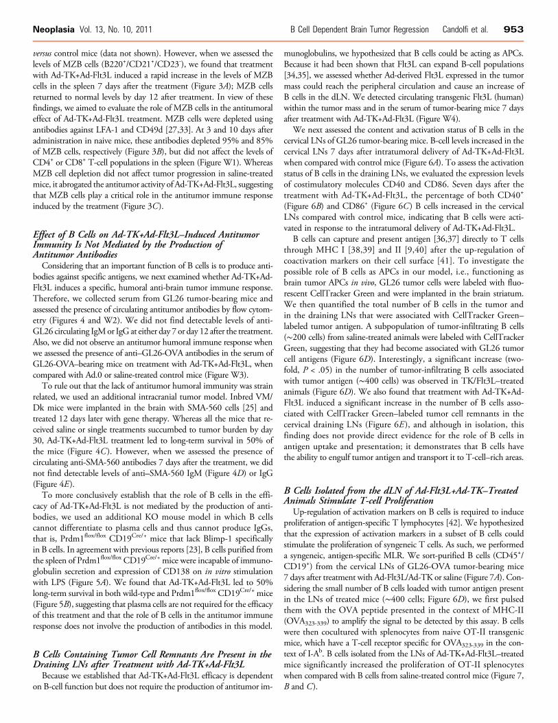
versus control mice (data not shown). However, when we assessed thelevels of MZB cells (B220+/CD21+/CD23-), we found that treatmentwith Ad-TK+Ad-Flt3L induced a rapid increase in the levels of MZBcells in the spleen 7 days after the treatment (Figure 3A); MZB cellsreturned to normal levels by day 12 after treatment. In view of thesefindings, we aimed to evaluate the role of MZB cells in the antitumoraleffect of Ad-TK+Ad-Flt3L treatment. MZB cells were depleted usingantibodies against LFA-1 and CD49d [27,33]. At 3 and 10 days afteradministration in naive mice, these antibodies depleted 95% and 85%of MZB cells, respectively (Figure 3B), but did not affect the levels ofCD4+ or CD8+ T-cell populations in the spleen (Figure W1). WhereasMZB cell depletion did not affect tumor progression in saline-treatedmice, it abrogated the antitumor activity of Ad-TK+Ad-Flt3L, suggestingthat MZB cells play a critical role in the antitumor immune responseinduced by the treatment (Figure 3C).
Effect of B Cells on Ad-TK+Ad-Flt3L–Induced AntitumorImmunity Is Not Mediated by the Production ofAntitumor Antibodies
Considering that an important function of B cells is to produce anti-bodies against specific antigens, we next examined whether Ad-TK+Ad-Flt3L induces a specific, humoral anti-brain tumor immune response.Therefore, we collected serum from GL26 tumor-bearing mice andassessed the presence of circulating antitumor antibodies by flow cytom-etry (Figures 4 and W2). We did not find detectable levels of anti-GL26 circulating IgMor IgG at either day 7 or day 12 after the treatment.Also, we did not observe an antitumor humoral immune response whenwe assessed the presence of anti–GL26-OVA antibodies in the serum ofGL26-OVA–bearing mice on treatment with Ad-TK+Ad-Flt3L, whencompared with Ad.0 or saline-treated control mice (Figure W3).
To rule out that the lack of antitumor humoral immunity was strainrelated, we used an additional intracranial tumor model. Inbred VM/Dk mice were implanted in the brain with SMA-560 cells [25] andtreated 12 days later with gene therapy. Whereas all the mice that re-ceived saline or single treatments succumbed to tumor burden by day30, Ad-TK+Ad-Flt3L treatment led to long-term survival in 50% ofthe mice (Figure 4C ). However, when we assessed the presence ofcirculating anti-SMA-560 antibodies 7 days after the treatment, we didnot find detectable levels of anti–SMA-560 IgM (Figure 4D) or IgG(Figure 4E).
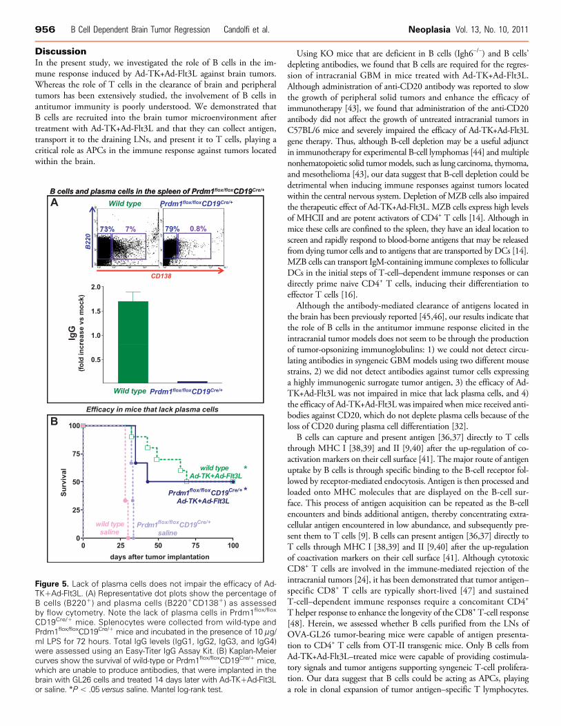
To more conclusively establish that the role of B cells in the effi-cacy of Ad-TK+Ad-Flt3L is not mediated by the production of anti-bodies, we used an additional KO mouse model in which B cellscannot differentiate to plasma cells and thus cannot produce IgGs,that is, Prdm1flox/flox CD19Cre/+ mice that lack Blimp-1 specificallyin B cells. In agreement with previous reports [23], B cells purified fromthe spleen of Prdm1flox/flox CD19Cre/+ mice were incapable of immuno-globulin secretion and expression of CD138 on in vitro stimulationwith LPS (Figure 5A). We found that Ad-TK+Ad-Flt3L led to 50%long-term survival in both wild-type and Prdm1flox/flox CD19Cre/+ mice(Figure 5B), suggesting that plasma cells are not required for the efficacyof this treatment and that the role of B cells in the antitumor immuneresponse does not involve the production of antibodies in this model.
B Cells Containing Tumor Cell Remnants Are Present in theDraining LNs after Treatment with Ad-TK+Ad-Flt3L
Because we established that Ad-TK+Ad-Flt3L efficacy is dependenton B-cell function but does not require the production of antitumor im-
munoglobulins, we hypothesized that B cells could be acting as APCs.Because it had been shown that Flt3L can expand B-cell populations[34,35], we assessed whether Ad-derived Flt3L expressed in the tumormass could reach the peripheral circulation and cause an increase ofB cells in the dLN. We detected circulating transgenic Flt3L (human)within the tumor mass and in the serum of tumor-bearing mice 7 daysafter treatment with Ad-TK+Ad-Flt3L (Figure W4).
We next assessed the content and activation status of B cells in thecervical LNs of GL26 tumor-bearing mice. B-cell levels increased in thecervical LNs 7 days after intratumoral delivery of Ad-TK+Ad-Flt3Lwhen compared with control mice (Figure 6A). To assess the activationstatus of B cells in the draining LNs, we evaluated the expression levelsof costimulatory molecules CD40 and CD86. Seven days after thetreatment with Ad-TK+Ad-Flt3L, the percentage of both CD40+
(Figure 6B) and CD86+ (Figure 6C ) B cells increased in the cervicalLNs compared with control mice, indicating that B cells were acti-vated in response to the intratumoral delivery of Ad-TK+Ad-Flt3L.
B cells can capture and present antigen [36,37] directly to T cellsthrough MHC I [38,39] and II [9,40] after the up-regulation ofcoactivation markers on their cell surface [41]. To investigate thepossible role of B cells as APCs in our model, i.e., functioning asbrain tumor APCs in vivo, GL26 tumor cells were labeled with fluo-rescent CellTracker Green and were implanted in the brain striatum.We then quantified the total number of B cells in the tumor andin the draining LNs that were associated with CellTracker Green–labeled tumor antigen. A subpopulation of tumor-infiltrating B cells(∼200 cells) from saline-treated animals were labeled with CellTrackerGreen, suggesting that they had become associated with GL26 tumorcell antigens (Figure 6D). Interestingly, a significant increase (two-fold, P < .05) in the number of tumor-infiltrating B cells associatedwith tumor antigen (∼400 cells) was observed in TK/Flt3L–treatedanimals (Figure 6D). We also found that treatment with Ad-TK+Ad-Flt3L induced a significant increase in the number of B cells asso-ciated with CellTracker Green–labeled tumor cell remnants in thecervical draining LNs (Figure 6E ), and although in isolation, thisfinding does not provide direct evidence for the role of B cells inantigen uptake and presentation; it demonstrates that B cells havethe ability to engulf tumor antigen and transport it to T-cell–rich areas.
B Cells Isolated from the dLN of Ad-Flt3L+Ad-TK–TreatedAnimals Stimulate T-cell Proliferation
Up-regulation of activation markers on B cells is required to induceproliferation of antigen-specific T lymphocytes [42]. We hypothesizedthat the expression of activation markers in a subset of B cells couldstimulate the proliferation of syngeneic T cells. As such, we performeda syngeneic, antigen-specific MLR. We sort-purified B cells (CD45+/CD19+) from the cervical LNs of GL26-OVA tumor-bearing mice7 days after treatment with Ad-Flt3L/Ad-TK or saline (Figure 7A). Con-sidering the small number of B cells loaded with tumor antigen presentin the LNs of treated mice (∼400 cells; Figure 6D), we first pulsedthem with the OVA peptide presented in the context of MHC-II(OVA323-339) to amplify the signal to be detected by this assay. B cellswere then cocultured with splenocytes from naive OT-II transgenicmice, which have a T-cell receptor specific for OVA323-339 in the con-text of I-Ab. B cells isolated from the LNs of Ad-TK+Ad-Flt3L–treatedmice significantly increased the proliferation of OT-II splenocyteswhen compared with B cells from saline-treated control mice (Figure 7,B and C).
Neoplasia Vol. 13, No. 10, 2011 B Cell Dependent Brain Tumor Regression Candolfi et al. 953
Figure 3. MZB cell depletion impairs the efficacy of the immunotherapy. (A) GL26 cells were implanted in the striatum of wild-type C57BL/6 mice. Mice were treated 14 days later with an intratumoral injection of Ad-TK+Ad-Flt3L, and as controls, with saline or an empty vector(Ad.0). At 7 and 12 days after the treatment, levels of MZB cells in the spleen were quantified by flow cytometry using anti-B220, anti-CD21,and anti-CD23 antibodies. *P < .05 versus saline, P̂ < .05 versus Ad.0. (B) Representative dot plots show efficacy of MZB cell depletionin the spleen of naive mice 3, 10, and 20 days after i.p. administration of anti-CD49d antibody (100 μg) and anti-LFA-1 antibody (100 μg)or the corresponding isotype controls (rat IgG2a and rat IgG2b, 100 μg each). *P < .05 versus isotype control (two-way ANOVA). (C) GL26cells were implanted in the striatum of C57BL/6 mice and treated 14 days later with an intratumoral injection of Ad-TK+Ad-Flt3L or saline.MZB cell depletion was performed at days 14 and 24 after tumor implantation. Kaplan-Meier survival curves show efficacy of Ad-TK+Ad-Flt3L–treated animals. *P < .05 versus saline. Mantel log-rank test.
954 B Cell Dependent Brain Tumor Regression Candolfi et al. Neoplasia Vol. 13, No. 10, 2011

Figure 4. Assessment of the antibody response against the tumor. (A and B) GL26 cells were implanted in the striatum of C57BL/6 miceand treated 14 days later with an intratumoral injection of Ad-TK+Ad-Flt3L, saline, or an empty vector (Ad.0). At 7 (A) and 12 (B) days afterthe treatment, serum was collected to evaluate the presence of circulating anti-GL26 cell IgM (A) and IgG (B), respectively. Histogramsand graphs show the fluorescence intensity of fixed GL26 cells that were incubated with normal C57BL/6 serum (red area) or serum fromtreated tumor-bearing mice (colored lines), followed by FITC-conjugated antimouse IgM or IgG. (C) Kaplan-Meier survival curves showthe survival of inbred VM/Dk mice bearing intracranial SMA-560 tumors and treated 12 days later with Ad-TK+Ad-Flt3L, saline, or Ad.0.*P < .05 versus saline (log-rank test). Seven days after the treatment, serum was collected to evaluate the presence of circulating anti–SMA-560 cell antibodies. (D and E) Histograms and graphs show the fluorescence intensity of fixed SMA-560 cells that were incubatedwith normal inbred VM/Dk mouse serum (red area) or serum from treated mice (colored lines), followed by FITC-conjugated antimouseIgM (D) or FITC–antimouse IgG (E).
Neoplasia Vol. 13, No. 10, 2011 B Cell Dependent Brain Tumor Regression Candolfi et al. 955
DiscussionIn the present study, we investigated the role of B cells in the im-mune response induced by Ad-TK+Ad-Flt3L against brain tumors.Whereas the role of T cells in the clearance of brain and peripheraltumors has been extensively studied, the involvement of B cells inantitumor immunity is poorly understood. We demonstrated thatB cells are recruited into the brain tumor microenvironment aftertreatment with Ad-TK+Ad-Flt3L and that they can collect antigen,transport it to the draining LNs, and present it to T cells, playing acritical role as APCs in the immune response against tumors locatedwithin the brain.
Using KO mice that are deficient in B cells (Igh6−/−) and B cells’depleting antibodies, we found that B cells are required for the regres-sion of intracranial GBM in mice treated with Ad-TK+Ad-Flt3L.Although administration of anti-CD20 antibody was reported to slowthe growth of peripheral solid tumors and enhance the efficacy ofimmunotherapy [43], we found that administration of the anti-CD20antibody did not affect the growth of untreated intracranial tumors inC57BL/6 mice and severely impaired the efficacy of Ad-TK+Ad-Flt3Lgene therapy. Thus, although B-cell depletion may be a useful adjunctin immunotherapy for experimental B-cell lymphomas [44] and multiplenonhematopoietic solid tumormodels, such as lung carcinoma, thymoma,and mesothelioma [43], our data suggest that B-cell depletion could bedetrimental when inducing immune responses against tumors locatedwithin the central nervous system. Depletion of MZB cells also impairedthe therapeutic effect of Ad-TK+Ad-Flt3L. MZB cells express high levelsof MHCII and are potent activators of CD4+ T cells [14]. Although inmice these cells are confined to the spleen, they have an ideal location toscreen and rapidly respond to blood-borne antigens that may be releasedfrom dying tumor cells and to antigens that are transported by DCs [14].MZB cells can transport IgM-containing immune complexes to follicularDCs in the initial steps of T-cell–dependent immune responses or candirectly prime naive CD4+ T cells, inducing their differentiation toeffector T cells [16].
Although the antibody-mediated clearance of antigens located inthe brain has been previously reported [45,46], our results indicate thatthe role of B cells in the antitumor immune response elicited in theintracranial tumor models does not seem to be through the productionof tumor-opsonizing immunoglobulins: 1) we could not detect circu-lating antibodies in syngeneic GBM models using two different mousestrains, 2) we did not detect antibodies against tumor cells expressinga highly immunogenic surrogate tumor antigen, 3) the efficacy of Ad-TK+Ad-Flt3L was not impaired in mice that lack plasma cells, and 4)the efficacy of Ad-TK+Ad-Flt3L was impaired when mice received anti-bodies against CD20, which do not deplete plasma cells because of theloss of CD20 during plasma cell differentiation [32].
B cells can capture and present antigen [36,37] directly to T cellsthrough MHC I [38,39] and II [9,40] after the up-regulation of co-activation markers on their cell surface [41]. The major route of antigenuptake by B cells is through specific binding to the B-cell receptor fol-lowed by receptor-mediated endocytosis. Antigen is then processed andloaded onto MHC molecules that are displayed on the B-cell sur-face. This process of antigen acquisition can be repeated as the B-cellencounters and binds additional antigen, thereby concentrating extra-cellular antigen encountered in low abundance, and subsequently pre-sent them to T cells [9]. B cells can present antigen [36,37] directly toT cells through MHC I [38,39] and II [9,40] after the up-regulationof coactivation markers on their cell surface [41]. Although cytotoxicCD8+ T cells are involved in the immune-mediated rejection of theintracranial tumors [24], it has been demonstrated that tumor antigen–specific CD8+ T cells are typically short-lived [47] and sustainedT-cell–dependent immune responses require a concomitant CD4+
T helper response to enhance the longevity of the CD8+ T-cell response[48]. Herein, we assessed whether B cells purified from the LNs ofOVA-GL26 tumor-bearing mice were capable of antigen presenta-tion to CD4+ T cells from OT-II transgenic mice. Only B cells fromAd-TK+Ad-Flt3L–treated mice were capable of providing costimula-tory signals and tumor antigens supporting syngeneic T-cell prolifera-tion. Our data suggest that B cells could be acting as APCs, playinga role in clonal expansion of tumor antigen–specific T lymphocytes.
Figure 5. Lack of plasma cells does not impair the efficacy of Ad-TK+Ad-Flt3L. (A) Representative dot plots show the percentage ofB cells (B220+) and plasma cells (B220+CD138+) as assessedby flow cytometry. Note the lack of plasma cells in Prdm1flox/flox
CD19Cre/+ mice. Splenocytes were collected from wild-type andPrdm1flox/floxCD19Cre/+ mice and incubated in the presence of 10 μg/ml LPS for 72 hours. Total IgG levels (IgG1, IgG2, IgG3, and IgG4)were assessed using an Easy-Titer IgG Assay Kit. (B) Kaplan-Meiercurves show the survival of wild-type or Prdm1flox/floxCD19Cre/+ mice,which are unable to produce antibodies, that were implanted in thebrain with GL26 cells and treated 14 days later with Ad-TK+Ad-Flt3Lor saline. *P < .05 versus saline. Mantel log-rank test.
956 B Cell Dependent Brain Tumor Regression Candolfi et al. Neoplasia Vol. 13, No. 10, 2011

Figure 6. B cells containing tumor cell remnants migrate to the cervical draining LNs. Wild-type C57BL/6 mice were implanted with GL26tumors and treated 14 days later with Ad-TK+Ad-Flt3L (Ad.TK/Flt3L), saline, or Ad.0. After 7 days, immune cells isolated from dLN wereanalyzed by flow cytometry. Representative dot plots and graphs show the content of B cells (CD19+/CD45+; A) and the percentage ofCD40+ (CD19+/CD45+CD40+; B) and CD86+ B cells (CD19+/CD45+/CD86+; C) in the dLN. *P < .05 versus saline, ^P < .05 versus Ad.0(one-way ANOVA followed by the Tukey test). (D and E) GL26 cells were labeled with CellTracker Green and implanted in the striatum ofC57BL/6 mice. Two days later, mice were treated with either saline or Ad-TK+Ad-Flt3L and immune cells were isolated from tumors anddraining LNs 7 days later. Representative dot plots and graphs show the content of B cells associated with CellTracker Green+ tumorprotein remnants within the tumors (D) and in the dLN (E). *P < .05 (Student’s t test).
Neoplasia Vol. 13, No. 10, 2011 B Cell Dependent Brain Tumor Regression Candolfi et al. 957
In support of this, we found that B cells were required for the clonalexpansion of tumor antigen–specific T cells. An alternative scenario isthat B cells are not involved directly in presenting tumor antigen toT cells but could provide costimulatory signals to T cells in contact withDCs. This is supported by other studies that highlight the role of B cellsto enhance the costimulatory signaling between DCs and T cells [49].We have previously shown that DCs play a pivotal role in the onset ofthe antitumor immune response induced by Ad-TK+Ad-Flt3L [2].Although DCs are the main APCs that trigger adaptive immune re-sponses [50], increasing evidence suggests that interactions betweenDCs and B cells promote optimal T-cell activation. For instance, co-operation between DC and B cells has been observed to be required forthe presentation of viral antigens to naive, antigen-specific T cells [51].B cells have been shown to collaborate with DCs in the presentation ofextracellular antigens that trigger the activation of effector T cells andthe generation of memory T cells [49]. Furthermore, B cells have beenshown to modulate the profile of cytokines released by activated DCs
[52]. Conversely, DCs have been shown to capture, retain, and transferunprocessed antigen to B cells [53]. Also, bidirectional proliferation andsurvival signaling have been detected between B cells and DCs [52,54].
Flt3L is a critical cytokine that induces differentiation of B cellsfrom early hematopoietic stem cell precursors [34]. In our model,expression of Flt3L within the tumor mass resulted in high levels ofsystemic Flt3L, which may be responsible for the expansion of MZBcells and the increased levels of B cells observed in the dLN of tumor-bearing mice treated with Ad-TK+Ad-Flt3L. We also observed in-creased expression of cell surface CD40 and CD86 on B cells in thedLN, which is indicative that B cells are activated [55] and capableof acting as tumor APCs [41]. Our data suggests that B cells mightbe transporting tumor antigen and promoting T-cell proliferation inthe draining LNs [36,56] after treatment with Ad-Flt3L/Ad-TK. Al-though the brain lacks classic lymph vessels, the migration of leukocytesfrom the brain to the cervical LNs has been shown to occur throughthe cribroid plate and the nasal mucosa [57]. Also, it is possible that
Figure 7. B cells isolated from the dLN of Ad-Flt3L– and Ad-TK–treated animals stimulate T-cell proliferation. (A) GL26 cells expressingthe surrogate antigen chicken ovoalbumin (OVA) were implanted in the striatum of wild-type C57BL/6 mice and treated 14 days later withAd-Flt3L and Ad-TK or saline. Seven days later, B cells were purified from the dLN by MACS. The purity of B-cell preparations was greaterthan 95%, as assessed by flow cytometry (B). (C) A syngeneic MLR was used to determine whether B cells could stimulate T-cell prolif-eration. B cells (stimulators) were primed with OVA, incubated with mitomycin, and then cocultured at different ratios with syngeneicsplenocytes collected from naive OT-II mice (responders) for 72 hours. BrdU incorporation into DNAwas used to determine cell proliferation.*P < .05 versus saline (randomization test). Inset: splenocytes were incubated in the absence of B cells and in the presence of anti-CD3antibody (1 μg/ml) or OVA323-339 (2 μg/ml), as positive controls.
958 B Cell Dependent Brain Tumor Regression Candolfi et al. Neoplasia Vol. 13, No. 10, 2011
recirculating B cells encounter antigen in the LNs. Although the Ig re-ceptor of B cells can bind soluble antigen, the efficiency of this processin vivo is very low. Nevertheless, it has been shown that, in the LNs,B cells can obtain antigens from subcapsular sinus macrophages andfrom follicular DCs, which can act as long-term depots for B-cellantigens [58]. These findings, and the fact that B cells from the LNsof Ad-Flt3L/Ad-TK–treated mice are able to stimulate the clonal ex-pansion of syngeneic tumor antigen–specific T cells, strongly supportthe notion that B cells could be acting as APCs in this model. Althoughthe number of B cells associated with tumor cell remnants that wedetected in the cervical LNs (∼400, i.e., ∼60 per 106 cells) during theantitumor immune response induced by immunotherapy may seemsmall, it is in agreement with data from other groups. Lindell et al.[59] have shown that around 50 antigen-specific B cells/106 total cellsreach the LNs to efficiently present cockroach antigen to T cells duringthe onset of allergic lung disease. It has also been demonstrated thatonly a few hundred skin-derived DCs are required to make their wayto the draining brachial LN to provide prolonged presentation androbust levels of CTL expansion [60–62].
Taken together, the results we present in this article strongly sug-gest that B cells are acting as APCs. The evidence we present herecan be summarized as follows: 1) B cells are required for antitumorimmunity triggered by our treatment because the treatment fails inB-cell–deficient mice Igh6−/− mice and in wild-type mice that havebeen depleted of total B cells or MZB cells; 2) B cells’ role in antitumorimmunity does not depend on antibody production; 3) B cells have theability to engulf tumor antigen, transport it to T-cell–rich areas, andoverexpress coactivation markers that support T-cell activation; and4) pure preparations of B cells from the LNs of Ad-Flt3L/Ad-TK–treated mice are able to stimulate the expansion of syngeneic tumorantigen–specific T cells.
The data presented herein indicate that B cells could be usefultherapeutic targets to increase the efficacy of immunotherapeutics forbrain cancer. It has been shown that CD40-activated B cells loaded withtumor RNA constitute potent antitumor vaccines, which can be readilygenerated from small amounts of blood and act as efficient APCs todrive the activation of antigen-specific T cells [22,63]. TLR9 agonistshave also been shown to stimulate the expression of MHC and co-activation markers in human B cells, enhancing their ability to cross-present antigens to autologous T cells [64], and could improve B-cellvaccine design for brain cancer immune-based therapeutics. Our resultshighlight the role of B lymphocytes during immunotherapy-dependentbrain tumor regression and suggest that they could act as APCs, leadingto effective antitumor immunity and immunologic memory.
References[1] Heimberger AB and Sampson JH (2011). Immunotherapy coming of age: what
will it take to make it standard of care for glioblastoma? Neuro Oncol 13, 3–13.[2] Curtin J, Liu N, Candolfi M, XiongW, Assi A, Yagiz K, EdwardsM,Michelsen K,
Kroeger K, Liu C, et al. (2009). HMGB1 mediates endogenous TLR2 activationand brain tumor regression. PLoS Med 6, e10.
[3] Chien PY, Wang J, Carbonaro D, Lei S, Miller B, Sheikh S, Ali SM, Ahmad MU,and Ahmad I (2005). Novel cationic cardiolipin analogue-based liposome forefficient DNA and small interfering RNA delivery in vitro and in vivo. Cancer GeneTher 12, 321–328.
[4] GhulamMuhammad AK, Candolfi M, King GD, Yagiz K, Foulad D, Mineharu Y,Kroeger KM, Treuer KA, Nichols WS, Sanderson NS, et al. (2009). Antigliomaimmunological memory in response to conditional cytotoxic/immune–stimulatorygene therapy: humoral and cellular immunity lead to tumor regression. Clin CancerRes 15, 6113–6127.
[5] Dong HP, Elstrand MB, Holth A, Silins I, Berner A, Trope CG, Davidson B,and Risberg B (2006). NK- and B-cell infiltration correlates with worse outcomein metastatic ovarian carcinoma. Am J Clin Pathol 125, 451–458.
[6] Nzula S, Going JJ, and Stott DI (2003). Antigen-driven clonal proliferation,somatic hypermutation, and selection of B lymphocytes infiltrating human ductalbreast carcinomas. Cancer Res 63, 3275–3280.
[7] Kotlan B, Simsa P, Teillaud JL, Fridman WH, Toth J, McKnight M, and GlassyMC (2005). Novel ganglioside antigen identified by B cells in human medullarybreast carcinomas: the proof of principle concerning the tumor-infiltrating B lym-phocytes. J Immunol 175, 2278–2285.
[8] de Visser KE, Korets LV, and Coussens LM (2005). De novo carcinogenesispromoted by chronic inflammation is B lymphocyte dependent. Cancer Cell 7,411–423.
[9] Rodriguez-Pinto D (2005). B cells as antigen presenting cells. Cell Immunol238, 67–75.
[10] Inoue S, Leitner WW, Golding B, and Scott D (2006). Inhibitory effects ofB cells on antitumor immunity. Cancer Res 66, 7741–7747.
[11] Shah S, Divekar AA, Hilchey SP, Cho HM, Newman CL, Shin SU, Nechustan H,Challita-Eid PM, Segal BM, Yi KH, et al. (2005). Increased rejection of primarytumors in mice lacking B cells: inhibition of anti-tumor CTL and TH1 cytokineresponses by B cells. Int J Cancer 117, 574–586.
[12] Perricone MA, Smith KA, Claussen KA, Plog MS, Hempel DM, Roberts BL,St George JA, and Kaplan JM (2004). Enhanced efficacy of melanoma vaccinesin the absence of B lymphocytes. J Immunother 27, 273–281.
[13] Li Q, Grover AC, Donald EJ, Carr A, Yu J, Whitfield J, Nelson M, Takeshita N,and Chang AE (2005). Simultaneous targeting of CD3 on T cells and CD40 onB or dendritic cells augments the antitumor reactivity of tumor-primed lymphnode cells. J Immunol 175, 1424–1432.
[14] Pillai S, Cariappa A, and Moran ST (2005). Marginal zone B cells. Annu RevImmunol 23, 161–196.
[15] Cinamon G, Zachariah MA, Lam OM, Foss FW Jr, and Cyster JG (2008).Follicular shuttling of marginal zone B cells facilitates antigen transport. NatImmunol 9, 54–62.
[16] Lopes-Carvalho T, Foote J, and Kearney JF (2005). Marginal zone B cells inlymphocyte activation and regulation. Curr Opin Immunol 17, 244–250.
[17] Serreze DV, Fleming SA, Chapman HD, Richard SD, Leiter EH, and TischRM (1998). B lymphocytes are critical antigen-presenting cells for the initiationof T cell–mediated autoimmune diabetes in nonobese diabetic mice. J Immunol161, 3912–3918.
[18] Chan OT, Hannum LG, Haberman AM, Madaio MP, and Shlomchik MJ (1999).A novel mouse with B cells but lacking serum antibody reveals an antibody-independent role for B cells in murine lupus. J Exp Med 189, 1639–1648.
[19] Takemura S, Klimiuk PA, Braun A, Goronzy JJ, and Weyand CM (2001).T cell activation in rheumatoid synovium is B cell dependent. J Immunol 167,4710–4718.
[20] Lapointe R, Bellemare-Pelletier A, Housseau F, Thibodeau J, and Hwu P(2003). CD40-stimulated B lymphocytes pulsed with tumor antigens are effec-tive antigen-presenting cells that can generate specific T cells. Cancer Res 63,2836–2843.
[21] von Bergwelt-Baildon M, Schultze JL, Maecker B, Menezes I, and Nadler LM(2004). Correspondence re R. Lapointe et al., CD40-stimulated B lymphocytespulsed with tumor antigens are effective antigen-presenting cells that can gener-ate specific T cells. Cancer Res 2003;63:2836–43. Cancer Res 64, 4055–4056;author reply 4056–4057.
[22] Schultze JL, Michalak S, Seamon MJ, Dranoff G, Jung K, Daley J, Delgado JC,Gribben JG, and Nadler LM (1997). CD40-activated human B cells: an alternativesource of highly efficient antigen presenting cells to generate autologous antigen-specific T cells for adoptive immunotherapy. J Clin Invest 100, 2757–2765.
[23] Shapiro-Shelef M, Lin KI, McHeyzer-Williams LJ, Liao J, McHeyzer-WilliamsMG, and Calame K (2003). Blimp-1 is required for the formation of immuno-globulin secreting plasma cells and pre-plasma memory B cells. Immunity 19,607–620.
[24] Yang J, Sanderson NSR, Wawrowsky K, Puntel M, Castro MG, and LowensteinPR (2010). Kupfer-type immunological synapse characteristics do not predictanti-brain tumor cytolytic T-cell function in vivo. Proc Natl Acad Sci USA 107,4716–4721.
[25] Sampson JH, Ashley DM, Archer GE, Fuchs HE, Dranoff G, Hale LP, andBigner DD (1997). Characterization of a spontaneous murine astrocytomaand abrogation of its tumorigenicity by cytokine secretion. Neurosurgery 41,1365–1372; discussion 1372–1363.
Neoplasia Vol. 13, No. 10, 2011 B Cell Dependent Brain Tumor Regression Candolfi et al. 959
[26] Yu S, Dunn R, Kehry MR, and Braley-Mullen H (2008). B cell depletioninhibits spontaneous autoimmune thyroiditis in NOD.H-2h4 mice. J Immunol180, 7706–7713.
[27] Lu TT and Cyster JG (2002). Integrin-mediated long-term B cell retention in thesplenic marginal zone. Science 297, 409–412.
[28] Candolfi M, Yagiz K, Foulad D, Alzadeh GE, Tesarfreund M, Muhammad AK,Puntel M, Kroeger KM, Liu C, Lee S, et al. (2009). Release of HMGB1 inresponse to proapoptotic glioma killing strategies: efficacy and neurotoxicity. ClinCancer Res 15, 4401–4414.
[29] Robertson JM, Jensen PE, and Evavold BD (2000). DO11.10 and OT-II T cellsrecognize a C-terminal ovalbumin 323-339 epitope. J Immunol 164, 4706–4712.
[30] Hussain SF, Yang D, Suki D, Aldape K, Grimm E, and Heimberger AB (2006).The role of human glioma-infiltrating microglia/macrophages in mediatingantitumor immune responses. Neuro Oncol 8, 261–279.
[31] Ngo VN, Cornall RJ, and Cyster JG (2001). Splenic T zone development isB cell dependent. J Exp Med 194, 1649–1660.
[32] DiLillo DJ, Hamaguchi Y, Ueda Y, Yang K, Uchida J, Haas KM, Kelsoe G, andTedder TF (2008). Maintenance of long-lived plasma cells and serologicalmemory despite mature and memory B cell depletion during CD20 immuno-therapy in mice. J Immunol 180, 361–371.
[33] Belperron AA, Dailey CM, Booth CJ, and Bockenstedt LK (2007). Marginalzone B-cell depletion impairs murine host defense against Borrelia burgdorferiinfection. Infect Immun 75, 3354–3360.
[34] Ray RJ, Paige CJ, Furlonger C, Lyman SD, and Rottapel R (1996). Flt3 ligand sup-ports the differentiation of early B cell progenitors in the presence of interleukin-11and interleukin-7. Eur J Immunol 26, 1504–1510.
[35] Buza-Vidas N, Cheng M, Duarte S, Nozad H, Jacobsen SE, and Sitnicka E(2007). Crucial role of FLT3 ligand in immune reconstitution after bone marrowtransplantation and high-dose chemotherapy. Blood 110, 424–432.
[36] Carrasco YR and Batista FD (2007). B cells acquire particulate antigen in amacrophage-rich area at the boundary between the follicle and the subcapsularsinus of the lymph node. Immunity 27, 160–171.
[37] Okada T, Miller MJ, Parker I, Krummel MF, Neighbors M, Hartley SB,O’Garra A, Cahalan MD, and Cyster JG (2005). Antigen-engaged B cells undergochemotaxis toward the T zone and form motile conjugates with helper T cells.PLoS Biol 3, e150.
[38] Hon H, Oran A, Brocker T, and Jacob J (2005). B lymphocytes participate incross-presentation of antigen following gene gun vaccination. J Immunol 174,5233–5242.
[39] Hoft DF, Eickhoff CS, Giddings OK, Vasconcelos JR, and Rodrigues MM(2007). Trans-sialidase recombinant protein mixed with CpG motif-containingoligodeoxynucleotide induces protective mucosal and systemic Trypanosoma cruziimmunity involving CD8+ CTL and B cell–mediated cross-priming. J Immunol179, 6889–6900.
[40] Clark MR, Massenburg D, Siemasko K, Hou P, and Zhang M (2004). B-cellantigen receptor signaling requirements for targeting antigen to the MHC classII presentation pathway. Curr Opin Immunol 16, 382–387.
[41] Evans DE, Munks MW, Purkerson JM, and Parker DC (2000). Resting B lym-phocytes as APC for naive T lymphocytes: dependence on CD40 ligand/CD40.J Immunol 164, 688–697.
[42] Croft M (1994). Activation of naive, memory and effector T cells. Curr OpinImmunol 6, 431–437.
[43] Kim S, Fridlender ZG, Dunn R, Kehry MR, Kapoor V, Blouin A, Kaiser LR, andAlbelda SM (2008). B-cell depletion using an anti-CD20 antibody augmentsantitumor immune responses and immunotherapy in nonhematopoetic murinetumor models. J Immunother 31, 446–457.
[44] Gadri Z, Kukulansky T, Bar-Or E, Haimovich J, and Hollander N (2009).Synergistic effect of dendritic cell vaccination and anti-CD20 antibody treatmentin the therapy of murine lymphoma. J Immunother 32, 333–340.
[45] Vasilevko V, Xu F, Previti ML, Van Nostrand WE, and Cribbs DH (2007).
Experimental investigation of antibody-mediated clearance mechanisms ofamyloid-β in CNS of Tg-SwDI transgenic mice. J Neurosci 27, 13376–13383.
[46] Ankeny DP and Popovich PG (2011). B cells and autoantibodies: complex rolesin CNS injury. Trends Immunol 31, 332–338.
[47] Dudley ME, Wunderlich J, Nishimura MI, Yu D, Yang JC, Topalian SL,Schwartzentruber DJ, Hwu P, Marincola FM, Sherry R, et al. (2001). Adoptivetransfer of cloned melanoma-reactive T lymphocytes for the treatment of patientswith metastatic melanoma. J Immunother 24, 363–373.
[48] Knutson KL, Schiffman K, Cheever MA, and Disis ML (2002). Immunizationof cancer patients with a HER-2/neu, HLA-A2 peptide, p369-377, results inshort-lived peptide-specific immunity. Clin Cancer Res 8, 1014–1018.
[49] Crawford A, Macleod M, Schumacher T, Corlett L, and Gray D (2006). PrimaryT cell expansion and differentiation in vivo requires antigen presentation by B cells.J Immunol 176, 3498–3506.
[50] Brigl M and Brenner MB (2004). CD1: antigen presentation and T cell func-tion. Annu Rev Immunol 22, 817–890.
[51] Diaz-de-Durana Y, Mantchev GT, Bram RJ, and Franco A (2006). TACI-BLySsignaling via B-cell–dendritic cell cooperation is required for naive CD8+ T-cellpriming in vivo. Blood 107, 594–601.
[52] Moulin V, Andris F, Thielemans K, Maliszewski C, Urbain J, and Moser M(2000). B lymphocytes regulate dendritic cell (DC) function in vivo: increasedinterleukin 12 production by DCs from B cell-deficient mice results in T helpercell type 1 deviation. J Exp Med 192, 475–482.
[53] Wykes M, Pombo A, Jenkins C, and MacPherson GG (1998). Dendritic cellsinteract directly with naive B lymphocytes to transfer antigen and initiate classswitching in a primary T-dependent response. J Immunol 161, 1313–1319.
[54] Wykes M and MacPherson G (2000). Dendritic cell–B-cell interaction: dendriticcells provide B cells with CD40-independent proliferation signals and CD40-dependent survival signals. Immunology 100, 1–3.
[55] Constant S, Schweitzer N, West J, Ranney P, and Bottomly K (1995). B lym-phocytes can be competent antigen-presenting cells for priming CD4+ T cells toprotein antigens in vivo. J Immunol 155, 3734–3741.
[56] Attanavanich K and Kearney JF (2004). Marginal zone, but not follicular B cells,are potent activators of naive CD4 T cells. J Immunol 172, 803–811.
[57] Goldmann J, Kwidzinski E, Brandt C, Mahlo J, Richter D, and Bechmann I(2006). T cells traffic from brain to cervical lymph nodes via the cribroid plateand the nasal mucosa. J Leukoc Biol 80, 797–801.
[58] Gonzalez SF, Degn SE, Pitcher LA, Woodruff M, Heesters BA, and Carroll MC(2011). Trafficking of B cell antigen in lymph nodes. Annu Rev Immunol 29,215–233.
[59] Lindell DM, Berlin AA, Schaller MA, and Lukacs NW (2008). B cell antigenpresentation promotes TH2 responses and immunopathology during chronicallergic lung disease. PLoS One 3, e3129.
[60] Stock AT, Mueller SN, van Lint AL, Heath WR, and Carbone FR (2004). Cuttingedge: prolonged antigen presentation after herpes simplex virus-1 skin infection.J Immunol 173, 2241–2244.
[61] Allan RS, Waithman J, Bedoui S, Jones CM, Villadangos JA, Zhan Y, Lew AM,Shortman K, Heath WR, and Carbone FR (2006). Migratory dendritic cellstransfer antigen to a lymph node–resident dendritic cell population for efficientCTL priming. Immunity 25, 153–162.
[62] Cumberbatch M and Kimber I (1995). Tumour necrosis factor-α is requiredfor accumulation of dendritic cells in draining lymph nodes and for optimal con-tact sensitization. Immunology 84, 31–35.
[63] Coughlin CM, Vance BA, Grupp SA, and Vonderheide RH (2004). RNA-transfected CD40-activated B cells induce functional T-cell responses against viraland tumor antigen targets: implications for pediatric immunotherapy. Blood 103,2046–2054.
[64] Jiang W, Lederman MM, Harding CV, and Sieg SF (2011). Presentation ofsoluble antigens to CD8+ T cells by CpG oligodeoxynucleotide-primed humannaive B cells. J Immunol 186, 2080–2086.
960 B Cell Dependent Brain Tumor Regression Candolfi et al. Neoplasia Vol. 13, No. 10, 2011
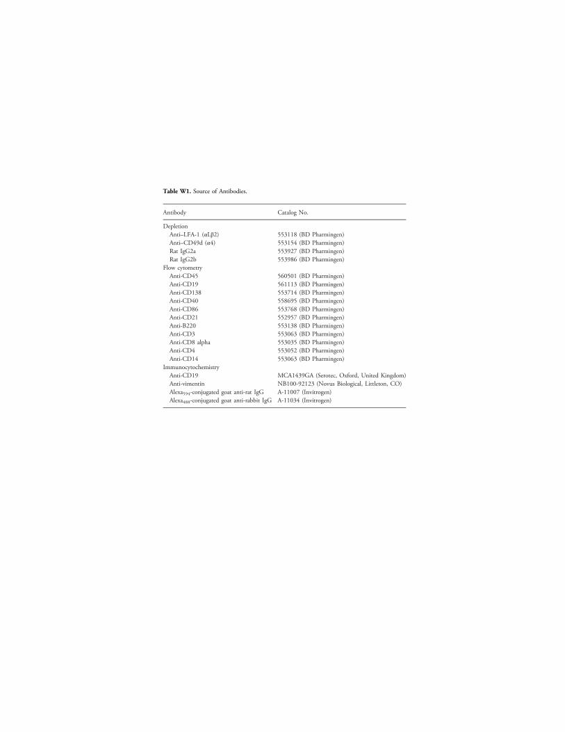
Table W1. Source of Antibodies.
Antibody Catalog No.
DepletionAnti–LFA-1 (αLβ2) 553118 (BD Pharmingen)Anti–CD49d (α4) 553154 (BD Pharmingen)Rat IgG2a 553927 (BD Pharmingen)Rat IgG2b 553986 (BD Pharmingen)
Flow cytometryAnti-CD45 560501 (BD Pharmingen)Anti-CD19 561113 (BD Pharmingen)Anti-CD138 553714 (BD Pharmingen)Anti-CD40 558695 (BD Pharmingen)Anti-CD86 553768 (BD Pharmingen)Anti-CD21 552957 (BD Pharmingen)Anti-B220 553138 (BD Pharmingen)Anti-CD3 553063 (BD Pharmingen)Anti-CD8 alpha 553035 (BD Pharmingen)Anti-CD4 553052 (BD Pharmingen)Anti-CD14 553063 (BD Pharmingen)
ImmunocytochemistryAnti-CD19 MCA1439GA (Serotec, Oxford, United Kingdom)Anti-vimentin NB100-92123 (Novus Biological, Littleton, CO)Alexa594-conjugated goat anti-rat IgG A-11007 (Invitrogen)Alexa488-conjugated goat anti-rabbit IgG A-11034 (Invitrogen)
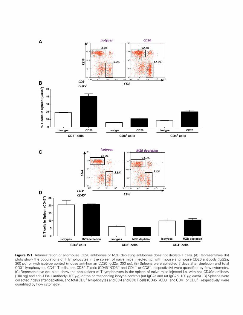
Figure W1. Administration of antimouse CD20 antibodies or MZB depleting antibodies does not deplete T cells. (A) Representative dotplots show the populations of T lymphocytes in the spleen of naive mice injected i.p. with mouse antimouse CD20 antibody (IgG2a,300 μg) or with isotype control (mouse anti-human CD20 IgG2a, 300 μg). (B) Spleens were collected 7 days after depletion and totalCD3+ lymphocytes, CD4+ T cells, and CD8+ T cells (CD45+/CD3+ and CD4+ or CD8+, respectively) were quantified by flow cytometry.(C) Representative dot plots show the populations of T lymphocytes in the spleen of naive mice injected i.p. with anti-CD49d antibody(100 μg) and anti–LFA-1 antibody (100 μg) or the corresponding isotype controls (rat IgG2a and rat IgG2b, 100 μg each). (D) Spleens werecollected 7 days after depletion, and total CD3+ lymphocytes and CD4 and CD8 T cells (CD45+/CD3+ and CD4+ or CD8+), respectively, werequantified by flow cytometry.
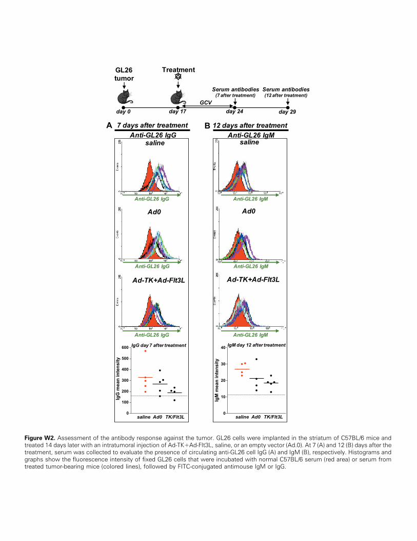
Figure W2. Assessment of the antibody response against the tumor. GL26 cells were implanted in the striatum of C57BL/6 mice andtreated 14 days later with an intratumoral injection of Ad-TK+Ad-Flt3L, saline, or an empty vector (Ad.0). At 7 (A) and 12 (B) days after thetreatment, serum was collected to evaluate the presence of circulating anti-GL26 cell IgG (A) and IgM (B), respectively. Histograms andgraphs show the fluorescence intensity of fixed GL26 cells that were incubated with normal C57BL/6 serum (red area) or serum fromtreated tumor-bearing mice (colored lines), followed by FITC-conjugated antimouse IgM or IgG.

Figure W3. Assessment of the antibody response against tumor cells expressing a surrogate antigen. GL26 cells expressing chickenovalbumin (GL26-OVA) were implanted in the striatumof C57BL/6mice and treated 14 days later with an intratumoral injection of Ad-TK+Ad-Flt3L, saline, or an empty vector (Ad.0). Seven days after the treatment, serum was collected to evaluate the presence of circulating anti–GL26-OVA cell IgM and IgG. Histograms and graphs show the fluorescence intensity of fixed GL26-OVA cells that were incubated withnormal C57BL/6 serum (red area) or serum from treated tumor-bearing mice (colored lines), followed by FITC-conjugated antimouse IgMor IgG.
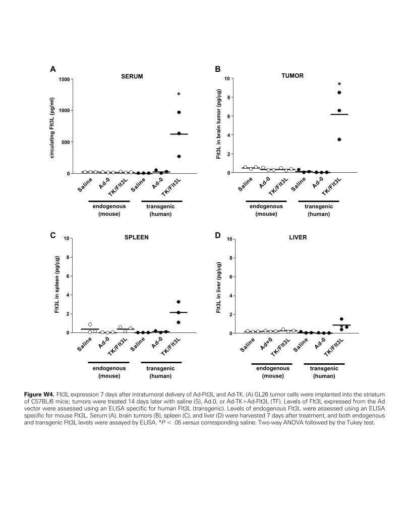
Figure W4. Flt3L expression 7 days after intratumoral delivery of Ad-Flt3L and Ad-TK. (A) GL26 tumor cells were implanted into the striatumof C57BL/6 mice; tumors were treated 14 days later with saline (S), Ad.0, or Ad-TK+Ad-Flt3L (TF). Levels of Flt3L expressed from the Advector were assessed using an ELISA specific for human Flt3L (transgenic). Levels of endogenous Flt3L were assessed using an ELISAspecific for mouse Flt3L. Serum (A), brain tumors (B), spleen (C), and liver (D) were harvested 7 days after treatment, and both endogenousand transgenic Flt3L levels were assayed by ELISA. *P < .05 versus corresponding saline. Two-way ANOVA followed by the Tukey test.