Embed Size (px)
Citation preview
KU LEUVEN
GROEP BIOMEDISCHE WETENSCHAPPEN
FACULTEIT BEWEGINGS- EN REVALIDATIEWETENSCHAPPEN
Longitudinal follow-up of children with Cerebral Palsy after Single-Event Multilevel Surgery
door Lise Loyensen Evie Moonemans
masterproef aangeboden, tot het behalen van de graad van Master of Science in de revalidatiewetenschappen en kinesitherapie
o.l.v.prof. dr. K. Desloovere, promotorprof. dr. A. Van Campenhout, copromotordr. L. Bar-On, copromotor
LEUVEN, 2016

KU LEUVEN
GROEP BIOMEDISCHE WETENSCHAPPEN
FACULTEIT BEWEGINGS- EN REVALIDATIEWETENSCHAPPEN
Longitudinal follow-up of children with Cerebral Palsy after Single-Event Multilevel Surgery
door Lise Loyensen Evie Moonemans
masterproef aangeboden, tot het behalen van de graad van Master of Science in de revalidatiewetenschappen en kinesitherapie
o.l.v.prof. dr. K. Desloovere, promotorprof. dr. A. Van Campenhout,
copromotordr. L. Bar-On, copromotor
LEUVEN, 2016Opgesteld volgens de richtlijnen van Gait & Posture
WOORD VOORAF
Deze masterproef zouden we niet gerealiseerd hebben zonder de hulp en steun van anderen. Graag
zouden we deze personen dan ook bedanken. Allereerst willen we onze promotor prof. dr. Kaat
Desloovere bedanken voor het uitzetten van de grote lijnen van onze masterproef en feedback te
geven waar nodig. Ook gaf je ons het vertrouwen om de masterproef verder uit te werken met hulp
van onze copromotoren, prof. dr. Anja Van Campenhout en dr. Lynn Bar-On.
Dank je wel prof. Van Campenhout om via jouw klinische inzichten dit interessante onderwerp aan te
brengen. Ondanks je drukbezette agenda, wist je toch steeds tijd voor ons vrij te maken en ons bij te
staan met je ervaring en feedback. Ook dank je wel dr. Lynn Bar-On voor je enorme inzet. We konden
altijd op je rekenen en hebben enorm veel gehad aan je hulp, tips en uitleg, want er kwam toch heel
veel bij kijken. Mede door jullie enthousiasme over dit onderwerp, waren wij ook vanaf het begin erg
gemotiveerd om samen tot een goed resultaat te komen.
Ook willen we graag de personen bedanken die werken in het laboratorium voor klinische
bewegingsanalyse in het Universitair Ziekenhuis Leuven, campus Pellenberg, voor het beantwoorden
van onze technische vragen en het ter beschikking stellen van de patiëntengegevens die we nodig
hadden om dit onderzoek uit te voeren.
We willen zeker en vast ook elkaar bedanken voor de leuke en goede samenwerking. Door het
vertrouwen in elkaar en omdat we beide even gemotiveerd en enthousiast zijn, hebben we er samen
alles aan gedaan om onze masterproef tot een goed einde te brengen.
Tot slot mogen we onze ouders, familie en vrienden niet vergeten. Jullie stonden altijd klaar als wij
het even niet meer zagen zitten. Bedankt voor jullie steun en vertrouwen de afgelopen jaren!
Val-Meer & Budel, 20 mei 2016 L.L. & E.M.
II
SITUERING
Deze masterproef kadert binnen een onderzoekslijn naar Cerebrale Parese (CP) in de afdeling
Neuromotorische, Pediatrische en Pelvische revalidatie van het Departement
Revalidatiewetenschappen. Het onderwerp van deze masterproef is voortgekomen uit klinische
bevindingen in het Universitair Ziekenhuis (UZ) Leuven, campus Pellenberg. In de praktijk werd
namelijk vastgesteld dat de resultaten van single-event multilevel chirurgie bij kinderen met CP zeer
uiteenlopend zijn. Er was bijgevolg nood aan bijkomend inzicht in de factoren die het effect van deze
interventie bepalen. Om de effecten van interventies te onderzoeken wordt er gebruik gemaakt van
driedimensionale ganganalyses, die worden uitgevoerd in het laboratorium voor klinische
bewegingsanalyse in UZ Leuven, campus Pellenberg. Dit laboratorium werkt nauw samen met het
Departement Revalidatiewetenschappen van de Faculteit Bewegings-en Revalidatiewetenschappen.
De objectieve driedimensionale (3D) ganganalyse omvat 3D video-opnames, krachtmetingen en
registratie van spieractiviteit. Het ganglaboratorium bestaat uit een geautomatiseerd 3D
videosysteem, een set van normale videocamera’s, apparatuur voor het meten van krachten en een
systeem voor de evaluatie van spieractiviteit tijdens het stappen. De meetapparatuur en bijhorende
software wordt gekenmerkt door een snelle technische ontwikkelingen. Voor het onderzoek worden
15 reflecterende merktekens en EMG-elektroden op het onderste lidmaat aangebracht. Vervolgens
stapt de patiënt 10 meter op blote voeten en eventueel ook met aangepast schoeisel. Met een
infraroodcamera wordt de positie van de retro-reflecterende merktekens op elk ogenblik van een
beweging geregistreerd. Aan de hand van deze bewegingstrajecten, in combinatie met kinematische
modellen, worden de bewegingen in de gewrichten van het onderstel lidmaat gestructureerd in kaart
gebracht. Deze objectieve evaluatiemethode is cruciaal voor het plannen en opvolgen van
behandelingen voor patiënten met gangproblemen [1].
Deze masterproef legt de focus op ganganalyse bij kinderen met CP. CP is een permanente
aandoening in de hersenen, waardoor primaire problemen ontstaan, zoals toegenomen
spierspanning, en afgenomen spierkracht, -controle en balans [2]. Deze primaire problemen kunnen
leiden tot spierverkortingen en botvervormingen, welke de kwaliteit en efficiëntie van de gang
aantasten [3]. Om dit te verbeteren ondergaan deze kinderen single-event multilevel chirurgie. Dit is
een aanpak waarbij verschillende ingrepen worden uitgevoerd tijdens één operatie, gevolgd door
één revalidatieperiode. Procedures die hierbij vaak uitgevoerd worden zijn skeletale ingrepen, zoals
derotatie van de femur of tibia en skeletale correcties ter hoogte van de voet, samen met weke-
III

delen ingrepen zoals spierverlengingen en peestransfers [4]. Om te bepalen welke ingrepen moeten
worden uitgevoerd en om de effecten van de ingrepen op te volgen, wordt de hierboven beschreven
ganganalyse toegepast [5]. In het verleden zijn regelmatig studies uitgevoerd naar de effecten van
deze operaties op korte termijn (< drie jaar). Deze effecten blijken positief. In de praktijk merkt men
echter dat er op langere termijn toch weer een achteruitgang optreedt. Dit is in de literatuur echter
nog niet vaak bestudeerd. Ook is het interessant om na te gaan welke factoren een positief lange-
termijn effect kunnen voorspellen. Om dit te onderzoeken, is deze masterproef opgestart.
Referenties:
[1] UZ Leuven, (n.d.). http://www.uzleuven.be/ganglabo (accessed April 4, 2016).
[2] P. Rosenbaum, N. Paneth, A. Leviton, M. Goldstein, M. Bax, D. Damiano, et al., A report: the definition and classification of cerebral palsy., Dev. Med. Child Neurol. Suppl. 109 (2007) 8–14.
[3] S. a. Rethlefsen, D.D. Ryan, R.M. Kay, Classification systems in cerebral palsy, Orthop. Clin. North Am. 41 (2010) 457–467.
[4] R. Norlin, H. Tkaczuk, one-session surgey for correction of lower extremity deformities in children with cerebral palsy, J. Pediatr. Orthop. 5 (1985) 208–211.
[5] F.M. Chang, J.T. Rhodes, K.M. Flynn, J.J. Carollo, The role of gait analysis in treating gait abnormalities in cerebral palsy, Orthop. Clin. North Am. 41 (2010) 489–506.
IV

Longitudinal follow-up of children with Cerebral Palsy after Single-Event Multilevel Surgery
5

Abstract
Background: Single-event multilevel surgery (SEMLS) is frequently used to treat secondary
musculoskeletal problems in children with spastic Cerebral Palsy (CP). Using three-dimensional gait
analysis (3DGA), objective decisions for and the evaluation of SEMLS can be made. The aims of this
retrospective study are to report the effect of SEMLS using 3DGA two to eight years post-surgery and
to explore the predictors of long-term results post SEMLS.
Methods: Thirty-one ambulant children with spastic CP were included. They all had SEMLS at an age
of five to 18 years, a preoperative and at least one postoperative 3DGA with a follow-up (FU) of two
to eight years. Primary outcome measures were the gait profile score (GPS), GPS in sagittal plane and
GPS in transverse plane. Secondary outcome measures were pelvic tilt, hip extension, hip rotation
and knee extension in stance while walking.
Results: GPS, GPS in sagittal plane, GPS in transverse plane, hip rotation and knee extension
improved significantly from 15.6° to 11.0°, 17.2° to 12,6°, 16.0° to 10.9°, 14.4° to -1.2° and 24.0° to
7.5° at 8 years FU respectively (p≤0.01). No change was observed in pelvic tilt and hip extension only
improved on the short-term from 10.6° to 6.3°. Positive long term outcome was associated with a
low GPS value and low knee angle at initial contact at baseline.
Conclusion: The effects of SEMLS on several gait parameters were found to be significant and
clinically relevant over the long-term. The results of the prediction can be used for further
investigation.
Keywords: Cerebral Palsy, Single-event multilevel surgery, 3D gait analysis, Long-term follow-up,
Prediction
1

1. IntroductionCerebral palsy (CP) is a neurological disorder in children. It occurs in about two to three per 1000
births, making it the most common cause of motor disability in childhood [1,2].
CP describes a group of permanent disorders of the development of movement and posture, causing
limitations in activity that are attributed to non-progressive disturbances that occurred in the
developing fetal or infant brain. The motor disorders of cerebral palsy are often accompanied by
disturbances of sensation, perception, cognition, communication and behavior, by epilepsy, and by
secondary musculoskeletal problems [3,4]. Preterm and low birth weight are risk factors which play a
role in developing CP [5].
The main problems in children with CP are abnormal muscle tone (mostly hypertonicity), muscle
weakness, poor selective motor control (inability to activate a specific pattern of muscles in an
isolated way [6]) and balance problems. Secondary to these main problems children with CP very
often develop musculoskeletal problems, such as muscle-tightness which is linked to decreased range
of motion (ROM) and bony deformities (e.g. femoral anteversion, tibial torsion, scoliosis, hip
subluxation). Together, these problems affect the quality and efficacy of gait [1,6]. Patients with CP
can be classified in several ways [6]: based on type of movement disorder, limb distribution, or by the
gross motor function using the Gross Motor Function Classification System (GMFCS). This latter
system rates the patients’ ambulatory function on a five-point ordinal scale [6].
Management of children with CP includes physiotherapy, casting, orthotic devices, and tone
reduction by oral medication (baclofen, diazepam, dantrolene sodium), Botulinum toxin A injections
or by neurosurgery (intrathecal baclofen pump or selective dorsal rhizotomy) [1,7]. Secondary
musculoskeletal problems are treated by orthopedic surgery, which includes soft tissue procedures
such as tendon lengthening, tendon transfers and conversion of biarticular to monoarticular muscles
and bony procedures with hip corrections, derotation of femur and tibia and foot surgery. In the
past, multiple procedures were performed spread over time (single level surgery). This was also
called ‘the birthday syndrome’, reflecting a yearly (or every other year) series of hospital admissions,
surgery and rehabilitation [1,8]. Nowadays, single-event multilevel surgery (SEMLS) is considered the
standard care for secondary musculoskeletal problems [9]. In this kind of surgery several procedures
are performed in one go, followed by one rehabilitation period. Most commonly combined surgical
procedures during SEMLS are bony corrections, such as derotation osteotomy of femur or tibia and
bony foot surgery, and soft tissue surgery, either muscle-tendon lengthening (psoas, adductors,
hamstrings, gastro-soleus complex and peronei) or transfers (rectus femoris, tibialis anterior and
posterior). The rehabilitation period includes a period of non-weight bearing and casting, followed by
intensive physical therapy to enhance strength, mobility and function [1,7,8,10,11]. Surgery to
2

improve gait is best performed after a child has developed a stable gait (after seven years) [12,13].
Given the invasive nature of these surgeries and the young age of the patients, it is detrimental to
make accurate evaluations regarding the need and timing of intervention. Once the intervention has
been decided upon, it is equally important to accurately evaluate the short and long-term outcome
and to realize the predictors of good outcome in order to improve any future interventions.
To make treatment decisions based on maximizing gait performance, three-dimensional gait analysis
(3DGA) techniques are important as they provide objective information [8,10,13,14]. To evaluate the
effect of treatment, gait data collected before and after surgery are compared. It has been
demonstrated that 3DGA can improve the long-term outcomes of different interventions in
ambulatory children with CP [15]. Additionally, it is important to know the natural progression of gait
in CP in comparison to that of typically developing children. In general, gait in children with CP will
deteriorate over time, especially during the adolescent growth spurt [1,12]. The deterioration of gait
is caused by progressively developing skeletal deformities and soft tissue contractures with growth
and increasing weight, because spastic and weak muscles fail to grow in proportion with the bone
[1,12]. For example, Bell et al. [16] reported an overall decrease in stride and temporal parameters,
gait kinematics and passive ROM over time in children with CP.
A comprehensive systematic review concluded that SEMLS had a large impact on gait in most
children with CP, but less impact on their gross motor function [11]. Nevertheless, several studies
examining the short-term effects (≤ three years follow-up) of SEMLS demonstrated improvements in
outcomes at body structure and body function as well as on the levels of activity and participation.
On body structure level, reduced static contractures at hip, knee and ankle post SEMLS were
reported, resulting in increased ROM [17–19].
At the activity level, improved gait kinematics [20], as measured by the Gillette Gait Index (GGI)
[19,21–23] and by the Gait Profile Score (GPS) [23] have been reported following SEMLS, but not in a
control group [23,24]. Most commonly reported kinematic improvements during gait include
reduced pelvic ROM, and increased knee and hip extension in stance [13,17–19]. Furthermore
improved gait distance [22,24], decreased Physical Cost Index (PCI) [13,22] indicating a more efficient
gait pattern and improved stabilization of gait [24] have been reported. Spatiotemporal parameters
showed improved stride length, but contradictory results on gait velocity and cadence [17,20].
Kinetic results have shown no change in the power generated at the knee and ankle, but the
maximum knee extensor moment during stance was reduced after surgical correction of crouch gait
[18]. Only minimal changes were found in Gross Motor Function Measurement (GMFM) [20,22,23].
Finally, on participation level, improvements were found in Health Related Quality of Live (HRQOL) as
measured by the Child Health Questionnaire and by the Pediatric Quality of Live Inventory [21–23]
and in the use of walking aids [24].
3
Despite the wealth of information on the short-term outcome of SEMLS, little is known about their
long-term effects (> three years follow-up). While several studies have reported follow-up three to
five years post-surgery [25–31], few provide follow-up of more than five years [12,32–34]. Those who
have reported on the long-term outcome, have shown that four to 10 years post operatively,
improved kinematic and temporospatial parameters are either maintained or further improved
[25,27,29,30,33,34]. Furthermore, in the existing literature with a follow-up period of 10 years, no
deterioration in gait parameters has been demonstrated [12,32,34]. In the study of Gannotti et al.
[32], which compared a group of 11 subjects who underwent multilevel surgery with a group of 12
subjects without intervention, it was demonstrated that while the GGI returned to pre-operative
values 10 years post-surgery, the improvements in individual kinematic parameters, such as knee
flexion at initial contact, timing of maximal knee flexion in swing and knee flexion range of motion,
were maintained in the multilevel group. Finally, Sung et al. [34] who studied a sample of 29 patients,
showed improvements in GDI and in kinematic parameters such as hip rotation, knee flexion at initial
contact and terminal swing that were larger after 10 years than after one year post-surgery.
Due to the high variability in outcome, it is also important to search for factors that are associated
with improved long-term results. For example, Švehlík et al. [12] demonstrated that the older the
child was at time of surgery, the better the long-term result on the GDI. Another study of Švehlík et
al. [35] reported that children aged 10 to 12 years with a GMFCS level III had the most improvement
on the GDI, but after correcting for other factors, age at surgery was the only predictive factor for
long-term results. On the contrary, Gannotti et al. [26] found that children with a faster
postoperative gait velocity four years after surgery were younger at time of surgery, had fewer
previous surgeries and a faster preoperative gait velocity. Harvey et al. [28] focused on the changes
in the use of assistive devices five years after SEMLS and concluded that children with higher GMFCS
scores were characterized by more changes in school or at home after surgery because of the greater
risk of declining gross motor function when they were adults. For example, if the children used
crutches at school or at home preoperatively, they tended to continue using them five years
postoperatively and if they used walkers they had more chance of changing to crutches
postoperatively.
In conclusion, while some positive results of SEMLS on the long-term have been shown, there is still
a lack of sufficient evidence and there are contradictory results regarding predictors of good
outcome. Further studies are needed to assess the long-term outcome of SEMLS in these children.
Therefore, the aims of this retrospective study are to: 1. report the effect of SEMLS based on results
from 3DGA two to eight years post-surgery; and 2. explore the predictors of long-term results post
SEMLS.
4

2. Methods
2.1 Subjects
Nine hundred forty-five children with CP were assessed for eligibility in this retrospective study. The
children were selected from the database of the Clinical Motion Analysis Laboratory of the University
Hospital of Leuven. The following inclusion criteria were used: (1) diagnosis of CP, (2) a SEMLS at UZ
Leuven, (3) aged between five and 18 years at date of operation, (4) GMFCS I-III, (5) presence of a
preoperative 3DGA, (6) presence of at least one postoperative 3DGA and (7) follow-up, including
3DGA, between two and ten years post-surgery. Children with the following criteria were excluded
from the study: diagnosis other than CP, intra-muscular Botulinum Toxin-A injections up to six
months before preoperative GA, prior orthopaedic surgery less than one year before preoperative
GA, selective dorsal rhizotomy (SDR) and the use of an intrathecal baclofen pump (ITB).
For the purpose of this study, SEMLS was defined as at least one surgical procedure performed at
two different anatomical levels (hip, knee or ankle) constituting both soft tissue and bony surgery or
only bony surgery [30]. To match the individual needs of the child, clinical examination and 3DGA
were carried out prior to surgery and a multidisciplinary team of clinicians determined which
procedures to perform. Ethical approval for this study was obtained from the Ethics Committee of
the University Hospital of Leuven (s5604).
2.2 Study design
All included children had a preoperative 3DGA and one or more analyses post-surgery. Follow-up
(FU) gait analyses were analyzed at two years (± 1.5), five years (± 1.5) and/or eight years (± 2) post-
surgery. Gait data were collected using an eight to 16 camera VICON system, operating at 100 Hz,
with 15 reflective markers located at specific anatomical landmarks of the lower limbs, according to
the lower limb Vicon PluginGait marker configuration (VICON, Oxford Metrics, Oxford, UK).
All children walked barefoot along a 10-meter walkway. Children were instructed to walk at a self-
selected walking speed. At least three successful trials were collected. A trial was considered
successful when there was good marker visibility.
2.3 Data analysis
Three dimensional kinematic data were calculated for the pelvis, hip, knee, ankle and foot bilaterally,
decomposed in the sagittal, coronal and transverse plane. All kinematic analyses were based on the
lower limb PlugInGait model (VICON, Oxford Metrics, Oxford, UK). Only the most affected leg was
selected for further analysis. The most affected leg was defined as the operated side if the surgical
procedure was performed unilaterally. If bilateral surgery was received, the most affected side was
5

determined based on the preoperative clinical examination. Gait cycles (GCs) were visually
determined by indicating the events of initial contact and toe off using Nexus software (Vicon, Oxford
Metrics Group, Oxford, UK). This was done for three to six trials to arrive at a minimal of three good
GCs per session per patient. An additional visual quality check of all the kinematic signals was carried
out in custom-made MATLAB software (Mathworks1, Natick, MA, USA), prior to further data analysis.
From the continuous waveforms organized in gait cycles for kinematics a set of discrete parameters
(maxima, minima, amplitudes, angles at specific time points in the gait cycle, timing of gait cycle
events) were selected based on clinical relevance. GPS was used as a summary statistic for gait
deviation. The GVS is the root mean square (RMS) difference between a kinematic variable from the
child and from data of typically developing children (n=55, age range 8-14 year) [36]. The GVS for
nine key relevant kinematic variables for the right and left legs can be combined to form a Movement
Analysis Profile (MAP). The RMS average of all the variable scores will then equal the GPS calculated
from the entire gait vector. [36]. All discrete parameters, as well as the GPS-values, were
automatically extracted from the waveforms using custom-made MATLAB software (Mathworks1,
Natick, MA, USA). Per subject, the value of each gait parameter for each of three or more GCs were
averaged for the selected side.
The primary outcome included post-operative GPS, GPS in the sagittal plane and GPS in the
transverse plane. Secondary outcome included the mean pelvic tilt during the gait cycle and
maximum hip extension, mean hip rotation and maximum knee extension during stance.
2.4 Statistical analysis
Means and standard deviations were calculated per parameter preoperatively and at two years, five
years and/or eight years FU, to evaluate how gait is influenced by SEMLS. Statistical analysis was
performed using SPSS Statistics version 20.0 (SPSS Inc, Chicago, USA). An alpha level below 0.05 was
used for all tests to indicate significance. To evaluate long-term outcomes, differences in kinematic
parameters preoperatively and between FU time instants were analyzed with generalized estimating
equation analysis, with different FU time instants as a within-subject factor. Exchangeable correlation
structures were assumed.
To explore possible predictors of the effect of SEMLS, correlations between subject characteristics
(age, GMFCS, and sex) and preoperative kinematic parameters to the primary outcome measures
were calculated by means of the Pearson’s correlation coefficients for normally distributed
parameters, and Spearman’s correlation coefficients for non-normally distributed parameters.
Normality was determined using the Shapiro-Wilk normality test. Parameters with a good, significant
correlation coefficient (r ≥0.50) were added to the GEE model as covariates.
6
3. Results
3.1 Subjects
Nine hundred forty-five children with CP from the database of the Clinical Motion Analysis
Laboratory of the University Hospital of Leuven were assessed for eligibility. Of those, 704 did not
meet the inclusion criteria and 102 subjects were excluded because the data were too old and not
compatible with the analysis software. Out of the remaining 139 subjects those with adequate
follow-up with GA were selected. Only the 44 subjects with a FU of five to eight years were retained,
according to the aim of the study. Thirteen of the 44 remaining subjects were excluded because of
software errors related to missing marker data. Thirty-one subjects were finally included in the study.
Characteristics of these children are summarized in Table 1.
The subjects underwent a total of 116 gait analyses. Of these gait analyses, 69 were selected based
on predefined FU time instants (2 ± 1,5 years FU, 5 ± 1,5 years FU and 8 ± 2 years). One gait analysis
was selected for each time point, but it was not feasible to select gait analyses for all participants at
all time points (Figure 1).
Table 1Characteristics of children pre Single-Event Multilevel Surgery (SEMLS) (n=31)
Characteristics SEMLS (n=31)
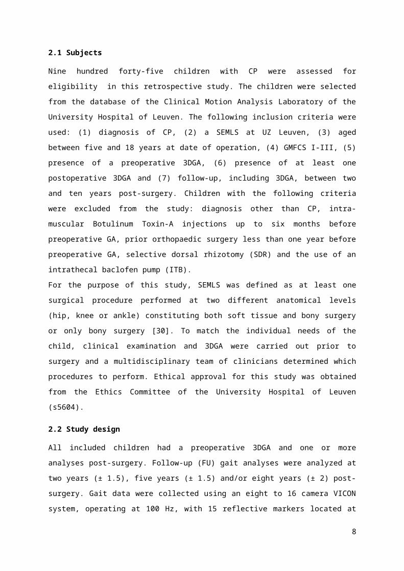
GMFCS I/II/III 12/14/5 Unilateral / bilateral CP 3/28 Gender, male/female 19/12 Mean (SD) [range] age at operation, years 11,7 (3,1) [6,5-18,0] Mean (SD) height before SEMLS, cm 141 (19) Mean (SD) weight before SEMLS, kg 36,1 (15,4)CP, Cerebral Palsy; GMFCS, Gross Motor Function Classification System; SD, Standard Deviation; cm, centimeter; kg, kilogram
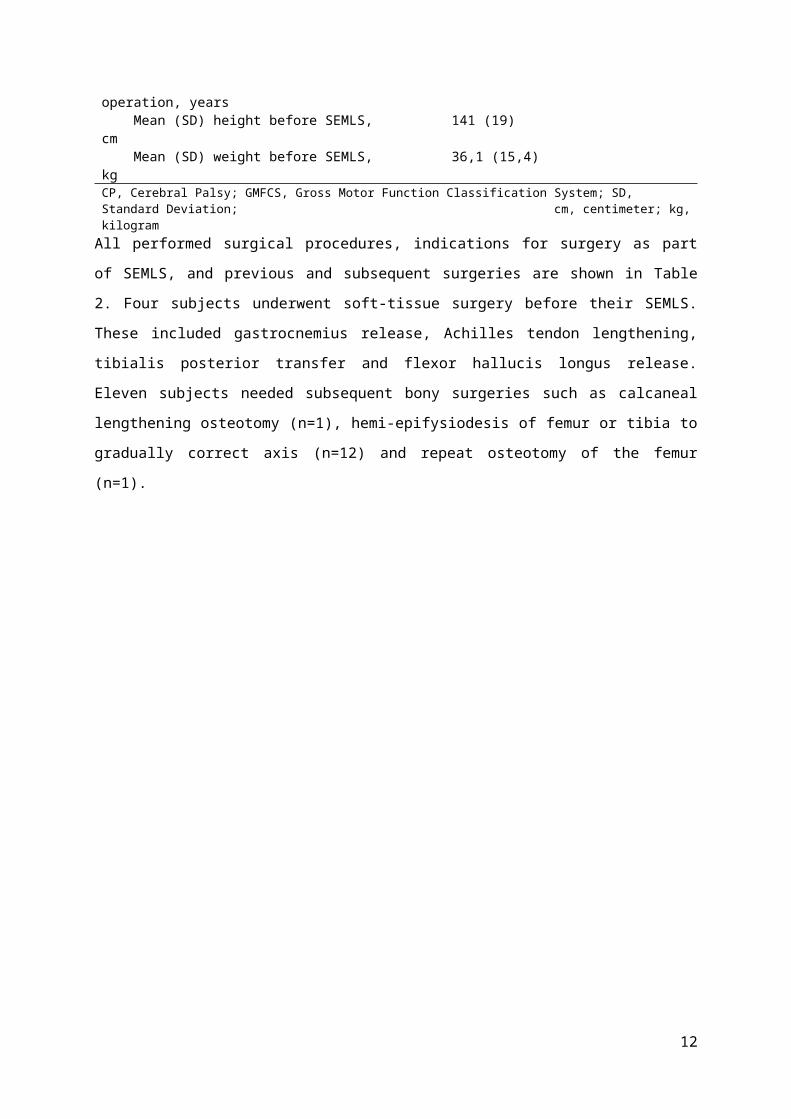
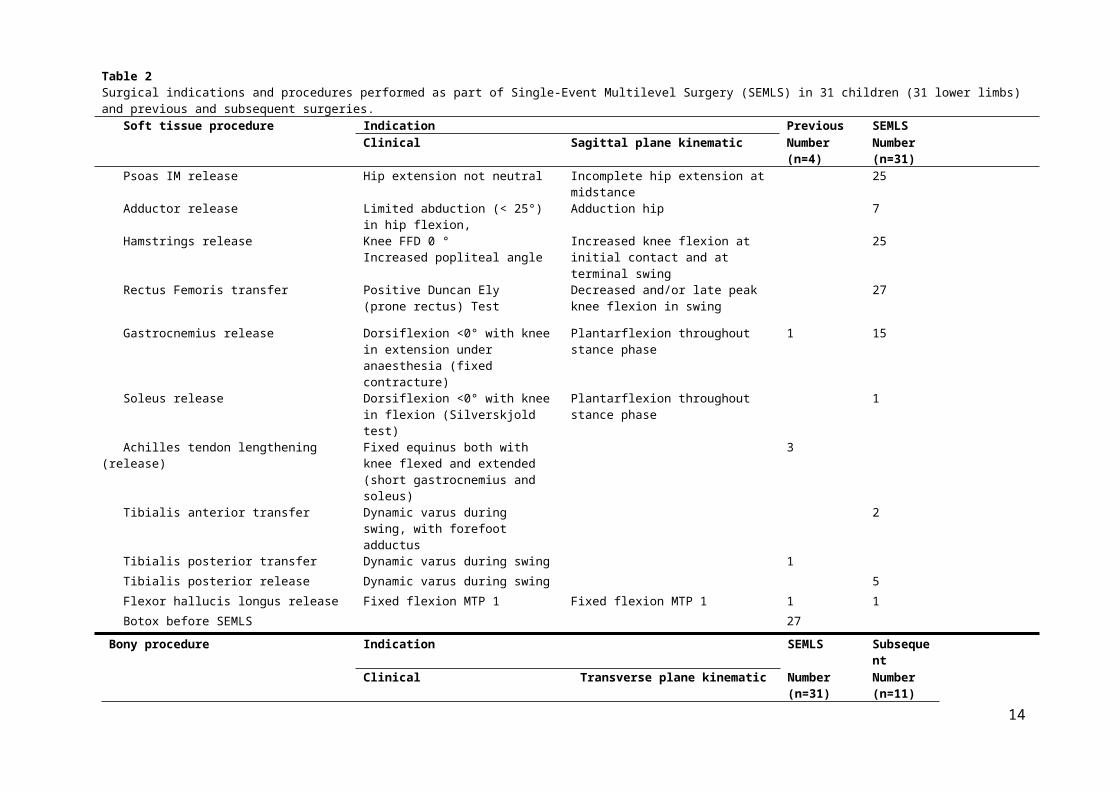
All performed surgical procedures, indications for surgery as part of SEMLS, and previous and
subsequent surgeries are shown in Table 2. Four subjects underwent soft-tissue surgery before their
SEMLS. These included gastrocnemius release, Achilles tendon lengthening, tibialis posterior transfer
and flexor hallucis longus release. Eleven subjects needed subsequent bony surgeries such as
calcaneal lengthening osteotomy (n=1), hemi-epifysiodesis of femur or tibia to gradually correct axis
(n=12) and repeat osteotomy of the femur (n=1).
7
8
Assessed for eligibility(n=945)
Out of the 139 remaining subjects only those with the longest FU (5-8 years) were retained
(n=44)
31 patients (n), 116 gait analyses (N) in total
Gait analyses selected according to FU range (N=69)
Patients (n) and gait analyses (N) analysed:n=31N=69
- 2±1,5 year FU: n= 29, N=29- 5±1,5 year FU: n= 20, N=20
- 8±2 year FU: n=20, N=20
1 gait analysis excluded because of a new SEMLS (N=115)
13 patients excluded(software errors related to missing
marker data)
Excluded (did not meet inclusion criteria)(n=704)
102 patients excluded (data too old to analyse in new
software)
Figure 1: Flow-chart of selection procedureFU, Follow-up; SEMLS, Single-Event Multilevel Surgery

Table 2Surgical indications and procedures performed as part of Single-Event Multilevel Surgery (SEMLS) in 31 children (31 lower limbs) and previous and subsequent surgeries. Soft tissue procedure Indication Previous SEMLS
Clinical Sagittal plane kinematic Number (n=4)
Number (n=31)
Psoas IM release Hip extension not neutral Incomplete hip extension at midstance 25 Adductor release Limited abduction (< 25°) in hip flexion, Adduction hip 7 Hamstrings release Knee FFD 0 °
Increased popliteal angleIncreased knee flexion at initial contact and at terminal swing
25
Rectus Femoris transfer Positive Duncan Ely (prone rectus) Test
Decreased and/or late peak knee flexion in swing
27
Gastrocnemius release Dorsiflexion <0° with knee in extension under anaesthesia (fixed contracture)
Plantarflexion throughout stance phase 1 15
Soleus release Dorsiflexion <0° with knee in flexion (Silverskjold test)
Plantarflexion throughout stance phase 1
Achilles tendon lengthening (release) Fixed equinus both with knee flexed and extended(short gastrocnemius and soleus)
3
Tibialis anterior transfer Dynamic varus during swing, with forefoot adductus
2
Tibialis posterior transfer Dynamic varus during swing 1 Tibialis posterior release Dynamic varus during swing 5 Flexor hallucis longus release Fixed flexion MTP 1 Fixed flexion MTP 1 1 1 Botox before SEMLS 27
Bony procedure Indication SEMLS SubsequentClinical Transverse plane kinematic Number
(n=31)Number(n=11)
Proximal femoral derotation osteotomy FNA >25°, increased internal hip rotation (>60°), decreased external hi rotation (<20°)
Hip rotation >10° internal throughout stance phase
24
Distal femoral extension/derotation osteotomy Fixed knee flexion contracture with or without high FNA
Knee flexion throughout stance with/without internal hip rotation
10
Tibial derotation osteotomy External tibial torsion >15° External foot progression angle throughout stance phase
19
Calcaneal lengthening osteotomy (of the feet) Planovalgus foot Limited push-off 3 1
Patellar distalisation Patella alta, usually in combination with fixed knee flexion contracture (distal femoral extension osteotomy); no active knee extension to passive ROM
Incomplete knee-extension in stance 20
Hemi-epifysiodesis femur/tibia Varus/valgus 12Flexion osteotomy femur Hyperextension Knee Hyperextension knee in stance 1
9
IM, intra-muscular; SEMLS, Single-Event Multilevel Surgery; FFD, Fixed Flexion Deformity; °, degree; FNA , Femoral Neck Anteversion; MTP, Metatarsal phalangeal joint
10
3.2 Long-term effects of SEMLS
The outcomes were evaluated preoperatively (mean 4.1 months before surgery with a range of 0.3
to 13.7 months) and two, five and eight years post-surgery (Table 3).
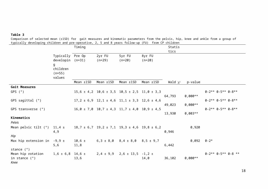
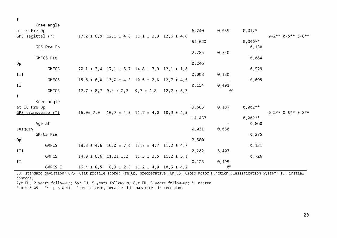
Primary outcomes
Evolution of MAPS and GPS post-operatively and over time is shown in Figure 2. GPS, GPS in sagittal
plane and GPS in transverse plane improved significantly over the long-term compared to
preoperative values (p ≤0.01). This improvement was seen at two years FU and was still present at
five and eight years FU. For GPS the mean difference between pre and 8 years follow-up was -4.6°
and for GPS in sagittal plane and GPS in transverse plane it was -4.6° and -5.1° respectively.
Secondary outcomes
The mean pelvic tilt showed no significant change postoperatively or over time. Minimal hip
extension in stance showed a significant improvement at two years FU, but the improvement was no
longer significant at five and eight years FU. Mean hip rotation in stance and maximal knee extension
in stance improved significantly at two years FU. These improvements were maintained at five and
eight years FU.
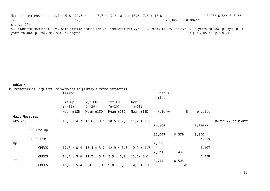
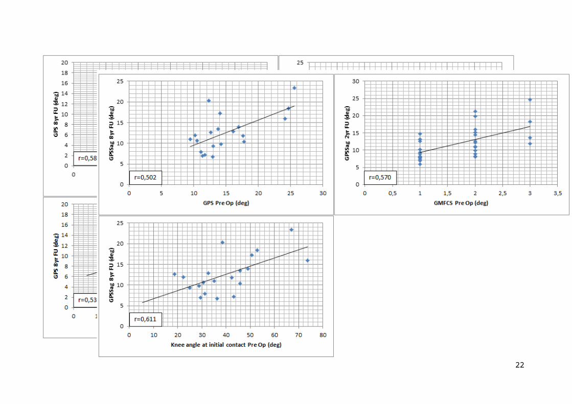
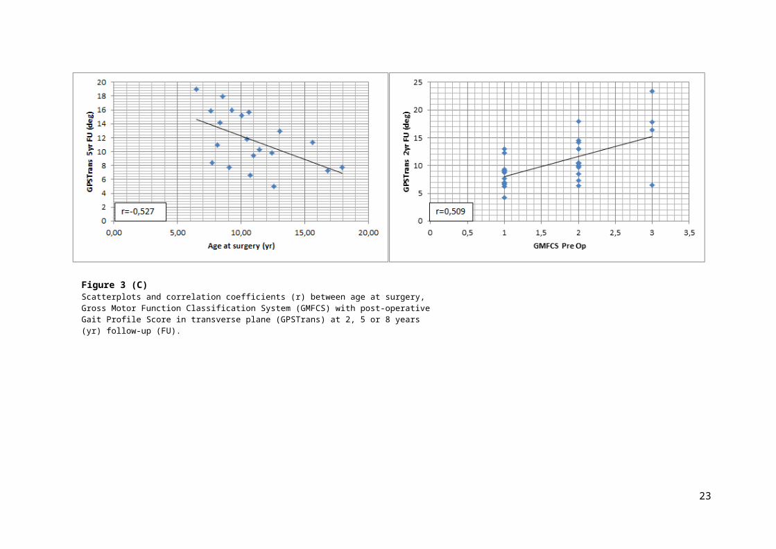
3.3 Predictors
Scatterplots of possible predictors with good (r ≥0.50) and significant correlation coefficients for the
primary outcome measures are shown in Figure 3. After univariate correlation analysis the following
predictors were retained: GPS (r=0.589), GMFCS (r=0.648) and knee angle at initial contact (r=0.537)
preoperatively for GPS (Figure 3 (A)), GPS (r=0.502), GMFCS (r=0.570) and knee angle at initial
contact (r=0.611) preoperatively for GPS in sagittal plane (Figure 3 (B)) and age at surgery (r=-0.527)
and GMFCS (r=0.509) preoperatively for GPS in transverse plane (Figure 3 (C)). The impact and
significance of these selected predictors are shown in Table 4.
Preoperative values for GPS, GMFCS and knee angle at initial contact were included as possible co-
variables of long-term improvements in GPS. When added to the GEE, preoperative GPS and knee
angle at initial contact were predictive for long term improvement in GPS.
For GPS in sagittal plane, the same three predictors were included in the model. For this outcome
parameter only knee angle at initial contact significantly contributed to long-term improvements.
Finally, for GPS in the transverse plane, age at surgery and preoperative GMFCS were significant in a
univariate analysis, but did not significantly contribute to long-term improvement in GPS in the
transverse plane.
11

Table 3Comparison of selected mean (±1SD) for gait measures and kinematic parameters from the pelvis, hip, knee and ankle from a group of typically developing children and pre-operative, 2, 5 and 8 years follow-up (FU) from CP children
Timing StatisticsTypically developing children (n=55) values
Pre Op (n=31) 2yr FU (n=29) 5yr FU (n=20) 8yr FU (n=20)
Mean ±1SD Mean ±1SD Mean ±1SD Mean ±1SD Wald p-valueGait MeasuresGPS (°) 15,6 ± 4,2 10,6 ± 3,5 10,5 ± 2,5 11,0 ± 3,3 64,793 0,000** 0-2** 0-5** 0-8**GPS sagittal (°) 17,2 ± 6,9 12,1 ± 4,6 11,1 ± 3,3 12,6 ± 4,6 49,023 0,000** 0-2** 0-5** 0-8**GPS transverse (°) 16,0 ± 7,0 10,7 ± 4,3 11,7 ± 4,0 10,9 ± 4,5 13,930 0,003** 0-2** 0-5** 0-8**KinematicsPelvisMean pelvic tilt (°) 11,4 ± 4,9 18,7 ± 6,7 19,2 ± 7,1 19,3 ± 4,6 19,8 ± 6,2 0,946 0,920HipMax hip extension in stance (°)
-9,9 ± 5,6 10,6 ± 11,8 6,3 ± 8,0 8,4 ± 8,0 8,5 ± 9,7 6,442 0,092 0-2*
Mean hip rotation in stance (°)
1,6 ± 6,8 14,6 ± 13,6 2,4 ± 9,9 2,6 ± 13,5 -1,2 ± 14,0 36,102 0,000** 0-2** 0-5** 0-8 **
KneeMax knee extension in stance (°)
1,7 ± 5,8 24,0 ± 19,5 7,7 ± 12,4 8,1 ± 10,3 7,5 ± 13,0 26,185 0,000** 0-2** 0-5** 0-8 **
SD, standard deviation; GPS, Gait profile score; Pre Op, preoperative; 2yr FU, 2 years follow-up; 5yr FU, 5 years follow-up; 8yr FU, 8 years follow-up; Max, maximum; °, degree * p ≤ 0.05 ** p ≤ 0.01
12

Table 4P Predictors of long term improvements in primary outcome parameters
Timing StatisticsPre Op (n=31) 2yr FU (n=29) 5yr FU (n=20) 8yr FU (n=20)
Mean ±1SD Mean ±1SD Mean ±1SD Mean ±1SD Wald B p-valueGait MeasuresGPS (°) 15,6 ± 4,2 10,6 ± 3,5 10,5 ± 2,5 11,0 ± 3,3 64,496 0,000** 0-2** 0-5** 0-8** GPS Pre Op 28,047 0,370 0,000** GMFCS Pre Op 2,699 0,259 GMFCS III 17,7 ± 0,4 15,4 ± 3,2 12,9 ± 3,5 10,9 ± 1,7 2,601 1,437 0,107 GMFCS II 14,3 ± 3,6 11,2 ± 3,0 9,9 ± 1,9 11,1± 3,6 0,744 0,505 0,388 GMFCS I 16,2 ± 5,4 8,4 ± 1,9 9,8 ± 1,9 10,8 ± 3,8 0a
Knee angle at IC Pre Op 6,240 0,059 0,012*GPS sagittal (°) 17,2 ± 6,9 12,1 ± 4,6 11,1 ± 3,3 12,6 ± 4,6 52,620 0,000** 0-2** 0-5** 0-8** GPS Pre Op 2,285 0,240 0,130 GMFCS Pre Op 0,246 0,884 GMFCS III 20,1 ± 3,4 17,1 ± 5,7 14,8 ± 3,9 12,1 ± 1,8 0,008 0,130 0,929 GMFCS II 15,6 ± 6,0 13,0 ± 4,2 10,5 ± 2,8 12,7 ± 4,5 0,154 -0,401 0,695 GMFCS I 17,7 ± 8,7 9,4 ± 2,7 9,7 ± 1,8 12,7 ± 5,7 0a
Knee angle at IC Pre Op 9,665 0,187 0,002**GPS transverse (°) 16,0± 7,0 10,7 ± 4,3 11,7 ± 4,0 10,9 ± 4,5 14,457 0,002** 0-2** 0-5** 0-8** Age at surgery 0,031 -0,038 0,860 GMFCS Pre Op 2,580 0,275 GMFCS III 18,3 ± 4,6 16,0 ± 7,0 13,7 ± 4,7 11,2 ± 4,7 2,282 3,407 0,131 GMFCS II 14,9 ± 6,6 11,2± 3,2 11,3 ± 3,5 11,2 ± 5,1 0,123 0,495 0,726 GMFCS I 16,4 ± 8,5 8,3 ± 2,5 11,2 ± 4,9 10,5 ± 4,2 0a
SD, standard deviation; GPS, Gait profile score; Pre Op, preoperative; GMFCS, Gross Motor Function Classification System; IC, initial contact; 2yr FU, 2 years follow-up; 5yr FU, 5 years follow-up; 8yr FU, 8 years follow-up; °, degree * p ≤ 0.05 ** p ≤ 0.01 a set to zero, because this parameter is redundant
13

14
Figure 2Movement Analysis Profile (MAPS) & Gait Profile Score (GPS) over time for all patients (n=31)RMS, Root Mean Square; pel, pelvic; ank, ankle; flex, flexion; dors, dorsiflexion; obl, obliquity; abd, abduction; rot, rotation; prog, progression;GPSSag, GPS in sagittal plane; GPSTrans, GPS in transverse plane; FU, follow-up; deg, degree
Pel tilt Hip flex Knee flex Ank dors Pel obl Hip abd Pel rot Hip rot Foot prog GPS GPS sag GPS trans0
5
10
15
20
25
pre SEMLS (n=31)2 year FU (n=29)5 year FU (n=20)8 year FU (n=20)
RMS
diffe
renc
e (d
eg)
15
Figure 3 (C)Scatterplots and correlation coefficients (r) between age at surgery, Gross Motor Function Classification System (GMFCS) with post-operative Gait Profile Score in transverse plane (GPSTrans) at 2, 5 or 8 years (yr) follow-up (FU).

4. DiscussionThis retrospective study examined the long-term effects of SEMLS by comparing preoperative to
postoperative values until eight years post-surgery. Furthermore, this study initiated an exploration
of possible predictors for the long-term effect of SEMLS.
To study the effects it is important to take into account the natural progression of gait. When a child
becomes older, gait often deteriorates because of increasing weight and failure of spastic and weak
muscles to grow in proportion with the bone [1,12,16] . Therefore maintenance of mobility in
children with CP is a valid long-term goal [35]. Eleven subjects in this study needed subsequent bony
surgeries after their SEMLS, although these were minor procedures. This could also be a
consequence of growth leading to recurrent deformities. In the study of Thomason et al. [30] it was
mentioned that ‘fine-tuning’ by additional surgeries is therefore often required because one SEMLS
cannot carry out all the procedures which are needed for the total growth period of the child.
When looking at long-term results, we demonstrated that overall gait improved significantly
postoperatively and remained stable over the long term. Improvements were two to three times the
minimal clinically important difference (MCID), which is 1.6° for the GPS [37]. These results confirmed
the study of Firth et al. [33] who, with a mean follow-up of 7,5 years, also found a significant
improvement in GPS that was still present at the end of the follow-up period. Comparable results
were seen in two studies of Švehlík et al. [12,35] and of Sung et al. [34] who followed subjects up to
10 years post-operatively.
Besides overall gait improvement, the evolution of clinical relevant kinematic parameters was
recorded based on the opinion of clinical experts. Selected parameters were mean hip extension
during stance and mean pelvic tilt during gait cycle, because high anterior pelvic tilt at adult age may
lead to subjective low back pain. Mean hip rotation is clinically important to verify whether
correction of rotational deformities is maintained over the long term and minimal knee angle in
stance is an important predictor of anterior knee problems. For these secondary outcome measures,
this study showed no significant change in mean pelvic tilt, but a tendency to a slight increase of
anterior tilt. A significant improvement in mean hip extension at two-year follow-up was found which
was not significant anymore on the long term. Furthermore a significant improvement was found
that remained constant over the long term for the mean hip rotation in stance towards external
rotation and the maximal knee extension in stance. Similar findings were reported in literature
similar results. Sung et al. [34] also showed no significant change in pelvic tilt and an improvement in
mean hip rotation towards external rotation after 10 years. The tendency to an increasing anterior
pelvic tilt in the results of our study could be a consequence of hamstring lengthening, which was
carried out in 25 subjects. Similar findings were reported by Rodda et al. [25], who found a significant
16
deterioration at five years follow-up. Furthermore, they found a significant increase in knee
extension during stance that was maintained until five years follow-up. They did not find any
improvement for hip extension in stance. A significant improvement in mean hip rotation was found
at one year, but during FU there was a deterioration back to preoperative values. This is in contrast
with the findings in this study. The difference could be explained by the very small sample size of only
10 subjects and no participants with a GMFCS I in the study of Rodda et al. [25]. We also compared
our study to the results of Ounpuu et al. [38], who specifically focused at the results of a femoral
derotation osteotomy and also included ambulatory children with a GMFCS I-III. They found a clear
improvement that continued until five years which is in accordance with our results. Saraph et al.
[29] studied the effects of SEMLS on maximum hip extension and maximum knee extension during
stance. They found that hip extension significantly improved on the short term (three years follow-
up). This was also observed in our study, but the results did not last on the long term. A possible
explanation for this may be that the spasticity of the psoas increased again. For maximum knee
extension, Saraph et al. [29] found a significant improvement until three years, although there was a
tendency to more flexion at three years compared to one year follow-up. The results of our study
showed that this improvement on the short term also existed on the long term.
The second aim of this study was to initiate the exploration of possible predictors for the effect of
SEMLS. For the prediction of the primary outcome measures, this study showed that age at time of
surgery, GMFCS-level, GPS and knee angle at initial contact before surgery, related to long-term
outcome. Contrary to our expectations, age at surgery was not retained as a predictor of long term
improvement on the primary outcome parameters. Only for GPS in the transverse plane the
correlation was good enough to include this as a covariate in the prediction model, but even then it
was not found to significantly contribute to the prediction. In contrast with our results, other studies
found age at surgery as a significant predictor of gait improvement [12,35] and of postoperative gait
velocity [26]. The small sample size of our study for prediction could have played a role in the
explanation of this difference. The level on the GMFCS, often used as an indication of severity, did
not have an influence on any of the outcome measures. This is in accordance with the study of
Švehlík et al. [35], who found that, after correction for age, GMFCS was no longer significant. Knee
flexion at initial contact and GPS before surgery were found to be very significant in the prediction
model in our study. GPS before surgery is objectively measured and is a more continuous measure of
severity compared to the GMFCS, which may increase its predictive capacity. Furthermore, both GPS
and knee flexion at initial contact are clinically important parameters. Therefore, it may be
interesting to explore these covariates in future research on a larger and more homogeneous
sample.
17
Besides the limited sample size due to the strict inclusion criteria, several other limitations of the
current study should be mentioned. The design of the study was retrospective. This explains why not
every subject had a 3DGA at every follow-up time instant and why duration of follow-up was not
consistent for all subjects. Therefore, there was a considerable incidence of missing data, which
limits the interpretation of the evolution of changes in gait on the long term. Another limitation was
the lack of a control group. This may lead to an underestimation of the results. However,, it is
considered ethically unjustifiable to create a group of patients with CP who do not receive, but do do
need surgical correction. This limitation is partially compensated by the use of 3DGA which is an
objective instrument to measure gait and is relatively immune to bias when strict selection criteria
are applied [30]. The fact that only ambulatory children were included, implicates that the results
cannot be generalized to all children with CP. Furthermore there was a variation in surgical
procedures between subjects, although all the combinations of procedures are covered by the
definition of SEMLS [30]. Finally, the study did not control for the effect of subsequent single-level
surgeries. This may interact with the outcome of SEMLS, but these procedures are inevitable in
retrospective study designs.
In conclusion, this study examined the long-term effect of SEMLS in ambulatory children with spastic
CP and found an overall improvement of gait, which was maintained over the long term. For the
prediction, only GPS and knee flexion at initial contact before surgery were found as significant
predictors for the effect of SEMLS, but this has to be further investigated in future research involving
larger samples.
Conflict of interest
The authors have no conflict of interest.
18

References[1] U. Narayanan, Management of Children With Ambulatory Cerebral Palsy: An Evidence-based
Review, J. Pediatr. Orthop. 32 (2012) 172–181.
[2] C. Cans, Surveillance of cerebral palsy in Europe: a collaboration of cerebral palsy surveys and registers. Surveillance of Cerebral Palsy in Europe (SCPE)., Dev. Med. Child Neurol. 42 (2000) 816–824.
[3] P. Rosenbaum, N. Paneth, A. Leviton, M. Goldstein, M. Bax, D. Damiano, et al., A report: the definition and classification of cerebral palsy., Dev. Med. Child Neurol. Suppl. 109 (2007) 8–14.
[4] M. Bax, M. Goldstein, P. Rosenbaum, A. Leviton, N. Paneth, B. Dan, et al., Proposed definition and classification of cerebral palsy, April 2005., Dev. Med. Child Neurol. 47 (2005) 571–6.
[5] A. Colver, C. Fairhurst, P.O.D. Pharoah, Cerebral palsy., Lancet. 383 (2014) 1240–9.
[6] S. a. Rethlefsen, D.D. Ryan, R.M. Kay, Classification systems in cerebral palsy, Orthop. Clin. North Am. 41 (2010) 457–467.
[7] J.M. Jacobs, Management options for the child with spastic cerebral palsy., Orthop. Nurs. 20 (2001) 53–59; quiz 59–61.
[8] F.M. Chang, J.T. Rhodes, K.M. Flynn, J.J. Carollo, The role of gait analysis in treating gait abnormalities in cerebral palsy, Orthop. Clin. North Am. 41 (2010) 489–506.
[9] R. Norlin, H. Tkaczuk, one-session surgey for correction of lower extremity deformities in children with cerebral palsy, J. Pediatr. Orthop. 5 (1985) 208–211.
[10] U.G. Narayanan, The role of gait analysis in the orthopaedic management of ambulatory cerebral palsy., Curr. Opin. Pediatr. 19 (2007) 38–43.
[11] J.L. McGinley, F. Dobson, R. Ganeshalingam, B.J. Shore, E. Rutz, H.K. Graham, Single-event multilevel surgery for children with cerebral palsy: A systematic review, Dev. Med. Child Neurol. 54 (2012) 117–128.
[12] M. Švehlík, G. Steinwender, T. Kraus, V. Saraph, T. Lehmann, W.E. Linhart, et al., The influence of age at single-event multilevel surgery on outcome in children with cerebral palsy who walk with flexed knee gait, Dev. Med. Child Neurol. 53 (2011) 730–735.
[13] A. Neve, G. Evans, J. Patrick, Simultaneous multiple operations for spastic diplegia, J Bone Jt. Surg. (1993) 488–494.
[14] P.A. DeLuca, R.B. Davis, S. Ounpuu, S. Rose, R. Sirkin, Alterations in surgical decision making in patients with cerebral palsy based on three-dimensional gait analysis., J. Pediatr. Orthop. 17 (1997) 608–14.
[15] P. Thomason, J. Rodda, M. Sangeux, P. Selber, Kerr Graham, Management of Children With Ambulatory Cerebral Palsy: An Evidence-based Review. Commentary by Hugh Williamson Gait Laboratory Staff., J. Pediatr. Orthop. 32 Suppl 2 (2012) 182–186.
[16] K.J. Bell, S. Ounpuu, P. a DeLuca, M.J. Romness, Natural progression of gait in children with cerebral palsy., J. Pediatr. Orthop. 22 (2002) 677–682.
[17] N.M. Bernthal, S.C. Gamradt, R.M. Kay, T. a L. Wren, A. V Cuomo, J. Reid, et al., Static and dynamic gait parameters before and after multilevel soft tissue surgery in ambulating children with cerebral palsy., J. Pediatr. Orthop. 30 (2010) 174–179.
[18] D. Metaxiotis, S. Wolf, L. Doederlein, Conversion of biarticular to monoarticular muscles as a component of multilevel surgery in spastic diplegia., J. Bone Joint Surg. Br. 86 (2004) 102–109.
[19] M. Gough, P. Schneider, a P. Shortland, The outcome of surgical intervention for early deformity in young ambulant children with bilateral spastic cerebral palsy., J. Bone Joint Surg.
19

Br. 90 (2008) 946–951.
[20] V. Saraph, E.-B. Zwick, G. Zwick, C. Steinwender, G. Steinwender, W. Linhart, Multilevel surgery in spastic diplegia: evaluation by physical examination and gait analysis in 25 children., J. Pediatr. Orthop. 22 (2002) 150–157.
[21] G.E. Gorton, M.F. Abel, D.J. Oeffinger, A. Bagley, S.P. Rogers, D. Damiano, et al., A prospective cohort study of the effects of lower extremity orthopaedic surgery on outcome measures in ambulatory children with cerebral palsy., J. Pediatr. Orthop. 29 (2009) 903–909.
[22] A. Akerstedt, O. Risto, P. Odman, B. Oberg, Evaluation of single event multilevel surgery and rehabilitation in children and youth with cerebral palsy--A 2-year follow-up study., Disabil. Rehabil. 32 (2010) 530–539.
[23] P. Thomason, R. Baker, K. Dodd, N. Taylor, P. Selber, R. Wolfe, et al., Single-event multilevel surgery in children with spastic diplegia: a pilot randomized controlled trial., J. Bone Joint Surg. Am. 93 (2011) 451–460.
[24] M. Gough, L.C. Eve, R.O. Robinson, A.P. Shortland, Short-term outcome of multilevel surgical intervention in spastic diplegic cerebral palsy compared with the natural history., Dev. Med. Child Neurol. 46 (2004) 91–97.
[25] J.M. Rodda, H.K. Graham, G.R. Nattrass, M.P. Galea, R. Baker, R. Wolfe, Correction of severe crouch gait in patients with spastic diplegia with use of multilevel orthopaedic surgery., J. Bone Joint Surg. Am. 88 (2006) 2653–2664.
[26] M.E. Gannotti, G.E. Gorton, M.T. Nahorniak, P.D. Masso, B. Landry, J. Lyman, et al., Postoperative gait velocity and mean knee flexion in stance of ambulatory children with spastic diplegia four years or more after multilevel surgery., J. Pediatr. Orthop. 27 (2007) 451–456.
[27] S.E. Adolfsen, S. Ounpuu, K.J. Bell, P. a DeLuca, Kinematic and kinetic outcomes after identical multilevel soft tissue surgery in children with cerebral palsy., J. Pediatr. Orthop. 27 (2007) 658–667.
[28] A. Harvey, P. Rosenbaum, S. Hanna, R. Yousefi-Nooraie, H.K. Graham, Longitudinal changes in mobility following single-event multilevel surgery in ambulatory children with cerebral palsy, J. Rehabil. Med. 44 (2012) 137–143.
[29] V. Saraph, E.-B. Zwick, C. Auner, F. Schneider, G. Steinwender, W. Linhart, Gait improvement surgery in diplegic children: how long do the improvements last?, J. Pediatr. Orthop. 25 (2005) 263–267.
[30] P. Thomason, P. Selber, H.K. Graham, Single Event Multilevel Surgery in children with bilateral spastic cerebral palsy: A 5 year prospective cohort study, Gait Posture. 37 (2013) 23–28.
[31] E.M. Godwin, C.R. Spero, L. Nof, R.R. Rosenthal, J.L. Echternach, The gross motor function classification system for cerebral palsy and single-event multilevel surgery: is there a relationship between level of function and intervention over time?, J. Pediatr. Orthop. 29 (2009) 910–915.
[32] M.E. Gannotti, G.E. Gorton, M.T. Nahorniak, P.D. Masso, Walking abilities of young adults with cerebral palsy: Changes after multilevel surgery and adolescence, Gait Posture. 32 (2010) 46–52.
[33] G.B. Firth, E. Passmore, M. Sangeux, P. Thomason, J. Rodda, B. Pt, et al., Multilevel Surgery for Equinus Gait in Children, J. Bone Jt. Surgery, Am. 95 (2013) 931–938.
[34] K.H. Sung, C.Y. Chung, K.M. Lee, B. Akhmedov, S.Y. Lee, I.H. Choi, et al., Long term outcome of single event multilevel surgery in spastic diplegia with flexed knee gait, Gait Posture. 37 (2013) 536–541.
20
[35] M. Švehlík, G. Steinwender, T. Lehmann, T. Kraus, Predictors of outcome after single-event multilevel surgery in children with cerebral palsy: a retrospective ten-year follow-up study., Bone Joint J. 98-B (2016) 278–81.
[36] R. Baker, J.L. McGinley, M.H. Schwartz, S. Beynon, A. Rozumalski, H.K. Graham, et al., The Gait Profile Score and Movement Analysis Profile, Gait Posture. 30 (2009) 265–269.
[37] M.L. McMulkin, B.A. MacWilliams, Application of the Gillette Gait Index, Gait Deviation Index and Gait Profile Score to multiple clinical pediatric populations, Gait Posture. 41 (2014) 608–612.
[38] S. Ounpuu, P. DeLuca, R. Davis, M. Romness, Long-term effects of femoral derotation osteotomies: an evaluation using three-dimensional gait analysis., J. Pediatr. Orthop. 22 (2002) 139–45.
APPENDICES
21
Appendix I: Graphs primary and secondary outcome measures over time
Appendix II: Populaire samenvatting
Appendix III: Author guidelines
Appendix I: Graphs primary and secondary outcome measures over time
22
23
pre 2yr 5yr 8yr0
5
10
15
20
25
30
GPS
Averagepatient 1-31
Timing
deg
pre 2yr 5yr 8yr05
10152025303540
GPS Sagittal
Averagepatient 1-31
Timing
deg
pre 2yr 5yr 8yr0
5
10
15
20
25
30
35
GPS Transverse
Averagepatient 1-31
Timing
deg
24
pre 2yr 5yr 8yr0
5
10
15
20
25
30
35
40
Mean pelvic tilt
AverageNo pathologypatient 1-31
Timing
deg
pre 2yr 5yr 8yr
-20
-10
0
10
20
30
40
50
60
Maximum hip extension in stance
AverageNo pathologypatient 1-31
Timing
deg
pre 2yr 5yr 8yr
-30
-20
-10
0
10
20
30
40
50
Mean hip rotation in stance
AverageNo pathologypatient 1-31
Timing
degl
25
pre 2yr 5yr 8yr
-40
-20
0
20
40
60
80
Maximum knee extension in stance
AverageNo pathologypatient 1-31
Timing
deg
26

Appendix II: Populaire samenvatting
27

Populaire samenvatting
Cerebrale Parese (CP) is een aandoening waarbij kinderen problemen hebben met houding en
beweging. Dit is het gevolg van een letsel dat optreedt in de ontwikkelende hersenen en is de meest
voorkomende oorzaak van motorische beperking bij kinderen. De hoofdproblemen zijn abnormale
spierspanning, spierzwakte, zwakke spiercontrole en balansproblemen. Secundair zorgen deze
problemen voor spierverkortingen en botvervormingen. Wanneer deze kinderen geen behandeling
krijgen, gaat de kwaliteit en de efficiëntie van hun stappatroon verder achteruit. De behandeling
bestaat uit kinesitherapie, gipsen, spalken en het verminderen van spierspanning. De secundaire
problemen worden behandeld via orthopedische chirurgie, waarbij spieren verlengd of verplaatst
worden en botten juist worden gepositioneerd. Vroeger werden deze kinderen bijna elk jaar
geopereerd. Nu probeert men zoveel mogelijk ingrepen in één operatie uit te voeren gevolgd door
één lange revalidatieperiode. Deze operatie noemt men ‘single-event multilevel chirurgie’ (SEMLS).
Om te beslissen welke ingrepen er moeten gebeuren, wordt een 3D ganganalyse uitgevoerd. Er
worden markers op het lichaam geplaatst die geregistreerd worden met infrarood camera’s om zo
het gangpatroon objectief in beeld te brengen. Ook na de operatie wordt dit op regelmatige
tijdstippen uitgevoerd om het effect van de operatie op te volgen.
Omdat in de literatuur vaak de korte termijn effecten van SEMLS werden onderzocht en nog niet
zoveel bekend is over de lange termijn, richt deze studie zich vooral op de lange termijn effecten.
Ook gaat deze studie na welke factoren eventueel een goede uitkomst kunnen voorspellen om aan
de hand hiervan te bepalen welke kinderen baat kunnen hebben van een operatie.
Voor het onderzoeken van de lange termijn effecten is er gebruik gemaakt van reeds beschikbare
data die verkregen werden bij 3D ganganalyses. De gangpatronen van 31 patiënten werden
geanalyseerd aan de hand van parameters die op basis van klinische relevantie werden geselecteerd.
Vooreerst werd gekeken naar ‘Gait Profile Score’ (GPS). Dit is een parameter die de afwijking van het
totale gangpatroon weergeeft ten opzichte van normaal ontwikkelende kinderen. In deze studie
werd een verbetering gevonden twee jaar na de operatie, die ook behouden bleef na vijf en acht
jaar. Verder werden ook enkele specifieke parameters bekeken, zoals de voor- achterwaartse
bekkenkanteling, het strekken en roteren van de heup en het strekken van de knie tijdens de
steunfase van het stappen. De rotatie van de heup en het strekken van de knie verbeterden op twee
jaar na operatie en ook hier bleef de verbetering aanwezig na vijf en acht jaar. Het strekken van de
heup verbeterde enkel op korte termijn en ter hoogte van het bekken werd geen verandering
gevonden.
Voor het tweede doel van de studie werden verbanden nagegaan tussen de GPS en mogelijke
voorspellers. De parameters waarbij een goed verband gevonden werd, werden gebruikt in een
28

statistisch model. Hieruit bleek dat enkel het aantal graden buiging van de knie bij het plaatsen van
de voet tijdens de gang en de GPS vóór de operatie mogelijk belangrijke voorspellers zijn.
Deze studie vond dus positieve en klinisch belangrijke effecten van SEMLS op lange termijn, maar
voor de predictie is verder onderzoek aangewezen. Toekomstige studies kunnen nagaan of deze
mogelijke factoren ook nog zinvol zijn bij een groter aantal proefpersonen voor het voorspellen van
de uitkomst van SEMLS.
29
Appendix III: Author guidelines
30
GUIDE FOR AUTHORS.
BEFORE YOU BEGIN
Ethics in publishingFor information on Ethics in publishing and Ethical guidelines for journal publication seehttps://www.elsevier.com/publishingethics and https://www.elsevier.com/journal- authors/ethics .
Conflict of interestAll authors must disclose any financial and personal relationships with other people or organizations that could inappropriately influence (bias) their work. Examples of potential conflicts of interest include employment, consultancies, stock ownership, honoraria, paid expert testimony, patent applications/registrations, and grants or other funding. If there are no conflicts of interest then please state this: 'Conflicts of interest: none'. See also https://www.elsevier.com/conflictsofinterest. Further information and an example of a Conflict of Interest form can be found at: http://service.elsevier.com/app/answers/detail/a_id/286/supporthub/publishing.
Submission declaration and verificationSubmission of an article implies that the work described has not been published previously (except in the form of an abstract or as part of a published lecture or academic thesis or as anelectronic preprint, see https://www.elsevier.com/sharingpolicy), that it is not under considerationfor publication elsewhere, that its publication is approved by all authors and tacitly or explicitly by the responsible authorities where the work was carried out, and that, if accepted, it will not be published elsewhere in the same form, in English or in any other language, including electronically without the written consent of the copyright-holder. To verify originality, your article may be checked by the originality detection service CrossCheck https://www.elsevier.com/editors/plagdetect.
ContributorsEach author is required to declare his or her individual contribution to the article: all authors must have materially participated in the research and/or article preparation, so roles for all authors should be described. The statement that all authors have approved the final article should be true and included in the disclosure.
AuthorshipAll authors should have made substantial contributions to all of the following: (1) the conception and design of the study, or acquisition of data, or analysis and interpretation of data, (2) drafting the article or revising it critically for important intellectual content, (3) final approval of the version to be submitted.
Changes to authorshipAuthors are expected to consider carefully the list and order of authors before submitting their manuscript and provide the definitive list of authors at the time of the original submission. Any addition, deletion or rearrangement of author names in the authorship list should be made only before the manuscript has been accepted and only if
31
approved by the journal Editor. To request such a change, the Editor must receive the following from the corresponding author: (a) the reason for the change in author list and (b) written confirmation (e-mail, letter) from all authors that they agree with the addition, removal or rearrangement. In the case of addition or removal of authors, this includes confirmation from the author being added or removed. Only in exceptional circumstances will the Editor consider the addition, deletion or rearrangement of authors after the manuscript has been accepted. While the Editor considers the request, publicationof the manuscript will be suspended. If the manuscript has already been published in an online issue, any requests approved by the Editor will result in a corrigendum.
Clinical trial resultsIn line with the position of the International Committee of Medical Journal Editors, the journal will not consider results posted in the same clinical trials registry in which primary registration resides to be prior publication if the results posted are presented in the form of a brief structured (less than 500 words) abstract or table. However, divulging results in other circumstances (e.g., investors' meetings) is discouraged and may jeopardise consideration of the manuscript. Authors should fully disclose all posting in registries of results of the same or closely related work.
Article transfer serviceThis journal is part of our Article Transfer Service. This means that if the Editor feels your article is more suitable in one of our other participating journals, then you may be asked to consider transferring the article to one of those. If you agree, your article will be transferred automatically on your behalf with no need to reformat. Please note that your article will be reviewed again by the new journal. More information about this can be found here: https://www.elsevier.com/authors/article-transfer-service.
CopyrightUpon acceptance of an article, authors will be asked to complete a 'Journal Publishing Agreement' (for more information on this and copyright, see https://www.elsevier.com/copyright). An e-mail will be sent to the corresponding author confirming receipt of the manuscript together with a 'Journal Publishing Agreement' form or a link to the online version of this agreement. Subscribers may reproduce tables of contents or prepare lists of articles including abstracts for internal circulation within their institutions. Permission of the Publisher is required for resale or distribution outside the institution and for all other derivative works, including compilations and translations (please consult https://www.elsevier.com/permissions). If excerpts from other copyrighted works are included, the author(s) must obtain written permission from the copyright owners and credit the source(s) in the article. Elsevier has preprinted forms for use by authors in these cases: please consult https://www.elsevier.com/permissions.For open access articles: Upon acceptance of an article, authors will be asked to complete an 'Exclusive License Agreement' (for more information see https://www.elsevier.com/OAauthoragreement). Permitted third party reuse of open access articles is determined by the author's choice of user license (see https://www.elsevier.com/openaccesslicenses).
Author rightsAs an author you (or your employer or institution) have certain rights to reuse your work. For more information see https://www.elsevier.com/copyright.
Role of the funding sourceYou are requested to identify who provided financial support for the conduct of the research and/or preparation of the article and to briefly describe the role of the sponsor(s), if any, in study design; in the collection, analysis and interpretation of data; in the writing of the report; and in the decision to submit the article for publication. If the funding source(s) had no such involvement then this should be stated.Funding body agreements and policiesElsevier has established a number of agreements with funding bodies which allow authors
32

to comply with their funder's open access policies. Some authors may also be reimbursedfor associated publication fees. To learn more about existing agreements please visithttps://www.elsevier.com/fundingbodies.After acceptance, open access papers will be published under a noncommercial license. For authors requiring a commercial CC BY license, you can apply after your manuscript is accepted for publication.
Open accessThis journal offers authors a choice in publishing their research:
Open access• Articles are freely available to both subscribers and the wider public with permitted reuse• An open access publication fee is payable by authors or on their behalf e.g. by their research funder or institutionSubscription• Articles are made available to subscribers as well as developing countries and patient groups through our universal access programs (https://www.elsevier.com/access).• No open access publication fee payable by authors.
Regardless of how you choose to publish your article, the journal will apply the same peer review criteria and acceptance standards.
For open access articles, permitted third party (re)use is defined by the following Creative Commons user licenses:Creative Commons Attribution-NonCommercial-NoDerivs (CC BY-NC-ND)For non-commercial purposes, lets others distribute and copy the article, and to include in a collective work (such as an anthology), as long as they credit the author(s) and provided they do not alter or modify the article.
The open access publication fee for this journal is USD 3300, excluding taxes. Learn more about Elsevier's pricing policy: https://www.elsevier.com/openaccesspricing.
Green open accessAuthors can share their research in a variety of different ways and Elsevier has a number of green open access options available. We recommend authors see our green open access page for further information (http://elsevier.com/greenopenaccess). Authors can also self-archive their manuscripts immediately and enable public access from their institution's repository after an embargo period. This is the version that has been accepted for publication and which typically includes author-incorporated changes suggested during submission, peer review and in editor-author communications. Embargo period: For subscription articles, an appropriate amount of time is needed for journals to deliver value to subscribing customers before an article becomes freely available to the public. This is the embargo period and it begins from the date the article is formally published online in its final and fully citable form.
This journal has an embargo period of 12 months.
Language (usage and editing services)Please write your text in good English (American or British usage is accepted, but not amixture of these). Authors who feel their English language manuscript may require editing to eliminate possible grammatical or spelling errors and to conform to correct scientific English may wish to use the English Language Editing service available from Elsevier's WebShop (http://webshop.elsevier.com/languageediting/) or visit our customer support site (http://support.elsevier.com) for more information.
SubmissionOur online submission system guides you stepwise through the process of entering your articledetails and uploading your files. The system converts your article files to a single
33

PDF file used in the peer-review process. Editable files (e.g., Word, LaTeX) are required to typeset your article for final publication. All correspondence, including notification of the Editor's decision and requests for revision, is sent by e-mail.
Submit your articlePlease submit your article via http://ees.elsevier.com/gaipos/.
PREPARATIONIntroductionState the objectives of the work and provide an adequate background, avoiding a detailed literature survey or a summary of the results.1. Article types accepted are: Original Article (Full paper or Short Communication), Review Article, Technical Note, Book Review. Word limits are as follows: Full paper 3,000 words plus no more than 5 figures/tables in total; Short Communication or Technical Note 1,200 words plus no more than 3 figures/tables in total. The word limits are non-inclusive of figures, tables, references, and abstracts. If the Editor feels that a paper submitted as a Full Paper would be more appropriate for the Short Communications section, then a shortened version will be requested. References should be limitedto 30 for Full Papers and Reviews, 15 for Short Papers and 10 for Technical Notes. An abstract not exceeding one paragraph of 250 words should appear at the beginning of each Article. The recommended word limit for Review Papers is 6,000 words. Authors must state the number of words when submitting.
2. All publications will be in English. Authors whose 'first' language is not English should arrange for their manuscripts to be written in idiomatic English before submission. A concise style avoiding jargon is preferred.
3. Authors should supply up to five keywords that may be modified by the Editors.
4. Acknowledgements should be included in the title page. Include external sources of support.
5. The text should be ready for setting in type and should be carefully checked for errors. Scripts should be typed double-spaced on one side of the paper only. Please do not underline anything, leave wide margins and number every sheet.
6. All illustrations should accompany the typescript, but not be inserted in the text. Refer to photographs, charts, and diagrams as 'figures' and number consecutively in order of appearance in the text. Substantive captions for each figure explaining the major point or points should be typed on a separate sheet.
7. Tables should be presented on separate sheets of paper and labelled consecutively but the captions should accompany the table.
8. Authors should also note that files containing text, figures, tables or multimedia data can be placed in a supplementary data file which will be accessible via ScienceDirect (see later section for further details).
9. When submitting your paper please ensure that you separate any identifying author or institution of origin names and details and place them in the title page (with authors and addresses). Submissions including identifying details in the manuscript text will be returned to the author.
IllustrationsAuthors are required to provide electronic versions of their illustrations.Information relating to the preferred formats for artwork may be found athttp://www.elsevier.com/wps/find/authors.authors/authorartworkinstructions.
What information to include with the manuscript
34

Having read the criteria for submissions, authors should specify in their letter of transmittal whether they are submitting their work as an Original Article (Full Paper or Short Communication), Review Article, Technical Note, or Book Review. Emphasis will be placed upon originality of concept and execution. Only papers not previously published will be accepted. Comments regarding articles published in the Journal are solicited and should be sent as "Letter to the Editor". Such Letters are subject to editorial review. They should be brief and succinct. When a published article is subjected to comment or criticism, the authors of that article will be invited to write a letter or reply.
A letter of transmittal must include the statement, "Each of the authors has read and concurs with the content in the final manuscript. The material within has not been and will not be submitted for publication elsewhere except as an abstract." The letter of transmittal must be from all co-authors. All authors should have made substantial contributions to all of the following: (1) the conception and design of the study, or acquisition of data, or analysis and interpretation of data, (2) drafting the article or revising it critically for important intellectual content, (3) final approval of the version tobe submitted.
All contributors who do not meet the criteria for authorship as defined above should be listed in an acknowledgements section. Examples of those who might be acknowledged include a person who provided purely technical help, writing assistance, or a department chair who provided only general support. Authors should disclose whether they had any writing assistance and identify the entity that paid for this assistance.
Work on human beings that is submitted to Gait & Posture should comply with the principles laid down in the Declaration of Helsinki; Recommendations guiding physicians in biomedical research involving human subjects. Adopted by the 18th World Medical Assembly, Helsinki, Finland, June 1964, amended by the 29th World Medical Assembly, Tokyo, Japan, October 1975, the 35th World Medical Assembly, Venice, Italy, October 1983, and the 41st World Medical Assembly, Hong Kong, September 1989. Themanuscript should contain a statement that the work has been approved by the appropriate ethical committees related to the institution(s) in which it was performed and that subjects gave informed consent to the work. Studies involving experiments with animals must state that their care was in accordance with institution guidelines. Patients' and volunteers' names, initials, and hospital numbers should not be used.
At the end of the text, under a subheading "Conflict of interest statement" all authors must disclose any financial and personal relationships with other people or organisations that could inappropriately influence (bias) their work. Examples of potential conflicts of interest include employment, consultancies, stock ownership, honoraria, paid expert testimony, patent applications/ registrations, and grants or other funding.
All sources of funding should be declared as an acknowledgement. Authors should declare the role of study sponsors, if any, in the study design, in the collection, analysis and interpretation of data; in the writing of the manuscript; and in the decision to submit the manuscript for publication. If the study sponsors had no such involvement, the authors should so state.
Authors are encouraged to suggest referees although the choice is left to the Editors. If you do, please supply their postal address and email address, if known to you.
Please note that papers are subject to single-blind review whereby authors are blinded to reviewers.
Randomised controlled trialsAll randomised controlled trials submitted for publication in Gait & Posture should include a completed Consolidated Standards of Reporting Trials (CONSORT) flow chart. Please refer to the CONSORT statement website at http://www.consort-statement.org for more information. The Journal has adopted the proposal from the International Committee of
35

Medical Journal Editors (ICMJE) which require, as a condition of consideration for publication of clinical trials, registration in a public trials registry. Trials must register at or before the onset of patient enrolment. The clinical trial registration number should be included at the end of the abstract of the article. For this purpose, a clinical trial is defined as any research project that prospectively assigns human subjects to intervention or comparison groups to study the cause-and-effect relationship between a medical intervention and a health outcome. Studies designed for other purposes, such as to study pharmacokinetics or major toxicity (e.g. phase I trials) would be exempt. Further information can be found at www.icmje.org.
Review and Publication Process1. You will receive an acknowledgement of receipt of the manuscript by the Editorial Office before the manuscript is sent to referees. Please contact the Editorial Office if you do not receive an acknowledgement.
Following assessment one of the following will happen:
A: The paper will be accepted directly. The corresponding author will be notified of acceptance by email or letter. The Editor will send the accepted paper to Elsevier for publication.
B: The paper will be accepted subject to minor amendments. The corrections should be made and the paper returned to the Editor for checking. Once the paper is accepted it will be sent to production.
C: The paper will be rejected outright as being unsuitable for publication in Gait and Posture.
2. By submitting a manuscript, the authors agree that the copyright for their articleis transferred to the publisher if and when the article is accepted for publication.(http://www.elsevier.com/wps/find/authorshome.authors/copyright).
3. Page proofs will be sent to the corresponding author for correction, although at this stage any changes should be restricted to typographical errors. Other than these, any substantial alterations may be charged to the authors. Proofs will be sent preferably by e-mail as a PDF file (although they can be sent by overland post) and must be rapidly checked and returned. Please ensure that all corrections are sent back in one communication. Subsequent corrections will not be possible.
4. An order form for reprints will accompany the proofs.
Essential title page information• Title. Concise and informative. Titles are often used in information-retrieval systems. Avoid abbreviations and formulae where possible.• Author names and affiliations. Please clearly indicate the given name(s) and family name(s) of each author and check that all names are accurately spelled. Present the authors' affiliation addresses (where the actual work was done) below the names. Indicate all affiliations with a lowercase superscript letter immediately after the author's name and in front of the appropriate address. Provide the full postal address of each affiliation, including the country name and, if available, the e-mail address of each author.• Corresponding author. Clearly indicate who will handle correspondence at all stages of refereeing and publication, also post-publication. Ensure that the e-mail address is given and that contact details are kept up to date by the corresponding author.• Present/permanent address. If an author has moved since the work described in the article was done, or was visiting at the time, a 'Present address' (or 'Permanent address') may be indicated as a footnote to that author's name. The address at which the author actually did the work must be retained as the main, affiliation address. Superscript Arabic numerals are used for such footnotes.
36

HighlightsHighlights are mandatory for this journal. They consist of a short collection of bullet points that convey the core findings of the article and should be submitted in a separate editable file in the online submission system. Please use 'Highlights' in the file name and make sure to strictly adhere to the following specifications: include 3 to 5 bullet points (maximum 85 characters (not words), including spaces, per bullet point). See http://www.elsevier.com/highlights for examples.
KeywordsImmediately after the abstract, provide a maximum of 6 keywords, using American spelling and avoiding general and plural terms and multiple concepts (avoid, for example, 'and', 'of'). Be sparing with abbreviations: only abbreviations firmly established in the field may be eligible. These keywords will be used for indexing purposes.
ArtworkElectronic artworkGeneral points• Make sure you use uniform lettering and sizing of your original artwork.• Embed the used fonts if the application provides that option.• Aim to use the following fonts in your illustrations: Arial, Courier, Times New Roman, Symbol, or use fonts that look similar.• Number the illustrations according to their sequence in the text.• Use a logical naming convention for your artwork files.• Provide captions to illustrations separately.• Size the illustrations close to the desired dimensions of the published version.• Submit each illustration as a separate file.A detailed guide on electronic artwork is available on our website:https://www.elsevier.com/artworkinstructions.You are urged to visit this site; some excerpts from the detailed information are given here.FormatsIf your electronic artwork is created in a Microsoft Office application (Word, PowerPoint, Excel) then please supply 'as is' in the native document format.Regardless of the application used other than Microsoft Office, when your electronic artwork is finalized, please 'Save as' or convert the images to one of the following formats (note the resolution requirements for line drawings, halftones, and line/halftone combinations given below): EPS (or PDF): Vector drawings, embed all used fonts.TIFF (or JPEG): Color or grayscale photographs (halftones), keep to a minimum of 300 dpi. TIFF (or JPEG): Bitmapped (pure black & white pixels) line drawings, keep to a minimum of 1000 dpi. TIFF (or JPEG): Combinations bitmapped line/half-tone (color or grayscale), keep to a minimum of 500 dpi.Please do not:• Supply files that are optimized for screen use (e.g., GIF, BMP, PICT, WPG); these typically have a low number of pixels and limited set of colors;• Supply files that are too low in resolution;• Submit graphics that are disproportionately large for the content.
ReferencesIndicate references to the literature in the text by superior Arabic numerals that run consecutively through the paper in order of their appearance. Where you cite a reference more than once in the text, use the same number each time. References should take the following form:1. Amis AA, Dawkins GPC. Functional anatomy of the anterior cruciate ligament. J Bone Joint Surg [Br] 1991; 73B: 260-2672. Insall JN. Surgery of the Knee. New York: Churchill Livingstone; 19843. Shumway-Cook A, Woollacott M. Motor Control: Theory and Practical Applications. Baltimore: Williams and Wilkins; 1995.
Please ensure that references are complete, i.e. that they include, where relevant,
37
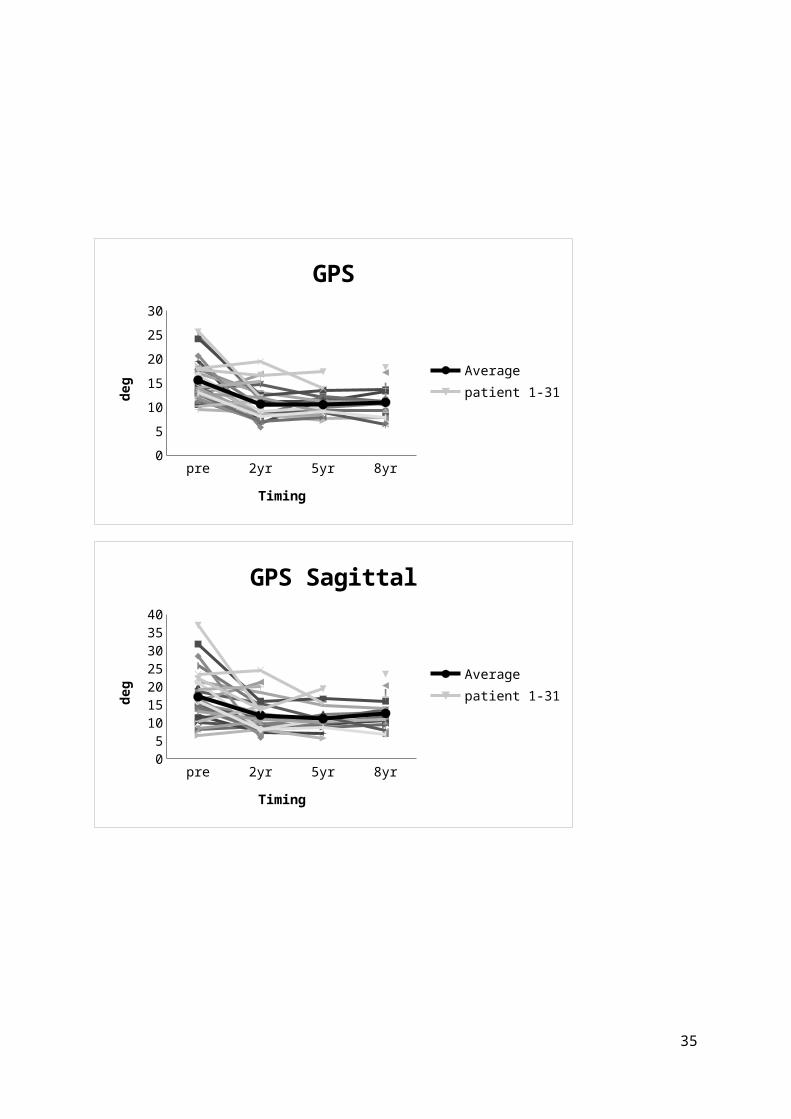
author's name, article or book title, volume and issue number, publisher, year and page reference and comply with the reference style of Gait Posture. Only salient and significant references should be included.
Reference management softwareMost Elsevier journals have their reference template available in many of themost popular reference management software products. These include all productsthat support Citation Style Language styles (http://citationstyles.org), such as Mendeley(http://www.mendeley.com/features/reference-manager) and Zotero (https://www.zotero.org/), as well as EndNote (http://endnote.com/downloads/styles). Using the word processor plug-ins from these products, authors only need to select the appropriate journal template when preparing their article, after which citations and bibliographies will be automatically formatted in the journal's style. If no template is yet available for this journal, please follow the format of the sample references and citations as shown in this Guide.
Users of Mendeley Desktop can easily install the reference style for this journal by clicking the following link:http://open.mendeley.com/use-citation-style/gait-and-postureWhen preparing your manuscript, you will then be able to select this style using the Mendeley plugins for Microsoft Word or LibreOffice.
Reference styleText: Indicate references by number(s) in square brackets in line with the text. The actual authors can be referred to, but the reference number(s) must always be given.List: Number the references (numbers in square brackets) in the list in the order in which they appear in the text.Examples: Reference to a journal publication:[1] Van der Geer J, Hanraads JAJ, Lupton RA. The art of writing a scientific article. J Sci Commun2010;163:51–9.Reference to a book:[2] Strunk Jr W, White EB. The elements of style. 4th ed. New York: Longman; 2000.Reference to a chapter in an edited book:[3] Mettam GR, Adams LB. How to prepare an electronic version of your article. In: Jones BS, Smith RZ, editors. Introduction to the electronic age, New York: E-Publishing Inc; 2009, p. 281–304.Note shortened form for last page number. e.g., 51–9, and that for more than 6 authors the first 6 should be listed followed by 'et al.' For further details you are referred to 'Uniform Requirements for Manuscripts submitted to Biomedical Journals' (J Am Med Assoc 1997;277:927–34) (see also http://www.nlm.nih.gov/bsd/uniform_requirements.html).Reference to a website:[4] Cancer Research UK. Cancer statistics reports for the UK, http://www.cancerresearchuk.org/aboutcancer/statistics/cancerstatsreport/; 2003 [accessed 13.03.03].
AudioSlidesThe journal encourages authors to create an AudioSlides presentation with their published article. AudioSlides are brief, webinar-style presentations that are shown next to the online article on ScienceDirect. This gives authors the opportunity to summarize their research in their own words and to help readers understand what the paper is about. More information and examples are available at https://www.elsevier.com/audioslides. Authors of this journal will automatically receive an invitation e-mail to create an AudioSlides presentation after acceptance of their paper.
Supplementary materialSupplementary material can support and enhance your scientific research. Supplementary files offer the author additional possibilities to publish supporting
38

applications, high-resolution images, background datasets, sound clips and more. Please note that such items are published online exactly as they are submitted; there is no typesetting involved (supplementary data supplied as an Excel file or as a PowerPoint slide will appear as such online). Please submit the material together with the article and supply a concise and descriptive caption for each file. If you wish to make any changes tosupplementary data during any stage of the process, then please make sure to provide an updated file, and do not annotate any corrections on a previous version. Please also make sure to switch off the 'Track Changes' option in any Microsoft Office files as these will appear in the published supplementary file(s). For more detailed instructions please visit our artwork instruction pages at https://www.elsevier.com/artworkinstructions.
Submission checklistThe following list will be useful during the final checking of an article prior to sending it to the journal for review. Please consult this Guide for Authors for further details of any item. Ensure that the following items are present:One author has been designated as the corresponding author with contact details:• E-mail address• Full postal addressAll necessary files have been uploaded, and contain:• Keywords• All figure captions• All tables (including title, description, footnotes)Further considerations• Manuscript has been 'spell-checked' and 'grammar-checked'• References are in the correct format for this journal• All references mentioned in the Reference list are cited in the text, and vice versa• Permission has been obtained for use of copyrighted material from other sources (including the Internet)Printed version of figures (if applicable) in color or black-and-white• Indicate clearly whether or not color or black-and-white in print is required.For any further information please visit our customer support site at http://support.elsevier.com.
AFTER ACCEPTANCEUse of the Digital Object IdentifierThe Digital Object Identifier (DOI) may be used to cite and link to electronic documents. The DOI consists of a unique alpha-numeric character string which is assigned to a document by the publisher upon the initial electronic publication. The assigned DOI never changes. Therefore, it is an ideal medium for citing a document, particularly 'Articles in press' because they have not yet received their full bibliographic information. Example of a correctly given DOI (in URL format; here an article in the journal Physics Letters B):http://dx.doi.org/10.1016/j.physletb.2010.09.059When you use a DOI to create links to documents on the web, the DOIs are guaranteed never to change.
Online proof correctionCorresponding authors will receive an e-mail with a link to our online proofing system, allowing annotation and correction of proofs online. The environment is similar to MS Word: in addition to editing text, you can also comment on figures/tables and answer questions from the Copy Editor. Web-based proofing provides a faster and less error-prone process by allowing you to directly type your corrections, eliminating the potential introduction of errors. If preferred, you can still choose to annotate and upload your edits on the PDF version. All instructions for proofing will be given in the e-mail we send to authors, including alternative methods to the online version and PDF.We will do everything possible to get your article published quickly and accurately. Please use this proof only for checking the typesetting, editing, completeness and correctness of the text, tables and figures. Significant changes to the article as accepted for publication will only be considered at this stage with permission from the Editor. It is important to
39
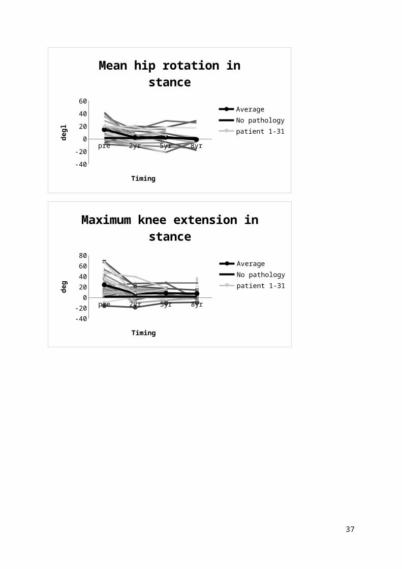
ensure that all corrections are sent back to us in one communication. Please check carefully before replying, as inclusion of any subsequent corrections cannot be guaranteed. Proofreading is solely your responsibility.
OffprintsThe corresponding author, at no cost, will be provided with a personalized link providing 50 days free access to the final published version of the article on ScienceDirect. This link can also be used for sharing via email and social networks. For an extra charge, paper offprints can be ordered via the offprint order form which is sent once the article is accepted for publication. Both corresponding and co-authors may order offprints at any time via Elsevier's WebShop (http://webshop.elsevier.com/myarticleservices/offprints).Authors requiring printed copies of multiple articles may use Elsevier WebShop's 'Create Your Own Book' service to collate multiple articles within a single cover (http://webshop.elsevier.com/myarticleservices/booklets).
Further InformationAuthors in Japan: please note that upon request, and if the author feels that it is necessary, Elsevier Japan will provide authors with a list of specialists who can check and improve the English of their manuscript (before submission). Please contact our Tokyo office: Elsevier K.K., 4F Higashi-Azabu, 1- Chome Bldg, 1-9-15 Higashi-Azabu, Minato-ku, Tokyo 106-0044, Japan. Tel: (+81)(3)5561-5037; Fax: (+81) (3) 5561 5047
AUTHOR INQUIRIESYou can track your submitted article at https://www.elsevier.com/track-submission.You can track your accepted article at https://www.elsevier.com/trackarticle. You are also welcome to contact Customer Support via http://support.elsevier.com.
© Copyright 2014 Elsevier | http://www.elsevier.com
40


![bildung Fort Der neuropathische Schmerz€¦ · oder intrathekale Baclofen-Pumpenthe-rapie) bleiben spezialisierten Zentren vor-behalten [7]. Zu den operativen Verfahren siehe Tabelle](https://img.pdfslide.tips/doc/110x75/5b9f969109d3f26e288d0e56/bildung-fort-der-neuropathische-oder-intrathekale-baclofen-pumpenthe-rapie.jpg)