Embed Size (px)
Citation preview
Kozłowska et al. Microb Cell Fact (2018) 17:71 https://doi.org/10.1186/s12934-018-0920-0
RESEARCH
Biotransformation of steroids by entomopathogenic strains of Isaria farinosaEwa Kozłowska1*, Natalia Hoc1, Jordan Sycz1, Monika Urbaniak2, Monika Dymarska1, Jakub Grzeszczuk3, Edyta Kostrzewa‑Susłow1, Łukasz Stępień2, Elżbieta Pląskowska3 and Tomasz Janeczko1*
Abstract
Background: Steroid compounds are very interesting substrates for biotransformation due to their high biological activity and a high number of inactivated carbons which make chemical modification difficult. Microbial transforma‑tion can involve reactions which are complicated and uneconomical in chemical synthesis, and searching for a new effective biocatalyst is necessary. The best known entomopathogenic species used in steroid modification is Beauveria bassiana. In this study we tested the ability of Isaria farinosa, another entomopathogenic species, to transform several steroids.
Results: Twelve strains of the entomopathogenic filamentous fungus Isaria farinosa, collected in abandoned mines located in the area of the Lower Silesian Voivodeship, Poland, from insects’ bodies covered by fungus, were used as a biocatalyst. All the tested strains effectively transformed dehydroepiandrosterone (DHEA). We observed 7α‑ and 7β‑hydroxy derivatives as well as changes in the percentage composition of the emerging products. Due to the similar metabolism of DHEA in all tested strains, one of them was selected for further investigation. In the culture of the selected strain, Isaria farinosa KCh KW1.1, transformations of androstenediol, androstenedione, adrenosterone, 17α‑methyltestosterone, 17β‑hydroxyandrost‑1,4,6‑triene‑3‑one and progesterone were performed. All the sub‑strates were hydroxylated with high yield and stereoselectivity. We obtained 6β‑hydroxyandrost‑4‑ene‑3,11,17‑trione, 15α,17β‑dihydroxy‑6β,7β‑epoxyandrost‑1,4‑diene‑3‑one and 6β,11α‑dihydroxyprogesterone. There is no evidence of either earlier microbial transformation of 17β‑hydroxyandrost‑1,4,6‑triene‑3‑one or new epoxy derivatives.
Conclusions: Isaria farinosa has a broad spectrum of highly effective steroid hydroxylases. The obtained 7‑hydroxy‑dehydroepiandrosterone has proven high biological activity and can be used in Alzheimer’s disease and as a key inter‑mediate in the synthesis of aldosterone antagonists. Transformation of progesterone leads to high yield of 6β,11α‑dihydroxyprogesterone and it is worth further study.
Keywords: Isaria farinosa, Biotransformation, DHEA, Progesterone, Hydroxylation
© The Author(s) 2018. This article is distributed under the terms of the Creative Commons Attribution 4.0 International License (http://creat iveco mmons .org/licen ses/by/4.0/), which permits unrestricted use, distribution, and reproduction in any medium, provided you give appropriate credit to the original author(s) and the source, provide a link to the Creative Commons license, and indicate if changes were made. The Creative Commons Public Domain Dedication waiver (http://creat iveco mmons .org/publi cdoma in/zero/1.0/) applies to the data made available in this article, unless otherwise stated.
BackgroundBiotransformation is an alternation of a xenobiotic sub-strate within an organism, as a result of its enzymatic activity. Enzymes can catalyse a wide spectrum of reac-tions like hydroxylation in inactivated positions. Micro-organisms can perform reactions which are complicated and economically unprofitable as chemical synthesis
because of their high regio-, chemo-, stereo and enanti-oselectivity [1–4]. Some specific microbial transforma-tions have been successfully incorporated into numerous syntheses of steroid drugs and key intermediates [2, 3, 5–7]. Applications of microorganism for steroid biotrans-formation have attracted considerable attention in the steroid chemistry community. In the literature, there are well-documented steroid transformations such as hydroxylation, dehydrogenation, redox reaction or Bae-yer–Villiger oxidation with numerous microorganisms [7–11]. Among them, hydroxylation of steroid com-pounds by filamentous fungi is a key reaction to obtain
Open Access
Microbial Cell Factories
*Correspondence: [email protected]; [email protected] 1 Department of Chemistry, Wrocław University of Environmental and Life Sciences, Norwida 25, 50‑375 Wrocław, PolandFull list of author information is available at the end of the article
Page 2 of 11Kozłowska et al. Microb Cell Fact (2018) 17:71
hydroxylated derivatives with higher biological proper-ties than their precursors [5, 12, 13]. The use of biotrans-formation methods is superior to chemical synthesis due to the difficulty of attaching an oxygen atom to an inacti-vated carbon atom in the steroid structure.
Through the development of biotransformation, it has been possible to develop methods for obtaining high yields of functionalized steroid compounds widely used in commercial production such as corticosteroids [14]. However, successful application of biotransformation for the industrial synthesis of known and novel steroids is still limited [7]. Therefore, further screening for new active catalytic strains is recommended.
The species Isaria farinosa belongs to the Cordycipi-taceae family in the Hypocreales order of Ascomycota. Many of the representatives of this family are parasitic, mainly infecting insects [15–17]. In recent years some of them, like Beauveria, Metarhizium and less fre-quently Isaria are used in biotransformation. Beauveria bassiana is well-known biocatalyst for the transforma-tion of steroid compounds. Hydroxylations at the C-7α and C-11α position and Baeyer–Villiger oxidation to d-homo lactones are described in the literature [18–24]. Also Metarhizium anisopliae have ability of hydroxyla-tion at 6β and 11α position [25]. The catalytic activity of Isaria fumosorosea towards selected steroid compounds with high substrate specificity was described [26]. The high virulence of Isaria farinosa, as other mentioned entomopathogenic strains, is closely connected with high chitinase, lipase and protease activities [12]. For this rea-sons, Isaria farinosa strains were used as biocatalysts in the biotransformation of steroid compounds for the first time. The high activity and specificity of the dehydroge-nases and hydroxylases present in their cells were also demonstrated.
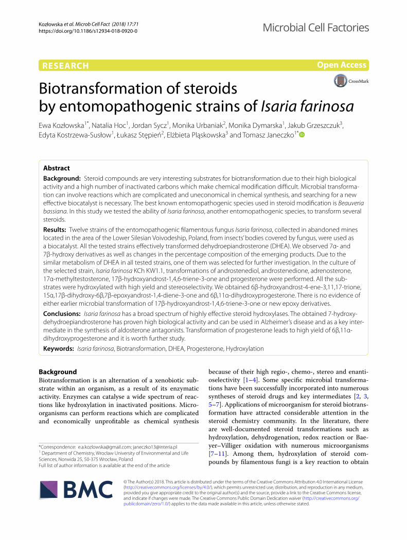
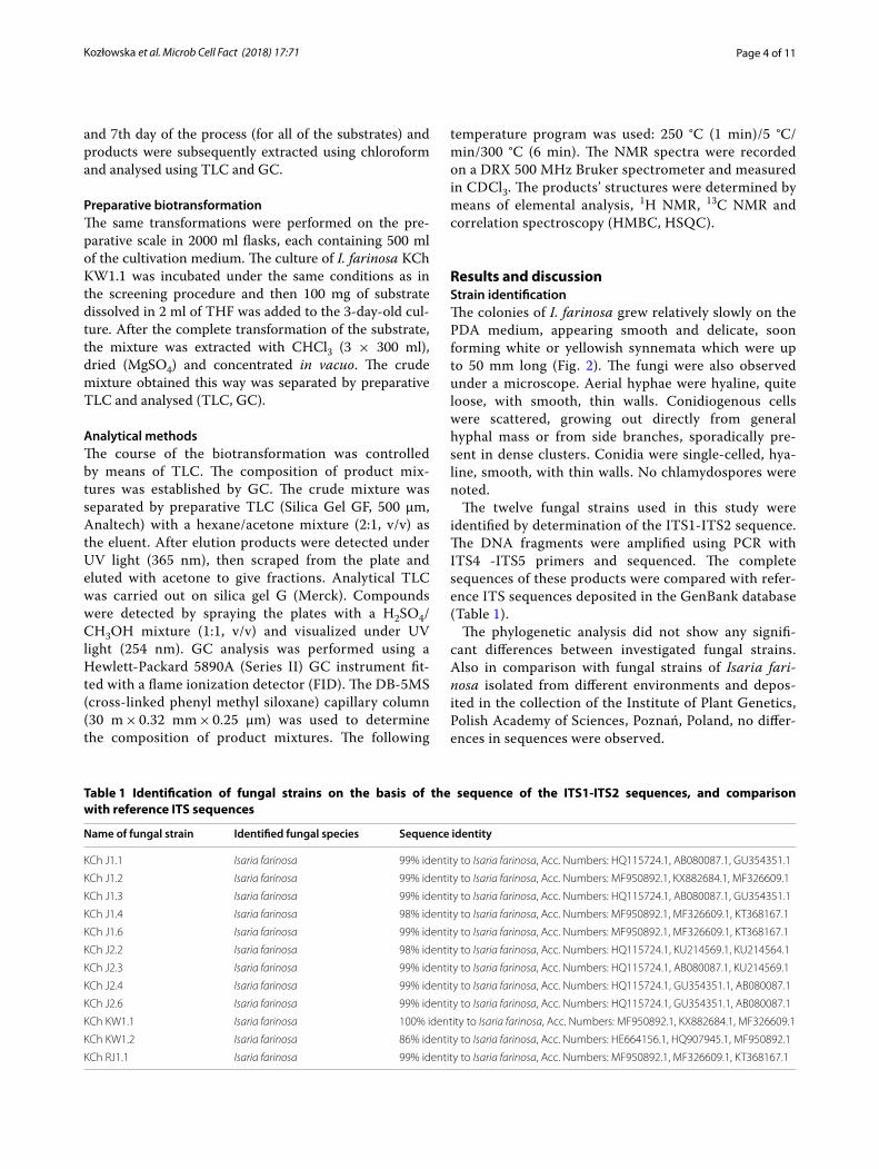
MethodsMaterialsThe following substrates were purchased from Sigma-Aldrich: dehydroepiandrosterone (DHEA), androstenediol (androst-5-ene-3β,17β-diol), androstenedione (androst-4-ene-3,17-dione, AD), adrenosterone (androst-4-ene-3,11,17-trione, ADR), pro-gesterone (P), 17α-methyltestosterone (17-mT) (Fig. 1.). 17β-Hydroxyandrost-1,4,6-triene-3-one (1,4,6-triene-T) was taken from the resources of the Department of Chemistry, Wrocław University of Environmental and Life Sciences.
Microorganisms: Twelve Isaria farinosa strains named KCh J1.1, KCh J1.2, KCh J1.3, KCh J1.4, KCh J1.6, KCh J2.2, KCh J2.3, KCh J2.4, KCh J2.6, KCh KW1.1, KCh KW1.2 and KCh RJ1.1 were obtained from the collection of the Department of Chemistry, Wrocław University of Environmental and Life Sciences, Wrocław, Poland. The strains were maintained on Sabouraud 4% dextrose-agar slopes and freshly subcultured before being used in the transformation experiments.
Isolation of strainsThe studied samples were collected in different aban-doned mines located in the area of the Lower Silesian Voivodeship, Poland. The samples named as KCh J*.* were collected in an adit near Ciechanowice, the sam-ples marked as KCh KW*.* were collected in an adit near Kletno, and those named as KCh RJ*.* were collected in an adit in the area of the Izera Crossroads in the Izera Mountains. The fungi were collected from these sites in the months of winter 2014/2015. After entering the adits, bodies of insects covered by fungal growths were searched for on the walls of the mines’ corridors. When found, cadavers of insects overgrown by the fungal
OH
O
OH
OH
O
O
O
OO
O
CH3
O
O
OHCH3 O
OH
DHEA Androstenediol
AD ADR 17-mT
P
1,4,6-trien-T
Fig. 1 Structures of substates
Page 3 of 11Kozłowska et al. Microb Cell Fact (2018) 17:71
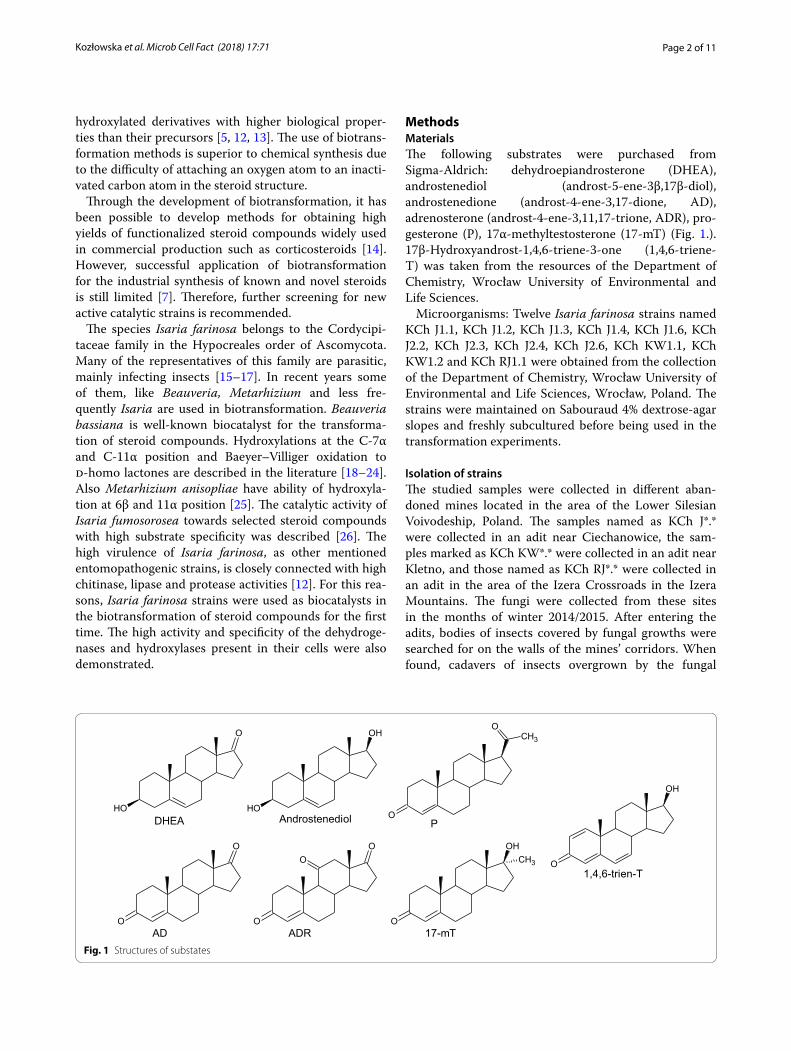
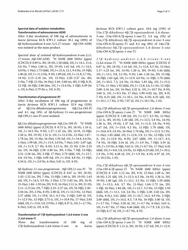
hyphae were placed in sterile plastic containers with the help of previously sterilized tweezers. Afterwards, the samples were transported to the laboratory of the Plant Pathology and Mycology Division of the Department of Plant Protection, Wrocław University of Environmen-tal and Life Sciences. There, pieces of hyphae from each sample were carefully pulled out from the insect’s cadav-ers and placed on Petri dishes with PDA medium. The growth of the fungi was observed for 4 weeks (Fig. 2).
DNA extraction and molecular identification of fungal strainsGrowing mycelia of individual fungal strains were main-tained in pure cultures for 7 days on PDA medium for genomic DNA extraction. Genomic DNA was extracted using a modified CTAB (hexadecyltrimethylammo-nium bromide) method, described earlier [27]. The DNA extracts were stored at – 20 °C until used. Species iden-tification was done on the basis of the sequences of the Internal Transcribed Spacers of the ribosomal DNA region (ITS1-ITS2).
Polymerase chain reactions were performed as described previously by Kozłowska et al. [9], using DreamTaq Green DNA polymerase (Thermo Scientific,
Espoo, Finland). The PCR amplification was done using ITS4 (5′-TCC TCC GCT TAT TGA TAT GC-3′) and ITS5 (5′-GGA AGT AAA AGT CGT AAC AAGG-3′) primers. Amplicons were electrophoresed in 1.5% agarose gel (Invitrogen) with GelGreen Nucleic Acid Stain (Biotium, Inc.).
For sequence analysis, PCR-amplified DNA fragments were purified as described previosly by Kozłowska et al. [9], DNA fragments were labelled using a forward primer and the BigDyeTerminator 3.1 kit (Applied Biosystems, Foster City, CA, USA), according to the producer’s rec-ommendations, and precipitated with 96% ethanol. Sequence reading was performed using Applied Bio-systems equipment. Sequences were analysed using the BLASTn algorithm.
Screening procedureErlenmeyer flasks (300 ml), each containing 100 ml of the cultivation medium (3% glucose, 1% bacteriological pep-tone), were inoculated with a suspension of the investi-gated strain and then incubated for 3 days at 24 °C on a rotary shaker. Then 10 mg of the substrate dissolved in 1 ml of THF was added. Samples were taken at 3, 6, 9, 12 h (only for DHEA as a substrate) and on the 1st, 3rd
Fig. 2 Isaria farinosa strains: a A moth cadaver found on a wall in an adit near Ciechanowice (Lower Silesian Voivodeship, Poland) overgrown by hyphae of Isaria farinosa © Jakub Grzeszczuk; b I. farinosa on PDA medium © Jakub Grzeszczuk; c Synnemata of I. farinosa grown on PDA medium © Jakub Grzeszczuk; d I. farinosa grown on cultivation medium © Tomasz Janeczko
Page 4 of 11Kozłowska et al. Microb Cell Fact (2018) 17:71
and 7th day of the process (for all of the substrates) and products were subsequently extracted using chloroform and analysed using TLC and GC.
Preparative biotransformationThe same transformations were performed on the pre-parative scale in 2000 ml flasks, each containing 500 ml of the cultivation medium. The culture of I. farinosa KCh KW1.1 was incubated under the same conditions as in the screening procedure and then 100 mg of substrate dissolved in 2 ml of THF was added to the 3-day-old cul-ture. After the complete transformation of the substrate, the mixture was extracted with CHCl3 (3 × 300 ml), dried (MgSO4) and concentrated in vacuo. The crude mixture obtained this way was separated by preparative TLC and analysed (TLC, GC).
Analytical methodsThe course of the biotransformation was controlled by means of TLC. The composition of product mix-tures was established by GC. The crude mixture was separated by preparative TLC (Silica Gel GF, 500 μm, Analtech) with a hexane/acetone mixture (2:1, v/v) as the eluent. After elution products were detected under UV light (365 nm), then scraped from the plate and eluted with acetone to give fractions. Analytical TLC was carried out on silica gel G (Merck). Compounds were detected by spraying the plates with a H2SO4/CH3OH mixture (1:1, v/v) and visualized under UV light (254 nm). GC analysis was performed using a Hewlett-Packard 5890A (Series II) GC instrument fit-ted with a flame ionization detector (FID). The DB-5MS (cross-linked phenyl methyl siloxane) capillary column (30 m × 0.32 mm × 0.25 μm) was used to determine the composition of product mixtures. The following
temperature program was used: 250 °C (1 min)/5 °C/min/300 °C (6 min). The NMR spectra were recorded on a DRX 500 MHz Bruker spectrometer and measured in CDCl3. The products’ structures were determined by means of elemental analysis, 1H NMR, 13C NMR and correlation spectroscopy (HMBC, HSQC).
Results and discussionStrain identificationThe colonies of I. farinosa grew relatively slowly on the PDA medium, appearing smooth and delicate, soon forming white or yellowish synnemata which were up to 50 mm long (Fig. 2). The fungi were also observed under a microscope. Aerial hyphae were hyaline, quite loose, with smooth, thin walls. Conidiogenous cells were scattered, growing out directly from general hyphal mass or from side branches, sporadically pre-sent in dense clusters. Conidia were single-celled, hya-line, smooth, with thin walls. No chlamydospores were noted.
The twelve fungal strains used in this study were identified by determination of the ITS1-ITS2 sequence. The DNA fragments were amplified using PCR with ITS4 -ITS5 primers and sequenced. The complete sequences of these products were compared with refer-ence ITS sequences deposited in the GenBank database (Table 1).
The phylogenetic analysis did not show any signifi-cant differences between investigated fungal strains. Also in comparison with fungal strains of Isaria fari‑nosa isolated from different environments and depos-ited in the collection of the Institute of Plant Genetics, Polish Academy of Sciences, Poznań, Poland, no differ-ences in sequences were observed.
Table 1 Identification of fungal strains on the basis of the sequence of the ITS1-ITS2 sequences, and comparison with reference ITS sequences
Name of fungal strain Identified fungal species Sequence identity
KCh J1.1 Isaria farinosa 99% identity to Isaria farinosa, Acc. Numbers: HQ115724.1, AB080087.1, GU354351.1
KCh J1.2 Isaria farinosa 99% identity to Isaria farinosa, Acc. Numbers: MF950892.1, KX882684.1, MF326609.1
KCh J1.3 Isaria farinosa 99% identity to Isaria farinosa, Acc. Numbers: HQ115724.1, AB080087.1, GU354351.1
KCh J1.4 Isaria farinosa 98% identity to Isaria farinosa, Acc. Numbers: MF950892.1, MF326609.1, KT368167.1
KCh J1.6 Isaria farinosa 99% identity to Isaria farinosa, Acc. Numbers: MF950892.1, MF326609.1, KT368167.1
KCh J2.2 Isaria farinosa 98% identity to Isaria farinosa, Acc. Numbers: HQ115724.1, KU214569.1, KU214564.1
KCh J2.3 Isaria farinosa 99% identity to Isaria farinosa, Acc. Numbers: HQ115724.1, AB080087.1, KU214569.1
KCh J2.4 Isaria farinosa 99% identity to Isaria farinosa, Acc. Numbers: HQ115724.1, GU354351.1, AB080087.1
KCh J2.6 Isaria farinosa 99% identity to Isaria farinosa, Acc. Numbers: HQ115724.1, GU354351.1, AB080087.1
KCh KW1.1 Isaria farinosa 100% identity to Isaria farinosa, Acc. Numbers: MF950892.1, KX882684.1, MF326609.1
KCh KW1.2 Isaria farinosa 86% identity to Isaria farinosa, Acc. Numbers: HE664156.1, HQ907945.1, MF950892.1
KCh RJ1.1 Isaria farinosa 99% identity to Isaria farinosa, Acc. Numbers: MF950892.1, MF326609.1, KT368167.1
Page 5 of 11Kozłowska et al. Microb Cell Fact (2018) 17:71
Spectral data of isolation metabolitesTransformation of adrenosterone (ADR)After 3-day incubation of 100 mg of adrenosterone in Isaria farinosa KCh KW1.1 culture 30.1 mg (30%) of 6β-hydroxyandrost-4-ene-3,11,17-trione (6β-OH-ADR) was isolated as the main product.
Spectral data of isolated 6β‑hydroxyandrost‑4‑ene‑3,11, 17‑trione (6β‑OH‑ADR) 1H NMR (600 MHz) (ppm) (CDCl3) δ: 0.89 (s, 3H, 18-H); 1.50 (ddd, 1H, J = 14.1, 11.6, 2.6 Hz, 7-Hα); 1.60 (s, 3H, 19-H); 1.62 (td, 1H, J = 14.5, 4.0 Hz, 1-Hα); 1.73 (ddd, 1H, J = 21.4, 12.1, 9.4 Hz, 15-Hβ); 1.86 (d, 1H, J = 11.5 Hz, 9-H); 1.89 (td, 1H, J = 11.8, 5.7 Hz, 14-H); 2.12–2.18 (m, 1H, 15-Hα); 2.20–2.37 (m, 4H, 2-Hα, 7-Hβ, 12-Hα, 16-Hα); 2.44–2.60 (m, 4H, 2-Hβ, 8-H, 12-Hβ, 16-Hβ); 2.80 (dm, 1H, J = 13.4 Hz, 1-Hβ); 4.39 (br s, 1H, 6-Hα); 5.79 (br s, 1H, 4-H).
Transformation of progesterone (P)After 3-day incubation of 100 mg of progesterone in Isaria farinosa KCh KW1.1 culture 32.9 mg (33%) of 6β,11α-dihydroxyprogesterone (6β,11α-OH-P) and 4.1 mg (4%) of 6β-hydroxy-11-oxo-progesterone (6β-OH,11-oxo-P) were isolated.
6β,11α‑dihydroxyprogesterone (6β,11α‑OH‑P) 1H NMR (600 MHz) (ppm) (CDCl3) δ: 0.73 (s, 3H, 18-H); 1.10 (t, 1H, J = 10.3 Hz, 9-H); 1.27–1.32 (m, 2H, 14-H, 15-Hβ); 1.50 (s, 3H, 19-H); 1.51 (t, 1H, J = 11.4 Hz, 12-Hα); 1.67–1.78 (m, 2H, 15-Hα, 16-Hα); 1.92 (td, 1H, J = 14.4, 4.4 Hz, 1-Hα); 1.99 (dt, 1H, J = 13.9, 3.0 Hz, 7-Hα); 2.01–2.07 (qd, 1H, J = 11.9, 2.7 Hz, 8-H); 2.13 (s, 3H, 21-H); 2.16–2.23 (m, 1H, 16-Hβ); 2.28–2.40 (m, 3H, 2-Hα, 7-Hβ, 12-Hβ); 2.51–2.58 (m, 2H, 2-Hβ, 17-Hα); 2.79 (ddd, 1H, J = 13.7, 4.8, 3.0 Hz, 1-Hβ); 4.09 (td, 1H, J = 10.6, 4.8 Hz, 11-Hβ); 4.34 (t, 1H, J = 2.8 Hz, 6-Hα); 5.81 (s, 1H, 4-H).
6β‑hydroxy‑11‑oxo‑progesterone (6β‑OH,11‑oxo‑P) 1H NMR (600 MHz) (ppm) (CDCl3) δ: 0.67 (s, 3H, 18-H); 1.42–1.52 (m, 2H, 7-Hα, 15-Hβ); 1.60 (s, 3H, 19-H); 1.63 (td, 1H, J = 14.3, 3.9 Hz, 1-Hα); 1.73–1.82 (m, 2H, 14-H, 15-Hα); 1.84–1.93 (m, 2H, 9-H, 16-Hα); 2.11 (s, 3H, 21-H); 2.11–2.13 (m, 1H, 7-Hβ); 2.23–2.27 (m, 1H, 16-Hβ); 2.30–2.44 (m, 2H, 2-Hα, 8-H); 2.48 (d, 1H, J = 12.3 Hz, 12-Hα); 2.57 (ddd, 1H, J = 19.8, 16.1, 6.3 Hz, 2-Hβ); 2.69 (d, 1H, J = 12.5 Hz, 12-Hβ); 2.73 (t, 1H, J = 10.8 Hz, 17-Hα); 2.83 (ddd, 1H, J = 13.6, 5.0, 2.9 Hz, 1-Hβ); 4.37 (t, 1H, J = 2.7 Hz, 6-Hα); 5.82 (s, 1H, 4-H).
Transformation of 17β‑hydroxyandrost‑1,4,6‑triene‑3‑one (1,4,6‑triene‑T)Three day transformation of 100 mg of 17β-hydroxyandrost-1,4,6-triene-3-one in Isaria
farinosa KCh KW1.1 culture gave: 18.6 mg (19%) of 15α,17β-dihydroxy-6β,7β-epoxyandrost-1,4-diene-3-one (15α-OH-6,7β-epoxy-1-ene-T), 5.4 mg (5%) of 15α,17β-dihydroxy-6β,7β-epoxyandrost-4-ene-3-one (15α-OH-6,7β-epoxy-T) and 8.2 mg (8%) of 14α,17β-dihydroxy-6β,7β-epoxyandrost-1,4-diene-3-one (14α-OH-6,7β-epoxy-1-ene-T)
1 7 β ‑ h y d r o x y ‑ a n d r o s t ‑ 1 , 4 , 6 ‑ t r i e n e ‑ 3 ‑ o n e (1,4,6‑triene‑T) 1H NMR (600 MHz) (ppm) (CDCl3) δ: 0.87 (s, 3H, 18-H); 1.14 (td, 1H, J = 12.8, 4.5 Hz, 12-Hα); 1.17–1.23 (m, 1H, 14-H); 1.20 (s, 3H, 19-H); 1.44 (ddd, 1H, J = 13.1, 9.8, 3.5 Hz, 9-H); 1.46–1.56 (m, 2H, 15-Hβ, 16-Hβ); 1.65 (qd, 1H, J = 12.9, 4.0 Hz, 11-Hβ); 1.78 (ddd, 1H, J = 10.5, 7.3, 2.6 Hz, 15-Hα); 1.83 (dq, 1H, J = 13.2, 3.7 Hz, 11-Hα); 1.92 (ddd, 1H, J = 12.8, 3.6, 3.1 Hz, 12-Hβ); 2.08–2.16 (m, 1H, 16-Hα); 2.32 (t, 1H, J = 10.7 Hz, 8-H); 3.68 (t, 1H, J = 8.5 Hz, 17-Hα); 5.99–6.02 (m, 2H, 4-H, 7-H); 6.23 (dd, 1H, J = 10.1, 2.6 Hz, 6-H); 6.25 (dd, 1H, J = 10.1, 1.7 Hz, 2-H); 7.07 (d, 1H, J = 10.1 Hz, 1-H).
15α,17β‑dihydroxy‑6β,7β‑epoxyandrost‑1,4‑diene‑3‑one (15α‑OH‑6,7β‑epoxy‑1‑ene‑T) 1H NMR (600 MHz) (ppm) (CDCl3) δ: 1.08 (td, 1H, J = 12.7, 4.2 Hz, 12-Hα); 1.12 (s, 3H, 18-H); 1.20 (dd, 1H, J = 12.5, 5.8 Hz, 14-H); 1.36 (s, 3H, 19-H); 1.37 (td, 1H, J = 11.9, 3.8 Hz, 9-H); 1.67 (td, 1H, J = 13.0, 4.2 Hz, 11-Hβ); 1.70 (ddd, 1H, J = 14.6, 8.9, 2.6 Hz, 16-Hα); 1.76 (dq, 1H, J = 13.3, 3.1 Hz, 11-Hα); 1.85 (ddd, 1H, J = 12.8, 3.9, 3.1 Hz, 12-Hβ); 2.55 (t, 1H, J = 12.0 Hz, 8-H); 2.67 (ddd, 1H, J = 14.7, 8.5, 7.8 Hz, 16-Hβ); 3.54 (d, 1H, J = 3.9 Hz, 7-Hβ); 3.59 (d, 1H, J = 3.9 Hz, 6-Hβ); 3.62 (t, 1H, J = 8.7 Hz, 17-Hα); 4.45 (ddd, 1H, J = 8.0, 5.8, 2.6 Hz, 15-Hβ); 6.23 (dd, 1H, J = 10.2, 1.9 Hz, 2-H); 6.48 (d, 1H, J = 1.9 Hz, 4-H); 6.97 (d, 1H, J = 10.2 Hz, 1-H).
15α,17β‑dihydroxy‑6β,7β‑epoxyandrost‑4‑ene‑3‑one (15α‑OH‑6,7β‑epoxy‑T) 1H NMR (600 MHz) (ppm) (CDCl3) δ: 1.05–1.12 (m, 2H, 9-H, 12-Hα); 1.09 (s, 3H, 18-H); 1.21 (dd, 1H, J = 12.3, 6.2 Hz, 14-H); 1.26 (s, 3H, 19-H); 1.40 (qd, 1H, J = 13.1, 3.2 Hz, 11-Hβ); 1.59 (ddd, 1H, J = 13.3, 6.8, 3.7 Hz, 11-Hα); 1.64–1.72 (m, 2H, 1-Hβ, 16-Hα); 1.84 (dt, 1H, J = 12.7, 3.3 Hz, 12-Hβ); 1.93 (ddd, 1H, J = 13.1, 5.0, 2.6 Hz, 1-Hβ); 2.38–2.45 (m, 2H, 2-Hα, 8-H,); 2.61 (ddd, 1H, J = 16.7, 14.2, 4.9 Hz, 2-Hβ); 2.68 (ddd, 1H, J = 14.5, 8.3, 7.8 Hz, 16-Hβ); 3.40 (d, 1H, J = 3.7 Hz, 7-Hα); 3.50 (d, 1H, J = 3.7 Hz, 6-Hα); 3.63 (t, 1H, J = 8.7 Hz, 17-Hα); 4.46 (ddd, 1H, J = 7.9, 5.8, 2.5 Hz, 15-Hβ); 6.17 (d, 1H, J = 0.7 Hz, 4-H).
14α,17β‑dihydroxy‑6β,7β‑epoxyandrost‑1,4‑diene‑3‑one (14α‑OH‑6,7β‑epoxy‑1‑ene‑T) 1H NMR (600 MHz) (ppm) (CDCl3) δ: 1.11 (s, 3H, 18-H); 1.27 (td, 1H, J = 12.9,
Page 6 of 11Kozłowska et al. Microb Cell Fact (2018) 17:71
4.2 Hz, 12-Hα); 1.33 (s, 3H, 19-H); 1.56–1.70 (m, 2H, 11-Hβ, 15-Hβ); 1.70–1.83 (m, 2H, 11-Hα, 15-Hα); 1.96 (dd, 1H, J = 12.5, 3.5 Hz, 9-H); 2.39–2.49 (m, 2H, 8-H, 16-Hα); 2.50–2.63 (m, 2H, 12-Hβ, 16-Hβ); 3.60 (d, 1H, J = 3.9 Hz, 7-Hβ); 3.63 (d, 1H, J = 3.9 Hz, 6-Hβ); 6.25 (dd, 1H, J = 10.2, 1.8 Hz, 2-H); 6.48 (d, 1H, J = 1.8 Hz, 4-H); 6.96 (d, 1H, J = 10.2 Hz, 1-H).
Interpretation of resultsThe purpose of this study was to investigate the cata-lytic ability of twelve newly isolated entomopathogenic filamentous fungal strains of Isaria farinosa against selected steroid compounds. The strains were isolated from insects’ cadavers and identified by determination of the ITS1-ITS2 rDNA sequence. Substrates in this study were: dehydroepiandrosterone (DHEA), andros-tenediol, androstenedione (AD), adrenosterone (ADR), progesterone (P), 17α-methyltestosterone (17-mT) and 17β-hydroxyandrost-1,4,6-triene-3-one (1,4,6-triene-T).
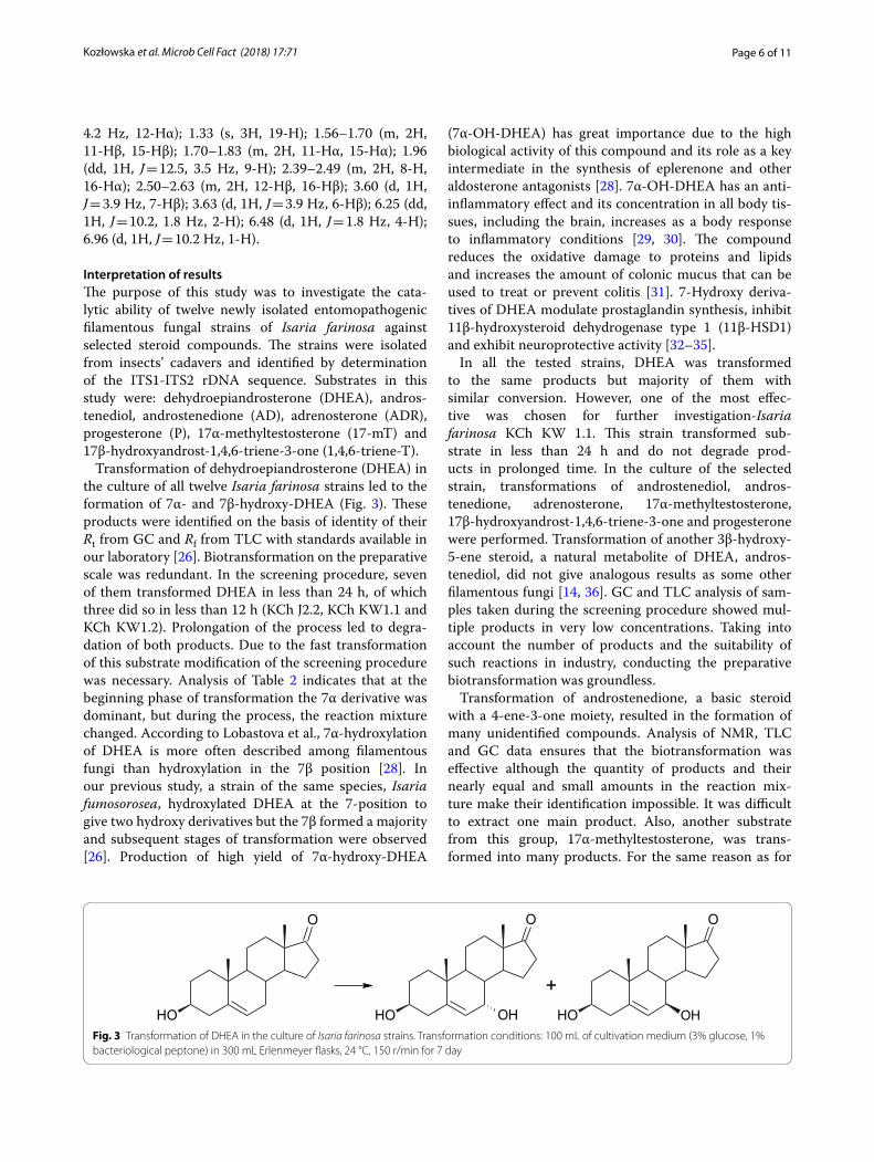
Transformation of dehydroepiandrosterone (DHEA) in the culture of all twelve Isaria farinosa strains led to the formation of 7α- and 7β-hydroxy-DHEA (Fig. 3). These products were identified on the basis of identity of their Rt from GC and Rf from TLC with standards available in our laboratory [26]. Biotransformation on the preparative scale was redundant. In the screening procedure, seven of them transformed DHEA in less than 24 h, of which three did so in less than 12 h (KCh J2.2, KCh KW1.1 and KCh KW1.2). Prolongation of the process led to degra-dation of both products. Due to the fast transformation of this substrate modification of the screening procedure was necessary. Analysis of Table 2 indicates that at the beginning phase of transformation the 7α derivative was dominant, but during the process, the reaction mixture changed. According to Lobastova et al., 7α-hydroxylation of DHEA is more often described among filamentous fungi than hydroxylation in the 7β position [28]. In our previous study, a strain of the same species, Isaria fumosorosea, hydroxylated DHEA at the 7-position to give two hydroxy derivatives but the 7β formed a majority and subsequent stages of transformation were observed [26]. Production of high yield of 7α-hydroxy-DHEA
(7α-OH-DHEA) has great importance due to the high biological activity of this compound and its role as a key intermediate in the synthesis of eplerenone and other aldosterone antagonists [28]. 7α-OH-DHEA has an anti-inflammatory effect and its concentration in all body tis-sues, including the brain, increases as a body response to inflammatory conditions [29, 30]. The compound reduces the oxidative damage to proteins and lipids and increases the amount of colonic mucus that can be used to treat or prevent colitis [31]. 7-Hydroxy deriva-tives of DHEA modulate prostaglandin synthesis, inhibit 11β-hydroxysteroid dehydrogenase type 1 (11β-HSD1) and exhibit neuroprotective activity [32–35].
In all the tested strains, DHEA was transformed to the same products but majority of them with similar conversion. However, one of the most effec-tive was chosen for further investigation-Isaria farinosa KCh KW 1.1. This strain transformed sub-strate in less than 24 h and do not degrade prod-ucts in prolonged time. In the culture of the selected strain, transformations of androstenediol, andros-tenedione, adrenosterone, 17α-methyltestosterone, 17β-hydroxyandrost-1,4,6-triene-3-one and progesterone were performed. Transformation of another 3β-hydroxy-5-ene steroid, a natural metabolite of DHEA, andros-tenediol, did not give analogous results as some other filamentous fungi [14, 36]. GC and TLC analysis of sam-ples taken during the screening procedure showed mul-tiple products in very low concentrations. Taking into account the number of products and the suitability of such reactions in industry, conducting the preparative biotransformation was groundless.
Transformation of androstenedione, a basic steroid with a 4-ene-3-one moiety, resulted in the formation of many unidentified compounds. Analysis of NMR, TLC and GC data ensures that the biotransformation was effective although the quantity of products and their nearly equal and small amounts in the reaction mix-ture make their identification impossible. It was difficult to extract one main product. Also, another substrate from this group, 17α-methyltestosterone, was trans-formed into many products. For the same reason as for
OH
O
OH
O
OH OH
O
OH
+
Fig. 3 Transformation of DHEA in the culture of Isaria farinosa strains. Transformation conditions: 100 mL of cultivation medium (3% glucose, 1% bacteriological peptone) in 300 mL Erlenmeyer flasks, 24 °C, 150 r/min for 7 day
Page 7 of 11Kozłowska et al. Microb Cell Fact (2018) 17:71
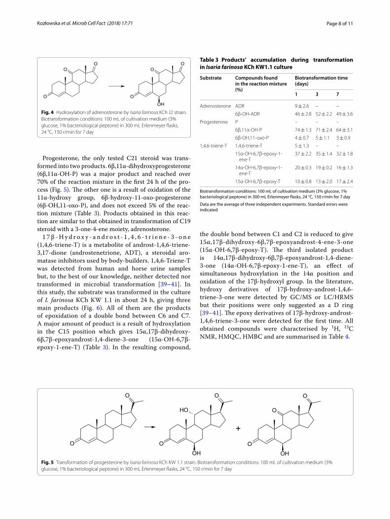
androstenediol, preparative biotransformation was not carried out. Only adrenosterone was transformed into one main product. The substrate was transformed in about 24 h giving mainly 6β-OH-ADR (Fig. 4). There were still some metabolites that had been isolated, but on the basis of NMR analysis, the steroid structure was not observed. None of them represented more than 10% of the reaction mixture. Compared to the adrenosterone
transformation in the culture of Isaria fumosorosea, the same product was obtained but much faster (one-day transformation vs. 7 days in I. fumosorosea culture) [26]. In contrast to Aspergillus tamarii, Cephalosporium aphidicola, Fusarium lini, Trichothecium roseum or Cun‑ninghamella elegans, no activity of 17β-hydroxysteroid dehydrogenase or similar enzyme was observed [2, 37, 38].
Table 2 Composition of the crude mixture obtained in the transformation of DHEA in Isaria farinosa strains
Biotransformation conditions: 100 mL of cultivation medium (3% glucose, 1% bacteriological peptone) in 300 mL Erlenmeyer flasks, 24 °C, 150 r/min for 7 day
Data are the average of three independent experiments. Standard errors were indicated
Isaria farinosa strain Compounds found in the reaction mixture (%)
Biotransformation time (h)
3 6 9 12 24 72 168
KCh J1.1 DHEA 100 ± 0.1 100 ± 0.1 99 ± 0.3 93 ± 0.5 74 ± 3.9 – –
7α‑OH‑DHEA – – – 5 ± 0.7 17 ± 2.7 60 ± 3.0 43 ± 4.6
7β‑OH‑DHEA – – – 2 ± 0.5 9 ± 2.3 38 ± 2.6 42 ± 3.8
KCh J1.2 DHEA 100 ± 0.1 100 ± 0.3 72 ± 3.3 39 ± 4.1 – – –
7α‑OH‑DHEA – – 21 ± 1.6 35 ± 1.8 64 ± 0.9 46 ± 1.1 14 ± 3.3
7β‑OH‑DHEA – – 7 ± 1.2 14 ± 2.0 36 ± 1.3 52 ± 1.1 46 ± 4.2
KCh J1.3 DHEA 100 ± 0.1 96 ± 0.5 63 ± 3.7 21 ± 3.9 1 ± 1.0 – –
7α‑OH‑DHEA – 3 ± 0.5 16 ± 2.5 49 ± 1.6 64 ± 0.7 35 ± 3.3 –
7β‑OH‑DHEA – 1 ± 0,2 5 ± 1.2 21 ± 1.8 25 ± 0.5 50 ± 3.2 –
KCh J1.4 DHEA 100 ± 0.2 96 ± 0.5 75 ± 2.1 44 ± 2.7 4 ± 0.6 – –
7α‑OH‑DHEA – 3 ± 0.1 12 ± 1.1 33 ± 0.9 61 ± 3.2 51 ± 4.3 –
7β‑OH‑DHEA – 1 ± 0.1 4 ± 0.2 13 ± 1.2 24 ± 1.9 36 ± 3.9 –
KCh J1.6 DHEA 100 ± 0.1 87 ± 0.8 36 ± 3.4 4 ± 0.5 1 ± 0.3 – –
7α‑OH‑DHEA – 8 ± 1.1 41 ± 2.9 66 ± 3.6 54 ± 1.1 26 ± 2.3 –
7β‑OH‑DHEA – 2 ± 0.3 16 ± 2.0 24 ± 1.7 30 ± 2.3 46 ± 4.2 –
KCh J2.2 DHEA 100 ± 0.3 88 ± 2.0 55 ± 3.1 – – – –
7α‑OH‑DHEA – 6 ± 0.8 31 ± 2.7 70 ± 0.9 67 ± 2.0 50 ± 1.6 –
7β‑OH‑DHEA – 2 ± 0.7 11 ± 1.1 26 ± 0.4 29 ± 2.6 47 ± 1.3 –
KCh J2.3 DHEA 100 ± 0.1 97 ± 0.4 90 ± 1.6 79 ± 0.9 – – –
7α‑OH‑DHEA – 2 ± 0.4 7 ± 1.3 16 ± 1.7 62 ± 0.3 45 ± 1.5 41 ± 4.3
7β‑OH‑DHEA – 1 ± 0.3 3 ± 0.8 6 ± 2.1 31 ± 0.7 47 ± 2.3 39 ± 2.8
KCh J2.4 DHEA 100 ± 0.6 84 ± 0.9 48 ± 3.2 3 ± 0.6 – – –
7α‑OH‑DHEA – 11 ± 2.4 28 ± 4.1 65 ± 1.6 62 ± 1.3 40 ± 1.7 2 ± 1.0
7β‑OH‑DHEA – 5 ± 0.7 10 ± 1.4 21 ± 2.0 32 ± 2.2 42 ± 2.2 13 ± 3.2
KCh J2.6 DHEA 100 ± 0.3 100 ± 0.5 81 ± 4.5 27 ± 3.8 6 ± 2.0 3 ± 1.1 –
7α‑OH‑DHEA – – 14 ± 3.1 38 ± 4.1 47 ± 1.4 34 ± 1.9 –
7β‑OH‑DHEA – – 5 ± 2.6 15 ± 3.1 28 ± 1.8 49 ± 2.1 –
KCh KW1.1 DHEA 100 ± 0.2 80 ± 2.1 53 ± 2.8 1 ± 0.6 1 ± 0.8 – –
7α‑OH‑DHEA – 12 ± 1.3 21 ± 2.6 68 ± 3.9 49 ± 1.3 34 ± 2.9 24 ± 2.7
7β‑OH‑DHEA – 4 ± 0.7 8 ± 1.5 26 ± 0.6 39 ± 0.4 56 ± 3.1 56 ± 3.0
KCh KW1.2 DHEA 100 ± 0.3 95 ± 1.7 68 ± 3.9 – – – –
7α‑OH‑DHEA – 4 ± 0.6 22 ± 2.0 69 ± 0.7 50 ± 1.3 34 ± 2.6 23 ± 2.3
7β‑OH‑DHEA – 1 ± 0.3 8 ± 1.9 27 ± 0.9 40 ± 0.5 56 ± 3.0 57 ± 4.4
KCh RJ1.1 DHEA 100 ± 0.1 100 ± 0.8 97 ± 0.7 27 ± 1.6 26 ± 3.3 – –
7α‑OH‑DHEA – – 2 ± 0.2 38 ± 0.9 46 ± 1.7 51 ± 0.2 40 ± 3.0
7β‑OH‑DHEA – – 1 ± 0.3 16 ± 0.7 23 ± 2.8 46 ± 1.5 57 ± 2.4
Page 8 of 11Kozłowska et al. Microb Cell Fact (2018) 17:71
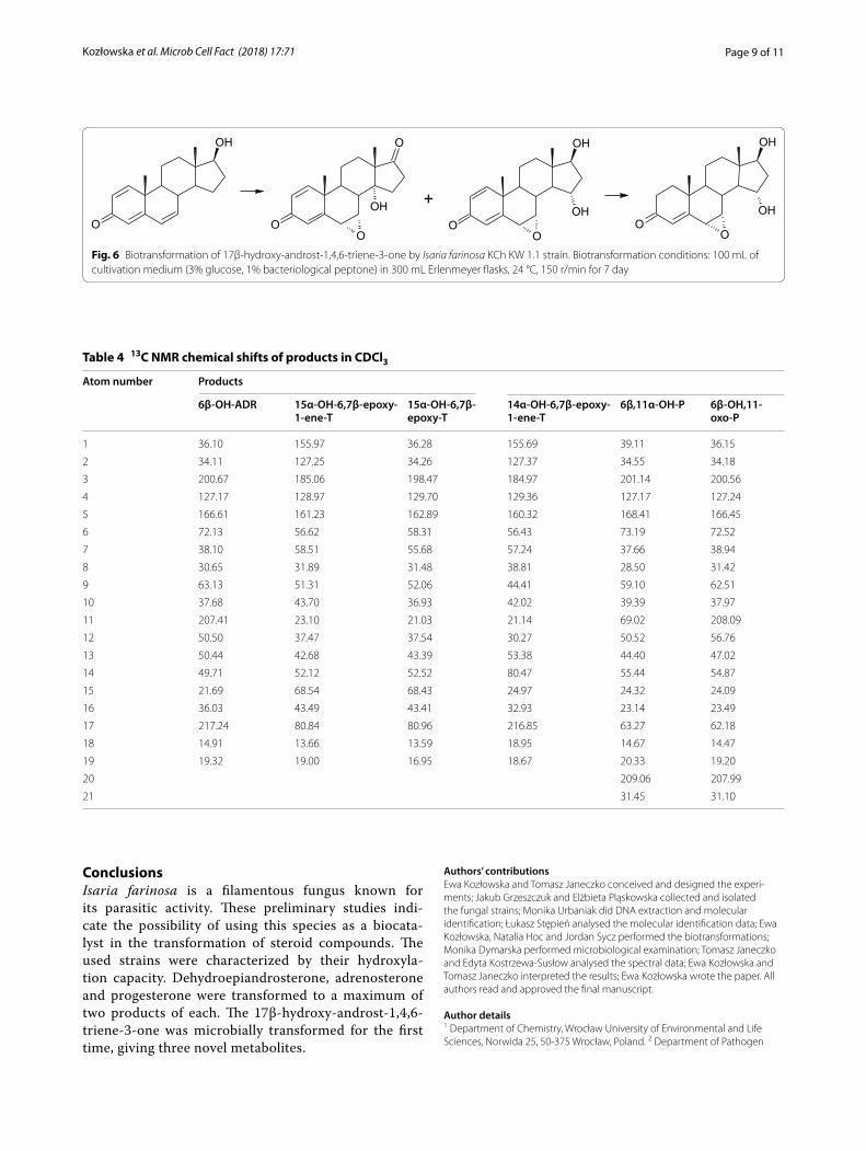
Progesterone, the only tested C21 steroid was trans-formed into two products. 6β,11α-dihydroxyprogesterone (6β,11α-OH-P) was a major product and reached over 70% of the reaction mixture in the first 24 h of the pro-cess (Fig. 5). The other one is a result of oxidation of the 11α-hydroxy group, 6β-hydroxy-11-oxo-progesterone (6β-OH,11-oxo-P), and does not exceed 5% of the reac-tion mixture (Table 3). Products obtained in this reac-tion are similar to that obtained in transformation of C19 steroid with a 3-one-4-ene moiety, adrenosterone.
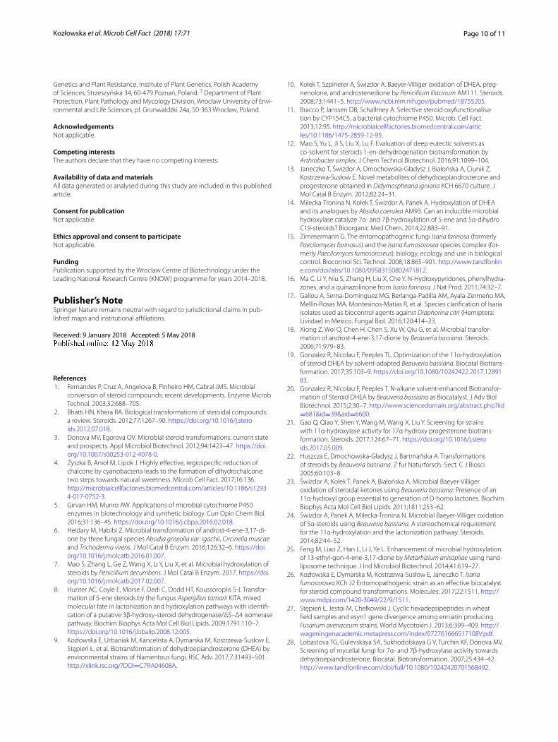
1 7 β - H y d r o x y - a n d r o s t - 1 , 4 , 6 - t r i e n e - 3 - o n e (1,4,6-triene-T) is a metabolite of androst-1,4,6-triene-3,17-dione (androstenetrione, ADT), a steroidal aro-matase inhibitors used by body-builders. 1,4,6-Triene-T was detected from human and horse urine samples but, to the best of our knowledge, neither detected nor transformed in microbial transformation [39–41]. In this study, the substrate was transformed in the culture of I. farinosa KCh KW 1.1 in about 24 h, giving three main products (Fig. 6). All of them are the products of epoxidation of a double bond between C6 and C7. A major amount of product is a result of hydroxylation in the C15 position which gives 15α,17β-dihydroxy-6β,7β-epoxyandrost-1,4-diene-3-one (15α-OH-6,7β-epoxy-1-ene-T) (Table 3). In the resulting compound,
the double bond between C1 and C2 is reduced to give 15α,17β-dihydroxy-6β,7β-epoxyandrost-4-ene-3-one (15α-OH-6,7β-epoxy-T). The third isolated product is 14α,17β-dihydroxy-6β,7β-epoxyandrost-1,4-diene-3-one (14α-OH-6,7β-epoxy-1-ene-T), an effect of simultaneous hydroxylation in the 14α position and oxidation of the 17β-hydroxyl group. In the literature, hydroxy derivatives of 17β-hydroxy-androst-1,4,6-triene-3-one were detected by GC/MS or LC/HRMS but their positions were only suggested as a D ring [39–41]. The epoxy derivatives of 17β-hydroxy-androst-1,4,6-triene-3-one were detected for the first time. All obtained compounds were characterised by 1H, 13C NMR, HMQC, HMBC and are summarised in Table 4.
O
OO
OHO
OO
Fig. 4 Hydroxylation of adrenosterone by Isaria farinosa KCh J2 strain. Biotransformation conditions: 100 mL of cultivation medium (3% glucose, 1% bacteriological peptone) in 300 mL Erlenmeyer flasks, 24 °C, 150 r/min for 7 day
O
O
O
OHO
OH
O
OH
O
O
+
Fig. 5 Transformation of progesterone by Isaria farinosa KCh KW 1.1 strain. Biotransformation conditions: 100 mL of cultivation medium (3% glucose, 1% bacteriological peptone) in 300 mL Erlenmeyer flasks, 24 °C, 150 r/min for 7 day
Table 3 Products’ accumulation during transformation in Isaria farinosa KCh KW1.1 culture
Biotransformation conditions: 100 mL of cultivation medium (3% glucose, 1% bacteriological peptone) in 300 mL Erlenmeyer flasks, 24 °C, 150 r/min for 7 day
Data are the average of three independent experiments. Standard errors were indicated
Substrate Compounds found in the reaction mixture (%)
Biotransformation time (days)
1 3 7
Adrenosterone ADR 9 ± 2.6 – –
6β‑OH‑ADR 46 ± 2.8 52 ± 2.2 49 ± 3.6
Progesterone P – – –
6β,11α‑OH‑P 74 ± 1.3 71 ± 2.4 64 ± 3.1
6β‑OH,11‑oxo‑P 4 ± 0.7 5 ± 1.1 3 ± 0.9
1,4,6‑triene‑T 1,4,6‑triene‑T 5 ± 1.3 – –
15α‑OH‑6,7β‑epoxy‑1‑ene‑T
37 ± 2.2 35 ± 1.4 32 ± 1.8
14α‑OH‑6,7β‑epoxy‑1‑ene‑T
20 ± 0.3 19 ± 0.2 16 ± 1.3
15α‑OH‑6,7β‑epoxy‑T 10 ± 0.8 13 ± 2.0 17 ± 2.4
Page 9 of 11Kozłowska et al. Microb Cell Fact (2018) 17:71
ConclusionsIsaria farinosa is a filamentous fungus known for its parasitic activity. These preliminary studies indi-cate the possibility of using this species as a biocata-lyst in the transformation of steroid compounds. The used strains were characterized by their hydroxyla-tion capacity. Dehydroepiandrosterone, adrenosterone and progesterone were transformed to a maximum of two products of each. The 17β-hydroxy-androst-1,4,6-triene-3-one was microbially transformed for the first time, giving three novel metabolites.
Authors’ contributionsEwa Kozłowska and Tomasz Janeczko conceived and designed the experi‑ments; Jakub Grzeszczuk and Elżbieta Pląskowska collected and isolated the fungal strains; Monika Urbaniak did DNA extraction and molecular identification; Łukasz Stępień analysed the molecular identification data; Ewa Kozłowska, Natalia Hoc and Jordan Sycz performed the biotransformations; Monika Dymarska performed microbiological examination; Tomasz Janeczko and Edyta Kostrzewa‑Susłow analysed the spectral data; Ewa Kozłowska and Tomasz Janeczko interpreted the results; Ewa Kozłowska wrote the paper. All authors read and approved the final manuscript.
Author details1 Department of Chemistry, Wrocław University of Environmental and Life Sciences, Norwida 25, 50‑375 Wrocław, Poland. 2 Department of Pathogen
OH
O
O
OO
OH
OH
OOH
O
OH
OOH
O
+
Fig. 6 Biotransformation of 17β‑hydroxy‑androst‑1,4,6‑triene‑3‑one by Isaria farinosa KCh KW 1.1 strain. Biotransformation conditions: 100 mL of cultivation medium (3% glucose, 1% bacteriological peptone) in 300 mL Erlenmeyer flasks, 24 °C, 150 r/min for 7 day
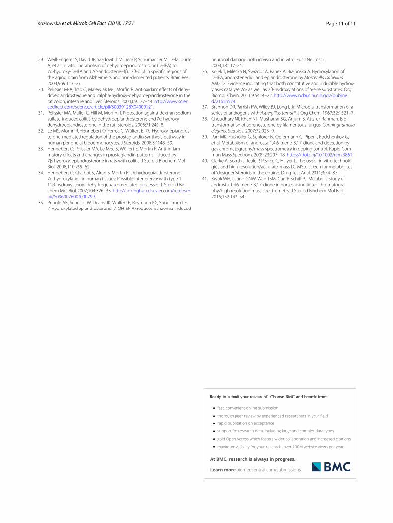
Table 4 13C NMR chemical shifts of products in CDCl3
Atom number Products
6β-OH-ADR 15α-OH-6,7β-epoxy-1-ene-T
15α-OH-6,7β-epoxy-T
14α-OH-6,7β-epoxy-1-ene-T
6β,11α-OH-P 6β-OH,11-oxo-P
1 36.10 155.97 36.28 155.69 39.11 36.15
2 34.11 127.25 34.26 127.37 34.55 34.18
3 200.67 185.06 198.47 184.97 201.14 200.56
4 127.17 128.97 129.70 129.36 127.17 127.24
5 166.61 161.23 162.89 160.32 168.41 166.45
6 72.13 56.62 58.31 56.43 73.19 72.52
7 38.10 58.51 55.68 57.24 37.66 38.94
8 30.65 31.89 31.48 38.81 28.50 31.42
9 63.13 51.31 52.06 44.41 59.10 62.51
10 37.68 43.70 36.93 42.02 39.39 37.97
11 207.41 23.10 21.03 21.14 69.02 208.09
12 50.50 37.47 37.54 30.27 50.52 56.76
13 50.44 42.68 43.39 53.38 44.40 47.02
14 49.71 52.12 52.52 80.47 55.44 54.87
15 21.69 68.54 68.43 24.97 24.32 24.09
16 36.03 43.49 43.41 32.93 23.14 23.49
17 217.24 80.84 80.96 216.85 63.27 62.18
18 14.91 13.66 13.59 18.95 14.67 14.47
19 19.32 19.00 16.95 18.67 20.33 19.20
20 209.06 207.99
21 31.45 31.10
Page 10 of 11Kozłowska et al. Microb Cell Fact (2018) 17:71
Genetics and Plant Resistance, Institute of Plant Genetics, Polish Academy of Sciences, Strzeszyńska 34, 60‑479 Poznań, Poland. 3 Department of Plant Protection, Plant Pathology and Mycology Division, Wrocław University of Envi‑ronmental and Life Sciences, pl. Grunwaldzki 24a, 50‑363 Wrocław, Poland.
AcknowledgementsNot applicable.
Competing interestsThe authors declare that they have no competing interests.
Availability of data and materialsAll data generated or analysed during this study are included in this published article.
Consent for publicationNot applicable.
Ethics approval and consent to participateNot applicable.
FundingPublication supported by the Wroclaw Centre of Biotechnology under the Leading National Research Centre (KNOW) programme for years 2014–2018.
Publisher’s NoteSpringer Nature remains neutral with regard to jurisdictional claims in pub‑lished maps and institutional affiliations.
Received: 9 January 2018 Accepted: 5 May 2018
References 1. Fernandes P, Cruz A, Angelova B, Pinheiro HM, Cabral JMS. Microbial
conversion of steroid compounds: recent developments. Enzyme Microb Technol. 2003;32:688–705.
2. Bhatti HN, Khera RA. Biological transformations of steroidal compounds: a review. Steroids. 2012;77:1267–90. https ://doi.org/10.1016/j.stero ids.2012.07.018.
3. Donova MV, Egorova OV. Microbial steroid transformations: current state and prospects. Appl Microbiol Biotechnol. 2012;94:1423–47. https ://doi.org/10.1007/s0025 3‑012‑4078‑0.
4. Żyszka B, Anioł M, Lipok J. Highly effective, regiospecific reduction of chalcone by cyanobacteria leads to the formation of dihydrochalcone: two steps towards natural sweetness. Microb Cell Fact. 2017;16:136. http://micro bialc ellfa ctori es.biome dcent ral.com/artic les/10.1186/s1293 4‑017‑0752‑3.
5. Girvan HM, Munro AW. Applications of microbial cytochrome P450 enzymes in biotechnology and synthetic biology. Curr Opin Chem Biol. 2016;31:136–45. https ://doi.org/10.1016/j.cbpa.2016.02.018.
6. Heidary M, Habibi Z. Microbial transformation of androst‑4‑ene‑3,17‑di‑one by three fungal species Absidia griseolla var. igachii, Circinella muscae and Trichoderma virens. J Mol Catal B Enzym. 2016;126:32–6. https ://doi.org/10.1016/j.molca tb.2016.01.007.
7. Mao S, Zhang L, Ge Z, Wang X, Li Y, Liu X, et al. Microbial hydroxylation of steroids by Penicillium decumbens. J Mol Catal B Enzym. 2017. https ://doi.org/10.1016/j.molca tb.2017.02.007.
8. Hunter AC, Coyle E, Morse F, Dedi C, Dodd HT, Koussoroplis S‑J. Transfor‑mation of 5‑ene steroids by the fungus Aspergillus tamarii KITA: mixed molecular fate in lactonization and hydroxylation pathways with identifi‑cation of a putative 3β‑hydroxy‑steroid dehydrogenase/Δ5–Δ4 isomerase pathway. Biochim Biophys Acta Mol Cell Biol Lipids. 2009;1791:110–7. https ://doi.org/10.1016/j.bbali p.2008.12.005.
9. Kozłowska E, Urbaniak M, Kancelista A, Dymarska M, Kostrzewa‑Susłow E, Stępień Ł, et al. Biotransformation of dehydroepiandrosterone (DHEA) by environmental strains of filamentous fungi. RSC Adv. 2017;7:31493–501. http://xlink .rsc.org/?DOI=C7RA0 4608A .
10. Kołek T, Szpineter A, Świzdor A. Baeyer‑Villiger oxidation of DHEA, preg‑nenolone, and androstenedione by Penicillium lilacinum AM111. Steroids. 2008;73:1441–5. http://www.ncbi.nlm.nih.gov/pubme d/18755 205.
11. Bracco P, Janssen DB, Schallmey A. Selective steroid oxyfunctionalisa‑tion by CYP154C5, a bacterial cytochrome P450. Microb. Cell Fact. 2013;12:95. http://micro bialc ellfa ctori es.biome dcent ral.com/artic les/10.1186/1475‑2859‑12‑95.
12. Mao S, Yu L, Ji S, Liu X, Lu F. Evaluation of deep eutectic solvents as co‑solvent for steroids 1‑en‑dehydrogenation biotransformation by Arthrobacter simplex. J Chem Technol Biotechnol. 2016;91:1099–104.
13. Janeczko T, Świzdor A, Dmochowska‑Gładysz J, Białońska A, Ciunik Z, Kostrzewa‑Susłow E. Novel metabolites of dehydroepiandrosterone and progesterone obtained in Didymosphearia igniaria KCH 6670 culture. J Mol Catal B Enzym. 2012;82:24–31.
14. Milecka‑Tronina N, Kołek T, Świzdor A, Panek A. Hydroxylation of DHEA and its analogues by Absidia coerulea AM93. Can an inducible microbial hydroxylase catalyze 7α‑ and 7β‑hydroxylation of 5‑ene and 5α‑dihydro C19‑steroids? Bioorganic Med Chem. 2014;22:883–91.
15. Zimmermann G. The entomopathogenic fungi Isaria farinosa (formerly Paecilomyces farinosus) and the Isaria fumosorosea species complex (for‑merly Paecilomyces fumosoroseus): biology, ecology and use in biological control. Biocontrol Sci. Technol. 2008;18:865–901. http://www.tandf onlin e.com/doi/abs/10.1080/09583 15080 24718 12.
16. Ma C, Li Y, Niu S, Zhang H, Liu X, Che Y. N‑Hydroxypyridones, phenylhydra‑zones, and a quinazolinone from Isaria farinosa. J Nat Prod. 2011;74:32–7.
17. Gallou A, Serna‑Domínguez MG, Berlanga‑Padilla AM, Ayala‑Zermeño MA, Mellín‑Rosas MA, Montesinos‑Matías R, et al. Species clarification of Isaria isolates used as biocontrol agents against Diaphorina citri (Hemiptera: Liviidae) in Mexico. Fungal Biol. 2016;120:414–23.
18. Xiong Z, Wei Q, Chen H, Chen S, Xu W, Qiu G, et al. Microbial transfor‑mation of androst‑4‑ene‑3,17‑dione by Beauveria bassiana. Steroids. 2006;71:979–83.
19. Gonzalez R, Nicolau F, Peeples TL. Optimization of the 11α‑hydroxylation of steroid DHEA by solvent‑adapted Beauveria bassiana. Biocatal Biotrans‑formation. 2017;35:103–9. https ://doi.org/10.1080/10242 422.2017.12891 83.
20. Gonzalez R, Nicolau F, Peeples T. N‑alkane solvent‑enhanced Biotransfor‑mation of Steroid DHEA by Beauveria bassiana as Biocatalyst. J Adv Biol Biotechnol. 2015;2:30–7. http://www.scien cedom ain.org/abstr act.php?iid=681&id=39&aid=6600.
21. Gao Q, Qiao Y, Shen Y, Wang M, Wang X, Liu Y. Screening for strains with 11α‑hydroxylase activity for 17α‑hydroxy progesterone biotrans‑formation. Steroids. 2017;124:67–71. https ://doi.org/10.1016/j.stero ids.2017.05.009.
22. Huszcza E, Dmochowska‑Gładysz J, Bartmańska A. Transformations of steroids by Beauveria bassiana. Z fur Naturforsch.‑Sect. C J Biosci. 2005;60:103–8.
23. Świzdor A, Kołek T, Panek A, Białońska A. Microbial Baeyer‑Villiger oxidation of steroidal ketones using Beauveria bassiana: Presence of an 11α‑hydroxyl group essential to generation of D‑homo lactones. Biochim Biophys Acta Mol Cell Biol Lipids. 2011;1811:253–62.
24. Świzdor A, Panek A, Milecka‑Tronina N. Microbial Baeyer‑Villiger oxidation of 5α‑steroids using Beauveria bassiana. A stereochemical requirement for the 11α‑hydroxylation and the lactonization pathway. Steroids. 2014;82:44–52.
25. Feng M, Liao Z, Han L, Li J, Ye L. Enhancement of microbial hydroxylation of 13‑ethyl‑gon‑4‑ene‑3,17‑dione by Metarhizium anisopliae using nano‑liposome technique. J Ind Microbiol Biotechnol. 2014;41:619–27.
26. Kozłowska E, Dymarska M, Kostrzewa‑Susłow E, Janeczko T. Isaria fumosorosea KCh J2 Entomopathogenic strain as an effective biocatalyst for steroid compound transformations. Molecules. 2017;22:1511. http://www.mdpi.com/1420‑3049/22/9/1511.
27. Stępień Ł, Jestoi M, Chełkowski J. Cyclic hexadepsipeptides in wheat field samples and esyn1 gene divergence among enniatin producing Fusarium avenaceum strains. World Mycotoxin J. 2013;6:399–409. http://wagen ingen acade mic.metap ress.com/index /07276 16665 17108 V.pdf.
28. Lobastova TG, Gulevskaya SA, Sukhodolskaya G V, Turchin KF, Donova MV. Screening of mycelial fungi for 7α‑ and 7β‑hydroxylase activity towards dehydroepiandrosterone. Biocatal. Biotransformation. 2007;25:434–42. http://www.tandf onlin e.com/doi/full/10.1080/10242 42070 15684 92.
Page 11 of 11Kozłowska et al. Microb Cell Fact (2018) 17:71
• fast, convenient online submission
•
thorough peer review by experienced researchers in your field
• rapid publication on acceptance
• support for research data, including large and complex data types
•
gold Open Access which fosters wider collaboration and increased citations
maximum visibility for your research: over 100M website views per year •
At BMC, research is always in progress.
Learn more biomedcentral.com/submissions
Ready to submit your research ? Choose BMC and benefit from:
29. Weill‑Engerer S, David JP, Sazdovitch V, Liere P, Schumacher M, Delacourte A, et al. In vitro metabolism of dehydroepiandrosterone (DHEA) to 7α‑hydroxy‑DHEA and Δ5‑androstene‑3β,17β‑diol in specific regions of the aging brain from Alzheimer’s and non‑demented patients. Brain Res. 2003;969:117–25.
30. Pelissier M‑A, Trap C, Malewiak M‑I, Morfin R. Antioxidant effects of dehy‑droepiandrosterone and 7alpha‑hydroxy‑dehydroepiandrosterone in the rat colon, intestine and liver. Steroids. 2004;69:137–44. http://www.scien cedir ect.com/scien ce/artic le/pii/S0039 128X0 40001 21.
31. Pélissier MA, Muller C, Hill M, Morfin R. Protection against dextran sodium sulfate‑induced colitis by dehydroepiandrosterone and 7α‑hydroxy‑dehydroepiandrosterone in the rat. Steroids. 2006;71:240–8.
32. Le MS, Morfin R, Hennebert O, Ferrec C, Wülfert E. 7b‑Hydroxy‑epiandros‑terone‑mediated regulation of the prostaglandin synthesis pathway in human peripheral blood monocytes. J Steroids. 2008;3:1148–59.
33. Hennebert O, Pelissier MA, Le Mee S, Wülfert E, Morfin R. Anti‑inflam‑matory effects and changes in prostaglandin patterns induced by 7β‑hydroxy‑epiandrosterone in rats with colitis. J Steroid Biochem Mol Biol. 2008;110:255–62.
34. Hennebert O, Chalbot S, Alran S, Morfin R. Dehydroepiandrosterone 7α‑hydroxylation in human tissues: Possible interference with type 1 11β‑hydroxysteroid dehydrogenase‑mediated processes. J. Steroid Bio‑chem Mol Biol. 2007;104:326–33. http://linki nghub .elsev ier.com/retri eve/pii/S0960 07600 70007 99.
35. Pringle AK, Schmidt W, Deans JK, Wulfert E, Reymann KG, Sundstrom LE. 7‑Hydroxylated epiandrosterone (7‑OH‑EPIA) reduces ischaemia‑induced
neuronal damage both in vivo and in vitro. Eur J Neurosci. 2003;18:117–24.
36. Kołek T, Milecka N, Świzdor A, Panek A, Białońska A. Hydroxylation of DHEA, androstenediol and epiandrosterone by Mortierella isabellina AM212. Evidence indicating that both constitutive and inducible hydrox‑ylases catalyze 7α‑ as well as 7β‑hydroxylations of 5‑ene substrates. Org. Biomol. Chem. 2011;9:5414–22. http://www.ncbi.nlm.nih.gov/pubme d/21655 574.
37. Brannon DR, Parrish FW, Wiley BJ, Long L Jr. Microbial transformation of a series of androgens with Aspergillus tamarii. J Org Chem. 1967;32:1521–7.
38. Choudhary MI, Khan NT, Musharraf SG, Anjum S. Atta‑ur‑Rahman. Bio‑transformation of adrenosterone by filamentous fungus, Cunninghamella elegans. Steroids. 2007;72:923–9.
39. Parr MK, Fußhöller G, Schlörer N, Opfermann G, Piper T, Rodchenkov G, et al. Metabolism of androsta‑1,4,6‑triene‑3,17‑dione and detection by gas chromatography/mass spectrometry in doping control. Rapid Com‑mun Mass Spectrom. 2009;23:207–18. https ://doi.org/10.1002/rcm.3861.
40. Clarke A, Scarth J, Teale P, Pearce C, Hillyer L. The use of in vitro technolo‑gies and high‑resolution/accurate‑mass LC‑MSto screen for metabolites of “designer” steroids in the equine. Drug Test Anal. 2011;3:74–87.
41. Kwok WH, Leung GNW, Wan TSM, Curl P, Schiff PJ. Metabolic study of androsta‑1,4,6‑triene‑3,17‑dione in horses using liquid chromatogra‑phy/high resolution mass spectrometry. J Steroid Biochem Mol Biol. 2015;152:142–54.