Embed Size (px)
Citation preview

Bundesinstitut für Risikobewertung
Herausgegeben von A. Schulte, U. Bernauer, S. Madle, H. Mielke, U. Herbst, H.-B. Richter-Reichhelm, K.-E. Appel, U. Gundert-Remy
Assessment of the Carcinogenicity of Formaldehyde [CAS No. 50-00-0] Bericht zur Bewertung der Karzinogenität von Formaldehyd

Impressum BfR-Wissenschaft Herausgegeben von A. Schulte, U. Bernauer, S. Madle, H. Mielke, U. Herbst, H.-B. Richter-Reichhelm, K.-E. Appel, U. Gundert-Remy Assessment of the Carcinogenicity of Formaldehyde [CAS No. 50-00-0] Bericht zur Bewertung der Karzinogenität von Formaldehyd Bundesinstitut für Risikobewertung Pressestelle Thielallee 88-92 14195 Berlin Berlin 2006 (BfR-Wissenschaft 02 /2006) 156 Seiten, 1 Abbildung, 24 Tabellen € 10,- Druck: Umschlag, Inhalt und buchbinderische Verarbeitung BfR-Hausdruckerei Dahlem ISSN 1614-3795 ISBN 3-938163-14-3

3 BfR-Wissenschaft
Contents
1 Introduction 7
2 Toxicological effect assessment: Hazard identification and dose response assessment 9
2.1 Absorption, metabolism and distribution 9 2.1.1 Overview on toxicokinetics, metabolism and distribution 9 2.1.2 Toxicokinetics in animals 10 2.1.2.1 Inhalation 10 2.1.2.1.1 Inhalation local 10 2.1.2.1.2 Inhalation systemic 12 2.1.2.2 Oral 13 2.1.2.2.1 Oral local 13 2.1.2.2.2 Oral systemic 13 2.1.2.3 Dermal 14 2.1.2.3.1 Dermal local 14 2.1.2.3.2 Dermal systemic 14 2.1.3 Toxicokinetics in humans 17 2.1.3.1 Inhalation 17 2.1.3.1.1 Inhalation local 17 2.1.3.1.2 Inhalation systemic 18 2.1.3.2 Oral 18 2.1.3.2.1 Oral local 18 2.1.3.2.2 Oral systemic 18 2.1.3.3 Dermal 18 2.1.3.3.1 Dermal local 18 2.1.3.3.2 Dermal systemic 19 2.1.4 Enzymatic metabolism of formaldehyde (detoxification) 19 2.1.4.1 Enzymes involved in the detoxification of formaldehyde 19 2.1.4.1.1 Formaldehyde dehydrogenase [EC 1.2.1.1] 19 2.1.4.1.2 Aldehyde dehydrogenase [EC 1.2.1.3] 21 2.1.4.1.3 S-Formylglutathione Hydrolase [EC 3.1.2.12] 21 2.1.4.1.4 Glyoxalase II [3.1.2.6] 21 2.1.4.1.5 Katalase 21 2.1.4.2 Species differences of enzymes involved in the metabolism of
formaldehyde 22 2.1.4.2.1 Formaldehyde dehydrogenase 22 2.1.4.2.2 Aldehyde dehydrogenase 22 2.1.5 Induction, Inhibition and Polymorphisms of enzymes involved in
the metabolism of formaldehyde 22 2.1.5.1 Formaldehyde dehydrogenase 22 2.1.5.1.1 Inducibility 22 2.1.5.1.2 Polymorphisms (with respect to FA metabolism) 22 2.1.5.2 Acetaldehyde dehydrogenase 23 2.1.5.2.1 Inducibility 23 2.1.5.2.2 Polymorphisms (with respect to FA metabolism) 23 2.2 Genetic toxicity 23 2.2.1 Overview on genetic toxicity 23 2.2.2 Systemic genetic toxicity in mammalian animals 24 2.2.3 Local genetic toxicity in mammalian animals 24 2.2.4 Systemic genetic toxicity of formaldehyde in humans 25 2.2.5 Local genetic toxicity in humans 26

4 BfR-Wissenschaft
2.2.6 Conclusion on genotoxicity 27 2.2.7 Tables on genotoxicity 30 2.3 Other relevant effects 42 2.3.1 Introduction 42 2.3.2 Sensory irritation 42 2.3.3 Pulmonary effects in asthmatic people 43 2.4 Carcinogenicity 44 2.4.1 Introduction 44 2.4.2 Relevant information from repeated-dose toxicity data 44 2.4.2.1 Inhalation 44 2.4.2.1.1 Studies in rodents 45 2.4.2.1.2 Studies in mice 47 2.4.2.1.3 Studies in monkeys 47 2.4.2.1.4 Human data 48 2.4.2.1.4.1 Nose 48 2.4.2.1.4.2 Lower respiratory tract 49 2.4.2.2 Oral 49 2.4.2.3 Conclusion on repeated dose toxicity 50 2.4.2.3.1 Inhalation 50 2.4.2.3.1.1 The major target tissue and the nature of the lesions in the
respiratory tract 50 2.4.2.3.1.2 The most sensitive subsite(s) in the nose 50 2.4.2.3.1.3 Gradient of extension and of severity of lesions 50 2.4.2.3.1.4 Time-response relationship (subacute to subchronic exposure) 51 2.4.2.3.1.5 Lowest effective formaldehyde concentrations in animals 51 2.4.2.3.1.6 Most sensitive species 51 2.4.2.3.1.7 Other target sites in the respiratory tract 52 2.4.2.3.1.8 Coherence of sites for cytotoxicity and cell proliferation 52 2.4.2.3.1.9 Coherence of sites for cell proliferation and tumor prevalence 52 2.4.2.3.2 Oral route 52 2.4.3 Systemic carcinogenicity in animals 53 2.4.3.1 Oral route 53 2.4.3.2 Conclusion on systemic carcinogenicity in animals 59 2.4.3.2.1 Hemopoietic neoplasias (HPN) 59 2.4.3.2.2 Tumors of the gastrointestinal tract 59 2.4.4 Systemic carcinogenicity in man 60 2.4.4.1 Cohort-studies 60 2.4.4.2 Case-control studies 68 2.4.4.3 Meta-analysis/Pooled studies: 69 2.4.4.4 Conclusion on data on systemic carcinogenicity in man 69 2.4.5 Local carcinogenicity in animals 72 2.4.5.1 Inhalation 72 2.4.5.1.1 Rat studies 72 2.4.5.1.2 Mouse studies 74 2.4.5.1.3 Hamster studies 75 2.4.5.2 Oral route 75 2.4.5.3 Dermal administration 75 2.4.5.4 Conclusion on the local carcinogenicity in experimental animals 75 2.4.5.4.1 Inhalation route 75 2.4.6 Local carcinogenicity in man 83 2.4.6.1 Inhalation 83 2.4.6.1.1 Cohort Studies 83 2.4.6.1.2 Case-control studies 93 2.4.6.2 Conclusion on data on local carcinogenicity in humans 97

5 BfR-Wissenschaft
3 Mode of action 101
3.1 Toxicokinetics 101
3.2 Genetic toxicity 101
3.3 Cytotoxicity and cell proliferation 102
3.4 Carcinogenicity 103 3.4.1 Systemic effects in animals 103 3.4.2 Local effects in animals 103 3.4.3 Systemic and local effects in humans 104 3.5 Discussion on the mode of action 104
4 Risk assessment 107
4.1 General considerations 107
4.2 Mechanistic aspects 108 4.2.1 Cell proliferation and histopathological effects 108 4.2.2 Genotoxicity 109 4.2.3 Other effects relevant for tumor formation 110 4.2.4 Dose-response relationship for tumors in the upper respiratory
tract 111 4.2.5 The mechanistic model 112 4.2.5.1 Genotoxic mechanisms 112 4.2.5.2 Cytotoxicity 112 4.2.5.3 Dose-response relationship 112 4.2.5.4 The “safe” level 113 4.2.5.5 Interspecies comparison 113 4.3 Derivation of a safe level 114 4.3.1 Choice of endpoint and point of departure 114 4.3.2 Human data 114 4.3.3 Animal data 115 4.3.4 Other derivations 116 4.3.5 Conclusion 116
5 Executive summary 119
6 Zusammenfassung 123
7 References 127
8 Appendix 143
9 List of tables 151
10 List of figures 153


7
BfR-Wissenschaft
1 Introduction
Formaldehyde is an ubiquitous compound in the environment, and it is an important endoge-nous chemical that occurs in most life forms, including humans (IARC, 1995). Formaldehyde is a colourless gas and has a pungent odour (Reuss et al. 1988). Commercially, it has been widely employed in the production of resins with urea, phenol and malamine and to a small extent, their derivatives. Because of its chemical reactivity it has been used for preservation and disinfection, as well as antimicrobial agent in consumer products such as cosmetic prod-ucts. As a result of its reactivity in target tissues with direct contact with the substance, formalde-hyde causes local irritation, acute and chronic toxicity and has genotoxic and carcinogenic properties. Recent up dated evaluations on the epidemiologic data concerning the tumor incidence in formaldehyde exposed populations have been re-analysed by the IARC (Cogliano et al., 2005). Based on that analysis IARC has now come to the decision to classify formaldehyde as „human carcinogen“. The BfR report on formaldehyde will consider the actual database of formaldehyde and comment whether the IARC/WHO classification is justified when these data are reviewed under the requirement of the current EU chemical legislation.

8 BfR-Wissenschaft

9
BfR-Wissenschaft
2 Toxicological effect assessment: Hazard identification and dose response assessment
2.1 Absorption, metabolism and distribution
2.1.1 Overview on toxicokinetics, metabolism and distribution
Formaldehyde is present at low levels in most living organisms. Physiological amounts of formaldehyde are endogenously formed from serine, glycine, methionine and choline by de-methylation of N-, O-, and S-methyl compounds (IPCS 2002; IARC, 1995). Formaldehyde in blood may be present in its free form (Mashford and Jones, 1982) and also bound to proteins such as serum albumin (Heck et al., 1982). Exogenous Formaldehyde can be absorbed after inhalative, dermal and oral exposure and the amount of absorption is dependent on the route of exposure. The overall uptake of inhaled formaldehyde by the nasal passages at resting minute volume airflow rates has been predicted to be 90% in rats, 67% in monkeys and 76% in humans (Kimbell et al., 2002). In humans, increasing airflow leads to a reduced percent-age uptake in the nasal passages with concomitant shift of flux to postnasal areas (Kimbell et al., 2001a). After inhalation uptake, nasal airflow pattern rather than metabolism of the parent compound, is decisive for deposition and the elicitation of effects (such as cell proliferation, formation of DPX). This has demonstrated/calculated to be true for all species investigated so far. Studies using radioactive (14C)-formaldehyde demonstrated, that after i.v. (Heck and Chin, 1982), i.p. (Mashford and Jones, 1982), and inhalative (Heck et al., 1982) exposure, radioactivity was extensively distributed in other tissues. In studies on several species, in-cluding humans, formaldehyde was rapidly metabolised after absorption. Therefore, in hu-mans, rats and monkeys, formaldehyde concentrations in blood after exposure to formalde-hyde were not elevated compared to physiological blood-levels of formaldehyde of about 0.1 mM (Casanova et al., 1988; Heck et al., 1982; Heck et al., 1985). This indicated, that formal-dehyde has a high „first-pass“ effect, so that systemic availability is extremely low. Hence, due to the reactivity of the compound and due to its rapid metabolism in the cells lining skin, gastro-intestinal tract and lung, local effects seem to play a more important role compared to systemic effects. There are several possible pathways, which formaldehyde – either from exogenous exposure or endogenously formed – might undergo, which are illustrated in Figure 1 (adopted from Conaway et al., 1996). The multi-step metabolism pathway of formaldehyde yielding formate and CO2 can be regarded as detoxification. Enzymes involved in this pathway seem to be ubiquitous enzymes and may therefore be present in those tissues, which can be reached by formaldehyde (nevertheless, up to now it has not been investigated, whether and to which extent formaldehyde-detoxifying enzymes are being present in human nasal mucosa). In addition, also glutathione as a cofactor for formaldehyde-dehydrogenase (FAD)-dependent formaldehyde detoxification, is ubiquitously present. It has been calculated that FAD-dependent oxidation of formaldehyde in rat nasal mucosa is half-saturated at an airborne concentration of approximately 2.6 ppm formaldehyde (Casanova et al., 1989). Enzymes involved in the detoxification of formaldehyde seem to be well conserved between species and in humans. Polymorphisms have only been reported for ALDH, which only plays a role at higher formaldehyde concentrations Radioactivity, which is distributed after application of radiolabelled formaldehyde into tissues distant from the site of entry, most probably is due to metabolic intake of radiolabelled for-maldehyde into the C1 pool and metabolic incorporation of radiolabel into biological macro-molecules.

10 BfR-Wissenschaft
Figure 1: Biological pathways of formaldehyde (adopted from Conaway et al., 1996
Briefly, there are the following possibilities: formaldehyde can be reversibly bound to cysteine to form thiazolidine-4-carboxylate (1), with urea to form hydroxymethyladducts (2) or with proteins (e.g. blood proteins such as serum albumin or mucus proteins in nasal mucosa) to form protein adducts (3). Irreversible reactions result from reaction with two proteins (protein-protein-crosslinks) (4) or from reaction of formaldehyde with protein and DNA (DNA-protein crosslinks (DPX)) (5). Formaldehyde reacts spontaneously and reversibly with glutathione (GSH) present in cells to form S-hydroxymethylglutathione (6). In the presence of NAD+, S-hydroxymethylglutathione can be enzymatically converted by formaldehyddehydrogenase (FAD) to formylglutathione (7). In the presence of water, formylglutathione can be cleaved by S-Formylglutathione hydrolase to glutathione and formic acid (8) (Uotila and Koivusalo, 1974). Formic acid can be excreted in urine as its sodium salt. Formic acid can also be cleaved to CO2, which can be exhaled. Either as formate or after binding of formaldehyde to tetrahydrofolic acid (9), uptake into the carbon one metabolic pathways is possible. Hereby, formaldehyde (as “activated” i.e. tetrahydrofolic acid bound formaldehyde) is an essential intermediate for the synthesis of purine, thymidine and certain amino acids (Rietbrock et al., 1971). The latter com-pounds can be incorporated into macromolecules as e.g. proteins or nucleic acids (summarised in Heck and Casanova, 2004).
2.1.2 Toxicokinetics in animals
2.1.2.1 Inhalation
2.1.2.1.1 Inhalation local
In rodents, which are obligate nose breathers, most of the inhaled formaldehyde is deposited and absorbed in regions of the upper respiratory tract. In monkeys, which are oronasal breathers, inhaled formaldehyde is deposited and absorbed in the nasal passages and oral mucosa and also in the trachea and bronchus. The extent and exact localisation of inhalative formaldehyde uptake is determined by nasal anatomy which exhibits species differences and also by mucus coating and clearance mechanisms (Kimbell et al., 2001a; Kepler et al., 1998). Nasal anatomy governs airflow patterns and therefore the sites which will come into contact with inhaled formaldehyde. By using nasal molds and computational fluid dynamics (CFD), it could be demonstrated for the rat, that a considerable proportion of flow intake at the nostrils passes into the middle and lateral meatuses with less flow to the dorsal and ven-tral medial pathways (Morgan et al., 1991; Kimbell et al., 1993). Results from such models
H
H
O
O
O
GS
GSH
C
CH
H
OH
NH2
NH2
O
+ Glutathione (GSH)
+ Tetrahydro-folic acid (THF)
+ CysteineThiazolidine-4-carboxylate
C-1 Pool, metabolic incorporation into biological macromolecules
+ Protein+ DNA
+ FAD+ NAD
+
+ H O2
(1)
(4)
(5)
(6)
(7)
(8)
(9)
(2)
(2)
(3)
+
+ Protein + Protein
+
S
N
H
C
OH
O
HO
HO
C
C C
C C
C
C
C
NH
NH
H
H H
H H
H
Protein Protein
Protein
Protein
DNA
O
OH
OH NH
NH
O
H
H H
H H
HCH2
OH
HO

11
BfR-Wissenschaft
were confirmed by experimental data which demonstrated histopathologic damage (see also Section 2.3.2) and DNA-protein crosslinks mainly in the anterior regions of the nose in areas lined by respiratory epithelium after acute inhalation exposure and – at formaldehyde con-centrations > 5.6 ppm - squamous cell carcinomas exclusively in the respiratory epithelial regions. In Rhesus monkeys, the largest streams of airflow passed to the ventral meatus from much of the medial nostril and to the middle meatus from the lateral nostril. A small, sharply defined region of the nostril provided flow to the dorsal region of the nose. Re-sponses in the Rhesus monkey after inhalative exposure to formaldehyde were most severe on the dorsal and ventral margins of the middle turbinate. These regions were also the points of direct impaction of anterior nasal flow streams which was also confirmed by CFD (Kepler et al., 1998). Therefore, in rats and Rhesus monkeys, the distribution of formaldehyde in-duced nasal lesions correlated well with regions of high bulk flow, secondary flows and turbu-lences (Morgan et al., 1991) with the exception of the lateral walls and dorsal aspects of posterior airways in Rhesus monkeys, where lesions were seen but flux was predicted to be low (Kepler et al., 1998). Nevertheless, the flow pattern in rats and Rhesus monkeys exhib-ited species differences. Schlosser (Schlosser, 1999) performed calculations in order to determine whether and to which extent the amount of formaldehyde which has been taken up in the rat nasal epithelial mucus could be eliminated by nasal mucus flow or by chemical reaction with amino groups (from proteins) present in the mucus. It could be concluded, that binding to amino groups was very low whereas elimination by nasal mucus flow was estimated to be 22-42% of in-haled formaldehyde. Due to the reversible impairment of mucociliary clearance by formalde-hyde which has been demonstrated in in vitro experiments (Hastie et al., 1990) a temporary increase of formaldehyde uptake because of reduced mucociliary clearance cannot be ex-cluded. In order to evaluate the contributions of metabolism and covalent binding to the disposition of inhaled (14C)-formaldehyde in the respiratory tract of rats and monkeys, a toxicokinetic model has been developed. The model suggests, that at low concentrations (up to 6 ppm), ap-proximately 93% of the formaldehyde in the rat nasal respiratory mucosa is eliminated via saturable metabolism, 7% is eliminated by non-saturable pathways other than formation of DNA-protein crosslinks (DPX) (e.g. non-saturable metabolism, covalent binding to mucus proteins) and only 7 x 10⁻⁶% is covalently bound as DNA-protein-crosslinks (DPX) (Heck and Casanova 1995; Heck and Casanova, 2004). It has been stated, that similar results were obtained for the middle turbinates, the lateral wall septum and nasopharynx of monkeys (Casanova et al., 1991; Heck et al., 1995). By using radioactive labelled formaldehyde, contributions of metabolic incorporation and co-valent binding as DPX to the radioactive labelling of DNA in the upper respiratory system of rats and Rhesus monkeys have been determined which is illustrated in the Table 1. In F344 rats exposed by inhalation for 6 h to 6 ppm to (3H)- and (14C)-formaldehyde, approximately 91% of the (14C) in the DNA of the respiratory mucosa was due to metabolic incorporation, which was found in thymidine (40%), deoxyguanosine (32%) and deoxyadenosine (19%). The remaining 9% of the (14C) in DNA was covalently bound as DPX (Casanova et al., 1989). In DNA obtained from the mucosa of the middle turbinates of the nose of Rhesus monkeys (where DPX were highest compared to anterior lateral wall/septum and nasopharynx) ex-posed by inhalation for 6 h to 6 ppm (14C)-formaldehyde, approximately 96% of the (14C) was due to metabolic incorporation, which was found in thymidine (89%), deoxyguanosine (3%) and deoxyadenosine (4%). 4% of the (14C) in the middle turbinate DNA of the monkey was covalently bound as DPX (Casanova et al., 1991). In Rhesus monkeys, after exposure to 0.71, 2.0 and 6.0 ppm (14C)-formaldehyde for 6 h, formation of DNA-protein crosslinks could also be demonstrated in extranasal tissues (in lar-

12 BfR-Wissenschaft
ynx, trachea, carina and in the major pulmonary airways) at exposure concentrations of 2.0 and 6.0 ppm. Table 1: Fate of [
14C] absorbed in the upper respiratory tract of animals after inhalative exposure to
(14
C)-formaldehyde (Adopted from Casanova et al., 1989 and Casanova et al., 1991)
Species Concentration of [14C]-form- aldehyde (ppm/6h)
Tissue source of DNA
Covalently bound [14C] [% of ab-sorbed dose]
Metabolically incorporated [14C] [% of ab-sorbed dose]
Distribution of metabolically incorporated [14C]
F344 rat 6 DNA from nasal respira-tory mucosal
9 91 40 % in thymidine 32 % in deoxyguanosine 19 % in deoxyadenosine
Rhesus monkey 0.71 DNA from middle turbin-ate of the nose
0.2 99.8 94.5 % in thymidine 1.5 % in deoxyguanosine 3.8 % in deoxyadenosine
Rhesus monkey 2 DNA from middle turbin-ate of the nose
1 99 84 % in thymidine 7 % in deoxyguanosine 8 % in deoxyadenosine
Rhesus monkey 6 DNA from middle turbin-ate of the nose
4 96 89 % in thymidine 3 % in deoxyguanosine 4 % in deoxyadenosine
2.1.2.1.2 Inhalation systemic
After inhalative exposure of rats, more than 93% of the inhaled formaldehyde was absorbed readily by the tissues of the respiratory tract (Patterson et al., 1986). In a more recent publi-cation it has been calculated by using CFD, that overall uptake of inhaled formaldehyde by the nasal passages was 90% for the rat and 67% for Rhesus monkey (Kimbell et al., 2001b). The fate of inhaled formaldehyde was studied in F344 rats exposed to [14C]-formaldehyde at 0.63 or 13.1 ppm for 6 h and is illustrated in the Table 2. About 40% of the inhaled formalde-hyde was eliminated as expired 14C carbon dioxide over a 70-h period. 17% of the radioactiv-ity was excreted in the urine, 5% of radioactivity was eliminated in the faeces and 35 – 39% of radioactivity remained in the tissues and carcass. Considerable radioactivity was detected in the oesophagus and trachea and lesser amounts were also found in kidney, liver, intestine and lung, indicating that formaldehyde or its me-tabolites have been swallowed and/or removed by the nasal mucosa (Heck and Chin, 1982). Elimination of radioactivity from the blood of rats after exposure by inhalation to 0.63 and 13.1 ppm to [14C]-formaldehyde is multiphasic. The half-life of radioactivity after inhalation was approximately 55 h (Heck and Chin, 1982). The possibility that inhaled formaldehyde may increase the formaldehyde concentrations in blood was investigated in F344 rats (Heck et al., 1985) and in Rhesus monkeys (Casanova et al., 1988) by using GC-MS analysis measuring both free and reversibly bound formalde-hyde. The blood formaldehyde concentrations of eight F344 rats exposed to 14.4 ppm for-maldehyde for 2 h (2.25 ± 0.7 µg/g blood) were identical to those from unexposed rats (2.24 ± 0.7 µg/g blood). In Rhesus monkeys exposed to high formaldehyde concentrations (6 ppm, 6h/d, 5 d/week, 4 weeks) the blood concentrations measured 7 min and 45 h after the last exposure were almost the same and statistically indistinguishable (7 min: 1.84 ± 0.15 µg/g blood; 45 h: 2.04 ± 0.40 µg/g blood).

13
BfR-Wissenschaft
Table 2: Distribution of radioactivity in rats after inhalative exposure to (14
C)-formaldehyde.
Species Dose (ppm/6h)
Distribution of radioactivity [percentage of totally recovered radio-activity; mean ± SD]
Time window Reference
Expired air: 39.4 ± 1.5 Urine: 17.6 ± 1.2 Faeces: 4.2 ± 1.5
Rat (n=4) 0.63
Tissues*) and carcass: 38.9 ± 1.2
Exposure duration 6h, animals were sacrificed 70 h after removal from the exposure chamber
Patterson et al., 1986 Heck and Chin, 1982
Expired air: 41.9 ± 0.8 Urine: 17.3 ± 0.6 Faeces: 5.3 ± 1.3
Rat (n=4) 13.1
Tissues*) and carcass: 35.2 ± 0.5
Exposure duration 6h, animals were sacrificed 70 h after removal from the exposure chamber
Heck and Chin, 1982
*) Nasal mucosa, trachea, oesophagus, lung, kidney, liver, intestine, spleen, heart, plasma, erythrocytes, brain, testes
The lack of elevation of plasma formaldehyde levels over physiological levels after inhalative exposure can be explained by rapid metabolism of formaldehyde (the half-life of formalde-hyde in plasma has been determined to be 1 min (Patterson et al., 1986)) in those cells/tissues, where the relevant enzymes (e.g. FAD) and cofactors (GSH) are present (see also section 2.1.4. - metabolism). In addition, analysis of the time course of residual radioac-tivity in plasma and erythrocytes after inhalation showed, that the radioactivity was due to incorporation of (14C) (as (14C)-formate) into serum proteins and subsequent release of la-belled proteins and cells into the circulation. The lack of elevated formaldehyde concentrations in blood after inhalative exposure may be the reason for the lack of well defined systemic effects after inhalative systemic exposure.
In rats, after inhalative exposure towards the high concentration of 20 ppm formaldehyde over 13 weeks, there was a slight increase in the levels of certain plasma enzymes sugges-tive of a hepatotoxic effect, but histopathologic examinations revealed no liver damage, and there were no changes in liver weight or liver GSH concentrations. The slight increase in plasma enzyme levels may have been caused by growth retardation (Heck et al., 1990; Woutersen et al., 1987).
There was no evidence of immunosuppression in mice or of impaired B-cell function in rats following exposure to formaldehyde (Dean et al., 1984; Adams et al., 1987; Holmstrom et al., 1989a; Heck et al., 1990).
2.1.2.2 Oral
2.1.2.2.1 Oral local
In rodents, formaldehyde is absorbed rapidly from the gastro-intestinal tract. In rats, about 40% of an oral dose of [14C] formaldehyde (7 mg/kg) was eliminated as (14C)-carbon dioxide within 12 h, while 10 % of radiolabel was excreted in the urine and 1 % of radiolabel was excreted in the faeces (Buss et al., 1964; Mashford and Jones, 1982). The induction of micronuclei and nuclear anomalies in cells of the gastro-intestinal epithelium of rats treated per os with formaldehyde (200 mg/kg) revealed effects in conjunction with se-vere local irritation. Induction of micronuclei and nuclear anomalies were elevated over un-treated controls in stomach, duodenum, ileum and colon thus suggesting uptake into cells of the gastro-intestinal tract (Migliore et al., 1989). 2.1.2.2.2 Oral systemic
After oral application of 7 mg/kg [14C] formaldehyde to rats, 40 % of the applied radioactivity was exhaled as [14C] carbon dioxide, 10% of radioactivity was excreted via kidneys and 1%

14 BfR-Wissenschaft
of radioactivity was excreted with faeces within 12 h. After application, radioactivity was dis-tributed rapidly. After 12 h, the lowest radioactivity was found in blood and the highest radio-activity was found in the bone marrow. Four days after application, radioactivity could be de-termined in brain, GI tract, liver, spleen, adrenal glands, kidneys and in fat (Buss et al., 1964). Migliore et al. (1989) demonstrated that in rats treated per os with formaldehyde (200 mg/kg), induction of micronuclei and nuclear anomalies were elevated over untreated con-trols in stomach, duodenum, ileum and colon. The observed effects were strongest in the stomach. Other tracts were clearly positive but to a lesser extent with effects declining with distance from the stomach. These data suggest that formaldehyde not only causes nuclear damage at the site of application, but also distant thereof. 2.1.2.3 Dermal
2.1.2.3.1 Dermal local
After application of [14C] formaldehyde to the skin of F344 rats, Dunkin-Hartley guinea pigs and cynomolgus monkeys, most of the formaldehyde evaporated. In rodents, 3.6-16% of (14C) and in monkeys, 9.5% of (14C) remained in the skin. It is assumed that substantial amounts of topically applied formaldehyde reacts reversibly or irreversibly with biomolecules in the nearby hair and skin. As time goes by, formaldehyde generated from reversible reac-tions migrates further away from the site of application (Jeffcoat et al., 1983). 2.1.2.3.2 Dermal systemic
An overview over absorption, distribution and excretion of (14C) formaldehyde after topical application to rats, monkeys, guinea pigs and rabbits is summarised in the Table 3. Toxicokinetics after dermal application of (14C)-formaldehyde was investigated in F344 rats, in Dunkin-Hartley guinea pigs and in cynomolgus monkeys (Jeffcoat et al., 1983). Rodents excreted about 6.6% of the dermally applied radioactivity in the urine over 72 h. 2-28% was collected in air traps. Less than 3% of the radioactivity (0.6–0.8% of the applied (14C)) was due to [14C] carbon dioxide. Therefore it was concluded, that the major part of the air-trapped radioactivity was due to evaporation from skin. Rodent carcass contained 22–28% of the [14C] and total blood about 0.1%. Between 3.6–16% of (14C) remained in the skin. In monkeys, after application of (14C)-formaldehyde, 0.24% of the applied radioactivity was excreted in the urine, 0.37% of radioactivity was determined as (14C)-carbon dioxide in air traps and about 0.015% of the radioactivity was found in total blood. 9.5% of the radioactivity remained in the skin at the site of application. Four hours after topical application of aqueous formaldehyde solutions onto rabbit skin via cloth patches at concentrations of 0.37, 3.7 and 37 mg formaldehyde per patch, the greatest amounts of radioactivity were found in the skin directly under the patch (72.1%, 64.1% and 57.7% at 0.37, 3.7, and 37 mg (14C)-formaldehyde). Smaller quantities of radioactivity were detected in liver, blood and expired carbon dioxide (Table 3) (Robbins et al., 1984). Further evidence that topically applied formaldehyde will be – at least partly – systemically available is given by the fact, that formaldehyde elicits positive responses in different meth-ods for investigation of contact sensitising properties in mice and guinea pigs (Hilton et al., 1996).

15
BfR-Wissenschaft
Table 3: Distribution of radioactivity in guinea pigs, cynomolgus monkeys, rabbits and rats after dermal application of [14
C] formaldehyde.
Species Dose/exposure conditions
Distribution of radio-activity
(% of applied dose) Time allowed for absorption Reference
Urine: 5.0 ± 0.6 (mean ± SD of 4 male and 5 female animals)
Faeces: 1.5 ± 0.5 Air traps: 28.3 ± 2.4 Carcass: 22.2 ± 1.2 Total Blood: 0.12 ± 0.01
Rat 0,1 mg (0,01 mg/µl aqueous solution, nonoccluded)
Skin: 16.2 ± 1.4
During the first 72 h after topical administration
Jeffcoat et al., 1983
Urine: 8.3 ± 1.0 (mean ± SD of 3 male and 5 female animals)
Faeces: 0.7 ± 0.1 Air traps: 22.1 ± 2.6 Carcass: 25.9 ± 1.9 Total Blood: 0.13 ± 0.01
Rat 11.2 mg (0.28 mg/µl aqueous solution, nonoccluded)
Skin: 3.4 ± 0.4
During the first 72 h after topical administration
Jeffcoat et al., 1983
Urine: 4.5 ± 1.0 (mean ± SD of 5 male and 6 female animals)
Faeces: 1.4 ± 0.2 Air traps: 21.4 ± 1.6
(<3% as CO2)
Carcass: 27.1 ± 1.7 Total Blood: 0.10 ± 0.02
Guinea pig 0.1 mg (0.01 mg/µl aqueous solution, nonoccluded)
Skin: 15.6 ± 2.5
During the first 72 h after topical administration
Jeffcoat et al., 1983
Urine: 6.8 ± 1.1 (mean ± SD of 5 male and 6 female animals)
Faeces: 1.2 ± 0.4 Air traps: 23.8 ± 3.1 Carcass: 28.4 ± 1.6 Total Blood: 0.09 ± 0.01
Guinea pig 11.2 mg (0.28 mg/µl aqueous solution, nonoccluded)
Skin: 3.8 ± 0.5
During the first 72 h after topical administration
Jeffcoat et al., 1983

16 BfR-Wissenschaft
continuation Table 3: Distribution of radioactivity in guinea pigs, cynomolgus monkeys, rabbits and rats after dermal application of [14
C] formaldehyde.
Species Dose/exposure conditions
Distribution of radio-activity
(% of applied dose) Time allowed for absorption Reference
Urine: 0.24 ± 0.1 (mean ± SD of 3 m animals (sex is not given)
Faeces: 0.20 ± 0.12 Air traps: 0.37 ± 0.17 Carcass: n.d. Total Blood: 0.015 ± 0.0006
Cynomolgus monkey
2.0 mg (0.01 mg/µl aqueous solution, nonoccluded)
Skin: 9.49 ± 3.90
During the first 72 h after topical administration
Jeffcoat et al., 1983
Skin:
72.14 ±44.3 (mean ± SD of 8 ani-mals)
Blood: 0.058 ± 0.058 exhaled CO2: 0.324 ± 0.413 Liver: 0.117 ± 0.140
Rabbit
0.37 mg formaldehyde per patch (0.37 mg/ml aqueous solution, occluded)
Remainder*): 0.058
During the first 4 h after topical administration via skin patch
Robbins et al., 1984
Skin:
64.095 ± 30.665
(mean ± SD of 8 ani-mals)
Blood: 0.095 ± 0.097 exhaled CO2: 0.518 ± 0.810 Liver: 0.204 ± 0.246
Rabbit
3.7 mg formaldehyde per patch (3.7 mg/ml aqueous solution, occluded)
Remainder*): 0.091
During the first 4 h after topical administration via skin patch
Robbins et al., 1984
Skin: 57.703 ± 29,735 (mean ± SD of 8 ani-mals)
During the first 4 h after topical administration via skin patch
Robbins et al., 1984
Blood: 0.079 ± 0.071 exhaled CO2: 0.261 ± 0.243 Liver: 0.205 ± 0.220
Rabbit
37 mg formaldehyde per patch (37 mg/ml aqueous solution, occluded)
Remainder*): 0.072
n.d.: not determined *) Muscle, Fat, Gonad, Kidney, Spleen, Brain **) Jeffcoat et al., (1983): „No significant differences between sexes were observed in any of the data“.

17
BfR-Wissenschaft
2.1.3 Toxicokinetics in humans
2.1.3.1 Inhalation
2.1.3.1.1 Inhalation local
Inhaled formaldehyde is deposited and absorbed in those regions of the upper respiratory tract where first contact occurs. In humans as oronasal breathers, those regions comprise primarily the nasal passages and oral mucosa, but also trachea and bronchus. In those regions, where contact of gaseous formaldehyde occurs, transport by diffusion into the mucus layer takes place. Formaldehyde which has been solubilised in the mucus layer, may (I) pass the mucus layer by diffusion and reach the underlying epithelium, (II) be re-versibly bound to mucus proteins and (III) be removed by mucus flow parallel to the epithelial surface (convective transport). In in vitro experiments it could be demonstrated, that within the first 60 minutes of an incubation of formaldehyde with human nasal mucus, formaldehyde reacts rapidly and reversibly with one component of nasal mucus which may most likely be albumin (Bogdanffy, 1987). Nevertheless, calculations on the removal of formaldehyde from rat nasal epithelial mucus demonstrated, that the rate of formaldehyde binding to amino groups is negligible and that convective transport is significant. Net mucus flow is a product of the thickness and the local velocity and this is available in the literature for rats but not for humans (Schlosser, 1999). Therefore, no numerical values can be obtained for the amount of inhaled formaldehyde which may be removed from mucus by binding to albumin or con-vective flow in humans. Different models (a three-dimensional anatomically accurate compu-tational fluid dynamics (CFD) model of nasal human airflow (Kimbell et al., 2001b; Kimbell et al., 2001a; Subramaniam et al., 1998), a mathematical single path model to predict mass flux in the lungs (Overton et al., 2001) or a combination thereof (Kimbell et al., 2002) have been used to derive mass flux (units of mass/(area-time)) as a measure of transport of formalde-hyde along the respiratory airways prior to disposition. The overall uptake of inhaled formal-dehyde by human nasal passages at resting minute volume was predicted to be 76% (Kimbell et al., 2001b). The maximum estimated mass flux of formaldehyde averaged over the breathing cycle for nonsquamous epithelium (at a resting cycling breathing rate of 7.5 l/min) was 2082 pmol/mm2*h*ppm. Nasal regions receiving maximal flux could be deter-mined by partitioning of flux regions between 0 and 2082 pmol/mm2*h*ppm into 20 incre-ments (flux bins). It could be calculated, that areas of maximal flux in the nose were small. Except for oronasal breathing (at minute volumes of about 50 l/min), the highest surface fluxes were predicted in the nasal airways, with formaldehyde penetrating further into the respiratory tract as minute volume increased. With oronasal breathing, on the other hand, nasal airflow was decreased compared to nasal breathing at 25 l/min but not at 7.5 and 9.0 l/min. Oronasal breathing (which occurs at higher activity levels) resulted in higher tra-cheobronchial flow compared to nasal breathing. Fluxes in deeper lung were predicted to be several orders of magnitude smaller than the maximum fluxes for the entire lower respiratory tract (Overton et al., 2001 ). CFD models were also used for combination with data of DPX formation in nasal mucosa from F344 rats and Rhesus monkeys in order to predict DPX formation in human nasal mu-cosa. Calculations were run for an arbitrary inhalation exposure scenario of 3 hr and formal-dehyde concentrations varying from 0.1 to 20 ppm. The results demonstrated, that predicted dose-response curves for DPX formation were similar for rats, monkeys and humans al-though significant interspecies differences in e.g. nasal anatomy or breathing rates exist (Conolly et al., 2000).

18 BfR-Wissenschaft
2.1.3.1.2 Inhalation systemic
After inhalation exposure of six human volunteers to 1.9 ppm [2.3 mg/m3] formaldehyde for 40 min, concentration of formaldehyde in blood was determined. Formaldehyde concentra-tions in blood measured immediately after exposure (2.77 ± 0.28 µg/g blood) were not differ-ent from endogenous formaldehyde concentrations in human blood (2.61 ± 0.14 µg/g blood) (Heck et al., 1985). In these investigations, free formaldehyde as well as reversibly bound formaldehyde (either as glutathione hemithioacetyl derivative or as N5,N10-methylene-tetrahydrofolic acid or bound to other compounds) has been detected simultaneously. After exposure of veterinary medicine students to formaldehyde concentrations in air of less than 0.4 ppm [0.5 mg/m3] over three consecutive weeks, no significant changes in formate con-centrations in urine could be observed. These investigations had been performed in order to determine whether formate concentrations in urine could be used as biomarkers for exoge-nous formaldehyde exposure. These investigations also showed, that endogenous formate levels (average: 12.5 mg/l) in urine of unexposed subjects exhibits considerable interindi-vidual variability (2.4 – 28.4 mg/l) (Gottschling et al., 1984). An explanation for the absence of an increase of blood formaldehyde levels after inhalative exposure is given by the fact, that formaldehyde converting enzymes such as formaldehyde dehydrogenase and aldehyde dehydrogenase are present in many human tissues and also in human erythrocytes (Malorny et al., 1965; Uotila and Koivusalo, 1987; Inoue et al., 1979). 2.1.3.2 Oral
2.1.3.2.1 Oral local
Ingested formalin toxicity has been documented because of accidental, suicidal or in homi-cidal attempts. Ingestion of formaldehyde may cause burning in the mouth and oesophagus, nausea and vomiting. Local gastrointestinal effects are due to the necrotic effects of formal-dehyde on mucus membranes. Early gastrointestinal damage from formaldehyde includes ulcers and perforation, whereas stricture formation is the most common late complication. The formalin-induced corrosive damage of the gastrointestinal tract depends upon the dura-tion of contact between formalin and the gastrointestinal tract. Oesophageal burns with for-malin is rare because of the rapid passage through the oesophagus. Nevertheless, dys-phagia may occur several weeks after ingestion to formaldehyde (overview in Pandey et al., 2000). After formaldehyde ingestion, some patients also show corrosive lesions in the jeju-num, ileum and in colon, because formalin passes into the lower part of the gastrointestinal tract with time and more surface area is available for contact but absorption is slowed down because tissue becomes fixed. 2.1.3.2.2 Oral systemic
In an overview by Pandey et al. (2000) it is stated that upon oral ingestion, formaldehyde can be absorbed into the bloodstream, where it is converted to formic acid within 90 seconds. High concentrations of formic acid can rapidly necrose cells in the liver, kidneys, heart and brain. Formic acid levels can accumulate in high concentrations as rapidly as 30 min after ingestion. The half-life of formic acid is reported to be 90 min. Formic acid can be excreted through the kidney as sodium salt or further oxidised to carbon dioxide and water (overview in Pandey et al., 2000). 2.1.3.3 Dermal
2.1.3.3.1 Dermal local
In in vitro assays with isolated human skin keratinocytes and fibroblasts, a 4- and 8 h expo-sure of cells to formaldehyde concentrations ranging between 0 and 100 µM induced DNA-protein crosslinks (DPX) or DNA-DNA-crosslinks (the assay did not distinguish between

19
BfR-Wissenschaft
DNA-DNA and DNA-protein crosslinks) as determined by the alkaline comet assay (Emri et al., 2004). The induction of DPX formation pointed to an absorption of formaldehyde into epi-dermal cells. 2.1.3.3.2 Dermal systemic
Dermal uptake of formaldehyde in human skin was determined in vitro by using a full thick-ness skin sample in a diffusion cell. The transcutaneous uptake of [14C] formaldehyde was measured. Dermal absorption rates from these experiments were 16.7 µg*cm-2*h-1 when a 3.7 % solution of formaldehyde was used and 319 µg*cm-2*h-1 when a 37% solution of for-maldehyde was used. In these experiments, it was unclear, whether the resorbed radioactiv-ity was due to formaldehyde or any other structure. Skin retention of formaldehyde-derived radioactivity represented a significant fraction of the total amount of formaldehyde absorbed (Loden, 1986). Further evidence for the dermal uptake of formaldehyde by human skin is given by the fact that formaldehyde can induce contact dermatitis in humans (Maibach, 1983) and because it is thought to be a significant hand allergen in women (Cronin, 1991). 2.1.4 Enzymatic metabolism of formaldehyde (detoxification)
2.1.4.1 Enzymes involved in the detoxification of formaldehyde
The enzymatic oxidation of formaldehyde leads to detoxification and is therefore thought to be a strategy for protection from endogenous and exogenous levels of formaldehyde. There are several enzymes which can contribute to the enzymatic oxidation of formaldehyde. 2.1.4.1.1 Formaldehyde dehydrogenase [EC 1.2.1.1]
In the presence of glutathione, formaldehyde forms spontaneously (non-enzymatically) the adduct S-hydroxymethylglutathione. S-Hydroxymethylglutathione is oxidised by glutathione-dependent formaldehyde dehydrogenase enzymes (FAD) to form S-formylglutathione with concomitant reduction of NAD+. Formaldehyde dehydrogenase belongs to the family of alco-holdehydrogenases (ALDH) which – in the absence of GSH - oxidizes long chain primary alcohols and which is also involved in the metabolism of ethanol, ω-hydroxy fatty acid and leukotriene. FAD and human class III alcohol dehydrogenase are the same enzymes (Holmquist and Vallee, 1991). Formaldehyde dehydrogenase is a ubiquitous enzyme. In hu-mans, it has been identified in a broad panel of different tissues as e.g. in liver, placenta, testes (summarised in Holmquist and Vallee, 1991), brain (Beisswenger et al., 1985), cells from oral mucosa (Hedberg et al., 2000) and erythrocytes (Malorny et al., 1965: Uotila and Koivusalo, 1987; Inoue et al., 1979). In an assay which detects both FAD and ALDH activity simultaneously, formation of formate as a marker of enzyme activities could be determined in normal human bronchial epithelial cells and in normal human bronchial explants (Ovrebo et al., 2002). Nevertheless, up to now it has not been investigated, whether and to which extent FAD is being expressed in human nasal tissue. In animals, FAD has also been determined in a broad panel of different tissues. FAD activity has been determined in all rat tissues investigated so far (Uotila and Koivusalo, 1997) with a 12-30 fold variation of activities among the tissues investigated. Activities were highest in liver, kidney, stomach, colon and small intestine, lower activities were determined in brain, spleen, heart, muscle, lung, testis, rectum, skin, oesophagus and in white and brown fat tis-sue. Immunohistochemically, FAD has been determined in rat tissues of the upper and lower res-piratory tract: in an attempt to investigate whether the distribution of FAD and GSH within the nose could account for the pattern of toxicity, which is observed after inhalative formaldehyde exposure (which is histopathologic damage and DNA-protein crosslinks mainly in the anterior regions of the nose in areas lined by respiratory epithelium after acute inhalation exposure

20 BfR-Wissenschaft
and – at formaldehyde concentrations > 5.6 ppm - squamous cell carcinomas exclusively in the respiratory epithelial regions), both FAD and GSH have been histologically localised in selected organs of the rat (Keller, 1990). The localisation of FAD in various tissues of the rat is summarised in the Table 4. By using a histochemical procedure, FAD could be qualita-tively localised in almost all tissues investigated. Table 4: Histochemical localisation of formaldehyde dehydrogenase in a variety of rat tissues (according to Keller, 1990). The results of the study indicated, that regional differences of FAD in the nose are insuf-ficient to account for the localised toxicity of inhaled formaldehyde.
Tissue Staining intensity*) Respiratory epithelium - ciliated - nonciliated - goblet cells
moderate reaction moderate reaction no reaction
Olfactory epithelium - sustentacular cell - sensory cell - basal cells - Bowman’s glands
moderate reaction moderate reaction (nuclei) moderate reaction (nuclei) weak to negligible reaction
Trachea and bronchi Bronchioles - Clara cells - ciliated cells
moderate reaction weak reaction
Lung parenchyma weak to negligible reaction Kidney strong reaction Liver (hepatocyte) strong reaction Brain - gray matter - white matter
moderate reaction (not perikaryon) strong reaction
Peripheral nerves strong reaction Olfactory nerves moderate reaction
*) based on the formation of a blue formazan precipitate after incubation of tissue section in a mixture of formaldehyde, GSH, NAD+, nitroblue tetrazolium, pyrazole and disulfiram. Due to differences in incubation times, a quantitative comparison is not possible.
Interestingly, sulfhydyl staining, which was also recorded in the respective organs, demon-strated the presence of GSH thus pointing to a likely detoxification of formaldehyde (one ex-ception were the olfactory sensory cells, which had sulfhydryl in the cytosol and FAD in the nuclei. Nevertheless, if S-hydroxymethylglutathione is able to cross the nuclear membrane, then formaldehyde could also be detoxified in the sensory cells). In the nose, FAD was found in similar amounts in both respiratory and olfactory epithelia ex-hibiting a prominent apical cytoplasmatic distribution in the olfactory epithelium. The results of the study indicated, that regional differences of FAD in the nose are insufficient to account for the localised toxicity of inhaled formaldehyde. Based on these results, formaldehyde in-duced nasal lesions might rather be attributable to regional disposition of formaldehyde as a result of nasal airflow characteristics and maybe partly to nonlinear detoxification of formal-dehyde at low concentrations. The sharp increase in toxicity and carcinogenicity, which can be observed at formaldehyde concentrations above 6 ppm has been interpreted as being due to saturation of FAD or de-pletion of GSH. This could be in line with the results obtained from calculations performed by Casanova et al. (Casanova et al., 1989): they concluded, that the formaldehyde detoxication pathway (oxidation by FAD) in rat nasal mucosa in a single exposure is half-saturated at an airborne concentration of approximately 2.6 ppm formaldehyde.

21
BfR-Wissenschaft
2.1.4.1.2 Aldehyde dehydrogenase [EC 1.2.1.3]
In addition to FAD, formaldehyde may also be metabolised by the NAD(P)+ dependent alde-hyde dehydrogenase (ALDH) which proceeds independent from GSH. In mammalians and humans, multiple forms of ALDH exist. Among the different forms, two enzymes have affinity for free formaldehyde, i.e. the cytosolic aldehyde dehydrogenase of class 1 (ALDH1) and the mitochondrial aldehyde dehydrogenase of class 2 (ALDH2). ALDH1 and ALDH2 have been determined in a variety of human and animal tissues (Uotila and Koivusalo, 1997). In an assay which detects both FAD and ALDH activity simultane-ously, formation of formate as a marker of enzyme activities could be determined in normal human bronchial epithelial cells and in normal human bronchial explants (Ovrebo et al., 2002). Nevertheless, expression of ALDH enzymes alone has not been determined so far in tissues of the upper respiratory tract of humans. In rats on the other hand, Aldehyde dehy-drogenase has been determined in respiratory and olfactory tissues (Casanova-Schmitz, 1984; Bogdanffy, 1986). Both ALDH1 and ALDH2 enzymes display Km values in the range of 0.5 mM for free formal-dehyde (Mukerjee et al., 1992), which is much higher compared to the Km of FAD towards formaldehyde. In another study using homogenates of respiratory and olfactory tissue from the rat nasal cavity, the kinetic parameters for the oxidation of formaldehyde have been in-vestigated (Casanova-Schmitz, 1984). Differentiation between FAD activity and ALDH activ-ity was achieved by either presence (FAD) or absence (ALDH) of GSH. For FAD, a Km of 2.6 µM has been determined in both respiratory and olfactory mucosa. For ALDH, Km values of 482 and 647 µM were obtained for the respiratory and olfactory mucosa, respectively. Therefore, at physiologically relevant concentrations, FAD will be the predominant enzyme for formaldehyde oxidation, with ALDH1 and ALDH2 becoming increasingly relevant at high formaldehyde concentrations (Dicker et al., 1986). 2.1.4.1.3 S-Formylglutathione Hydrolase [EC 3.1.2.12]
S-Formylglutathione hydrolase catalyzes the hydrolysis of S-formylglutathione to formate and GSH. It may be a ubiquitous enzyme, because its activity could be determined in 16 different rat tissues investigated. In most tissues, it exhibits 600-2000 fold higher activities in compari-son to FAD and 10 to 30 fold higher activities in comparison to glyoxalase II (Uotila and Koi-vusalo, 1997). 2.1.4.1.4 Glyoxalase II [3.1.2.6]
Glyoxalase II catalyses the hydrolysis of S-formylglutathione to formate and GSH. It may be a ubiquitous enzyme, because its activity could be determined in 16 different rat tissues in-vestigated. In most tissues, it exhibits lower activities in comparison to FAD with one excep-tion: in testis, higher specific activities for glyoxalase II in comparison to FAD could be de-termined (Uotila and Koivusalo, 1997). 2.1.4.1.5 Katalase
Formaldehyde can also be oxidised by Katalase. The Katalase-dependent pathway becomes important after GSH depletion.

22 BfR-Wissenschaft
2.1.4.2 Species differences of enzymes involved in the metabolism of formaldehyde
2.1.4.2.1 Formaldehyde dehydrogenase
Human formaldehyde dehydrogenase has been purified and characterised from e.g. liver (Holmquist and Vallee, 1991) or brain (Beisswenger et al., 1985). The enzyme consists of 373 amino acid residues and the orthologuos rat enzyme differs by 21 residues from the hu-man form. In the presence of GSH, the purified FAD enzyme isolated from human liver oxi-dises formaldehyde with a Km of 4 µM (Holmquist and Vallee, 1991), but also values of 5-6 µM (Heck et al., 1990) and 8 µM (Uotila and Koivusalo, 1974) have been reported. For spe-cies comparison, the following Km values of FAD from purified enzyme preparations from different species have been reported in the literature (Heck et al., 1990): 0.92 µM (rat liver) and 8.0 µM (bovine liver). It is assumed – although not determined experimentally up to now – that comparable Km values can be expected from nasal mucosa (Heck et al., 1990). 2.1.4.2.2 Aldehyde dehydrogenase
Hepatic cytosolic ALDH1 and mitochondrial ALDH2 have been isolated from human, rat and hamster and the oxidation of acetaldehyde has been determined. Whereas identical Km val-ues for acetaldehyde oxidation could be obtained from ALDH2 isolated from human, rat and hamster (Km = 0,2 µM), species differences in acetaldehyde oxidation could be observed with cytosolic ALDH1 (Km rat = 17 µM; Km hamster = 12 µM and Km human = 180 µM) (Klyosov et al., 1996). 2.1.5 Induction, Inhibition and Polymorphisms of enzymes involved in the metabolism of
formaldehyde
2.1.5.1 Formaldehyde dehydrogenase
2.1.5.1.1 Inducibility
Efforts have been undertaken to investigate the regulation of FAD (Barber et al., 1998). It could be demonstrated, that the transcription of a FAD gene (adhI) is regulated by the adhI promoter, whose activity is increased by both exogenous formaldehyde and metabolic sources of formaldehyde. It has been discussed, that either formaldehyde, hydroxymethylglu-tathione or another formaldehyde adduct (e.g. 5,10-methylenetetrahydrofolate) could be an inducer of adhI transcription. Nevertheless, in investigations comparing FAD activity in rat respiratory and olfactory mucosa from control animals and animals exposed to 15 ppm for-maldehyde (6 hours/day, 10 day), no differences in activity could be observed (Casanova-Schmitz, 1984). Recent work applying microarray technology investigated gene expression in F344 rat nasal respiratory epithelium after nasal instillation of formaldehyde (Hester et al., 2003). Although alterations in the expression of several genes, including those relevant for xenobiotic metabolising genes, have been reported after formaldehyde exposure, fad gene was not listed among the genes that were statistically significantly altered after treatment with formaldehyde.
2.1.5.1.2 Polymorphisms (with respect to FA metabolism)
Uotila and Koivusalo (1987) investigated the red blood cell samples from 217 Finns by an electrofocusing technique combined with a modified enzyme staining method and did not find genetic polymorphisms of FAD in this population. In a further study investigating blood sam-ples from Koreans, Chinese, Hungarian and Germans, a lack of existence of polymorphic forms of FAD could be demonstrated among different ethnic populations (Benkmann et al., 1991).
Investigations by Hedberg (Hedberg et al., 2001) demonstrated, that the FAD gene promoter sequence shows several polymorphisms that might influence its transcription, but such poly-morphisms were not observed in exons coding for the ALDH3 gene.

23
BfR-Wissenschaft
2.1.5.2 Acetaldehyde dehydrogenase
2.1.5.2.1 Inducibility
In investigations comparing ALDH activity in rat respiratory and olfactory mucosa from con-trol animals and animals exposed to 15 ppm formaldehyde (6 hours/day, 10 day), no differ-ences in activity could be observed (Casanova-Schmitz, 1984). Recent work applying mi-croarray technology as a modern approach investigated gene expression in F344 rat nasal respiratory epithelium after nasal instillation of formaldehyde (Hester et al., 2003). Although alterations in the expression of several genes, including those revelant for xenobiotic me-tabolising, have been reported after formaldehyde exposure, aldh genes were not listed among the genes that were statistically significantly altered after treatment with formalde-hyde.
2.1.5.2.2 Polymorphisms (with respect to FA metabolism)
For Aldehyde dehydrogenases, including ALDH2, genetic polymorphisms have been re-ported. In the case of ALDH2 certain polymorphic variants may cause a reduction in formal-dehyde oxidation up to 10 % compared to the wild-type activity (Wang et al., 2002).
2.2 Genetic toxicity
2.2.1 Overview on genetic toxicity
A wide variety of genotoxic endpoints is investigated in in vitro assays to assess the geno-toxic potential of formaldehyde (for overviews see IARC, 1995; IPCS, 2002). The vast major-ity of results demonstrates that formaldehyde is genotoxic to bacteria as well as to mammal-ian cells in culture, including nasal epithelial cells. In mammalian cells the positive genotoxic endpoints include structural chromosomal aberrations, sister-chromatid exchanges (SCE), gene mutations, DNA strand breaks, DNA protein crosslinks (DPX) and DNA repair. A fundamental aspect in the assessment of genotoxic effects of formaldehyde is whether genotoxicity in vivo is limited to directly exposed tissues or not. Here the terms 'local genotoxicity' and 'systemic genotoxicity' are used to differentiate between genotoxic effects in directly exposed and distant-site tissues. The focus of the following data analysis is whether the weight of evidence, considering all the available data, will give plausible evidence con-cerning the following aspects:
• 'systemic' genotoxic effects (2.2.2, animals, and 2.2.4, humans): Based on pharmacoki-netic information it is generally assumed that formaldehyde cannot express its genotoxic-ity in tissues which are distant from the sites of direct contact. In line with this, the major-ity of investigations on systemic genotoxicity resulted in negative findings. Nevertheless, in several publications positive findings were described. Therefore, a main focus is whether there is sufficient evidence for systemic genotoxic effects of formaldehyde in man.
• 'local' genotoxic effects (2.2.3, animals, and 2.2.5, humans): Here the main focus is on the dose-response relationship of genotoxic effects.
In general, no or only very few human data are available on the genotoxicity of chemical substances. This is due to fundamental methodological problems in the generation of reliable human genotoxicity data which are, among others, associated with the problem to precisely describe the exposure, co-exposure to other substances, the non-specificity of genotoxic endpoints (chemicals which are totally different in chemical structures and toxicological pro-files may induce the same genotoxic endpoints), the methodological problem that mutations can only be investigated in proliferating cells. Therefore, in the evaluation of the human

24 BfR-Wissenschaft
genotoxicity data on formaldehyde in Chapters 2.2.4 and 2.2.5 special emphasis is put on the reliability of the results. 2.2.2 Systemic genetic toxicity in mammalian animals
There is only a relatively small number of studies available which investigate systemic genotoxicity of formaldehyde in experimental animals. These studies were conducted with rats or mice which were exposed to formaldehyde by inhalation, by gavage (orally), or by intraperitoneal or intravenous injection. For details on these studies see Table 8. In 5 studies inhalation was used as route of exposure, peripheral lymphocytes and bone mar-row cells served as target cells. In 3 of these studies doses up to 10 or 15 ppm formaldehyde did not induce genotoxic effects, neither cytogenetic effects (such as structural or numerical chromosomal aberrations, micronuclei or SCE) nor direct DNA effects (DPX). In one of the studies (no. 2) a negative result was described for doses higher than 200 ppm, however, this paper cannot be adequately assessed because main parts are written in Korean language only. In study no. 3 positive increased frequencies of structural and numerical chromosomal aberrations are reported; but this study suffers from severe methodological limits and is not sufficiently reliable. Again 5 studies were conducted with others than the inhalation route of administration. In all studies negative findings were obtained for various genotoxic endpoints and target cells - except the dominant-lethal assay in study no. 8 which is described as a questionable positive finding, is of vague significance and is clearly overwhelmed by the negative findings. Therefore, it may be concluded that the genotoxic potential of formaldehyde is not expressed systemically in experimental animals. 2.2.3 Local genetic toxicity in mammalian animals
Seven investigations are published on local genotoxicity of formaldehyde in animals; all of the studies resulted in positive findings. For an overview on the data see Table 9. One of the studies (no. 17) was conducted with oral administration (gavage) of 200 mg/kg formaldehyde to rats; increased micronuclei frequencies were found along the gastrointesti-nal tract. The strongest genotoxic effect was obtained in the stomach, the weakest effect in the colon. Severe local irritation was seen in parallel to genotoxicity. Induction of structural chromosomal aberrations was investigated in pulmonary macrophages after inhalation exposure of formaldehyde to rats (study no. 12). Low doses of 0.5 and 3 ppm resulted in negative effects; after exposure to 15 ppm formaldehyde approximately doublings of the frequency of aberrant cells were found after 1 week- as well as after 8 week-exposure. Five studies (11 and 13 to 16) were inhalation studies on direct DNA effects by the Casanova group. In an early study (no. 16, 1984) covalent DNA binding to DNA of respiratory and olfac-tory mucosa cells was demonstrated. In papers published in 1987 and 1989 (studies 15 and 14) it was shown that DNA binding to respiratory mucosa cells was due to DPX. After single 6 h-exposure dose-dependent DPX formation was found for doses ranging from 0.3 to 10.0 ppm. From these studies Casanova et al. concluded that the yield of DPX is directly proportional to the intracellular formaldehyde concentration (whereas the intracellular formal-dehyde concentration is not directly correlated to the extracellular formaldehyde concentra-tion). Lateron Casanova et al. published extensive studies on formaldehyde-induced DPX forma-tion in respiratory tract cells of rats (1994, study no. 11) and Rhesus monkeys (1991, study no. 13).
25
BfR-Wissenschaft
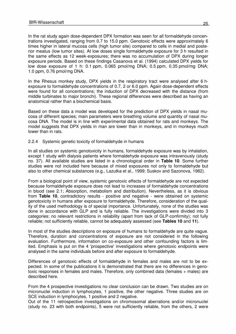
In the rat study again dose-dependent DPX formation was seen for all formaldehyde concen-trations investigated, ranging from 0.7 to 15.0 ppm. Genotoxic effects were approximately 6 times higher in lateral mucosa cells (high tumor site) compared to cells in medial and poste-rior meatus (low tumor sites). At low doses single formaldehyde exposure for 3 h resulted in the same effects as 12 week-exposures; there was no accumulation of DPX during longer exposure periods. Based on these findings Casanova et al. (1994) calculated DPX yields for low dose exposure of 1 h: 0.1 ppm, 0.065 pmol/mg DNA; 0.5 ppm, 0.35 pmol/mg DNA; 1.0 ppm, 0.76 pmol/mg DNA. In the Rhesus monkey study, DPX yields in the respiratory tract were analysed after 6 h-exposure to formaldehyde concentrations of 0.7, 2 or 6.0 ppm. Again dose-dependent effects were found for all concentrations; the induction of DPX decreased with the distance (from middle turbinates to major bronchi). These regional differences were described as having an anatomical rather than a biochemical basis. Based on these data a model was developed for the prediction of DPX yields in nasal mu-cosa of different species; main parameters were breathing volume and quantity of nasal mu-cosa DNA. The model is in line with experimental data obtained for rats and monkeys. The model suggests that DPX yields in man are lower than in monkeys, and in monkeys much lower than in rats. 2.2.4 Systemic genetic toxicity of formaldehyde in humans
In all studies on systemic genotoxicity in humans, formaldehyde exposure was by inhalation, except 1 study with dialysis patients where formaldehyde exposure was intravenously (study no. 37). All available studies are listed in a chronological order in Table 10. Some further studies were not included here because of mixed exposures not only to formaldehyde but also to other chemical substances (e.g., Lazutka et al., 1999; Suskov and Sazonova, 1982). From a biological point of view, systemic genotoxic effects of formaldehyde are not expected because formaldehyde exposure does not lead to increases of formaldehyde concentrations in blood (see 2.1; Absorption, metabolism and distribution). Nevertheless, as it is obvious from Table 10, contradictory results - positive and negative - were obtained on systemic genotoxicity in humans after exposure to formaldehyde. Therefore, consideration of the qual-ity of the used methodology is of special importance. Unfortunately, none of the studies was done in accordance with GLP and is fully reliable. The investigations were divided into 3 categories: no relevant restrictions in reliability (apart from lack of GLP-confirmity); not fully reliable; not sufficiently reliable, cannot be adequately assessed (see Tables 10 and 11). In most of the studies descriptions on exposure of humans to formaldehyde are quite vague. Therefore, duration and concentrations of exposure are not considered in the following evaluation. Furthermore, information on co-exposure and other confounding factors is lim-ited. Emphasis is put on the 4 'prospective' investigations where genotoxic endpoints were analysed in the same individuals before and after exposure to formaldehyde. Differences of genotoxic effects of formaldehyde in females and males are not to be ex-pected. In some of the publications it is demonstrated that there are no differences in geno-toxic responses in females and males. Therefore, only combined data (females + males) are described here. From the 4 prospective investigations no clear conclusion can be drawn. Two studies are on micronuclei induction in lymphocytes, 1 positive, the other negative. Three studies are on SCE induction in lymphocytes, 1 positive and 2 negative. Out of the 11 retrospective investigations on chromosomal aberrations and/or micronuclei (study no. 23 with both endpoints), 5 were not sufficiently reliable, from the others, 2 were

26 BfR-Wissenschaft
positive and 4 negative. Similarly out of the 7 SCE studies, 2 were not sufficiently reliable, from the others 2 were positive and 3 negative.
There is only one investigation on DPX which probably represents the most sensitive indica-tor for genotoxicity of formaldehyde. Shaham et al. (2003, study no. 19) reported on a 1.5-fold increase in DNA protein crosslinks (DPX) in human lymphocytes of 186 individuals after inhalation exposure to formaldehyde. The strength of the effect was not correlated with the formaldehyde concentration (0.4 vs. 2.24 ppm). The authors admit that 'no increase in its [formaldehyde] concentration in tissue or blood can be detected even moments after expo-sure', but they speculate that, nevertheless, formaldehyde may escape from metabolism. Concerning the negative DPX findings in rats by Casanova and colleagues (see Table 8) Shaham et al. argue that their methodology is far more sensitive. Although doubts arise con-cerning the reliability of these findings, this single positive finding on DPX cannot be evalu-ated finally. Balancing the data on systemic genotoxicity in humans, no clear evaluation can be made. In 2.2.2 it was concluded that the genotoxic potential of formaldehyde is not expressed sys-temically in vivo in animals. Altogether there is no sufficient evidence to reject the plausible assumption that formaldehyde does not induce systemic genotoxicity in man (Table 5). Table 5: Overview on the results obtained with systemic genetic toxicity testing of formaldehyde in hu-mans
study no. CAb MN SCE DPX other prospective investigations 22 Ying et al., 1999 -- 24 Ying et al., 1997 - 27 Suruda et al., 1993 + (weak) - 32 Yager et al., 1986 ++ (weak) retrospective investigations 18 Ye et al., 2005 ++ (weak) 19 Shaham et al., 2003 + 20 Shaham et al., 2002 + (weak) 21 Sari-Minodier et al., 2001 ++ (weak) 23 He et al., 1998 ? ? ? 25 Kitaeva et al., 1996 ? 26 Vasudeva and Anand, 1996 - 28 Jin and Zhu, 1992 ++ -- 29 Vargova et al., 1992 ? 30 Dobias et al., 1989 ? 31 Chebotarev et al., 1986 ? ? UDS ? 33 Bauchinger and Schmid, 1985 - - 34 Thomson et al., 1984 - -
35 Ward et al., 1984 sperm abn. -- 2F-bodies --
36 Fleig et al., 1982 -- 37 Goh and Cestero, 1979 ? num. chr. ab. ?
++ or --: no relevant restrictions in reliability (apart from lack of GLP-confirmity); + or -: not fully reliable; ?: not sufficiently reliable, cannot be adequately assessed; 2F-bodies, sperms with 2 F bodies (Y chromosomes); num.
chr. ab., numerical chromosome aberration; sperm abn., sperm abnormality; UDS, unscheduled DNA synthesis (DNA excision-repair).
2.2.5 Local genetic toxicity in humans
In all the 8 studies on local genotoxicity in humans formaldehyde exposure was by inhalation and MN were used as genotoxic endpoint. Five investigations were conducted retrospec-tively, 4 investigations were done prospectively (i.e. the same individuals were investigated

27
BfR-Wissenschaft
before and after formaldehyde exposure; in 1 paper both study types were performed). Six investigations were on nasal mucosa cells, 5 on buccal mucosa cells (in 3 papers both study types were performed). In the clear majority of studies positive results were reported. For an overview on local genetic toxicity in humans see Table 11. MN assays with nasal mucosa and buccal cells are no established routine tests; there are no internationally accepted guidelines. Main problems associated with these assays are dis-cussed by the Human Micronucleus Project (HuMN; Fenech et al.,1999; http://ehs.sph.berkeley.edu/holland/humn/). Repeated analyses show strong variations in MN frequencies of the same individuals. Exfoliated cell preparations contain a large number of degenerated cells. It is essential to differentiate cells with intact ('normal') nuclear structure from cells that have undergone kary-olysis and karyorrhexis in order to avoid artifacts in identifying MN. Some of the papers do not consider this aspect. Cell cycle kinetics has to be considered. MN observed in exfoliated buccal or nasal epithelial cells are not induced when the cells are at the epithelial surface, but only when they are in the basal layer. In general, cells take 7 - 16 days to emerge to the surface and exfoliate. Therefore, positive findings obtained 1 and 2 days after exposure will probably not be caused by formaldehyde (study no. 42). According to the review by Fenech et al. (1999), the average spontaneous MN frequency in healthy populations is approximately 0.1-0.3 %, with no significant variation between the cell types. In the 7 studies discussed here, an enormous variation is seen between MN frequen-cies in negative control individuals. Table 6: Variation of MN frequencies in negative control individuals
study no. Reference nasal mucosa cells (%)
buccal mucosa cells (%)
38 Ye et al., 2005 ca. 0.12* 39 Burgaz et al., 2002 0.3300 40 Burgaz et al., 2001 0.0610 41 Ying et al., 1997 0.1200 0.0568 42 Kitaeva et al., 1996 0.7700 43 Titenko-Holland et al., 1996 0.2000 0.0600 44 Suruda et al., 1993 0.0410 0.0046 45 Ballarin et al., 1992 0.0250
*1.2 MN/1000 cells (no frequency of MN cells given) Altogether, results obtained in these studies need very cautious interpretations. Seven out of the 8 studies (Table 6) are not fully or not sufficiently reliable and all of them are difficult to interprete. On the other hand, all studies led to positive findings in 1 or both cell types. In spite of the methodological insufficiencies, this must be interpreted as an indication that for-maldehyde can express its genotoxic potential in directly exposed human cells. However, quantitative data on exposure (and on MN frequencies) are not sufficiently reliable to derive relevant information on the dose-effect relationship. 2.2.6 Conclusion on genotoxicity
Formaldehyde is a highly reactive chemical with genotoxic properties. It induces various genotoxic effects; in cultured mammalian cells the induced mutations are mainly on the chromosomal level (such as structural aberrations, micronuclei), whereas there is no or a weak potential for induction of gene mutations (such as HPRT mutations or large colonies in the mouse lymphoma assay). Of the indicator endpoints induced by formaldehyde, DPX are of special importance. They represent primary DNA lesions which can be processed to muta-

28 BfR-Wissenschaft
tions. DPX were investigated in a number of studies in various cell locations and under vari-ous conditions. From the investigations on systemic mutagenicity of formaldehyde in mammals and man, it can be concluded that the genotoxic properties of formaldehyde are not expressed systemi-cally in distant site tissues. The data on local genotoxicity in humans need very cautious in-terpretation. Altogether it seems reasonable to conclude that formaldehyde can exhibit its genotoxic potential in directly exposed tissues in mammals and man; however, no reliable data on dose-effect relationships can be derived. The main focus is on data of local genotox-icity in the respiratory tract of mammals after inhalation exposure. Here it is demonstrated that formaldehyde induces DPX in the respiratory tract of rats and monkeys. The differences in DPX yields between species and cell locations seem to be based on an anatomical rather than a biochemical basis. In rats DPX were detected after inhalation of doses as low as 0.3 ppm mainly in the lateral meatuses (Casanova et al. 1989). In monkeys DPX are formed predominantly in the middle turbinates at an airborne concentration of 0.7 ppm formalde-hyde. Unlike for other toxicological effects induced by formaldehyde, for DPX no exposure concen-tration without effect can be derived. For low doses (e.g. up to 2 ppm) there seems to be a linear dose-effect relationship for DPX induction whereas at higher doses other factors (such as cytotoxicity) have strong influence on the DPX yield resulting in non-linearity of the dose-effect relationship. Based on their extensive investigation on formaldehyde-induced DPX, Casanova et al. (1991) developed a model according to which DPX yields in man are lower than in monkeys, and in monkeys much lower than in rats. For rat nasal mucosa cells, the following DPX yields were calculated for low dose exposure for 1 h (Casanova et al., 1994): 0.1 ppm, 0.065 pmol/mg DNA; 0.5 ppm, 0.35 pmol/mg DNA; 1.0 ppm, 0.76 pmol/mg DNA. Formaldehyde-induced DPX, i.e. cross-links between DNA and proteins that interact with DNA such as histones, are formed by a 2-step mechanism. In the first step formaldehyde reacts with amino groups in the side chains of amino acids (e. g. lysine, arginine) and subse-quently undergoes a reaction with further amino groups of DNA bases (reviewed in Barker et al., 2005). DPX may be removed by spontaneous hydrolysis as well as by partial proteolytic removal of proteins involved in DPX formation by proteosomes. Enzymatic DNA repair is possible by a nucleotide excision repair mechanism, but more than one pathway can be in-volved in the DNA repair of crosslinks (Quievryn and Zhitkovich, 2000; Speit et al., 2000). In several in vitro studies DPX-induction was analysed in parallel to mutation endpoints (Ta-ble 7), such as micronuclei and so-called small colonies in the mouse lymphoma assay (in-duced by clastogenic effects) (Merk and Speit, 1998; Speit et al., 2000, Speit and Merk, 2002). Thus, from the genotoxicity data a close relationship between DPX and mutations can be assumed. This is supported by the idea that crosslinks act as bulky helix-distorting ad-ducts which disturb replication (and transcription) of DNA, leading to DNA strand breaks and chromosomal aberrations. Hence, formation of DPX after formaldehyde exposure is consid-ered as a pre-mutagenic event. For better understanding investigations would be helpful which enable direct comparison of induction of DPX and mutational endpoints, e.g. micronu-clei, in vivo in rat nose cells. Formaldehyde is endogenously formed as by-product of regular mammalian metabolism. As mentioned in Section 2.1.1 the formaldehyde concentration was found to be approximately 100 µmol/l in blood of unexposed rats, monkeys or humans (Casanova et al., 1988; Heck et al., 1985); in livers or nasal mucosas of rats formaldehyde concentrations of 200 – 400 µmol/l were determined (Heck et al., 1982). Therefore, possible genotoxic effects of these formal-dehyde concentrations are of interest. From a number of studies it can be deduced that for-maldehyde concentrations in this range have the potential for induction of DPX and other genotoxic effects.

29
BfR-Wissenschaft
Table 7: Comparison of concentrations leading to DPX and other genotoxic effects in mammalian cells in culture
Cell type Genetic effect: DPX induction
Genetic effect: Others
Reference
Primary rat tracheal epithelial cells
DPX: dose-related increase from 50 to 400 µmol/l
DNA single-strand breaks: dose-related increase from 100 to 400 µmol/l
Cosma et al., 1988b
rat tracheal cell line DPX: dose-related increase from 50 to 400 µmol/l
DNA single-strand breaks: dose-related increase from 100 to 400 µmol/l
Cosma et al. 1988a
L5178Y mouse lymphoma cells
DPX: dose-related increase from 62.5 to 250 µmol/l
TK mutations (small colonies): dose-related increase from 125 to 250 µmol/l
Speit and Merk, 2002
MRC5CV1 (normal human cell line)
DPX: dose-related increase from 125 to 500 µmol/l
Micronuclei: dose-related increase from 125 to 500 µmol/l
XP12ROSV (exci-sion-repair deficient human cell line)
DPX: dose-related increase from 125 to 500 µmol/l
Micronuclei: dose-related increase from 125 to 500 µmol/l
GN06914 (crosslink-repair deficient cell line)
DPX: dose-related increase from 125 to 500 µmol/l
Micronuclei: dose-related increase from 125 to 500 µmol/l
Speit et al., 2000
human nasal epithelial cell line
DPX: dose-related increase from 333 to 16'667 µmol/l [10 to 500 µg/ml]
Zhong and Que Hee, 2004
Although these in vitro data cannot be directly extrapolated to the in vivo situation in man, a possible conclusion is that formaldehyde may contribute to the 'spontaneous' endogenous level of DPX and other genotoxic effects in human tissues. Then exogenous formaldehyde would add further genotoxic effects to a relative high endogenous level. All these studies do not allow a quantification of base-line and formaldehyde-induced DPX levels. Quievryn and Zhitkovitch (2000) calculated that (in vitro) 200 µmol/l formaldehyde induces approximately 2500 crosslinks per 108 base-pairs of DNA. Altogether, formaldehyde is a highly reactive compound, which exhibits its genotoxic poten-tial in directly exposed tissues in vitro and in vivo, in animals and in man. DPX are the best investigated genotoxic endpoint; they represent premutagenic DNA lesions and it was dem-onstrated in mammalian cell cultures that chromosomal effects, such as micronuclei, were induced in parallel in the same cells. After inhalation exposure formaldehyde-induced DPX were determined in the respiratory tract cells of rats (lowest observed effect concentration of 0.3 ppm), monkeys (lowest observed effect concentration of 0.7 ppm) and man (no lowest observed effect concentration can be derived). Concerning DPX induction, no exposure con-centration without effect can be derived. It might well be that endogenous formaldehyde con-centrations contribute to base-line levels of DPX and other genotoxic effects.

30 BfR-Wissenschaft
2.2.7 Tables on genotoxicity
Table 8: Systemic genetic toxicity of formaldehyde (FA) in mammalian animals
study no. References study no. 1 Dallas et al., 1992 study design inhalation study.
rats, exposed to 0.5 - 3 – 15 ppm FA for 1 or 8 weeks (5 days/week, 6 h/day) genotoxicity endpoint CAb bone marrow cells authors conclusion negative remarks frequencies of CAb cells after 1-week exposure: 3 % (negative control 0.8%
(0.5 ppm), 2.4% (3 ppm) and 4% (15 ppm) frequencies of CAb cells after 8-week exposure: 1.6% (negative control), 2.2% (0.5 ppm), 0.32% (3 ppm) and 1.6% (15 ppm) all CAb frequencies recalculated from columns in a figure
study no. 2 Kim et al., 1991 study design inhalation study.
mice, exposed to 181 – 229 ppm (males) and 253 - 273 ppm (females) for 2 h genotoxicity endpoint MN in bone marrow cells (PCE) authors conclusion negative remarks frequencies of MN cells in males: 0.11% (negative control), 0.2 % (181 ppm),
0.08% (229 ppm) frequencies of MN cells in females: 0.12% (negative control), 0.08% (253 ppm), 0.08% (273 ppm) Paper is written in Korean with english abstract and figures
study no. 3 Kitaeva et al., 1990 study design inhalation study.
rats, exposed to 0.42 - 1.25 ppm FA [0.5 - 1.5 mg/m3] for 4 months (5 days/week, 4 h/day)
genotoxicity endpoint CAb and numerical chromosome aberrations in bone marrow cells authors conclusion positive for both endpoints remarks CAb: increase in cells with CAb from 0.7% (negative control) to 2.4% (0.42 ppm)
and 4.0% (1.25 ppm) Numerical chromosome aberrations: increase in cells with hypoploidies from 7.0% (negative control) to 10.9% (0.42 ppm) and 13.6% (1.25 ppm); increase in cells with hyperploidies from 0.2% (negative control) to 0.8% (0.42 ppm) and decrease to 0.0% (1.25 ppm) The investigation suffers from severe methodological limits. E.g., it is stated that 40 rats were used and 100 mitoses were analysed per animal; however, the number of analysed cells for the 3 experimental groups varies between 600 and 785; this indicates that the methodology of chromosome preparation was inade-quate. Furthermore, the frequencies of hypoploidies are far higher than the fre-quencies of hyperploidies, again indicating limits in chromosome preparation. The results are not sufficiently reliable.
study no. 4 Casanova and Heck, 1987 study design inhalation study.
rats, exposed to 0.9 - 2.4 - 6.0 - 10.0 ppm for 2 d (3 h/d) genotoxicity endpoint DPX in bone marrow cells authors conclusion negative remarks study no. 5 Kligerman et al., 1984
study design inhalation study. rats, exposed to 0.5 - 6 – 15 ppm FA for 5 days (6 h/day)
genotoxicity endpoint CAb and SCE in peripheral lymphocytes authors conclusion negative for both endpoints remarks frequencies of CAb cells: 2.0% (negative control), 2.0% (15 ppm)
frequencies of SCE/metaphase: 9.5 (negative control), 9.7 (0.5 ppm), 8.5 (6 ppm), 9.9 (15 ppm)

31
BfR-Wissenschaft
continuation Table 8: Systemic genetic toxicity of formaldehyde (FA) in mammalian animals
study no. References study no. 6 Morita et al., 1997 study design oral by gavage
mice, 1 or 2 administrations of doses up to 200 mg FA/kg b.w; sampling 24, 48 and 72 h after treatment intravenous injection mice, treated with 2 doses up to 30 mg FA/kg b.w; sampling 24 - 48 – 72 h after treatment
genotoxicity endpoint MN in bone marrow cells (PCE) and peripheral retikulocytes authors conclusion negative under all experimental conditions remarks MN frequencies in PCE after 2 oral FA administrations, 24 h sampling: 0.14%
(negative control), 0.20% (100 mg/kg), 0.12% (200 mg/kg) MN frequencies in peripheral retikulocytes after single oral FA administration, 24 h sampling: 0.12% (negative control), 0.18% (100 mg/kg), 0.16% (200 mg/kg) MN frequencies in peripheral retikulocytes after single intravenous FA administra-tion, 24 h sampling: 0.10% (negative control), 0.14% (20 mg/kg), 0.08% (30 mg/kg) also negative for lower doses and after 48 and 72 h sampling
study no. 7 Natarajan et al., 1983 study design intraperitoneal injection
mice, treated with 6.25 - 12.25 - 25.00 mg FA/kg b.w; sampling 16 – 40 h after treatment
genotoxicity endpoint CAb in bone marrow (PCE) and spleen cells, MN in bone marrow cells authors conclusion negative remarks MN frequencies in PCE after single intraperitoneal FA administration, 16 h sam-
pling [number of MN per 100 PCE in males/females]: 0.8/0.4 (negative control), 0.6/0.6 (6.25 mg/kg), 1.6/1.2 (12.5 mg/kg), 1.6/1.6 (25 mg/kg) CAb frequencies in bone marrow cells after single intraperitoneal FA administra-tion, 16 h sampling [males/females]: 3.0/2.8% (negative control), 3.6/3.3% (6.25 mg/kg), 3.0/3.6% (12.5 mg/kg), 3.2/3.3% (25 mg/kg) CAb frequencies in spleen cells after single intraperitoneal FA administration, 16 h sampling [males/females]: 2.4/2.2% (negative control), 3.5/4.0% (6.25 mg/kg), 2.8/3.4% (12.5 mg/kg), 3.0/2.6% (25 mg/kg) also negative after 40 h sampling time
study no. 8 Fontignie-Houbrechts, 1981 study design intraperitoneal injection
mice, treated with 50 mg FA/kg bw genotoxicity endpoint CAb and numerical chromosome aberrations in spermatocytes-I, dominant-lethal
mutations authors conclusion negative for CAb and numerical chromosome aberrations, questionable positive
for dominant-lethal mutations remarks Dominant-lethal mutations: In the summary the dominant-lethal result is described
as negative; in the Discussion positive effects in the 1-week and 3-week mating periods are discussed. The results are based on only 10 males per experimental point. The results are not sufficiently reliable.
study no. 9 Gocke et al., 1981 study design intraperitoneal injection
mice, treated with 10 – 30 mg FA/kg b.w; sampling 30 h after treatment genotoxicity endpoint MN in bone marrow cells authors conclusion negative remarks

32 BfR-Wissenschaft
continuation Table 8: Systemic genetic toxicity of formaldehyde (FA) in mammalian animals
study no. References study no. 10 Epstein et al., 1972
study design intraperitoneal injection mice, treated with 16 - 20 - 32 – 40 mg FA/kg b.w genotoxicity endpoint dominant-lethal mutations
authors conclusion negative remarks
abbreviations: CAb, structural chromosomal aberrations; DPX, DNA-protein crosslinks; MN, micronuclei; SCE, sister-chromatid exchange(s)
Table 9: Local genetic toxicity of formaldehyde (FA) in mammalian animals
study no. References study no. 11 Casanova et al., 1994 study design inhalation
rats, exposed to 0.7 - 2 - 6 - 10 - 15 ppm FA for 3 h or 12 weeks genotoxicity endpoint DPX in nasal mucosa cells authors conclusion positive remarks dose-dependent DPX formation at all concentrations, after 3-h or 12-week expo-
sure, in all analysed nasal mucosa locations Comparison of the cell locations: in lateral mucosa cells (high tumor site) DPX yield was approximately 6 times higher than in medial and posterior meatus (low tumor sites) Comparison of exposure periods in lateral mucosa cells: no difference in DPX yields at low FA doses of 0.7 and 2.0 ppm; at high doses of 6.0 to 15.0 ppm ap-proximately 2 times higher DPX yield after acute 3h-exposure Based on these findings the following DPX yields were calculated for low dose exposure for 1 h: 0.1 ppm, 0.065 pmol/mg DNA; 0.5 ppm, 0.35 pmol/mg DNA; 1.0 ppm, 0.76 pmol/mg DNA Strong induction of cell proliferation in lateral mucosa at 6 and 15 ppm after 12 week-exposure
study no. 12 Dallas et al., 1992 study design inhalation
rats, exposed to 0.5 - 3 - 15 ppm FA for 1 or 8 weeks (5 days/week, 6 h/day) genotoxicity endpoint CAb in pulmonary macrophages authors conclusion positive remarks increases in CAb cells only at 15 ppm, from 3.5% (neg. contr., 1 week) and 4.4%
(neg. contr., 8 weeks) to 7.2% (1 week) and 9.2% (8 weeks). study no. 13 Casanova et al., 1991 study design inhalation
Rhesus monkeys, exposed to 0.7 - 2 - 6 ppm FA for 6 h genotoxicity endpoint DPX in respiratory tract mucosa cells authors conclusion positive
remarks dose-dependent DPX formation at all concentrations Comparison of the cell loca-tions: highest DPX yields in mucosa cells from middle turbinates; lower DPX yields in anterior lateral wall/septum and nasopharynx; very low DPX yields in larynx/trachea/carina and proximal portions of major bronchi. These regional differences were described as having an anatomical rather than a biochemical basis. Based on these data a model was developed for prediction of DPX yields in nasal mucosa of different species; main parameters were breathing volume and quan-tity of nasal mucosa DNA. The model is in line with experimental data obtained for rats and monkeys. The model suggests that DPX yields in man are lower than in monkeys, and in monkeys much lower than in rats.
study no. 14 Casanova et al., 1989
study design inhalation rats, exposed to 0.3 - 0.7 - 2.0 - 6.0 - 10.0 ppm FA for 6 h
genotoxicity endpoint DPX in nasal mucosa cells

33
BfR-Wissenschaft
continuation Table 9: Local genetic toxicity of formaldehyde (FA) in mammalian animals
study no. References authors conclusion positive
remarks dose-dependent DPX formation at all concentrations (non-linear dose-effect rela-tionship)
study no. 15 Casanova and Heck, 1987
study design inhalation rats, exposed to 0.9 - 2 - 4 - 6 - 10 ppm FA, 2 times for 3 h
genotoxicity endpoint DPX in respiratory mucosa cells authors conclusion positive
remarks dose-dependent DPX formation at 2-10 ppm study no. 16 Casanova et al., 1984
study design inhalation rats, exposed to 0.3 - 2 - 6 - 10 – 15 ppm FA, 2 times for 6 h
genotoxicity endpoint covalent DNA-binding in respiratory and olfactory mucosa cells authors conclusion positive
remarks positive in respiratory mucosa (dose-dependent effect at 2 – 15 ppm), negative in olfactory mucosa
study no. 17 Migliore et al., 1989
study design oral by gavage rats, treated with 200 mg FA/kg b.w; sampling 16 - 24 – 30 h after treatment
genotoxicity endpoint MN in gastro-intestinal tract cells authors conclusion positive
remarks increase in MN cells in all tissues. stomach: increase from 0.33% (neg. contr.) up to 6.5%; duodenum: increase from 0.33% (neg. contr.) up to 1.46; ileum: increase from 0.33% (neg. contr.) up to 1.80%; colon: increase from 0.33% (neg. contr.) up to 1.80%. Severe local irritation
Table 10: Systemic genetic toxicity of formaldehyde (FA) in humans
study no. References study no. 18 Ye et al., 2005 study design retrospective analysis.
exposed group 1: 18 factory workers; inhalation exposure to 0.985 mg/m3 with peaks of 1.694 mg/m3 for an average of 8.5 year (1 - 15 year) exposed group 2: 16 waiters; inhalation exposure to 0.107 mg/m3 with peaks of 0.300 mg/m3 for 12 weeks control group: 23 subjects: inhalation exposure to 0.011 mg/m3 with peaks of 0.015 mg/m3 It is stated that subjects of all groups had no smoking history, no medicine history for 3 weeks, no X-ray history for 0.5 year, no routine intake of any drugs. No GLP.
genotoxicity endpoint SCE in peripheral lymphocytes authors conclusion positive for exposed group 1, negative for exposed group 2 reliability of test result no relevant restrictions in reliability (apart from lack of GLP-conformity) remarks increase in SCE frequency from 6.41 SCE/metaphase (control group, S.D. 0.47)
to 8.26 (exposed group, S.D. 0.91) [recalculated from columns in figures] study no. 19 Shaham et al., 2003
study design retrospective analysis. exposed group: 186 workers from 14 hospital pathology departments; mean inha-lation exposure to 0.4 ppm (low-level-subgroup, range 0.04 - 0.70) or to 2.24 ppm (high-level-subgroup, range 0.72 - 5.60) for an average of 15.9 year control group: 213 subjects from administrative sections of the same hospitals; Adjustments were made for age, sex, origin and education. On the basis of a questionnaire it was concluded that nobody in the exposed group was occupationally exposed to substances known to form DPX except FA and nobody in the control group was ever occupationally exposed to FA. No GLP.
genotoxicity endpoint DPX in peripheral lymphocytes authors conclusion positive

34 BfR-Wissenschaft
continuation Table 10: Systemic genetic toxicity of formaldehyde (FA) in humans
study no. References reliability of test result not fully reliable remarks Increase in DPX (DPX DNA / total DNA) from 0.14 (control group) to 0.21 (ex-
posed group). No difference between low-level- and high-level subgroups. No difference between exposure groups up to 16 years and above 16 years [DPX values of 0.19 and 0.20]. Concerning quantitative DPX data, it is confusing that the same subgroup values of 0.19 and 0.20 were found for low-level- and high-level-groups as well as for the both exposure duration groups. Furthermore, the subgroup values of 0.19 and 0.20 do not lead the DPX value for the total group of 0.21. Pilot studies for this investigation were published a.o. in Shaham et al. 1996 and Shaham et al. 1997.
study no. 20 Shaham et al., 2002 study design retrospective investigation
exposed group: 90 workers from 14 hospital pathology departments; mean inha-lation exposure to 0.4 ppm (low-level-subgroup, range 0.04 - 0.70) or to 2.24 ppm (high-level-subgroup, range 0.72 - 5.60) for an average of 15.4 year control group: 52 subjects from administration sections of the same hospitals. It is stated that adjustments were made for age, sex, smoking habits, education and origin. No GLP.
genotoxicity endpoint SCE in peripheral lymphocytes authors conclusion positive reliability of test result not fully reliable remarks Increase in SCE frequency from 0.19 SCE/chromosome (control group) to 0.27
(exposed group). No difference between low level and high-level-subgroups [SCE frequency of 0.27 in both groups]. It is surprising that the subgroups 'low level exposure' and 'high level exposure' had the same means of FA exposure and the same ranges as in the 2003-publication on DPX, although the number of individuals varied from 90 to 186. The presentation of the SCE scoring methodology is confusing. In the tables SCE frequencies are given in SCE/chromosome (an unusual methodology going back to early days of SCE assays when adequate staining of whole metaphases was difficult); in the methods text it is stated: 'we examined cells that had 44-46 clearly visible chromosomes and normalised the SCE count to the frequency expected for 46 chromosomes' (which again is a quite unusual method). A pilot study for this investigation was published in Shaham et al. 1997.
study no. 21 Sari-Minodier et al., 2001
study design retrospective investigation exposed group: 10 workers in pathology labs (female non-smokers); mean inhala-tion exposure to 1.2 ppm for 9 year (1 - 16 year) control group: 27 age-matched persons (female non-smokers)
genotoxicity endpoint MN in peripheral lymphocytes authors conclusion positive
reliability of test result no relevant restrictions in reliability (apart from lack of GLP-confirmity) remarks weak increase in frequency of MN cells from 0.88% (S.D. 0.44%, control group)
to 1.88% (S.D. 1.31%, exposed group) study no. 22 Ying et al., 1999 study design prospective investigation
23 anatomy students (non-smokers) sampled before and after inhalation expo-sure to 0.508 mg/m3 (3 times per week) for 8 weeks No GLP.
Genotoxicity endpoint SCE in peripheral lymphocytes authors conclusion negative reliability of test result no relevant restrictions in reliability (apart from lack of GLP-confirmity) remarks SCE frequencies of 6.383 (before exposure) and 6.613 (after exposure).

35
BfR-Wissenschaft
continuation Table 10: Systemic genetic toxicity of formaldehyde (FA) in humans
study no. References study no. 23 He et al., 1998 study design retrospective investigation
exposed group: 13 anatomy students (non-smokers); mean inhalation exposure to 2.37 ppm (3.17 mg/m3, 10 h per week) for an average of 12 weeks control group: 10 unexposed students (non-smokers) from the same school Slides were not coded before analysis. No GLP.
genotoxicity endpoint CAb, MN and SCE in peripheral lymphocytes authors conclusion positive for all 3 endpoints reliability of test result not sufficiently reliable, cannot be adequately assessed remarks increase in frequency of cells with CAb incl. gaps from 3.4 % (control group) to
5.92% (exposed group). increase in frequency of MN cells from 0.315% (control group) to 0.638% (ex-posed group) increase in SCE frequency per cells from 5.26 (control group) to 5.91 (exposed group) The MN frequency in the control group of 0.315% is relatively low; even in the exposed group the MN frequency does not reach the normal level around 1%. The authors compared MN and CAb frequencies in exposed individuals and found a very close quantitative correlation; considering the relatively low number of cells analysed (100 cells per individual for CAb analysis) this is highly surpris-ing.
study no. 24 Ying et al., 1997 study design prospective investigation
23 anatomy students (non-smokers); inhalation exposure to 0.508 mg/m3 (3 times per week) for 8 weeks No GLP
genotoxicity endpoint MN in peripheral lymphocytes authors conclusion negative reliability of test result not fully reliable remarks frequencies of MN cells of 0.091% (before exposure) and 0.111 % (after expo-
sure). According to statistical evaluation this difference is not significant. The extremely low frequencies of MN cells make the findings difficult to evaluate.
study no. 25 Kitaeva et al., 1996 study design retrospective investigations
exposure group 1: 15 workers in a FA synthesis shop; inhalation exposure to 1.2 - 2.4 ml/m3 for an average of 9.7 year exposure group 2: 8 anatomy teachers; unspecified inhalation exposure for an average of 17.2 year control group: 6 unspecified subjects No GLP
genotoxicity endpoint CAb in peripheral lymphocytes authors conclusion positive for exposure group 1, no result stated for exposure group 2 reliability of test result not sufficiently reliable, cannot be adequately assessed remarks In exposed group 1 there was an increase in frequency of CAb cells from 1.8%
(control group) to 5.4% (exposed group). In exposed group 2 all cultures failed. The methodology used was obviously not adequate for CAb analysis. No meta-phases were obtained after 48-50 h-culture; after 72 h-culture only 148 meta-phases were scorable from 15 individuals.
study no. 26 Vasudeva and Anand, 1996
study design retrospective investigation exposed group: 30 anatomy students; inhalation exposure to <1 ppm for an aver-age of 15 months control group: 30 non-medical students No GLP
genotoxicity endpoint CAb in peripheral lymphocytes

36 BfR-Wissenschaft
continuation Table 10: Systemic genetic toxicity of formaldehyde (FA) in humans
study no. References authors conclusion negative
reliability of test result not fully reliable remarks CAb: aberration frequencies of 1.2% (exposed group) vs. 0.9 % (control group).
According to statistical evaluation this difference is not significant. Slides were not coded before analysis.
study no. 27 Suruda et al., 1993
study design prospective investigation exposed group: 29 embalmer students; inhalation exposure to 1.4 ppm with peaks up to 6.6 ppm for 85 d (mean of 6.9 embalmings for 125 min each) No GLP
genotoxicity study design MN and SCE in peripheral lymphocytes authors conclusion positive for MN, negative for SCE reliability of test result not fully reliable remarks increase in frequency of MN cells from 0.495% before exposure to 0.636% after
exposure slight decrease in SCE frequency per cell from 7.72 before exposure to 7.14 after exposure Considering the fact SCE are a quite sensitive indicator for the genotoxic potential of FA, it is not plausible that MN are reported as positive and SCE as negative.
study no. 28 Jin F. and Zhu R., 1992
study design retrospective investigation exposed group: 20 workers at vinylon factory (including 18 smokers); formalde-hyde exposure for an average of 10.5 year: annual average of 2.51 mg/m3 with FA concentrations below 3 mg/m3 during 11 months per year and 5.72 mg/m3 during the 12th month control group: 14 non-productive workers of the same factory (including 7 smok-ers)
genotoxicity endpoint CAb and SCE in peripheral lymphocytes authors conclusion positive for CAb, negative for SCE
reliability of test result no relevant restrictions in reliability (apart from lack of GLP-conformity) remarks increase in frequency of CAb cells (incl. gaps) from 1.57% (control group) to
5.08% (exposed group) [clear increases in chromosome fragments and chromatid breaks] SCE frequencies of 5.44 (control group) and 6.42 SCE per metaphase (exposed group). This difference was not statistically significant. [Increased SCE frequen-cies in smokers as compared to non-smokers.] No GLP.
study no. 29 Vargova et al., 1992
study design retrospective investigation exposed group: 20 workers of a wood-splinter materials factory (non-smokers); inhalation exposure to 0.55 - 10.36 mg/m3 for 5 - >16 years control group: 10 unexposed students (non-smokers) No GLP
genotoxicity endpoint CAb in peripheral lymphocytes authors conclusion negative
reliability of test result not sufficiently reliable, cannot be adequately assessed remarks lower CAb frequency in the exposed group (3.08 %) as compared to the neg.
contr. group (3.60%); both CAb frequencies were increased as compared with the general population

37
BfR-Wissenschaft
continuation Table 10: Systemic genetic toxicity of formaldehyde (FA) in humans
study no. References study no. 30 Dobias et al., 1989 study design 2 retrospective investigation
exposed group 1: 20 children; inhalation exposure to 0.317 mg/m3 for an unspeci-fied period exposed group 2: unspecified no. of children; inhalation exposure to 0.130 mg/m3
for an unspecified period exposed group 3: unspecified no. of children; inhalation exposure to 0.0365 mg/m3 for an unspecified period control group: unspecified No GLP
genotoxicity study design CAb in peripheral lymphocytes authors conclusion positive
reliability of test result not sufficiently reliable, cannot be adequately assessed remarks frequencies of CAb cells of 4.71% (exposed group 1), 2.82% (exposed group 2)
and 2.06% (exposed group 3) as compared to 1.37% in a control group. The paper is an abstract without adequate data on methodology and detailed result.
study no. 31 Chebotarev et al., 1986 [see also Chebotarev et al., 1985; abstract only]
study design retrospective investigation exposed group: 37 (or 36 for SCE analysis) workers of a wood-processing enter-prise; exposure not specified control group: 14 workers in an auxiliary shop at the same site No coding of slides, no GLP
genotoxicity endpoint CAb, SCE and UDS
authors conclusion positive for CAb, negative for SCE and UDS
reliability of test result not sufficiently reliable, cannot be adequately assessed remarks frequencies of CAb cells of 2.76% (exposed group) as compared to 1.64% in a
control group. SCE frequencies of 8.01 (exposed group) and 8.24 SCE per metaphase (control group) UDS (unscheduled DNA synthesis = excision-repair) was measured by incorpora-tion of 3H-Thymidine into DNA. A negative effect was observed. The main purpose of the study was to investigate whether lymphocytes from FA exposed individuals have an altered response to treatment with a mutagen in vitro. Exposure conditions are vague.
study no. 32 Yager et al., 1986
study design prospective investigation 8 anatomy students (non-smokers) sampled before and after inhalation exposure to 1.2 ppm [1.5 mg/m3] for 10 weeks
genotoxicity endpoint SCE in peripheral lymphocytes authors conclusion positive
reliability of test result no relevant restrictions in reliability (apart from lack of GLP-conformity) remarks weak increase in SCE frequency per cells from 6.39 (before exposure) to 7.20
(after exposure). No GLP
study no. 33 Bauchinger and Schmid, 1985
study design retrospective investigation exposed group: 20 papermakers (13 smokers); inhalation exposure to up to 3 ppm for 45 to 90 min per day for an average of 14.5 years control group: 20 workers (6 smokers) from the same factory without FA exposure No GLP
genotoxicity endpoint CAb and SCE in peripheral lymphocytes authors conclusion positive for CAb, negative for SCE
reliability of test result not fully reliable; the interpretation as 'positive' for CAb induction is not justified

38 BfR-Wissenschaft
continuation Table 10: Systemic genetic toxicity of formaldehyde (FA) in humans
study no. References remarks No increase in the frequency of cells with CAb [0.86% before exposure and
0.87% after exposure] The authors state an increase in frequencies of dicentric and ring chromosome, which was limited to 'supervisors' with long-term exposure for 18.9 years; i.e. exposure between 2 and 17 years was negative. Lymphocyte cultures were co-exposed to BrdUrd (a weak clastogen). SCE frequencies of 9.53 before and 8.87 SCE per cell after exposure [increased SCE frequencies for smokers in both groups]
study no. 34 Thomson et al., 1984
study design retrospective investigation exposed group: 6 pathology workers; inhalation exposure to 2.26 mg/m3 with peaks up to >11 mg/m3 for an unspecified long-term period control group: 5 unexposed subjects No GLP
genotoxicity endpoint CAb and SCE in peripheral lymphocytes authors conclusion negative for both endpoints
reliability of test result not fully reliable remarks mean frequencies of CAb cells and mean SCE frequencies not given
no consideration of smoking habit study no. 35 Ward et al., 1984
study design retrospective investigation exposed group: 11 hospital autopsy service workers; inhalation exposure to 0.61 - 1.32 ppm (TWA) for periods ranging from 1 month to several year control group: 11 non-exposed individuals from the same staff A number of confounding factors, a.o. smoking, was considered. No GLP
genotoxicity endpoint sperm abnormality and 2F-bodies in sperms
authors conclusion negative for both endpoints
reliability of test result no relevant restrictions in reliability (apart from lack of GLP-conformity) remarks frequencies of sperm abnormalities 44.5% (exposed group) vs. 53.5% (control
group). frequencies of sperms with 2 F-bodies 1.6% in the exposed group as well as in the control group [F-body are fluorescence-marked Y chromosomes, i.e. sperms with 2 F-bodies are aneuploid with respect to the Y chromosome]
study no. 36 Fleig et al., 1982
study design retrospective investigation exposed group: 15 workers in FA manufacture.; inhalation exposure below MAK value (<5 ppm before 1971, <1 ppm after 1971) for an average of 28 year control group: 15 unexposed employees from the same site A number of confounding factors, a.o. smoking, was considered. No GLP
genotoxicity endpoint CAb in peripheral lymphocytes authors conclusion negative
reliability of test result no relevant restrictions in reliability (apart from lack of GLP-confirmity) remarks aberration frequencies of 1.67% (exposed group) vs. 1.07% (control group). Ac-
cording to statistical evaluation this difference is not significant. study no. 37 Goh and Cestero, 1979
study design retrospective investigation exposed group: 40 hemodialysis patients; i.v.-exposure during dialysis of up to 126 mg per dialysis, 3 hemodialyses lasting 4 - 6 h per week; exposure period not specified control group: not specified No coding of slides, no GLP
genotoxicity endpoint CAb and numerical chromosomal aberrations in 'bone marrow aspirate' cells authors conclusion positive for both endpoints
reliability of test result not sufficiently reliable, cannot be adequately assessed

39
BfR-Wissenschaft
continuation Table 10: Systemic genetic toxicity of formaldehyde (FA) in humans
study no. References marks CAb: increase in frequency of acentric chromosome fragments from 2% (control
group) to 3.6% (exposed group); chromatid breaks per 100 cells: 0.7 (control group), 1.4 (exposed group). Numerical chromosomal aberrations: increase in frequency of cells with numerical chromosomal aberrations from 12% (control group) to 58% (exposed group). The 40 hemodialysis patients suffered from various diseases and they were co-exposed to a number of pharmaceuticals including the mutagen folic acid. No concurrent negative control. The presentation of CAb data is not plausible. Slide preparation was obviously not adequate for analysis of aneuploidies (e.g. 10x more hypoploidies as compared to hyperploidies). Altogether, the methodology is inappropriate.
Table 11: Local genetic toxicity of formaldehyde (FA) in humans
study no. References study no. 38 Ye et al., 2005
study design retrospective analysis. exposed group: 18 factory workers; inhalation exposure to 0.985 mg/m3 with peaks of 1.694 mg/m3 for an average of 8.5 year (1 - 15 year) exposed group 2: 16 waiters; inhalation exposure to 0.107 mg/m3 with peaks of 0.300 mg/m3 for 12 weeks control group: 23 subjects: inhalation exposure to 0.011 mg/m3 with peaks of 0.015 mg/m3 It is stated that subjects of all groups had no smoking history, no medicine history for 3 weeks, no X-ray history for 0.5 year, no routine intake of any drugs. 3000 cells per sample. No GLP.
Genotoxicity endpoint MN in nasal mucosa cells
authors conclusion positive for exposed group 1, negative for exposed group 2 reliability of test result no relevant restrictions in reliability (apart from lack of GLP-conformity) remarks increase in MN frequency from 1.2 MN per 1000 cells (control group, S.D. 0.6) to
2.7 MN per 1000 cells (exposed group 1, S.D. 1.1). no effect in exposed group 2: 1.8 MN per 1000 cells (S.D. 1.0) [MN frequencies recalculated from columns in figures]
study no. 39 Burgaz et al. 2002
study design retrospective investigation exposed group: 28 subjects working in pathology and anatomy laboratories (incl. 12 smokers); mean inhalation exposure to 2 – 4 ppm for 4.7 year control group: 18 subjects (incl. 7 smokers) 3000 cells per sample. No GLP.
Genotoxicity endpoint MN in buccal cells
authors conclusion positive
reliability of test result not fully reliable remarks MN: increase in frequency of MN cells from 0.33% (control group) to 0.71% (ex-
posed group) FA exposure to 2 – 4 ppm is known to lead to eye and respiratory tract irritation; the authors do not mention such effects.
study no. 40 Burgaz et al. 2001
study design retrospective investigation exposed group: 23 subjects in pathology and anatomy laboratories (incl. 14 smokers); mean inhalation exposure to 2 – 4 ppm for 5.06 year control group: 25 subjects (incl. 19 smokers) 3000 cells per sample. No GLP.
genotoxicity endpoint MN in nasal mucosa cells
authors conclusion positive
reliability of test result not fully reliable

40 BfR-Wissenschaft
continuation Table 11: Local genetic toxicity of formaldehyde (FA) in humans
study no. References remarks MN: increase in frequency of MN cells from 0.061% (control group; S.D. 0.027%)
to 0.101% (exposed group; S.D. 0.061%). Weak but statistically significant genotoxic effect. Considering that in the exposed group MN frequencies in females (0.082% MN cells) and males (0.119% MN cells) differed statistically significant, the 'weak positive' FA effect is difficult to interprete and may well be based on an extremely low MN frequency in the con-trol group (which is also quite different from the MN frequency in the buccal cell control group of 0.33%; see Burgaz et al. 2002). FA exposure to 2 - 4 ppm is known to lead to eye and respiratory tract irritation; the authors do not mention such effects.
study no. 41 Ying et al., 1997
study design prospective investigation exposed group: 23 anatomy students (non-smokers); inhalation exposure to 0.508 mg/m3 (3 times per week) for 8 weeks 3000 cells per sample. No GLP.
genotoxicity endpoint MN in nasal mucosa cells and buccal cells
authors conclusion positive for nasal mucosa cells and buccal cells reliability of test result not fully reliable remarks MN in nasal mucosa cells: increase in frequency of MN cells from 0.120% before
exposure to 0.384% after exposure MN in buccal cells: increase in frequency of MN cells from 0.0568% before expo-sure to 0.0857% after exposure An unusual type of data presentation makes this study difficult to interprete, e.g. the number of analyzed cells, given in terms like '2870 +/- 503', is not plausible.
study no. 42 Kitaeva et al., 1996
study design retrospective investigation exposed group 1: 13 'instructors in the Division of Normal Anatomy, who had been in contact with FA for a long period of time'; no FA concentration given control group: undefined group of 7 females prospective investigation exposed group 2: 12 'students after primary one-time contact with FA-containing anatomical preparations'; no FA concentration given
genotoxicity endpoint MN in buccal cells
authors conclusion positive
reliability of test result not sufficiently reliable, cannot be adequately assessed remarks exposed group (1):
- 8 females: 2.94% MN cells, which is statistically different from the MN fre-quency in a non-defined neg. contr. group of 7 females (0.64 %).
- 5 males: 1.18% MN cells, which is not statistically different from the MN fre-quency in the non-defined neg. contr. group.
- MN, exposed group (2):
- 6 females: increase in frequencies of MN cells from 0.58% before exposure to 2.50% (24 h after exposure) and 2.64% (48 h after exposure).
- 6 males: increase in frequencies of MN cells of from 0.77% before exposure to 2.02% (24 h after exposure) and 1.86% (48 h after exposure).
Based on the cell kinetics of the cells, the described effects 1 and 2 days after short-term exposure are not plausible [MN are induced while the cells are at the epithelial surface; 7 - 16 days are needed to emerge to the surface and exfoliate]. There is no mentioning of a differentiation between epithelial cells with intact nuclear structure and degenerated cells.
Presentation of methodology and data is poor.

41
BfR-Wissenschaft
continuation Table 11: Local genetic toxicity of formaldehyde (FA) in humans
study no. References study no. 43 Titenko-Holland et al., 1996
study design prospective investigation exposed group 1 (nasal mucosa cells): 13 mortuary science students; mean inha-lation exposure of 1.4 ppm/h for 90 d with peaks up to 2.4 ppm exposed group 2 (buccal cells): 19 mortuary science students; mean inhalation exposure of 1.4 ppm/h for 90 d with peaks up to 2.4 ppm
genotoxicity endpoint MN in nasal mucosa cells and buccal cells
authors conclusion negative in nasal mucosa cells, positive in buccal cells reliability of test result not sufficiently reliable, cannot be adequately assessed remarks MN in nasal mucosa cells: frequency of MN cells: 0.20% before exposure and
0.25% after exposure MN in buccal cells: increase in frequency of MN cells from 0.06% before expo-sure to 0.20% after exposure. Increase was higher for centromere-negative MN as compared to centromere-positive MN (i.e. MN origin was preferentially due to chromosome breakage) The same individuals were analysed as in Suruda 1993. Nevertheless, MN fre-quencies in control groups for nasal mucosa cells as well as for buccal cells dif-fered strongly; therefore, severe methodological problems have to be assumed. This seems to be supported by the fact that the investigation started with 28 indi-viduals, i.e. investigations failed for 9 or 15 individuals, respectively.
study no. 44 Suruda et al., 1993
study design prospective investigation exposed group: 29 mortician students; mean inhalation exposure to 1.4 ppm for 85 d with peaks up to 6.6 ppm
genotoxicity endpoint MN in nasal mucosa cells and buccal cells
authors conclusion negative for nasal mucosa cells, positive for buccal cells
reliability of test result not sufficiently reliable, cannot be adequately assessed remarks MN in nasal mucosa cells: 0.0410 % MN cells before exposure, 0.0500 % MN
cells after exposure MN in buccal cells: increase in frequency of MN cells from 0.0046% before expo-sure to 0.0600% after exposure The MN frequency in neg. contr. for buccal cells is extremely low; even in the exposed group the MN frequency does not reach the normal level of 0.1 - 0.3%.
study no. 45 Ballarin et al., 1992
study design retrospective investigation exposed group: 15 workers in a plywood factory; mean inhalation exposure to 0.1 - 0.39 mg/m3 for 6.8 year control group: matched group of 15 subjects
genotoxicity endpoint MN in nasal mucosa cells authors conclusion positive
reliability of test result not sufficiently reliable, cannot be adequately assessed remarks MN in nasal cells: increase in frequency of MN cells from 0.025% in neg. contr. to
0.090% in FA exposed group. No mentioning of a differentiation between epithelial cells with intact nuclear structure and degenerated cells. The MN frequency in neg. contr. is extremely low; even in the exposed group the MN frequency does not reach the normal level of 0.1 - 0.3%. Individuals in the exposed group were co-exposed to wood dust. Squamous metaplasia was found in 10/15 exposed persons (as compared to 1/15 in neg. controls); MN induction may be triggered by increased cell proliferation.

42 BfR-Wissenschaft
2.3 Other relevant effects
2.3.1 Introduction
Although sensory irritation is thought not to be involved in the carcinogenicity of formalde-hyde, this endpoint is considered because it is supposed to be the most sensitive adverse effect in humans linked to formaldehyde exposure. Formaldehyde exposure-related sensory irritation effects have also been observed in the deeper parts of the respiratory tract. A number of studies on occupationally exposed (non-asthmatic) workers reported effects such as sore throat, coughing and lung function distur-bance (e.g., Holmström and Wilhelmsson, 1988). Others did not find significant effects (e.g., Holness and Nethercott, 1989). The major weakness of many studies was the absence of representative quantitative data on exposure. Exposure concentrations related to pulmonary irritation were generally higher than those inducing sensory irritation of the upper respiratory tract. Thus, reports on pulmonary function observed under occupational exposure conditions were not considered here for the purpose to describe the most sensitive adverse effect. The question whether asthmatics are posed to higher risk by formaldehyde inhalation than healthy people is highlighted for consideration lateron in the risk assessment. 2.3.2 Sensory irritation
Exposure to formaldehyde vapour induces sensory irritation of the mucosae of the eyes and the respiratory tract. Related symptoms, morphological and pathopysiological effects are responses to irritation of the trigeminal and olfactory nerve endings. In their review on formaldehyde-related irritation, Paustenbach et al. (1997) calculated on data from numerous studies that about 50% of chamber-exposed persons will report eye irritation at 2 ppm formaldehyde, 25% at 1 ppm and few, if any, will report eye irritation at 0.5 ppm. They concluded that at 0.3 ppm or less, no irritation attributable to formaldehyde should occur, even if people are exposed for 8 h/d. The value of 0.3 ppm was recommended as occupational exposure limit (OEL) and was also established as the German MAK value (2000). In order to allow the application of a threshold value for sensory irritation for the whole popu-lation, data with controlled-exposure to low-ranges of formaldehyde are of importance. Until now, very few studies are available that elucidated the irritating effects at/below 0.5 ppm: Eye irritation was reported in 3/16 (19%) volunteers exposed to 0.24 ppm formaldehyde for 5 hours, the number of responders increased proportional with the concentration (Andersen and Mølhave, 1983). 5 out of 12 volunteers (42%) exposed to 0.35 ppm formaldehyde for 6 min had eye irritation, while none of the 28 people at 0 ppm formaldehyde reported eye irritation (Bender et al.,1983). Percentages of affected volunteers increased up to 74% with formaldehyde con-centrations up to 1 ppm. Accordingly, the latency time until the first symptoms were noted to decrease concentration-related (except the outlier at 0.7 ppm). Increase in eye irritation symptoms was observed at 1 ppm formaldehyde from 26%, but no increase above back-ground incidence at 0 ppm (which was 5% of 19 subjects) was reported at 0.5 ppm in non-smokers (0% of 10 subjects) (Kulle, 1993). Most human data on irritation were from studies bearing uncertainties as they used subjec-tive data from selfreporting of compliants that may be influenced by psychological and other factors. Examination of objective test parameters were included in only few studies:

43
BfR-Wissenschaft
In addition to subjected data, the irritating effects of increasing concentrations of formalde-hyde were determined by the eye-blinking rate of healthy volunteers in the study of Weber-Tschopp et al. (1977). Self-reporting of annoyance and eye irritation was significantly in-creased at 1 ppm and above either by formaldehyde exposure to continuously increasing concentrations up to 0.03-3.2 ppm (33 subjects) or to 5 periods of 1.5 minutes to 0.03, 1, 2, 3, or 4 ppm (48 subjects). The measured blinking frequencies/minute significantly increased above background values at 1.7 ppm, when formaldehyde was continuously exposed to in-creasing concentrations of formaldehyde (up to 3.2 ppm) by 33 subjects. No significant effect on the blinking frequencies was observed at 0.03, 0.5 and 1.2 ppm. However, response var-ied considerably between individuals and no blinking data were available for discontinuous exposure scenarios.
By measurement of a doubling of eye blinking rate it was not possible to objectively confirm eye irritation at 0.5 ppm, no other measured data were available for the low concentration range. No human data using modern techniques such as testing on anosmics, lateralisation technique or recording of chemosensory evoked brain activity are available.
Mucosal inflammation with increased vascular permeability (edema) and inflammatory cell infiltration is one of the hallmarks associated with sensory irritation. There were transiently increased symptoms of rhinitis and compared to the baseline pre-exposure values increases in eosinophiles, albumin, and total protein levels in the nasal washing fluid at 10 min, 4 and 18 hours following a 2-hour exposure to 0.4 ppm formaldehyde of 11 healthy non-smokers. The biomarkers of an inflammatory response of the nasal mucosa were still significantly ele-vated at 18 hours (Pazdrak et al., 1993).
Animal data on sensory irritation is not taken into consideration here. Probably due to me-thodical reasons for this endpoint, experimental data available reveal much higher irritation concentrations than controlled-exposure data from man (for review see Bos et al., 1992).
Objective data from measurement such as doubling of the eye blinking frequency or the mu-cociliary clearance confirm sensory irritation at concentrations of 1.7 ppm and above, but were negative below. However, based on the analysis of Paustenbach et al. (1997) and con-sidering additional subjective data from controlled exposure studies it could not be excluded that even at low concentrations of 0.2 to 0.3 ppm some individuals may respond by sensory irritation. Therefore 0.2 ppm is considered as a threshold for irritation effects of formalde-hyde. Although no data from exposure-controlled studies on formaldehyde concentrations below 0.24 ppm are available, 0.1 ppm is suspected to represent the no-observed effect concentration (NOAEC) .
2.3.3 Pulmonary effects in asthmatic people
Studies with controlled exposure to formaldehyde are given priority to substantiate whether there are specific vulnerabilities in asthmatics:
No significant bronchoconstriction was observed in 15 mild asthmatics exposed to 2.0 ppm formaldehyde for 40 min (Witek et al.,1987). Parameters of pulmonary functions (vital capac-ity, residual volume, total lung capacity, forced expiratory volume in one second (FEV1), forced vital capacity (FVC), peak expiratory flow rate, maximal flow at 50% of vital capacity) were examined at rest or during moderate exercise on separate days compared to room air exposition. The only functional effect seen was altered nonspecific airway reactivity to meta-choline. The threshold dose of metacholine required to produce a 20% reduction in FEV1 fell in 8 of 12 individuals after 40 min of formaldehyde exposure compared to their preexposure baseline values. Most common complaints were mild-moderate irritative effects such as bad odour, sore throat, and eye irritation that were reported during exposure to formaldehyde but were infrequent afterwards.

44 BfR-Wissenschaft
In another study on 15 asthmatic volunteers exposure for 90 min to low formaldehyde con-centrations of 0.0066, 0.009, or 0.7 ppm no significant effects on FEV1, airway resistance, specific airway resistance and flow-volume curves were observed (Harving et al., 1990). No indication on altered bronchial reactivity was determined in a histamine challenge test imme-diately following the formaldehyde exposure.
Sauder and coworkers (Sauder et al., 1987) did not found altered pulmonary function and airway reactivity during exposure of 9 nonsmoking asthmatics to 3 ppm formaldehyde for 3 hours. At intervals before, during and at the end of exposure, pulmonary function was as-sessed as FVC, FEV1, mean forced expiratory flow during the middle half of the FVC (25-75%), specific airway conductance or functional residual capacity). The nonspecific airway reactivity was determined by a metacholine callenge. Volunteers experienced mild to moder-ate irritation symptoms of the nose, eye and throat, but no formaldehyde-related effect on bronchoconstriction and no increase in methacholine-induced hyperreactivity.
Finally, the overall data on a specific reactivity in asthmatics under controlled exposure con-ditions are limited due to the small numbers of volunteers tested. The absence of significant formaldehyde-related differences in pulmonary function parameters comparing preexposure and exposure phases indicate that a formaldehyde-related increase in pulmonary dysfunction was not evident in asthmatics at concentrations of up to 3 ppm.
2.4 Carcinogenicity
2.4.1 Introduction
Experimental studies on carcinogenicity demonstrated that formaldehyde is a nasal carcino-gen in rats and – although data are limited – in mice. No tumors were observed in hamsters, but the study in hamsters was inadequate. Formaldehyde vapour exerts its tumorgenicity at the sites of contact. Cytotoxicity is the initial lesion, that induces increased cell replication, basal cell/epithelial hyperplasia, squamous metaplasia and dysplasia; lesions, which precede tumor development. Since decades there are some epidemiological studies that indicated formaldehyde might also have a carcinogenic potential for humans exposed. However, the data available were limited, the lack of consistency and of dose-response pattern and also the lack of or inade-quate estimation of exposure supported that there is insufficient evidence on formaldehyde’s potential being a human carcinogen. In 2002, IPCS reviewed the evidence for carcinogenicity in man in their risk assessment re-port. Since then, some updates of large cohort and other epidemiological studies have been delivered that viewed formaldehyde associated mortalities due to cancers in the na-sal/nasopharyngeal regions and in the hemopoietic system. In order to update the assessing of carcinogenicity, the IPCS report was used as a starting point for reevaluation of formaldehyde by considering all data that concern adverse effects following repeated or prolonged exposure to formaldehyde. A specific focus in this report is given on hemopoietic neoplasias (systemic carcinogenicity) and on tumors in the respiratory tract (local carcinogenicity). 2.4.2 Relevant information from repeated-dose toxicity data
2.4.2.1 Inhalation
IPCS reported in their evaluation on formaldehyde that ‘most short- and medium-term inhala-tion toxicity studies have been conducted in rats, with histopathological effects (e.g., hyper-

45
BfR-Wissenschaft
plasia, squamous metaplasia, inflammation, erosion, ulceration, disarrangement) and sus-tained proliferative response in the nasal cavity at concentrations of 3.1 ppm (3.7 mg/m3) and above (IPCS, 2002). Such effects were generally not observed at 1 or 2 ppm (1.2 or 2.4 mg/m3), although there have been occasional reports of small, transient increases in epithelial cell proliferation at lower concentrations (Swenberg et al., 1983; Zwart et al., 1988). Most chronic inhalation toxicity studies have also been conducted in rats, with the develop-ment of histopathological effects in the nasal cavity being observed at formaldehyde concen-trations of formaldehyde of 2 ppm (2.4 mg/m3) and higher (Swenberg et al., 1980; Kerns et al., 1983a; Rusch et al., 1983; Appelman et al., 1988; Woutersen et al., 1989; Monticello et al., 1996). No relevant new repeated dose study in experimental animals has been published since the release of the IPCS report. In addition to the excellent reporting in the IPCS publication, some of the already evaluated studies on non-neoplastic lesions and cell proliferation de-serve closer attention in this report to develop or confirm key events in formaldehyde-related tumor development. Data on local effects in the respiratory tract that were observed within carcinogenicity studies on experimental animals were reported in Section 2.4.5. The IPCS report refers to the hypothesis that formaldehyde-induced cytotoxicity is the initial lesion that precedes a proliferative response of the target tissue (e.g., hyper-/meta-/dys-plasia). If so, cell proliferation response should reflect the sites and the extent of cytotoxic lesions. The highest proliferation activities should be expected at sites with maximum le-sions, and proliferation should be accelerated at or above cytotoxic concentrations. For con-sideration later on in this report, the highest tumor response should be expected at sites with highest cell proliferation activity. 2.4.2.1.1 Studies in rodents
Short-term inhalation of 1 ppm formaldehyde on 6 h/d on three consecutive days did not induce changes or altered mitotic activities in the nasal mucosa of Wistar rats (Cassee et al.,1996), whereas significant increases in cell proliferation indices in the nasal mucosa of the nasal turbinates, maxillo-turbinates and of the lateral wall were observed with proliferating cell nuclear antigen (PCNA) expression at formaldehyde concentration of 3.2 ppm. Nasal changes at this concentration consisted of disarrangement, focal necrosis, thickening, des-quamation of degenerated cells, basal cell hyperplasia and/or increased numbers of mitotic figures in the respiratory epithelium. BrdU labelling of proliferating cell was less sensitive in this study and revealed only minimal elevation of cell proliferation rates at 3.2 ppm. In the less reliable study of Roemer et al. (1993) significant increases in the number of BrdU-labelled cells in a cell mixture from respiratory and olfactory epithelium (no details on the ex-act localisation of harvest, cells not characterised, cytometric evaluation) were found after acute exposure for 6 hours to 2, 6 and 20 ppm formaldehyde. Repeated exposure on three days, 6 hours/day revealed increased proportion of proliferating cells at 6 ppm and above compared to the control values, but no effect was seen at 2 ppm. Similarly, tracheal cells showed increased fractions of proliferating cells following a single 6 hour-exposure to 2, 6, and 20 ppm. At this site, exposures on three days revealed a significant decline in proliferat-ing cells at 2 and 6 ppm and a significantly higher proliferation response at 20 ppm. In con-trast to the results of other studies, the increase in the number of proliferating cells was not concentration-dependent and - comparing the results after single exposure to three expo-sures - not related to the duration. Increased rates of cell proliferation as indicated by (14C) from inhaled H(14C)HO into DNA nucleosides was found in F344 rats at exposure to concentrations of 6 ppm and above and no significant response was seen at 2 ppm (6 h/d, 5 d/wk, 4 d only or 11 wk + 4 d) (Casa-nova et al., 1994). Pretreatment with formaldehyde for 11 weeks revealed higher (14C)-DNA

46 BfR-Wissenschaft
incorporation rates than after 4-days exposure. In pretreated rats, the amount of (14C)-labelled DNA indicating de novo DNA synthesis was higher in the nasal lateral meatus (high tumor site) than in the medial and posterior meatuses (low tumor sites). The cell proliferation rates were significantly higher in the respiratory epithelium of F344 rats exposed for 3, 6, 12,18 or 24 months to 6 ppm (transiently up to 3 months) and above (per-manently at 10 and 15 ppm formaldehyde, indices estimated up to 18 months) (Monticello and Morgan, 1994; Monticello et al., 1996, original cancer bioassay reported in Section 2.3.5 as Kerns et al., 1983a). Comparing cell proliferation indices at different time points revealed that the increases for most mucosal sites were highest at 3 months and continuously drop to lower rates at 6, 12 and 18 months. After 3 months of exposure, the maximum labelling rate of proliferating cells was observed in the maxillo-turbinates (> the anterior lateral > anterior mid septum > posterior lateral meatus > posterior mid-septum), no response was seen in the area of maxillary sinus (Table 12). After exposure to ≥10 ppm for 18 months, the level of cell replication rates were still markedly above those in control animals. However, as in most ear-lier studies these data were not weighted for population density per unit length of basal membrane. Table 12: Mean labelling indices per unit length for each nasal site in rats
§ after 3 months of inhalation*
exposure to formaldehyde (Indices calculation without correction for number of cells/site; extracted from Monticello et al., 1996)
Formaldehyde concentration in ppm (mg/m³)
Anterior lateral meatus
Posterior lateral meatus
Anterior mid-septum
Posterior mid-septum
Anterior dorsal septum
Medial maxillo- turbinate
Maxillary sinus
0 10# 8 7 12 2 8 8 0.7 (0.84) 11 8 8 13 1 10 ND
2 (2.4) 10 11§§ 13 13§§ 3 11 3 6 (7.2) 16 10 4 11 4 9 ND
10 (12) 77 15 39 21 5 89 ND 15 (18) 93 60 76 52 6 115 11§§§
§ n=5 or 6, except §§ n=4 and §§§ n=3, * 6 h/d, 5d/wk, # No. of s-phase cells/mm basement membrane
Taking into account the total number of epithelial cells per area for the proliferative indices the authors found highest cell proliferation rates in the lateral meatus area (ante-rior > posterior) at concentrations of ≥10 ppm, a minimal response was seen at 6 ppm. Pro-liferation indices correlated well (R² = 0.88) with the tumor rates at the different sites when the proliferation indices were corrected for cell population. The majority of squamous cell carcinomas were contributed to the area of lateral meatus. In F344 rats exposed for 1 day, 4 days or 9 days or 6 weeks (6 hd/d, 5 d/wk), 6 ppm was confirmed as the formaldehyde concentration inducing significantly elevated cell proliferation rates in sites of the lateral meatus and maxillo-turbinate from day 1 onwards (indices not cor-rected for cell density as in Monticello et al., 1996). Corresponding histopathological findings were cell necrosis, inflammation, epithelial cell hyperplasia, and squamous metaplasia pref-erentially in the lateral meatus (> maxillo-turbinate > septum). Lesions extended to more pos-terior nasal levels and severity increased at higher concentrations and after longer duration of treatment (Monticello et al., 1991). Data on local effects on the respiratory tract after chronic inhalation exposure are reported in Section 2.4.5 (see also Table 18). The lowest concentration with purulent rhinitis, epithe-lial dysplasia and squamous metaplasia in the (anterior) level I of the nasal cavity of the rat was 2 ppm (2.4 mg/m³), the lesions extended to more posterior levels I to III at 5.6 ppm, and

47
BfR-Wissenschaft
were observed in all V levels of the nasal cavity at 14.3 ppm (Kerns et al., 1983a, Morgan et al., 1986). 2.4.2.1.2 Studies in mice
At 15 ppm formaldehyde, the only concentration tested, the cell proliferation rates were in-creased 8-fold in mice and 13-fold in rats 18 hours after a single 6-hour inhalation period (Chang et al., 1983). After short-term exposure on 5 consecutive days, the indices were 13-fold for mice and 23-fold for rats above control values. Proliferating cells were observed in the basal cell and intermediate layers of hyperplastic epithelium. Severe ulcerative rhinitis, inflammatory cell infiltrations, epithelial hyperplasia, increased numbers of mitosis and early karyomegaly were observed in rats after 5-day exposure to 15 ppm (6 h/d), while only focal ulceration and moderate hyperplasia were evident in the respiratory epithelium of mice at the same exposure regimen. Chronic exposure of mice to 2 ppm (2.5 mg/m³) formaldehyde for up to 24 months only in-duced serious rhinitis. Squamous metaplasia, epithelial dysplasia and seropurulent rhinitis were recorded after 24 months of exposure to 5.6 ppm (6.9 mg/m³) formaldehyde (Kerns et al., 1983a, see Section 2.4.5). 2.4.2.1.3 Studies in monkeys
Formaldehyde vapour induced lesions in the respiratory tract of Rhesus monkeys after 1 or 6 weeks of exposure to 6 ppm formaldehyde on 5 days a week (6 h/d, 6 ppm was the only concentration tested, Monticello et al., 1989). Mild degeneration, epithelial hyperplasia, in-flammation and squamous metaplasia was observed in the respiratory epithelium of the na-sal cavity, the trachea, and major bronchi. After 1 week of inhalation, lesions were mainly confined to level II to III (from five levels anterior-posterior) of the nasal passages where about one third to nearly the half of the areas were affected. The extension and severity of bilaterally symmetrical lesions increased when the inhalation period was 6 weeks, more than half of the area at the anterior levels II and III showed lesions and 20-40% of the posterior levels IV and V (including nasopharynx) were also damaged. Affected locations of induced lesions included all areas of the respiratory epithelium involving the nasal atrium, mid-septum, lateral wall, floor of the inferior meatus, dorsal and ventral angles of the middle tur-binate, and the medial aspect of the inferior turbinate. Lesions were most severe on the ven-tral and dorsal angles of the middle turbinate, no effects were seen in the maxillary sinuses. Also, erosions, epithelial hyperplasia, inflammatory cell inflammation and patchy hyperkera-tosis were identified in the transitional epithelium of the nasal vestibule. In the 6 week-treatment group, mild squamous metaplasia was observed in the olfactory/respiratory epithe-lial interface. In the area of the larynx/trachea, mild effects such as loss of cilia were seen in less than 3% after 1 week inhalation period, the portion of damaged area increased to 26% after 6 weeks of exposure and more extensive lesions consisting of loss of cilia and goblet cells, mild epithelial hyperplasia, early squamous metaplasia were observed. Increased rates of cell proliferation up to 18fold over control levels were observed in the re-gions where lesions were observed. The proliferation indices were significantly increased in the transitional epithelium (only at week 1), in the respiratory epithelium (week 6 > week 1), and in the olfactory epithelium (only week 6). The greatest elevation over control was found in the respiratory epithelium, their locations corresponded with the sites and extent of lesions described above. No increase in cell replication was found in the maxillary sinuses, which was consistent with the absence of any histological lesions at this site. Time-related in-creases in the proliferation rates were also seen in the nasopharynx, larynx, trachea and ca-rina after 1 week and 6 weeks of exposure. Due to interindividual variations, elevations after 6 weeks were not significant for the larynx, trachea and carina. No elevated cell replication was seen in the respiratory bronchioles. The authors concluded that comparing the extent of

48 BfR-Wissenschaft
lesions and cell proliferation data at 6 ppm formaldehyde, monkeys appeared to be more sensitive than rats. Another less comprehensive study on Rhesus monkeys revealed hoarseness, squamous metaplasia and hyperplasia was seen in the nasoturbinates (Rusch et al., 1983). Lesions were reported to be most clearly defined for the middle region, however, no description of severity and distribution at the anatomical structures were given. Following chronic exposure (7 d/wk, 22 h/d, 26 wk) lesions were observed in all of six animals at 3 ppm and in one of six animals at 1 ppm formaldehyde. 0.2 ppm was established as the no-observed-adverse-effect concentration (NOAEC). 2.4.2.1.4 Human data
2.4.2.1.4.1 Nose
Increased incidences of metaplasia and dysplasia of the nasal epithelium were found in populations occupationally exposed to formaldehyde from a number of survey studies on workers. Mean histological scores (range 0 (for normal respiratory epithelium) to 8) recording different stages of metaplastic alterations of the nasal respiratory epithelium were significantly in-creased in formaldehyde-exposed workers compared to a control group (Holmstrom et al., 1989b). Nasal tissue specimens were examined from a group of 70 workers in a chemical plant that produced formaldehyde and formaldehyde resins for impregnation of paper and a non exposed control group of 36 office workers without any history of occupational exposure to formaldehyde. A third group in this study was a group of 100 furniture workers working with particle board and glue components. They were coexposed to formaldehyde and wood dust and therefore not considered here. The workers from chemical industry were reported to be mainly exposed to formaldehyde as a single nasal irritant. Mean durations of employment in the groups were 10.4 years (SD 7.3, range 1-36 years) for the chemical workers. For 1985, estimates of personal breathing zone air concentrations ranged from 0.05 to 0.5 mg/m³ (median 0.3+0.16 ppm) for the chemical workers (with frequently occurring short peaks of exposure to >1 ppm), and from 0.09 to 0.16 mg/m3 in the late summer for the office workers with a year-round office worker median reported as 0.09 mg/m³. Annual exposure to formal-dehyde was calculated the measurements in 1985 and on prior measurements conducted in 1979 to 1984, on which no methodical and quantitative data are given. Nasal mucosa speci-mens were taken from the medial or inferior aspect of the middle turbinate. Limits of this study are that a coexposure to other substances was not completely ruled out, the exposure to wood dust was not analysed for the group of chemical workers. There was a large number of smokers in the exposed and control groups without significant differences between groups. No correlation between smoking habits and biopsy score was found, however, de-tails on percentage of smokers and quantitative data on smoking habits were not reported. Nasal histology scores ranged from 0 to 4 (mean 2.16, n=62) for the chemical workers, and from 0 to 4 (mean 1.56, n = 32) for the office workers. Among the chemical workers loss of cilia, squamous cell metaplasia replacing the columnar epithelium occurred more frequently than in the control group of office workers. An increased prevalence of metaplasia and dysplasia of the nasal epithelium, a higher inci-dence of hyperplasia of nasal mucosa and complaints were also reported in workers (Boysen et al.,1990). Nasal biopsies from the anterior curvature of the middle turbinate (respiratory epithelium) were taken from 37 workers with occupational exposure to formaldehyde for at least 3 years (range from 3-36 years, mean 20) and from a reference group of 37 age-matched office staff subjects. The histological scores contained five grades from 0 (normal epithelium) to 5 (dysplasia). Formaldehyde exposure was assessed by contribution to differ-ent working areas, where measurements were performed only during the last five years (no details given). The mean histological score was higher in the formaldehyde exposed group

49
BfR-Wissenschaft
(1.9 vs. 1.5 in the control group). In the exposed group, 3 volunteers with prolonged expo-sure to concentrations between 0.5 and 2 ppm had dysplasia (score 5), and keratinising metaplasia (score 4) was observed in 1 subject compared to the absence of such lesions in the controls. Non-keratinsing metaplasia (score 3) was more often found in the exposed (9 subjects versus 5 in controls). Additionally, rhinoscopy of the nasal mucosa revealed hyper-plastic nasal mucosa in 9 of the exposed group and in 5 of the control group. Subjective na-sal complaints were reported in 16 of the exposed subjects compared 2 in the control group.
In order to establish human data on exposure-related morphological damage of the nasal mucosa, for these as well as for other studies available (IPCS, 2002), it must be stated that none of them was conducted with sufficient methodical accuracy with respect to the quantita-tive exposure estimation, tissue sampling considering all relevant nasal and nasopharyngeal regions, sufficient number of samples of nasal tissue for statistical evaluation, and histopa-thological evaluation of incidences of abnormalities, and consideration of possible confound-ers (e.g., age, smoking habits, coexposure to other nasal carcinogens). If conducted small nasal biopsies from exposed workers were only taken from the anterior (turbinate) regions with the respiratory epithelium, samples from more distal regions including the nasopharyn-geal area, one of the putative tumor sites, were – assumably due to ethical reasons – not examined.
No data on cell proliferation are available from humans exposed to formaldehyde.
Due to the small populations investigated and methodical inaccuracies no firm conclusions on dose- or time-response relationships could be drawn for the nasal sites for humans ex-posed chronically/repeatedly to formaldehyde vapour. In general, we agree with the IPCS conclusion that the available data could be interpreted to be qualitatively consistent with the hypothesis that formaldehyde is primarily responsible for the induction of the histopathologi-cal lesions in the nose of long-term exposed subjects. 2.4.2.1.4.2 Lower respiratory tract
No human data on histomorphologic alterations in the lower respiratory tract after re-peated/chronic exposure to formaldehyde vapour are available. 2.4.2.2 Oral
Data on toxicological effects arising from short-term oral exposure are reported by IPCS (IPCS; 2002) to be limited to one study in which histopathological effects in the forestomach were not observed in Wistar rats receiving 25 mg/kg body weight per day in drinking-water over a period of 4 weeks (Til et al., 1989). Information on toxicological effects of the medium-term oral exposure of laboratory animals to formaldehyde is limited to single studies in rats and dogs (Johannsen et al., 1986). Reduction of weight gain in both species was observed at 100 mg/kg body weight per day; no-observed-effect levels (NOELs) were 50 and 75 mg/kg body weight per day, respectively. The principal non-neoplastic effect in animals exposed orally to formaldehyde is the devel-opment of histopathological changes within the forestomach and glandular stomach, such as erosion, ulceration, inflammation and hyperplasia with effects in rats at 82 mg/kg body weight per day and above (Til et al., 1989; Tobe et al., 1989) that were described in Section 2.4.3 (see Table 13 column ‘Local effects in the gastrointestinal tract’). To our knowledge, no rele-vant new data have been published on local effects after repeated oral administration.

50 BfR-Wissenschaft
2.4.2.3 Conclusion on repeated dose toxicity
2.4.2.3.1 Inhalation
Human data could be interpreted to be indicative of formaldehyde associated metaplastic and dysplastic changes in the respiratory epithelium of the nose. However due to the lack of fully reliable studies the weight of evidence is limited. As no valid information on the expo-sure concentrations and pattern were given, histopathology findings could not be related to effective formaldehyde concentrations or to a lowest-observed effect concentration.
No histomorphologic data on other sites of the respiratory tract than the nasal respiratory epithelium and no data on cell proliferation are available for humans exposed repeatedly to formaldehyde vapour. Repeated inhalation studies in animals revealed strong evidence on formaldehyde-related effects on the upper respiratory tract: 2.4.2.3.1.1 The major target tissue and the nature of the lesions in the respiratory tract
In rats and monkeys, formaldehyde-induced lesions and reactive cell proliferation response were most pronounced in the transitional and respiratory epithelium of the nasal passages. The nature of lesions observed as epithelial degeneration/cell necrosis and secondary to cytolethality, inflammatory cell response, squamous metaplasia and epithelial hyperpla-sia/dysplasia (furthermore referred as ‘non/pre-neoplastic lesions’1) and increased cell proliferative response were similar in rats and in monkeys.
Only few repeated inhalation data were available for the mouse revealing that the character of lesions is comparable to those seen in rats and monkeys. 2.4.2.3.1.2 The most sensitive subsite(s) in the nose
The three-dimensional distribution of subsites with maximum responses were slightly differ-ent among species: In rats, the lateral meatus was the site that reacted earliest, at lowest concentration and with highest severity with cytotoxic and hyper/metaplastic changes and elevated cell replication (Monticello et al., 1996, 1991, Casanova et al., 1994). In monkeys exposed to 6 ppm formaldehyde for 6 weeks, the ventral and dorsal angles of the middle turbinate of the anterior nasal passages were most severely affected at more than half to the area (Monticello et al., 1989). More distally, about 40% of the mucosa in the posterior level V (including the nasopharynx) was damaged, which indicated that this airway area was also highly susceptible. 2.4.2.3.1.3 Gradient of extension and of severity of lesions
Across species, there was an anterior-posterior gradient of non/pre-neoplastic lesions be-ing milder in the posterior regions. In the rat, the anterior portions of the nasal mucosa showed higher proliferative rates com-pared to the posterior sections (Monitcello et al., 1996) confirming the anterior-posterior gra-dient seen for non/preneoplastic lesions. This was also seen for the histological lesions that were concentrated on the cross-sectional levels II and III in the rat (Monticello et al., 1991). The distribution of non/pre-neoplastic lesions in monkeys was reported to be more wide-spread with posterior extensions to the nasopharynx, larynx, trachea and carina (Monticello et al., 1989). Anatomic locations with increases in cell replication were identical to the areas with lesions.
1 The term ‘non/pre-neoplastic lesions’ is used in this section as an operative term to avoid the repetition of the whole range of lesions within the cascade of events observed.

51
BfR-Wissenschaft
2.4.2.3.1.4 Time-response relationship (subacute to subchronic exposure)
In rats and monkeys, the distributions of non/pre-neoplastic lesions extended towards more posterior levels of the nose and in severity with treatment prolongation (Monticello et al., 1989,1991). The prolongation of treatment from 1 to 6 weeks further increased the cell proliferative re-sponse in the monkey. Similarly in rats, increases in cell proliferation were higher after 11 weeks plus 4 days treatment compared to the response after 4 days treatment period only (Casanova et al. 1994). Cell replication data after chronic exposure were not available for the monkey, data from the rat nose demonstrated that maximum proliferative activity was seen after 3 months (Monticello et al., 1996). At 6 months and lateron, the proliferation rates de-clined, but they remained significantly elevated compared to the control values. This curve flattening following the maximum after 3 months could be explained with the lower suscepti-bility of the respiratory epithelium after being replaced by squamous metaplasia during con-tinuous exposure. Kimbell et al. (1997) postulated that squamous metaplasia may be protec-tive by absorbing less formaldehyde than normal respiratory epithelium. It could be that the formaldehyde absorption by mucus-free squamous epithelium replacing respiratory epithe-lium reduces the local amount of absorbed water-soluble formaldehyde and/or squamous epitheliums is more resistant to the irritant effects of formaldehyde.
2.4.2.3.1.5 Lowest effective formaldehyde concentrations in animals
The lowest effective concentration inducing non/pre-neoplastic lesions after chronic expo-sure was 2 ppm (6 h/d, 5 d/wk, up to 24 months) (Kerns et al., 1983a, see Section 2.3.5). The lowest effect concentration from short-term exposure studies was 3.2 ppm (6 h/d, 3 d) in rats (Cassee et al., 1996). In monkeys, 6 ppm (6 h/d, 5 d/wk) (the only concentration tested, a NOAEC could not be established) was the lowest effective concentration for short-term exposure during 1 or 6 weeks (Monticello et al., 1989). Following chronic inhalation during 6 months (22 h/d, 7 d/wk, 26 wk) (Rusch et al., 1983), 3 ppm was the LOAEC for epithelial meta- and hyperplasia of the nasoturbinates, the NOAEC was 0.2 ppm formaldehyde. How-ever, the study was less meaningful than the study of Monticello and coworkers.
The lowest concentration that caused increases in cell proliferation indices following re-peated exposures was observed in rats exposed to 6 ppm for up to 3 months (6 h/d, 5 d/wk) (Monticello and Morgan, 1994; Casanova et al., 1994). Permanent increases in cell prolifera-tion rates were demonstrated for 10 ppm formaldehyde (Monticello et al., 1996). Significant elevations were also observed in monkeys exposed to 6 ppm for 1 week or 6 weeks (Monti-cello et al. 1989). No other concentrations were tested in this species.
Beyond the results from cell kinetic studies that might indicate that there would be no prolif-eration response after prolonged inhalation of formaldehyde at 2 ppm (Casanova et al., 1994, Monticello et al., 1996), squamous metaplasia and dysplasia were observed in the respiratory epithelium of rats exposed to 2 ppm for up to 24 months (Kerns et al., 1983a; Morgan et al., 1986). Histomorphology was more sensitive than the cell kinetic studies avail-able by demonstrating that 2 ppm was the lowest concentration associated with elevated cell proliferation.
2.4.2.3.1.6 Most sensitive species
Regarding cell proliferative response the monkey seemed to be the most sensitive species followed by the rat and then the mouse. The lowest concentration of formaldehyde where nasal lesions became obvious was 2 ppm in the rat and 3 ppm in the monkey. This might indicate the rat is the most sensitive species to formaldehyde-induced damage. Comparing the extent and severity of non/pre-neoplastic

52 BfR-Wissenschaft
lesions at 10 ppm among monkeys and rats it seems that the monkey is more sensitive than the rat (Monticello et al., 1989, 1991).
At formaldehyde concentration of 15 ppm (6 h/d, 5 d) degenerative and hyperplastic lesions were less prominent in mice than in rats (Chang et al., 1983). 2.4.2.3.1.7 Other target sites in the respiratory tract
Non/pre-neoplastic lesions of the same nature, but with lower extensions (in percentages of the affected surface area) were seen in the larynx, trachea and carina of the monkey ex-posed to 6 ppm formaldehyde (Monticello et al., 1989). The percentage of the affected area increased from <5 % after 1 week to about 26 % after 6 weeks. Hyperplasia and squamous metaplasia were observed in the larynx and proximal trachea of rats at ≥14.3 ppm (Kern et al., 1983 a, b; Sellakumar et al., 1985). Also, repeated formaldehyde inhalation exposure induced increased cell proliferation rates in more posterior regions such as larynx, trachea and carina in monkeys (Monticello et al., 1989). No cell kinetic data were found for the poste-rior parts of the respiratory tract. Thus, monkeys appeared to be more susceptible for lower airway toxicity than rats. 2.4.2.3.1.8 Coherence of sites for cytotoxicity and cell proliferation
In rats and monkeys, the sites of mucosal lesions corresponded to the areas with increased cell proliferation. In the monkey nose, the middle turbinate exhibited the most extended le-sions and the strongest response in replication rates (uncorrected for epithelial thickness). Comparing cytotoxity and mitogenicity at cross sectional levels, the grades of effects corre-sponded at levels II, III and V (highly affected) and at level IV (minor changes). In rats, the respiratory epithelium at the levels II and III and – considering the anatomical structures – at the lateral meatus was the most sensitive site for non/pre-neoplastic lesions as well as for the cell proliferation among different nasal regions with high proliferative re-sponse (corrected for cell number at a site).
Less severe non/pre-neoplastic lesions corresponded to lower increases in cell proliferation indices in the posterior areas of nasopharynx, larynx and trachea in monkeys.
In rats, lesions were also observed in the larynx and trachea, but no corresponding data from cell kinetic studies were available for this area.
2.4.2.3.1.9 Coherence of sites for cell proliferation and tumor prevalence
In rats, the sites of high cell proliferative activity were found to correspond to the mucosa areas where most frequently the tumor growth origined from (‘high tumor site’). In the rat, this was the lateral meatus (Casanova et al., 1994, Monticello et al, 1996). No tumor data are available for the monkey and no comparison could be drawn. 2.4.2.3.2 Oral route
Consistently to the cytotoxic effects seen at sites of contact for the inhalation route – re-peated administration of formaldehyde via the oral route induced erosion/ulceration in the forestomach and glandular stomach and hyperplasia of the limiting ridge and glandular stomach.

53
BfR-Wissenschaft
2.4.3 Systemic carcinogenicity in animals
2.4.3.1 Oral route
Fomaldehyde’s potential to induce systemic carcinogenicity in experimental animals was examined in several long-term studies using the oral route (Table 13). These studies were done in the 1980s and none of them is in full compliance to the actual standard requirements on carcinogenicity studies. The study of Til and coworkers (Til et al., 1989) is considered to be the most valid. Only Soffritti and coworkers reported increases in tumors of the gastrointestinal tract (see column ‘other tumor types’ in Table 13). The rates of adenomas/adenocarcinomas in the stomach gland were increased in male rats administered to 1500 mg/l (4% in the original study (Soffritti et al., 1989) and 14% after re-evaluation (Soffritti et al., 2002a). Tumor inci-dences in the intestine were 6% in males and females of the original study and raised to 10% in the reevaluation study. No other findings indicating cytotoxic or hyperplastic lesions were described. Incidences of tumors of the stomach and the intestine were in the same range in male and female rats of the parent generation in additional studies on transplacental car-cinogenesis (Soffritti et al., 1989). In contrast, erosive-ulcerative lesions and hyperplasia in the forestomach and glandular stomach were observed at high doses (≥1900 mg/l ≈ 109 mg/kg), but no tumor response was found in other rat carcinogenicity studies (Til et al., 1989; Tobe et al., 1989).

54 BfR-Wissenschaft
Table 13: Systemic carcinogenesis of formaldehyde in animals/oral studies
Study design Routinely performed histopathology on following organs/tissues in addi-tion to macroscopic abnormalies:
Local toxic effects in the gastro-intestinal tract
Indications of non-specific or systemic toxicity
Hemopoietic neo-plasias (HPN) (total)
Other tumor types Remark Reference
Main study: Groups of 50 male and female Sprague-Dawley rats adminis-tered drinking water cont. 0, 10, 50, 100, 500, 1000 or 1500 mg/l formaldehyde for 104 weeks, groups of 100 males and females as water control groups, study end = spontane-ous deaths
All groups: Brain, hypophysis, Zym-bal glands, salivary glands, harderian glands, head and face bones, oral and nasal cavities, tongue, thymus, mediastinal lymph nodes, lung, heart, diaphragm, liver, spleen, pan-creas, kidneys, adrenal glands, oe-sophagus, stomach, intestine (4 levels), bladder, prostate, uterus, vagina, gonads, interscapular fat pad, subcutaneous and mesenteric lymph nodes, sternum, femur
No data None, no effect on survival and body weight 1500 mg/l: Water consumption –30%
Significantly dose-related increases in males and females ≥50 mg/l (see Table 14); the majority of them were lym-phoblastic leuke-mias and lympho-sarcomas
1500 mg/l : ↑ Tumors in glan-dular stomach +4% in males vs. 0% in controls; ↑ tumors in intes-tine +6% in males and females vs. 0% in controls
No adjustments of the concen-trations to a constant dose/kg body weight; non-neoplastic effects were not reported; No historical control inci-dences of HPN neoplasias reported
Soffritti et al., 1989
Additional studies: Two groups of Sprague-Dawley rats administered drinking water containing 0 ppm (20 males and females) or 1500 ppm (18 males and females), two groups of their 12-day offsprings administered to drinking water containing 0 ppm (59 males and 49 fe-males) or 2500 ppm (36 males and 37 females) for 104 weeks, study end = spontane-ous deaths
Identical to the main study No data None on breeders, no effect on sur-vival and body weight. Body weight de-crease in offsprings (no data pre-sented)
Slight, non-significant increase in male and female breeders and male offsprings
2500 mg/l: Stomach tumors +6% in males and females vs. 0 % in controls; intestine neopla-sias +3% in males, +16% in females vs. 0% in controls
Non-neoplastic effects were not reported. no statistical evaluation
Soffritti et al., 1989

55
BfR-Wissenschaft
continuation Table 13: Systemic carcinogenesis of formaldehyde in animals/oral studies
Study design Routinely performed histopathology on following organs/tissues in addi-tion to macroscopic abnormalies:
Local toxic effects in the gastro-intestinal tract
Indications of non-specific or systemic toxicity
Hemopoietic neo-plasias (HPN) (total)
Other tumor types Remark Reference
Assumed to a re-evaluation of the main study of Soffritti et al., 1989: Groups of 50 male and female Sprague-Dawley rats administered to 0, 10, 50, 100, 500, 1000 or 1500 mg/l in drinking water, 100 males and females as control group (wa-ter), study end = spontaneous death (up to 163 weeks)
All groups: Skin, sub-cutaneous tissue, brain, pituitary gland, Zymbal glands, parotid glands, submaxillary glands, harderian glands, cranium (with oral and nasal cavities, external and internal ear ducts, (5 sections), tongue, thyroid, parathyroid, phar-ynx, larynx, thymus, mediastinal lymph nodes, trachea, lung and mainstem bronchi, heart, diaphram, liver, spleen, pancreas, kidneys, adrenal glands, oesophagus, stom-ach (fore and glandular), intestine (four levels), urinary bladder, pros-tate, gonads, inter-scapular fat pad, sub-cutaneous and mesenteric lymph nodes
No treatment-related non-neoplastic lesions ob-served (no detailed data reported)
No effect on sur-vival, feed con-sumption, body weight reduced water uptake at ≥500 mg/l
Significant ↑ in the number of animals with HPN at ≥100 mg/l for males and ≥1000 mg/l for females, non-sign. Increases in males at 50 mg/kg & females 500 mg/l (see Table 14)
1500 mg/l: ↑ Tumors in glan-dular stomach (14% of males vs. 0% in control); ↑ tumors in intes-tine (10% of males, 6% in females vs. 0% in control)
Doses were not corrected for reduced water uptakes. No dose-dependency for increase in tumors in males at 100, 500, 1000 mg/l and in females at 50, 100, 500 mg/l. Rates of HPN & gastrointestinal tumors nearly doubled in comparison to original evalua-tion
Soffritti et al., 2002a, 2002b

56 BfR-Wissenschaft
continuation Table 13: Systemic carcinogenesis of formaldehyde in animals/oral studies
Study design Routinely performed histopathology on following organs/tissues in addi-tion to macroscopic abnormalies:
Local toxic effects in the gastro-intestinal tract
Indications of non-specific or systemic toxicity
Hemopoietic neo-plasias (HPN) (total)
Other tumor types Remark Reference
Groups of 70 male and female Wistar rats administered to drinking water cont. formalde-hyde adjusted to achieve tar-get intakes of 0, 5, 25 and 125 mg/kg/bw/d for up to 2 years, mean doses were 0, 1.2, 15, or 82 mg/kg bw/d for males and 0, 1.8, 21 or 109 mg/kg/bw/d for females, selected animals (10/sex/group) killed after 12 or 18 months of treatment, all survivors killed in week 105. Doses in treatment groups ≈ average concentration of 0, 20, 260 or 1900 mg/l.
Extensive histopathology in rats of high dose + control groups: more than 30 organ/tissue samples (ex-cept thymus) All rats in low + mid dose groups: only liver, lungs, stomach, nose and, only for animals killed at week 105, adrenals, kidneys, spleen, testes thyroid, ovaries, pituitary, mammary glands.
High dose: Hyperplasia of limiting ridge, focal ulceration forestomach chronic atrophic gastritis, ulcera-tion glandular hyperplasia
No effects on gen-eral health & sur-vival high dose: ↓ food uptake ↓ body weight ↓ water uptake (-40%)
No treatment-related tumors
No treatment-related tumors
Higher inci-dence for papil-lary necrosis in high dose males and females, possi-bly related to reduced water consumption and reduced urine produc-tion
Til et al., 1989
Groups of 20 male and female Wistar rats administered drink-ing water containing 0, 0.02%, 0.1% or 0.5% (0, 200, 1000 or 5000 mg/l) formaldehyde for 24 months, 6 male and female rats of these groups were sacrificed after 12 or 18 months of treat-ment. Study ended at 24 months.
Brain, heart, lung, liver, kidney, spleen, adrenal, testis, ovary, pitui-tary, thyroid, stomach, small and large intestine, pancreas, uterus, lymph nodes
5000 mg/l: Erosion, ulcera-tion fore-/glandular stomach, hy-perplasia limit-ing ridge
5000 mg/l: Poor general health condition, sign. ↓ survival, ↓ body weight gain, ↓ water uptake ↓ diet intake
No treatment-related increase in tumors
No treatment-related increase in tumors
Small group size; limited organ spec-trum, no details reported except for non-neoplastic lesions in the stomach after 12 months
Tobe et al., 1989
Groups of 10 male Wistar rats administered to drinking water with 0% or 0.5% formaldehyde for 32 weeks
Limited to the stomach and other organs in the peritoneal cavity (not specified)
5000 mg/l: Erosion, ulcera-tion along the limiting ridge/ glandular sto-mach
5000 mg/l: ↓ Body weight gain,
No data 5000 mg/l: Fore-stomach papillomas 80% vs. 0% in controls
Small group size, short treatment dura-tion, limited to stomach find-ings
Takahashi et al., 1986

57
BfR-Wissenschaft
Soffritti and his coworkers (Soffritti et al., 1989, 2002a) reported systemic carcinogenicity from life-time studies in rats which was in contrast to the results of other studies (Til et al., 1989; Tobe et al., 1989). They found a significant increase in hemopoietic neoplasias (HPN, including leukemia and lymphoma, not otherwise specified) in Sprague-Dawley rats receiving formaldehyde with the drinking water at concentrations of 0, 10, 50, 500, 1000, or 1500 mg/l for 104 weeks. Animals were kept until their spontaneous death by week 160. At concentra-tions of 50 mg/l and above the incidences of HPN raised dose-dependently up to 22% for male rats and up to 14% for female rats versus 4% and 3% in control groups of males, re-spectively for the females (Table 14). Incidences of HPN were significantly higher than the control values for male rats at 1500 mg/l and for female rats at ≥1000 mg/l in the primary study (Soffritti et al. 1989). Tumor incidences increased dose-dependently (significant in trend tests) for both sexes. In their re-evaluation of the original study (Soffritti et al., 2002a) the authors reported significant increases in tumor rates, which were already evident at ≥100 mg/l in male rats, and were observed in female rats at ≥1000 mg/l. The maximum incidences of HPN reached 46% in male rats and 22% in female rats. Table 14: Oral lifetime carcinogenicity bioassay on formaldehyde in rats Data adopted from Soffritti et al., 1989 and Soffritti et al., 2002a
Groups % Animals with hemopoietic neoplasias Concentration (mg/l)#
Animals Soffritti et al., 1989& Soffritti et al., 2002a
p-trend p<0.01&
p-trend p<0.01&
p-trend p<0.01&
p-trend p<0.05&
Sex No. % p-value& % p-value 1,500 m 50 22 ** p<0.01 46 ** p<0.01
f 50 14 * p<0.05 20 * p<0.05 1,000 m 50 12 - 22 * p<0.05
f 50 14 * <0.05 22 * p<0.05 500 m 50 16 - 24 * p<0.05
f 50 8 - 14 -
100 m 50 10 - 26 ** p<0.01
f 50 8 - 16 - 50 m 50 10 - 20 -
f 50 8 - 14 -
10 m 50 2 - 8 - f 50 4 - 10 -
0 m 100 4 8 0 f 100 3 7
& Statistical significance calculated by the authors of this report * p<0.05 using χ²-test, ** p<0.01 using χ²-test, # administered with drinking water supplied ad libitum to male (m) and female (f) Sprague-Dawley rats
The statistical significance of the tumor response at high dose levels and its dose-relationship would strongly support that formaldehyde was associated with systemic carcino-genicity in Sprague-Dawley rats. But, for interpretation of the results from Soffritti’s studies some aspects should be considered which might limit the validity of the data: A major limit are the new data from Soffritti’s own reevaluation of his early study (Soffritti et al., 2002a). A review of the same tissues revealed marked excess in tumor rates in control and treated rats. The tumor incidences of hemopoietic neoplasias and of those in the gastro-intestinal tract were nearly twice compared to the original publication. Since no explanation (such as extended evaluations by higher numbers of tissue sections examined) were given, the extraordinary excess of tumors raises some concern on the validity of the study reevalu-ation and also on the credibility of the originial study.

58 BfR-Wissenschaft
Other oral studies using the Sprague-Dawley rat as the test strain were not available. Data from other oral carcinogenicity studies in different rat strains do not to confirm the findings of Soffritti. A valid cancer study in Wistar rats with a comparable study design as required by OECD TG 453 did not report increased tumor incidences in any organ (Til et al., 1989). More than 30 organs/tissues were examined by histopathology; data on non-neoplastic and neo-plastic effects were recorded and supplemented by parameters on hematology, clinical chemistry and urinalysis. If the incidences of leukemia or lymphoma would have been in-creased by treatment, this would not have been missed by the Til study. The study of Tobe and his coworkers is not appropriate as a study on carcinogenicity (Tobe et al., 1989). This study focussed on the non-neoplastic lesions in the stomach. Few organs (such as the liver, spleen, lymph nodes) but not all organs that may be relevant for tumors of the hemopoietic system were histopathologically examined. The negative outcome of this study is also not robust because of the single high concentration tested and the low numbers of animals that remained for lifetime treatment (8 males and females only/group). Like the study of Tobe and his coworkers, the study of Takahashi et al. (1986) was limited to the local chronic effects in the gastrointestinal tract. There is no evidence on increase in HPN from chronic or carcinogenicity studies with inhala-tion exposure to formaldehyde. The absence of leukemia/lymphomas does not mean there is no concern unless the study design was inadequate to catch the lesions at distant sites. A sufficient histopathology evaluation of organs/tissues relevant to detect HPN was only con-ducted in the inhalation study of Kerns and coworkers (Kerns et al., 1983a,b, see Table 13), where no indication on tumors of the hemopoietic system was observed. Unlike the requirements of today standard protocols for cancer bioassays (e.g., OECD TG 453) animals lived until their spontaneous deaths. Comparison with historical tumor data from other guideline-compliant studies in the same strain with sacrifices at the end of the treatment period at week 104 is difficult. No data were presented on the incidence of tumor-related mortalities and on the numbers of deaths associated with other non-neoplastic causes. Other limits of the study were the lack of dosing adjustment to increased body weight, the lack of data on non-neoplastic lesions, and the lack of tumor data on other organs than leukemias and gastro-intestinal tract. No historical control data on the incidence of HPN tumors for Sprague-Dawley rats main-tained under similar conditions (in-house control groups of other studies using the same strain at the same time period (±3-5 years)) were reported. This is not necessarily a limita-tion. Usually, the concurrent control incidences are the most relevant parameter in the study evaluation. Historical control data were particularly needed if there are indications that the tumor incidences in the control groups are unusual high or low. Any reasons (such as unex-pected infections or high mortality rates in the controls) to assume nonvalidity of spontane-ous incidences of the internal control groups were not identified. In this case, the lack of his-torical control data is thought to be of be lower magnitude. Feron and coauthors questioned the results from Soffritti’s study. In their literature research, data on historical incidences of leukemias in Sprague-Dawley rats of the 1980s were re-ported to reach 19% (males) or 14% (for males and females) (Feron et al., 1990). This would raise the possibility of an abnormal low value for the control groups. However, most literature data on historical control incidences in the same strain and the same decade are in the same range as that of Soffritti’s. The incidences of leukemias in control rats from cancer studies on other substances that was published by Soffritti’s coauthor Maltoni were 3-6% in males and 1-2% in females (Maltoni & Cotti, 1986, Maltoni et al., 1989). Similarly to the Soffritti study on formaldehyde, these animals were kept until their spontaneous death. Literature review from other chronic studies in the 1970s revealed very low incidences (<2% males, <3% in fe-males) (Stromberg, 1992). Chandra et al. (1992) evaluated control groups from 17 carcino-

59
BfR-Wissenschaft
genicity studies using Sprague-Dawley with 1340 males and 1329 females and found a low rate of HPN (total <4% in males, <3% in females). Also, the rates of the concurrent control in the Soffritti study which were 4% for males and 3% for females, are similar to those seen in Sprague Dawley rats of other studies in this publication where spontaneous incidences in breeders and their offsprings (0-5% for males and 5-6% for females) were similar to those of the lowest dose group of the main study (2% for males, 4% for females). Interestingly, the high percentage of 19% in male control rats that was referred by Feron came from Soffritti’s coauthor (Maltoni et al., 1988). In deed, a high variability in spontaneous incidences among several control groups was seen in this publication. In conclusion, the spontaneous rates of HPN in most reports were low and within a range of 0% up to 6%. The spontaneous rates in the control groups of Soffritti’s study were within the range that could be expected for this strain from the literature data of this century. This would not support the assumption of abnormal low rates of tumor incidences in the control. 2.4.3.2 Conclusion on systemic carcinogenicity in animals
2.4.3.2.1 Hemopoietic neoplasias (HPN)
In a carcinogenicity study on Sprague-Dawley rats oral administration of formaldehyde in-creased the rates of tumors of the hemopoietic system (Soffritti et al., 1989, 2002a). Due to the limitations of this study and the strong contrast of the findings to the negative results of another valid carcinogenicity study in the Wistar rat, a firm conclusion on a potential for for-maldehyde-associated induction of hemopoietic neoplasias in experimental animals can not be drawn. The contradictory response in the Soffritti studies might be attributable to a strain-specific response in the Sprague Dawley rat or to the unusual study design. If so, this needs further confirmation or support by other data to investigate strain specificity. In a reliable inhalative carcinogenicity study in rats no increase in tumors of the hemopoietic system has been observed. Thus, this does not support the concern from the Soffritti group. No oral carcinogenicity studies were available for the mouse as a second test species. Overall, the weight of evidence that formaldehyde has the potential to induce neoplasias of the hemopoietic system in experimental animals is insufficient. 2.4.3.2.2 Tumors of the gastrointestinal tract
Increased incidences of tumors of the glandular stomach and the intestine in rats of the Sof-fritti studies were contradictory to negative results of other studies of higher validity. The evi-dence on a formaldehyde associated tumor response in the gastrointestinal tract is - based on the data available - considered to be insufficient.

60 BfR-Wissenschaft
2.4.4 Systemic carcinogenicity in man
No (or weak) evidence for associations between formaldehyde exposure and cancers at or-gan sites different from the respiratory tract were seen in earlier reports. Recent studies seem to be more indicative for a possible association between the exposure to formaldehyde and increased mortalities from leukemia. The IPCS evaluation of epidemiological data (IPCS, 2002) was taken as the state of the art knowledge, the studies considered were documented in Appendix, Table 23. New data published since the release of this IPCS report were summarized in Table 15. The most relevant issue for consideration are the data on neoplasias of the hemopoietic system from formaldehyde exposed workers. New data that may give significance to other systemic tumor sites were not considered in this report. 2.4.4.1 Cohort-studies
A concern on an association between formaldehyde exposure and systemic carcinogenicity mainly came from a recently published follow-up study (Hauptmann et al., 2003). The study analysed formaldehyde exposure by the metrics peak exposure, average expo-sure intensity, cumulative exposure and duration of exposure. Exposure was estimated from work histories collected through 1980 on the basis of job titles, tasks, visits to the plants by study industrial hygienists, discussions with the workers and plant managers, and monitoring data. It was documented that no measurements of peak exposure were available. Peak ex-posures were calculated by an industrial hygienist from knowledge of the job tasks and a comparison with the 8-hour time-weighted average. The exposure matrices for each job-work area-calendar-year combination were derived by visiting each plant and considering the company’s monitored data and operations as a basis for exposure estimates. In addition, a monitoring of formaldehyde levels was conducted at each plant during the summer and win-ter of 1983-84 by three independent methods (Blair et al., 1986). Approximately 2000 air samples were used to standardise the plants’ monitoring data, to determine exposures of jobs, which had never been monitored, and to supplement historical estimates. Eight-hour TWA were calculated for jobs where air monitoring data are available. Where air samples were not available, current exposure levels were estimated from exposures of similar jobs in the same area. The sample size at each company, the type of measurement (personal/area) and measurement duration of the companies monitored exposure data were not specified (Blair et al., 1986). Among 8,486 mortalities of a total cohort of 25,619 workers employed in 10 industrial plants in the USA before 1966 and followed through 1994, the relative risks (RR) for tumor-related mortalities were significantly increased in two of four exposure metrics (see Table 16). Com-pared to the values for low peak exposure category, the RR for all hemopoietic malignancies (synonym for ‘lymphohematopoietic malignancies’) (at ≥2 ppm peak exposure), for leukemia (significantly at ≥2 ppm peak exposure) and myeloid leukemia (at ≥4 ppm peak exposure) were significantly increased. All effects were dose-dependent (positive trend test). The trend-test was also positive for Hodgkin’s disease, indicating that the incidence of Hodgkin’s dis-ease related mortalities increased dose-dependently, whereas the absolute value for RR did not reach significance. Significantly higher RRs were also observed for all hemopoietic malignancies at an average intensity of ≥0.5 ppm and for myeloid leukemia at an average intensity category ≥1 ppm and for the total numbers of hemopoietic neoplasia ≥0.5 ppm. The RR for deaths associated with

61
BfR-Wissenschaft
Hodgkin’s disease was significantly higher at an average of 0.5-0.9 ppm, but not in the high-est average intensity group (≥1 ppm). No significant effect was observed for the duration exposure groups and for cumulative ex-posure groups (for details, see Hauptmann et al., 2003). Comparing the mortality rates between the groups of nonexposed workers and the exposed workers of the total cohort did not reveal a significant increase for all hemopoietic malignan-cies or leukemias. The standard mortality rate (SMR) of exposed workers (total cohort) for all hemopoietic malignancies was 0.80 (95% CI 0.69 to 0.94) in comparison to 0.62 (95% CI 0.39 to 1.00) in the group of the unexposed. The mortality rate for leukemias was rather low in the unexposed group (0.38 (95% CI 0.14 to 1.00) and was higher without gaining signifi-cance in the exposure group (0.85 (95% CI 0.67 to 1.09). Exposures to 11 other agents including the leukemogenic compound benzene were identi-fied. However, no confounding effect was found when benzene-exposed workers were ex-cluded from analyses.

62 BfR-Wissenschaft
Table 15: New data on risk measures from cohort studies on leukemic cancers
Cohort exposed Cancer Risk measure SMR (95 %CI): No. of deaths
Remark (Additional findings, Parameters of exposure)
Reference
22418 workers in 10 plants (<1966-1994), compared to SMRs of un-exposed workers (total cohort) or low level expo-sure groups (for metrics on subgroups)
Leukemia (all)
Total cohort: 0.85 (0.67-1.09): 65 versus 0.38 (0.14-1.00):4 in unexposed workers
RR of exposure subgroups using the low exposure cate-gory group as a reference: Significantly increased relative risks in highest peak exposure categories and to a lesser extent in the high average exposure category: see Text and Table 16. Metrics: duration of exposure (years), cumulative expo-sure (ppm-years), higher peak exposure (ppm), average intensity exposure (ppm) each analysed as categorical variables of 4 categories and as continuous variables to evaluate trend. Categories based on 60th and 80th per-centile among all cancer deaths observed.
Hauptmann et al. 2003
14041 workers in 6 plants (5185 deaths in 1941-2000), compared to SMRs of gen-eral GB population
Leukemia (all) Hodgkin’s disease Non-Hodgkin’s lymphoma Multiple myeloma
Total cohort: 0.91 (0.62-1.29): 31 0.70 (0.26-1.53): 6 0.98 (0.67-1.39): 31 0.86 (0.48-1.41): 15
Exposure subgroup: No increase in relative risks in high exposure group. Metrics: average exposure (ppm, 5 categories; not ap-plied for LHS tumors), high exposure group (>2 ppm)
Coggon et al., 2003
11039 workers in 3 plants (1955-1998), compared to SMRs of gen-eral US population
Leukemia (all) Myeloid leukemia
1.09 (0.70-1.63): 24 (total period 1955-1998) 1.78 (1.04-2.96): 17 (MCOD, ≥10 years exposure) 1.92 (1.08-3.17): 15
(MCOD, ≥10 years exposure plus ≥20 years since 1st exposure) 1.44 (0.80-2.37): 15 (for 1968-1998) 2.24 (1.02-4.25): 9
(MCOD, ≥10 years exposure) 2.02 (1.13-3.34): 15 (MCOD, ≥20 years since 1st exposure) 2.55 (1.10-5.03): 8 (MCOD, ≥10 years exposure plus ≥20 years since 1st exposure)
Total cohort: Significantly increased SMRs for workers with time
since first exposure ≥≥≥≥ 20 years and for MCOD analy-
sis for ≥≥≥≥10 years exposure without or plus ≥≥≥≥20 years since 1
st exposure): see Text
Metrics: duration of exposure (years), time since first exposure (years), year of first exposure (year)
Pinkerton et al., 2004
CI Confidence interval; LHS hemoppoietic system; MCOD multiple cause of death analysis; bold findings with statistical significance

63
BfR-Wissenschaft
Table 16: Relative risks for mortality from hemopoietic malignancies (Extracted from Hauptmann et al., 2003)
RR (95 % confidence interval) α No. of deaths
RR (95 % confidence interval) α No. of deaths
Peak exposure, ppmβ
Average intensity, ppmβ
0 0.1-1.9 2.0-3.9 ≥4 0 0.1-0.4 0.5-0.9 ≥1 Person-years 135 396 335 923 194 468 199 921 135 396 454 927 139 628 135 757 Cause of death (ICD) P trend§ P trend⊕ P trend§ P trend⊕ Hemopoietic malig-nancies (200-209)
1.08 (0.60-1.94)
17
1.00 (Referent)
48
1.71 (1.14-2.58)
49
1.87 (1.27-2.75)
64
.002 .002 0.91 (0.52-1.59)
17
1.00 (Referent)
81
1.63 (1.11-2.37)
42
1.50 (1.01-2.24)
38
.050 .062
Hodgkin’s disease (201)
0.51 (0.06-4.52)
1
1.00 (Referent)
5
3.45 (0.98-12.16)
7
3.35 (0.97-11.59)
8
.014 .042 0.46 (0.05-3.93)
1
1.00 (Referent)
7
4.70 (1.61-13.77
8
3.12 (0.91-10.74)
5
.022 .031
Leukemia (204-207) 0.78 (0.25-2.43)
4
1.00 (Referent)
16
2.04 (1.04-4.01)
20
2.46 (1.31-4.62)
29
.001 .004 0.56 (0.19-1.66)
4
1.00 (Referent)
32
1.52 (0.83-2.79)
16
1.68 (0.91-3.08)
17
.193 .242
Myeloid leukemia (205)
0.67 (0.12-3.61)
2
1.00 (Referent)
6
2.43 (0.81-7.25)
8
3.46 (1.27-9.43)
14
.003 .009 0.41 (0.08-1.95)
2
1.00 (Referent)
14
1.15 (0.41-3.23)
5
2.49 (1.03-6.03)
9
.086 .088
ICD Codes of the International Classification of Diesease (ICD), 8th revision α Relative risks were derived from Poisson regression stratified for calendar year, age (both in 5-year intervals), sex, and race/ethnicity (black/white), and adjusted for pay category (salary/wage). Cut points for formaldehyde exposure categories were approximately the 60th and 80th percentiles of the distribution of the respective exposure measure in exposed subjects who died from cancer. These cut points ensured that there were sufficient numbers of cases in the exposed categories. β Exposure was calculated using a 2-year lag interval § Two-sided likelihood ratio test (1 degree of freedom) of zero slope for continuous formaldehyde exposure among unexposed and exposed person-years ⊕ Two-sided likelikood ratio test (1 degree for freedom) of zero slope for continuous formaldehyde exposure among exposed person-years only

64 BfR-Wissenschaft
For the interpretation of the data from Hauptmann the strengths and limitations of this study have to be considered: The strength of this study is the large number of subjects and a high median duration of fol-low-up of 35 years. A reanalysis by Marsh and Youk (2004) using the NCI cohort data revealed that positive trends in the Hauptmann analysis occurred because of baseline category deficits in deaths. It was criticised that the associations were exclusively based on internal mortality rate com-parison. In their alternative categorisation of average intensity exposure yielded SMRs (based on US and local counts rate-based standardised mortality ratio) for leukemia and myeloid leukemia to 1.0 in the highest exposure category and weaker evidence of a trend in RRs for leukemia and myeloid leukemia. They concluded that categorisations of formalde-hyde exposure and methods of data analysis caused uncertainty regarding the validity of the suggested association. While the categorisation affected the absolute values for RR, Hauptmann did not use categorised data for the trend test; the calculation based on continu-ous variables. Thus categorisation had no impact on the significance of trend tests. In contrast to the lack of a significant finding for average exposure categories in the Marsh reanalysis, their calculations revealed elevated SMRs for leukemia (1.24) and myeloid leu-kemia (1.42) in their high peak exposure group (≥4 ppm) in comparison to the SMR of gen-eral population (0.37 and 0.45, respectively). Although the scale was different the ratio be-tween the high and low exposure group was similar in the Marsh reanalysis (1.24/037=3.36) to the Hauptmann study (2.46/0.78= 3.15), the findings of Hauptmann were confirmed. Hauptmann and coworkers used the low exposure groups for the peak exposure category (0.1-1.9 ppm) and for average intensity (0.1-0.4 ppm), cumulative exposure (0.1-1.4 ppm-years) and duration of exposure (0.1-4.9 years) as the reference groups. The reason for us-ing the low-exposed as reference was to avoid bias by differences between exposed and unexposed workers with respect to unknown potential confounders. This may underestimate the RR for tumor-related mortalities. By this procedure, Hauptmann overcame the problem of low mortality rates in the groups of exposed and nonexposed workers of the total cohort, which were lower than in the general population (‘healthy worker’). Under the assumption that exposure to low doses of formaldehyde might be carcinogenic, a comparison with the unexposed group could result in much higher RR than those reported. Comparing high exposure categories with lower exposure categories resulted in higher weight of evidence than using the unexposed or even the general populations as reference group. In contrast to Marsh and Youk we think that the use of the internal low exposure groups as reference is one of the strengths of the Hauptmann study. Hauptmann could have also used the nonexposed group for comparison; this would be less strong than using the low dose categories. However, the case numbers in the nonexposed group – when normal-ised to 100 000 ppm-years - were smaller and would reduce the statistical power for detec-tion. Cole and Axten (2004) argued that subjects were assigned to peak exposure category groups, if the peak exposure was at least only once (generally for <15 minutes) above the 8-hour time-weighted average exposure intensity. Peak exposure was assessed from TWA-average exposure and job tasks. No measurements on peak exposure were available. Hauptmann et al. (2003) considered misclassification into categories unlikely to be responsi-ble for positive associations. They excluded workers with peak exposures in jobs of short duration (<1 year) or peaks that occurred less than daily to avoid that workers with few high peaks at the begin of their work history end up in the high exposure category. If short-term

65
BfR-Wissenschaft
jobs or jobs with exposure to infrequent peaks were excluded, this was reported to result in no significant changes of RRs for all leukemia associated with peak exposure. These data from workers routinely exposed to high peak levels could be of interest, but, unfortunately, no data were included. Beside this, we consider that short-term exposure to peak values may also be relevant for tumor initiation as formaldehyde acts as a genotoxic substance and these workers should not be excluded from the analyses. Cole and Axten (2004) questioned the value of a positive trend among exposed workers while the overall incidence of leukemia is below the norm of general population. To our view, it is likely that socio-economic similarities between the exposed worker groups were higher than similarities between exposed workers and the general population. Tumor-related mor-talities in a selected group of workers without exposure to a carcinogen (‘healthy workers’) might be lower than in the normal population with persons with chronic diseases that are un-able to work. Thus, a comparison with the general population represents a second-rate choice. Cole and Axten also concluded that a “weak” relationship between myeloid leukemia with average intensity of exposure exists because of the lack of a significant trend. Optimally, sig-nificant increases in RR for multiple dose metrics and duration together with a positive trend for dose relationship would support a stronger weight of evidence than an association to a single parameter. There was no increase in RR associated with the cumulative exposure (in ppm-years) or the duration of exposure (in years). Increases of RR associated with cumulative exposure have been shown to be a reliable indicator for risk associated with many carcinogens. Durations were commonly considered to be a poor exposure metric because it assumes constant in-tensities over time. The statistical power for both metrics was sufficiently high (>88 %) for detection of a twofold increase in malignancies for ‘all hemopoietic malignancies’ or ‘leukemia’ so that significant increases should have been recognised. The absence of significant findings of these pa-rameters for formaldehyde may be discussed to lower the weight of evidence for causality between formaldehyde exposure and leukemia. However, an alternative interpretation could also be that duration and cumulative exposure are not critical parameters for formaldehyde associated-tumor response. Therefore the absence of associations with cumulative exposure or duration of exposure as metrics does not weaken the strength of evidence from positive metrics. Exposures were calculated using a 2-year lag interval to account for latency. A mean latency period of two years may be adequate for some leukemic malignancies, such as AML (Smith et al., 1996), but might be considered too short for all tumor types of the hemopoietic system. This is not expected to result in an overestimation of the RR and thereby strengthened the evidence. Hauptmann explored lag intervals between 2 and 20 years that did not result in significant differences in goodness of model fit from the 2-year lag interval. Additional sources of formaldehyde such as cigarette smoke containing ≤10 ppm formalde-hyde were not adequately addressed in this study. Confounding by smoking could not totally be excluded since neither data on smoking history were recorded nor biomarkers of smoking were investigated. However, Hauptmann and his co-workers considered smoking as an un-likely cause of leukemia since no increased incidence of tobacco-related diseases including lung cancer occurred among the cohort. They assumed that smokers might have been dis-tributed equally among exposure groups. Smoking is only a weak risk factor for leukemia and is considered unlikely to cause a 3.5 fold elevated risk (for e.g. myeloid leukemia). It could be

66 BfR-Wissenschaft
stated that smoking appears not be a relevant confounding parameter for the increased risks for hemopoietic tumor in this study. The lack of significant findings in the total cohort comparing the mortality in the group of non-exposed workers to those of exposed workers could not be attributed to the statistical power (≥88% for leukemia and for all hemopoietic malignancies). The absence of a significant find-ing for the total cohort might be explained by the following: The significant effect at high ex-posure categories disappeared through the composition of the total group. The low exposure group that was used as reference group for all metrics was an integrated part of the total co-hort. The results of the Hauptmann study (2003) were not confirmed by another recent follow-up cohort study in the United Kingdom. Coggon et al. (2003) reported fewer leukemia-related deaths than expected among men exposed to formaldehyde (31 deaths observed versus 34.1 deaths expected) and among workers in high-exposed jobs (8 deaths observed versus 11.3 deaths expected) (see Table 15). 5 185 deaths were recorded among 14 014 men em-ployed after 1937 at six British factories and were followed up through 2000. Subjects were identified from employment records, and their jobs had been classified for potential exposure categories. No measurements were taken before 1970. From later measurements (no further details) and from workers recall of irritant symptoms, the workers were assigned to one of five categories. The background exposure corresponds to a time-weighted average concen-tration of less than 0.1 ppm, low exposure to 0.1-0.5 ppm, moderate exposure to 0.6-2 ppm and high exposure to >2 ppm, the fifth category was an unknown exposure category. Stan-dardised mortality ratios (SMRs) using the person-years method were calculated and com-pared with SMRs from national population or after adjustment for local mortalities. The authors found no increase in mortality from leukemia neither in the total cohort (31 ob-served deaths vs. 34.1 expected in the total) nor in the men with high average exposure (8 observed deaths vs. 11.3 expected in the total). The value of the Coggon study has to be discussed in comparison with the concern resulting from the Hauptmann study: Data for the individual average categories were not reported for the mortality-associated tu-mors of the hemopoietic system. Thus, no information on the dose-relationship among expo-sure groups is available from the Coggon study. Only two exposure matrices were applied for tumors of the hemopoietic system, the total cohort and men with high exposure >2 ppm. The high average exposure group was not stratified to focus on those with repeated exposure to the high (peak) concentration category as it was done by Hauptmann. Misclassification to the high exposure group might have been happened in the Coggon study, if workers were contributed to the highest average exposure category when they exceeded the highest concentration limit only once during their work history. A comparison of the study data of Coggon et al. (2003) with those of Hauptmann et al. (2003) is limited, because the category ‘peak exposure’ was not evaluated in the Coggon study. The selection of an appropriate control group is of high importance. Coggon compared mor-tality rates of exposed workers with the general population incidences and adjusted for local geographic variations. Using cancer-related mortalities of the national or local population may influence the risk ratio since it is unlikely that the worker groups are comparable to the general population. The overall rates of observed deaths for all causes or for all cancers were comparable or even higher than in the general or local population in the Coggon study.

67
BfR-Wissenschaft
This would not support the assumption of a general ‘healthy worker effect’ in the exposed group. However, a comparison with the internal background exposure group could have been more valuable to allow objection to the results from Hauptmann and co-workers. It is unclear why the workers of the Coggon study have equal or even higher SMRs for all death causes, for all cancers, for respiratory disease and digestive disease than the general population. Other factors confouding the mortality rates, e.g. co-exposure to other sub-stances or smoking were not considered en detail. The effect is even more pronounced for the high exposure category workers (86 % came from one plant), who had also much higher SMRs due to lung cancer and stomach cancers. The authors considered the increase in lung cancer mortalities too high to be explained by a confounding effect of smoking. Assuming formaldehyde is not associated with these „un-healthy workers“, other causes or co-exposure to other substances (not adequately ad-dressed in this study) are to be considered. In the Coggon study, data reported were restricted to few types of hemopoietic malignancies. Additional evaluations on those tumor entities that were positive in the Hauptmann study ‘all malignancies of the hemopoietic system’ and on ‘myeloid leukemia’ would also be of interest. To our calculation, the statistical power to find a twofold increase in leukemia compared to the control level (34.1 cases expected) was sufficient (>88%) for the total cohort (1941-2000), but very low (≈45%) to detect a twofold increase in men exposed to >2 ppm (11.3 cases expected). A statistical power of 80% is by convention considered to be sufficient. No exposure measurements were taken before 1970 and no data on the size and the type of samples on exposure measurements taken after 1970 were reported. The attribution to ex-posure categories by job histories and the workers recall of irritant symptoms may also have caused misclassifications. Although reported to have been relatively low, co-exposure to other substances occurred at some of the factories. Calculations of SMRs for formaldehyde-exposed workers were not done excluding a co-exposure to other substances. In a third recently published cohort study, the study of Pinkerton and her group (Pinkerton et al., 2004) leukemia- (all types) and myeloid leukemia-related mortalities were not increased in the total cohort of 11,039 workers from three garment plants compared to the general US population. Accumulated person years-at risk for workers after a minimum eligibility period of 3 months were calculated from 1955 in one plant and from 1959 in the other two plants through the end of 1998. Exposure was estimated for selected workers at different job areas in 1981 and 1984; no data were available for earlier years. Area monitoring revealed an overall geometric mean 8-hour time weighted average of 0.15 ppm. The levels at the job ar-eas ranged from 0.09 to 0.20 ppm and were found to be essentially constant without peaks or intermittent exposures. However, when the cohort was divided into subgroups with different time that has passed since first exposure a significant increase in myeloid leukemia mortality rates among workers with ‘more than 20 years time passed since first exposure’ (SMR 1.91, 13 deaths, compared to 0.40 (10-19 years) and 0.90 (<10 years) were seen. Additional cases were added by mul-tiple cause of death (MCOD) analysis, the excess was also significant for myeloid leukemia among workers with 10 or more years of exposure (SMR 2.24 (95% CI 1.02 to 4.25),9 cases) and among workers with 20 and more years since first exposure (SMR 2.02 (95% CI 1.13-3.34), 15 cases). SMR further increased when both metrics were combined (see Table 15). Considering the duration of exposure there were slight, but non-significant increases for the highest duration of exposure of ≥10 years for leukemia and myeloid leukemia. However,

68 BfR-Wissenschaft
when additional cases from multiple cause mortalities were included the rates for leukemia and myeloid leukemia among workers exposed more than 10 years reached significance. Among workers with both 10 or more years of exposure and 20 years or more since first ex-posure, multiple cause mortality from leukemia (15 deaths, SMR 1.92, 95% CI 1.08-3.17) and from myeloid leukemia (8 deaths, SMR 2.55, 95% CI 1.10-5.03)) was significantly in-creased. The evidence from the Pinkerton study should be considered in the light of:
The conclusion of the authors was that there is a possible relation between formaldehyde exposure and myeloid leukemia mortality. At first view, the evidence from this isolated signifi-cance on formaldehyde exposure that started 20 and more years before death appeared to be extremely weak since in general a shorter latency period for the hemopoietic malignan-cies should be expected. However, including all cases from multiple cause of death analysis, there was additional evidence from formaldehyde-exposure associated leukemia and its sub-type myeloid leukemia. The duration of exposure was the only parameter comparable to one of the two other stud-ies; however, the results among studies on this metric were inconsistent. While no significant association was found in the Hauptmann study the exposure for 10 years and more was positive for leukemia and myeloid leukemia in the Pinkerton study only after MCOD analysis. The same weakness as for the Coggon study existed through the comparison with the gen-eral population rates as an external control group. The standard mortality rate of exposed workers was significantly lower than expected. This confirms that the general population is less suitable for comparison and the workers were healthier than the non-exposed popula-tion. Pinkerton and her colleagues reported that the statistical power to detect a twofold increase in all hemopoietic malignancies was sufficiently high (99 %). For leukemias it was nearly suf-ficient (>76%) for the whole study period. Random monitoring of exposure did not reveal any substantial peak level of exposure. This might indicate a substantial difference in the exposure pattern at job areas among the Hauptmann study and the Pinkerton study. Irrespective of this, a peak exposure category was not established in the Pinkerton study, a comparison with the most sensitive exposure metric of the Hauptmann study is not possible. Although the overall conclusion drawn seems to be similar to the finding of the Hauptmann study, the measures of exposure to formaldehyde except the duration of exposure were dif-ferent between the studies and did not allow a comparison of the data. While Pinkerton ana-lysed for the year of first exposure and the time since first exposure, these parameters were not part of the Hauptmann’s calculations. 2.4.4.2 Case-control studies
There are a number of case-control studies that looked on putative tumor sites distant from the respiratory tract. From those case-control studies with focus on leukemia/lymphomas evaluated in the IPCS report (IPCS; 2002) none of them showed elevated relative risks for persons exposed to formaldehyde (see Appendix, Table 24). No additional case-control studies on the relationship between formaldehyde and leukemia were found to be published during the last five years.

69
BfR-Wissenschaft
2.4.4.3 Meta-analysis/Pooled studies:
In a meta-analytic review of 18 epidemiological studies published from 1975 to 2004 includ-ing the recently published studies of Hauptmann et al. (2003), Coggon et al. (2003) and Pinkerton et al. (2004) a small increase in the rate of leukemia was found among embalmers (metaRR 1.6, 95% CI 1.2-6.0) and pathologists/anatomists (metaRR 1.4, 95% CI 1.0-1.9), but not in industrial workers (Collins and Lineker, 2004). The authors concluded that the data do not provide consistent support for a relationship be-tween formaldehyde exposure and leukemia risk. Although the increase in relative risks of leukemia was consistent throughout the studies evaluated, the result was unexpected and questionable because of a lack of increases in leukemia for industrial workers who were ex-posed at higher categories. The exposure estimates were calculated as 8-hour TWA of 3.2 ppm and as peak exposure (95th percentile exposure level) of 10 ppm for industrial work-ers compared to 0.15 and 0.35 ppm 8 h-TWA and 5.5 ppm and 4.5 ppm peak levels for em-balmers and anatomists from an earlier study (Collins et al., 2001). To our consideration, the interpretation of the increase in metaRR by the authors appears questionable. The meta-analysis was conducted on different types of epidemiological studies using the total groups of exposed professionals and worker cohorts, it did not consider sub-groups with different exposure levels. It bears uncertainties regarding the exposure data that were used for interpretation and which originated from other sources (Collins et al., 2001, sampling mainly originated from Stewart et al., 1987) and were not compliant to the studies included in the meta-analysis. The upper limits of range of formaldehyde at job sites used for calculations (79 ppm, maximum median of 6.4 ppm) may be extremely high. Exposure calcu-lations for anatomists/pathologists appear to be underestimated comparing recent measure-ments of formaldehyde concentrations in the breathing zone of anatomy and pathology labo-ratory workers (range 2-4 ppm) (Burgaz et al., 2001). 2.4.4.4 Conclusion on data on systemic carcinogenicity in man
The present studies from occupational exposure evaluated the possible association between mortality-related tumors and formaldehyde exposure. Recently published large cohort stud-ies raised evidence that formaldehyde exposure might be associated with neoplasm of the hemopoietic system. Major concern comes from the recently published study of Hauptmann and co-workers (2003) who found increased relative risks for workers with highest peak ex-posure and a weaker increase for workers exposed to high average intensity. Tumor entities that seem to be associated with formaldehyde exposure were ‘hemopoietic malignan-
cies (all)’ (≥2 ppm peak exposure category, ≥0.5 ppm average intensity), ‘leukemia (all) (≥2 ppm peak exposure category, plus significant trend for peak exposure), ‘myeloid leu-
kemia’ (≥4 ppm peak exposure category, ≥0.1 ppm average intensity, plus significant trend for peak exposure) and Hodgkin’s disease (significant trend for peak exposure and average intensity). The significant increases in relative risk of these tumor types were summarised in Table 16 (indicated as ‘bold’). In their study, the duration and the cumulative exposure to formaldehyde were not linked to higher mortality rates from tumors of the hemopoietic sys-tem. Findings from the Pinkerton study (2004) may give some support, although the metrics linked to higher risks for tumor-associated mortalities were time-related. Pinkerton reported signifi-cant increases for myeloid leukemia for workers with time since first exposure of ≥20 years. This is not conflicting to the results of the Hauptmann study, where these parameters were not examined. Both studies revealed slightly increased risks for leukemia (all) and myeloid leukemia-related deaths for workers with longer exposure duration (≥10 years in the Pinker-ton and ≥15 years in the Hauptmann study), that did not reach significance. Significance in

70 BfR-Wissenschaft
the Pinkerton study was only seen for duration of exposure when MCOD (multiple cause of death) analysis was applied as a co-metric. When multiple causes of deaths were taken into consideration, risks for leukemia (all types) and myeloid leukemia significantly increased at ≥10 years of occupational exposure. MCOD analysis was not part of the Hauptmann study. Pinkerton did not examine the positive metrics in the Hauptmann study. No indication of an association between formaldehyde exposure and a higher risk for tumors in the hemopoietic system was found in a third cohort study (Coggon et al., 2003). Although the statistical power2 to detect a possible association between formaldehyde and leukemia was insufficient for high exposure category (workers exposed to ≥2 ppm), the negative out-come may appear uncertain due to insufficient statistical power (≤49%). The exposure met-rics (peak and average intensity) that demonstrated a positive link between formaldehyde exposure and hemopoietic malignancies in the Hauptmann study were not evaluated in the Coggon study. Moreover, the overall confidence to detect possible formaldehyde-related hemopoietic malignancies is judged to be low for this study because elevated mortality rates in workers compared to the general population indicated increased rates of deaths of other causes. Also, a co-exposure to other substances was poorly addressed in this study. Because of differences in the quantitative exposure estimations, exposure categorisations and in the metrics evaluated, the three studies are hardly comparable. When tumor-related mortalities for leukemia (all types) of the total cohort of exposed workers were compared with those in men without occupational exposure, the outcome was negative in three studies (Ta-ble 17). The statistical power to detect a two-fold increased risk in the total cohort was suffi-cient for the Coggon and Pinkerton study, who used general population mortality rates for comparison. But the power was below 80% and therefore insufficient in the Hauptmann study, where internal non-exposed workers were the reference group. Therefore, the ab-sence of an increase in tumor rates of leukemia in the total cohort of the Hauptmann study is considered uncertain. Overall, the significant positive findings of Hauptmann and co-workers indicating that highest peak exposure and average intensity is associated with ‘hemopoietic tumors’, leukemia (all)’ and myeloid leukemia’ is given more weight, since the comparisons to the internal low dose groups as reference represent a stronger proof of evidence than comparing tumor-related mortality rates to the unexposed group or to the general population. The mortality rates from general population, which is thought to have more socio-economic dissimilarities to the ex-posed workers, were used for comparison in the Coggon study and in the Pinkerton study. In general, the comparison with general population group appears less suitable as standard mortality rates may differ significantly from the unexposed workers or the total cohort of workers. No recent case-control studies were published that changed the previous IPCS evaluation. No formaldehyde-associated increased risk was estimated by the case studies available. In the light of the interpretation bias it cannot be excluded from the meta-analysis of the data that the observed increase in metaRR for leukemia is associated with formaldehyde expo-sure. The absence of an elevated risk from case-control studies could not be interpreted as a clear evidence of no association between formaldehyde exposure and systemic carcinogenicity. In 2 In order to assess the power of the relevant studies the following approach was used: As only the categorised data are avail-able, the power for detection of a doubling in proportion of the highest exposed group versus reference group was computed. As a standard, 80% was used as the threshold between sufficient and insufficient power.

71
BfR-Wissenschaft
earlier studies, the case numbers were very small and the retrospective exposure estimation by job history categorisation included bias. Persons were classified by questionnaire data in non-exposed (never exposed) or exposed when they worked in a work area with exposure to formaldehyde at least a minimum time period during their job history. Co-exposure to other substances and smoking may also be confounding factors. Table 17: Evaluation of the statistical power on the recently published cohort studies
Reference Parameter Reference group Hemopoietic neoplasm (all)
Leukemia (all) Myeloid leuke-mia
Hauptmann et al., 2003
Total cohort Non-exposed workers
Negative* power 66% insufficient#
Negative power 21% insufficient
No data
Highest peak exposure
Low peak expo-sure group
Positive**
≥2 ppm, dose related
Positive
≥2 ppm, dose related
Positive
≥4 ppm
Average inten-sity
Low average exposure group
Positive
≥0.5 ppm
Negative ≈90% power sufficient*
Positive ≥1 ppm
Coggon et al., 2003
Total cohort General popula-tion
No data Negative ≥92% power sufficient
No data
Workers ≥2 ppm General popula-tion
No data Negative ≤49% power insuffi-cient
No data
Pinkerton et al., 2004
Total cohort General popula-tion
Negative 99%& power sufficient
Negative 77%& power nearly sufficient
No data
MCOD§ analysis for ≥10 years
General popula-tion
No data Positive
Positive
for ≥20 years exposure
General popula-tion
No data Negative 59%
power insuffi-cient
Positive
* Negative = no significant change in relative risk, ** Positive = Significant increase in relative risk # ≥80 % to detect a two-fold increase in tumor-related mortality is considered to be sufficient statistical power; § by multiple cause of deaths (MCOD) analysis; All power values were calculated by the authors of the report using the function power.prop.test in R 2.2.0 (www.r-project.org). The statistical power was calculated based on the incidence in the reference group. For all calculations, the size of the control groups was assumed to be above 5000 people where the exact number does not affect the power calculation.

72 BfR-Wissenschaft
2.4.5 Local carcinogenicity in animals
2.4.5.1 Inhalation
No carcinogenicity studies on inhalation exposure to formaldehyde vapour were conducted during the last years. Therefore, the IPCS evaluation of animal data on local carcinogenic potential of formaldehyde is still valid. The results from earlier carcinogenicity studies in experimental animals are summarised in Table 18. 2.4.5.1.1 Rat studies
In F344 rats, a significant increase in nasal tumors was observed after exposure to 14.3 ppm formaldehyde vapour for up to 24 months. In this group, the survival rate sharply decreased from 12 months onwards, and only few rats were still alive after 24 months. This high rate of unscheduled deaths was associated with the development of nasal carcinomas (from 106 rats with nasal tumors 83 died unscheduled) (Kerns et al., 1983a,b). The overall incidences of nasal tumors (squamous cell carcinoma, undifferentiated carcinomas or polypoid ade-noma) were 47% in 115 females and 50% in 117 males. One squamous cell carcinoma in a male and in a female out of 41 rats that were killed at the 24-month interval was observed in 116 female and 119 male rats exposed to 5.6 ppm. No clear dose-response relationship ex-isted for the polypoid adenomas. Histopathological reanalysis of the nasal tissues from the rat carcinogenicity study of Kerns and co-workers revealed that the predominant localisation of tumor growth was on the ante-rior portion of the lateral side of the nasal turbinate and the adjacent lateral wall or the mid-ventral nasal septum (Morgan et al., 1986c). All sites where tumor origin was identified were lined by respiratory epithelium. Non-neoplastic lesions of the respiratory epithelium were seen at all exposure concentra-tions, severity of lesions were most intense at level I, were initially restricted to the ventral portion and distal tips of the nasoturbinates and maxillo-turbinates and progressed in severity and distribution to posterior parts with increased concentration and exposure duration. Al-tered epithelium was noted after 6 months of exposure in the rats at 14.3 ppm in anterior nose sections at level I to III. After 12 months epithelial dysplasia, squamous metaplasia and purulent rhinitis was seen in at level I (anterior part of the nose at the cross-sectional level of the incisor teeth) in animals exposed to 2 ppm. Similar lesions were also seen in rats at 5.6 ppm, and affected the respiratory epithelium at levels I, and the more posterior levels II and III. At the end of the study after 27 months, after a 3-month recovery period, there was a tendency for regression of squamous metaplasia. At the highest concentration tested (14.3 ppm), squamous metaplasia was evident at all lev-els during the study. A recovery measured as a reduction of percentage of affected animals was only seen at the posterior levels IV and V. In this group, squamous metaplasia pro-gressed to hyperkeratotic squamous hyperplasia and squamous papillary hyperplasia with foci of cellular atypia, carcinoma in situ and invasive squamous cell carcinomas. Nasal tu-mors were extremely osteolytic and associated with massive keratin production and purulent rhinitis. Only in rats exposed to 14.3 ppm, there were lesions in the proximal part of the trachea that were similar to those seen in the nasal mucosa. A recovery was seen in survivals of this group at the end of study (27th month). The carcinogenic potential in rats of this strain was confirmed by the lifetime studies of Mon-ticello et al. (1996). Non-neoplastic lesions in their rat study were primarily confined to the

73
BfR-Wissenschaft
anterior parts of the nasal cavity on the transitional and respiratory epithelium. They were symmetrically distributed and increased in severity from anterior to posterior parts of the na-sal mucosa. Minimal squamous metaplasia was the only finding at 6 ppm. Severe lesions consisting of epithelial hypertrophy, hyperplasia and metaplasia, mixed inflammatory cell infiltrates, turbinate adhesions, and in high dose animals, destructed turbinate architecture were seen in animals exposed to 10 and 15 ppm. In contrast to Kerns and co-workers they did not report non-neoplastic findings at 2 ppm and, in addition, degenerative lesions at the olfactory epithelium were recorded. The majority of nasal tumors were squamous cell carcinomas, in addition two adenocarcino-mas (one at 10 and 15 ppm each) and two rhabdomyosarcomas (one at 10 and 15 ppm each) were seen. The squamous cell carcinomas arose in regions lined by transitional or respiratory epithelium and were most common in the anterior sections of lateral meatus and of the mid-septum, both sites with high proliferative response (see Section 2.4.2). However, the majority of tumors observed in rats exposed to 15 ppm and many of them in the 10 ppm group were reported to be too large to allocate the site of origin. The lowest concentration with a significant increase in squamous metaplasia and epithelial cell hyperplasia of the nasal mucosa was 2.17 ppm in another bioassay in male F344 rats (Kamata et al., 1997). Occasionally, squamous metaplasia and epithelial cell hyperplasia were observed in rats exposed to 0.3 ppm with first appearance after 18 months (without epithelial cell hyperplasia) and after 24 months (with epithelial cell hyperplasia). Lesions at 2.17 ppm and 14.84 ppm were first recognised after 18 months and 6 months, respectively. They were mainly located in the respiratory epithelium of the naso- and maxillo-turbinates at the levels II and III and were rarely present in the nasal septum and the olfactory epithelium. Only one concentration (14.8 ppm) was investigated for lifetime exposure to 100 male Spra-gue-Dawley rats (Sellakumar et al., 1985; Albert et al., 1982). In the nasal cavity, epithelial hyperplasia or squamous hyperplasia was observed in 57 animals and rhinitis in 74 animals. Lesions were not restricted to the nasal mucosa. Epithelial hyperplasia was also present in the larynx and trachea (21 animals each). Squamous metaplasia was seen in the larynx (4 rats) and trachea (7 rats). Hyperplasia, but no squamous metaplasia, was found in the larynx of two control animals and in the trachea of six control animals. No details on the severity and extension of lesions were reported. The incidence of nasal cavity tumors (38 squamous cell carcinomas, 1 mixed carcinoma, 1 fibrosarcoma, 10 polyps or papilloma in 100 rats) was significantly increased (p<0.001 in Fischer’s exact probability test); none were seen in the control rats. Wistar rats that were exposed to 9.2 ppm (mean analytical concentration, nominal 10 ppm) for 3 months and kept unexposed during the following 25 months (stop study), still presented squamous metaplasia (65% of animals), basal cell/pseudoepithelial hyperplasia (15% of animals), and rhinitis (50% of animals) in the anterior part of nasal mucosa (level II) (Wout-ersen et al. 1989). No treatment-related non-neoplastic lesions were observed in groups of 30 rats exposed to 0, 0.1 or 1.0 ppm. The persistence of squamous metaplasia and other lesions during lifetime following a relative short period of formaldehyde inhalation is notewor-thy. Continuous exposure to 9.2 ppm during 28 months resulted in higher incidences for squamous metaplasia (96%), basal cell/pseudoepithelial hyperplasia (54%), and rhinitis (69%) compared to the effects in the the stop study. The lesions were most common at level II, and their incidences reduced towards the posterior section levels. A thinning and disar-rangement of the olfactory epithelium was observed in nearly a third of rats of this group (27% at level III). In comparison to the above mentioned studies in F344 and Sprague-Dawley rats, the increases in the rates of nasal tumors were unexpectedly low in the study with continuous exposure of Wouterson and his colleagues. One squamous cell carcinoma was found in each of the exposure groups (0.1, 1, 10 ppm, each 26-28 rats) that were evalu-

74 BfR-Wissenschaft
ated for nasal effects after 28 months of treatment. Although the incidences were low, their occurrence raises the question whether tumor development was related to such low concen-trations as 0.1 ppm formaldehyde. In rats of the stop study that were exposed for 3 months followed by a 25-month non-treatment period, one squamous cell carcinoma and one poly-poid adenoma was observed in the 9.8 ppm group. This observation might be interpreted that short periods of exposure followed by long-term periods of non-exposure can be suffi-cient to induce tumors of the nasal mucosa. However, these considerations are rather uncer-tain since the numbers of animals per group were only 30. In the study of Feron and co-workers formaldehyde vapour at concentrations of 0, 10 or 20 ppm was administered for short periods of 4, 8 or 13 weeks to Wistar rats. Non-neoplastic and tumor responses were assessed after a treatment-free period up to the week 131 (Feron et al., 1988). Although the observation period was at least 117 weeks and the treatment pe-riods were relatively short in relation to the lifetime, non-neoplastic lesions were still present at the end of the observation period. Dose-related significant increases in incidence and se-verity of hyperplasia and squamous cell metaplasia of the respiratory epithelium were seen in rats exposed to 20 ppm for 4, 8 or 13 weeks and in rats exposed to 10 ppm for 13 weeks. Only in rats of the 20 ppm groups with 8 and 13 weeks of treatment a replacement by respi-ratory-like epithelium or regenerating epithelium was observed in the olfactory epithelium. A marked increased incidence of nasal tumors was seen when rats were exposed for 13 weeks. 6/44 rats of this group had nasal tumors, four squamous cell carcinomas, 1 carci-noma in situ and 1 ameloblastoma. The interpretation of single squamous cell carcinomas and polypoid adenomas seen after 4 and 8 weeks of exposure to 20 ppm or one squamous cell carcinoma seen after 8 weeks of exposure to 10 ppm is difficult due to the unexpected high incidence of tumors in the control animals (2 squamous cell carcinomas/45 animals). Among a group of 16 female Sprague-Dawley rats exposed for 104 weeks to 12.4 ppm for-maldehyde, ten rats showed squamous metaplasia of the nasal mucosa, one rat developed a squamous cell carcinoma (Holmstrom et al., 1989c). 2.4.5.1.2 Mouse studies
Inhalation exposure for up to 24 months followed by a 3month-observation period to B6C3F1 mice revealed only low tumor response (Kerns et al., 1983a,b; Table 18). A total of 119-121 animals/sex were exposed to concentrations of 0, 2, 6 and 15 ppm formaldehyde, subgroups were killed at scheduled intervals after 6, 12, 18, 24 or 27 months. Among 25 male mice of the 14.3 ppm-group surviving 18 months or longer (17 male mice of them remained for the final sacrifice at 24 months), there were two squamous cell carcinomas. No tumors were ob-served in the nasal cavity of mice exposed to 0, 2.0, or 5.6 ppm. Non-neoplastic lesions in mice were less severe compared to similar lesions in rats from the same exposure groups. At 2 ppm, few mice had serous rhinitis in level II at 24 months. Few animals exposed to 5.6 ppm showed dysplastic changes and rhinitis (starting after 18 months of treatment) and metaplasia (only at 24 months). More than 90% of the animals of the high concentration group had dysplasia, metaplasia and serous rhinitis at level II of the nasal mu-cosa, lesions started at 12 months, reached maximum frequencies after 18 and 24 months. After 3 months of a treatment-free period the incidences of dysplasia and metaplasia de-creased to 50% and 20%, respectively. In an early mouse inhalation study with limited examinations of the lungs and tracheobron-chial epithelium, no lung tumors were observed (Horton et al., 1963). C3H mice were ex-posed to 0, 50 or 200 mg/m³ for three 1-hour periods weekly up to 35 weeks (59, 60 and 42 mice, respectively). 12 mice of 42 mice exposed to 200 mg/m³ died during the first three weeks. Lungs of 23 mice of the low dose group and of 35 mice of the high dose groups (in-cluding premature deaths) were histopathologically evaluated. Increased incidences of basal-

75
BfR-Wissenschaft
cell hyperplasia were observed in mice of both dose groups, in addition animals exposed to 200 mg/m³ had squamous metaplasia and atypical metaplasia. Remaining 36 mice of the 50 mg/m³ group were exposed to 150 mg/m³ during a prolongation of the inhalation period up to week 59. They showed metaplastic lesions at higher levels and extended lesions into the major bronchi (no details reported). 2.4.5.1.3 Hamster studies
Very limited data from lifetime studies in hamster did not reveal any tumor in the nasal cavity or lungs (Table 18). 88 hamsters inhaled formaldehyde at concentration of 10 ppm (5 h/d, 5 d/wk) during lifetime, 132 untreated hamsters were used as controls (Dalbey, 1982). No other concentrations of formaldehyde were tested. Histopathological examinations were lim-ited to 2 transverse sections of the nasal turbinates (level/tissue not specified), longitudinal sections of larynx and trachea, and all lung lobes. Hyperplastic and metaplastic areas were observed in the nasal epithelium of 5% of hamsters exposed to formaldehyde while none was observed in control animals. The negative outcome of this study is invalid due to me-thodical constraints. 2.4.5.2 Oral route
Local carcinogenicity after oral administration: Oral carcinogenicity studies in experimental animals were in described in Chapter 2.4.3 Systemic carcinogenicity in animals. 2.4.5.3 Dermal administration
No dermal carcinogenicity study with guideline compliance is available. From a mouse skin painting study, no skin tumors were observed in 16 male and 16 female mice with topical application of 200 µg formaldehyde twice a week at the end of the study after 60 weeks (Iversen, 1986). 2.4.5.4 Conclusion on the local carcinogenicity in experimental animals
2.4.5.4.1 Inhalation route
There is strong evidence that formaldehyde causes tumors in the upper respiratory tract of experimental animals. Several carcinogenicity studies in rats revealed that chronic formaldehyde inhalation induced nasal tumors. Significantly increased rates of tumors were seen at concentrations of 10 ppm (12.4 mg/m³) (Monticello et al., 1996) and above, the rate of tumors reached at maximum 53% at 15 ppm (Kamata et al., 1997). The most prominent tumor type was the squamous cell carcinoma, a low incidence of less differentiated tumors such as carcinomas or sarcomas or tumors with other main cell components such as adenocarcinoma or rhabdomyosarcoma were occasionally identified in the nasal cavity. Squamous metaplasia and epithelial cell hyperplasia/dysplasia are considered to be the pre-cursors of the squamous cell carcinomas. At concentrations of 14-15 ppm formaldehyde va-pour there were rat studies with rather high incidences of malignant nasal tumors (>40%) and others with relative low rates. The lower increase in incidences (4-6%) in the studies with long-term inhalation of Woutersen et al. (1989) and of Holmstrom et al. (1989c) do not weaken the strong evidence from the clearly positive cancer studies, but seeks for explana-tion. This might be contributed to a general diversity in biological responses or to strain dif-ferences (Wistar versus others). Also, the importance of the age at study begin should to be taken into consideration. Studies starting the treatment period at younger age appeared to result in higher tumor rates. However, these hypotheses need further elucidation.

76 BfR-Wissenschaft
The findings of Wouterson and co-workers and the results of the stop tests of Feron and his group (1988) support that short duration of exposure during the life time (4 to 12 weeks of exposure) at concentrations of ≥10 ppm may be sufficient for tumor development. Increased incidences of tumors of the nasal cavity were observed in studies in rats and but not in mice or hamsters. The only available study with lifetime inhalation in mice demon-strated that this species was less sensitive to the irritant effects of formaldehyde than rats. Squamous metaplasia and inflammation were less severe and less extended in mice than in rats at the same concentration (Kerns et al., 1983 a, b). Low tumor response in the mouse can be interpreted to reflect this lower grade of non-proliferative lesions. Comparing the breathing physiology of both species, mice are able reduce their minute volume up to 50% in response to irritants whereas the reduction reaches only 10% in rats (Chang et al., 1983). Considering this adaptive response of mice into dose calculation, the effects seen at 15 ppm in mice would correspond to those seen at 7-8 ppm in rats, a concentration which did not induce significant increases in tumor rates. The induction of 2 squamous cell carcinomas in 17 mice exposed to 14.3 ppm for 24 months indicated that there might also be a carcino-genic potential in mice. However, no clear conclusion could be drawn for this species due to the low numbers of tumors, the insufficient animal number in the 24 month group and proba-bly due to the low test concentration. The studies in hamsters are insufficient to derive any conclusion on formaldehyde’s potential to increase tumors in the nose or the respiratory tract.

77
BfR-Wissenschaft
Table 18: Inhalation carcinogenicity studies on formaldehyde in animals
Study design Exposure to formaldehyde in ppm (mg/m³)
Routinely performed his-topathology on following organs/tissues in addition to macroscopic abnormali-ties:
Local toxic effects on the respiratory tract
Tumors on the respira-tory tract
Indications of non-specific or systemic toxicity
Other tumor types at dis-tant sites
Remark Reference
Rat Groups of 119-120 male and 120 female F344 rats exposed to 0, 2.0, 5.6 or 14.3 ppm (mean concen-trations ≈ 0, 2.4, 6.7 or 17.2 mg/m³) 5 d/wk, 6h/d, up to 24 mo; observation period of 6 mo; interim sacrifices at 6, 12, 18, 24, 27 (except 15 ppm fe-males) , 30 (except 15 ppm groups). Whole body inhalation chamber, study onset at age of 7 weeks.
Five levels of nasal cavity (I to V) all animals, and 40 other organs /tissues from control and high dose animals
Nasal cavity: Epithelial dysplasia, squamous metaplasia, purulent rhinitis in level I at 2 ppm, in levels I to III at 5.6 ppm, all levels at 14.3 ppm Proximal trachea: epithelial hyperplasia, dysplasia, squamous metaplasia at 14.3 ppm
Nasal cavity: SCC or undifferenti-ated carcinoma /sarcoma: males: 0 %a, 0 %, 1 %, 47 %, females: 0 %, 0 %, 1 %, 46 %;
Polypoid adenomas in males 0 %, 3 %, 5 %, 3 %, in females 0 %, 3 %, 0 %,1 %
≥5.6 ppm: Exposure-related lower mean body weights in both sexes, significantly ↑ in mortality rates; 83 from 106 un-scheduled deaths with nasal tumors; tumor-bearing animals were ema-ciated and showed severe dyspnoe.
No other treatment related tumors
No indication on tumors on the hemopoi-etic system
Kerns et al., 1983a, b; Morgan et al., 1986
Groups of male and fe-male F344 rats exposed to 0, 0.7, 2, 6, 10 or 15 ppm (mean concentra-tions ≈ 0, 0.9, 2.5, 8.4, 12.4 or 18.6 mg/m³), 5 d/wk 6 h/d, up to 24 mo. 90 animals/group except 147 animals in the 15 ppm group. Whole body inhala-tion chamber, study onset at age of 8-9 weeks.
30 levels from six cross sectional tissue blocks of nasal cavity (I to VI) from all animals, no other organs/tissues examined
Nasal cavity: ≥6 ppm Squamous cell metaplasia, ≥10 ppm:epithelial hyper-trophy, hyperplasia, in-flammation, turbinate adhesion, olfactory de-generation
Nasal cavity: malig-nant tumors in males and females: 0 %, 0 %, 0 %, 3 %, 24 %, 47 % (SCCs and two AC, two RS) Polypoid adenomas in males and females: 0 %, 0 %, 0 %, 5.6 %, 9.5 %
15 ppm: Significantly ↑ in mortality rates
Buccal SCC in 3 rats at 15 ppm and in one rat at 2 ppm. No data re-ported on tumors at other sites.
No relevant data for the evaluation of systemic effects/ carcinogenic-ity. Com-puted analy-sis of dia-grams at 30 levels for mapping of tumor sites. No exact no. on males and females in groups.
Monticello et al., 1996

78 BfR-Wissenschaft
continuation Table 18: Inhalation carcinogenicity studies on formaldehyde in animals
Study design Exposure to formaldehyde in ppm (mg/m³)
Routinely performed histopathology on follow-ing organs/tissues in addition to macroscopic abnormalities:
Local toxic effects in the respiratory tract
Tumors in the respira-tory tract
Indications of non-specific or systemic toxicity
Other tumor types at distant sites
Remark Reference
Rat Groups of 32 male F344 rats exposed to 0, 0.3, 2.17, 14.85 ppm (0, 0.36, 2.6, 17.8 mg/m³) on 5d/wk, 6 h/d, for 28 mo, interim sacrifices of 5 rats/groups after 12, 18, and 24 mo. Whole body exposure, study onset at age of 5 weeks.
Five cross sections of the nose; pituitary, thy-roid, trachea, esopha-gus, stomach, small and large intestine, prostate gland, urinary bladder, muscle, femur, sciatic nerve, spinal cord, mes-enteric lymph nodes
Nasal cavity ≥2.17 ppm: Squamous metaplasia, epithelial hyperplasia 14.85 ppm: Hyperkeratosis, papillary hyperplasia
Nasal cavity: 0 %, 0 %, 0 %, 53 % (all nasal tumors) 15 ppm: 3 benign (squamous cell papil-loma) and 14 malignant (13 SCC, 1 sarcoma) tumors
14.85 ppm: Significant ↑ in mortality, signifi-cant ↓ in body weight and food consumption
No data Small number of animals per group, only one sex tested. Survivors at 28 mo: 0 ppm: 9, 0.3 ppm: 11, 2.17 ppm: 7, 14.85 ppm:0. Incomplete list of organs. In-sufficient data to evaluate systemic toxic-ity/carcinogenicity
Kamata et al., 1997
Groups of 99-100 male Sprague-Dawley rats exposed to 0 or 14.8 ppm (0, 17.8 mg/m³), 5 d/wk, 6 h/d, life time study (up to ≈ 26 mo for the exposed group). Whole body inha-lation chamber, study onset at age of 9 weeks.
Multiple cross sections of the head (not speci-fied); sections of the lung, trachea, larynx, liver, kidneys, bladder, testes
Nasal cavity: Larynx & Trachea: 14.8 ppm: Hyperplasia, squamous metaplasia
Nasal cavity: malignant tumors 0 %, 40 % (1 fibrosarcoma, 1 mixed carcinoma, 38 SCC) Polyps or papillomas: 0 % and 10 %
Average body weight lower and cumulative mor-tality higher than in controls during study, but no statistical com-parison of the exposed group to the control group.
Total number of tumors at other sites were 25 (incl. 2 malign lymphomas) in 99 controls, and 19 (incl. 3 malign. lym-phomas) in 100 exposed rats.
Small numbers of organs were examined for histopathology. Insufficient data to evaluate systemic toxic-ity/carcinogenicity.
Sellakumar et al., 1985; Albert et al., 1982

79
BfR-Wissenschaft
continuation Table 18: Inhalation carcinogenicity studies on formaldehyde in animals
Study design Exposure to formaldehyde in ppm (mg/m³)
Routinely performed histopathology on follow-ing organs/tissues in addition to macroscopic abnormalities:
Local toxic effects in the respiratory tract
Tumors in the respira-tory tract
Indications of non-specific or systemic toxicity
Other tumor types at distant sites
Remark Reference
Rat Groups of 16 female Sprague-Dawley rats exposed to 0 or 12.4 ppm, 5 d/w, 6 h/d, for 104 wk, study onset at age of 11 weeks.
Five cross sections of the nose lung
Nasal cavity: 12.4 ppm: Squamous metaplasia, dysplasia
Nasal cavity SCC: 0 %, 6
No effect on survival or growth
No data No data to evaluate sys-temic toxic-ity/carcinogenicity. Small number of animals per group, one sex only tested.
Holmstrom et al., 1989
Groups of 30 male Wistar rats exposed on 5 d/w, 6 h/d to 0, 0.1, 1.0 or 9.2 ppm (0, 0.12,1.2 or 11.4 mg/m³) for 28 mo or to 0, 0.1,1.0, 9.8 ppm (0, 0.12, 1.2 or 12.2 mg/m³) for 3 mo followed by 25 mo observation period. Whole body inhalation chamber, no data on age at study begin, rats weigh-ing 30-50 g 1 wk before onset of treatment.
Six cross sections of the nose; no other or-gans/tissues examined
Nasal cavity: 9.8 ppm/3 mo+25 mo recovery: Squamous metaplasia, hyperplasia, rhinitis at level II 9.2 ppm/28 mo: Squamous metaplasia, hyperplasia, rhinitis at level II-III
Nasal cavity 3 mo+25 mo recovery: SCC: 0 %, 3.8 %, 3.6 %, 3.8 % 28 mo: SCC 0 %, 0 %, 0 % 3.8 % ; polypoid adenoma: 0 %, 0 %, 0 %, 3.8 %
High dose: Significantly lower body weight during the first 3 mo
No data No data to evaluate sys-temic toxic-ity/carcinogenicity. Small number of animals per group, one sex only tested. Comparison of a stop test to lifetime treat-ment.
Wouterson et al., 1989

80 BfR-Wissenschaft
contiuation Table 18: Inhalation carcinogenicity studies on formaldehyde in animals
Study design Exposure to formaldehyde in ppm (mg/m³)
Routinely performed histopathology on follow-ing organs/tissues in addition to macroscopic abnormalities:
Local toxic effects in the respiratory tract
Tumors in the respira-tory tract
Indications of non-specific or systemic toxicity
Other tumor types at distant sites
Remark Reference
Rat Groups of 45 male Wistar rats exposed to 0, 10 or 20 ppm (0, 9.2 or 19.7 mg/m³) 5 d/wk, 6 h/d for 4, 8 or 13 wk followed by recovery of 126, 122 or 117 wk and sacrifice at wk 131. Study onset 1 week after initial weighing of 80 g, whole body exposure.
Six cross sections of the nose
Nasal cavity: 20 ppm: slight-severe hyperpla-sia and squamous metaplasia, hyperkera-tosis, rhinitis of the res-piratory and disar-rangement, thinning and cuboidal/squamous metaplasia of olfactory epithelium 10 ppm: similar, but less pro-nounced effects in the respiratory epithelium
Nasal cavity: 4 wk+recovery: 0 %, 0 %, 4.4 % (1 adenoma, 1 SCC) 8 wk+recovery: 4.4 %*, 2.3 % (1 SCC), 4.6 % (1 adenoma, 1 SCC) 13 wk+recovery: 0 %, 2.3 %, 11.4 % (4 SCC, 1 Carcinoma in situ)**
No significant effect on survival. Significant lower body weights in rats at 20 ppm during exposure periods of 4, 8 or 13 wk. Same effects for rats at 10 ppm exposed for 8 and 13 wk.
No data Stop tests, only one sex tested. *2 SCC from nasolacrimal duct or maxil-lary sinus. ** plus 1 ame-loblastoma No data to evaluate sys-temic toxic-ity/carcinogenicity.
Feron et al., 1988
Mouse Groups of 119-120 male and 120-121 female B6C3F1 mice exposed to 0. 2.0, 5.6 or 14.3 ppm (0, 2.5, 6.9 or 17.6 mg/m³) up to 24 mo followed by 6 mo recovery. 10 mice/sex/group sacri-ficed after 6 and 12 mo, 0-1 male and 19-20 fe-males at 18 mo, 17-21 males and 26-41 females at 24 mo, 0 male and 9-16/females at 27 mo.
Three levels of nasal cavity in all animals; 40 organs/tissues in con-trols and high dose animals
Nasal cavity: Respiratory epithelium: 2.0 ppm/24 mo: Serous rhinitis 5.6 ppm/after 18 mo: Serous rhinitis at level II, 5.6 ppm/24 mo: Squam-ous metaplasia, epithe-lial dysplasia, seropuru-lent rhinitis 14.3 ppm: Squamous metaplasia, epithelial dysplasia, seropululent rhinitis at level II and III, starting at 12 mo, maximal fre-
Nasal cavity: 14.3 ppm, at 24 mo: SCC in 2/17 males (11.7 %) (related to 25 mice surviving 18 mo or more: 8 %) Other groups. No nasal tumors
No significant effect on body weights, expo-sure related decrease in cu-mulative survival in male from 6th mo and in fe-males from 18th mo onwards, poor survival in males was attrib-uted to group housing and fighting & infec-tions of the uro-
No data No indication on hemopoeitic tumors. Maximum sur-vival of male mice was less than 27 mo.
Kerns et al., 1983a

81
BfR-Wissenschaft
Study design Exposure to formaldehyde in ppm (mg/m³)
Routinely performed histopathology on follow-ing organs/tissues in addition to macroscopic abnormalities:
Local toxic effects in the respiratory tract
Tumors in the respira-tory tract
Indications of non-specific or systemic toxicity
Other tumor types at distant sites
Remark Reference
Whole body exposure, age of 6 wk at study on-set.
quencies at 18 and 24 mo, tendency to regres-sion at 27 mo Olfactory epithelium: ≥5.6 ppm: Atrophy of ethmoturbi-nates Trachea: No lesions
genital tract.
40-60 Groups of C3H mice exposed to 0, 50, 100, and 200 mg/m³ (≈ 0, 40, 120, 161 ppm) for three 1-hour periods/wk for 35 wk, after wk 35 only the 50 mg/m³ was raised to 150 mg/m³ and expo-sure continued to wk 59.
Lungs examined, nose sections or other organs not examined
Nasal cavity: No data Tracheobronchial epi-thelium:
≥50 mg/m³: Basal-cell hyperplasia 200 mg/m³/wk 35: Squamous metaplasia of the trachea; atypical metaplasia in tracheo-bronchial epithelium of early deaths; 50/150 mg/m³/wk 59: extended squamous metaplasia to major bronchi
Lungs: No tumors ob-served. No data on other parts of the respiratory tract.
12 mice exposed to 200 mg/m³ died before 7th or 11th exposure during the 2nd or 3rd wk.
No data on other organs
Study period too short for evaluation of lung carcino-genicity. Uncommon dosing regimen with high con-centrations and few short expo-sure periods per week.
Horton et al., 1963
continuation Table 18: Inhalation carcinogenicity studies on formaldehyde in animals

82 BfR-Wissenschaft
continuation Table 18: Inhalation carcinogenicity studies on formaldehyde in animals
Study design Exposure to formaldehyde in ppm (mg/m³)
Routinely performed histopathology on follow-ing organs/tissues in addition to macroscopic abnormalities:
Local toxic effects in the respiratory tract
Tumors in the respira-tory tract
Indications of non-specific or systemic toxicity
Other tumor types at distant sites
Remark Referen ce
Hamster Groups of 88 male Syrian Golden hamsters exposed to 10 ppm (12.3 mg/m³) for 5 d/wk, 5 h/d for life, 132 controls.
Two cross sections of the nose, larynx, tra-chea, all lung lobes Only dense areas ≥1 mm in sections were scored as tumors.
Nasal cavity: Hyper-plastic and metaplastic areas in nasal epithe-lium in 5 % of hamsters
Nasal cavity: No tumors observed.
No effect on survival.
No data Major limits of the evaluation method, poorly documented study.
Dalbey, 1982
Group of 50 male Syrian golden hamster exposed to 30 ppm (36.9 mg/m³) for 5 h once a week for life, 50 controls. Whole body exposure.
Multiple cross-sections of the nose, lungs, tra-chea, larynx. Only dense areas ≥1 mm in sections were scored as tumors.
Nasal cavity: No tumors observed.
Reduced survival of exposed ani-mals
No data Major limits of the evaluation method, poorly documented study.
Dalbey, 1982
a If not specified % of tumor are related to the control group, low, mid and high dose groups as described in the study design
Abbreviations: mo months, wk week(s), SCC Squamous cell carcinoma, AC adenocarcinoma, RS rhabdomyosarcoma

83
BfR-Wissenschaft
2.4.6 Local carcinogenicity in man
2.4.6.1 Inhalation
Many epidemiological studies evaluated for the potential association between formaldehyde exposure and cancer of the respiratory tract had recently been reviewed in the IPCS report (IPCS, 2002). At the time of its publication, there was no convincing evidence on a formalde-hyde-related increase in tumors of nasal or nasopharyngeal regions and of the lung from cohort studies of populations of professionals or industrial workers (see Appendix, Table 23, adopted from the IPCS report). Some evidence came from an investigation of proportion-ate incidence in industrial workers, where a higher incidence ratio (SPIR=2.3 (1.3-4.0)) was found for tumors of the nasal cavity for exposed workers (Hansen and Olsen, 1995). The value further increased when only workers exposed above baseline levels were considered. (SPIR=3.0 (1.4-5.7). Other cohort studies had revealed some evidence, but they were not fully accepted due to their limits (Blair et al., 1986, 1987; Collins et al., 1988). Although exposure data were limited, some, but little evidence for increases in cancers of the nasal or nasopharyngeal area and of the lung was summarised by the IPCS from case-control studies (see Appendix, Table 24). There were some studies indicating that increas-ing rates of cancers in the nasal or nasopharyngeal tumors were related to high exposure or longer duration of formaldehyde exposure (Vaughan et al., 1986b, Roush et al., 1987, West et al., 1993, Hayes et al., 1986). Lack of consistency between the cohort studies available and little evidence of exposure – response on a small number of tumors observed supported the interpretation by the IPCS of little evidence of a causal relationship between exposure to formaldehyde and lung cancer and between exposure to formaldehyde and cancers in the nasal and nasopharyngeal area. In addition to the documentation of the IPCS report, new data with relevance for a possible causality between the exposure to formaldehyde and a) cancer of nasal and nasopharyngeal area and b) on cancer of the lungs were analysed. Where useful within this report, earlier studies were reconsidered as part of their follow-up studies. 2.4.6.1.1 Cohort Studies
Cancer of the nasal and nasopharyngeal areas (Table 20): In their update study, Hauptmann and his co-workers (2004) reported increased relative risks from cancer-related mortalities for formaldehyde-exposed workers from their study on 25 619 workers (Table 19). The total cohort is identical to that reported in the follow-up study on leukemia-related mortalities (Hauptmann et al., 2003, exposure assessment see 2.4.4). Among workers from 10 plants who started working at workplaces with formaldehyde expo-sure before 1966 and were followed through 1994, there was a significant increase in relative risks for a group of mortality-associated cancers of the upper respiratory tract (limited to can-cers of the salivary gland, floor of the mouth, other mouth, nasopharynx, nasal cavity and larynx) in the high average exposure group (≥1 ppm). Nine deaths from nasopharyngeal cancer occurred, seven among exposed workers (related to 125 090 person-years) and two among non-exposed workers (related to 409 074 person-years). While no tumor-related mortalities were seen in the low and mid peak category group, the relative risk for nasopharyngeal tumors causing mortality was increased nearly twofold (1.83 vs. 1 in the group of unexposed workers). All exposed cases had maximum peak expo-sure ≥4 ppm; six of them had high average intensity of exposure ≥1 ppm. A significant trend, indicating a dose-response relationship, was reported for the incidences of nasopharyngeal cancer associated mortalities for peak exposure (p-trend <0.05 including unexposed workers and p-trend <0.01 including low level exposure category) and for cumulative exposure (p-trends <0.05 including low level category or unexposed workers). However, the increases in relative risks for high average intensity (2.21 vs. 1) and high peak exposure categories (1.83

84 BfR-Wissenschaft
vs. 1) did not reach significance when compared to the non-exposed workers. No significant increases in relative risks were also reported when high exposure groups evaluated for cu-mulative exposure and duration of exposure were compared to ratios of the low exposure groups. The relative risk for high cumulative exposure category was 4.14fold increased in comparison to the low cumulative exposure group applied as reference group. Interestingly, the relative risk of the reference group was lower than those of the group of unexposed workers (RR 2.4). In addition three tumors of the nose and nasal cavity were reported for the exposed group versus none in the non-exposed groups. The comparison of SMR of the groups of exposed workers (total group of exposed workers without stratification) or unexposed workers to the mortality rate of the general population revealed a slightly lower SMR for exposed workers (0.96) and nonexposed workers (0.85). Also, SMR for non-cancer diseases were lower (albeit non-significantly) in the exposed and unexposed groups (e.g. for respiratory diseases 0.82 and 0.59, respectively). While SMRs for mortality at several cancer sites were also lower in both groups, the SMR for nasopharynx cancer was significantly increased (2.10, 95% CI 1.05-4.21, Table 20) and non-significantly increased for cancer of the nose and nasal cavity (1.19, 95% 0.38-3.68) and bone 1.57 (95% CI 0.75-3.29). Unexpectedly, a sight increase of the SMR was also recorded for cancer at the nasopharynx in the group of unexposed workers (1.56, 95% CI 0.38-6.23). The limits and strengths of the data from this study of Hauptmann and his co-workers (2004) to be considered are: The major strengths of this study were the high numbers of workers and the long observation period. The overall numbers of cancers of the nasopharynx and the nose and nasal cavities were very low. The statistical power to detect a twofold increase based on 2 cases in the control was <25%. That means there is low confidence that the non-significant increase in SMR was true. Ignoring the differences in ppm-years a calculation of the number of NPC related deaths (2 NPCs in 1991 deaths of unexposed, versus 7 NPCs plus 3 nasal tumors in 6495 deaths in exposed workers) would result that the increase was far lower (factor 1.5) resulting in addi-tional reduction of power. Thus, the absence of a significant increase in tumors remains un-certain.
At comparable size of the total cohort, the 15-year extension of the follow-up until 1980 in the original study of Blair and co-workers (1986) to the follow-up until 1994 resulted in a doubling of mortality cases (4396 versus 8486 death workers) and an observation period of 865 708 person-years compared to approximately 600 000 person-years in the Blair study, but re-vealed only three additional cases of nasopharyngeal tumors. Comparing the data from the early period up to 1980 (Blair et al., 1986) and the data from the Hauptmann study (2004) revealed that the one of the additional cases occurred in the unexposed group and two cases in the exposed groups. The number of added cases were in the range, which could be ex-pected from the Blair study. In the precursor study (Blair et al., 1986), four of six cases in exposed workers were ob-served in one plant. Two of these workers were short time employees, one was working only 3 months and the other 7 months in this plant (Collins et al., 1987). Based on the information from animal data (from so-called ‘stop studies’), short term exposure to formaldehyde might also be considered as relevant for NPC carcinogenesis in humans. Animal cancer bioassays with inhalation of formaldehyde for several weeks followed by a long-term period of non-treatment period were sufficient to increase nasal tumors (Wouterson et al., 1989; Feron et al., 1988).

85
BfR-Wissenschaft
Table 19: Relative risks and numbers of deaths for selected cancers in the respiratory tract (Mortality follow-up through 1994, extracted from Hauptmann et al., 2004)
RR (No. of deaths) RR (No. of deaths)
Peak exposure, ppm Average intensity, ppm 0 >0-<2.0 2.0-<4.0 ≥4 P trend‡ P trend§ 0 >0-<0.5 0.5-<1.0 ≥1.0 P trend‡ P trend§
person years Cause of death (ICD)
409,074 209,815 121,729 125,090 409,074 279,992 88,074 88,568
Upper respiratory tract†† (142, 144, 145, 147,
1.32 (11)
1.00 (14)
1.24 (12)
1.65 (18)
0.302 0.142 1.47 (11)
1.00 (18)
1.69 (11)
2.21** (15)
0.158 0.122
Nasopharynx‡‡ (147)
1.00§§ (2)
NA (0)
NA (0)
1.83 (7)
0.044 <0.001 1.00§§ (2)
NA (0)
0.38 (1)
1.67 (6)
0.126 0.066
Respiratory system (160-163)
1.06 (110)
1.00 (249)
1.43** (236)
0.93 (183)
-0.813 -0.572 1.03 (110)
1.00 (362)
1.14 (146)
1.16 (160)
0.873 0.726
Nose and nasal cavity (160)
NA (0)
1.00 (1)
1.55 (1)
1.47 (1)
0.414 0.779 NA (0)
1.00 (2)
1.48 (1)
NA (0)
-0.802 -0.562
Lung (162) 1.08 (103)
1.00 (237)
1.45** (227)
0.94 (177)
-0.874 -0.669 1.04 (103)
1.00 (348)
1.15 (141)
1.14 (152)
0.760 0.843
Cumulative exposure, (ppm-years) Duration of exposure (years) 0 >0-<1.5 1.5-<5.5 ≥5.5 P trend‡ P trend§ 0 >0-<5 5-<15 ≥15 P trend‡ P trend§
person years Cause of death (ICD)
409,074 319,418 82,630 54,586 409,074 324,912 90,046 41,676
Upper respiratory tract†† (142, 144, 145, 147,
1.24 (11)
1.00 (23)
1.92 (15)
0.86 (6)
0.744 0.765 0.96 (11)
1.00 (29)
1.00 (11)
0.46 (4)
-0.206 -0.159
Nasopharynx‡‡ (147)
2.40 (2)
1.00 (3)
1.19 (1)
4.14 (3)
0.029 0.025 1.77 (2)
1.00 (4)
0.83 (1)
4.18 (2)
0.206 0.147
Respiratory system (160-163)
0.93 (110)
1.00 (422)
0.92 (136)
0.82 (110)
-0.099 -0.076 0.88 (110)
1.00 (434)
0.79‡‡ (129)
0.77‡‡ (105)
-0.017 -0.008
Nose and nasal cavity (160)
NA (0)
1.00 (2)
1.32 (1)
NA (0)
-0.855 -0.715 NA (0)
1.00 (2)
1.08 (1)
NA (0)
-0.477 -0.250
Lung (162) 0.93 (103)
1.00 (407)
0.88 (125)
0.84 (109)
-0.165 -0.138 0.90 (103)
1.00 (414)
0.80‡‡ (123)
0.80 (104)
-0.045 -0.028
‡ Likelihood ratio test (1 df) of zero slope for continuous formaldehyde exposure for nonexposed person-years; -, negative slope estimate § Likelihood ratio test (1 df) of zero slope for continuous formaldehyde exposure for exposed person-years only; -, negative slope estimate ** 95 % confidence interval does not include 1.00 §§ Reference for this site because of no cases in the low-exposure category †† Cancer of the salivary gland, floor of the mouth, other mouth, nasopharynx, nasal cavity, larynx ‡‡ Cause of death corrected from cancer of the nasopharynx to that of the oropharynx for one extra death based on information from secondary sources other than death certificates

86 BfR-Wissenschaft
One case of NPC of originally eight cases in exposed workers was reclassified to be an oro-pharyngeal tumor (Lucas, 1994). The author considered that the risk of misclassification of rare tumors is high and causes overestimation of the risk for formaldehyde associated naso-pharyngeal tumors. Hauptmann and co-workers excluded this misclassified tumor in their risk calculations. If misclassfications are possible to both directions, it is not reasonable to ex-clude this one. At all, this may lead to an underestimation of the risk. Overall, the numbers of NPC in exposed and unexposed persons might be underestimated. It was reported that about 34% of NPC in hospital records were missed on the death certifi-cates (Percy et al., 1990). A multiple cause of death analysis could improve the data. In the follow-up study of Hauptmann, five of nine deaths from nasopharyngeal cancers oc-curred at one plant. Analyses of data of this plant only revealed increased relative risks for all four exposure metrics (see also Marsh et al., 2002, below). Unusual exposure concentrations at one plant alone does not give reliefe from the concern for exposure-related carcinogenicity unless no other reasons are valid to discharge these data. From nine workers in the Hauptmann study who died from nasopharyngeal tumors, two were not exposed. The other seven cases were exposed to formaldehyde and particulates. The role of co-exposure to particulates remains unknown. Hauptmann stated that exposed and formaldehyde-unexposed workers were similarly exposed to particulates. Wood dust as a known carcinogen was excluded as confounding factor, since none of the nasal or naso-pharyngeal cancer cases were exposed to wood dust. The positive trend of nasopharyngeal tumors was driven by seven tumors in workers of the high peak exposure group (≥4 ppm). The authors excluded peaks in jobs of short duration (<6 or <12 months) or rare peaks (less often than daily or weekly) and found two- to seven-fold increased risks in the high peak exposure category compared with the non-exposed category. This finding could support the importance of long term exposure with routinely re-exposure to peak concentrations and needs confirmation. Own calculations using the test on continuous trend confirmed the significant trend for naso-pharyngeal tumor related mortalities for the peak exposure category and for average expo-sure category. In addition to the calculations by Hauptmann, we found a significant positive trend on the sum of tumors in the nasopharynx (ICD3 147) and in the nose/nasal cavities (ICD 160) for peak exposure category (p<0.0004) and for average exposure category (p=0.0002) (Edler, 2004). Seven bone tumor associated mortalities were observed in the exposed workers. Although the number was as high as for the nasopharyngeal tumors, the authors found the interpreta-tion of bone tumors problematic due to the small numbers of tumors. No significant increase at higher dose metrics was observed except a significant trend for high cumulative exposure (≥5.5 ppm). The fact that no bone cancer related mortalities occurred in the nonexposed group could also be interpreted to increase a possible causal relation to formaldehyde expo-sure. With respect to the concern on the leukemogenic carcinogenicity, this needs further elucidation. It has been argued that Hauptmann introduced peak exposure category as a new metric be-cause of its significance. In fact, the peak exposure category has already been used in the precursor study of Blair and his co-workers (1986). At that time it resulted in no significant excess of nasopharyngeal cancer. However, the study used the SMR of the general popula-
3 ICD Codes of International Classification of Disease, 8th revision

87
BfR-Wissenschaft
tion as reference and the statistical power to detect a two-fold increase in tumors of the na-sopharynx was very low. While in the original study (Blair et al., 1986) the mortality rates of the general US population were used for comparison in the update of Hauptmann and his co-workers the mortality rates among exposed workers were compared to internal groups of workers who were exposed to the low level exposures or who were unexposed. This has been proposed by Collins et al. (1987) in order to minimise the interpretation problems of the original Blair data. The weight of evidence from the comparison of RRs of the high exposure groups to those of low exposure groups is higher than using the unexposed groups as a reference because it allows establishing an internal dose-response relationship. A lower level of evidence resulted when the SMRs of the general population were used for analysis because the SMR of other non-exposure related diseases might differ significantly among workers and the general population. It is assumed that their variability due to age, health status and socio-economic factors is more pronounced. Hauptmann confirmed this assumption by the low SMR for un-exposed workers compared to that of the general US population. Thus, using internal groups as the low exposure group, or in case of lack of cancers for a specific type, the internal un-exposed group as reference is the strength of this study. A 15-year lag interval has been integrated into exposure calculation to account for a mini-mum latency period of solid cancers. Alternative calculations using 2 to 20 years were re-ported to result in no significant differences from the 15-year lag. This supports the assump-tion of a long latency period for NPCs. In a reanalysis of the data from the Hauptmann study, Marsh and Youk (2005) questioned the validity of his results. 6 out of 10 NPC observed in unexposed and exposed workers were concentrated in plant 1 of the study. They documented that the exposure-response relation-ship was driven exclusively by plant 1. To their opinion other risk factors were not sufficiently addressed in the Hauptmann study, e.g. the possibility of a cluster of genetically-related cases of NPC, the influence of radiation (e.g. by a submarine base in the area) or hard wood dust exposure by hardwood or metal manufacturing in the area of plant 1. Due to the reanalysis of Marsh and Youk it was demonstrated that workers ever exposed to highest peak concentration were not equally distributed among the 10 plants. More than 92 % with this characteristic were concentrated in 4 of the plants, more than 31 % were con-centrated in plant 1 alone. In contrast to their reanalysis, Hauptmann considered the fre-quency exposed to highest peak exposure conditions into calculation. No significant differ-ence in relative risks was found when rare peaks were ignored. Different to Hauptmann, Marsh and Youk built categories based on tertiles among NPC deaths who were exposed to formaldehyde, included all plants combined and 10 deaths (in-cluding the one extra case reclassified as oropharyngeal tumor). In their comparison to the local county mortalities, they found significantly increased SMRs for highest peak category (≥4 ppm, SMR 5.53 (95% CI 2.39-10.9)), high average intensity (≥1.178 ppm, SMR 8.06 (95% CI, 1.66-23.55)) and high cumulative exposure (≥10.151 ppm-years, SMR 8.8 (95% CI 1.82-25.73)). Using identical categorisation of the Hauptmann study (based on 60th and 80th percentiles of formaldehyde exposure among (all) cancer deaths) confirmed significant effects for highest peak exposure category (SMR 4.84, 95 % CI 1.94-9.97) and high average intensity of expo-sure (SMR 8.36, 95 % CI 3.07-18.21) based on local county comparison of mortality ratios.

88 BfR-Wissenschaft
In a separate analysis on plant 1 of the Hauptmann study, 7329 workers employed from 1941 until 1984 were included in a follow-up study for the period from1945-1998. They were analysed for increased mortality rates due to malignancies of the upper and lower respiratory tract (Marsh et al., 2002, see Table 20). The exposure to formaldehyde was estimated by assignment to job categories based on sampling data and verbal job descriptions. Classifica-tion schemes chosen were reported to show maximal comparability with that of the Hauptmann study. Data on measurements of exposure were sporadically available between 1965 and 1987. During the study period (1945-1998), significant increases in mortality rates due to pharyngeal tumors (all tumors in the oro-, naso- and hypopharyngeal regions or un-specified, SMR 2.63 (95% CI 1.65-3.98)) and due to nasopharyngeal tumors (SMR 4.94 (95% CI 1.99-10.19) were observed in the total cohort in comparison to mortality rates of the general population. When the mortality rates were adjusted to the local county population, the SMRs were in the same range for all pharnygeal tumors (SMR 2.23 (95% CI 1.4-3.38)) and for nasopharyngeal tumors (SMR 5.00 (95% CI 2.01-10.30)). Among all workers of the cohort 22 pharyngeal cancers occurred (2 of them in unexposed workers); 7 of them were nasopharyngeal cancers (none in unexposed workers). In the exposed group, nasopharyn-geal cancer-related mortalities increased significantly through cumulative exposure of ≥0.004 ppm-year. Positive exposure measures with formaldehyde exposure-related mortality rate increases were also seen for the duration of exposure (>10 years), years of hire among 1947-1956, time since first exposure (20-29 years), exposure to average intensity of >0.2 ppm and of >0.7 ppm. The evaluation for most exposure measures was limited due to the low numbers of tumors (3 or less) in the subgroups and low numbers to be expected in the reference group. Overall the number of cases was very small and for all nasopharyngeal carcinoma-related-mortality rate calculations the statistical power was insufficient to detect positive significance. E.g., the combined metric ‘exposure to formaldehyde >0.2 ppm plus duration >10 years revealed sig-nificantly raised SMRs for pharyngeal tumors (all) (SMR 7.35 (95 % CI 2.39-17.16):5 cases and for nasopharyngeal tumors (SMR 27.61 (95 % CI 3.34-99.73): 2 cases. The combination of ‘exposure to >0.7 ppm’ and ‘duration >10 years did not gain significance due to the few cases (2 pharyngeal tumors and 1 nasopharyngeal tumor). A comparison to external SMRs was computed for the cohort instead of a more valuable in-ternal group of nonexposed workers. Although the comparison of mortality rates of exposed versus general population is different for a comparison of relative risks among exposed workers, the overall findings support an association with formaldehyde exposure. Exposure estimation of median average intensity exposure was ten times lower than esti-mated for the same workers in the Hauptmann study. Marsh argued that the difference could be explained by the fact that the NCI used data from several factories to estimate exposures in a single factory whereas Marsh and his co-workers assessed exposure categories only on the data from the single plant. Hauptmann rejected that this interpretation is not correct since the NCI’s exposure assessment was done separately for each of the 10 plants. No clear cause for the differences in the exposure assessment of plant 1 among both studies could be estimated except the difference in cohort sizes and the assumption of more exposure meas-urements conducted in the Hauptmann study where 2000 additional samples were taken in 1983/84. Although the classification schemes applied may have been similar to that in the Hauptmann study, this does not imply that the individual jobs were assigned to the same category. This may explain the ten-fold lower values for average intensity exposure.

89
BfR-Wissenschaft
Excess mortalities of pharnygeal and nasopharyngeal tumors were seen in workers hired during the period of 1947-1956. Higher formaldehyde exposure in the earlier period com-pared to the exposure in later periods was thought to be responsible. As no measurements were available for the early periods of the study, exposure calculations bear higher uncertain-ties for earlier periods than for later periods. However, it is likely that this error in exposure estimation would also apply for other studies. Positive metrics of the Hauptmann study were not considered in this Marsh study e.g. the peak exposure or show significant differences in their scaling (e.g. the high cumulative expo-sure category was >5.5 ppm-years in the Hauptmann study and >0.22 ppm in the Marsh study. Although there were quantitative differences in scaling of exposure categories, Marsh observed significantly increased SMRs for nasopharyngeal cancer-related mortalities in higher cumulative exposure subgroups. This metric showed a significant trend in the Hauptmann study. The second metric with significance in the Hauptmann study was not con-sidered by the Marsh analysis. The majority of NPC cases in the Hauptmann study was found in the cohort of 4261 workers of a single plant (plant 1). It is supposed that there is a large overlap with the larger plant 1 cohort (7328 workers) of the analysis of Marsh et al. (2002). 6 cases of nasopharyngeal car-cinomas were reported in exposed workers of the Hauptmann study (total workers exposed: 3715); Marsh reported 7 cases (out of 5665 exposed) in the exposed group of this plant. Al-though Marsh’ estimation of median average intensity exposure was ten times lower than that of the Hauptmann study, the comparison of mortality rates of exposed workers with SMRs of the local county population in the Marsh analysis revealed a positive association of several exposure measures with formaldehyde exposure. Marsh concluded that few expo-sure measures revealed some evidence of an association with formaldehyde, but overall the pattern of findings suggested no association of formaldehyde and nasopharyngeal cancer. In contrast to Marsh’ own conclusion, we consider the outcome of the Marsh study on the plant 1 as in line with the principle results of the Hauptmann study. Rather than simplifying the conclusion as a matter of the right ‘pattern’, the insufficient size of a single plant cohort to detect a significant increase in cancer-related mortalities due to a rare tumor type should be taken into account. Coggon in his study (Coggon et al., 2003, see Table 20) on 14014 men employed after 1937 at six British factories (follow-up period 1941-2000, see also 2.4.4) concluded that a small effect on sino-nasal or nasopharyngeal cancers could not be ruled out. The number of deaths from pharyngeal tumors (ICD classification for tumors of the nasopharynx <ICD 147> was included) was higher than expected (15 deaths observed versus 9.7 expected), but only one death were reported to be linked to a nasopharyngeal carcinoma. Two tumors of the nose and nasal sinuses were observed versus 2.3 expected, no one was found in workers of the high exposure category. In the subgroup of men with high exposure (>2 ppm) 6 pharyngeal tumors were observed instead of 3.1 expected. Even when assuming that all types of pharyngeal tumors were relevant, the statistical power based on 10 cases in the control group to detect a twofold increase was only 44%. The num-ber of cases was too low to find a two-fold higher rate with sufficient power. This uncertainty further raises (power <25%) for the cohort subgroups (deaths in 1990-2000 or men exposed >2ppm). This means the absence of significant findings in the Coggon study to be false negative is likely. In order to get information on the dose-response relationship the study considered a sub-group of men with high exposure separately. This does not allow an internal comparison

90 BfR-Wissenschaft
among groups of workers with different exposure characteristics, but may give some indica-tion on an increased SMR for a high exposure scenario. The exposure calculation is of limited quality as no exposure measurements were available for the exposure period between 1937 and 1970. No details were given on the exposure measurements after 1970. Before 1970, the exposure to formaldehyde was classified on the basis of job titles. Then, the categories were transformed into four classes according to the acute symptoms of irritation (Gardner et al., 1993). The susceptibility of people to symptoms is known to show variability. Thus, the contribution to the exposure categories may have in-troduced methodical inaccuracy. The high exposure group included workers exposed to concentrations higher than 2 ppm, which represents a high average intensity category. If a job was once assigned to this cate-gory (‘ever or never’ exposed to the average concentration of 2 ppm), it stayed there for all time periods. This metric did not consider the time spent in this job area or the change of average intensity during later periods. It is also not equivalent to a high peak exposure cate-gory. The average intensity appeared to be higher in the Coggon study for the subgroup of men > 2ppm) than in the highest average intensity group of Hauptmann study (> 1ppm). Re-garding the differences in exposure categorisation, the average categories used in both stud-ies were not directly comparable. Although no significant excess of NPC was observed, the authors concluded that a small effect on sino-nasal or nasopharyngeal cancer could not be ruled out. No mortalities with relation to nasal or nasopharyngeal tumors were reported in the Pinkerton study (Pinkerton et al. 2004, see Table 20). It can not be excluded that the disease categories were inadequate to identify mortality asso-ciated tumors in the nasal regions. Possible tumorgenicity in this area might be summarized among other effects on the respiratory tract as ‘all respiratory carcinomas’ and may be part of ‘other respiratory’ tumors of the nasopharynx might be integrated into pharyngeal tumors. However, the numbers of cancers in these categories were too small, nasal cancers were not seen among them. Also, the authors referred to the low statistical power of their study; a twofold increase in mortalities from nasopharyngeal cancer and from nasal cancer could be detected with a power of only 13% and 16%, respectively. Thus, the Pinkerton study is not informative on the evidence for formaldehyde associated tumors in the nasal regions. Lung cancer (Table 20): No association between formaldehyde exposure and lung cancer mortality was found in the cohort (total and in combination with any measure of the formaldehyde exposure) in the Hauptmann study (Hauptmann et al., 2004). The independent analysis of Marsh (Marsh et al., 2002) on workers in plant 1 of the Hauptmann study revealed significant higher mortality rates for cancer in bronchus, trachea and lung (ICD 162) (SMR=1.21, 95% CI 1.06-1.36, p<0.01) when standard rates of the local county were used. No significant change was observed (SMR = 1.11, 95% CI 0.98-1.25) when SMRs of the general US population were computed. Lung cancer and respiratory tract cancer related mortality rates were not elevated in the en-tire cohort of the Pinkerton study (Pinkerton et al., 2004). Mortality from cancers of the tra-chea and of the bronchus was inversely related to duration of exposure and highest among

91
BfR-Wissenschaft
workers with less than 10 years since first exposure. This fact and the assumption that for-maldehyde exposure levels should not have been raised during the last decades seem to argue against the relationship between formaldehyde exposure and lung cancer. The mortality from lung cancer in the cohort of Coggon and his co-workers (Coggon et al., 2003) was increased in workers with formaldehyde exposure. Compared with the general population, significantly elevated numbers of lung cancer-related deaths (594, SMR 1.22 (95 % CI 1.12-1.32) were observed in the total cohort versus 487 deaths expected from the general GB population and 272 deaths (SMR 1.58 (95 % CI 1.4-1.78) observed versus 172 deaths expected in men with high exposures >2ppm. Adjustment to the local population for men with high exposures also revealed significantly elevated mortality rates for lung cancer. In addition, there was a significant trend of the increase in SMRs for lung cancers among the exposure categories of workers (<0.1 ppm, 0.1-0.5 ppm and 0.6-2.0 ppm, >2 ppm). However, this effect was not related to the duration of exposure, since with duration of high exposure the risk of lung cancer mortality decreased non-significantly. Smoking as a confounder for lung tumors was not directly registered in any of the cohorts. The absence of any excess for tobacco-related tumors (e.g., lung, bladder) or diseases (e.g., ischaemic heart disease) suggested that smoking was not a confounder in the Hauptmann study. He assessed smoking in a small subgroup of 63 cases with cancer and 316 age-matched controls to demonstrate no correlation between smoking and formaldehyde expo-sure (Hauptmann et al., 2004). Smoking could not be ruled out as a possible confounder in the Coggon cohort (Coggon et al., 2003), where excesses of lung cancer mortalities and of respiratory disease mortalities were observed in the total cohort. For both, the SMRs increased with average exposure in-tensity when compared to the national mortality rates. No exposure-response was seen for locally adjusted mortality rates indicating that other factors may have contributed to the dis-eases. In total, these new data from follow-up studies revealed no indications of a formaldehyde-associated increase in lung-cancer related mortalities from two studies (Hauptmann et al., 2004; Pinkerton et al., 2004), and a significant increase in lung tumor rates in another study, where confounders could not be excluded as contributing to the excess tumor rates (Coggon et al., 2003). No clear conclusion could be drawn from the Marsh study (Marsh et al., 2002).

92 BfR-Wissenschaft
Table 20: New data on risk measures from cohort studies on cancers in the respiratory tract
Cohort exposed/Reference group
Cancer Risk measure SMR (95% CI): No. of deaths
Remark (Additional findings, Parameters of exposure)
Reference
22418 workers in 10 plants (<1966-1994), SMRs for selected cancers and other major causes of death compared to SMRs of the gen-eral population
For selected cancers in the respiratory tract: RR of higher exposure categories groups compared to RR of low exposure category group
Nasopharynx (Naso-pharyngeal carcinoma (NPC)) Upper respiratory tract (Cancer of salivary gland, floor of the mouth, other mouth, nasopharynx, nasal cavity, larynx)
Total cohort: 2.10 (1.05-4.21): 8 0.97 (0.90-1.04): 668
Significantly higher relative risks (RR) for mortalities from se-lected cancers in the upper respiratory tract at average intensity
≥≥≥≥1 ppm. No significantly increased RR for any specific region for all exposure metrics, non-significant increase of RR for NPC for high peak exposure (≥4 ppm), high average exposure category (≥1 ppm), and high cumula-tive exposure category (≥5.5 ppm-years), Significant p-trend on RR for cumulative exposure and for peak exposure categories: see Table 19. Metrics (4 categories): duration of exposure (years), cumulative expo-sure (ppm-years), higher peak exposure (ppm), average exposure (ppm)
Hauptmann et al. 2004
7328 workers in 1 plant (1941-1998) compared to local county SMRs
Pharyngeal cancer (all, ICD 146-149) Nasopharyngeal cancer (ICD 147) Trachea, bronchus, lung (ICD 162)
Total cohort: 2.42 (1.48-3.74): 20 6.03 (2.42-12.42): 7 1.21 (1.06-1.36): 262
Significantly higher RR for mortalities from pharyngeal cancers (oro-, naso- hypo-pharynx) and from nasopharyngeal cancers for
workers with exposure duration of ≥≥≥≥ 10 years. Nasopharyngeal cancer-related mortalities increased significantly through cumula-
tive exposure of ≥≥≥≥0.004 ppm-year, years of hire among 1947-1956, time since first exposure (20-29 years), exposure to average inten-sity of >0.2 ppm and of 0.7 ppm. Metrics: short/long term workers, year of hire, duration of employment (years), time since first employment (years), duration of exposure (years), cumulative exposure (ppm-years), average intensity of expo-sure (ppm), exposure >0.2 ppm or 0.7ppm, duration of exposure to >0.2 ppm or 0.7 ppm (years)
Marsh et al., 2002
14041 workers in 6 plants (5185 deaths in the total cohort (1941-2000), 1995 deaths in the high exposure group) compared to SMRs of general GB popula-tion
Lung (total cohort) Lung (men > 2ppm) Pharynx§ (total cohort) Pharynx§ (men >2ppm)
1.22 (1.12-1.32): 594 1.58 (1.40-1.78): 272 1.55 (0.87-2.56): 15 1.91 (0.70-4.17): 6
Metrics: average exposure (ppm, 5 categories) high exposure group (>2 ppm)
Coggon et al., 2003
11039 workers exposed in 3 plants (1955-1998), compared to SMRs of general US population
Respiratory cancers (all) Trachea, bronchus and lung Larynx Other respiratory Pharynx§
0.98 (0.83-1.14): 152 0.98 (0.82-1.15): 147 0.88 (0.18-2.59): 3 1.21 (0.15-4.37): 2 0.64 (0.13-1.86): 3
Metrics: duration of exposure (years), time since first exposure, year of first exposure
Pinkerton et al., 2004
ICD International Classification of Diseases, Eight Revision, SMR standardised mortality ratio, RR relative risk, NPC Nasopharyngeal carcinoma CI confidence interval § (nasopharynx (ICD 147) included)

93
BfR-Wissenschaft
2.4.6.1.2 Case-control studies
Cancer of the nasal, nasopharyngeal and laryngeal area (Table 21): Earlier studies were considered in the IPCS report (IPCS, 2002) and cited in Table 24, Ap-pendix. Some studies with significant increases in risks of nasopharyngeal cancer as well as some studies without an increase in risks for neoplasm in this area were reported, most of them with limitations. In three of four investigations (Vaughan et al., 1986b; Roush et al., 1987, West et al., 1993, Hayes et al., 1990) with significantly increased risks for nasopharyn-geal cancer (up to 5.5fold), increases were observed among workers with 10-25 years of exposure or in the highest exposure category. In addition to the case-control studies on cancer risks in humans considered in the IPCS re-port (2002), Marsh and co-workers recently published a nested case-control study (Marsh et al., 2002). No significant increase in risks for pharyngeal cancers was found in the cohort at the particular plant (Table 21), that was also analysed for cancer-related mortality ratios (Ta-ble 20). However, the number of cases were rather low (7 nasopharyngeal cancers out of a total of 22 pharyngeal cancers). Thus, false negative results are likely for this and other studies (e.g., Armstrong et al., 2000) with small numbers of cases. Relative risks slightly increased with duration of exposure and cumulative exposure in a large case-control study in Taiwan (Hildesheim et al., 2001), but did not achieve statistical signifi-cance as for other metrics such as years since first exposure or age at first exposure in 74 Asian patients with nasopharyngeal tumors and occupational exposure to formaldehyde. Clear evidence suggesting that formaldehyde exposure can be associated with increased risks for nasopharyngeal cancer came from a US case control study. Significant increases in risks for nasopharyngeal tumors for long duration (>18 years) and high cumulative exposure (>1.10 ppm-years) were reported, when the lowest probability category was excluded (Vaughan et al., 2000). In a pooled analysis of a total of 555 cases of sino-nasal cancers from 8 studies, formalde-hyde exposure was associated with a significantly higher risk (t’Mannetje et al., 1999). This result was confirmed by a recent meta-analysis of Luce et al. (2002) who pooled data of 12 case-control studies (including all 8 studies of the study of t’Mannetje and co-workers) to ex-amine the associations between sino-nasal cancer and occupational exposures other than wood dust and leather dust. The reanalysis resulted in a significantly increased risk of ade-nocarcinoma development with exposure to formaldehyde. Increases in odds ratios were significant for high level of formaldehyde exposure and adenocarcinomas in men (OR = 3.0, CI 1.5-5.7) and women (OR = 3.5, CI 1.2-10.5) and for high probability of exposure (>50 %) to formaldehyde and squamous cell carcinoma in women (OR = 6.2, CI 2.0-19.7), whereas men showed a non-significant elevation (OR = 2.5 CI=0.6-10.1). A positive association between the exposure of formaldehyde and laryngeal/hypopharyngeal tumors were suggested by Berrino and co-workers in a multi-centric case-control study in four European countries (Berrino et al., 2003). Significantly higher risks of laryn-geal/hypopharyngeal cancers were seen for workers <55 years who were exposed for 10-19 years. The risks for workers with longer duration of exposure (≥20 years) did not achieve significance. No significant increase in risks was observed, when a separate evaluation of tumors of the endolayrynx and hypopharynx was conducted. However, the number of tumors in the subgroups was not presented.

94 BfR-Wissenschaft
Table 21: New data on risk measures from case-control studies on cancers at the sites of contact (respiratory tract, pharynx and larynx)
Cancer Formaldehyde exposure Risk measure (95 % CI) Remark Reference Sino-nasal cancers (all) - adenocarcinoma - squamous cell carcinoma (104 female and 451 male cases) and 241 female and 1464 male controls
Occupational exposure women men - adenocarcinoma - squamous cell carcinoma
OR = 0.83 (0.41-1.69) OR = 1.66 (1.27-2.17) OR = 3.30 (1.98-5.49) OR = 1.27 (0.92-1.74)
Pooled analysis of eight Euro-pean studies
T’Mannetje et al., 1999
Nasopharyngeal cancers (196 cases) and 244 controls
Ever exposed (79 cases) Maximum exposure (ppm) <0.10 0.10-0.50 >0.50 Duration (years) 1-5 6-17 ≥18 Excluding the lowest category of prob-ability of exposure (61 cases): Duration (years) 1-5 6-17 ≥18 Cumulative exposure (ppm-years) 0.05-0.40 0.4-1.10 >1.10
OR = 1.3 (0.8-2.1) (p=0.57) OR = 1.4 (0.8-2.4) OR = 0.9 (0.4-2.3) OR = 1.6 (0.3-7.1) (p=.0.70) OR = 0.8 (0.4-1.6) OR = 1.6 (0.7-3.4) OR = 2.1 (1.0-4.5) (p=0.014) OR = 0.9 (0.4-2.1) OR = 1.9 = (0.9-4.4) OR = 2.7 (1.2-6.0) (p=0.033) OR = 0.9 (0.4-2.0) OR = 1.8 (0.8-4.1) OR = 3.0 (1.3-6.6)
Adjusted for smoking Vaughan et al., 2000

95
BfR-Wissenschaft
continuation Table 21: New data on risk measures from case-control studies on cancers at the sites of contact(respiratory tract, pharynx and larynx)
Cancer Formaldehyde exposure Risk measure (95 %CI) Remark Reference Larynx cancer (296 male cases) and hypophar-ynx cancer (201 male cases) and 296 male controls
Hypopharyngeal cancers: Probability of exposure to formalde-hyde, highest category Duration of exposure >20 years, after excluding subjects with a probability of <10 %) Larynx cancers: Probability of exposure to formalde-hyde, highest category Duration of exposure >20 years, after excluding subjects with a probability of <10 %)
OR = 3.78 (1.50-9.49) OR = 2.70 (1.08-6.73) OR = 1.04 (0.44-2.47) OR = 1.14 (0.47-2.74)
Adjusted for age, smoking, alcohol consumption, and other occupational exposures. Separate evaluation for both tumor sites. Only squamous cell carcinomas (>99 % of the tumors at the site of interest)
Laforest et al., 2000
Nasopharyngeal cancers 119 cases and 163 controls
Exposed to formaldehyde versus no history to 22 other occupational expo-sures
OR = 0.71 (0.34-1.43) Adjusted for co-exposure to diet, smoking and social class. Of the original group of 530 cases 47 % did not participate for interviewing due to death, severe illness or could not be located. Small number of cases – only 9 % of cases of the total group were exposed to formaldehyde.
Armstrong et al., 2000
Nasopharyngeal cancers (375 cases) and 325 controls
Occupational exposure: Ever exposed (74 cases) ≤10 years duration >10 years duration <25 cumulative exposure ≥25 cumulative exposure <20years since 1st expos. ≥20years since 1st expos. Age at 1st exposure <25 ≥25
OR = 1.4 (0.93-2.2)# OR = 1.3 (0.69-2.3)* OR = 1.6 (0.91-2.9)* OR = 1.3 (0.70-2.4) OR = 1.5 (0.88-2.7) OR = 2.3 (0.95-5.8) OR = 1.2 (0.76-2.0) OR = 1.3 (0.80-2.0) OR = 3.4 (0.94-12)
*Co-exposure to wood (<10 years, >10 years) did not result in a significant increase in risks. Information on smoking avail-able.
Hildesheim et al., 2001

96 BfR-Wissenschaft
continuation Table 21: New data on risk measures from case-control studies on cancers at the sites of contact(respiratory tract, pharynx and larynx)
Cancer Formaldehyde exposure Risk measure (95 %CI) Remark Reference Pharyngeal cancers (22 cases: oropharynx:5, nasopharynx:7, hypopharynx: 3, pharynx, unspecified:7) and 88 matched controls
Occupational exposure all, >0.2 ppm, >0.7 ppm Duration of exposure 10+ all, >0.2 ppm Cumulative exposure <0.004, 0.004-0.219, >0.22 ppm-years Average intensity exposure <0.003, 0.03-0.159, >0.16 ppm
OR = 1.88 (0.38-18.51) OR = 1.35 (0.45-4.25) OR = 1.60 (0.15-9.77) OR = 1.52 (0.34-6.23) OR = 3.28 (0.56-20.45) OR = 1.00 OR = 0.71 (0.20-2.43) OR = 0.79 (0.18-3.20) OR = 1.00 OR = 1.71 (0.47-6.10) OR = 0.99 (0.27-3.55)
No significant risks without any adjustment (or after adjustment for smoking and year of hire). Only a small number of cases.
Marsh et al. 2002
Sino-nasal cancer - Adenocarcinoma (169 men and 26 women) - Squamous cell carcinoma (330 men and 102 women) and 2349 men and 787 women as controls
Occupational exposure: High level of cumulative exposure High probability of exposure >50 % Duration of exposure >30 years
OR= 3.0 (1.5-5.7) men OR = 6.2 (2.0-19.7) women OR = 2.5 (0.6-10.1) men OR = 3.5 (1.2-10.5) women OR=1.4 (0.9-2.3)
Pooled analysis of twelve US and European studies Separate evaluation for each tumor type. Adjusted for age, study, wood dust, and leather dust or other occupational exposures when relevant.
Luce et al., 2002
Larynx and hypopharynx cancer (315 male cases) and 819 population controls, <55 years
Occupational exposure: all exposed above population back-ground versus low (exposed at popula-tion background) Duration of exposure: <10 years 10-19 years 20 years or more
OR = 1.6 (1.1- 2.3), after additional adjustment for other agents of the study: OR = 1.3 (0.8- 2.0) OR = 1.1 (0.5-2.1) OR = 2.2 (1.2-4.2) OR = 1.3 (0.6-2.8)
Adjusted for age, centre, to-bacco and alcohol consump-tion, other occupational expo-sures
Berrino et al., 2003
Laryngeal cancers (940 cases) and 1519 controls
Occupational exposure intensity: low mid high
OR = 1.1 (0.8-1.5) OD = 0.5 (0.2-1.3) OD = 0.7 (0.1-7.1)
Adjusted for age, smoking and alcohol consume.
Elci et al., 2003

97
BfR-Wissenschaft
Additional evidence came from studies in France, where workers exposed with high probabil-ity to formaldehyde showed nearly fourfold increased risks of hypopharyngeal tumors (Lafor-est et al., 2000). Exposure for more than 20 years did also increase the risks, when workers exposed to the low probability category (<10%). No higher risk for laryngeal tumors were observed. No excess of risk for laryngeal tumors were found for Turkish men with occupational expo-sure to formaldehyde (Elci et al., 2003). 2.4.6.2 Conclusion on data on local carcinogenicity in humans
No cohort study on tumor incidences in the respiratory tract of humans exposed to formalde-hyde is available. The present studies from occupational exposure evaluated the possible association between mortality-related tumors and formaldehyde exposure. Tumors of the upper respiratory tract: When IPCS reviewed the data, no consistent increases of cancer-related mortalities due to nasal or nasopharyngeal tumors were seen from cohort studies on exposed workers or pro-fessionals (IPCS, 2002; Appendix, Table 23). In those studies, where excesses of cancers were found, there were small numbers of observed tumors and little evidence on a dose-response pattern. The databases from earlier cohort studies have been significantly enlarged and their up-dates have been published during the last five years. Although the extension of the studies revealed only three additional cases and the overall numbers of mortalities of formaldehyde-associated nasopharyngeal and nasal carcinomas were still small, the recent updates of the cohort studies increased the weight of evidence supporting a causal relationship of formal-dehyde exposure to tumors of the nasal regions raised. Substantial concern was given by the largest of these studies, the study of Hauptmann et al. 2004, Table 19). In this study, there was a significantly elevated relative risk (RR 2.21) for tumors of the upper
respiratory tract at the high average intensity group (≥1 ppm). Among 15 deaths due to the selected tumor types in the upper respiratory tract (only cancers of the salivary gland, floor of the mouth, other mouth, nasopharynx, nasal cavity, larynx), 6 deaths were observed due to nasopharyngeal tumors. The relative risk of nasopharynx tumor-related mortalities for this metric was raised non-significantly to 1.67 compared to the reference. These findings alone would indicate a weak association to formaldehyde exposure. However, significant trends for two other metrics were observed for nasopharyngeal carci-noma related mortalities. Increased numbers of deaths due to nasopharyngeal carcinoma (NPC) were observed for exposed workers at high peak exposure (≥4 ppm) and at high cu-mulative exposure (≥5.5 ppm-years). Their relative risks increased 1.83fold and 4.14fold, respectively, which implies that the relative risk for nasopharyngeal cancer related mortality increased +83% for exposed workers at the high peak exposure (compared to the unex-posed workers) and +314% for exposed workers at high cumulative exposure (compared to the low exposure groups). Values for relative risks did not gain significance, but the trends for both metrics were significantly positive indicating that tumor-related deaths were dose-related. It is recognised that the overall numbers of cases of nasopharyngeal tumors were still small. Regarding the mortality rates in workers ever exposed to those never exposed in the total cohort, a 2.10fold (95% CI 1.05-4.21) increased mortality rate was observed in exposed workers through 7 nasopharyngeal carcinomas (plus an additional case which was reclassi-

98 BfR-Wissenschaft
fied as an oropharyngeal cancer) compared to the general population SMR. Due to two na-sopharyngeal cancers in the group of unexposed workers, the mortality rate for this tumor type appeared slightly higher (1.56 (95%CI, 0.39-6.23)) than expected by comparison with the external mortality of the US population. Since Hauptmann’s evaluation used internal comparison to low exposure category mortality rates and was not limited by comparing the mortality rates of exposed workers to the SMR of the general population, the possibility of abnormal high numbers of nasopharyngeal tumors in workers without formaldehyde expo-sure was not relevant for the outcome of the study or may even lead to an underestimation of the relative risks at higher exposure categories. Regarding the extremely rare spontaneous incidence of tumors in the nasal areas in humans (<1 per 100 000; Yu and Yuan, 2002) a low tumor-related mortality rate and a small size of the cohort could result in high statistical un-certainty. Thus, even when small – statistically non-significant - increases in the numbers of tumors in workers exposed to formaldehyde occur, this might pose a ‚toxicologically signifi-cant‘ increased tumor risk. Due to the lack of cases in the low peak exposure group (0-<2ppm), this group could not serve as reference group. Based on the two cases in the group of non-exposed workers the statistical power of the Hauptmann study to detect a doubling of nasopharyngeal cancer-related mortality was very low (e.g. for cumulative exposure <17% and for highest peak ex-posure <13%, see Table 22). Thus, the absence of a significant increase in relative risk for nasopharyngeal or nose/nasal cavity tumor-related mortalities remains uncertain or is even to be expected to be false negative. Taking the tumors of the nose/nasal cavity also into account as putative target site of formal-dehyde-associated carcinogenesis, additional calculations on the relative risks for mortality from nasopharyngeal carcinomas combined with carcinomas of the nose/nasal cavity re-vealed positive trends for peak exposure and for average intensity (Edler, 2004). Overall, the evidence from the Hauptmann study that formaldehyde exposure is associated with higher risks for naspharyngeal cancer-related mortalities is strengthened by the huge size of the cohort, its long follow-up period, the indication of a dose-response relationship among internal exposure categories and the use of the internal low or non-exposed exposure groups as reference instead of general population mortality rates. In line with results from exposure-response trends, the mortality rate of the total group of exposed workers for naso-pharynx cancer-related deaths was significantly elevated compared to the mortality rate of the general population. The absence of evidence on formaldehyde-related tumors of the nasopharyngeal regions in other large cohort studies (Coggon et al., 2003, Pinkerton et al., 2004) does not support the evidence of absence of a possible association. Rather the numbers of cases for the single tumor entities were too small to detect an effect with sufficient statistical power (see Table 22). The uncertainty of negative findings further raised for the cohort subgroups. Therefore, the absence of significant findings to be false negative is likely. Both studies are considered insufficient to prove evidence of formaldehyde related mortalities due to nasal or naso-pharyngeal tumors. Thus it appears that the lack of consistency among the outcome of studies of Coggon and co-workers and those of Pinkerton and co-workers and the findings of the Hauptmann study is not a criterion to reduce the weight of evidence from the Hauptmann study. A concentration of NPC cases in plant 1 of the Hauptmann study has been criticised as a weakness of his study. A cluster of cases, e.g., by genetic similarities among affected work-ers, could be discussed as a possible cause of NPC cases. Up to now no data on the genetic relationship among NPC cases were available for the Hauptmann cohort. The similarity of the SMRs for this tumor type between local county population and general population argues

99
BfR-Wissenschaft
against a possible cluster phenomenon for nasopharyngeal carcinomas in this area (Marsh et al., 2002). Marsh and Youk (2005) and Tarone and McLaughlin (2005) considered the outcome of the Hauptmann study as driven by the plant 1 cases. Hauptmann rejected the posterior critique and pointed out that the homogeneity test on nasopharyngeal cancer related mortalities was negative for all metrics but peak exposure when compared with SMRs of the general popula-tion (Hauptmann et al., 2005). In the absence of a clear cause for a concentration of the cases in plant 1 we share the assumption for tumors with rare occurrence of an uneven dis-tribution across plants (Hauptmann et al., 2005).
In principle, the formaldehyde-associated increase in relative risks for NPC-related mortali-ties in plant 1 of the Hauptmann study were in line with Marsh’ own analysis on plant 1 (Marsh et al., 2002). Marsh confirmed Hauptmann’s finding on a significantly increased SMR for nasopharyngeal cancer-related mortalities when compared to the general population. Increased NPC-related SMRs were in the same range when compared to the local county population. Positive exposure measures for formaldehyde exposure-related mortality in-creases were the higher cumulative exposure, the duration of exposure (>10 years), years of hire among 1947-1956, time since first exposure (20-29 years), exposure to average inten-sity of >0.2 ppm and of >0.7 ppm. As the peak exposure category of Hauptmann was not applied in the Marsh study and the number of cases were very small for several exposure categories in the Marsh study, the absence of a significant finding for several measures and cohort subgroups in the Marsh analysis is not contradictory to the results of the Hauptmann study.
The Marsh analysis on plant 1 (Marsh et al., 2002) revealed a significant increase in naso-pharyngeal cancer-related mortality rates for workers exposed for duration <1 year due to 4 cases. This is consistent to a similar observation of Hauptmann that 3 out of 6 workers with NPC in plant 1 were exposed for 8 months or less. While causality to formaldehyde exposure was questioned for these cases, e.g. by Tarone and Mc Laughlin (2005), we think that naso-pharyngeal tumors in workers with short-term exposure might occur. Squamous cell carci-nomas in rats tested in so-called stop-tests as well as early occurrence of nasal tumors in the conventional rat carcinogenicity studies (first observation at 12 months, Kerns et al. 1983a) give support on the idea that even a formaldehyde exposure for several months could be sufficient for the tumor development. Considering metaplasia/dysplasia/hyperplasia as the putative preneoplastic lesions of nasal tumors, their occurrence in animals as early as after few months of exposure would also be in line of taking these cases of nasopharyngeal can-cer caused mortalities into account. As long as no specific other cause of the nasopharyn-geal cancers could be contributed to the cases with short exposure duration, it could not be excluded that these cases were also linked to the formaldehyde exposure.
Earlier case-control studies (Appendix, Table 24), which showed increases in cancers of the nasal or nasopharyngeal area for workers with highest level or duration of exposure, were interpreted by IPCS to give weak evidence on formaldehyde inhalation associated to a tumor response. While some of the recently published case-control studies did not show sig-nificant effects, others revealed significant increases in risks for cancers in the naso-pharyngeal regions. Increasing risk with increasing duration or cumulative exposure indicate dose-response relationship. Although no consistent findings were seen across studies, increased risks from some large studies and pooled analysis, may give additional support to the evidence from the cohort studies on a possible association between the exposure to formaldehyde and tumors at the site of first contact in the nasal sinus, nasopharynx or hypopharynx. Where considered as an extra site in the few recent studies, no increased risk could be found for the larynx as a tumor site.

100 BfR-Wissenschaft
Table 22: Evaluation of the statistical power on the recently published cohort studies
Reference Parameter: Risk measure
Reference group
Respiratory tract tumors
Nasopharyngeal carcinoma (NPC)
Nose and nasal cavity tumors
Hauptmann et al., 2003
Total cohort: SMR
General popu-lation
Negative Power 100% sufficient for selected respiratory tract tumors
Positive**
Negative Power 9% insufficient
Highest peak exposure: Relative Risk
Low peak exposure group
Negative*,$$ power 58%# insufficient for selected upper respiratory tract tumors
Negative$ power 13% insufficient Note: Trend positive
Negative power 8% insufficient
Highest cumula-tive exposure: Relative Risk
Low cumula-tive exposure group
Negative$$ power 79% nearly sufficient for selected upper respiratory tract tumors
Negative power 17% insufficient Note: Trend positive
Negative power 13% insufficient
High average exposure: Relative Risk
Low average exposure group
Positive$$
≥1 ppm for selected upper respiratory tract tumors
Negative$ power 13% insufficient $
Negative power 13% insufficient
Marsh et al., 2002
Total cohort: SMR
General popu-lation
Negative power 100% sufficient
Positive No data
Coggon et al., 2003
Total cohort: SMR
General popu-lation
No data Negative power 44% insufficient for pharynx tumors including NPC
Negative power 14% insufficient
Workers ≥2 ppm: SMR
General popu-lation
No data Pharynx tumors including NPC: Negative power 17% insufficient
Negative power 7% insufficient
Pinkerton et al., 2004 (calculation cited in the reference)
Total cohort: SMR
General popu-lation
No data Negative power 13% insufficient
Negative power 16% insufficient
* Negative = no significant change in relative risk or cancer-related mortality rates in comparison to the general population ** Positive = Significant increase in relative risk among exposed groups or cancer-related mortality rates in comparison to
the general population $ non-exposed workers as reference group $$ selected upper respiratory cancers of the salivary gland, floor of the mouth, other mouth, nasopharynx, nasal cavity, larynx # ≥80% to detect a two-fold increase in tumor-related mortality is considered to be sufficient statistical power; All power values (except Pinkerton) were calculated by the authors of the report using the function power.prop.test in R 2.2.0 (www.r-project.org). The statistical power was calculated based on the incidence in the reference group. The size of the control groups was assumed to be above 5000 people where the exact number does not significantly affect the power calculation.
Tumors of the lower respiratory tract: No consistent increase in formaldehyde exposure related lung cancer mortalities were found in earlier studies as reported by IPCS, and also recent cohort studies did not indicate a sig-nificant increase in lung cancer related mortality. Thus, the absence of an association be-tween exposure to formaldehyde and lung cancer could be confirmed for the data available. No increase in risks for formaldehyde-associated lung cancer was demonstrated by the ear-lier case-control studies regarded by IPCS. New case-control studies on this issue were not found to be reported.

101
BfR-Wissenschaft
3 Mode of action
3.1 Toxicokinetics
Physiologically, formaldehyde occurs in most organisms, tissues and cells at very low con-centrations. In mammals, formaldehyde is found at values of about 0.1 mM in blood (man, monkey, rat). Tissues may absorb this substance from the inhaled air or, after dermal and oral exposure. The amount of absorption is dependent on the route and site of exposure (see Section 2.1). The uptake rates of formaldehyde in nasal passages after inhalation are calculated to be 90% in rats, 67% in monkeys and 76% in humans (Kimbell et al. 2002). Here, concentrations and the nasal airflow pattern rather than possible metabolites of formaldehyde appear to de-termine the elicitation of effects (e.g. formation of DNA protein crosslinks and changes in cell growth and differentiation patterns). Data from investigations with radiolabelled formaldehyde points to a rapid distribution and metabolisation after parenteral and inhalative administration in several species including man (Heck et al. 1982; Mashford et al. 1982; Heck et al. 1982). It needs to be stated that the physiological blood formaldehyde levels in humans, rats and monkeys are not elevated after parenteral exposure (Casanova et al. 1988; Heck et al., 1982; Heck et al., 1985) (see Sec-tion 2.1) indicating a very low systemic tissue and organ distribution of formaldehyde. These findings support evidence that formaldehyde shows local reactivity and elicits its toxic poten-tial focally and predominantly at deposition areas like epithelia of the upper respiratory tract, the oro-gastric tract as well as the skin.
3.2 Genetic toxicity
Data from relevant studies on formaldehyde genotoxicity are reviewed in Section 2.2 and may allow the conclusion that formaldehyde exhibits a genotoxic potential directly at the site of exposure in tissues of animals and man. However, no relevant data on dose-effect rela-tionships are established. Most studies focus on the local genotoxicity in the respiratory tract of mammals after inhala-tion exposure. Here, reliable data demonstrate that formaldehyde induces DNA protein crosslinks in rats and monkeys mainly at the anatomical site of exposure rather than follow-ing biochemical or metabolic systemic conversion. In the rat, these lesions in the epithelium are detected mainly in the mucosa of the lateral meatus in the nasal turbinates after inhala-tion of a concentration as low as 0.3 ppm formaldehyde (Casanova et al., 1989). In monkeys, these alterations are found predominantly in epithelial cells of the middle turbinate mucosa at a concentration of 0.7 ppm formaldehyde. Unlike for other endpoints of genotoxicity induced by formaldehyde a no-effect exposure concentration is not reported for DNA protein crosslinks. However, for concentrations up to 2 ppm, a linear dose-response relationship is seen for this change. At concentrations higher than 2 ppm, co-appearing factors such cytotoxicity may influence the yields of DNA protein crosslinks resulting in a non-linear dose-response curve. It may be of interest that a low con-tent of glutathione in cells of the respiratory epithelium or a depletion of glutathione in these cells induces an increased rate of DNA protein crosslinks indicating a dependency of genotoxic formaldehyde effects and the susceptibility of cells in the upper respiratory tract on the level of glutathione. By comparison of data on the induction of DNA protein crosslinks with data on mutation end-points, such as micronuclei and small colonies in the mouse lymphoma assay, indicative of

102 BfR-Wissenschaft
clastogenic effects (Merk and Speit 1998; Speit and Merk, 2002), some evidence of a close relationship between these genotoxic endpoints can be derived. This is interpreted to be in line with the assumption that cross links act as adducts which impair replication of DNA, leading to DNA-strand breaks and chromosomal aberration. However, whether formaldehyde induced DNA protein crosslinks represent early or pre-mutagenic events needs further inves-tigation.
3.3 Cytotoxicity and cell proliferation
Formaldehyde induced cytotoxicity is thought to be the initial lesion that precedes the prolif-eration of target tissues as revealed by data from short and long-term exposure to rodents and other species (see Section 2.4.2 of this report). There is no cellular injury in the nasal respiratory epithelium of Wistar rats after long-term exposure to formaldehyde (6h/d) up to concentrations of 2 ppm according to the mitotic in-dex. Measurements were performed in the epithelium of anatomical compartments of the nose where neoplastic lesions occur at high dose exposure. Short-term exposure on three consecutive days to 3.2 ppm formaldehyde (6h/d) induced significantly increased prolifera-tion in mucosal target areas such as nasal turbinates, maxillo-turbinates and lateral wall. While some studies showed a clear increase in nasal epithelial cell proliferation rates after inhalation exposure to 6 ppm formaldehyde (6h/, 5d/wk) for up to 11 weeks (Casanova et al., 1994), minimal response was evident at 6 ppm in other studies at 3 months (Monticello et al., 1996). Epithelial cell proliferation was permanently increased at 10 ppm and above and showed a maximum rate after 3 month of exposure and comparatively lower values at 6, 12 and 18 month (see Table 12). The most elevated cell proliferation index is located at the ma-jor target site (maxillo-turbinates > anterior lateral meatus> anterior mid septum > posterior mid-septum > posterior latereal meatus). The proliferation index after 18 months of >10 ppm formaldehyde exposure returned to lower values, but it still had rates clearly above control levels. Taking the total number of epithelial cells per mm² basal membrane into calculation the highest proliferative activities corresponded well to the major tumor sites in the rat nose, which is in the area of the lateral meatus (anterior > posterior). No elevated cell proliferation rates were observed at 2 ppm in the cell kinetic studies. How-ever, the evidence of hyper- and metaplastic lesions in the respiratory epithelium of rats ex-posed to 2 ppm for 24 months (Kerns et al., 1983a) demonstrated that 2 ppm is the lowest concentration associated with increased cell replication. In mice, increases in proliferation rates of nasal respiratory epithelial cells are observed at concentrations of 15 ppm formaldehyde (6h/d) after single and repeated (5d) inhalation. However, the mitotic index is only half of that found in the rat. Severity and extension of le-sions in the nasal respiratory epithelium are less extensive compared to those in rats. A 24-month exposure to 2 ppm formaldehyde induces only serious rhinitis in the mouse, whereas metaplasia and dysplasia are found at 5.6 ppm. In Rhesus monkeys, nasal epithelial lesions and cell proliferation develop after inhalation of 6 ppm, formaldehyde (5h/d) for one week and/or 6 weeks. Histopathology reveals epithelial degeneration, inflammation, hyperplasia, and metaplasia of the epithelium in nasal cavity, trachea and major bronchi. The lesions progressed with time and began in the anterior nasal compartments (after 1 week), then progressed further down in the respiratory tract towards the naso-pharynx, trachea and bronchi (after 6 weeks). In ventral and dorsal angles of the middle turbinates, lesions are most severe. Mild squamous cell metaplasia is found at the olfactory/respiratory border. No epithelial changes are seen in maxillary sinuses.

103
BfR-Wissenschaft
A highly increased proliferation index was found for the respiratory epithelium after 6 weeks of exposure at sites, where the most severe histomorphologic lesions occur. To a lesser ex-tent, such epithelial proliferation is observed in olfactory areas. The respiratory lesions ap-pear to be time-related but no statistical significance was evolved for larynx, trachea and ca-rina. The data suggests that the sensitivity of monkeys to respond with epithelial lesions and increased cell proliferation is similar to the rat. There are only few studies, which provide some information concerning the incidence of na-sal epithelial lesions in man after inhalation of formaldehyde. Differences in the possibility of examination and methodical deficiencies to establish exact exposure data for formaldehyde and other inhaled materials with adverse effects do not allow a firm conclusion that the epithelial alteration in the nose are precursor lesions for proliferative events. It can only be assumed as evidenced by the IPCS report on formaldehyde that inhalation of formaldehyde may lead to similar lesions in the epithelium of the upper respiratory tract in man as is found in experimental animals.
3.4 Carcinogenicity
3.4.1 Systemic effects in animals
The potential of formaldehyde to induce systemic neoplasm in animals is described in Sec-tion 2.4 of this report. The data may have some relevance and importance for data estab-lished by epidemiological methods in man. However, there is only one study performed in Sprague Dawley rats that yields significant incidences of hemopoietic neoplasms after for-maldehyde application via drinking water (Table 13 and 14). The results are of less scientific and regulatory value due methodical insufficiencies. Also, when data were re-examined 4 years later, and increased rate of hemopoietic neoplasms remained unexplained. Data from studies in Wistar rats given formaldehyde by oral administration do not show increased tumor incidences in any organ. In terms of a potential of formaldehyde for the induction of neo-plasms in the hemopoietic system, no evidence derives from the data of the many inhalation studies in rats (Table 18). 3.4.2 Local effects in animals
Results from formaldehyde inhalation studies with evidence for local carcinogenicity are de-scribed in Chapter 2.4.5 (Table 18). The majority of studies is performed in rats and, data show significant rates of squamous cell carcinomas at high exposure concentration (>10ppm). The site of tumor origin correlates with the anatomical site of changes described as epithelial precursor lesions in the nasal cavity. The neoplasms occur predominantly in areas lined by respiratory epithelium in the nasal turbinates and the adjacent wall of the lat-eral meatus. In some instances, they were apparently osteolytic. Thus, the tumor site corre-sponds to the area of enhanced cell proliferation induced by repeated formaldehyde toxicity (Chapter 2.4.2). This is also the site of cells with a high yield of formaldehyde-induced genotoxicity (Table 9). The data supports the local, bio-mechanistic mode of action thought to be applicable for formaldehyde-induced carcinogenicity in the rat. Concerning the precursor lesions, a progression is found for severity and extension. Data from studies with a relatively short (3 months) exposure period and a long observation period (117 weeks) show that lesions like squamous cell metaplasia persist up to the end of the study. The alterations progressed not only in severity but also from the anterior to the poste-rior nose and to the deeper respiratory tract as well as with increasing concentration and du-ration of formaldehyde exposure. Furthermore, some of these lesions are observed on the olfactory region, larynx, trachea and bronchi of rats and monkey.

104 BfR-Wissenschaft
The accumulated data on squamous metaplasia, epithelial hyperplasia and dysplasia support the thesis that they may well be precursor lesions in aetiology and pathogenesis of squam-ous cell carcinoma. They may persist for life as seen in short-term exposure studies and, may have the potential to progress and induce neoplasm. In the rat, the critical inhalation dosage for local tumor induction is a concentration of 6 ppm formaldehyde.
3.4.3 Systemic and local effects in humans
In quite a number of epidemiological studies, a possibly causal relation between the inhala-tion of formaldehyde and the development of local and systemic tumors in man is discussed (see Section 2.4.4 and 2.4.6). Since no direct exact data is available on tumor incidence, dose and duration of formaldehyde exposure, large cohort studies were performed (Hauptmann et al., 2003 and 2004) for tumor-related mortality in workers. Considering the updates from cohort studies of Hauptmann et al. (2004), sufficient evidence exists for a causal relation of formaldehyde exposure and nasopharyngeal tumors. The most important relative risks of mortality from selected tumors of the respiratory tract are listed in Table 19. An almost twofold rate (fourfold) of death due to nasopharyngeal cancer in ex-posed workers compared to internal, non-exposed (low exposed) groups is observed for high peak exposure (and cumulative exposure). Although their relative risks do not gain statistical significance, trends for both metrics are significantly positive indicating that death by tumor is formaldehyde dose-related. Apparently, the large cohort, a long follow-up period, some indi-cation of a dose response and the use of low or non-exposed groups as reference, instead of the mortality rate in the general population, explain and support the overall evidence in this study. Thus, the present epidemiological database reveals supporting evidence for an asso-ciation between the exposure to formaldehyde and tumors at the site of first contact i.e.: the nasal cavity of the rat and mouse and, the nasopharynx of man. No such association is found for other regions of the respiratory tract. In terms of a systemic effect, malignant tumors of the hemopoietic system (Table 16) also revealed a positive association with formaldehyde exposure. However, the facts that en-dogenous formaldehyde concentration could not be increased by exposure and the lack of a plausible mode of action, together with data from the evaluation of other cohort and case control studies, which do not find such a correlation for systemic tumors do not support a clear causality of formaldehyde exposure and hemopoietic neoplasms.
3.5 Discussion on the mode of action
The findings from quite a large number of studies in laboratory animals on the toxic effect of formaldehyde provide a huge database for different toxicological endpoints. Included are the probable toxicokinetic, reactivity at target site, genotoxic potential and the tissue changes after short- and long-term exposure by different routes of administration. The main target is apparently at the organ site exposed by the highest formaldehyde concentration. Formaldehyde reveals a high local reactivity in various tissues depending on the route of administration. DNA protein crosslinks already occur at low concentrations´of ≥0.3 ppm for-maldehyde. Following repeated exposure, dose-dependent tissue damage occurs at 2 ppm formaldehyde and above. In addition to lesions like ulceration and inflammation, epithelial hyperplasia, metaplasia and dysplasia are seen. It appears worth to mention that the lesions are progressing and may persist for life after a relatively short exposure period. At the same target site, where cells show changes associated with genotoxicity, malignant neoplasia may be found (>5 ppm). For example, the tumors are squamous cell carcinomas originating from the respiratory epithelium of the rat nasal cavity after formaldehyde inhalation. Since the animals are obligatory nose-breathers, the importance of airflow in the deposition of the test

105
BfR-Wissenschaft
article is emphasised. The experimental data are consistent to epidemiological findings in formaldehyde-exposed workers, who died by nasopharyngeal cancer. The observations cor-relate to the effects in rodents considering that men are nasal and oral breathers with differ-ent airflow and toxicant disposition. Significant numbers of tumors are observed in man after some decades, when the exposure is ≥4 ppm; in the rat it is ≥6 ppm for 2 years. It appears to be of importance that tumors occur at the exact site of genotoxicity, and at the exact ana-tomical site of non-neoplastic precursor change. All data indicate that formaldehyde is pre-dominantly a local carcinogen for rodents and man. Formaldehyde is absorbed and metabolised rapidly and, formaldehyde exposure does not increase formaldehyde concentrations in the blood. Thus, it may be expected that systemic genotoxicity is not found at anatomical sites distant from the port of entry. Nevertheless, there is some epidemiological evidence that the substance may act systemically in man. Similar experimental data may bear some methodical and evaluation errors, whereas the data on malignant neoplasms of the hemopoietic system of man gains statistic significance. In the light of all accumulated data, however, the causal relation between formaldehyde ex-posure and systemically induced cancer seems too inexplicable at present and questionable. From the experimental data and the epidemiological findings it may be concluded that the mode of action of formaldehyde as a locally acting carcinogen for rats and man is to be simi-lar, although the target tissue is not exactly the same. The exact site of tumor origin depends on the site of the highest substance deposition, which is determined by the airflow for inhala-tion. The criteria according to regulations of the European Community postulate for the classifica-tion of a category 1 carcinogen that there exist significant epidemiological data and support-ing genotoxic evidence. A plausible mode of action needs to be established. In the case of formaldehyde inhalation it is the cascading event from focal cytotoxicity, inflammatory irrita-tion and persistent as well as preneoplastic and proliferative change of the respiratory epithe-lium in the nasal cavity and nasopharynx, respectively. Thus, the epidemiological and ex-perimental evidence on formaldehyde fulfils the required regulatory criteria, and is sufficient to propose category 1 (human carcinogen, classification T, R49) according to the EU regula-tions.

106 BfR-Wissenschaft

107
BfR-Wissenschaft
4 Risk assessment
4.1 General considerations
The risk assessment of formaldehyde is focussed on the most important endpoint for ad-verse effects on human health, namely carcinogenicity. Data have been described and thoroughly assessed in the hazard part of this document. According to the assessment, there is sufficient evidence that formaldehyde causes tumors of the upper respiratory tract in humans, based on epidemiological data and on mechanistic considerations. Hence, risk assessment is performed for tumors in the upper respiratory tract. In epidemiological studies an association has also been observed between formaldehyde exposure and leukemia in humans. There are however facts which cast doubts on a causal relation between formaldehyde exposure and leukemia. First, inhalation of formaldehyde in concentrations up to 14 ppm (1.9 ppm for 40 min in humans, 6 ppm for 6 h in monkeys, 14.4 ppm for 2 h in rats) does not increase the blood concentration of formaldehyde and it can be assumed that there is no increased formaldehyde concentration in the bone marrow (the target tissue for leukemia) after formaldehyde exposure in the concentration range de-scribed (Casanova et al., 1988, Heck et al., 1985). Furthermore, inhaled formaldehyde does not increase protein adducts or DNA-protein cross links in bone marrow of rats (up to 15 ppm) or Rhesus monkeys (up to 6 ppm) and does not induce chromosomal aberrations in rat bone marrow (up to 15 ppm) (Heck and Casanova, 2004) indicating that formaldehyde related effects cannot be demonstrated in the target tissue. It can be concluded that inhaled formaldehyde is reacting at the site of entry and therefore will not reach the relevant target cells in the bone marrow via the blood stream. It has been speculated that stem cells circulat-ing in the peripheral blood could be targeted by formaldehyde when passing the lung or tis-sues in the upper respiratory tract. However, no experimental evidence has been provided to support this hypothesis. Hence, there is no biologically plausible mechanism which could explain the epidemiological findings. In summary, it is concluded that at the present state of knowledge the epidemiological findings describe an association rather than a causal rela-tionship between leukemia and formaldehyde exposure. This is why the risk assessment is referred at present to the induction of tumors in the upper respiratory tract and does not take leukemia into consideration. There are two main approaches to perform risk assessment. In the margin of exposure (MOE) approach the relevant no observed adverse effect level (NOAEL) is compared with the exposure level and the distance between the two levels is assessed with respect to the safety of human health. In the safe level approach, the NOAEL (used as ‘point of departure’) is divided by two safety factors. One accounts for the difference between animal and man, the other for the variability among humans. If the NOAEL can be derived from human data it may be appropriate to use modified safety factors or even no safety factor at all. Formaldehyde is acting at the site of entry and concentration is the relevant metric for the exposure. As formaldehyde is a chemical with a variety of use categories and use patterns the safe level approach is more appropriate than the MOE approach. It allows the compari-son of the measured concentration in the air (which is the relevant measure with regard to the exposure eliciting a local effect) with the defined safe level. It is thus possible to elaborate management options suited to the individual exposure scenario. In other contexts, acceptable exposure to formaldehyde is restricted to concentrations at which no odor is perceived. However, the perception of odor as such is judged not to be an adverse effect on human health (see: European Methodology for producing Acute Exposure

108 BfR-Wissenschaft
Threshold Levels, 2006) and hence, in risk assessment, odor perception is not an appropri-ate health endpoint. In this section, a safe level is derived with regard to the induction of tumors in the upper res-piratory tract including the pharyngeal region. The point of departure is cytotoxicity in rats and sensory irritation as a surrogate for this in man. It takes into consideration the dose-response relationship and mechanistic information of relevant steps in the development of formaldehyde induced tumors in this anatomical region.
4.2 Mechanistic aspects
Understanding the mechanisms underlying tumor induction by formaldehyde is critical to determine whether risks observed in animal experiments are of relevance to humans. Mechanistic considerations may also be used to define a practical threshold. A chemical agent can increase the risk of cancer either by damaging DNA or by increasing the number of cell replications, or both. Cell replication can rise due to increased cell division (by mito-genesis), due to inhibited apoptosis or by regeneration processes following cytotoxicity. Whether genotoxicity is caused by an endogenous process or due to exogenous exposure leading to genetic changes, DNA replication is required to fix the genetic errors permanently. Thus, DNA replication not only provides more opportunities for errors, but also fixes the er-rors into a permanent genomic alteration. An increased DNA replication can lead to an in-creased risk of cancer. Even for genotoxic chemicals, the dose response for tumor incidence is essentially dependent on cell proliferation. Increased cell proliferation is by itself the basis for carcinogenicity induced by non-genotoxic agents. For non-genotoxic chemicals, particu-larly those acting by non-receptor dependent mechanisms, a threshold phenomenon will usually be involved, in many cases associated with cytotoxicity. With respect to the induction of nasal tumors by formaldehyde it has to be considered (1) whether genotoxic events play the determining role or (2) whether the increased incidence of tumors is secondary to recurrent cytotoxicity only or (3) whether a combination of both processes may be involved. 4.2.1 Cell proliferation and histopathological effects
Increased cellular proliferation as a consequence of epithelial cell toxicity is the most signifi-cant determinant of uncontrolled cell growth. Formaldehyde induces irritation and cytotoxicity at the point of contact. An interaction of the carbonyl atom as the electrophilic site with amino groups in cellular proteins and nucleic acids is believed to be related to the germicidal prop-erties of formaldehyde. The same reaction may also be responsible for its irritating and cyto-toxic effects. Toxicity occurs when intracellular levels of formaldehyde saturate formaldehyde dehydrogenase activity, overwhelming the natural protection against formaldehyde and al-lowing the intact molecule to damage cell membranes, cytoplasmatic or nuclear components, as well as DNA. Therefore, high doses of formaldehyde are cytotoxic resulting in degenera-tion and necrosis of mucosal epithelial cell layers. A prevalent and constant observation in inhalation studies is the appearance of non-neoplastic lesions in different grades of severity and incidences. They have been observed in low severity grade but statistically significant at concentrations down to 2 ppm. Overall the results support the well-established pathogenesis that epithelial hyperplasia may precede squamous metaplasia which in turn may become a dysplasia known as precursor lesion of squamous cell carcinoma. In determining the extent of damage to the respiratory epithelium the relationship between concentration and total dose has been studied in ex-periments where rats were exposed to a range of concentrations for various duration of time so that the total inhaled dose was constant. It was concluded that formaldehyde concentra-tion in the inspired air might be more important than exposure duration (Wilmer et al. 1987,

109
BfR-Wissenschaft
1989). Nevertheless, the development of nasal squamous cell carcinoma is likely to require repeated and prolonged damage to the nasal epithelium. A single high dose (40 ppm) for short duration is probably not sufficient to induce squamous cell carcinoma cancer (Bhalla et al., 1991, Monteiro-Riviere and Popp 1986). The effect of formaldehyde exposure on cell proliferation within the respiratory epithelium of rats has been examined in a number of short-, medium-, and long-term studies and an ex-tensive data set is available in the F344 rat (Swenberg et al., 1986; Wilmer et al., 1987, 1989; Zwart et al., 1988; Monticello et al., 1991, 1996; Casanova et al., 1994). Rats inhaled formaldehyde (0, 0.7, 2.0, 6.0, 10 and 15 ppm) 6 h/day, 5 days/week for up to 2 years. The dose response for regenerative cell proliferation was non-linear, with the proliferation rates at 0.7 and 2 ppm not significantly different from control but showed even lower values. At higher concentrations (6, 10 or 15 ppm) increasing rates of cell turnover were seen (Monticello et al., 1991; Conolly et al. 2002). Also in another study the no-effect level in rats was approxi-mately 2 ppm (2.4 mg/m3) given for 6 h/day for 9 days (Swenberg et al., 1983). In addition, the relative increase in proliferative response is dependent on the specific site within the na-sal cavity and not always directly related to the duration of exposure. The extent of the car-cinogenic response following exposure to formaldehyde is correlated to the size of the target cell population within specific regions of the nasal cavity (Monticello et al., 1996). To illustrate more precisely a non-monotonic dose response relationship Gaylor et al. (2004) analysed the cell proliferation data from the study of Monticello (Monicello et al.,1996) with F344 rats exposed to formaldehyde by inhalation at concentrations of 0, 0.7, 2.0, 6.0, 10, 15 ppm for 6 h per day, 5 days per week, for up to 2 years. At the time of sacrifice of an ani-mal, measurement of the labeling index was obtained for several sites of the nasal respira-tory epithelium. In this analysis a statistically significant departure from a monotonic dose response was demonstrated for time-weighted average labeling indices with an estimated zero equivalent dose at a formaldehyde concentration of 5.4 ppm, with a lower 95% confi-dence limit of 2.7 ppm. For cancer risk assessment, the data supports the hypothesis that the threshold-type dose response for nasal tumor incidence is the result of minor genotoxicity at low doses which is superimposed by a non-linear dose response for cell proliferation at cyto-toxic dose levels. Increased epithelial cell proliferation (respiratory and olfactory epithelia) within the upper res-piratory tract has also been observed in monkeys exposed to airborne formaldehyde (Monti-cello et al., 1989; Casanova et al., 1991). Although comparable data in humans is not avail-able it can be assumed that similar responses will occur in the respiratory epithelium of hu-mans. 4.2.2 Genotoxicity
In rats and monkeys formaldehyde induces DNA-Protein-crosslinks (DPX) at the anatomical site of exposure. In the rat, these lesions are detected mainly in the mucosa of the lateral meatus in the nasal turbinates after inhalation of a concentration as low as 0.3 ppm formal-dehyde (Casanova et al., 1989). In monkeys, these alterations are found predominantly in epithelial cells of the middle turbinate mucosa beginning at an air borne concentration of 0.7 ppm. For concentrations up to 2 ppm formaldehyde, a linear dose-response-relationship is seen. At concentrations above 2 ppm, when the cellular content of glutathione becomes saturated, cytotoxicity influences the yields of DPX resulting in an increased DPX formation rate and finally a non-linear dose response curve. The available results lead to the conclu-sion that it is not possible to derive a defined exposure concentration below which DPX for-mation may not be induced. Therefore a NOAEL cannot be established for the generation of DPX.

110 BfR-Wissenschaft
As DPX have not been measured in human studies, a model for species extrapolation was developed according to which DPX yields in humans are lower than in monkeys, and in mon-keys much lower than in rats (Casanova et al., 1991). In human studies evidence has been most consistent for effects at the site of contact with an increased incidence of micronuleated cells of humans occupationally exposed to formaldehyde. In spite of methodological insuffi-ciencies the results can be interpreted as showing that formaldehyde can express its geno-toxicity in directly exposed cells. However, a dose-effect relationship cannot be derived from these studies. With regard to a mechanistic model it is important to consider whether DPX are pre-mutational lesions able to produce neoplasia (by initiating DNA replication errors resulting in mutations), or are most probably events leading to cell death or are effectively repaired. In-duction of DPX in parallel with mutation endpoints, such as micronuclei and so-called small colonies in the mouse lymphoma assay, indicative of clastogenic effects (Merk and Speit 1998; Speit et al. 2000), give some evidence of a close relationship between the genotoxic endpoints. This finding is interpreted as being in line with the assumption that cross links act as adducts which impair replication of DNA, leading to DNA strandbreaks and chromosomal aberrations. Although the interpretation that formaldehyde induced DPX represent pre-mutagenic events, is in need of further corroboration, presently the possible causal relation-ship between DPX and mutations should be considered as a crucial factor in tumor induction by formaldehyde. The generation of DPX depends on the concentration of formaldehyde present in the cell which in turn is related to the airborne concentration and the capacity of the cell to metabo-lize formaldehyde and to unspecific covalent binding of the chemical. Toxicokinetic modeling suggests that at concentrations up to 6 ppm approximately 93 % of formaldehyde in the rat nasal respiratory mucosa is eliminated via metabolic pathways, the capacity of which can be saturated at high concentrations, 7 % is eliminated by non-saturable pathways other than formation of DPX (e.g. covalent binding to mucus proteins) and only 7x 10⁻⁶ % is covalently bound as DPX (Heck and Casanova, 1994; Heck and Casa-nova, 2004). In Rhesus monkeys, results were obtained at several sites of the upper respira-tory tract (middle turbinates, lateral wall septum and nasopharynx) which were similar to the findings in rats (Casanova et al., 1991). In addition, after exposure of Rhesus monkeys to 6.0, 2.0 and 0.71 ppm (14C)-formaldehyde for 6 h, formation of DPX could also be demon-strated in other tissues of the upper respiratory tract (larynx, trachea, carina, and major pul-monary airways) at exposure concentrations of 2 ppm and 6 ppm. As formaldehyde is “de-toxified” involving GSH-dependent oxidation by formaldehyde dehydrogenase, the genera-tion of DPX will sharply increase at formaldehyde concentrations exceeding the intracellular capacity of glutathione. This will result in a non-linear concentration-effect relationship of air-borne formaldehyde and DPX generation with a disproportional increment when formalde-hyde concentrations exceed 2 ppm. 4.2.3 Other effects relevant for tumor formation
As has been pointed out, based on data in rats, cytotoxicity does play a critical role in the process of cell proliferation which contributes to tumor formation. Whereas for the rat a NOEAL can be derived concerning cytotoxicity, dose-response data in humans are not avail-able for the whole range of concentrations. In humans effects at low formaldehyde concen-trations are related to sensory irritation of mucus membranes of eyes, nose and throat. Ac-cording to Doty et al. (2004) airborne chemicals may elicit sensory irritation by stimulation of free nerve endings, particularly those on readily exposed mucus membranes of eye, nose, and the upper respiratory tract. Sensory correlates are sensations such as stinging, burning or irritation whereas physiological correlates include release of mediators such as substance P and calcitonin gene-related peptide and even inflammation as has been described for ace-

111
BfR-Wissenschaft
tone (Matshushita et al., 1969; Morgott, 2001). Given the physiological correlate to inflamma-tion, we feel that it is justified to use sensory irritation as a surrogate for cytotoxicity. Clinical studies and cross-sectional surveys in humans are sources for concentration-effect relationship concerning sensory irritation on eyes, nose, and throat. There is some variability among the results of different studies. This is partly because of variation in individual re-sponses to formaldehyde. However, exposure conditions (temperature, humidity, duration and co-exposure to other irritants) are also likely to influence response levels in well-conducted studies (Bender, 2002). Several short-term studies have been reported concerning irritation (Kulle et al., 1987; Kulle 1993). Analysis of the studies allows the conclusion that a LOAEL (lowest observed effect level) for mild and moderate eye irritation of 1.2 mg/m3 (1 ppm) and for nose/throat irritation of 2.5 mg/m3 (2 ppm) can be derived. The NOAEL in the studies was 0.6 mg/m3 (0.5 ppm). In their review Paustenbach et al. (1997) calculated from numerous studies that about 50% of chamber-exposed individuals will report eye irritation at 2 ppm formaldehyde, 25% at 1 ppm and few, if any, will report eye irritation at 0.5 ppm. They concluded that no irritation should occur at 0.3 ppm or less even with exposure for 8 h/d. In another study, eye irritation was reported in 3/16 (19%) volunteers exposed to 0.24 ppm formaldehyde for 5 h (Andersen and Molhave, 1983). From a long-term exposure study a LOAEL of 0.26 mg/m3 (0.21 ppm) and a NOAEL of 0.09 mg/m3 (0.08 ppm) can be established. The critical effects refer to nasal and eye irrita-tion, and nasal obstruction (Wilhelmsson and Holmstrom, 1992). Another study describes mild damage to nasal epithelium as well as mild irritation of the eyes and the upper respira-tory tract at a level of 0.3 mg/m3 (0.25 ppm) (Holmstrom et al. 1989). 4.2.4 Dose-response relationship for tumors in the upper respiratory tract
The majority of inhalation studies in rats show that significant tumor rates were obtained at exposure levels greater than or equal to 6 ppm. There is firm correlation between the most frequent tumor type (squamous cell carcinoma) and the predominant target epithelium for exposure and their anatomical localization in the nasal cavity. Results from inhalation studies in mice confirm the nasal cavity and the respiratory epithelium as the target sites for the tu-mor response. Considering all human data from cohort studies including the recent updates by Hauptmann et al. (2004), there is evidence for a causal relationship between the exposure to formalde-hyde and the generation of tumors in the naso-pharyngeal regions of humans. An almost 2-fold excess of death due to naso-pharyngeal cancer was observed in workers exposed to high peak exposure (≥4 ppm) as compared to the group of non-exposed workers and a 4-fold excess was observed for high cumulative amount exposure (≥5.5 ppm-years) as com-pared to low-exposed controls suggesting that tumor related death depends on concentra-tion/exposure levels. In the human studies the concentration-response relationship which could be assessed is limited to the relatively small range between 1 and 5 ppm. There are no data in humans for concentrations above 6 ppm, the concentration at which the animal ex-periments show clear increase in tumor rate. As the exposure analysis is retrospective and the exposure matrix expressed on categorial exposure scale there are some difficulties to apply a concentration-response model to the data. It can be concluded from the data in the cohort studies that the relative risk for naso-pharyngeal cancer was significantly increased when the exposure concentration was equal to or greater than 4 ppm (repeated peak expo-sure category).

112 BfR-Wissenschaft
In conclusion: DPX cross-links and tumors clearly show a dose-response relationship in vari-ous species. Cross-links have been observed at low exposure concentrations, with signifi-cantly increased rates at concentrations above 2 ppm and a dose-response relationship has also been demonstrated for tumors with statistically significant increases at exposure con-centrations higher than 6 ppm in the rat and 4 ppm in man. 4.2.5 The mechanistic model
4.2.5.1 Genotoxic mechanisms
The mechanisms by which inhaled formaldehyde induces nasal tumors are understood in the essential steps. Basically the induction of mutations, for which DPX are considered as pre-cursors, is one factor in inducing carcinogenicity. When discussing the role of inhalation ex-posure to formaldehyde it should be considered that formaldehyde is formed endogenously by catabolism of the amino acids glycine and serine. In its activated form formaldehyde is physiologically essential, e.g. for the synthesis of purine bases. Formaldehyde concentra-tions are approximately 100 µmol/l in the blood of several species, such as rats, monkeys and humans. Concentrations of 200 – 400 µmol/l were determined in the liver or the nasal mucosa of rats (Heck et al., 1982). Hence, there is “internal” exposure by endogenous for-maldehyde levels to which all tissues in the body are exposed. The question is whether there are genotoxic effects arising from physiological formaldehyde exposure. Studies with various cell lines show that formaldehyde concentrations in the range mentioned above (100 – 400 µmol/l) are able to induce DNA damage (e.g. DNA single strand breaks, micronuclei). In spite of the difficulty of extrapolating these results to the in vivo situation, one has to consider the possibility that endogenous formaldehyde may contribute to "spontaneous" base-line level of DPX. 4.2.5.2 Cytotoxicity
Nasal tumors are rare among control rats and mice. Their incidence is well under 1% and in humans ~10-6. The basal tumor rate might be due, for example, to DNA-damage, which re-maine unrepaired, or a spontaneous random infidelity in DNA replication. Normally the rates of DNA synthesis and mitosis in the adult nasal epithelium are low and similar in rodents and humans. There is considerable evidence that a sustained increase in epithelial cell regenera-tive proliferation resulting from formaldehyde-induced cytotoxic events is a prerequisite for the induction of nasal tumors. Regenerative cell proliferation increases the number of DNA replications and thus increases the probability of a DPX-initiated DNA replication error result-ing in a mutation. This proposed mode of action could be consistent with the observed point mutations at the GC base pairs in the p53 cDNA sequence in 5 of 11 squamous cell carci-nomas from rats exposed to 15 ppm formaldehyde for up to 2 years (Recio et al.,1992). Hence, we conclude that an increase in DNA replication in the rate of cellular proliferation probably contributes to increased genetic errors. 4.2.5.3 Dose-response relationship
Given the internal exposure to endogenous formaldehyde and the experimental results it is not feasible to derive an exposure concentration without DPX formation. However, there is evidence that a relevant increment of DPX formation is linked to intracellular glutathione de-pletion at higher exposure concentrations, which leads to a non-linear increase of intracellu-lar formaldehyde, and to sustained cell proliferation due to cytotoxic effects. The proposed mode of action is based on observations of consistent, parallel dose-response relationships for all three endpoints (sustained cell proliferation, generation of DPX and tumors) and on the concordance of the incidence of these effects across various regions of the nasal passages of the rat. While exposure to formaldehyde leads to both genotoxic effects and irrita-tion/cytotoxicity, neoplastic transformation is observable only if certain concentrations in the target cells are reached or exceeded, leading to a non-linear dose-response relationship

113
BfR-Wissenschaft
which is mainly determined by regenerative hyperplasia. The concentration-response-relationships for DPX formation, cytotoxic effects, proliferative response and tumors are highly non-linear, with a significant increase of the slopes at concentrations of around 4 ppm, a concentration at which glutathione-mediated metabolism is saturated (Casanova and Heck 1987). According to data from Swenberg et al. (1983) and Casanova et al. (1994), increased epithelial cellular proliferation, histological changes and DPX formation are more closely re-lated to exposure concentration than to total cumulative exposure. This finding is in line with the analysis of exposure matrices in the epidemiological studies where the observed tumors were related to peak exposure (Hauptmann et al., 2004). 4.2.5.4 The “safe” level
As the concentration-effect relationship of epithelial cellular proliferation is non-linear, a linear extrapolation of the tumor incidence from high to low dose exposure would markedly overes-timate the risk at low exposure and is inappropriate. Given a non-linear concentration-effect relationship and considering the background DNA damage and its variability, it is assumed that at lower concentrations the cancer incidence caused by formaldehyde cannot be distin-guished from background incidence. In those circumstances, a “safe” level is defined as the concentration at and below which there is no increased risk above background risk. As for-maldehyde increases the incidence of tumors in the upper respiratory tract only at concentra-tions which also induce cytotoxicity, a “practical” threshold is marked by this effect. Any risk in the concentration range at and below the so defined “safe” level is extremely low and can-not be distinguished from the background risk, and is, therefore, “practically” non-existent. 4.2.5.5 Interspecies comparison
The proposed underlying mechanism for the induction of tumors by formaldehyde and its biological plausibility is derived from animal data, mainly in rats. There are no arguments that the chain of events does not apply to the situation in humans. Interspecies comparison shows that cells lining the nasal airways of F344 rats and Rhesus monkeys are comparably sensitive to the cytotoxic effect of inhaled formaldehyde. Hence, there seems to be no great interspecies difference (Kimbell et al., 2001b). Although not sufficient in itself as a basis for inferring causality, direct evidence on histopathological lesions in the nose of humans ex-posed primarily to formaldehyde in the occupational environment is consistent with a qualita-tively similar response of the upper respiratory tract in humans and experimental animals to formaldehyde. However in rats exposed to moderate levels of formaldehyde, histopathologi-cal changes, increased epithelial cell proliferation, and DPX formation are restricted to the nasal cavity while in formaldehyde exposed monkeys (with a similar anatomical situation of the upper respiratory tract to man, serving as surrogates) effects have been observed deeper within the respiratory tract. As the anatomy of the upper respiratory tract in Rhesus monkeys is similar to the situation in man it can be assumed that human cells will react in the same way as the monkey’s cells, resulting in a sensitivity which is comparable to monkey. In summary, from all the evidence available, it can be concluded that inhalation of formalde-hyde presents a carcinogenic risk to humans at concentrations leading to irrita-tion/cytotoxicity. At irritating/cytotoxic concentrations inflammatory reactions and regenerative processes become the dominant risk factors, by promoting formaldehyde induced genotoxic events (DPX), increasing DNA damage and thus promoting malignant cell transformation. In the absence of cytotoxicity and regenerative processes, the theoretical increase in tumor incidence caused by formaldehyde is practically non-relevant.

114 BfR-Wissenschaft
4.3 Derivation of a safe level
4.3.1 Choice of endpoint and point of departure
Direct derivation of a “safe” level from epidemiological studies is not feasible as the availabil-ity of data in particular those describing the exposure parameters is limited, as is the number of tumor cases. The extrapolation to low concentrations based on a statistical model has a high degree of uncertainty because the result is determined more by the choice of the model than by the observed concentration response data (Edler, 2005). Therefore, an alternate approach has been chosen, based on mechanistic considerations described in Section 4.2.5. As pointed out irritation/cytotoxicity is the most important step in the chain of events. Hence, the NOAEL for this effect has been used as point of departure for the derivation of the “safe” level. As for humans the available data do not allow deriving a NOAEL for cytotoxicity, sensory irritation is selected as a surrogate, its selection has been justified in Section 4.2.3. The concentration at which no sensory irritation is observed is taken as point of departure. Supportive evidence is provided using data in rats and the con-centration at which no cytotoxicity has been observed in animal studies serves as point of departure. The “safe” level is derived by applying appropriate safety factors to the NOAEL in animals. 4.3.2 Human data
From the short-term studies a LOAEL can be derived for mild and moderate eye irritation of 1 ppm and for nose/throat irritation of 2 ppm. A NOAEL of 0.5 ppm is reported by Kulle et al. (1987); Kulle (1993). Based on the analysis of Paustenbach et al. (1997) and considering additional subjective data it cannot be excluded that even at concentrations of 0.2 - 0.3 ppm some individuals respond with sensory irritation after short-term exposure. In long term studies (10 years average) LOAELs of 0.2 - 0.3 ppm were observed and from the available data a NOAEL of about 0.1 ppm may be derived. The critical effect refers to nasal and eye irritations, which we use as surrogates for cytotoxic effects (Holmstrom et. al, 1989; Wilhelmsson and Holmstrom, 1992). The derivation is in agreement with the evaluation of WHO (2002) that only a very small proportion of the population experience symptoms of irritation following exposure to 0.1 ppm formaldehyde. An evaluation of acute sensory irritation data in humans has recently been made by Arts et al. (2006). Their final assessment on the level at which no marked effect is noted is based on a benchmark analysis of the data. Their model is characterized by a background incidence without formaldehyde exposure and a 10% response over a background response as the relevant effect size. The modeling approach of the authors results in a value of 1 ppm as the concentration without any relevant risk. However the interpretation of the results of the mod-eling approach as a “safe” level is not very convincing considering the assumptions sur-rounding the background response and the selected (10%) relevant effect size. As the point of departure for deriving the “safe” level is human data, no interspecies safety factor is needed. The level of 0.1 ppm is more than 10 times lower than a threshold for cyto-toxic damage to the nasal mucosa. Hence, we conclude that a safety factor accounting for intraspecies variability is not necessary. Given the described mechanism of action for tumor formation, the formaldehyde concentration of 0.1 ppm represents an exposure level at which there is practically no risk of upper respiratory tract cancer in humans.
115
BfR-Wissenschaft
4.3.3 Animal data
The derivation of a safe level of 0.1 ppm is supported by analysis of relevant animal data. In animals, cytotoxic response is observed at 2 ppm and cell proliferation is increased at 2.7 ppm (Gaylor et al., 2004) whereas the steep increase in DPX formation is seen at con-centrations above 2 ppm (Monticello et al., 1991; Swenberg et al., 1983). In the analysis of animal data by Arts et al. (2006) 1 ppm is the level in rats without producing nasal irritation, which we selected as appropriate effect to derive the “safe” level. The “safe” level in humans is calculated by applying safety factors to the NOAEL in rats. Regulatory bodies have discussed the appropriate choice of safety factors and results are presented in several scientific publications. Two recent documents are the result of interna-tional consensus (IPCS, 2006; ACUTEX, 2006). Two safety factors have to be considered when extrapolating from animal data. First, a factor which accounts for differences between animal and man (interspecies factor) and second, a factor which accounts for the variability among the human population (intraspecies factor). According to the documents, the default interspecies factor for systemic effects is 10 divided into a sub-factor accounting for differ-ences in systemic metabolism and a sub-factor accounting for differences in toxicodynamics. When extrapolating from rat to man, the sub-factor accounting for differences in systemic metabolism is 4 according to allometric scaling. As formaldehyde is acting locally, no sys-temic metabolism has to be considered; thus reducing the interspecies factor from 10 to 2.5 is appropriate. As has been demonstrated in the experimental studies, there is no great dif-ference in sensitivity between the species (Kimbell et al., 2001b). This finding further allows to reduce the interspecies sub-factor accounting for the toxicodynamics from 2.5 to 1. Hence, the resulting total interspecies safety factor is 1. According to the documents cited above, the default intraspecies factor of 10 is also divided into a sub-factor accounting for variability in systemic metabolism and a sub-factor accounting for variability in toxicodynamics. Both sub-factors are set at default values of 3.2 (3.2 x 3.2 = 10). With regard to formaldehyde systemic metabolism does not play a role because formalde-hyde is acting locally. Hence, the sub-factor need not be taken into consideration. Thus an intraspecies default factor of 3.2 remains which accounts for the variability in the toxicody-namics. From the short-term and long-term human studies a formaldehyde specific intraspe-cies/interindividual factor can be derived for the variability in toxicodynamic effects. Accord-ing to the experimental data, the interindividual variability of the NOAEL can be characterized by a factor of 4, and the interindividual variability of the LOAEL is characterized by a factor of 5. Hence, the intraspecies variability for the toxicodynamic factor is greater than the default value of 3.2. To be on the safe side, we selected the higher intraspecies factor of 5 for the derivation of the “safe” level. The resulting total intraspecies safety factor is 5. Starting with a concentration of 1 ppm and dividing by a safety factor of 5 results in a concentration of 0.2 ppm. Children, in particular small babies, are a special subpopulation with respect to in-halation exposure. However, the special sensitivity of children is related to the respiration rate and the immature metabolism leading to a higher internal exposure (Abraham et al., 2005a, b). There is, however, no indication that a higher sensitivity is present for locally act-ing substances at the portal of entry, in particular when the effect is related to the concentra-tion, as it is the case for formaldehyde. We are also aware of a study by Naya and Nakanishi (2005) on risk assessment of formal-dehyde for the general population in Japan. They recommend a reference concentration of 10 ppb in outdoor atmosphere for the general population in Japan. Atmospheric (outdoor) concentrations of formaldehyde in Japan were determined to be 2.5 to 3.2 ppb between 1998 and 2003.

116 BfR-Wissenschaft
4.3.4 Other derivations
Further evidence supporting that a level of 0.1 ppm would be safe comes from a study in which a modeling approach has been used to analyze experimental data. Key elements of this approach were: 1. the use of a three-dimensional computer reconstruction of the rat nasal passages and
computational fluid dynamics (CFD) modeling to predict regional dosimetry of formalde-hyde;
2. the association of the flux of formaldehyde into the nasal mucosa, as predicted by the CFD model, with formation of DPX and with regenerative cellular proliferation
3. the use of a two stage clonal growth model to link DPX and proliferation rates with tumor formation.
Based on the model structure, the prediction of the tumor dose response was extremely sen-sitive to cell kinetics. The raw dose-response data for proliferation rates are J-shaped, and use of these data led to a predicted J-shaped dose response for tumors, notwithstanding a concurrent low-dose-linear directly mutagenic effect of formaldehyde mediated by DPX (Co-nolly et al., 2003). This modeling approach was extended to humans. Regional dosimetry predictions for the entire respiratory tract were obtained by merging a three-dimensional CFD model for the human nose with a one dimensional typical path model for the lower respiratory tract. In other respects, the human model was structurally identical to the rat model. Addi-tional risks of respiratory tract cancer were predicted to be negative up to about 1 ppm for all three cases when the raw regenerative cellular proliferation data from the rat were used. When a J-shaped model was fit to the rat cellular proliferation data, positive maximum likeli-hood estimates (mle) of additional risks were obtained. These mle estimates were lower, for some comparisons by as much as a factor of 1000, than mle estimates from previous cancer dose-response assessments for formaldehyde. Breathing rate variations associated with dif-ferent physical activity levels did not markedly influence the predicted additional risks. In summary, this analysis of the human implications by modeling the experimental data indi-cates that - if the J-shaped predictions are accurate - the respiratory tract carcinogenicity of formaldehyde is not a human health concern below about 1 ppm. Furthermore from the modeling approach the predicted cancer risk associated with inhalation of formaldehyde is calculated to be below 10-6 at concentrations lower than 0.2 ppm. The authors conclude that protection from the irritant effects of formaldehyde on the eyes, nose and throat should be sufficient to protect also from its carcinogenic effect (Conolly et al., 2004). 4.3.5 Conclusion
The analysis of the available data leads to the assessment that formaldehyde exposure is carcinogenic in humans leading to tumors in the upper respiratory tract. The conclusion is based on epidemiological data and mechanistic considerations, confirming a plausible mechanism of action for the site of entry. Results from epidemiological studies also suggest a relation between formaldehyde expo-sure by inhalation and leukemia. However, at present a biologically plausible mechanism how formaldehyde exposure could be related to leukemia is not available. Therefore at the present state of knowledge the epidemiological findings describe only an association rather than a causal relationship. Concerning the tumors in the upper respiratory tract, the steps in the induction of tumors are understood and include non-genotoxic mechanisms, which in the low concentration range are the most critical events. Hence, it seems well founded that a safe level can be derived despite the fact that genotoxicity also plays a role in tumor formation. Our analysis of the available human data suggests that a level of 0.1 ppm formaldehyde is “safe” for the general

117
BfR-Wissenschaft
population. The proposed level of 0.1 ppm is 2 fold lower than the level derived from ani-mal data by applying appropriate safety factors. In the literature, a physiologically based model has been reported which has been applied to the animal data. From the reported cal-culations and their extrapolation to the human situation a level of 1 ppm, 10 times the level proposed by us, was considered to be safe. Hence, the proposed level of 0.1 ppm seems to be a conservative estimate. The level is in good agreement with the MAK value of 0.3 ppm which has been derived to protect humans at the working place (DFG, 2000).

118 BfR-Wissenschaft

119
BfR-Wissenschaft
5 Executive summary
Formaldehyde is absorbed after exposure by inhalation and after dermal or oral administra-tion. However, physiological formaldehyde levels (about 0.1 mM) in blood of humans, rats or monkeys are not elevated due to extensive reactivity at the site of entry and due to local me-tabolism leading to an extremely low systemic availability. Due to its local reactivity, formal-dehyde elicits effects predominantly at areas of its first deposition such as epithelia of the respiratory tract, the gastrointestinal system as well as the skin. In directly exposed tissues formaldehyde exerts genotoxic effects (DNA-protein cross-links (DPX), induction of DNA-strand breaks, chromosomal aberrations, micronuclei formation). After inhalation DPX were generated in the respiratory tract. In rats DPX were already de-tected at a concentration of 0.3 ppm formaldehyde, mainly in the lateral meatus of the nasal turbinates. In monkeys, the lowest formaldehyde concentration at which DPX was detected was 0.7 ppm. DPX were predominantly detected in the middle turbinates. Since no lower concentrations have been tested no exposure concentration without DPX formation could be derived. Up to 2 ppm the relationship between concentration and DPX formation is linear while at higher concentrations co-appearance of cytotoxicity has an important influence on DPX-formation resulting in a non-linear dose-effect relationship. DPX induction is related to mutation endpoints, such as induction of micronuclei and so- called small colonies in the mouse lymphoma assay (clastogenic effects) in vitro suggesting a causal relationship be-tween the events and indicating that formation of DPX should be considered as a pre-mutagenic effect of formaldehyde. Short-term exposure to 3.2 ppm formaldehyde for 6 hours (three days) significantly increased proliferation indices in mucosal areas of the target sites. Subchronic exposure to 6 ppm for-maldehyde ( h/d, 5 d/w, 11 wk) showed a clear increase of target-site specific cell prolifera-tion. The lowest effective concentration inducing cytotoxic and pre-neoplastic lesions after chronic exposure was 2 ppm. The amount of de novo DNA synthesis in epithelial cells of the anterior lateral nasal meatus (site of high tumor response) was higher than in medial or pos-terior lateral meatus (sites of low tumor response). Cell proliferation kinetics showed a maxi-mum after 3 months, and turned to lower activity thereafter. The most elevated cell prolifera-tion indices were located at the major target sites (maxillo-turbinates>anterior lateral mea-tus>anterior mid septum>posterior mid-septum>posterior lateral meatus) of cytotoxic effects. In rats and mice non-neoplastic lesions were observed not only in the respiratory epithelium but also in the olfactory region of the nose, and in the larynx, in the trachea and in the bron-chi. Chronic studies in rats show increased tumor rates in comparison to the control group at 6 ppm, differences between the groups gaining statistical significance at 10 ppm, and higher. There is a firm correlation between the predominant non-neoplastic effects (cytotoxicity and increased cell proliferation), hyperplastic/dysplastic lesions and the most frequent tumor type (squamous cell carcinoma), concerning target epithelium and anatomical location in the na-sal cavity. The observation supports the notion that mucosal degeneration and regenerative cell proliferation may precede squamous metaplasia and epithelial hyperplasia/dysplasia being precursor lesions of squamous cell carcinoma. Persistent precursor lesions such as squamous cell metaplasia even if resulting from limited or short-term exposure might have the potential to progress and induce neoplasia. In mice, also lesions in the respiratory epithelium of the nose were observed at formaldehyde concentrations of 2 ppm (inflammation) and 5.6 ppm (squamous metaplasia/dysplasia). Ele-vated proliferation indices were noted at 15 ppm, the only dose tested. Exposure of Rhesus monkeys to 6 ppm formaldehyde (5h/d) for one and for six weeks led to epithelial lesions (i.e. epithelial degeneration, inflammation, hyperplasia, metaplasia) in the

120 BfR-Wissenschaft
nasal cavity. In addition, similar lesions could be observed in the trachea and major bronchi. The lesions progressed from anterior nasal compartments (after 1 week) further down the respiratory tract towards the naso-pharynx, trachea and bronchi (after 6 weeks). Lesions were most severe in the ventral and dorsal angles of the middle turbinates. Based on cell proliferation indices and histopathological data it seems justified to conclude that monkeys may be of similar sensitivity to formaldehyde as rats. Likewise, data obtained from formaldehyde inhalation studies in human volunteers revealed that lesions induced in the upper respiratory tract are similar to those observed in monkeys and rats. Although earlier studies did not allow coming to a firm conclusion, new data from extended epidemiological studies point to the possible causal relationship of tumors in man and the exposure to formaldehyde. Recently published large cohort studies indicate associations between inhalation exposure to formaldehyde and tumor related mortalities. Systemic malig-nancies (hemopoietic system) and local tumors in the upper respiratory tract have been found to be associated with formaldehyde exposure by inhalation. The studies compared relative risks for tumor-associated mortality for several metrics (peak exposure, average in-tensity, cumulative exposure and duration of exposure) among workers of high exposure categories and those from the low exposure category, each metric categorised by different levels of exposure. An almost 2-fold excess of deaths due to naso-pharyngeal cancer was observed in workers with high peak exposure (≥4 ppm) as compared to the group of non-exposed workers and a 4-fold excess was observed for high cumulative exposure (≥5.5 ppm-years) as compared to low-exposed groups working at the same production plant. The in-creases in relative risks did not gain statistical significance. However, trend tests for both exposure metrics were significantly positive indicating that tumor-related deaths were dose-related. Furthermore, the relative risk for selected upper respiratory tract tumors (6 tumor types including nasopharyngeal cancer) was significantly increased when an average inten-sity concentration of 1 ppm was exceeded. In case-control studies inconsistent results have been found. Some of them, recently published, did not show significant effects, whereas oth-ers and meta-analysis revealed significant increases in risks for cancer in the naso-pharyngeal region. Concerning the association between formaldehyde exposure and neoplasms of the he-mopoietic system, some support is given by the results of recently published cohort studies. Other cohort studies and case control studies, however, had contradictory results. Since en-dogenous formaldehyde concentration does not increase by inhalation exposure levels re-ported in epidemiological studies, there is at present no plausible mechanistic basis which could explain the induction of hemopoietic tumors. In addition results from experimental stud-ies in rats give no firm indications that formaldehyde is able to induce neoplasms of the he-mopoietic system after inhalation or oral uptake. In conclusion there is sufficient evidence to assume a causal relationship between formalde-hyde exposure and induction of nasopharyngeal cancer in humans. Rodents and non-human primates show dose related cytotoxic-proliferative and metaplastic lesions with an anterior to posterior gradient and with species specific distribution in rats and monkeys. In the most ef-fected area squamous cell carcinoma were induced in rats. Considering the respiratory epi-thelium as the target tissue, along with the physiological and anatomical differences between rodents and humans (e.g. breathing pattern and architecture of the upper respiratory tract), recent results from cohort-studies showing enhanced mortality rates of nasopharyngeal can-cer in formaldehyde exposed workers are in line with the experimental data in rats. It is therefore proposed that according to the EU classification scheme air borne formalde-hyde can be classified as a Human Carcinogen (Category 1).

121
BfR-Wissenschaft
In this document risk assessment is focussed on formaldehyde induced naso-pharyngeal cancer. It is performed by using the “safe” level approach and based on the mode of action. Formaldehyde induced tumor induction requires concomitant occurrence of genotoxic and cytotoxic events and definite exposure conditions and concentrations at which cytotoxicity promotes sustained regenerative cell-proliferation in the respiratory epithelium of the nose (rodents) and the nasopharynx (humans). Analysis of the dose-response relationship reveals that in different species formation of DPX can be detected at the lowest exposure concentra-tions tested, with a non-linear increase above 2 ppm. Tumor formation, however, has been related to exposure concentrations higher than 6 ppm in the rat and 4 ppm in man. As direct derivation of a “safe” level from epidemiological studies has severe limitations due to the low number of cases and the information on the exposure conditions being not detailed enough, it would be necessary to extrapolate to low concentration based on a statistical model. This procedure, however, will produce results with a high degree of uncertainty because they are determined more by the choice of the model than by the observed concentration response data. Hence, an alternate way has been chosen to determine the “safe” level. In line with the mode of action of formaldehyde, we selected the most sensitive effect related to tumor for-mation that is epithelial irritation and cytotoxicity for which a dose-dependent relationship can only be derived from animal studies. We have chosen the sensory irritation of formaldehyde on eyes, nose, and throat as a surrogate. Concentration-effect data related to this effect can be obtained from experimental studies and from cross-sectional surveys in humans. It is supposed that the lowest concentration, which induces sensory irritation, will be markedly lower than the concentration inducing cytotoxicity and cell proliferation. From several short- and long-term human studies as the most informative sources for concentration effect rela-tionships we selected data from controlled exposure conditions resulting in concentrations of 0.2 - 0.3 ppm where a minimal sensory irritation can be observed. Furthermore based on these studies, one can state that ocular and upper respiratory tract sensory irritation is not present below 0.1 ppm. Thus the concentration of 0.1 ppm is proposed as a "safe" level with regard to the carcinogenicity of formaldehyde in humans. Since 0.1 ppm is more than 10 times below the threshold level observed for cytotoxic dam-age in the nasal mucosa, this level seems sufficiently low to eliminate any chance of causing adverse morphological effects within the target tissue. Hence, we conclude that an additional safety factor is not necessary and the level of 0.1 ppm seems to be suited to protect the whole population. Based on the proposed mode of action for tumor formation, a concentra-tion of 0.1 ppm formaldehyde represents an exposure level at which practically no risk of developing cancer in the upper respiratory tract of humans would be expected. The deriva-tion of a safe level of 0.1 ppm is also supported by analysis of relevant animal data and ap-plying appropriate uncertainty factors. Furthermore, in comparison to the results of a biologi-cally based mathematical model whose authors suggest up to 1 ppm as a level with no in-creased risk 0.1 ppm is a rather conservative estimate. The level of 0.1 ppm for the general population compares well with a MAK value of 0.3 ppm aimed to protect humans at the work-ing place.

122 BfR-Wissenschaft

123
BfR-Wissenschaft
6 Zusammenfassung
Formaldehyd wird durch Inhalation und über den dermalen und oralen Aufnahmeweg absor-biert. Bedingt durch seine starke Reaktivität am Ort des primären Kontaktes und durch loka-len Metabolismus kommt es im Blut von Menschen, Ratten oder Affen nicht zu einer Erhö-hung der physiologischen Konzentration von Formaldehyd, die 0,1 mM beträgt, und somit zu einer äußerst geringen systemischen Bioverfügbarkeit. Durch seine lokale Reaktivität wirkt Formaldehyd primär in den Bereichen seines ersten Kontaktes, z.B. auf Epithelien des Atem-traktes, des Magen-Darmtraktes oder der Haut. In direkt exponierten Geweben entfaltet Formaldehyd seine genotoxischen Wirkungen (DNA-Protein Cross-Links (DPX), Induktion von DNA-Strangbrüchen, Chromosomenabberationen, Bildung von Mikrokernen). Bei inhalativer Aufnahme von Formaldehyd werden DPX im Re-spirationstrakt gebildet. So wurden bei Ratten schon bei Konzentrationen von 0.3 ppm For-maldehyd DPX nachgewiesen, die vorwiegend im lateralen Meatus der Nasenmuscheln auf-traten. Bei Affen lag die niedrigste nachgewiesene Formaldehydkonzentration, bei der DPX gebildet wurden, bei 0.7 ppm. DPX wurden primär in den mittleren Nasenmuscheln nachge-wiesen. Da keine niedrigeren Konzentrationen geprüft wurden, kann eine Dosis ohne Effekte auf die DPX-Bildung nicht ermittelt werden. Die Dosis-Wirkungsbeziehung zwischen Formal-dehyd-Konzentration und DPX-Bildung verläuft bis zu 2 ppm linear, während in höheren Konzentrationen das Auftreten zytotoxischer Effekte die DPX-Bildung beeinflusst, woraus ein nicht-linearer Anstieg des Dosis-Wirkungsverhältnisses resultiert. Die Bildung von DPX steht in Beziehung zu anderen mutagenen Endpunkten, wie etwa die in-vitro-Induktion von Mikro-kernen und die Bildung sogenannter kleiner Kolonien des Maus-Lymphoma-Assays, die auf klastogene Wirkungen hindeuten. Diese Effekte sind indikativ dafür, dass eine kausale Be-ziehung zwischen den mutagenen Wirkungen besteht und dass die Bildung von DPX als prä-mutagene Wirkung von Formaldehyd zu betrachten ist. Im Tierexperiment erhöhte eine Exposition über 6 Stunden an 3 Tagen gegenüber 3.2 ppm Formaldehyd die Proliferationsindizes in Schleimhautarealen der Nasenmuscheln. Eine sub-chronische Inhalation von 6 ppm Formaldehyd über 11 Wochen, täglich an 5 Tagen pro Wo-che, führte zu einem deutlichen Anstieg der Zellproliferation in den Zielgeweben der Nase. Bei chronischer Inhalation betrug die niedrigste Konzentration, die zu zytotoxischen und prä-neoplastischen Läsionen führte, 2 ppm. In Bereichen mit hoher Tumorhäufung (im Bereich des anterialen lateralen Nasenganges) war die de novo DNA-Synthese höher als in Berei-chen mit geringerer Tumorhäufigkeit (mediale oder posteriale Bereiche des lateralen Mea-tus). In Studien zur Kinetik der zellproliferativen Aktivität erreichten die Indizes maximale Werte nach 3 Monaten und fielen danach ab. Die Schleimhautareale mit der höchsten Zellproliferationsaktivität entsprachen den primären Zielgeweben der Zytotoxizität (maxillare Turbinalien > anterialer lateraler Meatus > anteriales mittleres Septum > posteriales mittleres Septum > posterialer lateraler Meatus). Nicht-neoplastische Schleimhautläsionen wurden nicht nur im respiratorischen Epithel der Nase, sondern ebenso in deren Riechepithel, im Kehlkopf, in der Trachea und in den Bronchien beobachtet. In chronischen Untersuchungen an Ratten traten erhöhte Tumorinzidenzen bei 6 ppm For-maldehyd auf, die Unterschiede waren bei 10 ppm und darüber signifikant erhöht gegenüber der Kontrolle. Es gibt eine klare Übereinstimmung zwischen den vorherrschenden nicht neo-plastischen Wirkungen (Zytotoxizität und erhöhte Zellproliferation), den hyperplastischen und dysplastischen Läsionen sowie den häufigsten Tumortypen (Plattenepithelkarzinomen) der betroffenen Schleimhäute und ihrer anatomischen Lokalisation in der Nasenhöhle. Diese Beobachtungen unterstützen die Annahme, dass Schleimhautschädigung und regenerative Zellproliferation initiale Veränderungen sind, die der Plattenepithelmetaplasie und der Hy-perplasie oder Dysplasie des Epithels vorausgehen, die ihrerseits Vorstufen in der Entwick-lung von Plattenepithelkarzinomen sind.

124 BfR-Wissenschaft
Persistierende Vorstufen wie Plattenepithelmetaplasien können in ihrer Entwicklung fort-schreiten und Tumoren bilden, auch wenn sie aus kurzfritiger Exposition resultieren. Auch an Mäusen wurden Schädigungen des respiratorischen Epithels der Nase beobachtet. Bei 2 ppm Formaldehyd traten Entzündungen auf, ab 5,6 ppm wurden Plattenepithelme-taplasien und -dysplasien beobachtet. Erhöhte Zellproliferationsraten wurden bei 15 ppm festgestellt, die einzig untersuchte Konzentration für diesen Effekt bei der Spezies Maus. Bei Rhesusaffen, die 6 ppm Formaldehyd über 5 Stunden täglich für eine oder sechs Wo-chen mit der Atemluft aufnahmen, wurden Schleimhautschädigungen (Epitheldegeneration, Hyperplasien, Dysplasien) in der Nasenhöhle nachgewiesen. Vergleichbare Schädigungen traten in der Trachea und in den Hauptbronchien auf. Die Schädigungen nahmen von den vorderen Nasenbereichen, die nach einwöchiger Exposition betroffen waren, fortschreitend zu tieferen Abschnitten des Atemtraktes (Nasenrachen, Trachea und Bronchien) nach 6 Wo-chen Expositionszeit zu. Den höchsten Schweregrad erreichten die Läsionen in den ventra-len und dorsalen Winkelbereichen der mittleren Nasenmuschel. Die Zellproliferationsaktivität und die histopathologischen Ergebnisse rechtfertigen die Annahme, dass Affen in ihrer Emp-findlichkeit gegenüber den toxischen Wirkungen von Formaldehyd den Ratten gleichzustel-len sind. Untersuchungen an Freiwilligen zeigten, dass beim Menschen nach Inhalation von Formal-dehyd Schädigungen im oberen Atemtrakt auftreten, die denen bei Affen und Ratten ver-gleichbar sind. Obwohl ältere Studien keine eindeutigen Schlussfolgerungen erlaubten, weisen Daten aus neuen, erweiterten epidemiologischen Untersuchungen auf einen möglichen kausalen Zu-sammenhang zwischen Tumoren beim Menschen und der Exposition gegenüber Formalde-hyd hin. In jüngster Vergangenheit veröffentlichte, große Kohortenstudien zeigten eine Asso-ziation zwischen der inhalativen Aufnahme von Formaldehyd und Tumor-bedingten Todesfäl-len. In diesem Zusammenhang wurden systemische Tumoren des blutbildenden Systems und lokale Tumore am oberen Atemtrakt beobachtet. In den Untersuchungen wurde als Maß für die Exposition verschieden hohe wiederkehrende Spitzen(Peak)-Expositionen, kumulative Expositionen, die durchschnittliche Expositionshöhen und die Dauer der Expositionszeit er-fassten und die Tumor-abhängigen Mortalitätsraten zwischen Arbeitern der hohen Expositi-ons-Kategorien mit denen der niedrigen Kategorien verglichen. Es zeigte sich im Vergleich mit den nichtexponierten Arbeitern, dass die Zahl der Todesfälle durch Nasenrachentumoren bei den Arbeitern fast doppelt so hoch war, die hohen wiederkehrenden Spitzen-Expositionen (≥4 ppm) ausgesetzt waren. Für Arbeiter derselben Studie, die der Kategorie ‚hohe kumulative Exposition‘ (≥5,5 ppm-Jahre) angehörten, war das relative Risiko eines Nasenrachentumor-bedingten Tode, im Vergleich zu den Arbeitern mit geringer kumulativer Exposition, mehr als 4-fach erhöht. Diese Zunahmen des relativen Risikos erreichten keine statistische Signifikanz, jedoch weisen die positiven Trend-Tests für diese Expositionsmetri-ken eine Dosisabhängigkeit der Nasenrachentumor-bedingten Todesfälle nach. Darüber hin-aus war das relative Risiko für Todesfälle durch Tumoren des oberen Atemtraktes (eine Se-lektion von sechs Tumortypen, Nasenrachentumoren eingeschlossen) für Arbeiter mit einer Durchschnittsexposition von 1 ppm oder darüber signifikant erhöht. Keine konsistenten Ergebnisse wurden in Fall-Kontrollstudien beobachtet. Einige der kürzlich veröffentlichten Studien zeigten keine signifikanten Ergebnisse, während in anderen Studien und Meta-Analysen signifikante Zunahmen des Risikos für Krebserkrankungen in der Nasen-rachenregion ermittelt wurden. Für einen möglichen Zusammenhang zwischen Formaldehydexposition und Neoplasien des hämatopoetischen Systems gibt es unterstützende Ergebnisse aus den kürzlich veröffentlich-ten Studien, die im Widerspruch zu anderen Kohortenstudien und Fall-Kontrollstudien ste-

125
BfR-Wissenschaft
hen. Da die endogene Formaldehydkonzentration durch inhalative Exposition nicht erhöht wird, fehlt gegenwärtig ein plausibler Mechanismus für die Entstehung von hämatopoeti-schen Tumoren. Darüber hinaus gibt es aus den experimentellen Studien an Ratten keinen eindeutigen Hinweis darauf, dass Formaldehyd Tumore des hämatopoetischen Systems bei inhalativer oder oraler Exposition induzieren kann. Insgesamt gibt es damit ausreichend Evidenz für einen kausalen Zusammenhang zwischen einer Formaldehydexposition und der Entstehung von Nasenrachentumoren beim Men-schen. Nagetiere und niedrigere Primaten zeigten dosisabhängig zytotoxisch-proliferative und metaplastische Läsionen mit einem anterial-posterialen Gradienten im Schweregrad und mit einer spezies-spezifischen Verteilung der Läsionen. In den am schwersten betroffenen Bereichen entstanden bei der Ratte Plattenepithelkarzinome. Entsprechende Daten zum karzinogenen Potential für andere Spezies fehlen. Unter Berücksichtigung der physiologi-schen und anatomischen Unterschiede von Atmung und Anatomie des oberen Atemtraktes zwischen Nagetieren und Menschen wird das respiratorische Epithel als Zielgewebe ange-sehen, für das neuere Ergebnisse aus Kohortenstudien eine erhöhte Mortalität durch Nasen-rachentumore bei Formaldehyd-exponierten Arbeitern aufweisen, die im Einklang mit den experimentellen Daten der Ratte stehen. Nach den europäischen Vorgaben zur Einstufung von gefährlichen Stoffen und Zubereitun-gen sollte Formaldehyd deshalb als Karzinogen der Kategorie 1 bewertet werden. Die Bewertung der Risiken erfolgt im Hinblick auf Formaldehyd-induzierte Nasenrachentu-more. Basierend auf den Wirkmechanismus wird hierfür ein „Safe Level Approach“, ange-wendet, bei dem die für den Menschen sichere Konzentration, ermittelt wird. Für die Tumor-entstehung durch Formaldehyd ist das gleichzeitige Auftreten von genotoxischen und zytoto-xischen Wirkungen unabdingbar. Jedoch spielen Expositionsbedingungen und Konzentratio-nen, bei denen die Zytotoxizität eine anhaltend regenerative Zellproliferation im respiratori-schen Epithel der Nase (bei Nagetieren) und im Nasenrachen (beim Menschen) fördert, eine wichtige Rolle. Die Untersuchung der Dosis-Wirkungsbeziehungen weist darauf hin, dass in verschiedenen Spezies DPX in den niedrigsten getesteten Konzentrationen gebildet werden und dass diese Bildung in Konzentrationsbereichen bis 2 ppm linear verläuft. Die Entstehung von Tumoren ist in Konzentrationen ab 6 ppm für die Ratte und ab 4 ppm für den Menschen zu erwarten. Eine direkte Ableitung eines „Safe Levels“ aus den epidemiologischen Studien weist deutliche Beschränkungen auf. Wegen der insgesamt geringen Zahl der Tumoren und der nicht ausreichend detaillierten Angaben zu den Expositionsbedingungen wäre ein statis-tisches Modell für eine Extrapolation zu niedrigen Konzentrationen erforderlich. Mehr durch die Wahl des Modells als durch die beobachteten Konzentrations-Wirkungsdaten bestimmt würde dieses Vorgehen zu Ergebnissen führen, die mit einem hohen Maß an Unsicherheit behaftet wären. Daher wird alternativ der Weg gewählt, einen „Safe Level“ zu bestimmen. In Anlehnung an den Wirkmechanismus von Formaldehyd wurde der sensitivste Effekt, der im Bezug zur Tumorbildung steht, ausgewählt: Dies sind Epithelirritation und -zytotoxizität, für die Daten zu Dosis-Wirkungsbeziehungen nur aus tierexperimentellen Studien abgeleitet werden können. Als Surrogat für diese Wirkungen am Menschen wurde die sensorische Rei-zung von Formaldehyd auf Augen, Nase und Rachen herangezogen. Entsprechende Kon-zentrations-Wirkungsdaten stehen aus experimentellen und Querschnittsuntersuchungen an Menschen zur Verfügung. Es wird davon ausgegangen, dass die niedrigste Konzentration, die sensorische Reizung auslösen kann, deutlich unterhalb der Konzentration liegt, die zur Zytotoxizität und Zellproliferation führt. Aus mehreren kurz- und längerfristigen Untersuchun-gen am Menschen wurden Daten, die unter kontrollierten Expositionsbedingungen gewon-nen wurden, als Informationsquelle für die Ableitung von Konzentrations-Wirkungsbeziehungen herangezogen und ergaben eine Konzentration mit minimaler senso-rischer Reizwirkung von 0,2-0,3 ppm. Aus diesen Untersuchungen wurde ebenfalls deutlich, dass reizende Wirkungen am Auge und oberen Atemtrakt nicht für eine Konzentration von

126 BfR-Wissenschaft
0,1 ppm vorhanden waren. Folglich wird die Konzentration von 0,1 ppm als „Safe Level“ für die karzinogene Wirkung von Formaldehyd beim Menschen vorgeschlagen. Diese Konzentration von 0,1 ppm liegt zehnfach unterhalb der niedrigsten Konzentration, bei der Zytotoxizität in der Nasenschleimhaut auftrat. Sie erscheint ausreichend niedrig, um jede mögliche schädigende Wirkung auf das Zielgewebe auszuschließen. Hieraus ergibt sich, dass ein zusätzlicher Sicherheitsfaktor nicht erforderlich und ein „Safe Level“ von 0,1 ppm zum Schutz der gesamten Bevölkerung ausreichend ist. Mit Hinblick auf den vorgeschlage-nen Wirkmechanismus der Tumorentstehung stellt 0,1 ppm Formaldehyd eine Konzentration dar, bei deren Exposition praktisch kein Risiko der Tumorentwicklung im oberen Atemtrakt des Menschen zu erwarten ist. Die Ableitung eines „Safe Levels“ von 0,1 ppm wird ebenso durch die Analyse der relevanten tierexperimentellen Daten unter Anwendung eines geeig-neten Unsicherheitsfaktors unterstützt. Ein Vergleich zu den biologisch-basierten mathemati-schen Modellen anderer Autoren, die Gehalte bis zu 1 ppm als Konzentration ohne erhöhtes Krebsrisiko vorschlagen, ist der Wert von 0,1 ppm eine eher konservative Abschätzung. Die Konzentration von 0,1 ppm für die allgemeine Bevölkerung liegt im vergleichbaren Niveau mit dem MAK-Wert von 0,3 ppm, der Menschen am Arbeitsplatz schützen soll.

127
BfR-Wissenschaft
7 References
Abraham K, Mielke H, Huisinga W, Gundert-Remy U (2005 a) Elevated internal exposure of children in simulated acute exposure of volatile organic compounds: effect of concentration and duration. Arch. Tox. 79: 63-73.
Abraham K, Mielke H, Huisinga W, Gundert-Remy U (2005 b) Internal exposure of children by simulated acute inhalation of volatile organic compounds: The influence of chemical prop-erties on the child/adult concentration. Basic Clin. Pharmacol. Toxicol. 96: 242-243.
ACUTEX (2006) Methodology to develop acute exposure threshold levels in case of chemi-cal release. Final Report Acutex (2006) Contract Number: EVG1-CT-2002-00071 www.acutex.info
Adams DO, Hamilton TA, Lauer LD, Dean JH (1987) The effect of formaldehyde exposure upon the mononuclear phagocyte system of mice. Toxicol. Appl. Pharmacol. 88: 165-174.
Albert RE, Sellakumar AR, Laskin S, Kuschner M, Nelson N, Snyder CA (1982) Gaseous formaldehyde and hydrogen chloride induction of nasal cancer in the rat. J. Natl. Cancer Inst. 68: 597-603.
Andersen I, Molhave L (1983) Controlled human studies with formaldehyde. In: Gibson JE, ed. Formaldehyde toxicity. Washington DC, Hemisphere Publishing, pp. 155-165.
Andjelkovich DA, Shy CM, Brown MH, Jansen DB, Richardson RB (1994) Mortality of iron foundry workers. III. Lung cancer case-control study. J. Occup. Med. 36: 1301-1309.
Andjelkovich DA, Jansen DB, Brown MH, Richardson RB, Miller FJ (1995) Mortality of iron foundry workers. IV. Analysis of a subcohort exposed to formaldehyde. J. Occup. Environ. Med. 36: 1301-1309.
Armstrong RW, Imrey PB, Lye MS, Armstrong MJ, Yu MC, Sani S (2000) Nasopharyngeal carcinoma in Malaysian chinese: occupational exposures to particles, formaldehyde and heat. Intern. J. Epidem. 29: 991-998.
Arts JHE, Rennen MAJ, DeHeer C (2006) Inhaled formaldehyde: Evaluation of sensory irrita-tion in relation to carcinogenicity. Reg. Tox. Pharmacol. published online doi: 10.1016/j.ystph.2005.11.006.
Ballarin C, Sarto F, Giacomelli L, Bartolucci GB, Clonfero E (1992) Micronucleated cells in nasal mucosa of formaldehyde-exposed workers. Mutat. Res. 280: 1-7.
Barber RD, Donohue TJ (1998) Pathways for transcriptional activation of a gluta-thione-dependent formaldehyde dehydrogenase gene. J. Mol. Biol. 280: 775-784.
Barker S, Weinfeld M, Murray D (2005) DNA-protein crosslinks: their induction, repair, and biological consequences. Mutat. Res. 589: 111-135.
Bauchinger M, Schmid E (1985) Cytogenetic effects in lymphocytes of formaldehyde workers of a paper factory. Mutat. Res. 158: 195-199.
Beisswenger TB, Holmquist B, Vallee BL (1985) chi-ADH is the sole alcohol dehydrogenase isozyme of mammalian brains: implications and inferences. Proc. Natl. Acad. Sci. USA 82: 8369-8373.
Bender J (2002) The use of noncancer endpoints as a basis for establishing a reference concentration for formaldehyde. Regul. Toxicol. Pharmacol. 35: 23-31.
Bender FR, Mullin LS, Graepel GJ, Wilson WE (1983) Eye irritation response of humans to formaldehyde. Am. Ind. Hyg. Assoc. J. 44: 463-465.
Benkmann HG, Agarwal DP, Saha N, Goedd HW (1991) Monomorphism of formaldehyde dehydrogenase in different populations. Hum. Hered. 41: 276-278.

128 BfR-Wissenschaft
Berrino F, Richiardi L, Boffetta P, Estève J, Belletti I, Raymond L, Troschel L, Pisani P, Zubiri L, Ascunce N, Gubéran E, Tuyns A, Terracini B, Merletti F, Milan JEM working group (2003) Occupation and larynx and hypopharynx cancer: a job-exposure matrix approach in an inter-national case-control study in France, Italy, Spain and Switzerland. Cancer Causes Control. 14: 213-223.
Bertazzi PA, Pesatori A, Guercilena S, Consonni D, Zocchetti C (1989) Cancer risk among workers producing formaldehyde based resins: extension of follow-up. Med. Lav. 80: 111-122 (in Italian).
Bhalla DK, Mahavni V, Nguyen T, McClure T (1991) Effects of acute exposure to formalde-hyde on surface morphology of nasal epithelia in rats. J. Toxicol.Environ. Health 33: 171-188.
Blair A, Stewart P (1994) Comments on the Sterling and Weinkam analysis of data from the National Cancer Institute formaldehyde study. Am. J. Ind. Med. 25: 603-606.
Blair A, Stewart P, O’Berg M, Gaffey W, Walrath J, Ward J, Bales R, Kaplan S, Cubit D (1986) Mortality among industrial workers exposed to formaldehyde. J. Natl. Cancer Inst. 76: 1071-1084.
Blair A, Stewart PA, Hoover RN, Fraumeni JF Jr, Walrath J, O’Berg M, Gaffey W (1987) Cancers of the nasopharynx and oropharynx and formaldehyde exposure. J. Nat. Cancer Inst. 78: 191-193.
Blair A, Stewart PA, Hoover RN (1990) Mortality from lung cancer among workers employed in formaldehyde industries. Am. J. Ind. Med. 17: 683-699.
Blair A, Linos A, Stewart PA, Burmeister LF, Gibson R, Everett G, Schuman L, Cantor KP (1993) Evaluation of risks for non-Hodgkin’s lymphoma by occupation and industry expo-sures from a case-control study. Am. J. Ind. Med. 23: 301-312.
Boffetta P, Stellman SD, Garfinkel L (1989) A case-control study of multiple myeloma nested in the American Cancer Society prospective study. Int. J. Cancer 43: 554-559.
Bogdanffy MS (1986) Histochemical localization of aldehyde dehydrogenase in the respira-tory tract of the Fischer 344 rat. Toxicol. Appl. Pharmacol. 82: 560-567.
Bogdanffy MS (1987) Binding of formaldehyde to human and rat nasal mucus and bovine serum albumin. Toxicol. Lett. 38: 145-154.
Bond GG, Flores GH, Shellenberger RJ, Cartmill JB, Fishbeck WA, Cook RR (1986) Nested case-control study of lung cancer among chemical workers. Am. J. Epidemiol. 124: 53-66.
Bos PMJ, Zwart A, Reuzel PGJ, Bragt PC (1992) Evaluation of the sensory irritation test for the assessment of occupational health risk. Critical Reviews in Toxicology 21: 423-450.
Boysen M, Zadig E, Digernes V, Abeler V, Reith A (1990) Nasal mucosa in workers exposed to formaldehyde: a pilot study. Br. J. Ind. Med. 47: 116-121.
Brownson RC, Alavanja MCR, Chang JC (1993) Occupational risk factors for lung cancer among nonsmoking women: a case-control study in Missouri (United States). Cancer Causes Control. 4: 449-454.
Burgaz S, Cakmak G, Erdem O, Yilmaz M, Karakaya AE (2001) Micronuclei frequencies in exfoliated nasal mucosa cells from pathology and anatomy laboratory workers exposed to formaldehyde. Neoplasma 48: 144-147.
Burgaz S, Erdem O, Cakmak G, Erdem N, Karakaya A, Karakaya AE (2002) Cytogenetic analysis of buccal cells from shoe-workers and pathology and anatomy laboratory workers exposed to n-hexane, toluene, methyl ethyl ketone and formaldehyde. Biomarkers 7: 151-161.

129
BfR-Wissenschaft
Buss J, Koransky W, Kewitz H, Kuschinsky K (1964) Enterale Resorption von Formaldehyd. Naunyn-Schmiedebergs Archiv fur Experimentelle Pathologie und Pharmakologie 247: 380-381.
Callas PW, Pastides H, Hosmer DW (1996) Lung cancer mortality among workers in formal-dehyde industries. J. Occup. Environ. Med. 38: 747-748.
Casanova-Schmitz M, David RM, Heck Hd’A (1984) Oxidation of formaldehyde and acetal-dehyde by NAD+-dependent dehydrogenases in rat nasal mucosal homogenates. Biochem. Pharmacol. 33: 1137-1142.
Casanova M, Heck Hd'A (1987) Further studies of the metabolic incorporation and covalent binding of inhaled [3H]- and [14C] formaldehyde in Fischer-344 rats: Effects of glutathione deleption. Toxicol. Appl. Pharmacol. 89: 105-121.
Casanova M, Heck HD, Everitt JI, Harrington WW Jr, Popp JA (1988) Formaldehyde concen-trations in the blood of Rhesus monkeys after inhalation exposure. Food Chem. Toxicol. 26: 715-716.
Casanova M, Deyo DF, Heck Hd'A (1989) Covalent binding of inhaled formaldehyde to DNA in the nasal-mucosa of Fischer 344 Rats - Analysis of formaldehyde and DNA by high-performance liquid-chromatography and provisional pharmacokinetic interpretation. Fundam. Appl. Toxicol. 12: 397-417.
Casanova M, Morgan KT, Steinhagen WH, Everitt JI, Popp JA, Heck HD (1991) Covalent binding of inhaled formaldehyde to DNA in the respiratory tract of Rhesus monkeys: pharma-cokinetics, rat-to-monkey interspecies scaling, and extrapolation to man. Fundam. Appl. Toxicol. 17: 409-428.
Casanova M, Morgan KT, Gross EA, Moss OR, Heck DH (1994) DNA protein cross link and cell replication at specific sites in the nose of F344 rats exposed subchronically to formalde-hyde. Fundam. Appl. Toxicol. 23: 525-536.
Cassee FR, Groten JP, Feron VJ (1996) Changes in the nasal epithelium of rats exposed by inhalation to mixtures of formaldehyde, acetaldehyde, and acrolein. Fundam. Appl. Toxicol. 29: 208-218.
Chandra M, Riley MGI, Johnson DE (1992) Spontaneous neoplasms in aged sprague-dawley rats. Arch. Toxicol. 66: 496-502.
Chang JC, Gross EA, Swenberg JA, Barrow CS (1983) Nasal cavity deposition, histopathol-ogy, and cell proliferation after single or repeated formaldehyde exposures in B6C3F1 mice and F-344 rats. Toxicol. Appl. Pharmacol. 68: 161-176.
Chebotarev AN, Titenko NV, Selezneva TG (1985) Comparative-assessment of chromoso-mal-aberrations, sister-chromatid exchanges and unscheduled DNA-synthesis in blood-lymphocytes of workers exposed to formaldehyde. Mutat. Re. 147: 288-288
Chebotarev AN, Titenko NV, Selezneva TG, Fomenko VN, Katosova LM (1986) Comparison of chromosome aberrations, sister chromatid exchanges and unscheduled DNA synthesis in the evaluation of the mutagenicity of environmental factors. Tsitol. Genet. 20: 109-115
Coggon D, Harris EC, Poole J, Palmer KT (2003) Extended follow-up of a cohort of british chemical workers exposed to formaldehyde. J. Natl. Cancer Inst. 95: 1608-1615.
Cogliano VJ, Grosse Y, Baan RA, Straif K, Secretan MB, Ghissassi FE (2005) Meeting Re-port: Summary of IARC Monographs on formaldehyde, 2-butoxyehtanol, and 1-tert-butoxy-2-propanol. Environ. Health Perspect. 114: 1205-1208.
Cole P, Axten C (2004) Formaldehyde and leukemia: an improbable causal relationship. Regul. Toxicol. Pharmacol. 40: 107-112.

130 BfR-Wissenschaft
Collins JJ, Caporossi JC, Utidjian HM (1987) Letter to the Editor J. Natl. Cancer Inst. 78: 192-193.
Collins JJ, Caporossi JC, Utidjian HM (1988) Formaldehyde exposure and nasopharyngeal cancer: re-examination of the National Cancer Institute Study and an update of one plant. J. Natl. Cancer Inst. 80: 376-377.
Collins JJ, Esmen NA, Hall TA (2001) A review and meta-analysis of formaldehyde exposure and pancreatic cancer. Am. J. Ind. Med. 39: 336-345.
Collins JJ, Lineker GA (2004) A review and meta-analysis of formaldehyde exposure and leukemia. Regul. Toxicol. Pharmacol. 40: 81-91.
Conaway CC, WhysnerJ, Verna LK, Williams GM (1996) Formaldehyde mechanistic data and risk assessment: endogenous protection from DNA adduct formation. Pharmacol. Ther. 71: 29-55.
Conolly RB, Lilly PD, Kimbell JS (2000) Simulation modeling of the tissue disposition of for-maldehyde to predict nasal DNA-protein cross-links in Fischer 344 rats, Rhesus monkeys, and humans. Environ. Health Perspect. 108 Suppl 5: 919-924.
Conolly RB, Kimbell JS, Janszen DB, Miller FJ (2002) Dose response for formaldehyde-induced cytotoxicity in the human respiratory tract. Regul. Toxicol. Pharmacol. 35: 32-43.
Conolly RB, Kimbell JS, Janszen D, Schlosser PM, Kalisak D, Preston J, Miller FJ (2003) Biologically motivated computational modeling of formaldehyde carcinogenicity in the F344 rat. Toxicol. Sci. 75: 432-447.
Conolly RB, Kimbell JS, Janszen D, Schlosser PM, Kalisak D, Preston J, Miller FJ (2004) Human respiratory tract cancer risks of inhaled formaldehyde: dose-response predictions derived from biologically-motivated computational modeling of a combined rodent and human dataset. Toxicol. Sci. 82: 279-296.
Cosma GN, Marchok AC (1988a) Benzo[a]pyrene- and formaldehyde-induced DNA damage and repair in rat tracheal epithelial cells. Toxicology 51: 309-320.
Cosma GN, Jamasbi R, Marchok AC (1988b) Growth inhibition and DNA damage induced by benzo(a)pyrene and formaldehyde in primary cultures of rat tracheal epithelial cells. Mutation Res. 201: 161-168.
Cronin E (1991) Formaldehyde is a significant allergen in women with hand eczema. Contact Dermatitis 25: 276-282.
Dalbey WE (1982) Formaldehyde and tumors in hamster respiratory tract. Toxicology 24: 9-14.
Dallas CE, Scott MJ, Ward JB Jr, Theiss JC (1992) Cytogenetic analysis of pulmo- nary lavage and bone marrow cells after repeated formaldehyde inhalation. J. Appl. Tox. 12: 199-203.
Dean JH, Lauer LD, House RV, Murray MJ, Stillman WS, Irons RD, Steinhagen WH, Phelps MC, Adams DO (1984) Studies of immune function and host resistance in B6C3F1 mice ex-posed to formaldehyde. Toxicol. Appl. Pharmacol. 72: 519-529.
DFG (2000) Formaldehyde in: Occupational Toxicants, vol. 17: pp. 163-201.
Dicker E, Cederbaum AI (1986) Inhibition of the low-Km mitochondrial aldehyde dehydro-genase by diethyl maleate and phorone in vivo and in vitro. Implications for formaldehyde metabolism. Biochem. J. 240: 821-827.
Dobias L, Janca L, Lochman, Lochmanova A (1989) Genotoxic Action of Formaldehyde in Exposed Children. Mutation Research 216: 310.

131
BfR-Wissenschaft
Edler L (2004) Bewertung neuer epidemiologischer Studien-Konzentrations-Wir-kungsbeziehungen beim Formaldehyd. 1-41. Deutsches Krebsforschungszentrum.
Edler L (2005) Expertise: Assessment of New Epidemiological Studies – Concentration Re-sponse Relationships for Formaldehyde.
Edling CB, Jarvholm L, Andersson, Axelson 0 (1987) Mortality and cancer incidence among workers in an abrasive manufacturing industry. Br. J. Ind. Med. 44: 57-59.
Elci OC, Akpinar-Elci M, Blair A, Dosemeci M (2003) Risk of laryngeal cancer by occupa-tional chemical exposure in Turkey. J. Occup. Environ. Med. 45: 1100-1106.
Emri G, Schaefer D, Held B, Herbst C, Zieger W, Horkay I, Bayerl C (2004) Low concentra-tions of formaldehyde induce DNA damage and delay DNA repair after UV irradiation in hu-man skin cells. Exp. Dermatol. 13: 305-315.
Epstein SS, Arnold E, Andrea J, Bass W, Bishop Y (1972) Detection of chemical mutagens by the dominant lethal assays in the mouse. Toxicol. Appl. Pharmacol. 23: 288-325.
Fenech M, Holland N, Chang WP, Zeiger E, Bonassi S (1999) The Human Micro-nucleus Project: An international collaborative study on the use of the micronucleus technique for measuring DNA damage in humans. Mutat. Res. 428: 271-283.
Feron VJ, Bruyntjes JP, Woutersen RA, Immel HR, Appelman LM (1988) Nasal tumours in rats after short-term exposure to a cytotoxic concentration of formaldehyde. Cancer Lett. 39: 101-111.
Feron VJ, Til HP, Woutersen RA (1990) Formaldehyde in Sprague-Dawley rats. Toxicol. Ind. Health 6: 637-639.
Fleig I, Petri N, Stocker WG, Thiess AM (1982) Cytogenetic analyses of blood lymphocytes of workers exposed to formaldehyde manufacturing and processing. J. Occupat. Med. 24: 1009-1012.
Fontignie-Houbrechts N (1981) Genetic effects of formaldehyde in the mouse. Mutat. Res. 88: 109-114.
Gardner MJ, Pannett B, Winter PD, Cruddas AM (1993) A cohort study of workers exposed to formaldehyde in the British chemical industry: an update. Br. J. Ind. Med. 50: 827-834.
Gaylor DW, Lutz WK, Conolly RB (2004) Statistical analysis of nonmonotonic dose-response relationships: Research design and analysis of nasal cell proliferation in rats exposed to for-maldehyde. Toxicol. Sciences. 77: 158-164.
Gérin M, Siemiatycki J, Nadon L, Dewar R, Krewski D (1989) Cancer risks due to occupa-tional exposure to formaldehyde: results of a multi-site case-control study in Montreal. Int. J. Cancer 44: 53-58.
Gocke E, King MT, Eckhardt K, Wild D (1981) Mutagenicity of cosmetics ingredients licensed by the European Communities. Mutation Res. 90: 91-109.
Goh K-o, Cestero RVM (1979) Chromosomal abnormalities in maintenance hemodialysis patients. J. Medicine 10: 167-174.
Gottschling LM, Beaulieu HJ, Melvin WW (1984) Monitoring of formic acid in urine of humans exposed to low levels of formaldehyde. Am. Ind. Hyg. Assoc. J. 45: 19-23.
Hansen J, Olsen JH (1995) Formaldehyde and cancer morbidity among male em-ployees in Denmark. Cancer Causes Control 6: 354-360.
Harving H, Korsgaard J, Pedersen OF, Mølhave L, Dahl R (1990) Pulmonary function and bronchial reactivity in asthmatics during low-level formaldehyde exposure. Lung 168: 15-21.
Hastie AT, Patrick H, Fish JE (1990) Inhibition and recovery of mammalian respira-tory ciliary function after formaldehyde exposure. Toxicol. Appl. Pharmacol. 102: 282-291.

132 BfR-Wissenschaft
Hauptmann M, Lubin JH, Stewart PA, Hayes RB, Blair A (2003) Mortality from lymphohema-topoietic malignancies among workers in formaldehyde industries. J. Natl. Cancer Inst. 95: 1615-1623.
Hauptmann M, Lubin JH, Stewart PA, Hayes RB, Blair A (2004) Mortality from solid cancers among workers in formaldehyde industries. Am. J. Epidemiol. 159: 1117-1130.
Hauptmann M, Lubin J.H, Stewart PA, Hayes RB, Blair A (2005) Letters to the editor: The authors reply. Am. J. Epidemiol. 161: 1089-1091.
Hayes RB, Raatgever JW, de Bruyn A, Gerin M (1986) Cancer of the nasal cavity and para-nasal sinuses, and formaldehyde exposure. Int. J. Cancer 37: 487-492.
Hayes RB, Blair A, Stewart PA, Herrick RF, Mahar H (1990) Mortality of U.S. embalmers and funeral directors. Am. J. Ind. Med. 18: 641-652.
He JL, Jin LF, Jin HY (1998) Detection of cytogenetic effects in peripheral lymphocytes of students exposed to formaldehyde with cytokinesis-blocked micronucleus assay. Biomed. Environ. Sci. 11: 87-92.
Heck d'A H, Chin TY (1982) Distribution of 14C formaldehyde in rats after inhalation expo-sure. Gibson JE, 26-37. Washington DC, Hemisphere. Formaldehyde Toxicity.
Heck Hd’A, Casanova M, Starr TB (1990) Formaldehyde toxicity – new understanding. Crit. Rev. Toxicol. 20: 397-426.
Heck H, Casanova M (1995) Nasal dosimetry of formaldehyde modeling site specificity and the effects of preexposure In: Nasal toxicity and dosimetry of inhaled xenobiotics: implication for human health pp. 159-175.
Heck H, Casanova M (2004) The implausibility of leukemia induction by formaldehyde: a critical review of the biological evidence on distantsite toxicity. Regul. Toxicol. Pharmacol. 40: 92-106.
Heck HD, White EL, Casanova-Schmitz M (1982) Determination of formaldehyde in biologi-cal tissues by gas chromatography/mass spectrometry. Biomed. Mass. Spectrom 9: 347-353.
Heck HD, Casanova-Schmitz M, Dodd PB, Schachter EN, Witek TJ, Tosun T (1985) Formal-dehyde (CH2O) concentrations in the blood of humans and Fischer-344 rats exposed to CH2O under controlled conditions. Am. Ind. Hyg. Assoc. J. 46: 1-3.
Heck Hd'A, Casanova M (1994) Nasal dosimetry of formaldehyde: Modeling site specificity and the effects of preexposure. Inhal. Toxicol. 6: 159-175.
Hedberg JJ, Hoog JO, Nilsson JA, Xi Z, Elfwing A, Grafstrom RC (2000) Expression of alco-hol dehydrogenase 3 in tissue and cultured cells from human oral mucosa. Am. J. Pathol. 157: 1745-1755.
Hedberg JJ, Hansson A, Nilsson JA, Höög J-O, Grafström RC (2001) Functional polymor-phism in the alcohol dehydrogenase 3 (ADH3) promoter. Pharmacogenetics 11: 815-824.
Heineman EF, Olsen JH, Pottern LM, Gomez M, Raffn E, Blair A (1992) Occupational risk factors for multiple myeloma among Danish men. Cancer Causes Control 3: 555-568.
Hester SD, Benavides GB, Yoon L, Morgan KT, Zou F, Barry W, Wolf DC (2003) Formalde-hyde-induced gene expression in F344 rat nasal respiratory epithelium. Toxicology 187: 13-24.
Hildesheim A, Dosemeci M, Chan CC, Chen CJ, Cheng YJ, Hsu MM, Chen IH, Mittl BF, Sun B, Levine PH, Chen JY, Brinton LA, Yang CS (2001) Occupational exposure to wood, for-maldehyde, and solvents and risk of nasopharyngeal carcinoma. Cancer Epidemiol. Bio-markers Prev. 10: 1145-1153.

133
BfR-Wissenschaft
Hilton J, Dearman RJ, Basketter DA, Scholes EW, Kimber I (1996) Experimental assessment of the sensitizing properties of formaldehyde. Food. Chem. Toxicol. 34: 571-578.
Holly EA, Aston DA, Ahn DK, Smith AH (1996) Intraocular melanoma linked to occupations and chemical exposures. Epidemiology 7: 55-61.
Holmquist B, Vallee BL (1991) Human liver class III alcohol and glutathione dependent for-maldehyde dehydrogenase are the same enzyme. Biochem. Biophys. Res. Commun. 178: 1371-1377.
Holmstrom M, Wilhelmsson B (1988) Respiratory symptoms and pathophysiological effects of occupational exposure to formaldehyde and wood dust. Scand. J. Work Environ. Health 14: 306-311.
Holmstrom M, Rynnel-Dagoo B, Wilhelmsson B (1989a) Antibody production in rats after long-term exposure to formaldehyde. Toxicol. Appl. Pharmacol. 100: 328-333.
Holmstrom M, Wilhelmsson B, Hellquist H, Rosen G (1989b) Histological changes in the na-sal mucosa in persons occupationally exposed to formaldehyde alone and in combination with wood dust. Acta Otolaryngol. 107: 120-129.
Holmstrom M, Wilhelmsson B, Hellquist H (1989c) Histological changes in the nasal mucosa in rats after long-term exposure to formaldehyde and wood dust. Acta Otolaryngol. 108: 274-283.
Holness DL, Nethercott JR (1989) Health status of funeral service workers exposed to for-maldehyde. Arch. Environ. Health 44: 222-228.
Horton AW, Tye R, Stemmer KL (1963) Experimental carcinogenesis of the lung. Inhalation of gaseous formaldehyde or an aerosol of coal tar by C3H mice. J. Natl. Cancer Inst. 30: 31-43.
IARC (1995) IARC Monographs on the evaluation of carcinogenic risk of chemicals to hu-mans. Wood dust and formaldehyde. Vol. 62: 217-365.
Inoue K, Nishimukai H, Yamasawa K (1979) Purification and partial characterization of alde-hyde dehydrogenase from human erythrocytes. Biochim. Biophys. Acta 569: 117-123.
IPCS (2002) Concise international chemical assessment document No 40: Formaldehyde. WHO, Geneva. 2002, 1-75.
IPCS (2006) Chemical-specific adjustment factors for interspecies differences and human variability: Guidance document for use of data in dose/concentration-response assessment. Harmonization Project Document No. 2. WHO
Iversen OH (1986) Formaldehyde and skin carcinogenesis. Environment International 12: 541-544.
Jeffcoat AR, Chasalow F, Feldman DB, Marr H (1983) Disposition of [14C] Formaldehyde after topical exposure to rats, guinea pigs, and monkeys. In: Formaldehyde Toxicity (Gibson J.E, ed) Washington DC, Hemisphere Publishing Coperation, 38-50.
Jin F, Zhu R (1992) Cytogenetic effects of formaldehyde on the peripheral blood lymphocytes among industrial workers. Chinese Journal of industrial hygiene and occupational disease 10: 277-281
Johannsen FR, Levinskas GJ, Tegeris AS (1986) Effects of formaldehyde in the rat and dog following oral exposure. Toxicol. Lett. 30: 1-6.
Kamata E, Nakadate M, Uchida O, Ogawa Y, Suzuki S, Kaneko T, Saito M, Kurokawa Y (1997) Results of a 28-month chronic inhalation toxicity study of formaldehyde in male Fisher-344 rats. J. Toxicol. Sci. 22: 239-254.

134 BfR-Wissenschaft
Keller DS (1990) Histochemical localization of formaldehyde. Toxicol. Appl. Pharmacol. 106: 311-326.
Kepler GM, Richardson RB, Morgan KT, Kimbell JS (1998) Computer simulation of inspira-tory nasal airflow and inhaled gas uptake in a Rhesus monkey. Toxicol. Appl. Pharmacol. 150: 1-11.
Kerns WD, Pavkov KL, Donofrio DJ (1983a) Carcinogenicity of formaldehyde in rats and mice after long-term inhalation exposure. Cancer Res. 43: 4382-4392.
Kerns WD (1983b) The chronic effects of formaldehyde inhalation in rats and mice: a pre-liminary report. In: Formaldehyde Toxicity (Gibson J.E, ed) Washington DC, Hemisphere Publishing Coperation, 111-131.
Kim CY, Kim K, Shim JS, Kim YH, Roh KJ (1991) Acute toxicity and micronucleus formation study in mice exposed to formaldehyde by inhalation. Korean. J. Toxicol. 7: 61-71.
Kimbell JS, Gross EA, Joyner DR, Godo MN, Morgan KT (1993) Application of computational fluid dynamics to regional dosimetry of inhaled chemicals in the upper respiratory tract of the rat. Toxicol. Appl. Pharmacol. 121: 253-263.
Kimbell JS, Gross EA, Richardson RB, Conolly RB, Morgan KT (1997) Correlation of regional formaldehyde flux predictions with the distribution of formaldehyde-induced squamous meta-plasia in F344 rat nasal passages. Mutat. Res. 380: 143-154.
Kimbell JS, Overton JH, Subramaniam RP, Schlosser PM, Morgan KT, Conolly RB, Miller FJ (2001a) Dosimetry modeling of inhaled formaldehyde: binning nasal flux predictions for quan-titative risk assessment. Toxicol. Sci. 64: 111-121.
Kimbell JS, Subramaniam RP, Gross EA, Schlosser PM, Morgan KT (2001b) Dosimetry modeling of inhaled formaldehyde: comparisons of local flux predictions in the rat, monkey, and human nasal passages. Toxicol. Sci. 64: 100-110.
Kimbell JS, Schlosser PM, Conolly RB, Miller FJ (2002) Dosimetry modeling of inhaled for-maldehyde. CITT activities 22: 1-12.
Kitaeva LV, Kitaev EM, Pimenova MN (1990) Cytopathic and cytogenetic effects of chronic inhalation of formaldehyde on the female rats germ and marrow cells. Cytologia 32: 1216-121.
Kitaeva LV, Mikheeva EA, Shelomova LF, Shvartsman PI (1996) Genotoxic effect of formal-dehyde in somatic human cells in vivo. Genetika 32: 1287-1290.
Kligerman AD, Phelps MC, Erexson GL (1984) Cytogenetic analysis of lymphocytes from rats following formaldehyde inhalation. Toxicol. Lett. 21: 241-246.
Klyosov AA, Rashkovetsky LG, Tahir MK, Keung WM (1996) Possible role of liver cytosolic and mitochondrial aldehyde dehydrogenases in acetaldehyde metabolism. Biochemistry 35: 4445-4456.
Kulle TJ, Sauder LR, Hebel JR, Green DJ, Chatham MD (1987) Formaldehyde dose-response in healthy nonsmokers. J. Air Pollut. Control. Assoc. 37: 919-924.
Kulle TJ (1993) Acute odor and irritation response in healthy nonsmokers with formaldehyde exposure. Inhal. Toxicol. 5: 323-332.
Laforest L, Luce D, Goldberg P, Bégin D, Gérin M, Demers PA, Brugère J, Leclerc A (2000) Laryngeal and hypopharyngeal cancers and occupational exposure to formaldehyde and various dusts: a case-control study in France. Occup. Environ. Med. 57: 767-773.
Lazutka JR, Lekevicius R, Dedonyte V, Maciuleviciute-Gervers L, Mierauskiene J, Rudaitiene S, Slapsyte G (1999) Chromosomal aberrations and sister-chromatid exchanges in Lithua-nian populations: effects of occupational and environmental exposures. Mutat. Res. 445: 225-239

135
BfR-Wissenschaft
Loden M (1986) The in vitro permeability of human skin to benzene, ethylene glycol, formal-dehyde, and n-hexane. Acta Pharmacol. Toxicol. (Copenh) 58: 382-389.
Lucas LJ (1994) Misclassification of nasopharyngeal cancer. J. Natl. Cancer Inst. 19: 1556-1558.
Luce D, Gérin M, Leclerc A, Morcet J-F, Brugère J, Goldberg M (1993) Sinonasal cancer and occupational exposure to formaldehyde and other substances. Int. J. Cancer 53: 224-231.
Luce D, Leclerc A, Begin D, Demers PA, Gérin M, Orlowski E, Kogevinas M, Beli S, Bugel I, Bolm-Audorff U, Brinton LA, Comba P, Hardell L, Hayes RB, Magnani C, Merler E, Preston-Martin S, Vaughan TL, Zheng W, Boffetta P (2002) Sinonasal cancer and occupational expo-sures: a pooled analysis of 12 case-control studies. Cancer Causes Control 13: 147-157.
Maibach H (1983) Formaldehyde: Effects on animal and human skin. In: Gibson JE ed. For-maldehyde Toxicity, Washington DC, Hemisphere: 166-174
Malorny G, Ritbrock N, Schneider M (1965) The oxidation of Formaldehyde to formic acid in the blood: a contribution to the metabolism of Formaldehyde. Naunyn Schmiedebergs Arch. Pharmacol. 250: 419-436.
Maltoni C, Lefemine G, Cotti G, Perino G (1988) Long-term carcinogenicity bioassays on trichloroethylene administered by inhalation to Sprague-Dawley rats and Swiss and B6C3F1 mice. Ann. N Y Acad. Sci. 534: 316-342.
Maltoni C, Ciliberti A, Cotti G, Bonti B, Belpoggi F (1989) Benzene, an experimental multipo-tential carcinogen: Results of the long-term bioassays performed at the Bologna Institue of Oncology. Environ, Health Perspect. 82: 109-124.
Maltoni C, Cotti G (1986) Results of long-term carcinogenicity bioassays of tetrachloroethyl-ene on Sprague-Dawley rats administered by ingestion. Acta Oncologica 7: 11-26.
Marsh GM, Stone RA, Esmen NA, Henderson VL, Lee K (1996) Mortality among chemical workers in a factory where formaldehyde was used. Occup. Environ. Med. 53: 613-627.
Marsh GM, Youk AO (2005) Reevaluation of mortality risks from nasopharyngeal cancer in the formaldehyde cohort study of the National Cancer Institute. Regul. Toxicol. Pharmacol. 42: 275-283.
Marsh GM, Youk AO, Buchanich JM, Cassidy LD, Lucas LJ, Esmen NA, Gathuru IM (2002) Pharyngeal cancer mortality among chemical plant workers exposed to formaldehyde. Toxi-col. Ind. Health 18: 257-268.
Marsh GM, Youk AO (2004) Reevaluation of mortality risks from leukemia in the formalde-hyde cohort study of the National Cancer Institute. Regul. Toxicol. Pharmacol. 40: 113-124.
Mashford PM, Jones AR (1982) Formaldehyde metabolism by the rat: a re-appraisal. Xeno-biotica 12: 119-124.
Matanoski GM (1989) Risks of pathologists exposed to formaldehyde. National Institute for Occupational Safety and Health, Cincinnati, Ohio. 45 pp, (PB91-173682)
Merk O, Speit G (1998) Significance of formaldehyde-induced DNA-protein crosslinks for mutagenesis. Environ. Molec. Mutag. 32: 260-268.
Merletti F, Boffetta P, Ferro G, Pisani P, Terracini B (1991) Occupation and cancer of the oral cavity or oropharynx in Turin, Italy. Scand. J. Work Environ. Health 17: 248-254.
Migliore L, Ventura L, Barale R, Loprieno N, Castellino S, Pulci R (1989) Micronuclei and nuclear anomalies induced in the gastro-intestinal epithelium of rats treated with formalde-hyde. Mutagenesis 4: 327-334.

136 BfR-Wissenschaft
Monteiro-Riviere NA, Popp JA (1986) Ultrastructural evaluation of acute nasal toxicity in the rat respiratory epithelium in response to formaldehyde gas. Fundam. Appl. Toxicol. 6: 251-262.
Monticello TM, Morgan KT, Everitt JI, Popp JA (1989) Effects of formaldehyde gas on the respiratory tract of Rhesus monkeys. Am. J. Pathol. 134: 515-527.
Monticello TM, Miller FJ, Morgan KT (1991) Regional increases in rat nasal epithelial cell proliferation following acute and subchronic inhalation of formaldehyde. Toxicol. Appl. Phar-macol. 409-421.
Monticello TM, Morgan KT (1994) Cell proliferation and formaldehyde-induced respiratory carcinogenesis. Risk Anal. 14: 313-319.
Monticello TM, Swenberg JA, Gross EA, Leininger JR, Kimbell JS, Seilko S, Starr TB, Gib-son JE, Morgan KT (1996) Correlation of regional and nonlinear formaldehyde-induced nasal cancer with proliferating populations of cells. Cancer Res. 56: 1012-1022.
Morgan KT, Jiang XZ, Starr TB, Kerns WD (1986) More precise localization of nasal tumors associated with chronic exposure of F-344 rats to formaldehyde gas. Toxicol. Appl. Pharma-col. 82: 264-271.
Morgan KT, Kimbell JS, Monticello TM, Patra AL, Fleishman A (1991) Studies of inspiratory airflow patterns in the nasal passages of the F344 rat and Rhesus monkey using nasal molds: relevance to formaldehyde toxicity. Toxicol. Appl. Pharmacol. 110: 223-240.
Morita T, Asano N, Awogi T, Sasaki YF, Sato SI, Shimada H, Sutou S, Suzuki T, Wakata A, Sofuni T, Hayashi M (1997) Evaluation of the rodent micronucleus assay in the screening of IARC carcinogens (groups 1, 2A and 2B) the summary report of the 6th collaborative study by CSGMT/JEMS MMS. Collaborative Study of the Micronucleus Group Test. Mammalian Mutagenicity Study Group. Mutat. Res. 389: 3-122.
Mukerjee N, Pietruszko R (1992) Human mitochondrial aldehyde dehydrogenase substrate specificity: comparison of esterase with dehydrogenase reaction. Arch. Biochem. Biophys. 299: 23-29.
Naya M, Nakanishi J (2005) Risk assesment of formaldehyde for the general popula-tion in Japan. Regul. Toxicol. Pharmacol. 43:232-248.
Natarajan AT, Darroudi F, Bussmann CJM, van Kesteren-van Leeuwen AC (1983) Evalua-tion of the mutagenicity of formaldehyde in mammalian cytogenetic assays in vivo and in vitro. Mutation Res. 122: 355-360.
Olsen JH, Asnaes S (1986) Formaldehyde and the risk of squamous cell carcinoma of the sinonasal cavities. Br. J. Ind. Med. 43: 769-774.
Overton JH, Kimbell JS, Miller FJ (2001) Dosimetry modeling of inhaled formaldehyde: the human respiratory tract. Toxicol. Sci. 64: 122-134.
Ovrebo S, Haugen A, Skaug V (2002) Biotransformation of formaldehyde in cultured human bronchus. Environ. Res. 89: 38-42.
Pandey CK, Agarwal A, Baronia A, Singh N (2000) Toxicity of ingested formalin and its man-agement. Hum. Exp. Toxicol. 19: 360-366.
Partanen T, Kauppinen T, Hernberg S, Nickels J, Luukkonen R, Hakulinen T, Pukkala E (1990) Formaldehyde exposure and respiratory cancer among woodworkers – an update. Scand. J. Work Environ. Health 16: 394-400.
Patterson DL, Gross EA, Bogdanffy MS, Morgan KT (1986) Retention of formaldehyde gas by the nasal passages of F-344 rats. Toxicologist 6: 55.
137
BfR-Wissenschaft
Paustenbach D, Alarie Y, Kulle T, Schachter N, Smith R, Swenberg J, Witschi H, Horowitz S.B (1997) A recommended occupational exposure limit for formaldehyde based on irritation. J. Toxicol. and Environ. Health 50: 217-263.
Pazdrak K, Gorski P, Kradowiak A, Ruta U (1993) Changes in nasal lavage fluid due to for-maldehyde inhalation. Int. Arch. Occup. Environ. Health 64:515-519.
Percy CL, Miller BA, Gloeckler Ries LA (1990) Effect of changes in cancer classification and the accuracy of cancer death certificates on trends in cancer mortality. Ann. N.Y. Acad. Sci. 609: 87-97.
Pinkerton LE, Hein MJ, Stayner LT (2004) Mortality among a cohort of garment workers ex-posed to formaldehyde: an update. Occup. Environ. Med. 61: 193-200.
Pottern LM, Heineman EF, Olsen JH, Raffn E, Blair A (1992) Multiple myeloma among Dan-ish women: employment history and workplace exposures. Cancer Causes Control. 3: 427-432.
Quievryn G, Zhitkovich A (2000) Loss of DNA-protein crosslinks from formaldehyde-exposed cells occurs through spontaneous hydrolysis and an active repair process linked to pro-teosome function. Carcinogenesis 21: 1573-1580.
Recio L, Sisk S, Pluta L, Bermudez E, Gross EA, Chen Z, Morgan K, Walker C (1992.) p53 mutations in formaldehyde-induced nasal squamous cell carcinomas in rats. Cancer Res. 52: 6113-6116.
Reuss G, Disteldorf W, Grundler O, Holt A (1988) Formaldehyde. In: Ullmann’s Encyclopedia of Industrial Chemistry 5th Rev.Ed., Vol. A11: pp. 619-651. VCH Publishers, Weinheim.
Rietbrock N, Herken W, Abshagen U (1971) [Folate-catalyzed elimination of formic acid dur-ing methanol poisoning]. Biochem. Pharmacol. 20: 2613-2622.
Robbins JD, Norred WP, Bathija A, Ulsamer AG (1984) Bioavailability in rabbits of formalde-hyde from durable-press textiles. J. Toxicol. Environ. Health 14: 453-463.
Roemer E, Anton HJ, Kindt R (1993) Cell proliferation in the respiratory tract of the rat after acute inhalation of formaldehyde or acrolein. J. Appl. Toxicol. 13: 103-107.
Roush GC, Walrath J, Stayner LT, Kaplan SA, Flannery JT, Blair A (1987) Nasopharyngeal cancer, sinonasal cancer, and occupations related to formaldehyde: a case-control study. J. Natl. Cancer Inst. 79: 1221-1224.
Rusch GM, Clary JJ, Rinehart WE, Bolte HF (1983) A 26-week inhalation toxicity study with formaldehyde in the monkey, rat and hamster. Toxicol. Appl. Pharmacol. 68: 329-343.
Sari-Minodier I, Orsiere T, Auquier P, Pompili J, Gelin C, Patelis G, Gazazian G, Francois N, Botta A (2001) Le test des micronoyaux dans l'évaluation du risque mutagène: étude auprès de 10 salariés au formaldéhyde. Arch. Mal. Prof. 62: 75-82
Sauder LR, Green DJ, Chatham MD, Kulle TJ (1987) Acute pulmonary response of asthmat-ics to 3.0 ppm formaldehyde. Toxicol. Ind. Health 3: 569-578.
Schlosser PM (1999) Relative roles of convection and chemical reaction for the disposition of formaldehyde and ozone in nasal mucus. Inhal. Toxicol. 11: 967-980.
Sellakumar AR, Snyder CA, Solomon JJ, Albert RE (1985) Carcinogenicity of formaldehyde and hydrogen chloride in rats. Toxicol. Appl. Pharmacol. 81: 401-406.
Shaham J, Bomstein Y, Meltzer A, Kaufman Z, Palma E, Ribak J (1996) DNA-protein crosslinks, a biomarker of exposure to formaldehyde - in vitro and in vivo studies. Carcino-genesis 17: 121-125.

138 BfR-Wissenschaft
Shaham J, Bomstein Y, Melzer A, Ribak J (1997) DNA-Protein Crosslinks and Sister Chro-matid Exchanges as Biomarkers of Exposure to Formaldehyde. Int. J. Occup. Environ. Health 3: 95-104.
Shaham J, Gurvich R, Kaufman Z (2002) Sister chromatid exchange in pathology staff occu-pationally exposed to formaldehyde. Mutation Res. 514: 115-123.
Shaham J, Bomstein Y, Gurvich R, Rashkovsky M, Kaufman Z (2003) DNA-protein crosslinks and p53 protein expression in relation to occupational exposure to formaldehyde. Occup. Environ. Med. 60: 403-409.
Smith MA, McCaffrey RP, Karp JE (1996) The secondary leukemias: Challenges and re-search directions. JNCI 88: 407-418.
Soffritti M, Maltoni C, Maffei F, Biagi R (1989) Formaldehyde: an experimental multipotential carcinogen. Toxicol. Ind. Health 5: 699-730.
Soffritti M, Belpoggi F, Lambertin L, Lauriola M, Padovani M, Maltoni C (2002a) Results of long-term experimental studies on the carcinogenicity of formaldehyde and acetaldehyde in rats. Ann. N Y Acad. Sci. 982: 87-105.
Soffritti M, Belpoggi F, Cevolani D, Guarino M, Padovani M, Maltoni C (2002b) Results of long-term experimental studies on the carcinogenicity of methyl alcohol and ethyl alcohol in rats. Ann. N Y Acad. Sci. 982: 46-69.
Speit G, Merk O (2002) Evaluation of mutagenic effects of formaldehyde in vitro:detection of crosslinks and mutations in mouse lymphoma cells. Mutagenesis 17: 183-187.
Speit G, Schutz P, Merk O (2000) Induction and repair of formaldehyde-induced DNA-protein crosslinks in repair-deficient human cell lines. Mutagenesis 15: 85-90.
Stayner LT, Elliott L, Blade L, Keenlyside R, Halperin W (1988) A retrospective cohort mortal-ity study of workers exposed to formaldehyde in the garment industry. Am. J. Ind. Med. 13: 667-681.
Sterling TD, Weinkam JJ (1994) Mortality from respiratory cancers (including lung cancer) among workers employed in formaldehyde industries. Am. J. Ind. Med. 25: 593-602.
Stewart PA, Cubit D, Blair A (1987) Formaldehyde levels in seven industries. Appl. Ind. Hyg. 2: 231-236.
Stromberg PC (1992) Changes in hematologic system. In: Pathobiology of the aging rat. Mohr U, Dungworth DL, Capen CC (Eds), ILSI Press, Washington, Vol. 1,
Stroup NE, Blair A, Erikson GE (1986) Brain cancer and other causes of death in anatomists. J. Natl. Cancer Inst. 77: 1217–1224.
Subramaniam RP, Richardson RB, Morgan KT, Guilmette RA, Kimbell JS (1998) Computa-tional fluid dynamics simulations of inspiratory airflow in the human nose and nasopharynx. Inhal. Toxicol. 10: 91-120.
Suruda A, Schulte P, Boeniger M, Hayes RB, Livingston GK, Steeland K, Stewart P, Herrick R, Doulhit D, Fingerhut MA (1993) Cytogenetic effects of formaldehyde exposure in students of mortuary science. Cancer Epidemiol. Biomarkers Prev. 2: 453-460.
Suskov II, Sazonova LA (1982) Cytogenetic effects of epoxy, phenolformaldehyde and poly-vinylchloride resins in man. Mutation Res. 104: 137-140.
Swenberg JA, Kerns WD, Mitchell RI, Gralla EJ, Pavkov KL (1980) Induction of squamous cell carcinomas of the rat nasal cavity by inhalation exposure to formaldehyde vapor. Cancer Res. 40: 3398-3402.

139
BfR-Wissenschaft
Swenberg JA, Barrow CS, Borreiko CJ, Heck Hd’A, Levine RJ, Morgan KT, Starr TB (1983) Non-linear biological responses to formaldehyde and their implications for carcinogenic risk assessment. Carcinogenesis 4: 945-952.
Swenberg JA, Gross EA, Martin J, Popp JA (1983) Mechanism of formaldehyde toxicity. In: ed. Formaldehyde toxicity. Washington, DC, Hemisphere Publishing132-147.
Swenberg JA, Gross EA, Martin J, Randall HA (1986) Localization and quantitation of cell proliferation following exposure to nasal irritants. In: Barrow, C.S., ed. Toxicology of the nasal passages. Washington, DC, Hemisphere Publishing, 291-300.
t'Mannetje A, Kogevinas M, Luce D, Demers PA, Bégin D, Bolm-Audorff U, Comba P, Gérin M, Hardell L, Hayes RB, Leclerc A, Magnani C, Merler E, Tobias A, Boffetta P (1999) Si-nonasal cancer, occupation, and tobacco smoking in European women and men. Am. J. Ind. Med. 36: 101-107.
Takahashi M, Hasegawa R, Furukawa F, Toyoda K, Sato H, Hayashi Y (1986) Effects of ethanol, potassium metabisulfite, formaldehyde and hydrogen peroxide on gastric carcino-genesis in rats after initiation with N-methyl-N'-nitro-N-nitrosoguanidine. Jpn. J. Cancer Res. 77: 118-124.
Tarone RE, McLaughlin JK (2005) Letters to the editor. Re: “Mortality from solid cancers among workers in formaldehyde industries”. Am. J. Epidemiol. 161: Thomson EJ, Shackleton S, Harrington JM (1984) Chromosome aberrations and sister-chromatid exchange frequen-cies in pathology staff occupationally exposed to formaldehyde. Mutat. Res. 141: 89-93.
Til HP, Woutersen RA, Feron VJ, Hollanders VH, Falke HE, Clary JJ (1989) Two-year drink-ing-water study of formaldehyde in rats. Food Chem. Toxicol. 27: 77-87.
Titenko-Holland N, Levine AJ, Smith MT, Quintana PJE, Boeniger M, Hayes R, Suruda A, Schulte P (1996) Quantification of epithelial cell micronuclei by fluorescence in situ hybridiza-tion (FISH) in mortuary science students exposed to formaldehyde. Mutat. Res. 371: 237-248.
Tobe M, Naito K, Kurokawa,Y (1989) Chronic toxicity study on formaldehyde administered orally to rats. Toxicology 56: 79-86.
Uotila L, Koivusalo M (1974) Formaldehyde dehydrogenase from human liver. Purification, properties, and evidence for the formation of glutathione thiol t'Mannetje A et al (1999) Si-nonasal cancer, occupation, and tobacco smoking in European women and men. Am. J. Ind. Med. 36: 101-107.
Uotila L, Koivusalo M (1987) Multiple forms of formaldehyde dehydrogenase from human red blood cells. Hum. Hered. 37: 102-106.
Uotila L, Koivusalo M (1997) Expression of formaldehyde dehydrogenase and S-formylglutathione hydrolase activities in different rat tissues. Adv. Exp. Med. Biol. 414: 365-371.
Vargova M, Janota S, Karelova J, Barancokova M, Sulcova M (1992) Analysis of the Health Risk of Occupational Exposure to Formaldehyde Using Biological Markers. Analysis 20: 451-454.
Vasudeva N, Anand C (1996) Cytogenetic evaluation of medical students exposed to formal-dehyde vapor in the gross anatomy dissection laboratory. J. Am. Coll. Health 44: 177-179.
Vaughan TL, Strader C, Davis S, Daling JR (1986a) Formaldehyde and cancers of the phar-ynx, sinus and nasal cavity I. Occupational exposures. Int. J. Cancer 38: 677-683.
Vaughan TL, Strader C, Davis S, Daling JR (1986b) Formaldehyde and cancers of the phar-ynx, sinus and nasal cavity: II. Residential exposures. Int. J. Cancer 38: 685-688.

140 BfR-Wissenschaft
Vaughan TL, Stewart PA, Teschke K, Lynch CF, Swanson GM, Lyon JL, Berwick M (2000) Occupational exposure to formaldehyde and wood dust and nasopharyngeal carcinoma. Oc-cup. Environ. Med. 57: 376-384.
Wang RS, Nakajima T, Kawamoto T, Honma T (2002) Effects of aldehyde dehydrogenase-2 genetic polymorphisms on metabolism of structurally different aldehydes in human liver. Drug. Metab. Dispos. 30: 69-73.
Ward JB Jr, Hokanson JA, Smith ER, Chang LW, Pereira MA, Whorton EB Jr, Legator MS (1984) Sperm count, morphology and fluorescent body frequency in autopsy service workers exposed to formaldehyde. Mutat. Res. 130: 417-424.
Weber-Tschopp A, Fischer T, Grandjean E (1977) Reizwirkungen des Formaldehyds (HCHO) auf den Menschen. Int. Arch. Occup. Environ. Health 39: 207-218.
West S, Hildesheim A, Dosemeci M (1993) Non-viral risk factors for nasopharyngeal carci-noma in the Philippines: results from a case-control study. Int. J. Cancer 55: 722-727.
WHO (2002) Concise International Chemical Assessment Document 40: Formaldehyde. Ge-neva.
Wilhelmsson B, Holmström M (1992) Possible mechanisms of formaldehyde-induced dis-comfort in the upper airways. Scand. J. Work Environ. Health 18: 403-407.
Wilmer JWGM, Woutersen RA, Appelman LM, Leemann WR, Feron VJ (1987) Subacute (4-week) inhalation toxicity study of formaldehyde in male rats: 8-hour intermittent versus 8-hour continuous exposures. J. Appl. Toxicol. 7: 15-16.
Wilmer JW, Woutersen RA, Appelman LM, Leeman WR, Feron VJ (1989) Subchronic (13-week) inhalation toxicity study of formaldehyde in male rats: 8-hour intermittent versus 8-hour continuous exposures. Toxicol. Lett. 47: 287-293.
Witek TJ, Schachter EN, Tosun T, Beck GJ, Leaderer BP (1987) An evaluation of respiratory effects following exposure to 2.0 ppm formaldehyde in asthmatics: Lung function, symptoms, and airway reactivity. Arch. Environ. Health 42: 230-237.
Wortley P, Vaughan TL, Davis S, Morgan MS, Thomas DB (1992) A case-control study of occupational risk factors for laryngeal cancer. Br. J. Ind. Med. 49: 837-844.
Woutersen RA, Appelman LM, Wilmer JW, Falke HE, Feron VJ (1987) Subchronic (13-week) inhalation toxicity study of formaldehyde in rats. J. Appl. Toxicol. 7: 43-49.
Woutersen RA, Garderen-Hoetmer A, Bruijntjes JP, Zwart A, Feron VJ (1989) Nasal tumours in rats after severe injury to the nasal mucosa and prolonged exposure to 10 ppm formalde-hyde. J. Appl. Toxicol. 9: 39-46.
Yager JW, Cohn KL, Spear RC, Fisher JM, Morse L (1986) Sister-chromatid exchanges in lymphocytes of anatomy students exposed to formaldehyde-embalming solution. Mutat. Res. 174: 135-139.
Ye X, Yan W, Xie H, Zhao M, Ying C (2005) Cytogenetic analysis of nasal mucosa cells and lymphocytes from high-level long-term formaldehyde exposed workers and low-level short-term exposed waiters. Mutat. Res. 588: 22-27.
Ying CJ, Yan WS, Zhao MY, Ye XL, Xie H, Yin SY, Zhu XS (1997) Micronuclei in nasal mu-cosa, oral mucosa and lymphocytes in students exposed to formaldehyde vapor in anatomy class. Biomed. Environ. Sci. 10: 451-455.
Ying CJ, Ye XL, Xie H, Yan WS, Zhao MY, Xia T, Yin SY (1999) Lymphocyte subsets and sister-chromatid exchanges in the students exposed to formaldehyde vapor. Biomed. Envi-ron. Sci. 12: 88-94.
Yu M, Yuan J-M (2002) Epidemiology of nasopharyngeal carcinoma. Seminars in Cancer. Biology 12: 421-429.

141
BfR-Wissenschaft
Zhong W, Que Hee SS (2004) Formaldehyde-induced DNA adducts as biomarkers of in vitro human nasal epithelial cell exposure to formaldehyde. Mutat. Res. 563: 13-24.
Zwart A, Woutersen RA, Wilmer JW, Spit BJ, Feron VJ (1988) Cytotoxic and adaptive effects in rat nasal epithelium after 3-day and 13-week exposure to low concentrations of formalde-hyde vapour. Toxicology 51: 87-99.


143
BfR-Wissenschaft
8 Appendix
Table 23: Summary of risk measures from cohort studies (adopted from IPCS, 2002)
Cancer Cohort exposed Risk measure (95 % CI)* Reference (comments) Brain Leukemia “Other lymphatic tissues” Nasal cavity and sinus Larynx Lung
male anatomists SMR = 2.7 (1.3-5.0): 10 SMR = 1.5 (0.7-2.7): 10 SMR = 2.0 (0.7-4.4): 6 SMR = 0 (0.7-7.2): 0 SMR = 0.3 (0-2): 1 SMR = 0.3 (0.1-0.5): 12
Stroup et al., 1986 (Likely exposure to other substances; no quantitative data on exposure)
Multiple myeloma Lymphoma Pancreas Lung
male abrasives production workers SIR = 4 (0.5-14): 2 SIR = 2 (0.2-7.2): 2 SIR = 1.8 (0.2-6.6): 2 SIR = 0.57 (0.1-2.1): 2
Edling et al., 1987 (Increases based on only two cases each)
Buccal cavity Connective tissue Trachea, bronchus and lung Pharynx
garment manufacturing workers SMR = 343 (118-786)2..4 SMR = 364 (123-825)2..4 SMR = 114 (86-149)2..39 SMR = 111 (20-359)2..2
Stayner et al., 1988
Alimentary tract Stomach Liver Lung
resin manufacturing workers SMR = 134 (p>0.05): 11 SMR = 164 (p>0.05): 5 SMR = 244 (p>0.05): 2 SMR = 69: 6
Bertazzi et al., 1989 (Small cohort exposed primarily to low concentra-tions; few deaths during oberservation period)
Buccal cavity and pharynx Respiratory system Hypopharynx Pancreas Leukemia
male pathologists SMR = 0.52 (0.28-0.89): 13 SMR = 0.56 (0.44-0.77): 77 SMR = 4.7 (0.97-13.4): 3 SMR = 1.4 (1.04-1.88): 47 SMR = 1.68 (1.14-2.38): 31
Matanoski, 1989
Buccal cavity and pharynx Nasopharynx Lymphatic and hematopoietic Colon Trachea, bronchus and lung
Male mortuary workers PMR = 120 (81-171): 30 PMR = 216 (59-554): 4 PMR = 139 (115-167): 115 PMR = 127 (104-153): 111 PMR = 94.9: 308
Hayes et al., 1990
Lung Buccal cavitiy Pharynx Lung
male chemical workers employed before 1965 workers exposed to >2.4 mg formalde-hyde/m3 at one specific plant
SMR = 123 (110-136): 348 SMR = 137 (28-141): 3 SMR = 147 (59-303): 7 SMR = 126 (107-147): 165
Gardner et al., 1993 (35% of cohort exposed to >2 ppm [2.4 mg/m3])

144 BfR-Wissenschaft
continuation Table 23: Summary of risk measures from cohort studies (adopted from IPCS, 2002)
Cancer Cohort exposed Risk measure (95 %CI)* Reference (comments) Nasal cavity Nasopharynx Lung Larynx Oral cavity and pharynx Nasal cavitiy
male industrial workers male industrial workers exposed above baseline levels
SPIR = 2.3 (1.3-4.0): 13 SPIR = 1.3 (0.3-3.2): 4 SPIR = 1.0 (0.9-1.1): 410 SPIR = 0.9 (0.6-1.2): 32 SPIR = 1.1 (0.7-1.7): 23 SPIR = 3.0 (1.4-5.7): 9
Hansen and Olsen, 1995
Buccal cavitiy and pharynx Trachea, bronchus and lung
male automotive foundry workers SMR = 131 (48-266): 6 SMR = 120 (89-158): 51
Andjelkovich et al., 1995 (25% of cohort exposed to >1.5 ppm [1.8 mg/m3])
Buccal cavitiy and pharynx Trachea, bronchus and lung
male automotive foundry workers SMR = 131 (48-266): 6 SMR = 120 (89-158): 51
Andjelkovich et al., 1995 (25% of cohort exposed to >1.5 ppm [1.8 mg/m3])
Nasopharynx white male industrial workers exposed to >0.1 ppm formaldehyde
SMR = 2.7 (p<0.05): 6 Blair et al., 1986 (4% of cohort exposed to >2 ppm [2.4 mg/m3])
Nasopharynx white male industrial workers with cumula-tive exposures of: 0 ppm-years <0.5 ppm-years 0.51-5.5 ppm-years >5.5 ppm-years
SMR = 530: 1 SMR = 271 (p>0.05): 2 SMR = 256 (p>0.05): 2 SMR = 433 (p>0.05): 2
Blair et al., 1986 (4% of cohort exposed to >2 ppm [2.4 mg/m3])
Nasopharynx white male industrial workers co-exposed to particulates with cumulative formalde-hyde exposures of: 0 ppm-years <0.5 ppm-years 0.5-<5.5 ppm-years >5.5 ppm-years
SMR = 0: 0 SMR = 192: 1 SMR = 403: 2 SMR = 746: 2
Blair et al., 1987
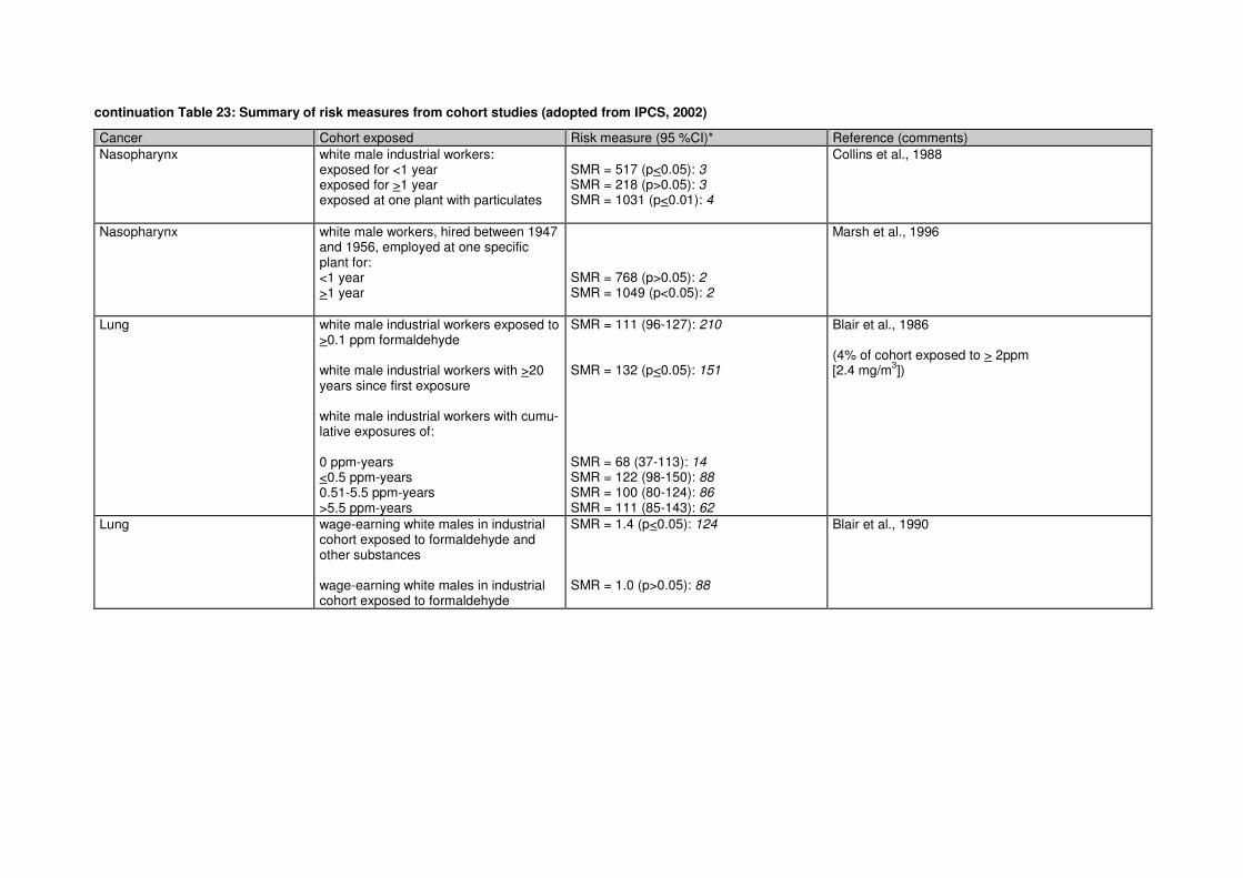
145
BfR-Wissenschaft
continuation Table 23: Summary of risk measures from cohort studies (adopted from IPCS, 2002)
Cancer Cohort exposed Risk measure (95 %CI)* Reference (comments) Nasopharynx white male industrial workers:
exposed for <1 year exposed for >1 year exposed at one plant with particulates
SMR = 517 (p<0.05): 3 SMR = 218 (p>0.05): 3 SMR = 1031 (p<0.01): 4
Collins et al., 1988
Nasopharynx white male workers, hired between 1947 and 1956, employed at one specific plant for: <1 year >1 year
SMR = 768 (p>0.05): 2 SMR = 1049 (p<0.05): 2
Marsh et al., 1996
Lung white male industrial workers exposed to >0.1 ppm formaldehyde white male industrial workers with >20 years since first exposure white male industrial workers with cumu-lative exposures of: 0 ppm-years <0.5 ppm-years 0.51-5.5 ppm-years >5.5 ppm-years
SMR = 111 (96-127): 210 SMR = 132 (p<0.05): 151 SMR = 68 (37-113): 14 SMR = 122 (98-150): 88 SMR = 100 (80-124): 86 SMR = 111 (85-143): 62
Blair et al., 1986 (4% of cohort exposed to > 2ppm [2.4 mg/m3])
Lung wage-earning white males in industrial cohort exposed to formaldehyde and other substances wage-earning white males in industrial cohort exposed to formaldehyde
SMR = 1.4 (p<0.05): 124 SMR = 1.0 (p>0.05): 88
Blair et al., 1990

146 BfR-Wissenschaft
continuation Table 23: Summary of risk measures from cohort studies (adopted from IPCS, 2002)
Cancer Cohort exposed Risk measure (95 %CI)* Reference (comments) Lung subjects in industrial cohort less than 65
years of age with cumulative exposures of: <0.1 ppm-years 0.1-0.5 ppm-years 0.5-2.0 ppm-years >2.0 ppm-years males in industrial cohort less than 65 years of age with cumulative exposures of: <0.1 ppm-years 0.1-0.5 ppm-years 0.5-2.0 ppm-years >2.0 ppm-years
RR = 1.0 RR = 1.47 (1.03-2.12)2
RR = 1.08 (0.67-1.70)2
RR = 1.83 (1.09-3.08)2
RR = 1.0 RR = 1.50 (1.03-2.19)2
RR = 1.18 (0.73-1.90)2
RR = 1.94 (1.13-3.34)2
Sterling and Weinkam, 1994
Lung white wage-earning males in industrial cohort with >2 ppm-years of cumulative exposure durations of: <1 year 1-<5 years 5-<10 years >10 years
(no observed deaths) SMR = 1.1 (p>0.05): 9 SMR = 2.8 (p<0.05): 17 SMR = 1.0 (p>0.05): 10
Blair and Stewart, 1994
Lung white male workers employed at one specific plant for: <1 year >1 year
SMR = 134 (p<0.05): 63 SMR = 119 (p>0.05): 50
Marsh et al., 1996 (25% exposed to >0.7 ppm [0.9 mg/m3])
Lung white males in industrial cohort with cumulative exposures of: 0 ppm-years 0.05-0.5 ppm-years 0.51-5.5 ppm-years >5.5 ppm-years
RR = 1.00 RR = 1.46 (0.81-2.61)
RR = 1.27 (0.72-2.26)
RR = 1.38 (0.77-2.48)
Callas et al., 1996
1 Unless otherwise noted, values in parentheses are 95 % confidence interval or level of statistical significance. Risk measures are presented here in the format reported in the references cited. Values in italics are the number of oberserved deaths or cases, when specified in the reference cited. Abbreviations are as follows: SMR = standardized mortality ratio as reported in the original study; SIR = standardized incidence ratio; PMR = proportionate mortality ratio; SPIR = standardized proportionate incidence ration; RR = relative risk. 2 Values in parentheses represent 90 % confidence interval.

147
BfR-Wissenschaft
Table 24: Summary of risk measures from case-control studies (adopted from IPCS, 2002)
Cancer1 Formaldehyde exposure Risk measure (95 %CI) Reference (comments) Oropharynx or hypopharynx SEER population based – Wash-ington State
>10 years occupational exposure occupational exposure score of >20
OR = 1.3 (0.7-2.5) OR = 1.5 (0.7-3.0)
Vaughan et al., 1986a (IARC Working Group noted that different proportions of inter-views conducted with next-of-kin cases and controls may have affected odds ratios)
Nasopharynx SEER population based – Wash-ington State
exposure score of >20 OR = 2.1 (0.6-7.8) Vaughan et al., 1986a (IARC Working Group noted that different proportions of inter-views conducted with next-of-kin cases and controls may have affected odds ratios)
Nasopharynx SEER population based – Wash-ington State
residential exposure of >10 years residential exposure of <10 years
OR = 5.5 (1.6-19.4) OR = 2.1 (0.7-6.6)
Vaughan et al., 1986b (IARC Working Groupc onsidered living in a mobile home a poor proxy for exposure)
Nasal squamous cell carcinoma Hospital based - Netherlands
occupational exposure assessment A occupational exposure assessment B
OR = 3.0 (1.3-6.4)2 OR = 1.9 (1.0-3.6)2
Hayes et al., 1986 (IARC Working Group noted that a greater proportion of cases than controls were dead and variable numbers of next-of-kin were interviewed. 10 % of controls but none of cases, by telephone. Noted also that, although different, results for as-sessments A & B were both positive)
Squamous cell carcinoma of nasal cavity/paranasal sinus Danish Cancer Registry
occupational exposure without exposure to wood dust
OR = 2.0 (0.7-5.9) Olsen and Asnaes, 1986 (IARC Working Group noted possibly incomplete adjustment for confounding for wood dust for adenocarcinoma; felt that squamous cell carcinoma less likely to be affected, since no clear association with wood dust) (Small number of cases)
Nasopharynx Connecticut Tumour Registry
highest potential exposure category highest potential exposure category and dying at 68+ years of age
OR = 2.3 (0.9-6.0) OR = 4.0 (1.3-12)
Roush et al., 1987
Oral/oropharynx Population based – Turin, Italy
“any” occupational exposure “probable or definite” occupational exposure
OR = 1.6 (0.9-2.8) OR = 1.8 (0.6-5.5)
Merletti et al., 1991 (Small number of cases with “definite” exposure to formalde-hyde)
Larynx SEER population based – Wash-ington State
“high” occupational exposure occupational exposure of >10 years occupational exposure score of >20
OR = 2.0 (0.2-19.5) OR = 1.3 (0.6-3.1) OR = 1.3 (0.5-3.3)
Wortley et al., 1992

148 BfR-Wissenschaft
continuation Table 24: Summary of risk measures from case-control studies (adopted from IPCS, 2002)
Cancer1 Formaldehyde exposure Risk measure (95 %CI) Reference (comments) Nasal cavity/paranasal sinus (adenocarcinoma) Population based - France
„any” exposure without exposure to wood dust „any” exposure with medium to high expo-sure to wood dust „no” exposure but medium to high exposure to wood dust
OR = 8.1 (0.9-72.9) OR = 692 (91.9-5210) OR = 130 (14.1-1191)
Luce et al., 1993 (IARC Working Group noted possible residual confounding by exposure to wood dust)
Nasopharynx Hospital based – Philippines
<15 years of exposure >25 years since first exposure <25 years of age at first exposure
OR = 2.7 (1.1-6.6) OR = 2.9 (1.1-7.6) OR = 2.7 (1.1-6.6)
West et al., 1993 (IARC Working Group noted no control for the presence of Epstein-Barr viral antibodies, for which previous strong asso-ciation with nasopharyngeal cancer was oberserved)
Lung Nested – cohort of chemical work-ers – Texas
likely occupational exposure OR = 0.62 (0.29-1.36) Bond et al., 1986
Lung Lung (adenocarcinoma) Population based – Montréal, Que-bec
„long-high” occupational exposure/ (cancer controls/population controls) „long-high” occupational exposure/ (cancer controls/population controls)
OR = 1.5 (0.8-2.8)/ OR = 1.0 (0.4-2.4) OR = 2.3 (0.9-6.0) OR = 2.2 (0.7-7.6)
Gérin et al., 1989
Respiratory cancer Nested – cohort of Finnish wood-workers
cumulative exposure of >3.6 mg/m3 - months, without minimum 10-year induction period cumulative exposure of >3.6 mg/m3 - months, without minimum 10-year induction period exposure to formaldehyde in wood dust
OR = 0.69 (0.21-2.24)2 OR = 0.89 (0.26-3.0)2 OR = 1.19 (0.31-4.56)2
Partanen et al., 1990 (IARC Working Group noted that there were too few cancers at sites other than the lung for meaningful analysis)
Lung Population based - Missouri
potentially exposed non-smokers OR = 0.9 (0.2-3.3) Brownson et al., 1993
Lung Nested – cohort of U.S. automotive foundry workers
occupational exposure with latency period of: 0 years 10 years 15 years 20 years
OR = 1.31 (0.93-1.85) OR = 1.04 (0.71-1.52) OR = 0.98 (0.65-1.47) OR = 0.99 (0.60-1.62)
Andjelkovich et al., 1994
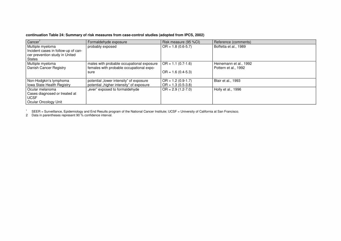
149
BfR-Wissenschaft
continuation Table 24: Summary of risk measures from case-control studies (adopted from IPCS, 2002)
Cancer1 Formaldehyde exposure Risk measure (95 %CI) Reference (comments) Multiple myeloma Incident cases in follow-up of can-cer prevention study in United States
probably exposed OR = 1.8 (0.6-5.7) Boffetta et al., 1989
Multiple myeloma Danish Cancer Registry
males with probable occupational exposure females with probable occupational expo-sure
OR = 1.1 (0.7-1.6) OR = 1.6 (0.4-5.3)
Heinemann et al., 1992 Pottern et al., 1992
Non-Hodgkin’s lymphoma Iowa State Health Registry
potential „lower intensity” of exposure potential „higher intensity” of exposure
OR = 1.2 (0.9-1.7) OR = 1.3 (0.5-3.8)
Blair et al., 1993
Ocular melanoma Cases diagnosed or treated at UCSF Ocular Oncology Unit
„ever” exposed to formaldehyde OR = 2.9 (1.2-7.0) Holly et al., 1996
1 SEER = Surveillance, Epidemiology and End Results program of the National Cancer Institute; UCSF = University of California at San Francisco. 2 Data in parentheses represent 90 % confidence interval.

150 BfR-Wissenschaft

151
BfR-Wissenschaft
9 List of tables
Table 1: Fate of [14C] absorbed in the upper respiratory tract of animals after inhalative exposure to (14C)-formaldehyde (Adopted from Casanova et al., 1989 and Casanova et al., 1991) 12
Table 2: Distribution of radioactivity in rats after inhalative exposure to (14C)-formaldehyde. 13
Table 3: Distribution of radioactivity in guinea pigs, cynomolgus monkeys, rabbits and rats after dermal application of [14C] formaldehyde. 15
Table 4: Histochemical localisation of formaldehyde dehydrogenase in a variety of rat tissues (according to Keller, 1990). The results of the study indicated, that regional differences of FAD in the nose are insufficient to account for the localised toxicity of inhaled formaldehyde. 20
Table 5: Overview on the results obtained with systemic genetic toxicity testing of formaldehyde in humans 26
Table 6: Variation of MN frequencies in negative control individuals 27
Table 7: Comparison of concentrations leading to DPX and other genotoxic effects in mammalian cells in culture 29
Table 8: Systemic genetic toxicity of formaldehyde (FA) in mammalian animals 30
Table 9: Local genetic toxicity of formaldehyde (FA) in mammalian animals 32
Table 10: Systemic genetic toxicity of formaldehyde (FA) in humans 33
Table 11: Local genetic toxicity of formaldehyde (FA) in humans 39
Table 12: Mean labelling indices per unit length for each nasal site in rats§ after 3 months of inhalation* exposure to formaldehyde (Indices calculation without correction for number of cells/site; extracted from Monticello et al., 1996) 46
Table 13: Systemic carcinogenesis of formaldehyde in animals/oral studies 54
Table 15: New data on risk measures from cohort studies on leukemic cancers 62
Table 16: Relative risks for mortality from hemopoietic malignancies (Extracted from Hauptmann et al., 2003) 63
Table 17: Evaluation of the statistical power on the recently published cohort studies 71
Table 18: Inhalation carcinogenicity studies on formaldehyde in animals 77
Table 19: Relative risks and numbers of deaths for selected cancers in the respiratory tract (Mortality follow-up through 1994, extracted from Hauptmann et al., 2004) 85
Table 20: New data on risk measures from cohort studies on cancers in the respiratory tract 92
Table 21: New data on risk measures from case-control studies on cancers at the sites of contact (respiratory tract, pharynx and larynx) 94
Table 22: Evaluation of the statistical power on the recently published cohort studies 100
Table 23: Summary of risk measures from cohort studies (adopted from IPCS, 2002) 143
Table 24: Summary of risk measures from case-control studies (adopted from IPCS, 2002) 147

152 BfR-Wissenschaft

153
BfR-Wissenschaft
10 List of figures
Figure 1: Biological pathways of formaldehyde (adopted from Conaway et al., 1996 10
Danksagung Ein herzliches Dankeschön an Frau Christa Hensel und an Frau Silvia Becker für die Recherchen und die Unterstützung bei der Erstellung dieses Berichtes.

154 BfR-Wissenschaft

155
BfR-Wissenschaft
Publications which have already appeared in the BfR-Wissenschaft series 01/2004 Published by L. Ellerbroek, H. Wichmann-Schauer, K. N. Mac
Methoden zur Identifizierung und Isolierung von Enterokokken und deren Resistenzbestimmung
€ 5 02/2004 Published by M. Hartung Epidemiologische Situation der Zoonosen in Deutschland im Jahr 2002 € 15 03/2004 Published by A. Domke, R. Großklaus, B. Niemann. H. Przyrembel, K. Richter,
E. Schmidt, A. Weißenborn, B. Wörner, R. Ziegenhagen Verwendung von Vitaminen in Lebensmitteln Toxikologische und ernährungsphysiologische Aspekte, Teil 1 € 15
04/2004 Published by A. Domke, R. Großklaus, B. Niemann, H. Przyrembel, K. Richter,
E. Schmidt, A. Weißenborn, B. Wörner, R. Ziegenhagen Verwendung von Mineralstoffen in Lebensmitteln Toxikologische und ernährungsphysiologische Aspekte, Teil 2 € 15
05/2004 Published by M. Hartung
Epidemiologische Situation der Zoonosen in Deutschland im Jahr 2003 Übersicht über die Meldungen der Bundesländer € 15
01/2005 Published by A. Weißenborn, M. Burger, G.B.M. Mensink, C. Klemm, W. Si-chert-Hellert, M. Kersting, H. Przyrembel Folsäureversorgung der deutschen Bevölkerung Abschlussbericht zum Forschungsvorhaben € 10
02/2005 Published by R. F. Hertel, G. Henseler
ERiK - Entwicklung eines mehrstufigen Verfahrens der Risikokommunikation € 10
03/2005 Published by P. Luber, E. Bartelt Campylobacteriose durch Hähnchenfleisch Eine quantitative Risikoschätzung € 5 04/2005 Published by A. Domke, R. Großklaus, B. Niemann, H. Przyrembel, K. Richter,
E. Schmidt, A. Weißenborn, B. Wörner, R. Ziegenhagen Use of Vitamins in Foods Toxicological and nutritional-physiological aspects € 15 01/2006 Published by A. Domke, R. Großklaus, B. Niemann. H. Przyrembel, K. Richter,
E. Schmidt, A. Weißenborn, B. Wörner, R. Ziegenhagen Use of Vitamins in Foods Toxicological and nutritional-physiological aspects € 15

156 BfR-Wissenschaft
Die Hefte der Reihe BfR-Wissenschaft sind erhältlich beim: Bundesinstitut für Risikobewertung Pressestelle Thielallee 88-92 D-14195 Berlin Fax: 030-8412 4970 E-Mail: [email protected]