Embed Size (px)
Citation preview
Hindawi Publishing CorporationCase Reports in MedicineVolume 2010, Article ID 871278, 4 pagesdoi:10.1155/2010/871278
Case Report
Giant-Cell Tumor of the Distal Ulna Treated by Wide Resectionand Ulnar Support Reconstruction: A Case Report
Akio Minami, Norimasa Iwasaki, Kinya Nishida, Makoto Motomiya,Katsuhisa Yamada, and Daisuke Momma
Department of Orthopaedic Surgery, Hokkaido University Graduate School of Medicine, Sapporo 060-8638, Japan
Correspondence should be addressed to Akio Minami, [email protected]
Received 26 January 2010; Accepted 16 May 2010
Academic Editor: Valerae O. Lewis
Copyright © 2010 Akio Minami et al. This is an open access article distributed under the Creative Commons Attribution License,which permits unrestricted use, distribution, and reproduction in any medium, provided the original work is properly cited.
Giant-cell tumor of bone occurred in the distal end of the ulna is extremely uncommon. A 23-year-old male had a giant-celltumor occurred in the distal end of the ulna. After wide resection of the distal segment of the ulna including giant-cell tumor,ulnar components of the wrist joint were reconstructed with modified Sauve-Kapandji procedure using the iliac bone graft,preserving the triangular fibrocartilage complex and ulnar collateral ligament in order to maintain ulnar support of the wrist,and the proximal stump of the resected ulna was stabilized by tenodesis using the extensor carpi ulnaris tendon. One year afteroperation, the patient’s wrist was pain-free and had a full range of motion. Postoperative X-rays showed no abnormal findingsincluding recurrence of the giant-cell tumor and ulnar translation of the entire carpus. The stability of the proximal stump of thedistal ulna was also maintained.
1. Introduction
Giant-cell tumor (GCT) of the bone is a rare, benign, andlocally invasive tumor. It is accounting for about 3% to 5%of all primary bone tumors [1]. GCTs of the bone usuallyoccur at the epiphysis of the long bone such as femur, tibia,humerus, and radius. GCTs occurred at the distal end of theulna are extremely rare, accounting for 0.45% to 3.2% of allthe cases of GCTs [2]. This paper described a young malewith a GCT of the distal end of the ulna treated by a wideresection and ulnar support reconstruction of the wrist.
2. Case Report
A 23-year-old male, manual laborer, noticed a movementalpain and swelling around the ulnar head of the left wrist onJanuary, 2008. Pain suddenly increased two months after theonset without any particular event. The patient was seen toa clinic on March, 2008. In there, the patient was informedthat there was an abnormal shadow in the ulnar head of theleft wrist. There was no history of any other swelling in thebody, fever, and loss of weight. The patient was introducedand first seen in our hospital on May, 2008.
Physical examinations revealed that there was an ovalswelling of 4 × 3 cm in the distal end of the ulna. Therewas no color change and redness on the overlying skin.The swelling was diffusely tender and uniformly elasticallyhard. There was no adherence of the skin to the under lyingbone. The range of motion of the patient’s left wrist waslimited to 60◦ (contralateral side: 80◦) in dorsiflexion and50◦ (80◦) in palmar flexion, 60◦ (90◦) in pronation and 80◦
(90◦) in supination. Moderate movemental pain was presentat the extremes in all directions. The grip strength of hisnondominant left wrist showed 27 kgf compared with 42 kgfof the unaffected dominant hand.
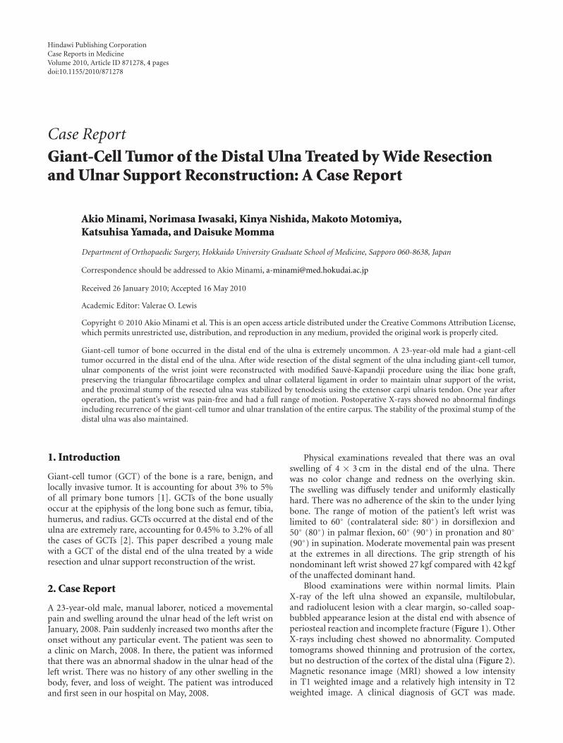
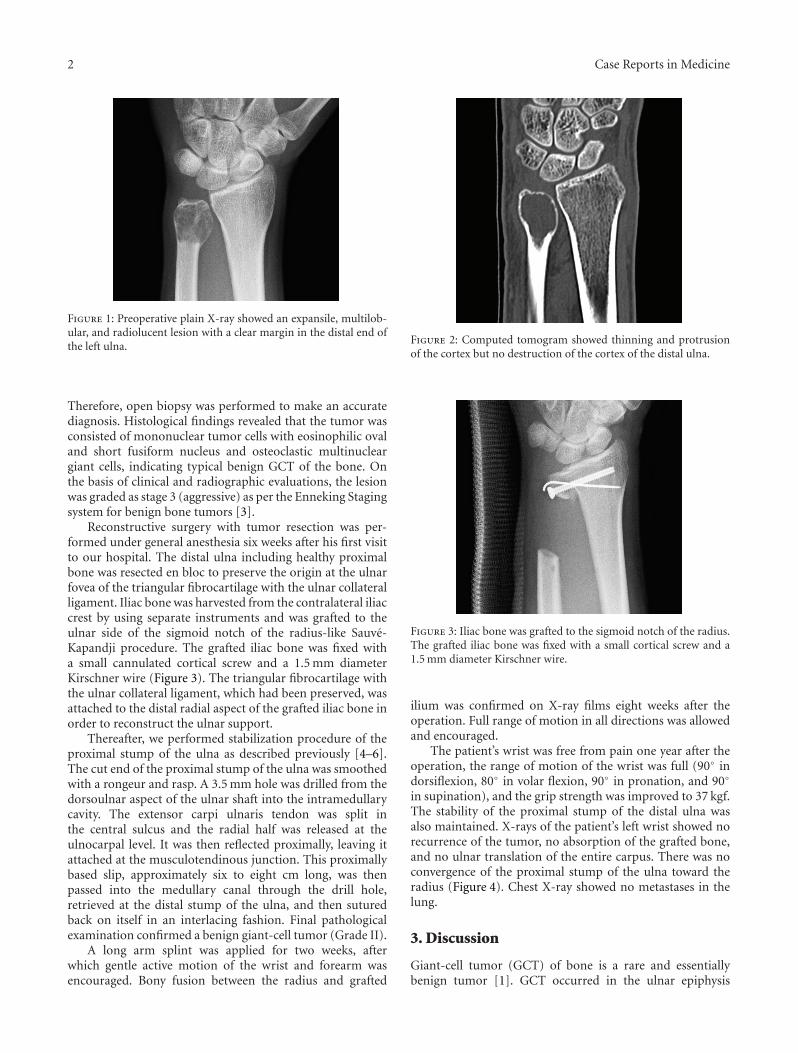
Blood examinations were within normal limits. PlainX-ray of the left ulna showed an expansile, multilobular,and radiolucent lesion with a clear margin, so-called soap-bubbled appearance lesion at the distal end with absence ofperiosteal reaction and incomplete fracture (Figure 1). OtherX-rays including chest showed no abnormality. Computedtomograms showed thinning and protrusion of the cortex,but no destruction of the cortex of the distal ulna (Figure 2).Magnetic resonance image (MRI) showed a low intensityin T1 weighted image and a relatively high intensity in T2weighted image. A clinical diagnosis of GCT was made.
2 Case Reports in Medicine
Figure 1: Preoperative plain X-ray showed an expansile, multilob-ular, and radiolucent lesion with a clear margin in the distal end ofthe left ulna.
Therefore, open biopsy was performed to make an accuratediagnosis. Histological findings revealed that the tumor wasconsisted of mononuclear tumor cells with eosinophilic ovaland short fusiform nucleus and osteoclastic multinucleargiant cells, indicating typical benign GCT of the bone. Onthe basis of clinical and radiographic evaluations, the lesionwas graded as stage 3 (aggressive) as per the Enneking Stagingsystem for benign bone tumors [3].
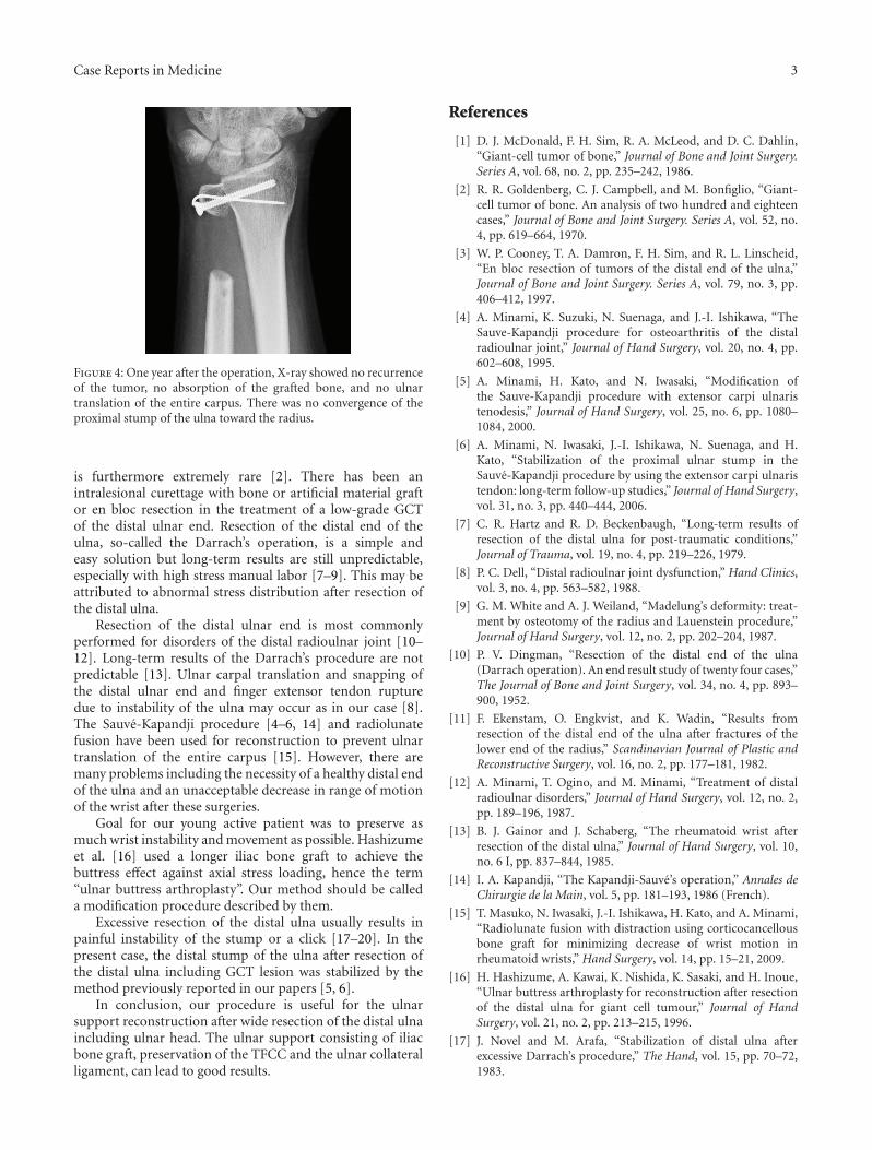
Reconstructive surgery with tumor resection was per-formed under general anesthesia six weeks after his first visitto our hospital. The distal ulna including healthy proximalbone was resected en bloc to preserve the origin at the ulnarfovea of the triangular fibrocartilage with the ulnar collateralligament. Iliac bone was harvested from the contralateral iliaccrest by using separate instruments and was grafted to theulnar side of the sigmoid notch of the radius-like Sauve-Kapandji procedure. The grafted iliac bone was fixed witha small cannulated cortical screw and a 1.5 mm diameterKirschner wire (Figure 3). The triangular fibrocartilage withthe ulnar collateral ligament, which had been preserved, wasattached to the distal radial aspect of the grafted iliac bone inorder to reconstruct the ulnar support.
Thereafter, we performed stabilization procedure of theproximal stump of the ulna as described previously [4–6].The cut end of the proximal stump of the ulna was smoothedwith a rongeur and rasp. A 3.5 mm hole was drilled from thedorsoulnar aspect of the ulnar shaft into the intramedullarycavity. The extensor carpi ulnaris tendon was split inthe central sulcus and the radial half was released at theulnocarpal level. It was then reflected proximally, leaving itattached at the musculotendinous junction. This proximallybased slip, approximately six to eight cm long, was thenpassed into the medullary canal through the drill hole,retrieved at the distal stump of the ulna, and then suturedback on itself in an interlacing fashion. Final pathologicalexamination confirmed a benign giant-cell tumor (Grade II).
A long arm splint was applied for two weeks, afterwhich gentle active motion of the wrist and forearm wasencouraged. Bony fusion between the radius and grafted
Figure 2: Computed tomogram showed thinning and protrusionof the cortex but no destruction of the cortex of the distal ulna.
Figure 3: Iliac bone was grafted to the sigmoid notch of the radius.The grafted iliac bone was fixed with a small cortical screw and a1.5 mm diameter Kirschner wire.
ilium was confirmed on X-ray films eight weeks after theoperation. Full range of motion in all directions was allowedand encouraged.
The patient’s wrist was free from pain one year after theoperation, the range of motion of the wrist was full (90◦ indorsiflexion, 80◦ in volar flexion, 90◦ in pronation, and 90◦
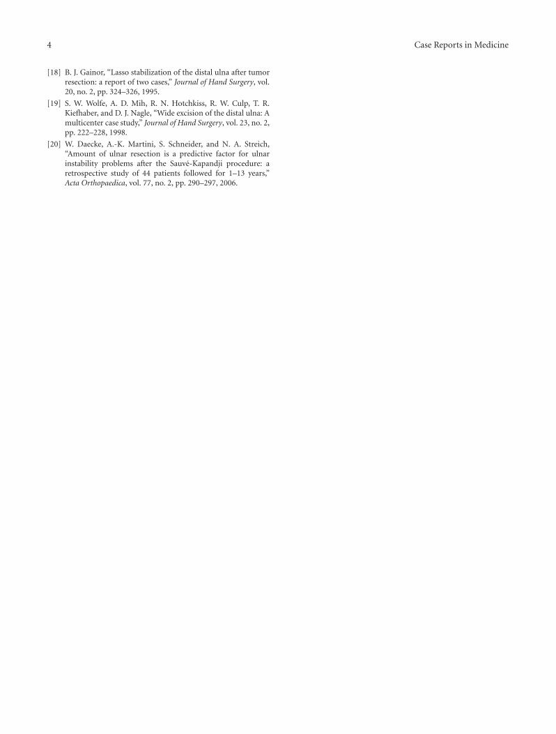
in supination), and the grip strength was improved to 37 kgf.The stability of the proximal stump of the distal ulna wasalso maintained. X-rays of the patient’s left wrist showed norecurrence of the tumor, no absorption of the grafted bone,and no ulnar translation of the entire carpus. There was noconvergence of the proximal stump of the ulna toward theradius (Figure 4). Chest X-ray showed no metastases in thelung.
3. Discussion
Giant-cell tumor (GCT) of bone is a rare and essentiallybenign tumor [1]. GCT occurred in the ulnar epiphysis
Case Reports in Medicine 3
Figure 4: One year after the operation, X-ray showed no recurrenceof the tumor, no absorption of the grafted bone, and no ulnartranslation of the entire carpus. There was no convergence of theproximal stump of the ulna toward the radius.
is furthermore extremely rare [2]. There has been anintralesional curettage with bone or artificial material graftor en bloc resection in the treatment of a low-grade GCTof the distal ulnar end. Resection of the distal end of theulna, so-called the Darrach’s operation, is a simple andeasy solution but long-term results are still unpredictable,especially with high stress manual labor [7–9]. This may beattributed to abnormal stress distribution after resection ofthe distal ulna.
Resection of the distal ulnar end is most commonlyperformed for disorders of the distal radioulnar joint [10–12]. Long-term results of the Darrach’s procedure are notpredictable [13]. Ulnar carpal translation and snapping ofthe distal ulnar end and finger extensor tendon rupturedue to instability of the ulna may occur as in our case [8].The Sauve-Kapandji procedure [4–6, 14] and radiolunatefusion have been used for reconstruction to prevent ulnartranslation of the entire carpus [15]. However, there aremany problems including the necessity of a healthy distal endof the ulna and an unacceptable decrease in range of motionof the wrist after these surgeries.
Goal for our young active patient was to preserve asmuch wrist instability and movement as possible. Hashizumeet al. [16] used a longer iliac bone graft to achieve thebuttress effect against axial stress loading, hence the term“ulnar buttress arthroplasty”. Our method should be calleda modification procedure described by them.
Excessive resection of the distal ulna usually results inpainful instability of the stump or a click [17–20]. In thepresent case, the distal stump of the ulna after resection ofthe distal ulna including GCT lesion was stabilized by themethod previously reported in our papers [5, 6].
In conclusion, our procedure is useful for the ulnarsupport reconstruction after wide resection of the distal ulnaincluding ulnar head. The ulnar support consisting of iliacbone graft, preservation of the TFCC and the ulnar collateralligament, can lead to good results.
References
[1] D. J. McDonald, F. H. Sim, R. A. McLeod, and D. C. Dahlin,“Giant-cell tumor of bone,” Journal of Bone and Joint Surgery.Series A, vol. 68, no. 2, pp. 235–242, 1986.
[2] R. R. Goldenberg, C. J. Campbell, and M. Bonfiglio, “Giant-cell tumor of bone. An analysis of two hundred and eighteencases,” Journal of Bone and Joint Surgery. Series A, vol. 52, no.4, pp. 619–664, 1970.
[3] W. P. Cooney, T. A. Damron, F. H. Sim, and R. L. Linscheid,“En bloc resection of tumors of the distal end of the ulna,”Journal of Bone and Joint Surgery. Series A, vol. 79, no. 3, pp.406–412, 1997.
[4] A. Minami, K. Suzuki, N. Suenaga, and J.-I. Ishikawa, “TheSauve-Kapandji procedure for osteoarthritis of the distalradioulnar joint,” Journal of Hand Surgery, vol. 20, no. 4, pp.602–608, 1995.
[5] A. Minami, H. Kato, and N. Iwasaki, “Modification ofthe Sauve-Kapandji procedure with extensor carpi ulnaristenodesis,” Journal of Hand Surgery, vol. 25, no. 6, pp. 1080–1084, 2000.
[6] A. Minami, N. Iwasaki, J.-I. Ishikawa, N. Suenaga, and H.Kato, “Stabilization of the proximal ulnar stump in theSauve-Kapandji procedure by using the extensor carpi ulnaristendon: long-term follow-up studies,” Journal of Hand Surgery,vol. 31, no. 3, pp. 440–444, 2006.
[7] C. R. Hartz and R. D. Beckenbaugh, “Long-term results ofresection of the distal ulna for post-traumatic conditions,”Journal of Trauma, vol. 19, no. 4, pp. 219–226, 1979.
[8] P. C. Dell, “Distal radioulnar joint dysfunction,” Hand Clinics,vol. 3, no. 4, pp. 563–582, 1988.
[9] G. M. White and A. J. Weiland, “Madelung’s deformity: treat-ment by osteotomy of the radius and Lauenstein procedure,”Journal of Hand Surgery, vol. 12, no. 2, pp. 202–204, 1987.
[10] P. V. Dingman, “Resection of the distal end of the ulna(Darrach operation). An end result study of twenty four cases,”The Journal of Bone and Joint Surgery, vol. 34, no. 4, pp. 893–900, 1952.
[11] F. Ekenstam, O. Engkvist, and K. Wadin, “Results fromresection of the distal end of the ulna after fractures of thelower end of the radius,” Scandinavian Journal of Plastic andReconstructive Surgery, vol. 16, no. 2, pp. 177–181, 1982.
[12] A. Minami, T. Ogino, and M. Minami, “Treatment of distalradioulnar disorders,” Journal of Hand Surgery, vol. 12, no. 2,pp. 189–196, 1987.
[13] B. J. Gainor and J. Schaberg, “The rheumatoid wrist afterresection of the distal ulna,” Journal of Hand Surgery, vol. 10,no. 6 I, pp. 837–844, 1985.
[14] I. A. Kapandji, “The Kapandji-Sauve’s operation,” Annales deChirurgie de la Main, vol. 5, pp. 181–193, 1986 (French).
[15] T. Masuko, N. Iwasaki, J.-I. Ishikawa, H. Kato, and A. Minami,“Radiolunate fusion with distraction using corticocancellousbone graft for minimizing decrease of wrist motion inrheumatoid wrists,” Hand Surgery, vol. 14, pp. 15–21, 2009.
[16] H. Hashizume, A. Kawai, K. Nishida, K. Sasaki, and H. Inoue,“Ulnar buttress arthroplasty for reconstruction after resectionof the distal ulna for giant cell tumour,” Journal of HandSurgery, vol. 21, no. 2, pp. 213–215, 1996.
[17] J. Novel and M. Arafa, “Stabilization of distal ulna afterexcessive Darrach’s procedure,” The Hand, vol. 15, pp. 70–72,1983.
4 Case Reports in Medicine
[18] B. J. Gainor, “Lasso stabilization of the distal ulna after tumorresection: a report of two cases,” Journal of Hand Surgery, vol.20, no. 2, pp. 324–326, 1995.
[19] S. W. Wolfe, A. D. Mih, R. N. Hotchkiss, R. W. Culp, T. R.Kiefhaber, and D. J. Nagle, “Wide excision of the distal ulna: Amulticenter case study,” Journal of Hand Surgery, vol. 23, no. 2,pp. 222–228, 1998.
[20] W. Daecke, A.-K. Martini, S. Schneider, and N. A. Streich,“Amount of ulnar resection is a predictive factor for ulnarinstability problems after the Sauve-Kapandji procedure: aretrospective study of 44 patients followed for 1–13 years,”Acta Orthopaedica, vol. 77, no. 2, pp. 290–297, 2006.

Submit your manuscripts athttp://www.hindawi.com
Stem CellsInternational
Hindawi Publishing Corporationhttp://www.hindawi.com Volume 2014
Hindawi Publishing Corporationhttp://www.hindawi.com Volume 2014
MEDIATORSINFLAMMATION
of
Hindawi Publishing Corporationhttp://www.hindawi.com Volume 2014
Behavioural Neurology
EndocrinologyInternational Journal of
Hindawi Publishing Corporationhttp://www.hindawi.com Volume 2014
Hindawi Publishing Corporationhttp://www.hindawi.com Volume 2014
Disease Markers
Hindawi Publishing Corporationhttp://www.hindawi.com Volume 2014
BioMed Research International
OncologyJournal of
Hindawi Publishing Corporationhttp://www.hindawi.com Volume 2014
Hindawi Publishing Corporationhttp://www.hindawi.com Volume 2014
Oxidative Medicine and Cellular Longevity
Hindawi Publishing Corporationhttp://www.hindawi.com Volume 2014
PPAR Research
The Scientific World JournalHindawi Publishing Corporation http://www.hindawi.com Volume 2014
Immunology ResearchHindawi Publishing Corporationhttp://www.hindawi.com Volume 2014
Journal of
ObesityJournal of
Hindawi Publishing Corporationhttp://www.hindawi.com Volume 2014
Hindawi Publishing Corporationhttp://www.hindawi.com Volume 2014
Computational and Mathematical Methods in Medicine
OphthalmologyJournal of
Hindawi Publishing Corporationhttp://www.hindawi.com Volume 2014
Diabetes ResearchJournal of
Hindawi Publishing Corporationhttp://www.hindawi.com Volume 2014
Hindawi Publishing Corporationhttp://www.hindawi.com Volume 2014
Research and TreatmentAIDS
Hindawi Publishing Corporationhttp://www.hindawi.com Volume 2014
Gastroenterology Research and Practice
Hindawi Publishing Corporationhttp://www.hindawi.com Volume 2014
Parkinson’s Disease
Evidence-Based Complementary and Alternative Medicine
Volume 2014Hindawi Publishing Corporationhttp://www.hindawi.com


![CaseReport - Hindawi Publishing Corporationdownloads.hindawi.com/journals/criot/2017/4592783.pdf · ConflictsofInterest eauthorshavenoconictsofinteresttodeclare. References [1] P](https://img.pdfslide.tips/doc/110x75/5c0de1a809d3f27c728c0531/casereport-hindawi-publishing-conflictsofinterest-eauthorshavenoconictsofinteresttodeclare.jpg)


![Case Report - Hindawi Publishing Corporationdownloads.hindawi.com/journals/crirh/2020/8899391.pdf · plasty for rheumatoid arthritis patients with large tibial ... [28] S. Roser-Page,](https://img.pdfslide.tips/doc/110x75/608a0eceb7b47936b0748e21/case-report-hindawi-publishing-plasty-for-rheumatoid-arthritis-patients-with-large.jpg)