Embed Size (px)
Citation preview

Vo1.187, No. 1,1992
August 31,1992
BIOCHEMICAL AND BIOPHYSICAL RESEARCH COMMUNICATIONS
Pages 507-5]4
Cell Cycle Dependent Gene Expressions and Activities of
Protein Phosphatases PPI and PP2A in Mouse NIH3T3
Fibroblasts
Koji Nakamura I, Toshiaki Koda 2, Mitsuaki Kakinuma 2, Shu-ichi
Matsuzawa I, Kazuki Kitamura I, Yusuke Mizuno I, and Kunimi Kikuchi I,*
Section of iBiochemistry and 2Bacterial Infection, Institute of
Immunological Science, Hokkaido University, Kita-ku, Sapporo
060, Japan
Received July 17, 1992
SUMMARY: We determined the mRNA levels and the activities in nuclear and non-nuclear fractions of protein phosphatase type 1 (PPI) and type 2A (PP2A) through the cell cycle in synchronized mouse NIH3T3 fibroblasts. The mRNA level for PPI~ was gradually elevated in late G1 phase, began to decrease in M phase, and reached the control level with entering into the next G1 phase. The mRNA level for PP2A was rapidly increased in early G1 phase, kept at the high level, and decreased after S phase. In nuclear fractions of cells, spontaneous activities of both PPI and PP2A were gradually increased until M phase and rapidly decreased with entering the next G1 phase, while in non-nuclear fraction such dramatic alterations were not observed. Potential activities of PPI in both fractions revealed by Co2+-trypsin treatment showed an oscillaion patterns similar to those of the spontaneous activities. These results strongly suggest that cell cycle dependent gene expressions and activities of PPI and PP2A play roles in DNA synthesis and mitosis during the cell cycle. ® 1992 Academic Press, Inc.
In the past several years there has been increasing evidence
indicating specific phosphorylation and dephosphorylation of
nuclear proteins during cell growth. Although it is well-known
that the phosphorylation plays crucial roles in signal
transduction for cell proliferation, differentiation and
carcinogenesis, little has been focused on the implication of
protein phosphatases for the mitotic events.
Four principa ! serine/threonine specific protein phosphatases
termed PPI, PP2A, PP2B and PP2C and several protein tyrosin
*To whom correspondence should be addressed.
507
0006-291X/92 $4.00 Copyright © 1992 by Academic Press, Inc.
All rights of reproduction in any form reserved.

Vol. 187, No. 1, 1992 BIOCHEMICAL AND BIOPHYSICAL RESEARCH COMMUNICATIONS
phosphatases have been identified (i, 2). Recently it has been
reported that the protein phosphatases play important roles in
the cell division cycle. CDC25, a tyrosin phosphatase,
dephosphorylates and activates p34 cdc2 kinase in fission yeast
(3) and human (4). Histone HI phosphorylated by p34 cdc2 is
dephosphorylated by PP2AI (5). Inhibition of mitosis by okadaic
acid suggests the possibility of involvement of PP2A in the
transition from metaphase to anaphase (6). Okadaic acid also
induces cell cycle arrest at either GI/S or G2/M depending on
dose in myeloid leukemic cells (7). It was also reported that
okadaic acid induces mitotic arrest and enhances nuclear protein
phosphorylation in human leukemia K562 cells (8).
We have recently reported remarkable elevations in PPI and
PP2A mRNA levels at preneoplastic stages during
hepatocarcinogenesis and in regenerating livers (9). We also
found the specific overexpression of PPI~ mRNA in a
transplantable hepatoma cell line, AHI3(10) . Very recently, we
reported that the potential activity of nuclear PPI was
increased 2.4-fold at 12 h after partial hepatectomy compared to
the control level (ii).
To clarify further the mechanism and the significance of these
phenomena in regulation of cell proliferation, we have examined
the mRNA levels and activities of PPI and PP2A in nuclear and
non-nuclear fractions during the cell cycle using synchronized
mouse NIH3T3 fibroblasts.
MATERIALS AND METHODS
Cell culture: Mouse NIH3T3 fibroblasts were grown in Dulbecco's modified Eagle's medium (DMEM) containing 10% calf serum. In experiments on serum stimulation of cell growth, cells were initially grown to confluency and then placed in serum-free DMEM for 36 h. The cells were then stimulated by replacing the serum-free DMEM with fresh DMEM containing 10% calf serum, harvested at appropriate times and counted. The cells were subjected to flow cytometry, Northern blot analysis and phosphatase activity assay. Cell cycle analysis: The harvested cells were washed with phosphate-buffered saline (PBS), fixed with PBS-ethanol mixture (I:i v/v), then rewashed with PBS. After centrifugation, the cells were resuspended in PBS containing 50 ~g/ml propidium iodide, 0.5 mg/ml RNase and 0.1% NAN3, and incubated at room temperature for 15 min. After the incubation, the suspension was mixed with 1 vol. of PBS and passed through a nylon mesh. GI, S and G2+M phases of the cell cycle were analyzed by FACScan flow cytometer (Becton Dickinson). cDNA probes: The 1.4 kb full-length cDNA of rat PPIu and the 410 bp BstEII-BglII fragment of rat PP2A cDNA (12) were labeled
with [~-32p]dCTP by using Multiprime DNA Labeling Kit (Amersham International plc, England). Northern blot analysis: Total cellular RNA (20 ~g) prepared by the procedure of Chomczynski et al. (13) was separated by
508

Vol. 187, No. 1, 1992 BIOCHEMICAL AND BIOPHYSICAL RESEARCH COMMUNICATIONS
electrophoresis on 1% agarose gel containing 6% formaldehyde and transferred to a nitrocellulose membrane (Schleicher & Schuell, Germany) . After being baked at 80°C for 2 h under vacuum, the membranes were prehybridized in 5 x SSC, 1 x Denhardt's solution, 50 mM NaH2PO4 pH 6.5, 1% glycine, I00 ~g/ml denatured
E.coli DNA, 50% formamide for 1 h at 42°C. Hybridization was performed in the same solution containing the radioactive cDNA probes at 42°C overnight. The membranes were washed twice in 2 x SSC and 0.1% SDS at room temperature for 5 min, followed by washing twice in 0.i x SSC and 0.1% SDS at 50°C for 15 min. The membranes were exposed to XRP-5 X-ray film (Eastman Kodak Company, NY) at -80°C with intensifying screens. The membranes were reused after the old probes had been washed out by incubating the membranes in 2.5 mM Tris-HCI, pH 8.0, 0.i mM EDTA, 0.05% sodium pyrophosphate and 0.05 x Denhardt's solution at 70°C for 1 h. Preparation of nuclear and non-nuclear fractions: The
harvested cells were adjusted at 107/ml and lysed with NP-40 lysis buffer (i0 mM Tris-HCI, pH 7.4, I0 mM NaCI, 3 mM MgCI2,
0.5% (v/v) NP-40) at 107 cells/ml and centrifuged at 500 x g for 5 min at 4°C (14). The supernatant was termed non-nuclear fraction. The pellet after being washed with the same buffer
was termed nuclear fraction, which was then lysed at 2 x 10⁷ cells/ml by the method of Kret et al. (15) and used for enzyme assays. Protein phosphatase assay: Activities of PPI and PP2A in the nuclear and non-nuclear fractions were measured by the method of Cohen et al. (16) with a slight modification (17). PP2A activity was determined in the presence of inhibitor-2 to inhibit PPI activity. PPI activity was calculated by deducting the phosphatase activity measured in the presence of inhibitor-2 from the one in its absence. PPI activity was also measured
after Co2+-trypsin treatment as described previously (17, 18). PPI activities before and after the treatment were termed spontaneous and potential activities, respectively. One unit of activity (U) was defined as the amount of enzyme which catalyzes the release of 1 ~mol of phosphate per min. Activities of PPI
and PP2A were presented as milli unit per 107 cells.
RESULTS
Expression of PPI and PP2A in mouse NIH3T3 fibroblasts
during the cell cycle: Cell cycle dependent expressions of
PPI and PP2A after stimulation of cell growth by serum addition
were investigated.
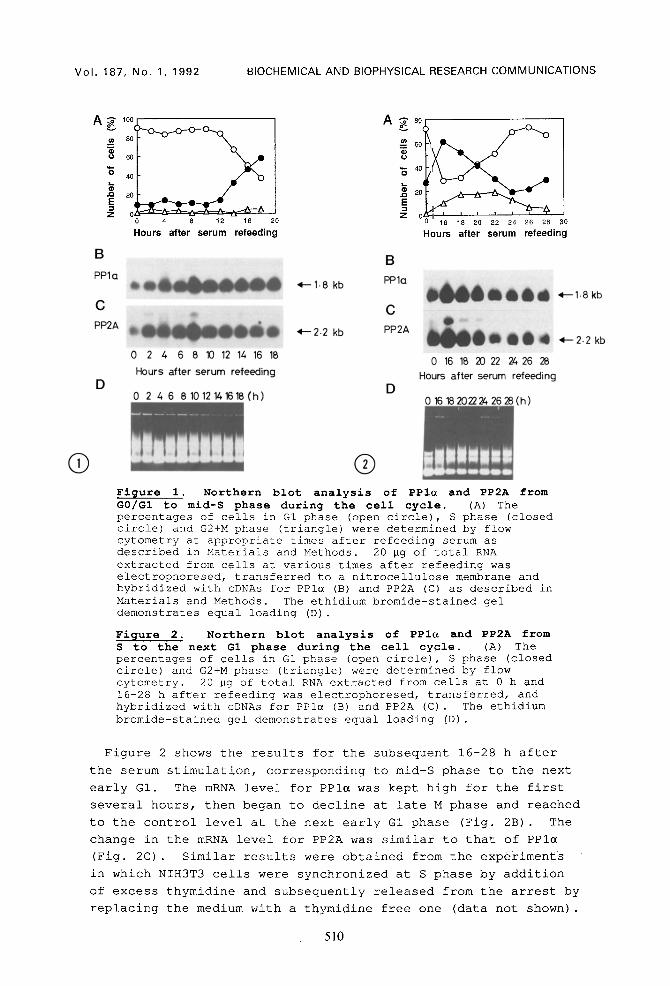
Figure 1 shows the results for the stages of the cell cycle
from G0/GI to mid-S phase. Figure IA shows the number of cells
at GI, S and G2+M phases. Figure IB and IC show alterations in
the mRNA levels of PPI~ and PP2A, respectively, for 18 hours
after serum re-addition. The mRNA level for PPI~ began to
increase from 2 h after the serum stimulation and reached a
maximum at 8-10 h, corresponding to late G1 phase. The mRNA
level for PP2A was increased and reached a maximum 2 h after the
stimulation, and remained at the high level until late S phase.
509
Vol . 187, No. 1, 1992 BIOCHEMICAL AND BIOPHYSICAL RESEARCH COMMUNICATIONS
A ~ 100(
¢n 80
60
40
~ 20 E
4 8 12 16
B
PPlo
2O
Hours after serum refeeding
C PP2A
D
0 2 4 6 8 10 12 14 16 18
Hours after serum refeeding
0 2 4 6 81012141618(h)
4- -1 .8 kb
~v- 2.2 kb
A ? 8 °
-~ 6o
~- 40 0
~ 20 I
E Z
B PPlc
C PP2A
D
16 18 20 22 24 26 28 30
Hours after serum refeeding
0 16 1B 20 22 24 26 28 Hours after serum refeeding
0 16 18202224 26 28 (h)
,~-- 1.8 kb
*'--2.2 kb
Q ® Figure i. Northern blot analysis of PPIu and PP2A from G0/GI to mid-S phase during the cell cycle. (A) The percentages of cells in G1 phase (open circle), S phase (closed circle) and G2+M phase (triangle) were determined by flow cytometry at appropriate times after refeeding serum as described in Materials and Methods. 20 Bg of total RNA extracted from cells at various times after refeeding was electrophoresed, transferred to a nitrocellulose membrane and hybridized with cDNAs for PPI~ (B) and PP2A (C) as described in Materials and Methods. The ethidium bromide-stained gel demonstrates equal loading (D) .
Figure 2. Northern blot analysis of PPI~ and PP2A from S to the next G1 phase during the cell cycle, (A) The percentages of cells in G1 phase (open circle), S phase (closed circle) and G2+M phase (triangle) were determined by flow cytometry. 20 Bg of total RNA extracted from cells at 0 h and 16-28 h after refeeding was electrophoresed, transferred, and hybridized with cDNAs for PPI~ (B) and PP2A (C) . The ethidium bromide-stained gel demonstrates equal loading (D) .
Figure 2 shows the results for the subsequent 16-28 h after
the serum stimulation, corresponding to mid-S phase to the next
early GI. The mRNA level for PPI~ was kept high for the first
several hours, then began to decline at late M phase and reached
to the control level at the next early G1 phase (Fig. 2B) . The
change in the mRNA level for PP2A was similar to that of PPI~
(Fig. 2C) . Similar results were obtained from the experiments
in which NIH3T3 cells were synchronized at S phase by addition
of excess thymidine and subsequently released from the arrest by
replacing the medium with a thymidine free one (data not shown).
510

Vol. 187, No. 1, 1992 BIOCHEMICAL AND BIOPHYSICAL RESEARCH COMMUNICATIONS
20 A / ~
0 10 20 Hours after serum refeeding
30
I°°I
< ol ~
v
10 20 30 Hours after serum refeeding
Figure 3. Protein phosphatase activities of PPI and PP2A in nuclear and non-nuclear fractions during the cell cycle. (A) Spontaneous activities of PPI (open symbol) and PP2A (closed symbol) in nuclear (circle) and non-nuclear fractions (square) were measured during the cell cycle as described in Materials and Methods. (B) Potential activities of PPI revealed by Co2+-trypsin treatment in nuclear (open circles) and non-nuclear fractions (closed circles) were measured as described in Materials and Methods. Spontaneous activities of PPI in nuclear (open squares) and non-nuclear (closed squares) fractions were also presented for comparison. Each point represents average of duplicate determinations. These results were confirmed by determinations for another series of cell extractions at the same time points.
Activities of PPI and PP2A in nuclear and non-nuclear
fractions: Whether the increases in the mRNA levels of PPI and
PP2A during the cell cycle are reflected in their enzyme
activity levels was examined. The phosphatase activities are
presented in Fig. 3 as milli unit per 107 cells, not per mg
protein, because the protein concentrations in nuclear fractions
varied during the cell cycle.
In nuclear fraction, activities of both PPI and PP2A were
increased from 6 h after serum stimulation, peaking at 24 h
corresponding to mid-M phase, and then rapidly declined to the
control levels (Fig. 3A) . The activities at the peaks of PPI
and PP2A were approximately 3.9- and ll-fold, respectively,
greater than those before the stimulation. On the other hand,
in non-nuclear fraction, there were only slight increases in
activities of PPI and PP2A, 1.9- and 1.8-fold, respectively,
compared to the control levels.
Potential activity of PPI revealed by Co2+-trypsin
treatment: It has been reported that the treatment of the
crude extracts with Co2+-trypsin gives the highest activity of
PPI (17, 19), which reflects the total amount of PPI catalytic
subunit. The PPI activities in both nuclear and non-nuclear
fractions were activated 5- to 10-fold by the treatment, while
the oscillation patterns were similar to those of their
spontaneous activity before the treatment (Fig. 3B) .
511

Vol. 187, No. 1, 1992 BIOCHEMICAL AND BIOPHYSICAL RESEARCH COMMUNICATIONS
DISCUSSION
In this paper, we demonstrated that, in NIH3T3 fibroblasts,
PPI gene was highly expressed at S phase and that the activities
of PPI and PP2A reach a peak at mitosis.
PPI activity in nuclei was greatly activated by Co2+-trypsin
treatment in the present work, which is contradictory to the
previous report that PPI in rat liver nuclei exists as a free
catalytic subunit (15). We also confirmed that PPI activity in
rat liver nuclei was not activated upon Co2+-trypsin treatment
(ii) . Therefore, the present results suggest a possibility that
PPI in nuclei of mouse NIH3T3 fibroblasts exists as a complex
with regulatory subunits, such as inhibitor-2. This possibility
is consistent with the previous report that inhibitor-2
oscillates and peaks at S and M phases in both cytosol and
nucleus of rat fibroblasts (20).
The activity of PPI in nuclear fraction was increased 3.9-fold
higher at M phase than the control level, whereas the increase
in non-nuclear fraction was only 1.8-fold (Fig. 3A) . There are
at least two possible mechanisms to explain this fact. One is
that PPI protein at S to M phase translocates from cytosol into
nucleus immediately after its translation. The fact that the
increase in mRNA of PPI preceded the increase in the activity
might support this mechanism. Fernandez et al. have
demonstrated using synchronized rat embryo fibroblasts that PPI
undergoes a marked relocation from cytosol into nucleus at the
end of G2 phase (21). Whether previously synthesyzed PPI or
newly synthesized one relocates is not clear. Another
possibility is a phosphorylation-dephosphorylation mechanism in
cell cycle dependent regulation of nuclear PPI.
The mRNA for PP2A was continuously expressed from early G1
phase until M phase. This observation is compatible with the
previous report presenting constant expression of PP2A in
Western blots and the activity assay during the mitotic cell
cycle using embryonic bovine tracheal cells (22). In the
present work, however, the PP2A activity in nuclei was increased
at M phase, whereas that in non-nuclear fraction was kept
constant at a high level, about 10-fold that in nuclear fraction
except at M phase (Fig. 3A) . It might be possible that PP2A in
cytosol redistributes into nucleus or that PP2A in nucleus is
activated by some modifications, such as phosphorylation, at M
phase. In any case, the increase in nuclear PP2A activity
before mitosis is consistent with the fact that okadaic acid
induces CDC2 kinase activation (23).
512

Vol . 187, No. 1, 1992 BIOCHEMICAL AND BIOPHYSICAL RESEARCH COMMUNICATIONS
The present work together with others suggests that PPI and
PP2A participate in mitotic events, however, their roles in
mitosis are not yet clear. To elucidate these points and get
further insight into roles of the protein phosphatases in
regulation of the cell cycle, analyses of these protein
phosphatases at protein levels are now in progress.
ACKNOWLEDGMENTS
We thank Mrs. Eiko Yoshida for her skillful assistance. This work was supported in part by a Grant-in-Aid for Cancer Research from the Ministry of Education, Science and Culture of Japan and by a Grant-in-Aid for General Scientific Research (B) from the Ministry of Education, Science and Culture of Japan.
REFERENCES
i. Cohen, P. (1989) Annu. Rev. Biochem. 58, 453-508. 2. Saito, H. and Streuli, M. (1991) Cell Growth Differ. 2, 59-
65 3. Millar, J.B.A., McGowan, C.H., Lenaers, G., Jones, R. and
Russell, P. (1991) EMBO J. i0, 4301-4309. 4. Strausfeld, U.,Labb6, J.C., Fesquet, D. Cavadore, J.C.,
Picard, A., Sadhu, K., Russell, P. and Dor6e, M. (1991) Nature 351, 242-245.
5. Sola, M.M., Langan, T., Cohen, P. (1991) Biochim. Biophys. Acta 1094, 211-216.
6. Vandr@, D.D. and Wills, V.L. (1992) J. Cell Sci. I01, 79-91. 7. Ishida, Y., Furukawa, Y., Decaprio, J.A., Saito, M. and
Griffin, J.D. (1992) J. Cell. Physiol. 150, 484-492. 8. Zheng, B., Woo, C.F. and Kuo, J.F. (1991) J. Biol. Chem 266,
10031-10034. 9. Kitamura, K., Mizuno, Y., Sasaki, A., Yasui, A., Tsuiki, S.
and Kikuchi, K. (1991) J. Biochem. (Tokyo) 109, 307-310. i0. Kitamura, K., Mizuno, Y., Hatayama, I., Sato, K., Tamura,
S., Nagao, M., Tsuiki, S. and Kikuchi, K. (1992) Jpn. J. Cancer Res. 83, 66-71.
ii. Kakinoki, Y., Kitamura, K., Matsuzawa, S., Mizuno, Y., Miyazaki, T. and Kikuchi, K. (1992) Biochem. Biophys. Res. Commun. 185, 291-297.
12 Kitagawa, Y., Tahira, T., Ikeda, I., Kikuchi, K. Tsuiki, S., Sugimura, T. and Nagao, M. (1988) Biochim. Biophys. Acta 951, 123-129.
13. Chomczynski, P. and Sacchi, N. (1987) Anal. Biochem. 162, 156-159.
14 Greenberg, M.E. and Ziff, E.B. (1984) Nature 311, 433-437. 15. Kuret, J., Bell, H. and Cohen, P. (1986) FEBS Lett. 203,
197-202. 16. Cohen, P., Alemany, S., Hemmings, B.A., Resink, T.J.,
Stralfors, P. and Tung, H.Y.L. (1988) Methods. Enzymol. 159, 390-408.
17. Matsuzawa, S., Tamura, T., Mizuno, Y., Kobayashi, S., Okuyama, H., Tsukitani, Y., Uemura, D. and Kikuchi, K. (1992) J. Biochem. (Tokyo) IIi, 472-477.
18. Brautigan, D.L., Ballou, L.M. and Fischer, E.H. (1982) Biochemistry 21, 1977-1982.
513
Vol. 187, No. 1, 1992 BIOCHEMICAL AND BIOPHYSICAL RESEARCH COMMUNICATIONS
19. Martin, B.L., Shriner, C.L. and Brautigan, D.L. (1991) FEBS Lett. 285, 6-10.
20. Brautigan, D.L., Sunwoo, J., Labb~, J.C., Fernandez, A. and Lamb, N.J.C. (1990) Nature 344, 74-78.
21. Fernandez, A., Brautigan, D.L. and Lamb, N.J.C. (1992) J. Cell Biol. 116, 1421-1430.
22. Ruediger, R., Vanwarthood, J.E., Mumby, M. and Walter, G. (1991) Mol. Cell. Biol. ii, 4282-4285.
23. F61ix, M., Cohen, P. and Karsenti, E. (1990) EMBO J. 9, 675- 683.
514


![Mesenchymal Stem Cells Induce Epithelial to Mesenchymal ... · carcinoma-associated fibroblasts (CAFs), promote tumor growth and metastasis [4–6]. We previously reported that mesenchymal](https://img.pdfslide.tips/doc/110x75/5f46bbee76a15e19dd11d352/mesenchymal-stem-cells-induce-epithelial-to-mesenchymal-carcinoma-associated.jpg)