Embed Size (px)
Citation preview
Vol:.(1234567890)
The Protein Journal (2018) 37:290–299https://doi.org/10.1007/s10930-018-9775-9
1 3
Chloroplast Proteome of Nicotiana benthamiana Infected by Tomato Blistering Mosaic Virus
Esau Megias1 · Lílian Silveira Travassos do Carmo1 · Cícero Nicolini2 · Luciano Paulino Silva1 · Rosana Blawid3 · Tatsuya Nagata3 · Angela Mehta1
Published online: 25 May 2018 © Springer Science+Business Media, LLC, part of Springer Nature 2018
AbstractTymovirus is a genus of plant pathogenic viruses that infects several dicotyledonous plants worldwide, causing serious dis-eases in economically important crops. The known cytopathic effect on the host cell organelles involves chloroplast membrane deformation and the induction of vesicles in its periphery. These vesicles are known to be the location where tymoviral genomic RNA replication occurs. Tomato blistering mosaic virus (ToBMV) is a tymovirus recently identified in tomato plants in Brazil, which is able to infect several other plants, including tobacco. In this work, we investigated the chloroplast proteomic profile of ToBMV-infected N. benthamiana using bidimensional electrophoresis (2-DE) and mass spectrometry, aiming to study the virus-host interaction related to the virus replication and infection. A total of approximately 200 spots were resolved, out of which 36 were differentially abundant. Differential spots were identified by mass spectrometry includ-ing photosynthesis-related and defense proteins. We identified proteins that may be targets of a direct interaction with viral proteins, such as ATP synthase β subunit, RNA polymerase beta-subunit, 50S ribosomal protein L6 and Trigger factor-like protein. The identification of these candidate proteins gives support for future protein–protein interaction studies to confirm their roles in virus replication and disease development.
Keywords Plant proteomics · Plant-virus interaction · ToBMV · 2-DE · Mass spectrometry
1 Introduction
Tymovirus are single-stranded RNA viruses with positive sense that infect mainly dicotyledonous plants, including those from families Cucurbitaceae, Brassicaceae and Sola-naceae, and cause serious diseases [1]. The genus Tymovi-rus comprises a total of 35 confirmed species, and belongs to the Tymoviridae family, which includes the Maculavirus and Marafivirus genera. In Brazil, five Tymovirus species have been reported: Eggplant mosaic virus [2], Petunia
vein-banding virus [3], Passion fruit yellow mosaic virus [4], cassia yellow mosaic-associated virus [5], and tomato blistering mosaic virus (ToBMV) [6].
ToBMV was isolated from Santa Catarina state, Brazil and tentatively named as tomato blistering mosaic virus (ToBMV) [7]. ToBMV was also able to infect several other plants, including Nicotiana tabacum, N. benthamiana, Cap-sicum annuum and Solanum violaefolium [8, 9]. This virus species was always found in virome studies in tomato and pepper plants using Next-generation sequencing (Tatsuya Nagata, unpublished data) and, therefore it is essential to investigate and understand the effects of this virus in the host plant since it represents a serious threat to tomato and pepper producing areas. A recent report revealed the detec-tion of this virus affecting tomato in Argentina, which has a serious epidemiological impact [10]. A phylogenetic analy-sis was also performed and showed that ToBMV clustered with other common tymoviruses that infect solanaceous hosts [6, 7]. Recently, the complete genome of ToBMV was sequenced [6, 8, 9] revealing a size of about 6.3 kb and three ORFs: ORF 1 encodes the RNA replication polyprotein,
Esau Megias and Lílian Silveira Travassos do Carmo have contributed equally to the work.
* Angela Mehta [email protected]
1 Embrapa Recursos Genéticos e Biotecnologia, Av. W5 Norte final, Brasília, DF 70770-917, Brazil
2 Universidade Estadual do Piauí, Teresina, PI, Brazil3 Departamento de Biologia Celular, Instituto de Biologia,
Universidade de Brasília, Brasília, DF, Brazil

291Chloroplast Proteome of Nicotiana benthamiana Infected by Tomato Blistering Mosaic Virus
1 3
(cleaved to metyltransferase/protease, helicase, and RNA-dependent RNA polymerase), ORF 2 the movement pro-tein, and ORF 3 the coat protein. ToMBV causes the typical cytopathic effects usually seen on tymovirus infected tissue, including double-membrane vesicles on the periphery of the chloroplasts [7]. The vesicles, caused by tymoviruses, are the sites where viral genome replication occurs. In general, an RNA virus replicates on the surface of the organelle and (in many cases) viral replication-related proteins, such as helicase, methyltransferase or viral RNA-dependent RNA polymerase, interact directly with the host organelle mem-brane proteins. This replication site known as the virus replication complex (VRC) often forms vesicles, escaping from the RNA degradation pressure of plant cells by RNAi machinery [11].
During the last 10 years, more focus has been given to identify the host gene expression changes during viral infec-tion using different functional genomic techniques, including proteomics based on bidimensional electrophoresis (2-DE) and liquid chromatography (LC) coupled to mass spectrom-etry (MS). Research in proteomics of plant-virus interac-tions has been extensively reviewed in Di Carli et al. [12]. In order to get a better view of plant responses to biotic and abiotic stresses, recent studies have focused on organelle subproteomes, such as those of chloroplast and mitochon-dria. 2-DE has proven to successfully reveal the protein pro-files of both luminal and peripheral thylakoid proteins from Pisum sativum [13] and Arabidopsis thaliana [14]. However, much less is known about the effects that different biotic and abiotic-stress factors have on the chloroplast proteome. In virus-plant interaction, a study of the interaction between N. benthamiana and pepper mild mottle virus (PMMoV, genus Tobamovirus) pointed out changes in the abundance of several photosynthetic proteins as well as proteins of the Benson-Calvin cycle, nitrogen metabolism, and protein syn-thesis [15].
The most common symptoms of viral infection observed in plants are chlorosis and reduction in photosynthesis. In the case of ToBMV, due to the strong chloroplast interac-tion, photobleaching (whitening leaves) is also observed when the symptoms are very severe, probably as a result of compromised chlorophyll synthesis [7]. Several reports showed the down-regulation of photosynthetic proteins by viral proteins (reviewed in [16]), and the identification of these targets is one on the main goals in plant-virus interac-tion studies. ROS production and scavenging, for example, are important responses during plant-virus interaction and are crucial for disease resistance. Brizard et al. [17] co-purified different peroxidases with viruses from Rice yel-low mottle virus resistant and susceptible rice and showed that viruses recruit many host proteins for their develop-ment. Chloroplasts are also the crucial site for hormone production, such as salicylic (SA) and jasmonic (JA) acids
[18, 19], involved in plant defense systems against viruses [20]. Proteins involved in photosynthetic electron-transport chain and the Benson-Calvin cycle were also reported in N. benthamiana in response to the PMMoV Spanish strain by analyzing the chloroplast proteome by 2-DE [15]. Although some chloroplast proteins interacting with virus proteins have been identified, the knowledge of this interaction is still limited. The understanding of chloroplast-virus interac-tion can certainly bring new insights for the elucidation of the tymovirus replication and infection mechanisms. Thus, considering the importance of plant chloroplast proteins in viral infection and development, the objective of the present work was to study the chloroplast subproteome of the host plant N. benthamiana (model plant for plant virology) upon ToBMV infection by 2-DE in order to obtain a new insight of the processes affected by this virus.
2 Materials and Methods
2.1 ToBMV Plant Inoculation and Chloroplast Enrichment
Nicotiana benthamiana plants cultivated in glasshouse at ± 25 °C for 30 days were mechanically inoculated with ToBMV infected sap in phosphate buffer pH 7.2 with 0.2% of sodium sulfide and carborundum. The same mechanical damage with phosphate buffer and abrasive carborundum was applied to non-inoculated control plants. Three biologi-cal replicates were obtained for each sample and used for chloroplast enrichment.
Leaves were collected 10 days after inoculation, when plants started showing intense systemic symptoms, as deter-mined by de Oliveira et al. [7]. Plants were maintained in a dark room for 24 h before leaves were harvested to reduce starch accumulation. Chloroplast enrichment was performed on collected leaves using Chloroplast Isolation Kit (Sigma-Aldrich, St. Louis, USA), with minor modifications. Fresh leaves were blended two to five times for 5 s in Chloroplast Isolation Buffer (CIB) 6:1 v:w (g) (0.3 M sorbitol, 5 mM MgCl2, 5 mM EGTA, 5 mM EDTA, 20 mM HEPES/KOH, pH 8.0, 10 mM NaHCO3) [21]. The blended leaf sap was filtered in nylon cloth and centrifuged in 50 mL tubes at 1000×g at 4 °C for 7 min to sediment the chloroplasts. The supernatant was discarded and the green pellet was suspended in CIB. A Percoll (GE HealthCare) gradient 40%/80% diluted in CIB was prepared by centrifugation at 3200×g for 15 min at 4 °C. The intact chloroplasts at the interface between the 40 and 80% Percoll layers were col-lected, visualized by light microscope and stored at − 80 °C.
292 E. Megias et al.
1 3
2.2 Chloroplast Protein Extraction and Quantification
Chloroplast protein extraction was performed according to Carmo et al. [22]. For each 100 µL of purified chloroplast 750 µL of extraction buffer (0.7 M sucrose, 0.5 M Tris–HCl, 30 mM HCl, 50 mM EDTA, 0.1 M KCl, 40 mM DTT) were used. Samples were incubated for 15 min at room tempera-ture and 750 µL of phenol were added. Samples were main-tained under agitation in a vortex mixer for 15 min and cen-trifuged at 8050×g for 3 min. This step was repeated with 500 µL of phenol. Proteins were precipitated with 0.1 M ammonium acetate in methanol and washed with 80% ace-tone. Proteins were suspended in solubilization buffer (7 M urea; 1 M thiourea; 4% m/v CHAPS; 2% IPG buffer pH 3–10 NL; 40 mM DTT). Protein quantification was performed using Bio-Rad Protein Assay Dye Reagent Concentrate (Bio-Rad), according to the manufacturer’s instructions.
2.3 2‑DE and Image Analysis
A total of 400 µg of extracted proteins were used to rehydrate 13 cm Immobiline DryStrips, pH 3–10 NL for 16 h. Isoelec-tric focusing was performed using GE™ Healthcare Ettan™ IPGphor™ 3 Isoelectric Focusing System (GE Healthcare Life Sciences, Issaquah, USA), according to the manufac-turer’s instructions (step 1: 500 V, 60 min, 0.5 kVh; step 2: 1000 V, 60 min, 0.8 kVh; step 3: 8000 V, 150 min, 11.3 kVh; step 4: 8000 V, 55 min, 7.4 kVh). IPG strips were main-tained in equilibration buffer [1.5 M Tris–HCl pH 8.8; 6 M urea; 30% (v/v) glycerol; 2% (w/v) SDS; 1% (v/v) bromo-phenol blue] with 1 M DTT for 15 min followed by 15 min in the same buffer containing 2.5% (w/v) iodoacetamide. The second dimension was performed on 12% polyacryla-mide gel and the electrophoresis was run in a vertical system (Biometra V2) with glycine buffer [20 mM Tris HCl pH 8.3, 192 µM glycine and 0.1% (w/v) SDS]. Benchmark Protein Ladder (Thermo Fisher Scientific, Waltham, USA) was used as molecular mass marker. Gels were stained overnight with colloidal Coomassie Blue [0.1% (w/v) Coomassie G250; 2% (v/v) phosphoric acid; 10% (w/v) ammonium sulphate; and 20% (v/v) methanol] and destained with Milli-Q water.
One gel from each biological replicate was digitalized with the ImageScanner III (GE Healthcare Life Sciences) according to the manufacturer’s instructions. Images were analyzed using the software ImageMaster™ 2D Platinum v7.0 (GE Healthcare Life Sciences). Spots were automati-cally detected and a manual adjustment was performed to minimize possible technical artifacts. Automated match-ing was performed and matches were manually checked in order to minimize possible errors. Only proteins present in, at least, two out of three replicates were considered for analysis. Spot quantification was performed using intensity,
area, volume, and relative volume, according to ImageMas-ter™ 2D Platinum v7.0. Proteins were considered as differ-entially abundant only when differences were significant by Student’s t test at a significance level of 95%.
2.4 Protein Identification by MS
The differentially abundant proteins were excised from the gel and hydrolyzed using Trypsin Profile IGD kit (Sigma-Aldrich), according to manufacturer’s instructions. A total of 0.4 µg of trypsin was added to each spot and the diges-tion was incubated overnight at 37 °C. After the digestion, 1 µL of the solution was mixed with 1 µL of alpha-cyano-4-hydroxycinnamic acid (10 mg/mL in 50% acetonitrile and 0.1% trifluoroacetic acid) and applied manually onto an Anchorchip 800/384 MALDI target plate. Peptides were analyzed using Ultra-Flex III or Auto-Flex Speed MALDI TOF–TOF mass spectrometers (Bruker Daltonics, Billerica, USA) operating in positive reflector (MS) and LIFT TM (MS/MS) modes.
MS and MS/MS peak lists were generated using the FlexAnalysis 3.3 software (Bruker Daltonics), with a qual-ity factor threshold of 30 and 3 as S/N, and were individually searched using the MASCOT server (Matrix Science, Lon-don, UK; http://www.matri xscie nce.com/searc h_form_selec t.html) in NCBIprot database against Viridiplantae (or Plant ESTs) and Virus. The protein identification parameters for peptide mass fingerprinting (PMF) searches were: 150 ppm mass tolerance, 1 missed cleavage and carbamidomethyla-tion of cysteine residues as fixed modification and oxidation of methionine residues as variable modification. For MS/MS, the parameters used were the same described for PMF with an ion mass tolerance of 0.6 Da and charge state +1. When pI and molecular mass (Mw) were not available, these values were calculated using ExPASy Compute pI/Mw tool (http://ca.expas y.org/tools /pi_tool.html). The cutoff value for the Probability Based Mowse score calculated by MASCOT (at p < 0.05) was used to accept the identification.
3 Results and Discussion
In the present study, the effect of ToBMV infection in pro-tein accumulation in chloroplasts of N. benthamiana plants was analyzed by 2-DE and mass spectrometry. Inoculated plants showed typical symptoms, including veinal clear-ing, mottling, leaf distortion and stunting [7] (Fig. 1). Leaf samples from infected and control plants were collected and used for chloroplast isolation followed by protein extrac-tion. The analysis of the obtained 2D maps from infected and non-infected plants showed approximately 210 spots per gel, varying in mass from 15 to 120 kDa. These results showed a number of proteins similar to those previously

293Chloroplast Proteome of Nicotiana benthamiana Infected by Tomato Blistering Mosaic Virus
1 3
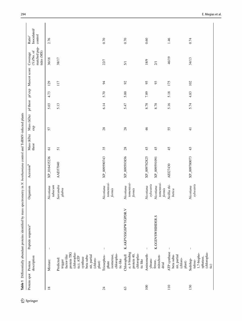
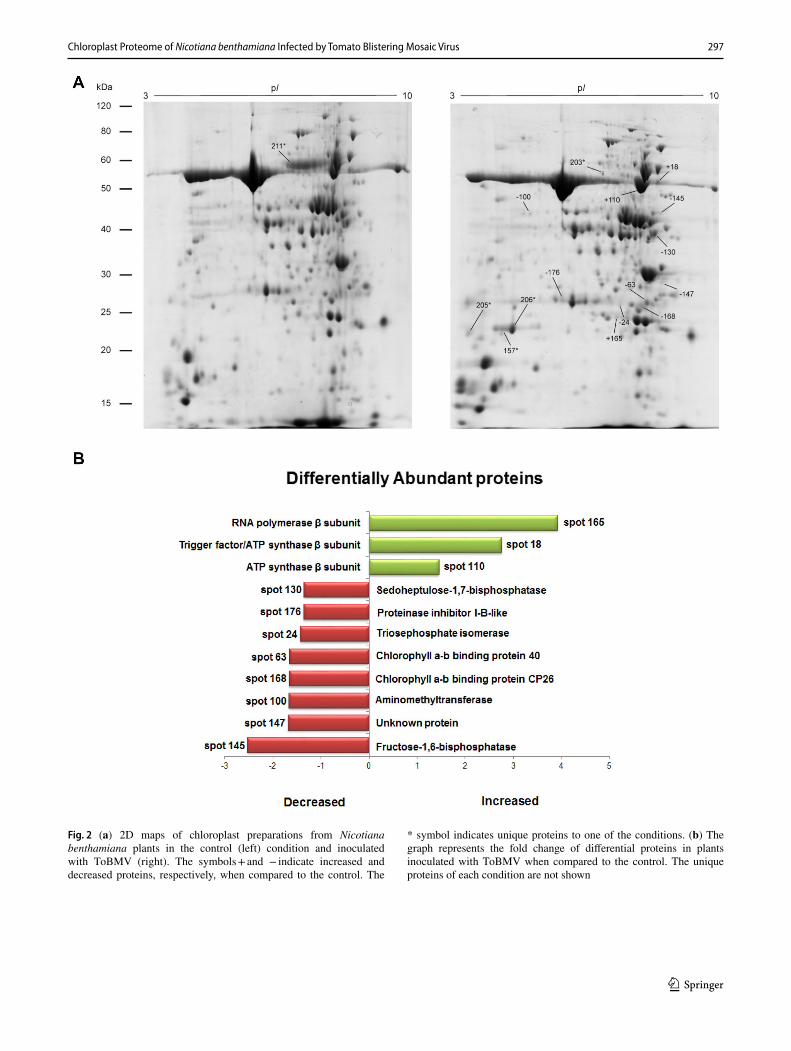
described for N. bethamiana chloroplasts [15]. The compara-tive analysis between both treatments (infected and control plants) showed a total of 36 differentially abundant proteins, including 12 increased, 17 decreased and 4 unique proteins in infected samples, as well as 3 unique proteins in control plants. All differential spots were excised for identification by mass spectrometry (MS), and a total of 16 differential proteins were identified (Table 1).
The cytopathic effect of the ToBMV on the host cell organelles involves chloroplast deformation and the induc-tion of vesicles on its periphery. In general, it is well known that plant viral infection affects plant chloroplasts causing the reduction of several chloroplastic proteins [16, 23–27]. In this study, as expected, these proteins were, indeed, decreased in infected plants when compared to control plants, such as a Sedoheptulose-1,7-bisphosphatase (spot 130; Table 1; Fig. 2a, b) and Ribulose 1,5-bisphosphate carboxylase/oxygenase large subunit (RubL) (Spot 211; Table 1; Fig. 2a), which was unique to control plants. The lower abundance of Sedoheptulose-1,7-bisphosphatase and RubL in infected plants was previously described in differ-ent plant-pathogen interactions [28, 29], and it has been shown that oxidative alterations affect the photosynthetic proteins causing their degradation during stress [30, 31]. This is consistent with the low levels of these proteins during PMMoV—N. benthamiana interaction [15] and also with the data obtained in our study. These results suggest that ToBMV also compromises the abundance of chloroplast-associated proteins and affects the photosynthetic apparatus.
Interestingly, among the increased proteins in plants infected by ToBMV was a mixture of proteins, which included Trigger factor-like protein and ATP synthase β subunit (spot 18; Table 1; Fig. 2a, b). It has been reported that ATP synthase β subunit interacts with viral proteins [16]. Moreover, the Trigger factor-like protein may help
viral infection, since it is a folding chaperone [32, 33]. Host chaperones can contribute to viral infection by interacting with viral proteins [32, 33]. Another interest-ing protein is RNA polymerase beta-subunit (spot 165; Table 1; Fig. 2), which showed an increased fold change of 3.93 in infected plants when compared to the control. The involvement of this enzyme in viroid replication has been reported. Plant viroids, which have circular RNAs, encode no proteins and are able to redirect a host polymerase for its replication in the chloroplast (reviewed in Ahlquist [34]). Differently, the ToBMV genome presents an ORF that encodes the RNA replicase polyprotein. It is possible that ToBMV interacts with chloroplastic RNA polymerase to change the gene expression in chloroplast genes to bene-fit virus infection. Therefore, these proteins are interesting candidates to further investigation to establish their role in viral pathogenesis during ToBMV infection.
It is noteworthy that some proteins were unique in infected samples. Among them are 50S ribosomal protein L6 (spot 205; Table 1; Fig. 2a), ATP synthase F1 subu-nit 1—mitochondrion (spot 203; Table 1; Fig. 2a), and ToBMV coat protein (spot 157 and 206; Table 1; Fig. 2a). Studies showed that the protein synthesis by chloroplasts generally can be inhibited by the virus [35], compromising even the levels of chloroplast ribosomal RNA [36]. How-ever, it is also known that viruses recruit host ribosomal subunits to translate viral mRNAs (reviewed in Walsh and Mohr [37]). Therefore, it is possible that the abundance of 50S ribosomal protein in infected plants could be caused by ToBMV to promote viral infection.
Fig. 1 Symptoms caused by ToBMV in Nicotiana bentha-miana plants. (a) Infected plant, showing necrotic spot in inoculated leaves, vein clearing and top distortion in upper leaves. (b) Infected leaf showing vein clearing and mottling. (c) Healty N. benthamiana plant
294 E. Megias et al.
1 3
Tabl
e 1
Diff
eren
tially
abu
ndan
t pro
tein
s ide
ntifi
ed b
y m
ass s
pect
rom
etry
in N
. ben
tham
iana
con
trol a
nd T
oBM
V-in
fect
ed p
lant
s
Prot
ein
spot
Prot
ein
desc
riptio
nPe
ptid
e se
quen
cea
Org
anis
mA
cess
ionb
Mas
s (kD
a)th
eor
Mas
s (kD
a)ex
ppI
theo
rpI
exp
Mas
cot s
core
Cov
erag
e (%
)/No.
of
mat
ched
pep
-tid
es (M
S)
Rat
ioc
Inoc
ulat
ed/
cont
rol
18M
ixtu
re:
–N
icot
iana
ta
bacu
mX
P_01
6435
236
6157
5.03
4.73
129
36/1
82.
76
Pred
icte
d:
trigg
er
fact
or-li
ke
prot
ein
TIG
(c
hlor
opla
s-tic
); A
TP
synt
hase
be
ta su
bu-
nit,
parti
al
(chl
oro-
plas
t)
Sarc
andr
a gl
abra
AA
D37
040
515.
1311
738
/17
24Tr
iose
phos
-ph
ate
isom
eras
e,
chlo
ropl
as-
tic-li
ke
–N
icot
iana
to
men
tosi
-fo
rmis
XP_
0095
9074
335
286.
145.
7094
22/7
0.70
63C
hlor
ophy
ll a,
b b
indi
ng
prot
ein
40,
chlo
ropl
as-
tic li
ke
K.A
KPV
SSG
SPW
YG
PDR
.VN
icot
iana
to
men
tosi
-fo
rmis
XP_
0095
9385
628
285.
475.
0092
5/1
0.70
100
Am
inom
eth-
yltra
ns-
fera
se,
mito
chon
-dr
ial
–N
icot
iana
sy
lves
tris
XP_
0097
8282
545
468.
787.
8995
18/9
0.60
K.G
GD
VSW
HIH
DER
.SN
icot
iana
to
men
tosi
-fo
rmis
XP_
0095
9109
145
8.78
932/
1
110
ATP
synt
hase
be
ta su
bu-
nit,
parti
al
(chl
oro-
plas
t)
–Ne
illia
thi-
betic
aA
EI27
430
4555
5.16
5.18
175
46/1
91.
46
130
Sedo
hep-
tulo
se-
1,7-
bisp
ho-
spha
tase
, (c
hlor
opla
s-tic
)
–N
icot
iana
sy
lves
tris
XP_
0097
8007
343
415.
744.
8310
234
/13
0.74
295Chloroplast Proteome of Nicotiana benthamiana Infected by Tomato Blistering Mosaic Virus
1 3
Tabl
e 1
(con
tinue
d)
Prot
ein
spot
Prot
ein
desc
riptio
nPe
ptid
e se
quen
cea
Org
anis
mA
cess
ionb
Mas
s (kD
a)th
eor
Mas
s (kD
a)ex
ppI
theo
rpI
exp
Mas
cot s
core
Cov
erag
e (%
)/No.
of
mat
ched
pep
-tid
es (M
S)
Rat
ioc
Inoc
ulat
ed/
cont
rol
145
Fruc
tose
-1,
6-bi
spho
-sp
hata
se
chlo
ropl
as-
tic-li
ke
–N
icot
iana
at
ttenu
ata
XP_
0192
4293
645
465.
464.
6780
19/6
0.40
K.Y
IDD
LKD
PGPS
GK
PYSA
R.Y
Nic
otia
na
taba
cum
XP_
0164
6140
945
5.26
554/
1
147
Unk
now
n pr
otei
n D
S12
from
2D
-PA
GE
of le
af,
chlo
ropl
as-
tic-li
ke
–N
icot
iana
sy
lves
tris
XP_
0097
6279
631
315.
204.
6182
27/5
0.59
157
Puta
tive
coat
pr
otei
nK
.TPT
ATL
QIR
.GTo
mat
o bl
ister
ing
mos
aic
viru
s
AIG
0582
920
248.
608.
6369
4/1
Uni
que
to
inoc
ulat
edK
.LLA
YSP
APT
TPSK
.T71
6/1
165
RN
A p
oly-
mer
ase
beta
su
buni
t (c
hlor
o-pl
ast)
–Sa
rtid
ia
dew
inte
riY
P_00
9141
152
1726
7.16
5.78
865/
83.
93
168
Chl
orop
hyll
a, b
bin
ding
pr
otei
n C
P26,
chl
o-ro
plas
tic
K.Y
QA
YEL
IHA
R.W
Nic
otia
na
taba
cum
XP_
0164
5880
030
285.
735.
1366
3/1
0.60
176
Prot
eina
se
inhi
bito
r I-
B-li
ke
–So
lanu
m
tube
rosu
mX
P_00
6354
531
1328
5.23
7.08
8059
/50.
74
203
ATP
synt
hase
F1
subu
nit
1 (m
ito-
chon
drio
n)
–N
icot
iana
ta
bacu
mBA
D83
524
5660
5.84
6.14
8014
/7U
niqu
e to
in
ocul
ated
R.E
AFP
GD
VFY
LHSR
.LAr
abid
opsi
s th
alia
na55
6.01
642/
1
296 E. Megias et al.
1 3
Tabl
e 1
(con
tinue
d)
Prot
ein
spot
Prot
ein
desc
riptio
nPe
ptid
e se
quen
cea
Org
anis
mA
cess
ionb
Mas
s (kD
a)th
eor
Mas
s (kD
a)ex
ppI
theo
rpI
exp
Mas
cot s
core
Cov
erag
e (%
)/No.
of
mat
ched
pep
-tid
es (M
S)
Rat
ioc
Inoc
ulat
ed/
cont
rol
205
50S
ribos
o-m
al p
rote
in
L6, c
hlor
o-pl
astic
–N
icot
iana
sy
lves
tris
XP_
0097
8546
825
239.
999.
0115
530
/12
Uni
que
to
inoc
ulat
edK
.KLQ
LVG
VG
YR
.ASp
inac
ia
oler
acea
5MLC
_H25
10.0
965
4/1
K.S
EIG
QFA
ASI
R.R
Lupi
nus
angu
stifo
-liu
s
OIW
0802
427
10.0
863
4/1
206
Puta
tive
coat
pr
otei
nK
.TPT
ATL
QIR
.GTo
mat
o bl
ister
ing
mos
aic
viru
s
AIG
0582
920
248.
608.
2972
4/1
Uni
que
to
inoc
ulat
ed
211
Rib
ulos
e-1,
5-bi
spho
-sp
hate
car
-bo
xyla
se/
oxyg
enas
e la
rge
subu
-ni
t, pa
rtial
(p
lasti
d)
–Q
uara
ribe
a sp
atul
ata
AFJ
9623
549
616.
126.
1011
525
/12
Uni
que
to
cont
rol
a The
sym
bol “
−”
in th
e pe
ptid
e se
quen
ce c
olum
n in
dica
tes t
hat p
eptid
e m
ass fi
nger
prin
ting
(PM
F) w
as p
erfo
rmed
and
ther
efor
e no
pep
tide
sequ
ence
was
gen
erat
edb M
ore
than
one
acc
essi
on c
ited
indi
cate
s diff
eren
t hits
for p
eptid
es id
entifi
ed fr
om th
e sa
me
prot
ein
c Rat
io i
nocu
late
d/co
ntro
l va
lues
ove
r 1
dete
rmin
e in
crea
sed
prot
eins
, whi
le v
alue
s be
low
1 c
orre
spon
d to
dec
reas
ed p
rote
ins.
Uni
que
prot
eins
to
each
tre
atm
ent
are
indi
cate
d. T
he p
rote
in
spot
abu
ndan
ce is
exp
ress
ed a
s a sp
ot ra
tio, c
ompa
ring
inoc
ulat
ed a
nd c
ontro
l spo
t vol
umes
297Chloroplast Proteome of Nicotiana benthamiana Infected by Tomato Blistering Mosaic Virus
1 3
Fig. 2 (a) 2D maps of chloroplast preparations from Nicotiana benthamiana plants in the control (left) condition and inoculated with ToBMV (right). The symbols + and − indicate increased and decreased proteins, respectively, when compared to the control. The
* symbol indicates unique proteins to one of the conditions. (b) The graph represents the fold change of differential proteins in plants inoculated with ToBMV when compared to the control. The unique proteins of each condition are not shown
298 E. Megias et al.
1 3
4 Conclusions
This is the first comparative report showing chloroplast proteins involved in the plant response to ToBMV infec-tion. The results indicate that this virus has an effect similar to other viruses, affecting severely the protein metabolism in chloroplasts. Proteins potentially involved in the infection process were also identified, including an RNA polymerase beta-subunit and a 50S ribosomal pro-tein L6, which were highly abundant or unique in infected tissues analyzed. Furthermore, this study identified some proteins that may be targets of a direct interaction with viral proteins, such as ATP synthase β subunit and Trig-ger factor-like protein. The identification of these targets is highly important in understanding the pathogenicity of ToBMV since they may be recruited by the virus to favor the infection. Therefore, it would be interesting to further investigate these chloroplast-related proteins to elucidate their roles in ToBMV pathogenesis.
Acknowledgements This work was sponsored by Embrapa, UnB, CAPES and CNPq.
Compliance with Ethical Standards
Conflict of interest Authors have no conflict of interest to declare.
References
1. Hull R (2002) CHAPTER 1 - introduction. In: Matthews’ plant virology, 4th edn. Academic Press, London, pp 1–12 https ://doi.org/10.1016/B978-01236 1160-4/50052 -9
2. Ribeiro S, Kitajima E, Oliveira R, Koenig R (1996) A strain of eggplant mosaic virus isolated from naturally infected tobacco plants in Brazil. Plant Dis 80(4):446–449
3. Alexandre MAV, Duarte LML, Rivas EB, Chagas CM, Barra-das MM, Koenig R (2000) Petunia vein banding virus: charac-terization of a new tymovirus from petunia × hybrida. Plant Dis 84(7):739–742. https ://doi.org/10.1094/pdis.2000.84.7.739
4. Crestani O, Kitajima E, Lin M, Marinho V (1986) Passion fruit yellow mosaic virus, a new tymovirus found in Brazil. Phytopa-thology 76(9):951–955
5. Nicolini C, Pio-Ribeiro G, Andrade GP, Melo FL, Oliveira VC, Guimarães FC, Resende RO, Kitajima EW, Rezende JAM, Nagata T (2012) A distinct tymovirus infecting cassia hoffmannseggii in Brazil. Virus Genes 45(1):190–194. https ://doi.org/10.1007/s1126 2-012-0750-9
6. Nicolini C, Inoue-Nagata AK, Nagata T (2015) Complete genome sequence of a proposed new tymovirus, tomato blistering mosaic virus. Arch Virol 160(2):609–612. https ://doi.org/10.1007/s0070 5-014-2289-7
7. de Oliveira VC, Nagata T, Guimarães FC, Ferreira FA, Kitajima EW, Nicolini C, de Oliveira Resende R, Inoue-Nagata AK (2013) Characterization of a novel tymovirus on tomato plants in Bra-zil. Virus Genes 46(1):190–194. https ://doi.org/10.1007/s1126 2-012-0830-x
8. Blawid R, Hayashi EAI, Rezende JAM, Kitajima EW, Nagata T (2016) A highly divergent isolate of tomato blistering mosaic virus from Solanum violaefolium. Virus Genes 52(2):294–298. https ://doi.org/10.1007/s1126 2-016-1288-z
9. Melo FL, Fernandes JEA, Ribeiro BM, Ribeiro SG (2014) Com-plete genome sequence of a tobacco-infecting, tomato-blistering mosaic virus. Genome Announcements 2(4):e00701–e00714.
10. Ferrand L, Nome C, Orílio AF, García ML, Nagata T, Ronco LB, Dal Bó E (2015) First report of tomato blistering mosaic virus infecting tomato in Argentina. Plant Dis 100(5):1026. https ://doi.org/10.1094/pdis-07-15-0782-pdn
11. Verchot J (2011) Wrapping membranes around plant virus infec-tion. Curr Opin Virol 1(5):388–395. https ://doi.org/10.1016/j.covir o.2011.09.009
12. Di Carli M, Benvenuto E, Donini M (2012) Recent insights into plant–virus interactions through proteomic analysis. J Prot Res 11(10):4765–4780. https ://doi.org/10.1021/pr300 494e
13. Peltier J-B, Friso G, Kalume DE, Roepstorff P, Nilsson F, Adam-ska I, van Wijk KJ (2000) Proteomics of the chloroplast: system-atic identification and targeting analysis of lumenal and peripheral thylakoid proteins. Plant Cell 12(3):319–342
14. Peltier J-B, Emanuelsson O, Kalume DE, Ytterberg J, Friso G, Rudella A, Liberles DA, Söderberg L, Roepstorff P, von Heijne G, van Wijk KJ (2002) Central functions of the lumenal and periph-eral thylakoid proteome of Arabidopsis determined by experimen-tation and genome-wide prediction. Plant Cell 14(1):211–236. https ://doi.org/10.1105/tpc.01030 4
15. Pineda M, Sajnani C, Barón M (2010) Changes induced by the Pepper mild mottle tobamovirus on the chloroplast proteome of Nicotiana benthamiana. Photosynth Res 103(1):31. https ://doi.org/10.1007/s1112 0-009-9499-y
16. Zhao J, Zhang X, Hong Y, Liu Y (2016) Chloroplast in plant-virus interaction. Front Microbiol 7:1565. https ://doi.org/10.3389/fmicb .2016.01565
17. Brizard JP, Carapito C, Delalande F, Van Dorsselaer A, Brugidou C (2006) Proteome analysis of plant-virus interactome: compre-hensive data for virus multiplication inside their hosts. Mol Cell Proteomics 5(12):2279–2297
18. Boatwright JL, Pajerowska-Mukhtar K (2013) Salicylic acid: an old hormone up to new tricks. Mol Plant Pathol 14(6):623–634. https ://doi.org/10.1111/mpp.12035
19. Wasternack C, Hause B (2013) Jasmonates: biosynthesis, per-ception, signal transduction and action in plant stress response, growth and development. An update to the 2007 review in annals of botany. Ann Bot 111(6):1021–1058. https ://doi.org/10.1093/aob/mct06 7
20. Alazem M, Lin N-S (2015) Roles of plant hormones in the regula-tion of host–virus interactions. Mol Plant Pathol 16(5):529–540. https ://doi.org/10.1111/mpp.12204
21. Kubis SE, Lilley KS, Jarvis P (2008) Isolation and preparation of chloroplasts from Arabidopsis thaliana plants. In: Posch A (ed) 2D PAGE: sample preparation and fractionation. Humana Press, Totowa, pp 171–186 https ://doi.org/10.1007/978-1-60327 -210-0_16
22. Carmo LST, Resende RO, Silva LP, Ribeiro SG, Mehta A (2013) Identification of host proteins modulated by the virulence factor AC2 of Tomato chlorotic mottle virus in Nicotiana benthami-ana. Proteomics 13(12–13):1947–1960. https ://doi.org/10.1002/pmic.20120 0547
23. Seo S, Okamoto M, Iwai T, Iwano M, Fukui K, Isogai A, Naka-jima N, Ohashi Y (2000) Reduced levels of chloroplast FtsH pro-tein in tobacco mosaic virus–infected tobacco leaves accelerate the hypersensitive reaction. Plant Cell 12(6):917–932
24. Bhat S, Folimonova SY, Cole AB, Ballard KD, Lei Z, Watson BS, Sumner LW, Nelson RS (2013) Influence of host chloroplast proteins on tobacco mosaic virus accumulation and intercellular

299Chloroplast Proteome of Nicotiana benthamiana Infected by Tomato Blistering Mosaic Virus
1 3
movement. Plant Physiol 161(1):134–147. https ://doi.org/10.1104/pp.112.20786 0
25. Mochizuki T, Ogata Y, Hirata Y, Ohki ST (2014) Quantitative transcriptional changes associated with chlorosis severity in mosaic leaves of tobacco plants infected with Cucumber mosaic virus. Mol Plant Pathol 15(3):242–254
26. Kundu S, Chakraborty D, Kundu A, Pal A (2013) Proteomics approach combined with biochemical attributes to elucidate com-patible and incompatible plant-virus interactions between Vigna mungo and Mungbean yellow mosaic India Virus. Proteome Sci 11:15. https ://doi.org/10.1186/1477-5956-11-15
27. Wu L, Wang S, Chen X, Wang X, Zu X, Chen Y (2013) Proteomic and phytohormone analysis of the response of maize (Zea mays L.) seedlings to sugarcane mosaic virus. PLoS ONE 8(7):e70295. https ://doi.org/10.1371/journ al.pone.00702 95
28. Pan X, Zhu B, Luo Y, Fu D (2013) Unraveling the protein network of tomato fruit in response to necrotrophic phytopathogenic Rhiz-opus nigricans. PLoS ONE 8(9):e73034. https ://doi.org/10.1371/journ al.pone.00730 34
29. Ji X, Gai Y, Zheng C, Mu Z (2009) Comparative proteomic analy-sis provides new insights into mulberry dwarf responses in mul-berry (Morus alba L.). Proteomics 9(23):5328–5339. https ://doi.org/10.1002/pmic.20090 0012
30. Vass I, Cser K, Cheregi O (2007) Molecular mechanisms of light stress of photosynthesis. Ann NY Acad Sci 1113:114–122. https ://doi.org/10.1196/annal s.1391.017
31. Houtz RL, Portis AR (2003) The life of ribulose 1,5-bisphosphate carboxylase/oxygenase–posttranslational facts and mysteries. Arch Biochem Biophys 414(2):150–158
32. Ellis RJ (2013) Assembly chaperones: a perspective. Philos Trans R Soc Lond B Biol Sci 368(1617):20110398. https ://doi.org/10.1098/rstb.2011.0398
33. Gorovits R, Moshe A, Ghanim M, Czosnek H (2013) Recruitment of the host plant heat shock protein 70 by tomato yellow leaf curl virus coat protein is required for virus infection. PLoS ONE 8(7):e70280. https ://doi.org/10.1371/journ al.pone.00702 80
34. Ahlquist P (2002) RNA-dependent RNA polymerases, viruses, and RNA silencing. Science 296(5571):1270–1273. https ://doi.org/10.1126/scien ce.10691 32
35. Pérez-Bueno ML, Rahoutei J, Sajnani C, García-Luque I, Barón M (2004) Proteomic analysis of the oxygen-evolving complex of photosystem II under biotec stress: studies on Nicotiana benthami-ana infected with tobamoviruses. Proteomics 4(2):418–425. https ://doi.org/10.1002/pmic.20030 0655
36. Fraser RS (1969) Effects of two TMV strains on the synthesis and stability of chloroplast ribosomal RNA in tobacco leaves. Mol Gen Genet 106(1):73–79
37. Walsh D, Mohr I (2011) Viral subversion of the host protein syn-thesis machinery. Nat Rev Microbiol 9(12):860–875. https ://doi.org/10.1038/nrmic ro265 5