Embed Size (px)
Citation preview
Hindawi Publishing CorporationProstate CancerVolume 2013, Article ID 705865, 6 pageshttp://dx.doi.org/10.1155/2013/705865
Clinical StudyAnalysis of Preoperative Detection for Apex Prostate Cancer byTransrectal Biopsy
Tomokazu Sazuka,1 Takashi Imamoto,1 Takeshi Namekawa,1,2 Takanobu Utsumi,1
Mitsuru Yanagisawa,1 Koji Kawamura,1 Naoto Kamiya,3 Hiroyoshi Suzuki,3 Takeshi Ueda,2
Satoshi Ota,4 Yukio Nakatani,4 and Tomohiko Ichikawa1
1 Department of Urology, Graduate School of Medicine, Chiba University, 1-8-1 Inohana, Chuou-ku, Chiba 260-8670, Japan2Division of Urology, Chiba Cancer Center, Chiba 260-8717, Japan3Department of Urology, Toho University Sakura Medical Center, Sakura 285-8741, Japan4Department of Pathology, Graduate School of Medicine, Chiba University, Chiba 260-8670, Japan
Correspondence should be addressed to Tomokazu Sazuka; [email protected]
Received 11 November 2012; Accepted 21 January 2013
Academic Editor: Manfred P. Wirth
Copyright © 2013 Tomokazu Sazuka et al. This is an open access article distributed under the Creative Commons AttributionLicense, which permits unrestricted use, distribution, and reproduction in any medium, provided the original work is properlycited.
Background. The aim of this study was to determine concordance rates for prostatectomy specimens and transrectal needle biopsysamples in various areas of the prostate in order to assess diagnostic accuracy of the transrectal biopsy approach, especiallyfor presurgical detection of cancer in the prostatic apex. Materials and Methods. From 2006 to 2011, 158 patients whose radicalprostatectomy specimens had been evaluated were retrospectively enrolled in this study. Concordance rates for histopathologyresults of prostatectomy specimens and needle biopsy samples were evaluated in 8 prostatic sections (apex, middle, base, andtransitional zones bilaterally) from 73 patients diagnosed at this institution, besides factors for detecting apex cancer in total 118true positive and false negative apex cancers. Results. Prostate cancer was foundmost frequently (85%) in the apex of all patients. Of584 histopathology sections, 153 (49%) from all areas were false negatives, as were 45% of apex biopsy samples. No readily availablepreoperative factors for detecting apex cancer were identified. Conclusions. In Japanese patients, the most frequent location ofprostate cancer is in the apex. There is a high false negative rate for transrectal biopsy samples. To improve the detection rate,transperitoneal biopsy or more accurate imaging technology is needed.
1. Introduction
One of the most frequent location of cancer in the prostategland is in the apex. Iremashvili et al. showed the incidence ofcarcinoma in prostatectomy specimens; 65.4% of all patientshad apex carcinoma, 56.6% had middle carcinoma, 47.3%had base carcinoma [1]. Apex core specimens obtained byneedle biopsy have been associated with the highest cancerdetection rates [2]. However, to the best of our knowledge,there have been no previous reports of assessments of thesensitivity and specificity of transrectal biopsy proceduresfor detection of apical prostate cancer through determiningcorrelations between histopathologic diagnoses of preop-erative transrectal biopsy and subsequently resected tissue
specimens, especially with regard to presurgical detection ofprostate cancer localized to the apex.
Recently, in Japan, prostate cancer (PCA) screening hasspread and diagnostic imaging technology has improved.Detection of early stage PCA has been increasing [3, 4].Kikuchi et al. reported that, in the United States after 1995,many smaller PCAs detected were located in the apex ofthe prostate: the frequency of apical cancer detection after1995 had risen to 46% from 26%, a significant increase[5, 6]. Takashima et al. in 2002 reported that in Japanesemen, 82.3% of all T1c prostate tumors were located in theapex and were significantly denser compared to midprostatetumors [7]. Because of such recent diagnostically related data,
2 Prostate Cancer
determination of precise tumor location is now a useful toolfor patient care.
The protocol for systematic transrectal biopsy was intro-duced by Hodge et al. more than 20 years ago [8]; use ofthis technique has increased the PCA detection rate. Huo etal. reported that accuracy of biopsy core analysis, when cor-related with prostatectomy specimens, had an average sensi-tivity and specificity for location of 48% and 84%, respectively[9], and Rogatsch et al. found a positive predictive value ofonly 71.1% [10]. Thus, predicting location by core specimenanalysis has not been particularly reliable.
Here we report results of a study of 14-core transrectalprostate biopsy specimens, 3 peripheral zone at regularintervals X 2 and 1 TZ X 1-X 2 bilaterally. The location ofeach cancer was determined from examination of subsequentradical prostatectomy (RP) specimens, and then concordancerates for prostatectomy specimens and preoperative needlebiopsy samples of 8 prostate areas (bilateral apex, middle,base, and TZ) were determined, with special attention paidto detection of apex cancers by transrectal apex biopsy.
2. Materials and Methods
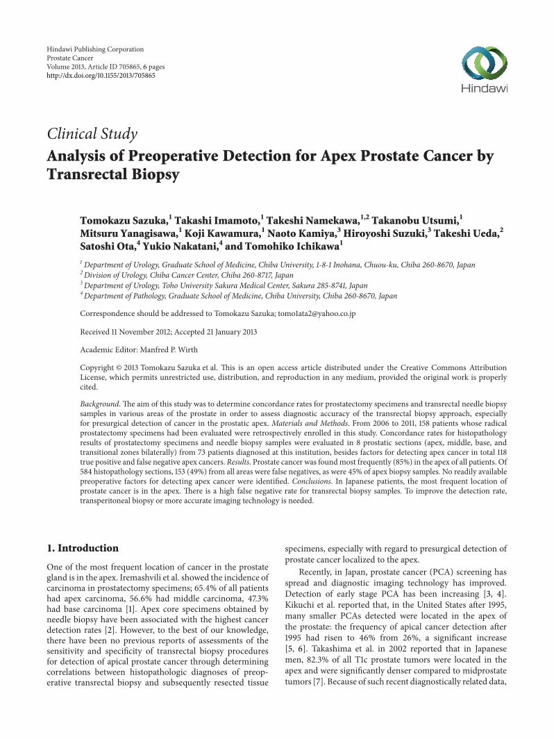
A total of 158 patients whose RP specimens had been eva-luated appropriately in 203 underwent RP patients at ChibaUniversity Graduate School of Medicine, Japan, from 2006to 2011 were retrospectively enrolled in this study. The studywas performed with approval of the hospital ethics com-mittee, and informed consent was obtained from patients.All patients had increased prostate specific antigen (PSA)levels (3.0 ng/mL or greater) and/or abnormal digital rectalexamination (DRE) findings, and PCA diagnosed by needlebiopsy. Patients who received neoadjuvant androgen depriva-tion therapy were excluded.
The indication for RP was clinically localized prostatecancer in patients aged 75 years or younger. Clinical stage T3was also considered an indication for surgery. The cliniciansconsidered not only clinical stage but also the Gleason scoreand PSA level.
Initial histopathology results were reported by experi-enced uropathologists after assessment of each prostate spe-cimen, all of which were fully embedded and sectionedat 5mm intervals for analysis. The anatomical locations oftumor foci were reproduced on a prostate cancermap. Tumorvolumeswere calculated using ImageProcessing andAnalysisin JAVA (Image J, NIH, United States). We defined the pro-static apex tumor as all or a part of tumor located within 1 cmfrom distal end of radical prostatectomy specimen.
Transrectal ultrasound (TRUS) was performed using theSSD-2000 System and a 7.5-MHz transducer (Aloka, Japan).All patients received a local anesthesia injection (5mL 1%lidocaine) to the apex of the prostate. Prostate needle biopsieswere performed transrectally using an 18-gauge biopsy needleand a biopsy gun under TRUS guidance, providing 17mmlong tissue cores. For the 14-core biopsy, 12 specimens weretaken from the peripheral zone at regular intervals and 2specimens were taken from the TZs. All biopsy specimenswere labeled according to the biopsy site (apex, middle, orbase of the peripheral zone or TZ, and left or right lobe) and
Table 1: Patients’ characteristics.
Characteristic Study population (n = 158)Age, mean ± SD years 65.26 ± 5.11
PSA, mean ± SD ng/mL 8.86 ± 5.09
PSA F/T, mean ± SD % 14.26 ± 7.66
Clinical T stageT1c 127 casesT2a–c 26 casesT3a 5 cases
Biopsy Gleason score6 48 cases7 86 cases⩾8 24 casesProstate volume, mean ± SD mL 30.97 ± 15.20
OperationORP 50 casesLRP 108 cases
Pathologic T stageT2a–c 98 casesT3ab 59 casesT4 1 case
RP Gleason score6 16 cases7 120 cases⩾8 22 cases
PSA: prostate specific antigen, F/T: free-to-total PSA ratio, RP: radicalprostatectomy, ORP: open radical prostatectomy, LRP: laparoscopic radicalprostatectomy.
were then submitted in separate formalin-filled containers tothe Department of Pathology, Chiba University Hospital.
The location of each cancer was determined in all cases,and concordance rates for prostatectomy specimens andneedle biopsy samples from 8 sections (bilateral apex,middle,base, and TZ) were determined for 73 patients diagnosed atour institution. Clinicopathological factors possibly correlat-ing with detection of apex cancer using transrectal biopsywere assessed in total 118 cancers, 65 true positive and 53 falsenegative apex cancers.
Statistical analysis was performed using the Student’s 𝑡-test, 𝜒2 test, Mann-Whitney 𝑈 test, and logistic regressionanalysis. 𝑃 values <0.05 were considered significant. SPSSversion 12.0 software (SPSS, Chicago, Illinois, USA) was usedfor all analyses.
3. Results
All 158 consecutive patients receiving RP were included inthis study. Clinical and pathological features are summarizedin Table 1.
The mean age was 65 years, mean PSA was 8.86 ng/mL,mean free to total PSA ratio was 14.26%, and mean prostatevolume was 30.97mL. Clinical T1c patients were the mostcommon, and 127 cases (80%) and 5 cases (3%) of clinical T3awere included. The biopsy Gleason score was 6 in 48 cases
Prostate Cancer 3
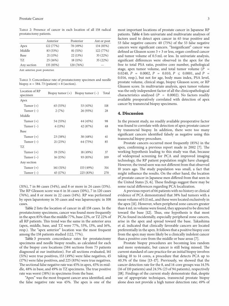
Table 2: Presence of cancer in each location of all 158 radicalprostatectomy patients.
Anterior Posterior Ant or postApex 122 (77%) 78 (49%) 134 (85%)Middle 83 (53%) 81 (51%) 122 (77%)Base 21 (13%) 22 (13%) 35 (22%)TZ 25 (16%) 18 (11%) 35 (22%)Any section 135 (85%) 120 (76%) —Ant: anterior, post: posterior.
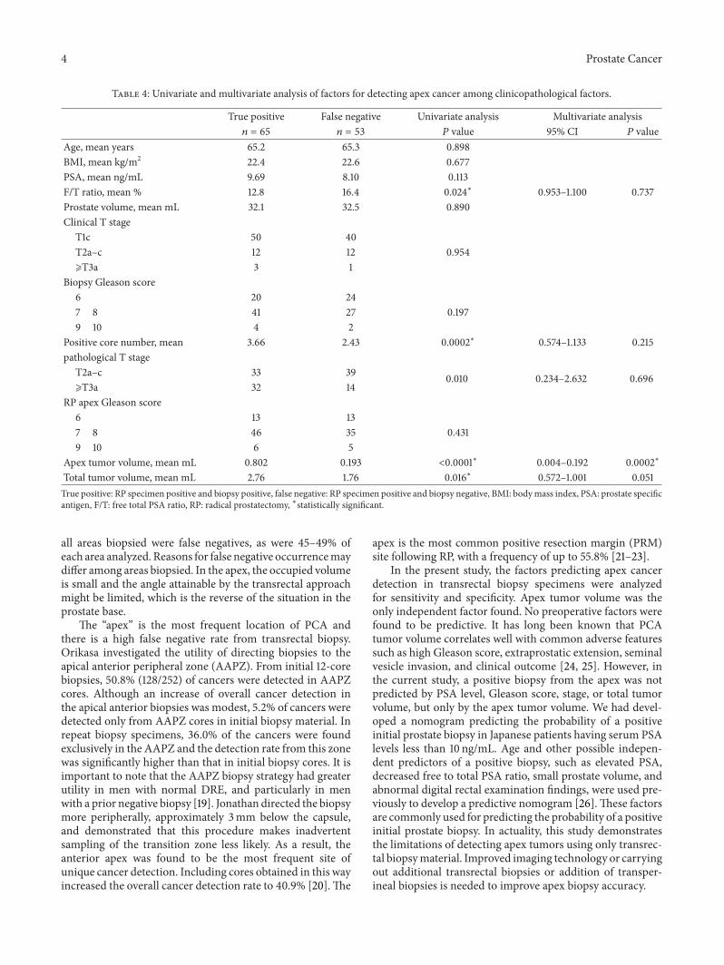
Table 3: Concordance rate of prostatectomy specimen and needlebiopsy. 𝑛 = 584. 73 (patient) × 8 (section).
Location of RPspecimen Biopsy tumor (+) Biopsy tumor (−) Total
ApexTumor (+) 65 (55%) 53 (45%) 118Tumor (−) 2 (7%) 26 (93%) 28
MiddleTumor (+) 54 (55%) 44 (45%) 98Tumor (−) 6 (13%) 42 (87%) 48
BaseTumor (+) 23 (38%) 38 (48%) 61Tumor (−) 21 (25%) 64 (75%) 85
TZTumor (+) 19 (51%) 18 (49%) 37Tumor (−) 16 (15%) 93 (85%) 109
Any sectionTumor (+) 161 (51%) 153 (49%) 314Tumor (−) 45 (17%) 225 (83%) 270
(31%), 7 in 86 cases (54%), and 8 or more in 24 cases (15%).The RP Gleason score was 6 in 16 cases (10%), 7 in 120 cases(76%), and 8 or more in 22 cases (14%). RP was performedby open laparotomy in 50 cases and was laparoscopic in 108cases.
Table 2 lists the location of cancer in all 158 cases. In theprostatectomy specimens, cancer was found more frequentlyin the apex 85% than themiddle 77%, base 22%, or TZ 22% ofall RP patients. This trend was the same in the anterior area(apex, middle, base, and TZ were 77%, 53%, 13%, and 16%,resp.). The “apex anterior” location was the most frequentamong the 158 patients studied (122, 77%).
Table 3 presents concordance rates for prostatectomyspecimens and needle biopsy results, as calculated for eachof the biopsy core locations (584 sections from 73 patientsdiagnosed at our institution). For all sections evaluated, 161(51%) were true positives, 153 (49%) were false negatives, 45(17%) were false positives, and 225 (83%) were true negatives.The sectional false negative rate was 45% in apex, 45% inmid-dle, 48% in base, and 49% in TZ specimens.The true positiverate was worst (38%) in specimens from the base.
“Apex” was the most frequent cancer area identified, andthe false negative rate was 45%. The apex is one of the
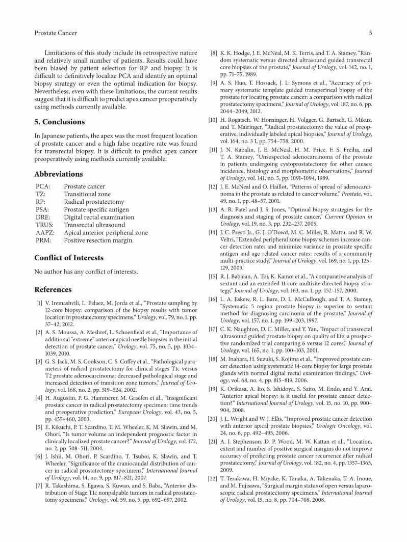
most important locations of prostate cancer in Japanese RPpatients. Table 4 lists univariate and multivariate analyses offactors used to detect apex cancer in 65 true positive and53 false negative cancers: 40 (75%) of the 53 false negativecancers were significant cancers. “Insignificant” cancer wasdefined as Gleason score 3 + 3 or less, organ-confined cancerand tumor volume of 0.5mL or less. In univariate analysis,significant differences were observed in the apex for thefree to total PSA ratio, positive core number, pathologicalstage, apex tumor volume, and total tumor volume (𝑃 =0.0240, 𝑃 = 0.0002, 𝑃 = 0.010, 𝑃 ≤ 0.0001, and 𝑃 =0.016, resp.), but not for age, body mass index, PSA level,prostate volume, clinical stage, biopsy Gleason score, or RPGleason score. In multivariate analysis, apex tumor volumewas the only independent factor of all the clinicopathologicalcharacteristics analysed (𝑃 = 0.0002). No factors readilyavailable preoperatively correlated with detection of apexcancer by transrectal biopsy specimens.
4. Discussion
In the present study, no readily available preoperative factorwas found to correlate with detection of apex prostate cancerby transrectal biopsy. In addition, there were too manysignificant cancers identified falsely as negative using thistransrectal biopsy procedure.
Prostate cancers occurred most frequently (85%) in theapex, confirming a previous report made in 2002 [7]. Theworking hypothesis leading to this study was that, becauseof widespread screening for PCA and improved imagingtechnology, the RP patient population might have changed.However, the trend seen was not different from that observed10 years ago. The study population was small, a fact thatmight influence the results. On the other hand, the locationof prostate cancer in Japanese men differed from that seen inthe United States [5, 6]. These findings suggest there may besome racial differences regarding PCA localization.
A previous report of 66 patients with no history or clinicalevidence of PCA demonstrated that 38% had tumors with amean volume of 0.11mL, and these were located exclusively inthe apex [11]. However, when peripheral zone cancers greaterthan 4mL in volumewere found, they appeared to be directedtoward the base [12]. Thus, one hypothesis is that mostPCAs found incidentally, especially peripheral zone cancers,arise in the apex and spread toward the base. Takashimaet al. indicated that clinically favorable cancers are locatedpreferentially in the apex. It follows that a positive biopsy corefrom the apex may more likely be a clinically indolent cancerthan a positive core from the middle or base areas [7].
Prostate biopsy procedures are becoming less randomand more systematic, but cancer is still being missed. Thecurrent standard of care practice for an initial biopsy involvestaking 10 to 14 cores, a procedure that detects PCA up to40.3% of the time [13–17]. Previously, we showed that thecancer detection rate for the 8, and 14-core groups was 14.5%(16 of 110 patients) and 24.5% (23 of 94 patients), respectively[18]. Findings of the current study demonstrate that, despiteuse of appropriate techniques, transrectal prostate biopsyalone does not provide a high tumor detection rate; 49% of
4 Prostate Cancer
Table 4: Univariate and multivariate analysis of factors for detecting apex cancer among clinicopathological factors.
True positive False negative Univariate analysis Multivariate analysis𝑛 = 65 𝑛 = 53 𝑃 value 95% CI 𝑃 value
Age, mean years 65.2 65.3 0.898BMI, mean kg/m2 22.4 22.6 0.677PSA, mean ng/mL 9.69 8.10 0.113F/T ratio, mean % 12.8 16.4 0.024∗ 0.953–1.100 0.737Prostate volume, mean mL 32.1 32.5 0.890Clinical T stage
T1c 50 40T2a–c 12 12 0.954⩾T3a 3 1
Biopsy Gleason score6 20 247 8 41 27 0.1979 10 4 2
Positive core number, mean 3.66 2.43 0.0002∗ 0.574–1.133 0.215pathological T stage
T2a–c 33 39 0.010 0.234–2.632 0.696⩾T3a 32 14
RP apex Gleason score6 13 137 8 46 35 0.4319 10 6 5
Apex tumor volume, mean mL 0.802 0.193 <0.0001∗ 0.004–0.192 0.0002∗
Total tumor volume, mean mL 2.76 1.76 0.016∗ 0.572–1.001 0.051True positive: RP specimen positive and biopsy positive, false negative: RP specimen positive and biopsy negative, BMI: bodymass index, PSA: prostate specificantigen, F/T: free total PSA ratio, RP: radical prostatectomy, ∗statistically significant.
all areas biopsied were false negatives, as were 45–49% ofeach area analyzed. Reasons for false negative occurrencemaydiffer among areas biopsied. In the apex, the occupied volumeis small and the angle attainable by the transrectal approachmight be limited, which is the reverse of the situation in theprostate base.
The “apex” is the most frequent location of PCA andthere is a high false negative rate from transrectal biopsy.Orikasa investigated the utility of directing biopsies to theapical anterior peripheral zone (AAPZ). From initial 12-corebiopsies, 50.8% (128/252) of cancers were detected in AAPZcores. Although an increase of overall cancer detection inthe apical anterior biopsies was modest, 5.2% of cancers weredetected only from AAPZ cores in initial biopsy material. Inrepeat biopsy specimens, 36.0% of the cancers were foundexclusively in the AAPZ and the detection rate from this zonewas significantly higher than that in initial biopsy cores. It isimportant to note that the AAPZ biopsy strategy had greaterutility in men with normal DRE, and particularly in menwith a prior negative biopsy [19]. Jonathan directed the biopsymore peripherally, approximately 3mm below the capsule,and demonstrated that this procedure makes inadvertentsampling of the transition zone less likely. As a result, theanterior apex was found to be the most frequent site ofunique cancer detection. Including cores obtained in this wayincreased the overall cancer detection rate to 40.9% [20].The
apex is the most common positive resection margin (PRM)site following RP, with a frequency of up to 55.8% [21–23].
In the present study, the factors predicting apex cancerdetection in transrectal biopsy specimens were analyzedfor sensitivity and specificity. Apex tumor volume was theonly independent factor found. No preoperative factors werefound to be predictive. It has long been known that PCAtumor volume correlates well with common adverse featuressuch as high Gleason score, extraprostatic extension, seminalvesicle invasion, and clinical outcome [24, 25]. However, inthe current study, a positive biopsy from the apex was notpredicted by PSA level, Gleason score, stage, or total tumorvolume, but only by the apex tumor volume. We had devel-oped a nomogram predicting the probability of a positiveinitial prostate biopsy in Japanese patients having serum PSAlevels less than 10 ng/mL. Age and other possible indepen-dent predictors of a positive biopsy, such as elevated PSA,decreased free to total PSA ratio, small prostate volume, andabnormal digital rectal examination findings, were used pre-viously to develop a predictive nomogram [26].These factorsare commonly used for predicting the probability of a positiveinitial prostate biopsy. In actuality, this study demonstratesthe limitations of detecting apex tumors using only transrec-tal biopsymaterial. Improved imaging technology or carryingout additional transrectal biopsies or addition of transper-ineal biopsies is needed to improve apex biopsy accuracy.
Prostate Cancer 5
Limitations of this study include its retrospective natureand relatively small number of patients. Results could havebeen biased by patient selection for RP and biopsy. It isdifficult to definitively localize PCA and identify an optimalbiopsy strategy or even the optimal indication for biopsy.Nevertheless, even with these limitations, the current resultssuggest that it is difficult to predict apex cancer preoperativelyusing methods currently available.
5. Conclusions
In Japanese patients, the apex was the most frequent locationof prostate cancer and a high false negative rate was foundfor transrectal biopsy. It is difficult to predict apex cancerpreoperatively using methods currently available.
Abbreviations
PCA: Prostate cancerTZ: Transitional zoneRP: Radical prostatectomyPSA: Prostate specific antigenDRE: Digital rectal examinationTRUS: Transrectal ultrasoundAAPZ: Apical anterior peripheral zonePRM: Positive resection margin.
Conflict of Interests
No author has any conflict of interests.
References
[1] V. Iremashvili, L. Pelaez, M. Jorda et al., “Prostate sampling by12-core biopsy: comparison of the biopsy results with tumorlocation in prostatectomy specimens,”Urology, vol. 79, no. 1, pp.37–42, 2012.
[2] A. S. Moussa, A. Meshref, L. Schoenfield et al., “Importance ofadditional “extreme” anterior apical needle biopsies in the initialdetection of prostate cancer,” Urology, vol. 75, no. 5, pp. 1034–1039, 2010.
[3] G. S. Jack, M. S. Cookson, C. S. Coffey et al., “Pathological para-meters of radical prostatectomy for clinical stages T1c versusT2 prostate adenocarcinoma: decreased pathological stage andincreased detection of transition zone tumors,” Journal of Uro-logy, vol. 168, no. 2, pp. 519–524, 2002.
[4] H. Augustin, P. G. Hammerer, M. Graefen et al., “Insignificantprostate cancer in radical prostatectomy specimen: time trendsand preoperative prediction,” European Urology, vol. 43, no. 5,pp. 455–460, 2003.
[5] E. Kikuchi, P. T. Scardino, T. M. Wheeler, K. M. Slawin, and M.Ohori, “Is tumor volume an independent prognostic factor inclinically localized prostate cancer?” Journal of Urology, vol. 172,no. 2, pp. 508–511, 2004.
[6] J. Ishii, M. Ohori, P. Scardino, T. Tsuboi, K. Slawin, and T.Wheeler, “Significance of the craniocaudal distribution of can-cer in radical prostatectomy specimens,” International Journalof Urology, vol. 14, no. 9, pp. 817–821, 2007.
[7] R. Takashima, S. Egawa, S. Kuwao, and S. Baba, “Anterior dis-tribution of Stage T1c nonpalpable tumors in radical prostatec-tomy specimens,” Urology, vol. 59, no. 5, pp. 692–697, 2002.
[8] K. K. Hodge, J. E. McNeal, M. K. Terris, and T. A. Stamey, “Ran-dom systematic versus directed ultrasound guided transrectalcore biopsies of the prostate,” Journal of Urology, vol. 142, no. 1,pp. 71–75, 1989.
[9] A. S. Huo, T. Hossack, J. L. Symons et al., “Accuracy of pri-mary systematic template guided transperineal biopsy of theprostate for locating prostate cancer: a comparison with radicalprostatectomy specimens,” Journal of Urology, vol. 187, no. 6, pp.2044–2049, 2012.
[10] H. Rogatsch, W. Horninger, H. Volgger, G. Bartsch, G. Mikuz,and T. Mairinger, “Radical prostatectomy: the value of preop-erative, individually labeled apical biopsies,” Journal of Urology,vol. 164, no. 3 I, pp. 754–758, 2000.
[11] J. N. Kabalin, J. E. McNeal, H. M. Price, F. S. Freiha, andT. A. Stamey, “Unsuspected adenocarcinoma of the prostatein patients undergoing cystoprostatectomy for other causes:incidence, histology and morphometric observations,” Journalof Urology, vol. 141, no. 5, pp. 1091–1094, 1989.
[12] J. E. McNeal and O. Haillot, “Patterns of spread of adenocarci-noma in the prostate as related to cancer volume,” Prostate, vol.49, no. 1, pp. 48–57, 2001.
[13] A. R. Patel and J. S. Jones, “Optimal biopsy strategies for thediagnosis and staging of prostate cancer,” Current Opinion inUrology, vol. 19, no. 3, pp. 232–237, 2009.
[14] J. C. Presti Jr., G. J. O’Dowd, M. C. Miller, R. Mattu, and R. W.Veltri, “Extended peripheral zone biopsy schemes increase can-cer detection rates and minimize variance in prostate specificantigen and age related cancer rates: results of a communitymulti-practice study,” Journal of Urology, vol. 169, no. 1, pp. 125–129, 2003.
[15] R. J. Babaian, A. Toi, K. Kamoi et al., “A comparative analysis ofsextant and an extended 11-core multisite directed biopsy stra-tegy,” Journal of Urology, vol. 163, no. 1, pp. 152–157, 2000.
[16] L. A. Eskew, R. L. Bare, D. L. McCullough, and T. A. Stamey,“Systematic 5 region prostate biopsy is superior to sextantmethod for diagnosing carcinoma of the prostate,” Journal ofUrology, vol. 157, no. 1, pp. 199–203, 1997.
[17] C. K. Naughton, D. C. Miller, and Y. Yan, “Impact of transrectalultrasound guided prostate biopsy on quality of life: a prospec-tive randomized trial comparing 6 versus 12 cores,” Journal ofUrology, vol. 165, no. 1, pp. 100–103, 2001.
[18] M. Inahara, H. Suzuki, S. Kojima et al., “Improved prostate can-cer detection using systematic 14-core biopsy for large prostateglands with normal digital rectal examination findings,” Urol-ogy, vol. 68, no. 4, pp. 815–819, 2006.
[19] K. Orikasa, A. Ito, S. Ishidoya, S. Saito, M. Endo, and Y. Arai,“Anterior apical biopsy: is it useful for prostate cancer detec-tion?” International Journal of Urology, vol. 15, no. 10, pp. 900–904, 2008.
[20] J. L. Wright andW. J. Ellis, “Improved prostate cancer detectionwith anterior apical prostate biopsies,” Urologic Oncology, vol.24, no. 6, pp. 492–495, 2006.
[21] A. J. Stephenson, D. P. Wood, M. W. Kattan et al., “Location,extent and number of positive surgical margins do not improveaccuracy of predicting prostate cancer recurrence after radicalprostatectomy,” Journal of Urology, vol. 182, no. 4, pp. 1357–1363,2009.
[22] T. Terakawa, H. Miyake, K. Tanaka, A. Takenaka, T. A. Inoue,andM. Fujisawa, “Surgical margin status of open versus laparo-scopic radical prostatectomy specimens,” International Journalof Urology, vol. 15, no. 8, pp. 704–708, 2008.
6 Prostate Cancer
[23] L. Salomon, A. G. Anastasiadis, O. Levrel et al., “Location ofpositive surgical margins after retropubic, perineal, and lap-aroscopic radical prostatectomy for organ-confined prostatecancer,” Urology, vol. 61, no. 2, pp. 386–390, 2003.
[24] P. A. Humphrey, “Tumor amount in prostatic tissues in relationto patient outcome andmanagement,” American Journal ofClinical Pathology, vol. 131, no. 1, pp. 7–10, 2009.
[25] J. I. Epstein, “Prognostic significance of tumor volume in radi-cal prostatectomy and needle biopsy specimens,” Journal of Uro-logy, vol. 186, no. 3, pp. 790–797, 2011.
[26] K. Kawamura, H. Suzuki, N. Kamiya et al., “Development ofa new nomogram for predicting the probability of a positiveinitial prostate biopsy in Japanese patients with serum PSAlevels less than 10 ng/mL,” International Journal of Urology, vol.15, no. 7, pp. 598–603, 2008.

Submit your manuscripts athttp://www.hindawi.com
Stem CellsInternational
Hindawi Publishing Corporationhttp://www.hindawi.com Volume 2014
Hindawi Publishing Corporationhttp://www.hindawi.com Volume 2014
MEDIATORSINFLAMMATION
of
Hindawi Publishing Corporationhttp://www.hindawi.com Volume 2014
Behavioural Neurology
EndocrinologyInternational Journal of
Hindawi Publishing Corporationhttp://www.hindawi.com Volume 2014
Hindawi Publishing Corporationhttp://www.hindawi.com Volume 2014
Disease Markers
Hindawi Publishing Corporationhttp://www.hindawi.com Volume 2014
BioMed Research International
OncologyJournal of
Hindawi Publishing Corporationhttp://www.hindawi.com Volume 2014
Hindawi Publishing Corporationhttp://www.hindawi.com Volume 2014
Oxidative Medicine and Cellular Longevity
Hindawi Publishing Corporationhttp://www.hindawi.com Volume 2014
PPAR Research
The Scientific World JournalHindawi Publishing Corporation http://www.hindawi.com Volume 2014
Immunology ResearchHindawi Publishing Corporationhttp://www.hindawi.com Volume 2014
Journal of
ObesityJournal of
Hindawi Publishing Corporationhttp://www.hindawi.com Volume 2014
Hindawi Publishing Corporationhttp://www.hindawi.com Volume 2014
Computational and Mathematical Methods in Medicine
OphthalmologyJournal of
Hindawi Publishing Corporationhttp://www.hindawi.com Volume 2014
Diabetes ResearchJournal of
Hindawi Publishing Corporationhttp://www.hindawi.com Volume 2014
Hindawi Publishing Corporationhttp://www.hindawi.com Volume 2014
Research and TreatmentAIDS
Hindawi Publishing Corporationhttp://www.hindawi.com Volume 2014
Gastroenterology Research and Practice
Hindawi Publishing Corporationhttp://www.hindawi.com Volume 2014
Parkinson’s Disease
Evidence-Based Complementary and Alternative Medicine
Volume 2014Hindawi Publishing Corporationhttp://www.hindawi.com

![Oracle APEX 5.1 Installations Oracle XE 11.2, Apex 5.1.3, Apache … · 2019-08-28 · Oracle APEX 5.1 – Installations Oracle XE 11.2, Apex 5.1.3, Apache Tomcat et ORDS [591 d]](https://img.pdfslide.tips/doc/110x75/5f79f240bba1e439d43cde1b/oracle-apex-51-installations-oracle-xe-112-apex-513-apache-2019-08-28-oracle.jpg)