Embed Size (px)
Citation preview

231
Journal of Hard Tissue Biology 29[4] (2020) 231-2382020 The Hard Tissue Biology Network AssociationPrinted in Japan, All rights reserved.CODEN-JHTBFF, ISSN 1341-7649
OriginalEffects of Glucocorticoids on Diurnal Variations in Experimental Tooth Movement
Sae Kusafuka1,2), Hisataka Kondo2), Kaori Hayashi1,2), Kazunori Hamamura2), Takuma Sato1), Ken Miyazawa1), Shigemi Goto1) and Akifumi Togari2)
1) Department of Orthodontics, School of Dentistry, Aichi Gakuin University, Nagoya, Japan2) Department of Pharmacology, School of Dentistry, Aichi Gakuin University, Nagoya, Japan(Accepted for publication, September 7, 2020)
Abstract: Diurnal variations in bone remodeling, which are regulated by the central circadian clock in the suprachiasmatic nuclei, have previously been identified. Glucocorticoids induce a circadian rhythm in osteoclasts, causing a circadian rhythm in bone resorption. It has been reported that the extent of experimental tooth movement (ETM) also exhibits diurnal variations, but this has not been thoroughly investigated. The aim of this study was to examine the role of glucocorticoids in the diurnal variations in the extent of ETM. Male C57BL6/J mice were divided into three groups: the whole-day group (WDG); ETM was performed during both the light and dark periods, the light period group (LPG); ETM was performed during the light period (7:00–19:00), and the dark period group (DPG); ETM was performed during the dark period (19:00–7:00). Orthodontic force was applied for a total of 48 hr in all groups. A piece of an orthodontic elastic band was inserted between the right upper first and second molars (M1 and M2) according to the Waldo method. During the study period, the distance between M1 and M2 increased by 185.1±7.0 μm in the WDG, 186.0±6.6 μm in the LPG, and 152.7±9.9 μm in the DPG. The amount of ETM-induced tooth movement was significantly larger in the WDG and LPG than in the DPG. The osteoclast surface/bone surface (compressed side) and osteoclast number/bone surface (compressed side) ratios were also significantly larger in the WDG and LPG than in the DPG. Consistent with the results of osteoclast parameters, the immune reactivity of receptor activator of nuclear factor-κB ligand (RANKL) was higher in the WDG and LPG than in the DPG. Adrenalectomy eliminated the differences in osteoclast parameters, RANKL immune reactivity and the extent of ETM be-tween the LPG and DPG. These differences were restored by the daily administration of the synthetic glucocorticoid dexa-methasone to adrenalectomized mice. These results suggest that circulating glucocorticoids contribute to the diurnal varia-tions in the osteoclast parameters which result in the diurnal variation of the tooth movement, and the diurnal variation of RANKL immune reactivity may contribute to it.
Key words: Diurnal variation, Glucocorticoids, Tooth movement, Osteoclast, Adrenalectomy
IntroductionAutonomic nervous system activity, metabolism, and hormonal se-
cretion display circadian rhythms, which contribute to the maintenance of homeostasis in physiological functions1–5). Circadian rhythms are de-rived from the suprachiasmatic nucleus, which is located in the hypo-thalamus and controls the peripheral clock of each tissue6, 7). The central clock is controlled by light stimulation, and central time information is thought to be transmitted to peripheral tissues by the sympathetic nerv-ous system and glucocorticoids8). We and others have shown that gluco-corticoids contribute to the transmission of central time information to bone tissue9, 10).
Glucocorticoids, which are steroid hormones produced in the adrenal cortex, raise the blood glucose level in the liver by promoting gluconeo-genesis11). Mineralocorticoids, which induce the reabsorption of sodium ions, promote potassium ion excretion, and regulate electrolyte and wa-ter levels in the body, are secreted from the adrenal cortex12). Fujihara et al.9) reported that there are diurnal variations in the expression levels of osteoclast-related genes. Adrenalectomized (ADX) mice lose such vari-
ations, but they can be restored by administering dexamethasone (DEX) to ADX mice. These results indicate that glucocorticoids influence the circadian rhythms of bone metabolism by regulating the diurnal varia-tions in osteoclast activity.
Orthodontic force stimulates local bone remodeling, which results in tooth movement13). In studies in which an open coil was placed between the upper first molars, Igarashi et al.14) and Miyoshi et al.15) showed that to induce lateral expansion of the maxilla, applying orthodontic force during the daytime was more effective than applying it at night in rats. Their results indicated that diurnal variations are also seen in the extent of experimental tooth movement (ETM), but it is not clear whether glu-cocorticoids are involved in this.
The receptor activator of nuclear factor-κB ligand (RANKL) regu-lates osteoclast differentiation and activity, which is upregulated by ETM in periodontal ligament16-18). We have previously reported that the mRNA expression of RANKL shows circadian rhythms in bone tissues and ETM based on the Waldo method is controlled by the sympathetic nervous system9, 19, 20). In the Waldo method, teeth are moved by insert-ing an elastic band between the first and second molars. In the present study, we investigated whether the extent of ETM induced by the Waldo method exhibits diurnal variations, and we also elucidated the effects of
Correspondence to: Dr. Hisataka Kondo, Aichi Gakuin University, 1-100 Kusumoto-cho, Chikusa-ku, Nagoya, 464-8650, Japan; Tel: +81-52-757-6743; Fax: +81-752-5988; E-mail: [email protected]

232
J.Hard Tissue Biology Vol. 29(4): 231-238, 2020
adrenalectomy on such tooth movement and the effects of glucocorti-coid administration on such tooth movement after ADX.
Materials and MethodsAnimals
Male C57BL6/J mice (Japan SLC Inc., Hamamatsu, Japan) were raised in our animal facility and kept under a 12-hr light/12-hr dark cy-cle, with lights on at 7:00 and off at 19:00. All animals were supplied with food and water ad libitum. The experimental protocols were re-viewed and approved by the animal experiment committee of the School of Dentistry, Aichi Gakuin University (AGUD-415-1,2,3 and AGUD-458-1,2).
Experimental tooth movementSeven- or 8-week-old mice were randomized by weight and divided
into the following three groups: the whole-day group (WDG), in which orthodontic force was applied continuously for 48 hr; the light period group (LPG), in which orthodontic force was applied throughout the light period (7:00–19:00) for 4 days; and the dark period group (DPG), in which orthodontic force was applied throughout the dark period (19:00–7:00) for 4 days. Then, the mice were anesthetized using an an-esthetic mixture of 0.375 mg/kg medetomidine (Medetomin, Meiji Sei-ka Pharma Co., Ltd., Tokyo, Japan), 2 mg/kg midazolam (Dormicum, Astellas Pharma Inc., Tokyo, Japan), and 2.5 mg/kg butorphanol (Vetor-phale, Meiji Seika Pharma Co., Ltd., Tokyo, Japan) (MMB), and a piece of elastic band (No. 404-136, Unitek Co., Ltd., Saint Paul, USA) was inserted between the upper right first molar (M1) and second molar (M2) according to the Waldo method21). After the piece of rubber band had been inserted, atipamezole (Mepatia, Meiji Seika Pharma Co., Ltd, To-kyo, Japan) was injected immediately to help the mice recover from the anesthesia. The mice were sacrificed via carbon dioxide inhalation at the end of the study period. The body weights of the mice were assessed every day during the ETM (FY-3000; A and D Co., Ltd, Tokyo, Japan) (Table 1-3).
AdrenalectomyUsing the MMB anesthetic mixture, a dorsal incision was made, and
the bilateral adrenal glands were removed from 5-week-old mice9). The ADX mice were supplied with food ad libitum and 0.9% NaCl to pre-vent hyponatremia and kept under a 12-hr light/12-hr dark cycle, with lights on at 7:00 and off at 19:00, for at least two weeks, before being subjected to ETM.
Three-dimensional micro-computed tomography (3D-μCT) analysis The amount of tooth movement induced by the ETM was measured
using 3D-μCT (an R-mCT μCT scanner; Rigaku Co., Ltd, Tokyo, Japan) and the TRI/3D-Bon software (Ratoc Systems, Inc., Tokyo, Japan). The maxilla was scanned using an X-ray current of 90 keV, a tube voltage of 88 µA, and an isotropic voxel size of 10-µm. In order to measure the amount of tooth movement induced by the ETM, objects were adjusted to ensure that the buccal view in which the distance between M1 and M2 was shortest was shown (Fig. 1a, 2a, 3a)22).
Bone histomorphometryThe right maxilla was dissected out and fixed in 4% paraformalde-
hyde. The fixed samples were put into 20% ethylenediaminetetraacetic acid (EDTA) for two weeks for decalcification. Five-μm-thick occlusal sections were obtained from the region located between 50 μm and 125 μm above the furcation. Osteoclasts were stained for tartrate-resistant
acid phosphatase (TRAP). The number of TRAP-positive multinucleat-ed cells was counted to determine the number of osteoclasts on the bone surface (compressed side) (the Oc.N/BS ratio) and the relative size of the osteoclast surface area compared with the bone surface area (com-pressed side) (the Oc.S/BS ratio), as reported previously23). Osteoblasts were stained with hematoxylin and eosin (HE), and the number of oste-oblasts on the bone surface (tensioned side) (Ob.N/BS) was counted. For immunohistochemistry, antigen inactivation treatment was carried out in a hot bath at 90°C for 40 min, and tissue sections were incubated with the primary antibody; i.e., anti-receptor activator of nuclear fac-tor-κB ligand (RANKL) mouse monoclonal antibody (ab45039, Abcam Plc., Cambridge, UK) (diluted 1:100), at 4°C over night, before being incubated with the secondary antibody; i.e., simple stain mouse MAX-PO (414322, Nichirei Co., Tokyo, Japan), at room temperature for 10 min. For negative control, mouse IgG1 antibody (X0931, Agilent, Santa Clara, USA) (diluted 1:100), at 4°C over night. The number of RANKL positive cells were counted and expressed that as a percentage of the to-tal number of cells present on the compressed side.
Statistical analysisData are shown as mean±SEM values (For body weight, data are
shown as mean±SDM). Statistical analyses were conducted with the Student’s t-test or ANOVA using GraphPad Prism version 6.0 for Win-dows (GraphPad Software, San Diego, USA). Post-hoc analyses, which were performed using Tukey’s multiple comparisons test, were conduct-ed on parameters that exhibited interactive effects, as described previ-ously19). P-values of <0.05 or <0.01 were considered to be statistically significant.
ResultsThe amount of ETM induced via the Waldo method exhibited diurnal variations
To investigate the diurnal variations in the amount of ETM induced using the Waldo method, mice were kept under a 12-hr light/12-hr dark cycle for at least two weeks before tooth movement was induced and then were divided into three groups (the WDG, LPG, and DPG). In the WDG, force was applied continuously for 48 hr. In the LPG, force was only applied during the light period (7:00–19:00) for 4 days (force was applied for a total of 48 hr). In the DPG, force was only applied during the dark period (19:00–7:00) for 4 days (force was applied for a total of 48 hr). The amount of tooth movement achieved was measured using 3D-μCT (Fig. 1a). The mean increase in the distance between M1 and M2 induced by the ETM (hereafter referred to as the tooth movement distance) was 185.1±7.0 μm in the WDG, 186.0±6.6 μm in the LPG, and 152.7±9.9 μm in the DPG. In the DPG, the mean tooth movement distance was 17.5% lower than that seen in the WDG and 17.9% lower than that seen in the LPG (Fig. 1b). There was no significant difference in the mean tooth movement distance between the WDG and LPG.
To examine the role played by osteoclasts in the diurnal variations in the tooth movement distance, we measured the Oc.S/BS and Oc.N/BS ratios as osteoclast parameters. The mean Oc.S/BS ratio was 10.8±0.9% in the WDG, 16.0±1.2% in the LPG, and 4.2±1.6% in the DPG (Fig. 1c). In the DPG, the mean Oc.S/BS ratio was 61.2% lower than that seen in the WDG and 73.7% lower than that seen in the LPG (Fig. 1c). The mean Oc.N/BS ratio was 3.2±0.4/mm in the WDG, 4.4±0.5/mm in the LPG, and 1.2±0.5/mm in the DPG. In the DPG, the mean Oc.N/BS ratio was 62.5% lower that seen in the WDG and by 72.7% lower than that seen in the LPG (Fig. 1c). The mean Ob.N/BS ratio was 3.0±0.5/mm in the WDG, 4.9±1.0/mm in the LPG, and 5.3±0.6/mm in the DPG.

233
Sae Kusafuka et al.: Glucocorticoids Regulate the Diurnal Variations in Tooth Movement
There were no significant differences in the mean Ob.N/BS between any of the three groups (Fig. 1d). The immunohistological examinations showed that there were 14.4±0.7/mm2, 14.9±0.8/mm2, and 7.7±0.7/mm2 RANKL-positive cells in the WDG, LPG, and DPG, respectively (Fig. 1e). In the DPG, the number of RANKL-positive cells was 46.5% lower than that seen in the WDG and 48.3% lower than that seen in the LPG (Fig. 1e).
The ETM led to reductions in body weight in the LPG and DPG. The mean body weight of the mice in the WPG was higher than those of the mice in the LPG and DPG (Table 1). There was no significant differ-ence in body weight between the LPG and DPG (Table 1).
Figure 1. Diurnal variations were detected in the tooth movement distance achieved using the Waldo method. (a) 3D-μCT images of the right upper molars. Scale bars: 500 μm. (b) Mean tooth movement distance. The WDG and LPG exhibited greater tooth movement than the DPG. (c) Representa-tive images of the staining of tartrate-resistant acid phosphatase (TRAP) in osteoclasts. Scale bars: 50 μm. The osteoclast (stained red) surface/bone surface (Oc.S/BS) (%) and osteoclast number/bone surface (Oc.N/BS) (number/mm) ratios were analyzed. Both ratios were lower in the DPG than in the WPG and LPG. (d) Representative images of the hematoxy-lin-eosin (HE) staining of osteoblasts. The osteoblast number/bone surface (Ob.N/BS) (number/mm) ratio was analyzed. There were no significant differences in the Ob.N/BS ratio between the groups. Scale bars: 50 μm. (e) Immunohistochemistry of receptor activator of nuclear factor-κB ligand (RANKL). The ratio of RANKL-positive cells (%) was lower in the DPG than in the WPG and LPG. Arrow indicates RANKL-positive cells. IgG: negative control. Scale bars: 50 μm. In the WDG, force was applied contin-uously for 48 hr. In the LPG, force was only applied during the light period (7:00–19:00) for 4 days. In the DPG, force was only applied during the dark period (19:00–7:00) for 4 days. Six, six, and seven animals were used in the WDG, LPG, and DPG, respectively. The error bar indicates the mean±SEM. *, ** statistically significant difference vs. the WDG at p<0.05 and p<0.01, respectively, according to ANOVA; #, ##statistically significant difference vs. the LPG at p<0.05 and p<0.01, respectively, according to ANOVA; T: teeth, B: bone.

234
J.Hard Tissue Biology Vol. 29(4): 231-238, 2020
The diurnal variations in the tooth movement distance disappeared in the ADX mice
To elucidate the effects of endogenous glucocorticoids on the diurnal variations in the tooth movement distance, mice were subjected to adre-nalectomy, before being kept under a 12-hr light/12-hr dark cycle for two weeks, and then ETM was conducted. The tooth movement distance was measured using 3D-μCT (Fig. 2a). The mean tooth movement dis-tance was 141.0±3.6 μm in the WDG, 149.4±4.6 μm in the LPG, and 158.7±6.2 μm in the DPG. There were no significant differences in the mean tooth movement distance between the three groups (Fig. 2b). The mean Oc.S/BS ratio was 10.4±1.1% in the WDG, 13.1±1.0% in the LPG, and 12.9±1.1% in the DPG. The mean Oc.N/BS ratio was 2.1±0.3/mm in the WDG, 3.3±0.5/mm in the LPG, and 3.2±0.7/mm in the DPG. The mean Ob.N/BS ratio was 4.8±0.4/mm in the WDG, 4.1±1.0/mm in the LPG, and 4.1±0.4/mm in the DPG. There were no significant differ-ences in the Oc.S/BS, Oc.N/BS, or Ob.N/BS ratio between the three
Figure 2. The diurnal variations in the tooth movement distance disap-peared in the adrenalectomized (ADX) mice. (a) 3D-μCT images of the right upper molars. Scale bars: 500 μm. (b) Mean tooth movement distance. There were no significant differences between the groups. (c) Representa-tive images of the staining of tartrate-resistant acid phosphatase (TRAP) in osteoclasts. The Oc.S/BS (%) and Oc.N/BS (number/mm) ratios were ana-lyzed. There were no significant differences between the groups. Scale bars: 50 μm. (d) Representative images of the HE staining of osteoblasts. The Ob.N/BS (number/mm) ratio was analyzed. There were no significant differences between the groups. Scale bars: 50 μm. (e) Immunohistochem-istry of RANKL. There were no significant differences between the groups. Arrow indicates RANKL-positive cells. IgG: negative control. Scale bars: 50 μm. In the WDG, force was applied continuously for 48 hr. In the LPG, force was only applied during the light period (7:00–19:00) for 4 days. In the DPG, force was only applied during the dark period (19:00–7:00) for 4 days. Six, seven, and seven ADX animals were used in the WDG, LPG, and DPG, respectively. The error bar indicates mean±SEM. T: teeth, B: bone.
235
Sae Kusafuka et al.: Glucocorticoids Regulate the Diurnal Variations in Tooth Movement
groups (Figs. 2c, d). The mean number of RANKL-positive cells was 5.8±0.4% in the WDG, 5.8±0.3% in the LPG, and 5.7±0.5% in the DPG (Fig. 2e). There were no significant differences in the number of RANKL-positive cells between the three groups (Fig. 2e). ETM led to reductions in body weight in the LPG. There were no significant differ-ences in body weight between the three groups (Table 2).
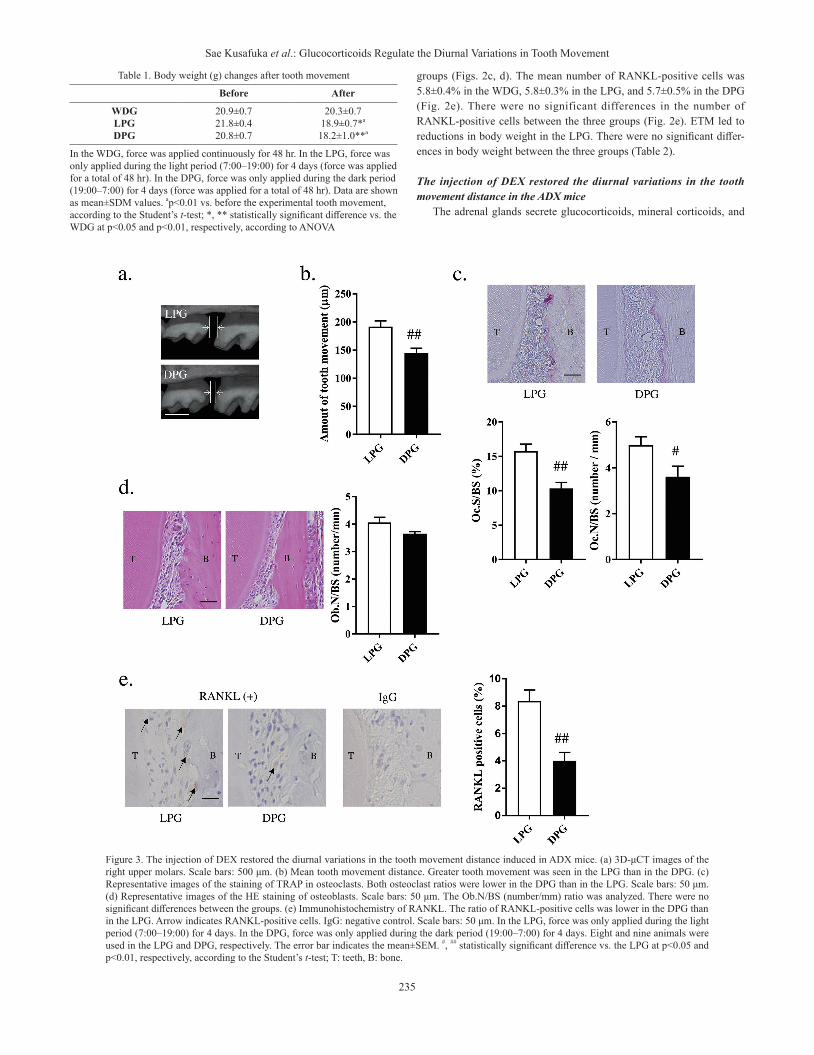
The injection of DEX restored the diurnal variations in the tooth movement distance in the ADX mice
The adrenal glands secrete glucocorticoids, mineral corticoids, and
Table 1. Body weight (g) changes after tooth movement
Before After
WDGLPGDPG
20.9±0.721.8±0.420.8±0.7
20.3±0.718.9±0.7*a
18.2±1.0**a
In the WDG, force was applied continuously for 48 hr. In the LPG, force was only applied during the light period (7:00–19:00) for 4 days (force was applied for a total of 48 hr). In the DPG, force was only applied during the dark period (19:00–7:00) for 4 days (force was applied for a total of 48 hr). Data are shown as mean±SDM values. ap<0.01 vs. before the experimental tooth movement, according to the Student’s t-test; *, ** statistically significant difference vs. the WDG at p<0.05 and p<0.01, respectively, according to ANOVA
Figure 3. The injection of DEX restored the diurnal variations in the tooth movement distance induced in ADX mice. (a) 3D-μCT images of the right upper molars. Scale bars: 500 μm. (b) Mean tooth movement distance. Greater tooth movement was seen in the LPG than in the DPG. (c) Representative images of the staining of TRAP in osteoclasts. Both osteoclast ratios were lower in the DPG than in the LPG. Scale bars: 50 μm. (d) Representative images of the HE staining of osteoblasts. Scale bars: 50 μm. The Ob.N/BS (number/mm) ratio was analyzed. There were no significant differences between the groups. (e) Immunohistochemistry of RANKL. The ratio of RANKL-positive cells was lower in the DPG than in the LPG. Arrow indicates RANKL-positive cells. IgG: negative control. Scale bars: 50 μm. In the LPG, force was only applied during the light period (7:00–19:00) for 4 days. In the DPG, force was only applied during the dark period (19:00–7:00) for 4 days. Eight and nine animals were used in the LPG and DPG, respectively. The error bar indicates the mean±SEM. #, ## statistically significant difference vs. the LPG at p<0.05 and p<0.01, respectively, according to the Student’s t-test; T: teeth, B: bone.

236
J.Hard Tissue Biology Vol. 29(4): 231-238, 2020
sex steroids. Therefore, we examined whether the effects of adrenalecto-my were dependent on glucocorticoids. ADX mice were maintained un-der a 12-hr light/12-hr dark cycle for two weeks, and then the adminis-tration of DEX was started. DEX (3 mg/kg/day) was administered every day at 19:00 during the tooth movement. The tooth movement distance was measured using 3D-μCT (Fig. 3a). The mean tooth movement dis-tance was 191.8±10.3 μm in the LPG and 144.9±8.3 μm in the DPG. The mean tooth movement distance was 24.5% lower in the DPG than in the LPG (Fig. 3b). The mean Oc.S/BS ratio was 15.8±1.0% in the LPG and 10.5±0.8% in the DPG. The mean Oc.N/BS ratio was 5.0±0.4/mm in the LPG and 3.6±0.5/mm in the DPG. In the DPG, the mean Oc.S/BS and Oc.N/BS ratios were 33.5% and 28.0% lower than those seen in the LPG, respectively (Fig. 3c). The mean Ob.N/BS ratio was 4.1±0.2/mm in the LPG and 3.7±0.1/mm in the DPG. There were no significant differences in the Ob.N/BS ratio between the LPG and DPG (Fig. 3d). The mean number of RANKL-positive cells was 8.4±0.7% in the LPG and 4.0±0.6% in the DPG. The mean number of RANKL-posi-tive cells was 52.4% lower in the DPG than in the LPG (Fig. 3e). ETM led to reductions in body weight in the LPG and DPG. There were no significant differences in body weight between the groups (Table 3).
DiscussionThe present study revealed that: 1) There were diurnal variations in
the tooth movement distance, 2) the ablation of the adrenal glands abro-gated the diurnal variations in the tooth movement distance, 3) the ad-ministration of DEX restored the diurnal variations the tooth movement distance and 4) The changes of osteoclast parameters and RANKL im-mune reactivity were consistent with the tooth movement distance. These results indicate that glucocorticoids play a role in the mediation of the signaling responsible for the diurnal variations in the osteoclast activity resulting in the diurnal variation of the tooth movement, and the diurnal variation of RANKL immune reactivity may contribute to it.
In this study, we showed for the first time that diurnal variations ex-ist in the tooth movement distance achieved using the Waldo method. The amount of tooth movement achieved was much larger in the LPG
than in the DPG, which is consistent with the findings of previous stud-ies14, 15). As mice are nocturnal animals, these results suggest that per-forming ETM during the rest period is more effective than performing it in the active period. Although tooth movement-induced weight loss was observed, there were no significant differences in body weight between the LPG and DPG, suggesting that the observed differences in tooth movement were not due to the effects of weight loss. In addition, the mean body weight of the mice after the ETM was lower in the LPG and DPG than in the WDG, which might have been due to the differences in the number of total doses of anesthetic that were administered and the length of the experimental period. Histological examinations also showed that osteoclast parameters, such as the Oc.S/BS and Oc.N/BS ratios, exhibited larger values in the light period than in the dark period, which suggests that tooth movement occurred due to increased osteo-clast activity and/or osteoclastogenesis. Bone resorption and calcium re-lease display diurnal variations and are most active when animals are resting9, 24, 25). It is considered that the diurnal variations in such osteo-clast activity and/or osteoclastogenesis contribute to the diurnal varia-tions in the tooth movement distance.
Miyoshi et al.15) reported that intermittent force moves teeth more ef-fectively than sustained force. In our study, no significant differences in the tooth movement distance between the WDG and the LPG. However, there was significant difference in Oc.S/BS between the WDG and the LPG. This indicates that intermittent force induce Oc.S/BS more effec-tively than sustained force. The partial discrepancies between Mi-yoshi’s15) and our experimental results might have been due to differenc-es in the animal species and orthodontic devices used, but further investigation of these issues is needed. They also reported that on day 7, greater osteogenesis was seen in the LPG than in the DPG, as was found for bone resorption. In our experiments, there were no such differences in osteoblast parameters, but this could be explained by the short experi-mental period (4 days). These results suggest that ETM using the Waldo method might not have any effect on 4 days tooth movement-induced bone formation changes.
Fujihara et al. reported that glucocorticoids mediate signals from the central circadian clock to peripheral bone tissue9). As glucocorticoids are secreted from the adrenal gland, we investigated whether adrenalectomy affected the diurnal variations in the tooth movement distance induced by ETM. As a result, it was found that adrenalectomy eliminated the differences in osteoclast parameters, RANKL immune reactivity and the diurnal variations in the tooth movement distance between the LPG and DPG. As adrenalectomy causes the loss of glucocorticoids, mineral cor-ticoids, and sex steroids, we examined whether the daily administration of a synthetic DEX affected the diurnal variations in the tooth move-ment distance in ADX mice. The administration of DEX restored the differences in osteoclasts parameters, RANKL immune reactivity and diurnal variations in the tooth movement distance came between the LPG and DPG in the ADX mice. These findings suggest that glucocorti-coids transmit central circadian rhythms to peripheral osteoclasts, which results in the diurnal variations in the tooth movement distance induced by ETM. In addition, our data indicates that glucocorticoids also trans-mit central circadian rhythms to RANKL produced cells such as osteo-blasts and osteocytes.
In summary, these results suggest that circulating glucocorticoids contribute to the diurnal variations in the osteoclast parameters which result in the diurnal variation of the tooth movement, and the diurnal variation of RANKL immune reactivity may contribute to it. In this study, it was clarified that an efficient orthodontic tooth movement method that takes diurnal rhythms into consideration and the distur-
Table 3. Body weight (g) changes after tooth movement in dexamethasone (DEX)-injected adrenalectomy (ADX) mice
Before After
LPGDPG
23.6±1.824.0±1.4
20.6±2.0 a
20.5±1.6 a
In the WDG, force was applied continuously for 48 hr. In the LPG, force was only applied during the light period (7:00–19:00) for 4 days (force was applied for a total of 48 hr). In the DPG, force was only applied during the dark period (19:00–7:00) for 4 days (force was applied for a total of 48 hr). Data are shown as mean±SDM values. ap<0.01 vs. before the tooth movement according to the Student’s t-test.
Table 2. Body weight (g) changes after the tooth movement in ADX mice
Before After
WDGLPGDPG
23.9±1.323.9±1.122.1±2.0
22.4±1.721.6±1.1 a
21.4±1.8
In the WDG, force was applied continuously for 48 hr. In the LPG, force was only applied during the light period (7:00–19:00) for 4 days (force was applied for a total of 48 hr). In the DPG, force was only applied during the dark period (19:00–7:00) for 4 days (force was applied for a total of 48 hr). Data are shown as mean±SDM values. None of the parameters analyzed with ANOVA exhibited significant differences. ap<0.01 vs. before the tooth movement, according to the Student’s t-test.

237
Sae Kusafuka et al.: Glucocorticoids Regulate the Diurnal Variations in Tooth Movement
bance of circadian rhythms due to stress and irregular lifestyles could affect orthodontic tooth movement.
AcknowledgementsResearch in the author’s laboratory is partially supported by Grants-
in-Aid for Scientific Research (15K11061 and 19K10416 to Hisataka Kondo) from the Japan Society for the Promotion of Science.
Conflict of InterestThe authors declare no potential conflicts of interest with respect to
the authorship and/or publication of this article.
References1. Suzuki S, Toyabe S, Moroda T, Tada T, Tsukahara A, Iiai T, Mina-
gawa M, Maruyama S, Hatakeyama K, Endoh K and Abo T. Circa-dian rhythm of leucocytes and lymphocytes subsets and its possible correlation with the function of the autonomic nervous system. Clin Exp Immunol 110(3): 500-508, 1997
2. Herzog ED. Neurons and networks in daily rhythms. Nat Rev Neu-rosci 8(10): 790-802, 2007
3. Son GH, Chung S, Choe HK, Kim HD, Baik SM, Lee H, Lee HW, Choi S, Sun W, Kim H, Cho S, Lee KH and Kim K. Adrenal periph-eral clock controls the autonomous circadian rhythm of glucocorti-coid by causing rhythmic steroid production. Proc Natl Acad Sci USA 105(52): 20970-20975, 2008
4. Huang W, Ramsey KM, Marcheva B and Bass J. Circadian rhythms, sleep, and metabolism. J Clin Invest 121(6): 2133-2141, 2011
5. Panda S. Circadian physiology of metabolism. Science 354(6315): 1008-1015, 2016
6. Silver R, LeSauter J, Tresco PA and Lehman MN. A diffusible cou-pling signal from the transplanted suprachiasmatic nucleus con-trolling circadian locomotor rhythms. Nature 382(6594): 810–813,1996
7. Inouye ST and Kawamura H. Persistence of circadian rhythmicity in amammalian hypothalamic “island” containing the suprachias-matic nucleus. Proc Natl Acad Sci U S A 76(11): 5962-5966, 1979
8. Czeisler CA, Kronauer RE, Allan, JS, Duffy JF, Jewett ME, Brown EN and Ronda JM. Bright light induction of strong (type 0) reset-ting of the human circadian pacemaker. Science 244(4910): 1328–1333, 1989
9. Fujihara Y, Kondo H, Noguchi T and Togari A. Glucocorticoids mediate circadian timing in peripheral osteoclasts resulting in the circadian expression rhythm of osteoclast-related genes. Bone 61: 1-9, 2014
10. Takarada T, Xu C, Ochi H, Nakazato R, Yamada D, Nakamura S, Kodama A, Shimba S, Mieda M, Fukasawa K, Ozaki K, Iezaki T, Fujikawa K, Yoneda Y, Numaro R, Hida A, Tei H, Takeda S and Hi-noi E. Bone resorption is regulated by circadian clock in osteoblasts. J Bone Miner Res 32(4): 872-881, 2017
11. Kuo T, McQueen A, Chen TC and Wang JC. Regulation of Glucose Homeostasis by Glucocorticoids. Adv Exp Med Biol 872: 99-126,
201512. Kleeman CR, Levi J and Better O. Kidney and adrenocortical hor-
mones. Nephron 15(3-5): 261-278, 197513. Xiaoyun Ma, Zhu Mengjiao, Mi Xiaohui and Chen Fengshan. Role
of FGF23 c.35C>A in Bone Remodeling during Orthodontic Tooth Movement. J Hard Tissue Biol 29(2): 55-62, 2020
14. Igarashi K, Miyoshi K, Shinoda H, Saeki S and Mitani H. Diurnal variation in tooth movement in response to orthodontic force in rats. Am J Orthod Dentofacial Orthop 114(1): 8-14, 1998
15. Miyoshi K, Igarashi K, Saeki S, Shinoda H and Mitani H. Tooth movement and changes in periodontal tissue in response to ortho-dontic force in rats vary depending on the time of day force is ap-plied. Eur J Orthod 23: 329-338, 2001
16. Lacey DL, Timms E, Tan HL, Kelley MJ, Dunstan CR, Burgess T, Elliott R, Colombero A, Elliott G, Scully S, Hsu H, Sullivan J, Hawkins N, Davy E, Capparelli C, Eli A, Qian YX, Kaufman S, Sa-rosi I, Shalhoub V, Senaldi G, Guo J, Delaney J and Boyle WJ. Os-teoprotegerin ligand is a cytokine that regulates osteoclast differen-tiation and activation. Cell 93(2): 165-176, 1998
17. Shoji-Matsunaga A, Ono T, Hayashi M, Takayanagi H, Moriyama K and Nakashima T. Osteocyte regulation of orthodontic force-me-diated tooth movement via RANKL expression. Sci Rep 7(1): 8753, 2017
18. Yamaguchi M. RANK/RANKL/OPG during orthodontic tooth movement. Orthod Craniofac Res 12(2): 113-119, 2009
19. Kondo M, Kondo H, Miyazawa K, Goto S and Togari A. Experi-mental tooth movement-induced osteoclast activation is regulated by sympathetic signaling. Bone 52(1): 39-47, 2013
20. Hayashi K, Kondo H, Kusafuka S, Tanaka K, Hamamura K, Hirai T, Kodama D, Sato T, Miyazawa K, Goto S and Togari A. Alpha 1B-adrenergic receptor signaling contributes to experimental tooth movement. Aichi-Gakuin Dental Science 31: 31-41, 2018
21. Waldo CM and Rothblatt JM. Histologic response to tooth move-ment in the laboratory rat; procedure and preliminary observations. J Dent Res 33(4): 481-486, 1954
22. Mizuno M, Miyazawa K, Tabuchi M, Tanaka M, Yoshizako M, Mi-namoto C, Torii Y, Tamaoka Y, Kawatani M, Osada H, Maeda H and Goto S. Reveromycin A administration prevents alveolar bone loss in osteoprotegerin knockout mice with periodontal disease. Sci Rep 5: 16510, 2015
23. Parfitt AM, Drezner MK, Glorieux FH, Kanis JA, Malluche H, Me-unier PJ, Ott SM and Recker RR. Bone histomorphometry: Stand-ardization of nomenclature, symbols, and units. Report of the asbmr histomorphometry nomenclature committee. J Bone Miner Res 2(6): 595-610, 1987
24. Shao P, Ohtsuka-Isoya M and Shinoda H. Circadian rhythms in se-rum bone markers and their relation to the effect of etidronate in rats. Chronobiol Int 20: 325–336, 2003
25. Shinoda H and Stern PH. Diurnal rhythms in Ca transfer into bone, Ca release from bone, and bone resorbing activity in serum of rats. Am J Physiol 262(2 Pt 2): 235-240, 1992

238
J.Hard Tissue Biology Vol. 29(4): 231-238, 2020


![CODEN [USA]: IAJPBB ISSN: 2349-7750 INNDDOO AAMMEER](https://img.pdfslide.tips/doc/110x75/619847d4ba8d4e373f7d46ec/coden-usa-iajpbb-issn-2349-7750-innddoo-aammeer-.jpg)
![CODEN [USA]: IAJPBB ISSN: 2349-7750 INDO …](https://img.pdfslide.tips/doc/110x75/61b330c931d7e92b471d12a3/coden-usa-iajpbb-issn-2349-7750-indo-.jpg)