Embed Size (px)
Citation preview
pubs.acs.org/Biochemistry Published on Web 04/06/2010 r 2010 American Chemical Society
4060 Biochemistry 2010, 49, 4060–4067
DOI: 10.1021/bi100181a
Cooperation between Subunits Is Essential for High-Affinity Bindingof N-Acetyl-D-hexosamines to Dimeric Soluble and Dimeric Cellular
Forms of Human CD69†
Daniel Kavan,‡,§,^ Monika Kubı́�ckov�a, ),^ Jan Bı́l�y,‡ Ond�rej Van�ek,‡,§ Kate�rina Hofbauerov�a,§ Hynek Mr�azek,‡,§
Daniel Rozbesk�y,‡,§ Pavla Bojarov�a,‡,§ Vladimı́r K�ren,§ Luk�a�s �Zı́dek, ) Vladimı́r Sklen�a�r, ) and Karel Bezou�ska*,‡,§
‡Department of Biochemistry, Faculty of Science, Charles University, 12840 Prague, Czech Republic, §Institute ofMicrobiology v.v.i.,Academy of Sciences of Czech Republic, 14220 Prague, Czech Republic, and )National Centre for Biomolecular Research, Faculty of
Science, Masaryk University, 61137 Brno, Czech Republic ^These authors contributed equally to the experiments
Received July 21, 2009; Revised Manuscript Received April 6, 2010
ABSTRACT: CD69 is an earliest lymphocyte activation antigen and a universal leukocyte triggering moleculeexpressed at sites of active immune response. The binding ofGlcNAc to the dimeric humanCD69was followedby equilibrium dialysis, fluorescence titration, and NMR. Clear cooperation was observed in the high-affinitybinding (Kd=4.0� 10-7M) of the carbohydrate to two subunits of the dimeric CD69 (Hill coefficient 1.94). Acontrol monosaccharideManNAc was not bound by human CD69, and both monosaccharides had no effectson the structure of the receptor. However, a monomeric CD69 obtained by mutating Q93 and R134 at thedimer interface exhibited a much lower affinity for GlcNAc (Kd=1.3� 10-5M) and no cooperativity (Hill co-efficient 1.07). Perturbation of the dimer interface resulted in a severe impairment of the signaling ability ofcellular CD69 when cross-linked with an antibody or with a bivalent high-affinityN-acetylhexosamine dimer-based ligand. The availability of stable preparations of soluble CD69 receptor with well-documented ligandbinding properties will be beneficial for immunological experiments evaluating the role of this antigen in thecomplex environment of the immune system.Moreover, such preparations in combinationwith efficient ligandmimetics able to both activate CD69þ lymphocytes and to block undesired hyperactivation caused by othercellular ligands will also become indispensable tools in explaining the exact role of the CD69 antigen in theinteraction between the tumor cell and the effector natural killer lymphocyte.
CD69 is an early lymphocyte activationmarker and a universalleukocyte triggering molecule expressed at sites of active immuneresponse and chronic inflammation (1, 2). Initial in vitro studiessuggested that CD69 may function as an activating molecule inmany leukocyte subsets including γ/δ T-cells and natural killer(NK)1 cells (3, 4). The CD69 gene is located within the NK genecomplex on human chromosome 12. It codes a type II calcium-dependentmembrane lectin, amember of one important family ofabundant NK cell surface receptors (5, 6) participating in theformation of the receptor “zipper” at the NK cell-tumor cellinterface (7). Most receptors of NK cells that recognize targetstructures at the surface of tumor or virally infected cells mediatetheir activation or inhibitory effects through their sequentiallydiverse cytoplasmic domains (8). However, the cytoplasmicdomain of CD69 is short and lacks the prominent function-associated peptide motifs. Previous studies have shown that thedownstream activation processes initiated by CD69 engagement
occur through Src-dependent activation of Syk, activation ofphospholipase Cγ2 and Vav, and the subsequent transmission ofsignals through the Rac-ERK pathway (9, 10). Alternatively,CD69 can also propagate activation signals through heterotri-meric G proteins and the subsequent intracellular signalingpathways coupled to these molecular switches (11, 12). Recently,in vivo studies in CD69-deficient mice have added yet anotherdimension into the biology of this receptor revealing its non-redundant role in downregulation of the immune responsethrough the production of the pleiotropic cytokine TGF-β andthrough interactions with regulatory T-cells (2). Using the sameexperimental model, it has been shown recently that CD69 formsa complex with sphingosine 1-phosphate receptor, negativelyregulates its function, and thus inhibits lymphocyte egress fromlymphoid organs downstream of interferon-R/β, known media-tors of transient egress shut down (13). Furthermore, results ofmany immunological studies indicate that CD69may be involvedin pathogenesis of several diseases including rheumatoid arthritis,chronic inflammatory liver diseases, mild asthma, and acquiredimmunodeficiency syndrome (14).
The identification of the natural ligand for CD69 is a keycritical step for further advancement of our knowledge on thebiology of this receptor. The initial findings that CD69 binds tocalcium and certain N-acetyl-D-hexosamines (15) could not belater reproduced using a somewhat different expression con-struct (16). Since then, these discrepancies have been at leastpartially explained by careful structural evaluations of therecombinant proteins used for binding studies, as well as by
†Supported by grants from Ministry of Education of Czech Re-public (MSM_21620808, 1M0505, and AVOZ50200510 to K.B. andMSM0021622413 and LC0603 to V.S.) and from Czech Science Foun-dation (303/09/0477 and 305/09/H008 to K.B. and 203/09/P024 to P.B.)and by EU Project Spine 2 (Contract LSHG-CT-2006-02/220 to K.B.).*To whom correspondence should be addressed at Charles University.
Phone: þ420-2-2195-1272. Fax: þ420-2-2195-1283. E-mail: [email protected].
1Abbreviations:DSS, disucinimidyl suberate;Gal, D-galactose;GlcNAc,N-acetyl-D-glucosamine; ManNAc, N-acetyl-D-mannosamine; MES buf-fer, 10 mMMES with 150 mMNaCl and 1 mMNaN3; MESþ C buffer,10mMMES, 150mMNaCl, 1 mMCaCl2, and 1mMNaN3; NK, naturalkiller.
This paper was retracted on April 15, 2014 (Biochemistry 2014, DOI: 10.1021/bi500367g).
Retrac
ted

Article Biochemistry, Vol. 49, No. 19, 2010 4061
establishing a direct link between the binding of calcium andcarbohydrates (17). The proper folding of CD69 produced basedon the recently suggested constructs encompassing G70-K199of the entire receptor was established using detailed structur-al experiments based on both NMR (18) and protein crystal-lography (19). The most recent development of efficient structur-al mimetics of the high-affinity ligand for CD69 opened theway for manipulating with numerous activities of CD69 at themolecular and cellular level (20, 21) and provided efficientcompounds for further in vivo testing of their immunomodulatingproperties (22-24).
Here we report new findings indicating that the binding ofN-acetyl-D-hexosamines to soluble CD69 is highly cooperative atmolecular level, and this cooperativity is not seen for Q93A andR134Amutantswith disturbed formation of noncovalent dimers.Similarly at the cellular level, efficient signaling after CD69 cross-linking by antibody or bivalent ligand is diminished for the abovemutants with a damaged subunit cross-talk more dramaticallythan for CD69 bearing C68A mutation, and thus lacking thedisulfide bridge forming the covalent dimer identified previouslyas the critical signaling element.
EXPERIMENTAL PROCEDURES
Materials.All chemicals were analytical grade reagents of thebest quality available commercially and were obtained fromSigma unless indicated otherwise. The preparation of dimericN-acetylhexosamine disaccharide with the chemical composi-tion (GalNAcβ1-4GlcNAcβ-NH-CS-NH-CH2-)2 by a combi-nation of chemical and enzymatic steps has been describedpreviously (25).Preparation of Soluble Dimeric CD69. Preparation of
soluble dimeric CD69 using protocol II has been describedpreviously (18). For the preparation of a uniformly 15N-labeledform of the receptor, a producing culture of Escherichia coli BL-21 Gold (Stratagene) harboring the expression plasmid was used,grown on a standard M9 minimal medium containing 15NH4Cl.Identification of Key Amino Acid Residues Disrupting
the Receptor Dimer Interface.We examined the three-dimen-sional structure of the crystallized soluble dimeric CD69 depos-ited into the RCSB Protein Databank under accession code3CCK (18). Glutamine Q93 appeared to be involved in two keyhydrogen bonds between the amide group of its side chain andtwo adjacent acidic residues belonging to the opposite subunit,Asp88 and Glu87. With R134, the intertwining with the secondsubunit is even more profound, and the guanidyl group of thisamino acid forms three hydrogen bonds with A136 and Y135 ofthe opposing subunit and is also involved in the stackinginteraction with the phenyl ring of Y135.Site-Directed Mutagenesis and Expression of the Mu-
tated CD69 Proteins. Mutated forms of CD69 in which theQ93 and/or and R135 were mutated into A were produced usingthe CD69 expression plasmid in pRSETB (18). Mutations wereintroduced using the QuickChange site-directed mutagenesis kit(Stratagene) in combination with the following oligonucleotidepairs: CD69Q93F, 50-GAGGACTGGGTTGGCTACGCGAG-GAAATGCTACTTTATT-30, and CD69Q93R, 50-AATAAA-GTAGCATTTCCTCGCGTAGCCAACCCAGTCCTC-30, andCD69R134F, 50-GACATGAACTTTCTAAAAGCATACGC-AGGTAGAGAGGAA-30 and CD69R134R, 50-TTCCTCTCT-ACCTGCGTATGCTTTTAGAAAGTTCATGTC. The intro-duced mutations were verified by DNA sequencing using an
ABI Prism 3130 genetic analyzer (Applied Biosystems). TheCD69Q93A/R134A double mutant was prepared sequentially,applying the R134A mutation process on the Q93A mutant.Mutated CD69 proteins were prepared using the same protocol(protocol II) used for the production of thewild-type protein (18).Moreover, the proper refolding of the protein was verified usingNMRmeasurementwith the homogenously 15N-labeled proteinsas described previously (18).Gel Filtration.Gel filtration was performed using a Superdex
200 HR 10/30 column (GE Healthcare) connected to the pro-tein purification system BioSys510 (Beckman Coulter) andequilibrated with MES buffer at room temperature. In order toexamine the effect of monosaccharide binding to CD69 on thehydrodynamic volume of the CD69 protein, the protein sampleswere incubated overnight at 4 �C in the presence of 1 mMManNAc or 1 mM GlcNAc and then injected onto the gelfiltration column equilibrated in MES þ C buffer containing1 mM concentrations of the respective monosaccharides.AnalyticalUltracentrifugation. Sedimentation velocity and
sedimentation equilibrium experiments were performed using aProteomeLabXL-I analytical ultracentrifuge (BeckmanCoulter)equipped with an An50Ti rotor and dual absorbance and laserinterference optics. Before the experiment, 0.5 mL samples ofCD69 proteins diluted to 0.4 mg 3mL-1 were dialyzed for 20 hagainst 2 L of MES þ C buffer, with or without the mono-saccharides, in concentration indicated in the text, and thedialysis buffer was used as a reference and sample dilution buffer.The sedimentation velocity experiment was conducted at 48000rpm for dimeric CD69 at 20 �C. Data were analyzed with theprogram SEDFIT (26, 27). Based on buffer composition andamino acid sequence using the program SEDNTERP (www.jphilo.mailway.com), buffer density and CD69 partial specificvolume for CD69NG70 were estimated as 1.00309 g 3mL-1 and0.7183 mL 3 g
-1, respectively.Protein Stability Experiments. CD69 proteins were diluted
to 0.5 mg/mL, and UV spectra were taken in the 200-300 nmrange in a Beckman DU-70 spectrophotometer (BeckmanCoulter) equipped with a heated cuvette. The initial UV scanwas taken at 25 �C, after which the temperature in the cuvettewasincreased in 5 �C increments. Experiments were performedroutinely in MES þ C buffer. Alternatively, protein stabilitywas verified using differential scanning calorimetry and FTIRspectroscopy as has been previously described (17, 18, 28).NMRTitrations.AllNMRexperimentswere run at 300K in
a Bruker Avance 600 MHz spectrometer equipped with acryogenic H/C/N TCI probehead. 1H-15N HSQC spectra of0.3 mM 15N-labeled wild-type CD69 protein CD69NG70 (18)were used as a routine check of protein folding and stability. Thesample buffer consisted of 10 mM MES, pH 5.8, with 49 mMNaCl, 1 mM NaN3, and 10% D2O. During NMR titration, a0.1 mM solution of the unlabeled wild-type CD69 protein (7) wastitrated. In an initial experiment, aliquots of the GlcNAc ligandcorresponding to 25%, 50%, 75%, 100%, 200%, and 500% ofsaturation were added, and signals of the free GlcNAc ligandwere observed at 2.2 ppm in the 1D proton spectra and used forthe estimation of the free ligand concentration. In a separateexperiment aimed at estimating the binding constant, smallerligand additions were used as the equivalence was appoached.The protein was titrated to 75% of the estimated number ofbinding sites, after which the amount of ligand was increased inincrements of 5% of the estimated number of binding sites untilthe equivalence point was reached. All spectra were processed
Retrac
ted
4062 Biochemistry, Vol. 49, No. 19, 2010 Kavan et al.
using the software NMRPIPE (29). The dissociation constantKd, defined asKd= (cp- cLþ [L])[L]/(cL- [L]), was obtained bya nonlinear fitting of the [L] vs cL titration curves (Figure 2A,B).Equilibrium Dialysis. N-Acetyl-D-[1-3H]glucosamine (speci-
fic activity 500 GBq/mmol) and N-acetyl-D-[1-3H]mannosamine(specific activity 650 GBq/mmol) were prepared as describedpreviously (17) or purchased from Amersham. To set up equi-librium dialysis experiments, a rotating apparatus with glassblocks containing separate sealable chambers with externalaccess was used as described previously (17). Aliquots (200 μL)of 0.1 μM solutions of CD69 proteins in MES þ C buffer wereincubated with varying amounts of ligand at 27 �C (300 K) for48 h. After equilibration, 100 μL aliquots were withdrawn fromthe control and from the protein-containing chambers. Theresults were calculated and plotted according to Scatchard asdescribed previously (17).Tryptophan Fluorescence Quenching. Tryptophan fluore-
scence quenching experiments were performed according to thedescribed methodology (30) with minor modifications. In initialexperiments, 100 nmol aliquots of CD69 protein were pipettedinto multiple wells of a UV Star plate (Greiner, Germany) andmixed with 10-fold serial dilutions of the GlcNAc ligand.Incubation proceeded for 1 h at 27 �C (300K) in the thermostatedchamber of a Safire2 plate reader (Tecan,Austria), afterwhich thefluorescence of tryptophan residues was measured in duplicatewells using the bottom fluorescence measurements and thefollowing settings: λex = 275 nm, λem = 350 nm, excitation andemission slits were set to 5 and 20, respectively, and the fluore-scence gain was manually set to 66. After finding the lowestconcentration of ligand that still caused the quenching of trypto-phan fluorescence, detailed dilutions of the ligand by 10%saturation steps were performed, and the concentration of freeand bound ligand was calculated as described previously (17, 30).Preparation of the Eukaryotic Expression Constructs,
Transfection into Jurkat Cells, and Selection of the Trans-fectants. In order to mutate the dimerization cystein C68 (15) toA, site-directed mutagenesis was performed using the originalexpression plasmid (15) as described above using oligonucleotideprimers CD69C68F, 50-TCAGTGGGCCAATACAATGCTC-CAGGCCAATACACATTC-30, and CD69C68R, 50-GAATG-TGTATTGGCCTGGAGCATTGTATTGGCCCACTGA-30,and the pCDA401 plasmid (15). Single mutation CD69C68A,double mutations CD69C68A/Q93A and CD69C68A/R134A,and the triple mutation CD69C68A/Q93A/R134A were pre-pared by applying the mutagenesis protocol onto expressionplasmids for wild-type CD69 (8) and for the respective dimeriza-tion mutants described above. After the mutagenesis and DNAsequencing, DNA fragments coding the C-terminal extracellularsegments of CD69 were linked with the DNA fragment codingthe N-terminal part of the receptor (31) using linking PCR (8).The X construct corresponded to the religated pCR3 (mock) andwas used as a control (8). The eukaryotic expression vectors weresequenced and transfected into a Jurkat T lymphoblastoid cellline maintained in RPMI1640 and supplemented with 10% fetalcalf serum (8).Precipitation of Cellular Forms of CD69 Using Anti-
bodies and Dimeric N-Acetylhexosamines. Transfected Jur-kat cells (1 � 106) were surface radioiodinated using lactoper-oxidase (31), washed three times with medium, and then incu-bated with 1 mM concentrations of dimeric N-acetylhexosaminedisaccharides for 1 h at room temperature. The incubation wasfollowed by the addition of 100 mM DSS and by cross-linking
of the receptors for another 1 h at 4 �C. Thereafter, cells werelysed, and CD69 receptor complexes were immunoprecipitated
using G protein beads (GE Healthcare) coated with monoclonal
antibodies against CD69, BL-KFB/B1 (31). Beads were washed
extensively, boiled in sample buffer for SDS-PAGE, and
analyzed using 15% SDS-polyacrylamide gels followed by
autoradiography.Cellular Activation Assays and Production of IL-2.
Transfected Jurkat cells (106) were incubated with dimericN-acetylhexosamine disaccharides as described in the precedingsection or with saturating concentrations of monoclonal anti-bodies against CD69 for 5 min (cellular activation) or 12 h(IL-2 production) and used to determine the free cytoplasmiccalcium (11) or IL-2 production (8).
RESULTS AND DISCUSSION
Evaluation of Calcium and Carbohydrate Binding Activ-ity of Highly Stable CD69 Proteins. We and others havepreviously generated several constructs optimized for the pre-paration of highly stable soluble recombinant CD69 proteinssuitable for ligand identification experiments (Supporting Infor-mation Table S1). Preliminary ligand binding experiments wereperformed to evaluate the ability of these constructs to bindcalcium andmonosaccharides shown to be important ligands forthe receptor (15, 17).With regard to the binding of calcium, therehas been no difference in the ability to bind calcium between thecovalent dimeric protein CD69CQ65 and noncovalent dimericproteins CD69NG70 and CD69NV82 when compared to themonomeric protein CD69MS100: each of these proteins bound1 mol of calcium/mol of CD69 subunit withKd of approximately58 μM (ref 17 and Supporting Information Figure S1). On theother hand, significant differences between these protein con-structs were observed with regard to the binding of N-acetyl-D-hexosamines. While the IC50 values for the soluble monomericCD69, CD69MS100, with regard to binding of the two activeN-acetyl-D-hexosamines, D-GlcNAc and D-GalNAc, were eachapproximately 10-5 M, these values were about 10 times lowerfor the dimeric proteinCD69NV82 and about 100 times lower forthe other two highly stable dimeric proteins, CD69CQ65 andCD69NG70 (Supporting Information Figure S2). The latterprotein has been selected for all of the subsequent binding experi-ments and will be referred to as soluble dimeric CD69. Thehomogeneity and monodispersity were routinely evaluated foreach batch of the produced soluble dimeric CD69 using SDSelectrophoresis under both reducing and nonreducing conditionsand gel filtration on a Superdex 200 HR column (ref 18 andFigure 1). Moreover, the identity, quality, and proper refoldingof each batch of the produced protein were also verified asdescribed previously (18) using high-resolution ion cyclotronresonance mass spectrometry, one-dimensional proton NMR,thermal stability experiments, and tests of the biochemicalstability (ref 18 and Table 1).Cooperativity of GlcNAc Binding Proved by Direct
BindingExperiments.The detailed binding studies with solubledimeric CD69 were performed using D-GlcNAc as the high-affinity carbohydrate ligand, together with D-ManNAc and, insome experiments, D-Gal as negative controls. The initial evi-dence for the interaction of the soluble dimeric CD69 withGlcNAc was obtained by NMR titration. A 0.1 mM solutionof the dimeric receptor was titrated up to equivalence assumingthe existence of two high-affinity binding sites per receptor
Retrac
ted
Article Biochemistry, Vol. 49, No. 19, 2010 4063
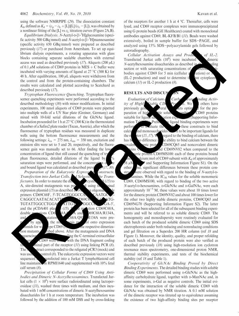
dimer (17). The results of this experiment (Figure 2B andSupporting Information Figure S3B) confirmed the specificbinding of GlcNAc to the receptor (2 mol of GlcNAc boundto a receptor dimer) and provided an affinity estimation in thelow micromolar range (Kd = 4.0� 10-7 M). On the other hand,no interaction could be seen with the ManNAc negative controlunder the same experimental conditions (Figure 2A and Support-ing Information Figure S3A). However, NMRdid not enable thefraction of the bound ligand to be measured.
In order to confirm the results obtained by NMR titration,additional direct binding experiments were performed. When thebound and the unbound ligands had been separated by dialysisunder equilibrium, two binding sites per receptor dimer weredetected. Direct binding experiments enabled the degree ofsaturation at each particular ligand concentration to be calcu-lated. The resulting saturation curve, showing the saturation(fraction bound normalized per receptor subunit) dependence onthe ligand concentration (Figure 2C), clearly revealed a strikingcooperativity in the highly specific (Kd = 4.0� 10-7 M) binding
of GlcNAc to the receptor with the Hill coefficient approachingthe maximum theoretical value (theory 2.00, experiment 1.95; seeFigure 2C). These results were also independently confirmed bythe third binding assay, the fluorescent titration, which gavebinding parameters essentially identical to those obtained by theequilibrium dialysis (Figure 2D). On the other hand, very littlespecific binding for both ManNAc and Gal control monosac-charides could be seen in both of the latter assays (Figure 2C,D).Binding of N-Acetyl-D-hexosamines Did Not Result in
Significant Conformational Change in CD69 Protein. Toanalyze the structural changes of soluble CD69 upon ligandbinding, variations in the hydrodynamic properties of thereceptor were investigated. The molecular size of the receptor,which had been saturated with an excess of GlcNAc, was studiedby gel filtration on Superdex 200 HR and by analytical ultra-centrifugation and compared with the size of the receptorpreincubated in the ManNAc control. The elution time decreasefrom 36.5 to 31.7min in the gel filtration would indicate a changein the molecular size of CD69 upon GlcNAc binding whencompared to the presence of ManNAc (Supporting InformationFigure S4). However, the detailed analysis of soluble dimericCD69 in the absence of any ligand, in the presence of 1 mMManNAc, and in the presence of 1 mM GlcNAc did not revealany changes in hydrodynamic properties since the value of theexperimentally determined sedimentation coefficient was identi-cal (Supporting Information Figure S5).Binding of N-Acetyl-D-glucosamine to the Stable Mono-
mericCD69Follows a Single SiteModel and Proceeds withMuch Lower Affinity. In the next step, the interactions ofGlcNAc with the monomeric subunit of CD69 were studied.Since it proved extremely difficult to prepare the monomericform of the receptor by dissociation of the CD69 dimer (18), weused the available crystal structure of the CD69 dimer andanalyzed the dimer interface for critical residues participatingin the dimerization. Two such critical residues, namely Q93 andR134, both interacting with two residues of the other sub-unit, could be identified (Supporting Information Figure S6).
FIGURE 1: Analysis of wild-type and mutant CD69 proteins bySDS-PAGE and gel filtration. In (A) and (B), these proteins wereanalyzed under reducing and nonreducing conditions, respectively.Analyzed proteins, from left to right, were wild-type CD69,CD69Q93Amutant, CD69R134Amutant, and CD69QRDM.Mar-ker proteins were BSA (65 kDa), ovalbumin (44 kDa), lactoglobulin(18 kDa), lysozyme (14 kDa), and aprotinin (6 kDa). In (C) to (F),these proteins were analyzed by gel filtration on a Superdex 200 HRcolumn, and the four respective panels contain chromatograms forwild-type CD69, CD69Q93A mutant, CD69R134A mutant, andCD69QRDM double mutant.
Table 1: Summary of Stability Properties ofWild-TypeDimeric CD69 and
CD69 Dimerization Mutants
protein characteristics Tda (�C) Td
b (�C) Tdc (�C)
CD6CD69WT noncovalent dimers 65 67 65
CD69Q93A dimer/monomer equilibrium 63 62 64
CD69R134A dimer/monomer equilibrium 62 60 60
CD69QRDM monomeric 60 62 61
aDetermined from thermal UV denaturation measurements. bDeter-mined from differential scanning calorimetry. cDetermined from FTIRspectroscopy.
FIGURE 2: Measurements of direct interaction of soluble CD69NG70with ManNAc and GlcNAc. (A, B) NMR titration of soluble CD69with ManNAc and GlcNAc, respectively. (C, D) Concentrationdependence of receptor saturation measured by equilibrium dialysisand tryptophan fluorescence quenching, respectively, using GlcNAc,ManNAc, and Gal as indicated.
Retrac
ted
4064 Biochemistry, Vol. 49, No. 19, 2010 Kavan et al.
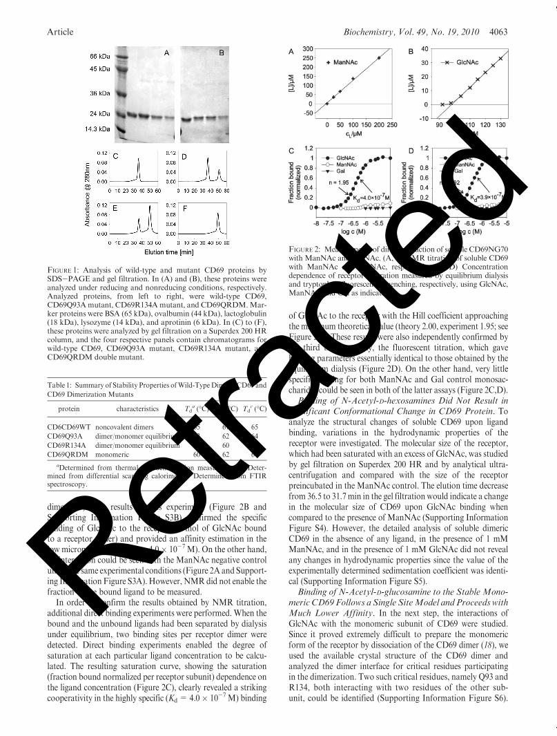
These amino acid residues were mutated to alanine, singly or incombination. All three produced mutated proteins were aftertheir refolding and purification extensively verified using themethodology described previously (18) for the wild-type proteinCD69NG70 using SDS electrophoresis and ion cyclotron reso-nance mass spectrometry of the entire protein, as well as nuclearmagnetic resonance to check the proper folding of these proteins(Figure 1 and results not shown). The stability of the mutantsoluble CD69 proteins was comparable to that of the wild-typeprotein, indicating that the introduced mutations did not resultin any decrease of protein stability. Only the double mutantbehaved as a monomeric protein (Figure 1), with stabilitycomparable to that of the dimer receptor (Table 1). This proteinwas used to analyze the binding of GlcNAc to the monomericprotein. The results clearly showed that binding to the mono-meric subunit of CD69 was much weaker and noncooperative(Hill coefficient of 1.07; Figure 3).Q93/R134/C68 TripleMutation Is Necessary to Disrupt
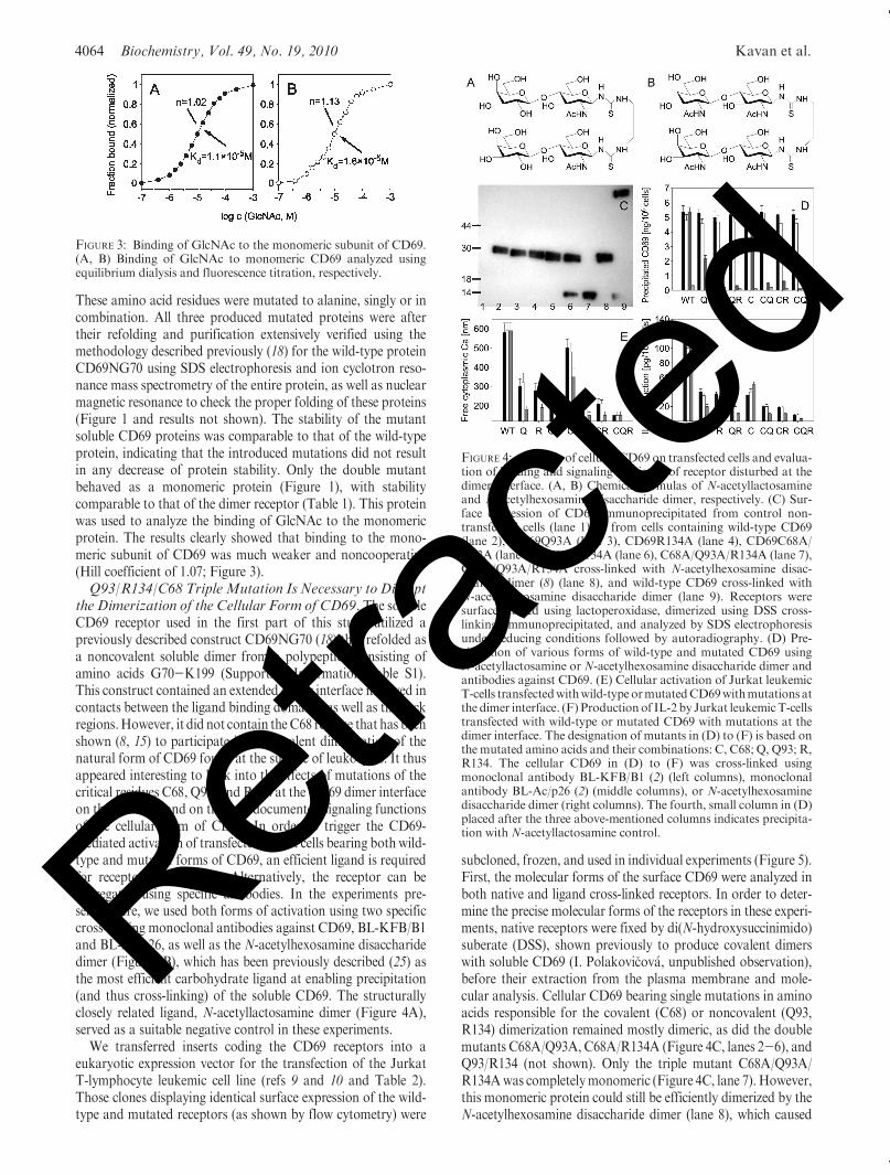
the Dimerization of the Cellular Form of CD69. The solubleCD69 receptor used in the first part of this study utilized apreviously described construct CD69NG70 (18) that refolded asa noncovalent soluble dimer from a polypeptide consisting ofamino acids G70-K199 (Supporting Information Table S1).This construct contained an extended dimer interface involved incontacts between the ligand binding domains, as well as the neckregions.However, it did not contain theC68 residue that has beenshown (8, 15) to participate in the covalent dimerization of thenatural form of CD69 found at the surface of leukocytes. It thusappeared interesting to look into the effects of mutations of thecritical residues C68, Q93, and R134 at the CD69 dimer interfaceon the structure and on the well-documented signaling functionsof the cellular form of CD69. In order to trigger the CD69-mediated activation of transfected Jurkat cells bearing both wild-type and mutated forms of CD69, an efficient ligand is requiredfor receptor cross-linking. Alternatively, the receptor can beaggregated using specific antibodies. In the experiments pre-sented here, we used both forms of activation using two specificcross-linking monoclonal antibodies against CD69, BL-KFB/B1and BL-Ac/p26, as well as the N-acetylhexosamine disaccharidedimer (Figure 4B), which has been previously described (25) asthe most efficient carbohydrate ligand at enabling precipitation(and thus cross-linking) of the soluble CD69. The structurallyclosely related ligand, N-acetyllactosamine dimer (Figure 4A),served as a suitable negative control in these experiments.
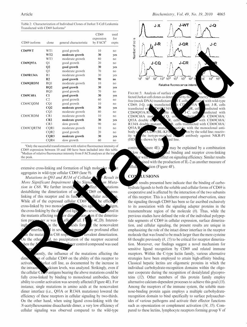
We transferred inserts coding the CD69 receptors into aeukaryotic expression vector for the transfection of the JurkatT-lymphocyte leukemic cell line (refs 9 and 10 and Table 2).Those clones displaying identical surface expression of the wild-type and mutated receptors (as shown by flow cytometry) were
subcloned, frozen, and used in individual experiments (Figure 5).First, the molecular forms of the surface CD69 were analyzed inboth native and ligand cross-linked receptors. In order to deter-mine the precise molecular forms of the receptors in these experi-ments, native receptors were fixed by di(N-hydroxysuccinimido)suberate (DSS), shown previously to produce covalent dimerswith soluble CD69 (I. Polakovi�cov�a, unpublished observation),before their extraction from the plasma membrane and mole-cular analysis. Cellular CD69 bearing single mutations in aminoacids responsible for the covalent (C68) or noncovalent (Q93,R134) dimerization remained mostly dimeric, as did the doublemutants C68A/Q93A, C68A/R134A (Figure 4C, lanes 2-6), andQ93/R134 (not shown). Only the triple mutant C68A/Q93A/R134Awas completelymonomeric (Figure 4C, lane 7). However,this monomeric protein could still be efficiently dimerized by theN-acetylhexosamine disaccharide dimer (lane 8), which caused
FIGURE 3: Binding of GlcNAc to the monomeric subunit of CD69.(A, B) Binding of GlcNAc to monomeric CD69 analyzed usingequilibrium dialysis and fluorescence titration, respectively.
FIGURE 4: Analysis of cellular CD69 on transfected cells and evalua-tion of binding and signaling efficiency of receptor disturbed at thedimer interface. (A, B) Chemical formulas of N-acetyllactosamineand N-acetylhexosamine disaccharide dimer, respectively. (C) Sur-face expression of CD69 immunoprecipitated from control non-transfected cells (lane 1) or from cells containing wild-type CD69(lane 2), CD69Q93A (lane 3), CD69R134A (lane 4), CD69C68A/Q93A (lane 5), C68A/R134A (lane 6), C68A/Q93A/R134A (lane 7),C68A/Q93A/R134A cross-linked with N-acetylhexosamine disac-charide dimer (8) (lane 8), and wild-type CD69 cross-linked withN-acetylhexosamine disaccharide dimer (lane 9). Receptors weresurface-labeled using lactoperoxidase, dimerized using DSS cross-linking, immunoprecipitated, and analyzed by SDS electrophoresisunder reducing conditions followed by autoradiography. (D) Pre-cipitation of various forms of wild-type and mutated CD69 usingN-acetyllactosamine or N-acetylhexosamine disaccharide dimer andantibodies against CD69. (E) Cellular activation of Jurkat leukemicT-cells transfectedwithwild-typeormutatedCD69withmutations atthe dimer interface. (F) Productionof IL-2 by Jurkat leukemicT-cellstransfected with wild-type or mutated CD69 with mutations at thedimer interface. The designation of mutants in (D) to (F) is based onthe mutated amino acids and their combinations: C, C68; Q, Q93; R,R134. The cellular CD69 in (D) to (F) was cross-linked usingmonoclonal antibody BL-KFB/B1 (2) (left columns), monoclonalantibody BL-Ac/p26 (2) (middle columns), or N-acetylhexosaminedisaccharide dimer (right columns). The fourth, small column in (D)placed after the three above-mentioned columns indicates precipita-tion with N-acetyllactosamine control.
Retrac
ted
Article Biochemistry, Vol. 49, No. 19, 2010 4065
extensive cross-linking and formation of high molecular weightaggregates in wild-type cellular CD69 (lane 9).Mutations in Q93 and R134 of Cellular CD69 Result in
More Significant Impairment in Its Signaling than Muta-tion in C68. We further investigated the effect of mutationsdestabilizing the dimerization of cellular CD69 on the cross-linking of this receptor and its function in cellular activation.While all of the expressed cellular CD69 could be efficientlycross-linked by two monoclonal antibodies against this antigen,the cross-linking by the dimerized ligandwas severely impaired inthe mutants affecting receptor dimerization even if the dimeriza-tion process per se was not affected (cf. Figure 4C,D). Interest-ingly, mutations in the amino acids forming the noncovalentdimer interface had in several instances a more profound effectthan the mutation in C68 responsible for covalent dimerization.On the other hand, no precipitation of the receptor occurredwhen theN-acetyllactosamine dimer control compoundwas used(Figure 4D).
Subsequently, the influence of the mutations affecting thedimerization of cellular CD69 on the ability of this receptor toactivate the Jurkat cell line, as documented by the increase inthe intracellular calcium levels, was analyzed. Strikingly, even ifthe cellular CD69 antigens bearing the above mutations could befully cross-linked by binding to monoclonal antibodies, theirability to confer activation was severely affected (Figure 4E). Forinstance, single mutations in amino acids at the noncovalentdimer interface (i.e., Q93A or R134A mutations) lowered theefficiency of these receptors in cellular signaling by two-thirds.On the other hand, when using ligand cross-linking with theN-acetylhexosamine disaccharide dimer, a very low efficiency ofcellular signaling was observed compared to the wild-type
controls (Figure 4E). This may be explained by a combinationof low efficiency of ligand binding and receptor cross-linkingtogether with a direct effect on signaling efficiency. Similar resultswere obtained with the production of IL-2 as another measure ofcellular activation (Figure 4F).
CONCLUSIONS
The results presented here indicate that the binding of carbo-hydrate ligands to both the soluble and cellular forms of CD69 iscooperative and is affected by the interaction of the two subunitsof this receptor. This is a hitherto unreported observation, sincethe signaling through CD69 has been so far ascribed exclusivelyto its association with the signaling adapter proteins in thetransmembrane region of the molecule (8). Thus, while theprevious studies have defined the role of the individual polypep-tide segments of CD69 in cellular expression, surface dimeriza-tion, and cellular signaling, the present results are unique inemphasizing the role of the intact dimer interface in the receptormolecule that was found to bemuch larger than themere cysteine68 thought previously (8, 15) to be critical for receptor dimeriza-tion. Moreover, our findings suggest a novel mechanism forsensitive ligand recognition by CD69 and related immunereceptors. Within the C-type lectin family, various alternativestrategies have been employed to attain high-affinity binding.Classical hepatic lectins are oligomeric proteins in which theindividual carbohydrate-recognition domains within the oligo-mer cooperate during the recognition of desialylated glycopro-teins (32). Other members of this protein family employalternative calcium-dependent processes to achieve this goal (33).Among the receptors of the immune system, the soluble man-nose-binding protein again employs a multiple carbohydrate-recognition domain to bind specifically to surface polysacchar-ides of various pathogens and activate their effector functionssuch as opsonization or complement activation (34, 35). Com-pared to these lectins, lymphocyte receptors forming group V of
Table 2: Characterization of Individual Clones of Jurkat T-Cell Leukemia
Transfected with CD69 Isoformsa
CD69 isoform clone general characteristic
CD69
expression
by FACSb
used
for
expts
CD69WT WT1 good growth 10 no
WT2 moderate growth 30 yes
WT3 moderate growth 80 no
CD69Q93A Q1 good growth 20 no
Q2 good growth 30 yes
Q3 moderate growth 70 no
CD69R134A R1 moderate growth 30 yes
R2 good growth 90 no
CD69QRDM RQ1 moderate growth 10 no
RQ2 good growth 30 yes
RQ3 good growth 70 no
CD69C68A C1 good growth 30 yes
C2 good growth 60 no
CD69CQDM CQ1 good growth 10 no
CQ2 moderate growth 30 yes
CQ3 moderate growth 80 no
CD69CRDM CR1 moderate growth 10 no
CR2 moderate growth 30 yes
CR3 slow growth 90 no
CD69CQRTM CQR1 moderate growth 10 no
CQR2 good growth 20 no
CQR3 moderate growth 30 yes
CQR4 slow growth 60 no
aOnly the successful transformants with relative fluorescence intensity ofCD69 expression between 10 and 100 have been included into this table.bMedian of relative fluorescence intensity from FACS analyses at the top ofthe peak.
FIGURE 5: Analysis of surface expression of cellular CD69 in trans-fected Jurkat cell clones as determined by flow cytometry: J-X, insert-free (mockDNA) transfectant; J-WT, cells transfectedwithwild-typeCD69; J-Q, cells transfected with CD69Q93A mutant; J-R, cellstransfected with CD69R134A mutant; J-QR, cells transfected withCD69Q93A/R134A double mutant; J-C, cells transfected withCD69C68A mutant; J-CQ, cells transfected with CD69C68A/Q93A double mutant; J-CR, cells transfected with CD69C68A/R134A double mutant; J-CQR, cells transfected with CD69C68A/Q93A/R134A triple mutant. Reactivity with the monoclonal anti-body against CD69 (BL-KFB/B1) is shown by the solid line; reactiv-ity with the isotype-matched control antibody against NKR-P1(HD14) is shown by the dotted line.
Retrac
ted
4066 Biochemistry, Vol. 49, No. 19, 2010 Kavan et al.
theC-type lectin family aremuch smaller andmostly dimeric, andthus they seem to have developed alternative strategies for theirrecognition of specific ligands. These are based either on oligo-merization of the receptor within the specialized plasma mem-brane microdomains of the immune cell (36) or on alternativestrategies that may be used by some of these receptors (37). Themolecular mechanism that we propose based on the results of thecurrent work is unique in rapid propagation of the activationsignal using the mechanism of rapid cooperative receptor cross-linking and oligomerization based on the positive cooperativityin binding of the multivalent ligands.
ACKNOWLEDGMENT
We thank Angela Risso for assistance and helpful discussions,Iva Polakovi�ckov�a for help with DSS cross-linking experiments,and Michal Navr�atil for help in the development of cellularreceptor precipitation assays.
SUPPORTING INFORMATION AVAILABLE
A description of additional experimental procedures with sup-porting references, binding of calcium to the fourth generationsoluble CD69 proteins (Figure S1), carbohydrate inhibition experi-ments for these proteins (Figure S2), primary data for NMR titra-tions with ManNAc and GlcNAc (Figure S3), elution profilesfor gel filtration of soluble dimeric CD69 (CD69NG70) in theabsence of ligand as well as in the presence of 1 mMManNAc and1 mM GlcNAc (Figure S4), sedimentation velocity experimentswith CD69NG70 in the absence of ligand and in the presence of1 mM ManNAc and 1 mM GlcNAc (Figure S5), identificationof amino acids for the design of the monomeric CD69 mutant(Figure S6), and characterization of CD69 expression con-structs (Table S1). This material is available free of charge viathe Internet at http://pubs.acs.org.
REFERENCES
1. Testi, R., D’Ambrosio, D., DeMaria, R., and Santoni, A. (1994) TheCD69 receptor: a multipurpose cell surface trigger for hematopoieticcells. Immunol. Today 15, 479–483.
2. Sancho, D., Gom�ez, M., and S�anchez-Madrid, F. (2005) CD69 is animmunoregulatory molecule induced following activation. TrendsImmunol. 26, 136–140.
3. Moretta, A., Poggi, A., Pende, D., Tripodi, G., Orengo, A. M., Pella,N., Augugliaro, R., Bortino, C., Ciccone, E., and Moretta, L. (1991)CD69-mediated pathway of lymphocyte activation: anti-CD69monoclonal antibodies trigger the cytolytic activity of differentlymphoid effector cells with the exception of cytolytic T lymphocytesexpressing T cell receptor R/β. J. Exp. Med. 174, 1393–1398.
4. Borrego, F., Robertson, M. J., Ritz, J., Pena, J., and Solana, R. (1999)CD69 is a stimulatory receptor for natural killer cells and its cytotoxiceffect is blocked byCD94 inhibitory receptor. Immunology 97, 159–165.
5. Lopez-Cabrera, M., Santis, A. G., Fernandez-Ruiz, F., Blacher, R.,Esch, F., Sanchez-M�ateos, P., and S�anchez-Madrid, F. (1993)Molecular cloning, expression, and chromosomal localization of thehuman earlies activation antigen AIM/CD69, a new member ofC-type lectin superfamily of signal-transmitting receptors. J. Exp.Med. 178, 537–547.
6. Lanier, L. L. (2008) Up on the tightrope: natural killer cell activationand inhibition. Nat. Immunol. 9, 495–502.
7. Vivier, E., Tomasello, E., Baratin, M., Walzer, T., and Ugolini, S.(2008) Functions of natural killer cells. Nat. Immunol. 9, 503–510.
8. Sancho,D., Santis, A.G., Alonso-Lebrero, J. L., Viedma, F., Tejedor,R., and S�anchez-Madrid, F. (2000) Functional analysis of ligand-binding and signal transduction domains of CD69 and CD23 C-typelectin leukocyte receptors. J. Immunol. 165, 3868–3875.
9. Pisegna, S., Zignoni, A., Pirozzi, G., Cinque, B., Cifoni, M. G.,Morrone, S., Picolli, M., Frati, L., Palmieri, G., and Santoni, A.(2002) Src-dependent Syk activation controls CD69 mediated signal-ing and function of human NK cells. J. Immunol. 169, 68–74.
10. Zingoni, A., Palmieri, G., Morrone, S., Carretero, M., Lopez-Botet,M., Picolli,M., Frati, L., and Santoni, A. (2000)CD69-triggered ERKactivation and functions are negatively regulated by CD94/NKG2Ainhibitory receptors. Eur. J. Immunol. 30, 644–651.
11. Risso, A., Smilowich, D., Capra, M. C., Baldissarro, I., Yan, G.,Bargilessi, A., and Cosulich, M. E. (1991) CD69 in resting andactivated T lymphocytes. Its association with a GTP binding proteinand biochemical requirements for its expression. J. Immunol. 146,4105–4114.
12. Bikah, G., Pogue-Caley, R. R., and McHeyzer-Williams, M. G.(2000) Regulating T helper cell immunity through antigen respon-siveness and calcium entry. Nat. Immunol. 1, 402–412.
13. Shiow, R. L., Rosen, D. B., Brde�ckov�a, N., Xu, Y., An, J., Langer,L. L., Cyster, J. G., and Matloubian, M. (2006) CD69 acts down-stream of interferon-R/β to inhibit S1P1 and lymphocyte egress fromlymphoid organs. Nature 440, 540–544.
14. Marzio, R., Mauel, J., and Betz-Corradin, S. (1999) CD69 andregulation of the immune functions. Immunopharmacol. Immunotox-icol. 21, 565–582.
15. Bezou�ska, K., Nepovı́m, A., Horv�ath, O., Pospı́�sil, M., Hamann, J.,and Feizi, T. (1995) CD69 antigen of human leukocyte is a calcium-dependent carbohydrate-binding protein. Biochem. Biophys. Res.Commun. 208, 68–74.
16. Childs, R. A.,Galustian, C., Lawson,A.M.,Douf�an,G., Benwell, K.,Frankel, G., and Feizi, T. (2000) Recombinant soluble human CD69dimer produced in Escherichia coli: reevaluation of saccharide bind-ing. Biochem. Biophys. Res. Commun. 266, 19–23.
17. Pavlı́�cek, J., Sopko, B., Ettrich, R., Kopeck�y, V., Baumruk, V., Man,P., Havlı́�cek, V., Vrback�y,M.,Martı́nkov�a, L., K�ren, V., Pospı́�sil,M.,and Bezou�ska, K. (2003) Molecular characterization of binding ofcalcium and carbohydrates by an early activation antigen of lympho-cytes CD69. Biochemistry 42, 9295–9306.
18. Van�ek, O., N�alezkov�a,M., Kavan, D., Borovi�ckov�a, I., Pompach, P.,Nov�ak, P., Kumar, V., Vannucci, L., Hude�cek, J., Hofbauerov�a, K.,Kopeck�y, V., Brynda, J., Kolenko, P., Dohn�alek, J., Kade�r�avek, P.,Chmelı́k, J., Gor�cı́k, L., �Zı́dek, L., Sklen�a�r, V., and Bezou�ska, K.(2008) Soluble recombinant CD69 receptors optimized to havean exceptional physical and chemical stability display prolongedcirculation and remain intact in the blood of mice. FEBS J. 275,5589–5606.
19. Kolenko, P., Sk�alov�a, T., Van�ek, O., �St�ep�ankov�a, A., Du�skov�a, J.,Ha�sek, J., Bezou�ska, K., and Dohn�alek, J. (2009) The high-resolutionstructure of the extracellular domain of human CD69 using a novelpolymer. Acta Crystallogr. F65, 1258–1260.
20. Pavlı́�cek, J., Kavan, D., Pompach, P., Nov�ak, P., Luk�san, O., andBezou�ska, K. (2004) Lymphocyte activation receptors: new structuralparadigma in group V of C-type animal lectins. Biochem. Soc. Trans.32, 1124–1126.
21. Bezou�ska, K., �Snajdrov�a, R., K�renek, K., Van�curov�a,M., K�adek, A.,Ad�amek, D., Lhot�ak, P., Kavan, D., Hofbauerov�a, K., Man, P.,Bojarov�a, P., and K�ren, V. (2010) Carboxylated calixarenes bindstrongly to CD69 and protect CD69þ killer cells from suicidal celldeath induced by tumor cell surface ligands. Bioorg. Med. Chem. 18,1434–1440.
22. K�renek, K.,Kuldov�a,M., Hulı́kov�a,K., Stibor, I., Lhot�ak, P., Dudi�c,M., Budka, J., Pelantov�a, H., Bezou�ska, K., Fi�serov�a, A., and K�ren,V. (2007)N-acetyl-D-glucosamine substituted calix[4]arenes as stimu-lators of NK cell-mediated antitumor immune response. Carbohydr.Res. 342, 1781–1792.
23. Hulı́kov�a, K., Benson, V., Svoboda, J., �Sı́ma, P., and Fi�serov�a, A.(2009) N-Acetyl-D-glucosamine-coated polyamidoamine dendrimermodulates antibody formation via natural killer cell activation. Int.Immunopharmacol. 9, 792–799.
24. Benson, V., Grob�arov�a, V., Richter, J., and Fi�serov�a, A. (2010)Glycosylation regulates NK cell-mediated effector function throughPI3K pathway. Int. Immunol. 22, 167–177.
25. Bojarov�a, P., K�renek, K., Wetjen, K., Adamiak, K., Pelantov�a, H.,Bezou�ska, K., Elling, L., and K�ren, V. (2009) Synthesis of LacdiNAc-terminated glycoconjugates by mutant galactosyltransferase;A wayto new glycodrugs and materials. Glycobiology 19, 509–517.
26. Schuck, P. (2000) Size distribution analysis of macromolecules bysedimentation velocity ultracentrifugation and Lamm equation mod-elling. Biophys. J. 78, 1606–1619.
27. Schuck, P. (2003) On the analysis of protein self-association bysedimentation velocity analytical ultracentrifugation. Anal. Biochem.320, 104–124.
28. Dousseau, F., Therrien, M., and P�ezole, M. (1989) On the spectralsubtraction of water from the FT-IR spectra of aqueous solution ofproteins. Appl. Spectrosc. 43, 538–542.
Retrac
ted
Article Biochemistry, Vol. 49, No. 19, 2010 4067
29. Delaglio, F., Grzesiek, S., Vuister, G. W., Zhu, G., Pfeifer, J., andBax, A. (1995) NMRPipe: a multidimensional spectral processingsystem based on an UNIX pipes. J. Biomol. NMR 6, 277–293.
30. Chipman, D. M., Grisaro, V., and Sharon, N. (1967) The binding ofoligosaccharides containingN-acetylglucosamine and N-acetylmuramicacid to lysozyme. J. Biol. Chem. 242, 4388–4394.
31. Hamann, J., Fiebig, H., and Strauss, M. (1993) Expression cloningof the early activation antigen CD69, a type II integral membraneprotein with a C-type lectin domain. J. Immunol. 150, 4920–4927.
32. Blomhoff, R., Tolleshaug, H., and Berg, T. (1982) Binding of calciumions to the isolated asialoglycoprotein receptor. Implication for receptorfunction in suspended hepatocytes. J. Biol. Chem. 257, 7456–7459.
33. Kimura, T., Imai, Y., and Irimura, T. (1995) Calcium-dependent con-formation of a mouse macrophage calcium-type lectin. Carbohydrate
binding activity is stabilized by an antibody specific for a calcium-dependent epitope. J. Biol. Chem. 270, 16056–16062.
34. Weis, W. I., and Drickamer, K. (1994) Trimeric structure of a C-typemannose-binding protein. Structure 15, 1227–1240.
35. Wallis, R., and Drickamer, K. (1999) Molecular determinant sofoligomer formation and complement fixation in mannose-bindingproteins. J. Biol. Chem. 274, 3580–3589.
36. Diefenbach, A., and Raulet, D. H. (2001) Strategies for target cellrecognition by natural killer cells. Immunol. Rev. 181, 170–184.
37. Coombs, P. J., Harrison, R., Pemberton, S., Quintero-Martinez, A.,Parry, S., Haslam, S. M., Dell, A., Tailor, M. E., and Drickamer, K.(2010) Identification of novel contributions to high-affinity glyco-protein-receptor interactions using engineered ligands. J. Mol. Biol.396, 685–696.
Retrac
ted