Embed Size (px)
Citation preview
RESEARCH ARTICLE Open Access
Coordinated increase of g-secretase reactionproducts in the plasma of some female Japanesesporadic Alzheimer’s disease patients: quantitativeanalysis of p3-Alca with a new ELISA systemTomoko Konno1†, Saori Hata1†, Yukiko Hamada2, Yuko Horikoshi-Sakuraba2, Tadashi Nakaya1, Yuhki Saito1,Tohru Yamamoto1, Takayuki Yamamoto3, Masahiro Maeda2, Takeshi Ikeuchi4, Sam Gandy5,6,7†, Hiroyasu Akatsu3†
and Toshiharu Suzuki1*†, for the Japanese Alzheimer’s Disease Neuroimaging Initiative
Abstract
Background: Aggregatable amyloid b-peptide (Ab) and non-aggregatable p3-Alca are metabolic products of the g-secretase cleavage of amyloid b-protein precursor (APP) and Alcadeina (Alca), respectively. Familial AD (FAD) -linkedmutations in the presenilin 1 or 2 (PS1 or PS2) component of g-secretase can cause alternative intramembranousprocessing of APP and Alca, leading to a coordinated generation of variants of both Ab and p3-Alca. Variant Alcapeptides have been observed in the cerebrospinal fluid (CSF) of patients with mild cognitive impairment and sporadicAlzheimer’s disease (AD). Since, like APP, Alca is largely expressed in brain, one might predict that alternative processingof Alca would be reflected in body fluids of some AD patients. These patients with misprocessing of multiple g-secretasesubstrates might define an endophenotype of p3-Alca, in whom AD is due either to dysfunction of g-secretase or to adisorder of the clearance of hydrophobic peptides such as those derived from transmembrane domains.
Results: We developed a simple procedure for extraction of p3-Alca from plasma and for analyzing this extract ina sensitive, p3-Alca-specific sandwich enzyme-linked immunosorbent assay (ELISA) system. Plasma p3-Alca levelsand Ab40 levels were examined in sporadic AD subjects from two independent Japanese cohorts. In some ofthese patients, levels of plasma p3-Alca were significantly higher, and were accompanied by parallel changes inAb40 levels. This AD-related difference was more marked in female subjects, but this phenomenon was notobserved in subjects with frontotemporal lobar degeneration (FTLD).
Conclusion: Reagents and procedures have been established that enable extraction of p3-Alca from plasma andfor quantification of plasma p3-Alca levels by ELISA. Some populations of AD subjects apparently show increasedlevels of both p3-Alca and Ab40. Quantification of p3-Alca level may be useful as a readily accessible biomarkerfor a population of sporadic AD patients in which disease pathogenesis is associated with either dysfunction of g-secretase or with a disorder of the clearance of transmembrane domain-derived peptides.
BackgroundAlcadein proteins (Alcs) represent a family of neuronaltype I transmembrane proteins composed of Alcadeina(Alca), Alcadeinb (Alcb) and Alcadeing (Alcg). The Alcsare encoded by three independent genes that are highly
conserved among mammals, as is the gene encoding theAlzheimer’s amyloid b-protein precursor (APP). Alcsand APP are largely co-localized in brain neurons wherethey are also frequently cross-linked by the X11L adap-tor. Both Alc and APP accumulate in the dystrophicneurites surrounding senile plaques of Alzheimer’s dis-ease (AD) brain and participate in the molecular patho-biology of the disease [1-5]. Although Alcs undergoalternative processing in sporadic AD [5], our data
* Correspondence: [email protected]† Contributed equally1Laboratory of Neuroscience, Graduate School of Pharmaceutical Sciences,Hokkaido University, Sapporo 060-0812, JapanFull list of author information is available at the end of the article
Konno et al. Molecular Neurodegeneration 2011, 6:76http://www.molecularneurodegeneration.com/content/6/1/76
© 2011 Konno et al; licensee BioMed Central Ltd. This is an Open Access article distributed under the terms of the Creative CommonsAttribution License (http://creativecommons.org/licenses/by/2.0), which permits unrestricted use, distribution, and reproduction inany medium, provided the original work is properly cited.
indicate that Alcs are not aggregatable (unpublishedobservations), and Alcs do not appear in amyloid depos-its [1].Both APP and the Alcs are substrates for primary pro-
teolytic cleavage by the APP a-secretases ADAM10 andADAM17. These cleavages are directed toward the holo-proteins within their respective intralumenal juxtamem-brane domains, causing shedding of the ectodomainsand retention of membrane-bound carboxyl terminalfragments (CTFs) [4]. Next, the identical g-secretase cancleave either the APP-CTF or the Alc-CTF within thetransmembrane region. This sequential a- and g-clea-vage of APP yields the p3 peptide, and, by analogy, thea- and g-cleavage of Alc generates the p3-Alc peptide(amino acid sequence of p3-Alca along with Ab and p3are shown in Additional file 1, Figure S1A).A substantial body of research has established that
pathogenic mutations in the presenilin 1 (PS1) and pre-senilin 2 (PS2) genes underlie most familial Alzhemer’sdisease (FAD: [6]), and that PS1 or PS2 forms the cata-lytic site of the g-secretase aspartyl proteinase complex[7]. There are ~200 known pathogenic PS1/PS2 muta-tions, and all are believed to act by shifting registrationof the intramembranous cleavage of APP within the cat-alytic site of g-secretase so as to increase the ratio of thelevel of the minor and more aggregation prone Ab spe-cies (Ab42) relative to that of the major and less aggre-gatable species (Ab40) (also known as the Ab42/40ratio; [8,9]). While there is controversy around whetherPS mutations act by elevating Ab42, by decreasingAb40, or both, there is consensus around the conceptthat increases in the Ab42/40 ratio are believed to leadto AD by permitting the formation of neurotoxic Ab oli-gomer(s) [10,11].Recently, low cerebrospinal fluid (CSF) Ab42 [12,13]
and low plasma Ab42/40 [14-16] have been identified aspossible biomarkers for AD. Ab42 levels are determinedby a composite of Ab42 generation, aggregation, anddeposition. In order to study the role of g-secretase cata-lysis apart from the effects of Ab42 aggregation, we [4,5]and others [17] have studied metabolism of non-APP-derived, nonamyloidogenic g-secretase reaction products.Using a cell culture model, we have demonstrated thatAlca cleavage by g-secretase is modulated by a panel ofFAD-linked PS1 mutations, all of which increased theproportionate representation of minor p3-Alca specieswith variant C-termini [4]. Thus, we have proposed thatlevels and/or ratios of p3-Alca species in CSF may haveclinical utility as surrogate markers of g-secretase dys-function [5]. Conceivably, patients with AD due to g-secretase dysfunction might be most likely to respond tog-secretase modulator therapies.Reduction in clearance of CSF Ab has recently been
associated with sporadic AD (SAD; [18]), a phenomenon
that might well be explained by Ab aggregation or oligo-merization, since there is clear evidence that aggregatedAb is cleared more slowly than non-aggregated forms. Aless likely, but not impossible, scenario that could leadto elevated levels of Ab and p3-Alc species involves aformulation wherein SAD might, in some cases, be attri-butable to a defect in clearance of transmembrane (TM)domain-derived peptides. This formulation would dove-tail well with evidence that apolipoprotein E (APOE) iso-type modulates Ab clearance [19,20] and raises thepossibility that APOE genotype may modulate clearanceof many TM domain-derived peptides, including Ab andp3-Alc. In separate work still in progress, we are study-ing whether there exists any relationship between APOEgenotype in the levels of plasma and CSF p3-Alc. In anyevent, there is substantial support for the possibility thatalterations in Ab generation and/or clearance are con-sidered to be possible underpinnings for at least somecases of SAD. These alternative hypotheses are notmutually exclusive and could underlie different bio-chemical endophenotypes that all lead to clinical AD.Here we report the establishment of the reagents and
procedures that will enable the extraction of p3-Alcafrom plasma and the quantification of plasma p3-Alcalevels by ELISA. We also investigate the possibility thatquantification of plasma levels of both p3-Alca andAb40 may be useful as readily accessible biomarkers fordefining endophenotypes of sporadic AD that are asso-ciated with either a dysfunction of g-secretase or with adisorder of the clearance of TM domain-derivedpeptides.
ResultsExtraction and quantification of serum and plasma p3-AlcsSupplementary Figure S1A displays the amino acidsequences and cleavage sites of human p3-Alca and Ab.Non-aggregatable p3-Alc peptides are detectable in CSF[4], and changes in their quality and quantity have beenproposed to represent evidence for g-secretase dysfunc-tion in SAD ([5]; Hata et al submitted for publication).Because circulating biomarkers offer advantages overCSF biomarkers for noninvasive and/or serial sampling,we sought to detect p3-Alca in serum and plasma.In a pilot study, efforts focused on detecting endogen-
ous p3-Alca in serum. Healthy human serum (HS), new-born calf serum (CS), fetal bovine serum (FBS) andbovine serum albumin solution (BSA, 10 mg/mL inPBS), or these same preparations containing syntheticp3-Alca35 (10 ng) were subjected to immunoprecipita-tion after samples were depleted of immunoglobulins bypre-clearing with protein-G Sepharose beads (GEHealthcare). The immunoprecipitates were probed byWestern blotting. Endogenous p3-Alca was detected in
Konno et al. Molecular Neurodegeneration 2011, 6:76http://www.molecularneurodegeneration.com/content/6/1/76
Page 2 of 12
FBS, while it was not detected in HS, CS, or the controlBSA solution. These data indicate that, at least some-times, endogenous p3-Alca is detectable in serum (Addi-tional file 1, Figure S1B).Although endogenous p3-Alca was detectable in
serum, this fluid contained factor(s) that interfered withthe immunodetection of p3-Alca (Additional file 1, Fig-ure S1B). When serum spiked with synthetic p3-Alca35peptide (10 ng) was analyzed, a strong signal wasdetected in the FBS and BSA solutions. However, p3-Alca35 peptide was not initially detectable in HS or CSeven after spiking with synthetic p3-Alca35 peptide,although overexposure of the film revealed a weak signalin HS (Additional file 1, Figure S1B). Therefore, thispilot study suggested that; (i) endogenous p3-Alc is pre-sent in serum, but that (ii) serum contains factor(s)other than IgG that interfere with one or more steps inthe immunoprecipitation/immunodetection protocol fordetecting either endogenous or exogenous p3-Alca.Next, we developed an ELISA system that was specific
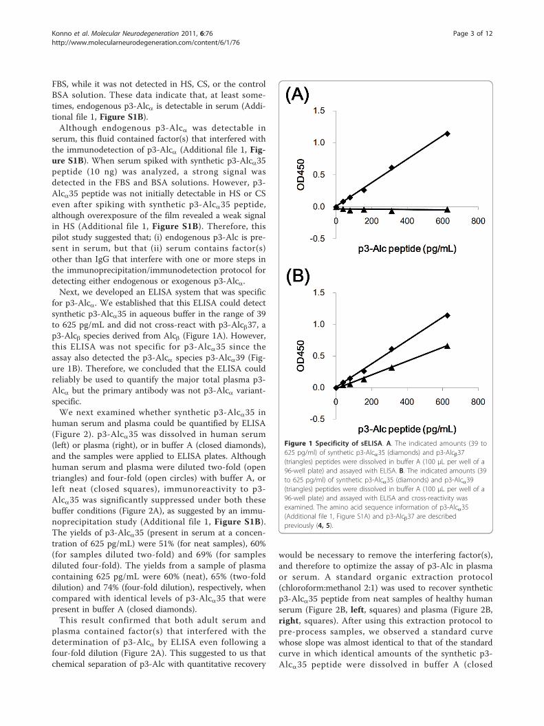
for p3-Alca. We established that this ELISA could detectsynthetic p3-Alca35 in aqueous buffer in the range of 39to 625 pg/mL and did not cross-react with p3-Alcb37, ap3-Alcb species derived from Alcb (Figure 1A). However,this ELISA was not specific for p3-Alca35 since theassay also detected the p3-Alca species p3-Alca39 (Fig-ure 1B). Therefore, we concluded that the ELISA couldreliably be used to quantify the major total plasma p3-Alca but the primary antibody was not p3-Alca variant-specific.We next examined whether synthetic p3-Alca35 in
human serum and plasma could be quantified by ELISA(Figure 2). p3-Alca35 was dissolved in human serum(left) or plasma (right), or in buffer A (closed diamonds),and the samples were applied to ELISA plates. Althoughhuman serum and plasma were diluted two-fold (opentriangles) and four-fold (open circles) with buffer A, orleft neat (closed squares), immunoreactivity to p3-Alca35 was significantly suppressed under both thesebuffer conditions (Figure 2A), as suggested by an immu-noprecipitation study (Additional file 1, Figure S1B).The yields of p3-Alca35 (present in serum at a concen-tration of 625 pg/mL) were 51% (for neat samples), 60%(for samples diluted two-fold) and 69% (for samplesdiluted four-fold). The yields from a sample of plasmacontaining 625 pg/mL were 60% (neat), 65% (two-folddilution) and 74% (four-fold dilution), respectively, whencompared with identical levels of p3-Alca35 that werepresent in buffer A (closed diamonds).This result confirmed that both adult serum and
plasma contained factor(s) that interfered with thedetermination of p3-Alca by ELISA even following afour-fold dilution (Figure 2A). This suggested to us thatchemical separation of p3-Alc with quantitative recovery
would be necessary to remove the interfering factor(s),and therefore to optimize the assay of p3-Alc in plasmaor serum. A standard organic extraction protocol(chloroform:methanol 2:1) was used to recover syntheticp3-Alca35 peptide from neat samples of healthy humanserum (Figure 2B, left, squares) and plasma (Figure 2B,right, squares). After using this extraction protocol topre-process samples, we observed a standard curvewhose slope was almost identical to that of the standardcurve in which identical amounts of the synthetic p3-Alca35 peptide were dissolved in buffer A (closed
Figure 1 Specificity of sELISA. A. The indicated amounts (39 to625 pg/ml) of synthetic p3-Alca35 (diamonds) and p3-Alcb37(triangles) peptides were dissolved in buffer A (100 μL per well of a96-well plate) and assayed with ELISA. B. The indicated amounts (39to 625 pg/ml) of synthetic p3-Alca35 (diamonds) and p3-Alca39(triangles) peptides were dissolved in buffer A (100 μL per well of a96-well plate) and assayed with ELISA and cross-reactivity wasexamined. The amino acid sequence information of p3-Alca35(Additional file 1, Figure S1A) and p3-Alcb37 are describedpreviously (4, 5).
Konno et al. Molecular Neurodegeneration 2011, 6:76http://www.molecularneurodegeneration.com/content/6/1/76
Page 3 of 12

diamonds). This indicated that factor(s) interfering withthe immunoreaction were largely excluded by thechloroform:methanol extraction (see Materials andMethods). This procedure resulted in yields (accordingto ELISA) that were 82% for peptide (625 pg/mL) dis-solved in serum (left) and 91% for that dissolved inplasma (right) (Figure 2B), relative to the identical con-centration of peptide dissolved in buffer A. Recovery ofp3-Alca35 from plasma via the extraction protocol washigh enough (> 90%) to be considered quantitative.Once extraction conditions were established, we
turned to the measurement of endogenous p3-Alcalevels in serum and plasma from various clinical popu-lations. Serum and plasma samples derived from thesame subjects (healthy normal, n = 5) were comparedfor p3-Alca levels (Table 1). The p3-Alca levels inserum samples were lower (67-87%) than thoseobserved in plasma samples, suggesting that 10 to 30%
of p3-Alca may be trapped in clots or degraded by theproteases involving in clotting. Similar issues have alsoplagued Ab ELISAs, and we therefore used plasma sam-ples for analyzing the cohorts of subjects with variousneurological diagnoses.
Plasma p3-Alca levels in AD and FTLD subjects (Japanesecohort 1)As a first trial, we examined levels of p3-Alca, Ab40,and Ab42 levels in plasma from a cohort of patients(designated “Japanese cohort 1”) with either AD (n =49) or FTLD (n = 15). There were no remarkable differ-ences for p3-Alca, Ab40, and Ab42 levels between ADand FTLD subjects, except that there was a trendtoward higher plasma Ab40 values in some subjects(Table 2and Additional file 1, Figure S2). In order tocharacterize the “high Ab40 subjects” and “low Ab40subjects” in further detail, a cut-off value of Ab40 (340pg/mL, the average of Ab40 value in AD subjects), wasimposed, and the p3-Alca, Ab40 and Ab42 levels werere-analyzed (Figure 3). When populations were stratifiedby Ab40 levels ("low Ab40” indicates < 340 pg/mL;“high Ab40” indicates > 340 pg/mL of middle panels ofFigure 3), the high Ab40 population was noted to con-tain samples with a significantly higher Ab42 levelswhen compared to the Ab42 levels from the low Ab40population, and this was true in both AD (right of Fig-ure 3A) and FTLD (right of Figure 3B) populations.There were no significant differences between the lowand high Ab40 populations with respect to age, Ab40/42 ratio, and MMSE score distributions (Additional file1, Figure S3). However, the p3-Alca levels were signifi-cantly higher in high Ab40 samples when comparedwith those present in low Ab40 samples (left of Figure3A), indicating that levels of p3-Alca, Ab40 and Ab42were correlated in some subgroups. This coordinatedalteration in p3-Alca levels was observed in AD but notin FTLD subjects despite of the covariance of Ab42 andAb40 levels in both populations (left of Figure 3B).
Figure 2 Effect of p3-Alca extraction from serum and plasmaon the quantification of p3-Alca with sELISA. A. Quantification ofp3-Alca35 peptide in human serum (left) and plasma (right) withoutextraction. Synthetic p3-Alca35 peptide was dissolved in humanserum and plasma and the samples in the indicated concentrations,and samples were assayed by ELISA alongside samples in whichstandard p3-Alca35 peptide was dissolved in buffer A (closeddiamonds in each respective panel). The p3-Alca35 peptide wasadded to undiluted serum or plasma [closed squares], or to serumor plasma diluted 2-fold [open triangles] or 4-fold [open circles]with buffer A. B. p3-Alca35 peptide was added to undiluted serum(left) or plasma (right), and extracted using the procedure describedin “Materials and Methods “. Extracted samples (open squares inpanel B) were also examined alongside a standard p3-Alca35peptide dissolved in buffer A (closed diamonds). Endogenous p3-Alca levels in serum and plasma (no addition of synthetic p3-Alca)were subtracted in order to determine the specific curve forquantifying levels of synthetic p3-Alca.
Table 1 p3-Alca levels in serum and plasma derived fromthe same subjects.
p3-Alca (pg/mL)
Serum Plasma
Mean ± SD Mean ± SD
1 129 ± 3 148 ± 6
2 103 ± 8 154 ± 15
3 142 ± 2 165 ± 0
4 151 ± 24 178 ± 13
5 182 ± 12 245 ± 10
Serum and plasma samples were prepared from five non-AD healthyvolunteers respectively and p3-Alca was quantified with ELISA in duplicatesafter extraction, as described in “Materials and Methods“. SD, standarddeviation.
Konno et al. Molecular Neurodegeneration 2011, 6:76http://www.molecularneurodegeneration.com/content/6/1/76
Page 4 of 12

Despite the fact that FTLD serves as a useful “otherneurological disease” (OND) control, Japanese cohort 1does not include non-demented subjects, and FTLDsubjects do not match AD subjects with regard to aver-age age (Table 2), therefore, we next examined plasmap3-Alca levels in Japanese cohort 2 which included age-matched nondemented controls.
Plasma p3-Alca levels in AD and non-AD subjects(Japanese cohort 2)We examined p3-Alca levels in the extracted plasma ofa second group of AD subjects and age-matched elderlynondemented controls (total n = 60). This cohortincluded AD patients of CDR 2 and CDR 3, and theseAD patients showed relatively higher Ab40 levels (n =39, 378 ± 113 pg/mL) in plasma when compared tothose from the age-matched normal control subjects (n= 21, 254 ± 63 pg/mL) (Table 3). The plasma of theseAD subjects (n = 39, average age 76 ± 7) contained 232± 59 pg/mL p3-Alca, while that of age-matched normalcontrols (n = 21, average age 73 ± 6) contained 163 ±70 pg/mL of p3-Alca (Table 3, Figure 4A, upper; P =0.0017 by the Mann-Whitney U-test)). Surprisingly, thestatistical difference was largely restricted to female sub-jects. The plasma from female AD subjects (n = 24)contained 245 ± 58 pg/mL of p3-Alca, while that offemale elderly nondemented controls (n = 14) contained142 ± 77 pg/mL (P = 0.0006) (Figure 4A, middle). Incontrast, plasma of male AD subjects (212 ± 54 pg/mL,n = 15) and male elderly normal controls (205 ± 19 pg/
Table 2 Summary of subject information (Japanesecohort 1)
N Age p3-Alca(pg/mL)
Ab 40(pg/mL)
Ab 42(pg/mL)
AD Total 49 75 ± 7 197 ± 50 335 ± 82 26 ± 7
Male 18 76 ± 6 193 ± 57 342 ± 77 28 ± 8
Female 31 75 ± 7 200 ± 47 331 ± 86 25 ± 7
FTLD Total 15 64 ± 12 190 ± 31 366 ± 51 30 ± 6
Male 8 65 ± 14 197 ± 20 371 ± 57 29 ± 7
Female 7 61 ± 9 183 ± 41 361 ± 47 31 ± 7
Average age, gender, and average values of p3-Alca, Ab40 and Ab42 of ADand FTLD subjects are summarized. Numbers indicate means ± standarddeviation. Details of individual subjects are shown in Additional file 2, TablesS1 (AD) and S2 (FTLD).
Figure 3 Levels of p3-Alca and Ab in plasma from “low Ab40” and “high Ab40” populations of AD and FTLD subjects (Japanesecohort 1). Subjects with AD and FTLD were separated into two populations, who showed low (< 340 pg/mL) and high (> 340 pg/ml) Ab40levels (cut-off line is shown in the middle panels with dotted line). AD (A) and FTLD (B) are respectively indicated in two populations for p3-Alca(left), Ab40 (middle) and Ab42 (right). Statistical analysis was performed using the Mann-Whitney U-test. *, P < 0.05; **, P < 0.01; ***, P < 0.001. N.S, not significant.
Konno et al. Molecular Neurodegeneration 2011, 6:76http://www.molecularneurodegeneration.com/content/6/1/76
Page 5 of 12

mL, n = 7) showed similar levels of p3-Alca and no sta-tistical significance was detected (Figure 4A, lower).Unextracted plasma samples from this cohort were
also studied. Ab40 levels in AD subjects distributedwithin a higher range than that observed for age-matched normal controls (Figure 4B, upper). The higherplasma Ab40 levels in female, but not male, AD subjectswere readily obvious to visual inspection (Figure 4B,middle, lower). Levels of p3-Alca and Ab40 wereincreased in parallel in AD subjects and were higherthan the levels observed in elderly normal controls.The p3-Alca levels within a particular individual did
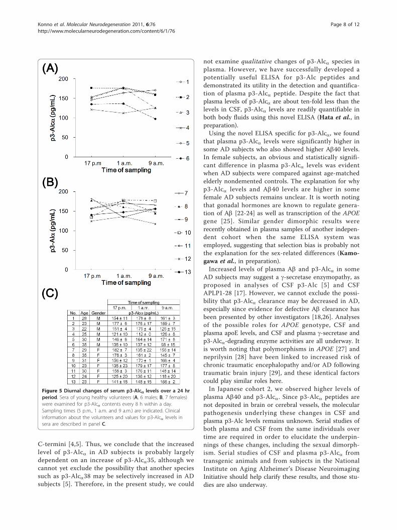
not substantially change when blood samples were takenevery 8 h over a 24 h period (Figure 5). Therefore, thepossibility that the time of day when blood was sampledwas excluded as a significant factor.
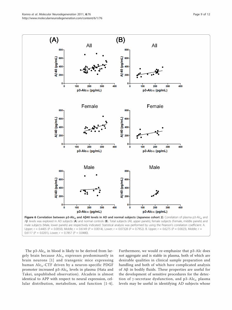
Correlation between levels of p3-Alca and Ab40We anticipated that the level of non-aggregatable p3-Alca in plasma might parallel that of Ab, at least withregard to Ab40, which is less aggregatable than Ab42.Therefore, we tested for the existence of any correlationbetween levels of p3-Alca and those of Ab40 in Japanesecohort 2. In AD subjects (n = 39), a significant correla-tion was observed between levels of p3-Alca and Ab(Figure 6A upper; r = 0.4405, P = 0.0050). However, thisstrong correlation was restricted to samples from femalesubjects (n = 24) (Figure 6A middle; r = 0.6149, P =0.0014). No significant correlation was observed betweenlevels of p3-Alca and Ab in samples from male subjects(n = 15) (Figure 6A, lower; r = 0.07328, P = 0.7952).Analysis of levels of p3-Alca and Ab in samples from
normal subjects revealed that there was also a strongcorrelation in normal subjects (n = 21) between levels ofthese two peptides (Figure 6B, upper; r = 0.6275, P =0.0023). The correlation between levels of p3-Alca andAb in samples from both normal females (n = 14) andthose from normal males (n = 7) was statistically signifi-cant (for female subjects, Figure 6B, middle; r = 0.6117,
P = 0.0201: for male subjects, Figure 6B, lower; r =0.7857, P = 0.0480). Overall, these analyses indicatedthat there was a significant correlation between levels ofp3-Alca and those of Ab40 in the plasma of normalsubjects, and that this correlation was more robust inthe plasma of female AD subjects.
DiscussionThe pathogenesis of sporadic AD is thought to begenetically heterogeneous. The precise identities andrelative importance of the many contributory genesremain unclear, and additional loci have recently beenidentified [21]. This is in contrast to the situation forFAD in which pathogenic mutations of APP and PSgenes cause alterations in Ab speciation [6,7].From the analyses of p3-Alca levels in CSF, we pre-
viously proposed that alternative processing of multipleg-secretase substrates may occurs in some sporadic ADpopulations. This earlier study was performed using acombination of immunoprecipitation and MALDI-TOF/MS to isolate and quantify p3-Alc in 300 μL of CSF[4,5]. Although the procedure showed semi-quantitativeaccuracy and was effective for testing changes in theabundance of p3-Alc species in the brain, CSF samplingis relatively invasive (when compared with blood sam-pling) and presents a challenge for large and/or longitu-dinal studies that involve repeated sampling over time.Therefore, we developed a system by which p3-Alccould be quantified in relatively small plasma samples.The combination of simple p3-Alc extraction and highlyselective ELISA enabled the quantification of p3-Alca in100 μL of plasma (200 μL in duplicate assay). Successfulextraction of p3-Alca from plasma with chloroform/methanol solvent suggests that the majority of p3-Alcain plasma may be bound to lipid or lipoproteins. Whilewe have not performed further characterization of p3-Alca in plasma, apolipoprotein E would be a potentialsuspect and association could vary by APOE isotype. Astudy of the effect of this variable in both CSF andplasma samples is underway.We have not yet developed an ELISA system that can
perfectly discriminate levels of each individual p3-Alcaspecies (e.g., p3-Alca35, p3-Alca38). Such an assayrequires antibodies against the neoepitopes createdwhen the specific C-termini are generated by g-cleavage.Since the polyclonal 839 antibody was prepared usingthe C-terminal amino acid sequence of p3-Alca35 asantigen, the ELISA used in this study displayed a relativespecificity for detecting synthetic p3-Alca35, but therewas also crossreactive detection of p3-Alca39 syntheticpeptide at about 50-60% of the sensitivity that wasobserved for detection of p3-Alca35 (Figure 1B).p3-Alca35 with a C-terminal Thr851 is the major
(~90%) peptide among the p3-Alca species with various
Table 3 Summary of subject information (Japanesecohort 2)
N Age p3-Alca(pg/mL)
Ab 40(pg/mL)
AD Total 39 76 ± 7 232 ± 59 378 ± 113
Male 15 74 ± 7 212 ± 54 366 ± 143
Female 24 78 ± 7 245 ± 58 386 ± 92
Normal Total 21 73 ± 6 163 ± 70 254 ± 63
Male 7 71 ± 7 205 ± 19 267 ± 49
Female 14 75 ± 6 142 ± 77 247 ± 69
Average age, gender, and average values of p3-Alca and Ab40 aresummarized. Numbers indicate means ± standard deviation. Details ofindividual subjects are shown in Additional file 2, Tables S3 (AD) and S4(Normal).
Konno et al. Molecular Neurodegeneration 2011, 6:76http://www.molecularneurodegeneration.com/content/6/1/76
Page 6 of 12
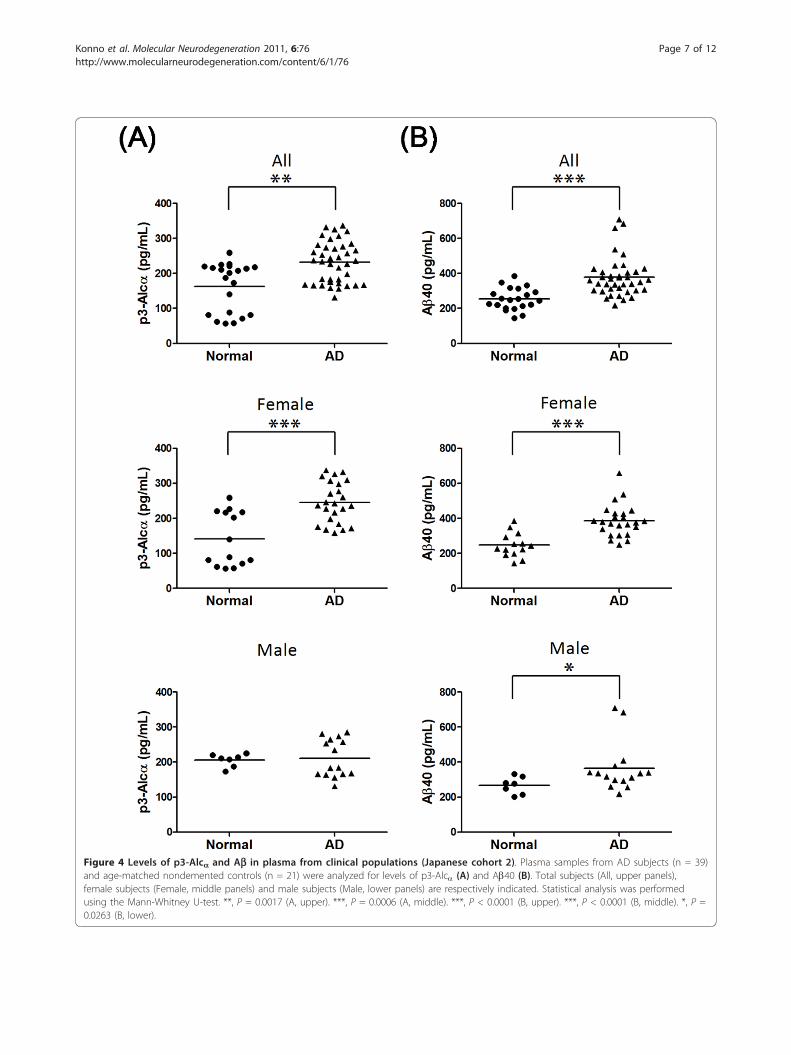
Figure 4 Levels of p3-Alca and Ab in plasma from clinical populations (Japanese cohort 2). Plasma samples from AD subjects (n = 39)and age-matched nondemented controls (n = 21) were analyzed for levels of p3-Alca (A) and Ab40 (B). Total subjects (All, upper panels),female subjects (Female, middle panels) and male subjects (Male, lower panels) are respectively indicated. Statistical analysis was performedusing the Mann-Whitney U-test. **, P = 0.0017 (A, upper). ***, P = 0.0006 (A, middle). ***, P < 0.0001 (B, upper). ***, P < 0.0001 (B, middle). *, P =0.0263 (B, lower).
Konno et al. Molecular Neurodegeneration 2011, 6:76http://www.molecularneurodegeneration.com/content/6/1/76
Page 7 of 12
C-termini [4,5]. Thus, we conclude that the increasedlevel of p3-Alca in AD subjects is probably largelydependent on an increase of p3-Alca35, although wecannot yet exclude the possibility that another speciessuch as p3-Alca38 may be selectively increased in ADsubjects [5]. Therefore, in the present study, we could
not examine qualitative changes of p3-Alca species inplasma. However, we have successfully developed apotentially useful ELISA for p3-Alc peptides anddemonstrated its utility in the detection and quantifica-tion of plasma p3-Alca peptide. Despite the fact thatplasma levels of p3-Alca are about ten-fold less than thelevels in CSF, p3-Alca levels are readily quantifiable inboth body fluids using this novel ELISA (Hata et al., inpreparation).Using the novel ELISA specific for p3-Alca, we found
that plasma p3-Alca levels were significantly higher insome AD subjects who also showed higher Ab40 levels.In female subjects, an obvious and statistically signifi-cant difference in plasma p3-Alca levels was evidentwhen AD subjects were compared against age-matchedelderly nondemented controls. The explanation for whyp3-Alca levels and Ab40 levels are higher in somefemale AD subjects remains unclear. It is worth notingthat gonadal hormones are known to regulate genera-tion of Ab [22-24] as well as transcription of the APOEgene [25]. Similar gender dimorphic results wererecently obtained in plasma samples of another indepen-dent cohort when the same ELISA system wasemployed, suggesting that selection bias is probably notthe explanation for the sex-related differences (Kamo-gawa et al., in preparation).Increased levels of plasma Ab and p3-Alca in some
AD subjects may suggest a g-secretase enzymopathy, asproposed in analyses of CSF p3-Alc [5] and CSFAPLP1-28 [17]. However, we cannot exclude the possi-bility that p3-Alca clearance may be decreased in AD,especially since evidence for defective Ab clearance hasbeen presented by other investigators [18,26]. Analysesof the possible roles for APOE genotype, CSF andplasma apoE levels, and CSF and plasma g-secretase andp3-Alca-degrading enzyme activities are all underway. Itis worth noting that polymorphisms in APOE [27] andneprilysin [28] have been linked to increased risk ofchronic traumatic encephalopathy and/or AD followingtraumatic brain injury [29], and these identical factorscould play similar roles here.In Japanese cohort 2, we observed higher levels of
plasma Ab40 and p3-Alca. Since p3-Alca peptides arenot deposited in brain or cerebral vessels, the molecularpathogenesis underlying these changes in CSF andplasma p3-Alc levels remains unknown. Serial studies ofboth plasma and CSF from the same individuals overtime are required in order to elucidate the underpin-nings of these changes, including the sexual dimorph-ism. Serial studies of CSF and plasma p3-Alca fromtransgenic animals and from subjects in the NationalInstitute on Aging Alzheimer’s Disease NeuroimagingInitiative should help clarify these results, and those stu-dies are also underway.
Figure 5 Diurnal changes of serum p3-Alca levels over a 24 hrperiod. Sera of young healthy volunteers (A, 6 males; B, 7 females)were examined for p3-Alca contents every 8 h within a day.Sampling times (5 p.m., 1 a.m. and 9 a.m.) are indicated. Clinicalinformation about the volunteers and values for p3-Alca levels insera are described in panel C.
Konno et al. Molecular Neurodegeneration 2011, 6:76http://www.molecularneurodegeneration.com/content/6/1/76
Page 8 of 12
The p3-Alca in blood is likely to be derived from lar-gely brain because Alca expresses predominantly inbrain neurons [1] and transgenic mice expressinghuman Alca-CTF driven by a neuron-specific PDGFpromoter increased p3-Alca levels in plasma (Hata andTakei, unpublished observation). Alcadein is almostidentical to APP with respect to neural expression, cel-lular distribution, metabolism, and function [1-4].
Furthermore, we would re-emphasize that p3-Alc doesnot aggregate and is stable in plasma, both of which aredesirable qualities in clinical sample preparation andhandling and both of which have complicated analysisof Ab in bodily fluids. These properties are useful forthe development of sensitive procedures for the detec-tion of g-secretase dysfunction, and p3-Alca plasmalevels may be useful in identifying AD subjects whose
Figure 6 Correlation between p3-Alca and Ab40 levels in AD and normal subjects (Japanese cohort 2). Correlation of plasma p3-Alca andAb levels was explored in AD subjects (A) and normal controls (B). Total subjects (All, upper panels), female subjects (Female, middle panels) andmale subjects (Male, lower panels) are respectively indicated. Statistical analysis was performed by using the Pearson’s correlation coefficient. A.Upper; r = 0.4405 (P = 0.0050), Middle; r = 0.6149 (P = 0.0014), Lower; r = 0.07328 (P = 0.7952). B. Upper; r = 0.6275 (P = 0.0023), Middle; r =0.6117 (P = 0.0201), Lower; r = 0.7857 (P = 0.0480).
Konno et al. Molecular Neurodegeneration 2011, 6:76http://www.molecularneurodegeneration.com/content/6/1/76
Page 9 of 12
clinical phenotype is caused by a functional alteration ofg-secretases and/or by defective clearance of transmem-brane domain-derived peptides. This endophenotypingmay be important for selecting subjects for trials of g-secretase modulators or plasminogen activator inhibitor-1 inhibitors [30,31], respectively.
Materials and methodsBlood collection and processing, and quantification ofplasma p3-AlcaInformed consent for the use of all human plasma andserum in this study was obtained from the patientsand/or their families and approved by the appropriateethical boards at each institution. Alzheimer’s diseasewas clinically diagnosed based on two major criteria;Diagnostic and Statistical Manual of Mental Disorders:4th Edition (DSM-IV) and National Institute of Neuro-logical and Communicational Disorders and Stroke -Alzheimer’s Disease and Related Disorders Association(NINCDS-ADRDA). In Japanese cohort 1, the clinicaldiagnoses of patients with FTLD were made on thebasis of established clinical criteria [32]. A compilationof clinical characteristics and data is shown in Table 2.Detailed descriptions of all subjects are shown in Addi-tional files 2, Tables S1 and S2.In Japanese cohort 2, all subjects with AD are inpati-
ents and showed MMSE scores consistent with a diag-nosis of either moderate or severe dementia (CDR 2 orCDR 3). Clinical characteristics and data are shown inTable 3. Detailed descriptions of all subjects includingthe raw values of p3-Alca are shown in Additional files2, Tables S3 and S4.For plasma samples, blood was collected into tubes
containing EDTA and centrifuged. For serum samples,blood was collected into tubes with no EDTA andallowed to coagulate. To extract p3-Alca peptides, afour-fold volume of organic reagent (800 μL of chloro-form: methanol [2:1] mixture) was added to plasma orserum (usually 200 μL for duplicate assay) in a conicaltube (1.5 mL), agitated well by vortexing or sonicationfor 10 s, and then left to stand for 1 h at room tempera-ture (18-23°C). Next, a 160 μL aliquot of distilled waterwas added, and the samples were mixed by vortexing.The samples were centrifuged at 15,000 rpm for 15 min,and the aqueous phase was recovered and dried using aSpeedVac system. The dried sample was dissolved in250 μL of PBS containing 1% (w/v) BSA and 0.05% (v/v)Tween-20 (buffer A). The samples (×1) or samplesfurther diluted with buffer A (2- and 4-fold) were usedfor ELISA. Aliquots of 100 μL were studied in duplicate.Generally the undiluted sample (×1) was examined forquantification.
Antibodies and ELISA systemThe major p3-Alca species, p3-Alca35, is a peptide thatincludes the sequence from Ala817 to Thr852 of Alca1(for the amino acid sequence of p3-Alca, see Additionalfile 1, Figure S1A). Polyclonal rabbit antibodies wereraised against a peptide containing the sequencebetween position 817 and 822 (#817 antibody) and apeptide containing the sequence between position 839and 852 (#839 antibody), and affinity purified with theeach respective peptide antigen resin. The antibody 839was used to capture p3-Alca, and horseradish peroxi-dase-conjugated pan p3-Alca antibody 817 and tetra-methyl benzidine were used to detect the captured p3-Alca colormetrically at OD450. Synthetic p3-Alca35 pep-tide was used as a standard assay protein. The specificityfor p3-Alca in the ELISA was confirmed using syntheticp3-Alca35 (major species of CSF p3-Alca), p3-Alca39and p3-Alcb37 peptides [4,5]. Ab in neat samples with-out extraction was quantified with sELISA (Wako PureChemical Industries Co Ltd for cohort 1 and IBL CoLtd for cohort 2).
Additional material
Additional file 1: Figure S1. Amino acid sequences of p3-Alca, andrecovery of p3-Alca in sera by immunoprecipitation and detectionof p3-Alca by Western blotting. A. Amino acid sequences andcleavage sites of p3-Alca and Ab in human. The amino acid sequencesof p3-Alca species and the primary a- and secondary g-cleavage sites ofAlca1 are indicated. “35” indicates major g-cleavage site to generate p3-Alca35 while “38” indicates minor g-cleavage site to generate p3-Alca38.The amino acid sequences of p3 and Ab peptides are also shown.Primary a- and b- cleavage sites of APP695 are shown, “40” indicatesmajor g-cleavage site to generate Ab40 while “42” indicates minor g-cleavage site to generate Ab42. Putative transmembrane region isindicated with box. B. Detection of endogenous and synthetic p3-Alcapeptides. Synthetic p3-Alca35 peptide (10 ng) was added to humanserum (HS), calf serum (CS), fetal bovine serum (FBS), and bovine serumalbumin solution in PBS (BSA; 10 mg/mL). The samples with (+) orwithout (-) addition of p3-Alca35 peptide were subject toimmunoprecipitation with anti-pan p3-Alca 3B5 antibody andimmunoprecipitates were analyzed by Western blotting with UT135antibody. The far right lane is a loading control sample containing 4 ngof p3-Alca35 peptide. “Overexposure“ (lower row) indicates overexposureof film. The pan p3-Alca mouse monoclonal antibody 3B5 and polyclonalrabbit antibody UT135 have been described (J. Biol. Chem. [2009] 284,36024-36033). Figure S2. Levels of p3-Alca in plasma of AD and FTLDsubjects (Japanese cohort 1). Plasma samples from AD (n = 49) andFTLD (n = 15) subjects were analyzed for levels of p3-Alca. Statisticalanalysis was performed using the Mann-Whitney U-test. N.S, notsignificant. Figure S3. Age, Ab40/42 ratio and MMSE scoredistribution in subjects of low and high Ab40 populations(Japanese cohort 1). Age (upper left), Ab40/42 ratio (upper right) andMMSE score (lower left) of AD and FTLD subjects of low Ab40population are compared to these of high Ab40 population. Statisticalanalysis was performed using the Mann-Whitney U-test. N.S, notsignificant.
Additional file 2: Table S1. Information on AD subjects (Japanesecohort 1). The subjects (n = 49) were clinically diagnosed using CDR(clinical dementia rating) criteria. Table S2. Information on FTLDsubjects (Japanese cohort 1). The patients (n = 15) were clinically
Konno et al. Molecular Neurodegeneration 2011, 6:76http://www.molecularneurodegeneration.com/content/6/1/76
Page 10 of 12
diagnosed as described in “Materials and Methods”. Table S3.Information on AD subjects (Japanese cohort 2). The subjects (n =39) were clinically diagnosed with AD at stages CDR 2 or CDR 3. TableS4. Information of normal controls (Japanese cohort 2). Age-matched normal elderly controls (n = 21) are indicated. The subjectswere clinically non-demented.
AbbreviationsAD: Alzheimer’s disease; Aβ: amyloid β-protein; Alc: alcadein; APP: amyloid β-protein precursor; p3-Alc: small peptide generated by serial primary andsecondary cleavages of Alc; CSF: cerebrospinal fluid: FAD: familial Alzheimer’sdisease; SAD: sporadic Alzheimer’s disease; PS: presenilin; sELISA: sandwichenzyme-linked immunosorbent assay.
AcknowledgementsThis work was supported in part by Grants-in-aid for Scientific Research fromthe Ministry of Education, Culture, Sports, Science and Technology (MEXT), agrant from the Ministry of Health, Labor and Welfare (MHLW), and a grantfrom the New Energy and Industrial Technology Development Organization(NEDO) in Japan. S.G. was supported by the NIA Alzheimer’s DiseaseResearch Center grant P50 05138 to Mary Sano.
Author details1Laboratory of Neuroscience, Graduate School of Pharmaceutical Sciences,Hokkaido University, Sapporo 060-0812, Japan. 2Immuno-BiologicalLaboratories Co., Ltd. (IBL), Fujioka 375-0005, Japan. 3Choju Medical Institute,Fukushimura Hospital, Toyohashi 441-8124, Japan. 4Department of MolecularNeuroscience, Brain Research Institute, Niigata University, Niigata 951-8585,Japan. 5Department of Neurology, Mount Sinai School of Medicine,Alzheimer’s Disease Research Center, New York, NY 10029, USA. 6Departmentof Psychiatry, Mount Sinai School of Medicine, Alzheimer’s Disease ResearchCenter, New York, NY 10029, USA. 7James J. Peters Veterans AdministrationMedical Center, NY 10468, USA.
Authors’ contributionsTK and SH carried out all of the experiments. YH, YH-S and MM establishedand modified the p3-Alcα sELISA system. TY, TI and HA collected the bloodsamples for this study. TN, YS, TY, HA, SG and TS participated in the designof the study, and TS conceived the study. SG, HA and TS are co-seniorauthors. All authors read and approved the final manuscript.
Competing interestsThe authors declare that they have no competing interests.
Received: 12 July 2011 Accepted: 8 November 2011Published: 8 November 2011
References1. Araki Y, Tomita S, Yamaguchi H, Miyagi N, Sumioka A, Kirino Y, Suzuki T:
Novel cadherin-related membrane proteins, Alcadeins, enhance the X11-like protein mediated stabilization of amyloid β-protein precursormetabolism. J Biol Chem 2003, 278:49448-49458.
2. Araki Y, Miyagi N, Kato N, Yoshida T, Wada S, Nishimura M, Komano H,Yamamoto T, De Strooper B, Yamamoto K, Suzuki T: Coordinatedmetabolism of Alcadein and amyloid β-protein precursor regulatesFE65-dependent gene transactivation. J Biol Chem 2004, 279:24343-24354.
3. Araki Y, Kawano T, Taru H, Saito Y, Wada S, Miyamoto K, Kobayashi H,Ishikawa OH, Ohsugi Y, Yamamoto T, Matsuno K, Kinjyo M, Suzuki T: Thenovel cargo receptor Alcadein induces vesicle association of kinesin-1motor components and activates axonal transport. EMBO J 2007,26:1475-1486.
4. Hata S, Fujishige S, Araki Y, Kato N, Araseki M, Nishimura M, Hartmann D,Saftig P, Fahrenholz F, Taniguchi M, Urakami K, Akatsu H, Martins RN,Yamamoto K, Maeda M, Yamamoto T, Nakaya T, Gandy S, Suzuki T:Alcadein cleavages by APP α- and γ-secretases generate small peptidesp3-Alcs indicating Alzheimer disease-related γ-secretase dysfunction. JBiol Chem 2009, 284:36024-36033.
5. Hata S, Fujishige S, Araki Y, Taniguchi M, Urakami K, Peskind E, Akatsu H,Araseki M, Yamamoto K, Martins RN, Maeda M, Nishimura M, Levey A,Chung KA, Montine T, Leverenz J, Fagan A, Goate A, Bateman R,Holtzman DM, Yamamoto T, Nakaya T, Gandy S, Suzuki T: Alternativeprocessing of γ-secretase substrates in common forms of mild cognitiveimpairment and Alzheimer’s disease: Evidence for γ-secretasedysfunction. Ann Neurol 2011, 69:1026-1031.
6. De Strooper B: Loss-of-function presenilin mutations in Alzheimerdisease. Taking point on the role of presenilin mutations in Alzheimerdisease. EMBO Rep 2007, 8:141-146.
7. Steiner H, Fluher R, Haass C: Intramembrane proteolysis by γ-secretase. JBiol Chem 2008, 283:29627-29631.
8. Sherrιngton R, Rogaev EI, Liang Y, Rogaeva EA, Levesque G, Ikeda M, Chi H,Lin C, Li G, Holman K, Tsuda T, Mar L, Foncin J-F, Bruni AC, Montesi MP,Sorbi S, Rainero I, Pinessi L, Nee L, Chumakov I, Pollen D, Brookes A,Sanseau P, Polinsky RJ, Wasco W, Da Silva HAR, Haines JL, Pericak-Vance MA, Tanzi RE, Roses AD, et al: Cloning of a gene bearing missensemutations in early-onset familial Alzheimer’s disease. Nature 1995,375:754-760.
9. Borchelt DR, Thinakaran G, Eckman CB, Lee MK, Davenport F, Ratovitsky T,Prada CM, Kim G, Seekins S, Yager D, Slunt HH, Wang R, Seeger M,Levey AI, Gandy SE, Copeland NG, Jenkins NA, Price DL, Younkin SG,Sisodia S: Familial Alzheimer’s disease-linked presenilin 1 variants elevateAβ1-42/1-40 ratio in vitro and in vitro. Neuron 1996, 17:1005-1013.
10. Haass C, Selkoe DJ: Soluble protein oligomers in neurodegeneration:lessons from the Alzheimer’s amyloid β-peptide. Nat Rev Moll Cell Biol2007, 8:101-112.
11. Shankar GM, Li S, Methta TH, Garcia-Munoz A, Shepardson NE, Smith I,Brett FM, Farrell MA, Rowan MJ, Lemere CA, Regan CM, Walsh DM,Sabatini BL, Selkoe DJ: Amyloid-β protein dimers isolated directly fromAlzheimer’s brain impair synaptic plasticity and memory. Nat Med 2008,14:837-842.
12. Shaw LM, Vanderstichele H, Knapik-Czajka M, Figurski M, Coart E,Blennow K, Soares H, Simon AJ, Lewczuk P, Dean RA, Siemers E, Potter W,Lee VM, Trojanowski JQ: Alzheimer’s Disease Neuroimaging Initiative:Qualification of the analytical and clinical performance of CSF biomarkeranalyses in ADNI. Acta Neuropathol 2011, 121:597-609.
13. Frankfort SV, Tulner LR, van Campen JP, Verbeek MM, Jansen RW,Beijnen JH: Amyloid β protein and tau in cerebrospinal fluid and plasmaas biomarkers for dementia: a review of recent literature. Curr ClinPharmacol 2008, 3:123-1331.
14. Lambert JC, Schhraen-Maschke S, Richard F, Fievet N, Rouaud O, Berr C,Dartigues JF, Tzourio C, Alperovitch A, Buee L, Amouyel A: Association ofplasma amyloid with risk of dementia: The prospective three-city study.Exp Neurol 2009, 73:847-853.
15. Graff-Radford NR, Crook JE, Lucas J, Boeve BT, Knopman DS, Ivnik RJ,Smith GE, Younkin LH, Petersen RC, Younkin SG: Association of low plasmaAβ42/Aβ40 ratios with increased imminent risk for mild cognitiveimpairment and Alzheimer disease. Arch Neurol 2007, 64:354-362.
16. Yaffe K, Weston A, Graff-Radford NR, Satterfield S, Simonsick EM,Younkin SG, Younkin LH, Kuller L, Ayonayon HN, Ding J, Harris TB:Association of plasma β-amyloid level and cognitive reserve withsubsequent cognitive decline. JAMA 2011, 305:261-266.
17. Yanagida K, Okochi M, Tagami S, Nakayama T, Kodama TS, Nishitomi K,Jiang J, Mori K, Tatsumi S, Arai T, Ikeuchi T, Kasuga K, Tokuda T, Kondo M,Ikeda M, Deguchi K, Kazui H, Tanaka T, Morihara T, Hashimoto R, Kudo T,Steiner H, Haass C, Tsuchiya K, Akiyama H, Kuwano R, Takeda M: The 28-amino acid form of an APLP1-derived Aβ-like peptide is a surrogatemarker for Aβ42 production in the central nervous system. EMBO MolMed 2009, 1:223-35.
18. Mawuenyega KG, Sigurdson W, Ovod V, Munsell L, Kasten T, Morris JC,Yarasheski KE, Bateman RJ: Decreased clearance of CNS β-amyloid inAlzheimer’s disease. Science 2010, 330:1774.
19. Yang DS, Small DH, Seydel U, Smith JD, Hallmayer J, Gandy SE, Martins RN:Apolipoprotein E promotes the binding and uptake of β-amyloid intoChinese hamster ovary cells in an isoform-specific manner. Neuroscience1999, 90:1217-1226.
20. Sharman MJ, Morici M, Hone E, Berger T, Taddei K, Martins IJ, Lim WL,Singh S, Wenk MR, Ghiso J, Buxbaum JD, Gandy S, Martins RN: APOEgenotype results in differential effects on the peripheral clearance of
Konno et al. Molecular Neurodegeneration 2011, 6:76http://www.molecularneurodegeneration.com/content/6/1/76
Page 11 of 12
amyloid-β42 in APOE knock-in and knock-out mice. J Alzheimers Dis 2010,21:403-409.
21. Naj AC, Jun G, Beecham GW, Wang LS, Vardarajan BN, Buros J, Gallins PJ,Buxbaum JD, Jarvik GP, Crane PK, Larson EB, Bird TD, Boeve BF, Graff-Radford NR, De Jager PL, Evans D, Schneider JA, Carrasquillo MM, Ertekin-Taner N, Younkin SG, Cruchaga C, Kauwe JS, Nowotny P, Kramer P, Hardy J,Huentelman MJ, Myers AJ, Barmada MM, Demirci FY, Baldwin CT, et al:Common variants at MS4A4/MS4A6E, CD2AP, CD33 and EPHA1 areassociated with late-onset Alzheimer’s disease. Nat Genet 2011, 43:436-41.
22. Xu H, Gouras GK, Greenfield JP, Vincent B, Naslund J, Mazzarelli L, Fried G,Jovanovic JN, Seeger M, Relkin NR, Liao F, Checler F, Buxbaum JD, Chit BT,Thinakaran G, Sisodia SS, Wang R, Greengard P, Gandy S: Estogen reducesneural generation of Alzheimer β-amyloid peptides. Nat Med 1998,4:447-451.
23. Zheng H, Xu H, Uljon SN, Gross R, Hardy K, Gaynor J, Lafrancois J,Simpkins J, Refolo LM, Petanceska S, Wang R, Duff K: Modulation of Aβpeptides by estrogen in mouse models. J Neurochem 2002, 80:191-196.
24. Nord LC, Sundgvist J, Anderson E, Fried G: Analysis of oestrogenregulation of alpha-, beta- and gamma-secretase gene and proteinexpression in cultured human neuronal and glial cells. Neurodegener Dis2010, 7:349-364.
25. Verghese PB, Castellano JM, Holzman DM: Apolipoprotein E in Alzheimer’sdisease and other neurological disorders. Lancet Neurol 2011, 10:241-252.
26. Yasojima K, Akiyama H, McGeer EG, McGeer PL: Reduced neprilysin in highplaque areas of Alzheimer brain: a possible relationship to deficientdegradation of β-amyloid peptide. Neurosci Lett 2001, 297:97-100.
27. Jordan BD, Relkin NR, Ravdin LD, Jacobs AR, Bennett A, Gandy S:Apolipoprotein E epsilon4 associated with chronic traumatic brain injuryin boxing. JAMA 1997, 278:136-140.
28. Johnson VE, Stewart W, Graham DI, Stewart JE, Praestgaard AH, Smith DH:A neprilysin polymorphism and amyloid-beta plaques after traumaticbrain injury. J Neurotrauma 2009, 26:1197-1202.
29. DeKosky ST, Ikonomovic MD, Gandy S: Traumatic brain injury: football,warfare, and long-term effects. N Engl J Med 2010, 363:1293-1296.
30. Melchor JP, Pawlak R, Strickland S: The tissue plasminogen activator-plasminogen proteolytic cascade accelerates amyloid-beta (Abeta)degradation and inhibits Abeta-induced neurodegeneration. J Neurosci2003, 23:8867-8871.
31. Melchor JP, Pawlak R, Chen Z, Strickland S: The possible role of tissue-typeplasminogen activator (tPA) and tPA blockers in the pathogenesis andtreatment of Alzheimer’s disease. J Mol Neurosci 2003, 20:287-289.
32. Neary D, Snowden JS, Gustafson L, Passant U, Stuss D, Black S, Freedman M,Kertesz A, Robert PH, Albert M, Boone K, Miller BL, Cummings J, Benson DE:Frontotemporal lobar degeneration: a consensus on clinical diagnosticcriteria. Neurology 1998, 51:1546-1554.
doi:10.1186/1750-1326-6-76Cite this article as: Konno et al.: Coordinated increase of g-secretasereaction products in the plasma of some female Japanese sporadicAlzheimer’s disease patients: quantitative analysis of p3-Alca with a newELISA system. Molecular Neurodegeneration 2011 6:76.
Submit your next manuscript to BioMed Centraland take full advantage of:
• Convenient online submission
• Thorough peer review
• No space constraints or color figure charges
• Immediate publication on acceptance
• Inclusion in PubMed, CAS, Scopus and Google Scholar
• Research which is freely available for redistribution
Submit your manuscript at www.biomedcentral.com/submit
Konno et al. Molecular Neurodegeneration 2011, 6:76http://www.molecularneurodegeneration.com/content/6/1/76
Page 12 of 12