Embed Size (px)
Citation preview

Norikane et al. EJNMMI Res (2021) 11:42 https://doi.org/10.1186/s13550-021-00785-8
ORIGINAL RESEARCH
Correlation of 4′-[methyl-11C]-thiothymidine PET with Gd-enhanced and FLAIR MRI in patients with newly diagnosed gliomaTakashi Norikane1, Katsuya Mitamura1, Yuka Yamamoto1* , Yukito Maeda2, Kenichi Tanaka1, Tetsuhiro Hatakeyama3, Keisuke Miyake3, Jun Toyohara4 and Yoshihiro Nishiyama1
Abstract
Purpose: To elucidate the biological association between tumor proliferation, tumor infiltration and neovasculariza-tion, we analyzed the association between volumetric information of 4′-[methyl-11C]thiothymidine (4DST) positron emission tomography (PET) and fluid-attenuated inversion recovery (FLAIR) and T1-weighted gadopentetate dimeg-lumine (Gd)-enhanced magnetic resonance imaging (MRI), in patients with newly diagnosed glioma.
Methods: A total of 23 patients with newly diagnosed glioma who underwent both 4DST PET/CT and Gd-enhanced MRI before therapy were available for a retrospective analysis of prospectively collected data. The maximum standard-ized uptake value (SUVmax) for tumor (T) and the mean SUV for normal contralateral hemisphere (N) were calculated, and the tumor-to-normal (T/N) ratio was determined. Proliferative tumor volume (PTV) from 4DST PET and the vol-ume of Gd enhancement (GdV) and hyperintense region on FLAIR (FLAIRV) from MRI were calculated.
Results: All gliomas but 3 diffuse astrocytomas and one anaplastic astrocytoma had 4DST uptake and Gd enhance-ment on MRI. There was no significant difference between PTV and GdV although the exact edges of the tumor dif-fered in each modality. The FLAIRV was significantly larger than PTV (P < 0.001). Significant correlations between PTV and GdV (ρ = 0.941, P < 0.001) and FLAIRV (ρ = 0.682, P < 0.001) were found.
Conclusion: These preliminary results indicate that tumor proliferation assessed by 4DST PET is closely associated with tumor-induced neovascularization determined by Gd-enhanced MRI in patients with newly diagnosed glioma.
Keywords: 4DST, PET, Glioma, MRI
© The Author(s) 2021. Open Access This article is licensed under a Creative Commons Attribution 4.0 International License, which permits use, sharing, adaptation, distribution and reproduction in any medium or format, as long as you give appropriate credit to the original author(s) and the source, provide a link to the Creative Commons licence, and indicate if changes were made. The images or other third party material in this article are included in the article’s Creative Commons licence, unless indicated otherwise in a credit line to the material. If material is not included in the article’s Creative Commons licence and your intended use is not permitted by statutory regulation or exceeds the permitted use, you will need to obtain permission directly from the copyright holder. To view a copy of this licence, visit http:// creat iveco mmons. org/ licen ses/ by/4. 0/.
IntroductionTumor proliferation is considered an important indica-tor of tumor growth and therapeutic effectiveness [1]. Positron emission tomography (PET) using 2-deoxy-2-[18F]fluoro-D-glucose (FDG) is an established functional imaging modality for diagnostic oncologic imaging of various tumors [2]. However, since FDG directly reflects
the glucose metabolism, it can provide only an indirect measure of cellular proliferation.
The most direct indicator of cellular proliferation is deoxyribonucleic acid (DNA) synthesis, which can be measured using radiolabeled thymidine or its analogs. A thymidine analog, 3′-deoxy-3′-[18F]-fluorothymidine (FLT), has been used to assess the tumor proliferative activity of various types of tumor including brain tumor [3–6]. In brain gliomas, FLT has been validated for evalu-ation of cellular proliferation and tumor grade [3]. Toy-ohara et al. developed 4′-[methyl-11C] thiothymidine (4DST) for cellular proliferation imaging, which is resist-ant to degradation by thymidine phosphorylase and is
Open Access
*Correspondence: [email protected] Department of Radiology, Faculty of Medicine, Kagawa University, 1750-1 Ikenobe, Miki-cho, Kita-gun, Kagawa 761-0793, JapanFull list of author information is available at the end of the article

Page 2 of 7Norikane et al. EJNMMI Res (2021) 11:42
incorporated into DNA [7]. 4DST PET has been shown to be helpful in noninvasive evaluation of the prolifera-tion of several types of tumor including brain tumor [8, 9].
Anatomical imaging using magnetic resonance imag-ing (MRI) has been the most commonly used modality for obtaining brain tumor information. Gadopentetate dimeglumine (Gd) enhancement in MRI is due to pas-sive diffusion of the paramagnetic contrast agent across a damaged blood–brain barrier (BBB) and has been cor-related with tumor neovascularization and proliferation [10]. Radiologically, glioblastoma usually manifests as a Gd-enhancing mass with surrounding non-enhancing tissue marked by abnormal fluid-attenuated inversion recovery (FLAIR) signal [11]. Pathology studies demon-strate microscopic tumor infiltration throughout the per-itumoral edema, which appears hyperintense on FLAIR sequences [12]. More than 90% of tumor recurrences will occur within FLAIR envelope [12]. To elucidate the bio-logical association between tumor proliferation, tumor infiltration and neovascularization, we analyzed the association between volumetric information of 4DST PET and FLAIR and T1-weighted Gd-enhanced MRI in patients with newly diagnosed glioma.
Materials and methodsPatientsWe conducted a retrospective analysis of prospec-tively collected data. The prospective study consisted of 220 patients with brain tumors who underwent 4DST PET/CT study between August 2011 and July 2017. All patients included gave written informed consent, and the study protocol was approved by our institutional ethics committee. From these patients, 23 (9 males, 14 females; mean age, 59.1 years; age range, 21–84 years) were selected for this retrospective analysis. Eligible patients fulfilled all of the following criteria: (1) histologically con-firmed glioma with treatment-naïve and (2) both 4DST PET/CT and T1-weighted Gd-enhanced and FLAIR MRI were performed within 2 weeks before treatment. The tumor numbers and types were as follows: 4 diffuse astrocytomas, 4 anaplastic astrocytomas, 1 anaplastic oligodendroglioma and 14 glioblastomas. This retrospec-tive data collection was compliant with our institutional ethics committee, with the need for informed consent waived. Some of the data from 16 of these patients were used in a previous study [8].
PET/CT and MRI studiesThe 4DST was manufactured using an automated syn-thesis system with HM-18 cyclotron (QUPID; Sumitomo Heavy Industries Ltd, Tokyo, Japan) and was synthe-sized using the method described by Toyohara et al. [7].
All acquisitions were performed using a Biograph mCT 64-slice PET/CT scanner (Siemens Medical Solutions USA Inc., Knoxville, TN, USA). Data acquisition began with low-dose CT at the following settings: no contrast agent, 120 kV, 50 mA, 1.0-s tube rotation time, 3-mm slice thickness, 3-mm increments and pitch 0.55. PET emission scanning of the head region with a 15-min acquisition of one bed position was performed 15 min after intravenous injection of 4DST (6 MBq/kg). The PET data were acquired in 3D mode and were recon-structed with an ordered subsets expectation maximiza-tion, incorporating correction with point spread function and time-of-flight model (5 iterations, 21 subsets) on a 256 × 256 matrix, with a 21.6 cm axial field of view and 3 mm slice thickness.
MRI was performed using a 1.5 T MRI scanner (Achieva, Philips Medical Systems, Eindhoven, Neth-erlands). The MRI protocol for this study included the acquisition of 2D T2 FLAIR (repetition time/echo time = 8000/100 ms, 7 mm slice thickness with 1 mm interslice gap, matrix: 512 × 512, FOV 210 × 210 mm2) and 2D T1-weighted imaging (repetition time/echo time = 440/9 ms, 7 mm slice thickness with 1 mm inter-slice gap, matrix: 512 × 512, FOV 210 × 210 mm2) after administration of 0.1 mmol/kg Gd.
Image analysisPET images were visually assessed by two experienced nuclear physicians independently. Any difference of opinion was resolved by consensus. Tumor lesions were identified as areas of focally increased uptake, exceed-ing that of normal brain background. Semi-quantitative analysis using the standardized uptake value (SUV) was performed by an experienced nuclear physician using a syngo.via (Siemens Healthcare, Erlangen, Germany). The SUV was calculated using the following formula: SUV = cdc/(di/w), where cdc is the decay-corrected tracer tissue concentration (Bq/g); di, the injected dose (Bq); and w, the patient’s body weight (g). The maximum SUV (SUVmax) for tumor and the mean SUV (SUVmean) for reference tissue were calculated. For the reference tis-sue, a circular region of interest (ROI) of 10 × 10 mm was manually placed on the uninvolved contralateral hemi-sphere. The tumor-to-contralateral normal brain tissue (T/N) ratio was determined by dividing the tumor SUV-max by the contralateral hemisphere SUVmean. Prolif-erative tumor volume (PTV) was defined as the volume with a threshold of 40% of the SUVmax [7]. When the radioactivity associated with the tumor could not be vis-ually identified, the mean SUV of the tumor on the basis of MRI studies was measured and PTV was assumed to be zero.

Page 3 of 7Norikane et al. EJNMMI Res (2021) 11:42
In the MRI, ROI was manually placed over the Gd enhancing part of the tumor and the hyperintense region on FLAIR MRI. The volumes of the Gd enhanc-ing part of the tumor (GdV) and the hyperintense FLAIR (FLAIRV) were calculated by summing the volumes (area multiplied by interslice distance) of the ROIs.
Statistical analysisDifferences in semi-quantitative parameters were ana-lyzed by nonparametric Wilcoxon signed-rank test. Spearman correlation coefficients were used to examine the relationship between PTV and GdV and FLAIRV. For all analyses, P values < 0.05 were considered statisti-cally significant.
ResultsAll gliomas but 3 diffuse astrocytomas and one anaplas-tic astrocytoma had 4DST uptake and Gd enhancement on MRI. The mean (± SD) of the normal brain paren-chyma showed low 4DST uptake with an SUVmean of 0.40 ± 0.11. The mean (± SD) of the SUVmax for tumor and T/N ratio was 2.37 ± 1.65 and 5.82 ± 4.10, respectively.
Table 1 shows the results of 4DST PET and MRI parameters in patients with newly diagnosed gliomas. In all patients, there was no significant difference between PTV and GdV although the exact edges of the tumor differed in each modality (Figs. 1, 2). The FLAIRV was significantly larger than PTV (P < 0.001). A significant correlation between PTV and GdV was found (ρ = 0.941, P < 0.001) (Fig. 3a). A significant correlation between PTV and FLAIRV was also found (ρ = 0.682, P < 0.001) (Fig. 3b).
In a subgroup analysis of patients with glioblastoma only, there was no significant difference between PTV and GdV. The FLAIRV was significantly larger than PTV (P < 0.001). Significant correlations between PTV and GdV (ρ = 0.921, P < 0.001) and FLAIRV (ρ = 0.754, P = 0.002) were also found.
DiscussionWe analyzed the association between volumetric infor-mation of 4DST PET and FLAIR and T1-weighted Gd-enhanced MRI in patients with newly diagnosed glioma. The results showed that the volume of 4DST
Table 1 4DST PET and MRI parameters in patients with newly diagnosed gliomas
PTV Proliferative tumor volume, GdV volume of Gd enhancement, FLAIRV volume of hyperintense FLAIR* P < 0.001 compared with PTV
Parameter All patients (n = 23) Glioblastoma (n = 14)
Median volume in cm3
PTV (range) 14.47 (0–66.79) 23.94 (2.03–66.79)
GdV (range) 6.79 (0–84.35) 31.05 (3.35–84.35)
FLAIRV (range) 50.10 (11.02–196.16)* 112.45 (11.02–196.16)*
Fig. 1 Radiologic images of a 60-year-old female with newly diagnosed glioblastoma. Transverse 4DST PET image a shows inhomogeneous uptake in the right occipital lobe (SUVmax = 5.36, T/N ratio = 13.74, PTV = 44.58 cm3). Transverse T1-weighted MRI with Gd enhancement b shows inhomogeneously enhanced lesion in tumor (GdV = 48.28 cm3). Region of interest shows proliferation area drawn on 4DST PET image from a. The proliferation area on 4DST PET image and Gd enhancement area on MRI differed very slightly. The hyperintense area identified on transverse FLAIR MRI c is larger than the proliferation area on 4DST PET image (FLAIRV = 137.52 cm3)
Page 4 of 7Norikane et al. EJNMMI Res (2021) 11:42
uptake was closely correlated to the volume of Gd enhancement in MRI, but the exact edges of the tumor differed in each modality. These findings are similar to those from a previous study using FLT PET [4]. On the other hand, the FLAIRV was significantly larger than PTV.
FLAIR and Gd-enhanced MRI are commonly used to obtain anatomical information especially in the central nervous system. However, MRI provides nonspecific information of the tumor as it does not always differ-entiate brain tumors from non-tumorous tissue or low-grade tumors from high-grade ones. PET can provide valuable molecular information and may improve the ability of diagnostic procedures to determine the malig-nancy and the extent of tumor invasion.
Increased cellular proliferation and DNA replication are characteristic of malignant transformation [13]. The evaluation of cellular proliferation rate by means of PET is important as a noninvasive clinical approach. FLT, a fluorinated thymidine analogue, has emerged as a PET tracer for assessing tumor-proliferating activity in various tumors including brain gliomas [3–5]. How-ever, little FLT is actually incorporated into DNA syn-thesis, and FLT uptake in the tumor does not reflect the whole DNA synthesis [14]. Due to the drawbacks of FLT, efforts were made to produce a more attractive PET tracer that can be used to accurately assess tumor malignancy and cellular proliferation. Toyohara et al. focused on the 4DST because of its metabolic stability and close similar structure to native thymidine [7].
After intravenous injection, 4DST distributes rapidly in the extracellular fluid and is transported from there into the cytosol by human nucleoside transporters, probably mainly by the human equilibrative nucleoside transporter-1 (hENT1) [15]. Plotnik et al. reported that proliferating cells have higher levels of hENT1 than non-proliferating cells [15]. 4DST is trapped within the cytosol after being monophosphorylated by thymidine kinase-1, one of the main enzymes in the salvage path-way of DNA synthesis [7, 15]. In the present study, the FLAIRV was significantly larger than PTV although there was no significant difference between GdV and PTV. The heterogeneity of glioblastoma is apparent on standard MRI sequences [12]. The peritumoral edema, which appears hyperintense on FLAIR sequence, appears to develop in response to angiogenic and vas-cular permeability factors associated with infiltrating tumor [12]. Gd enhancement in MRI is due to passive diffusion of the paramagnetic contrast agent across the damaged BBB and has been correlated with the tumor vascularization and proliferation [10]. In the present study, four gliomas without Gd enhancement could not be visualized with 4DST PET suggesting that 4DST does not easily cross the intact BBB. The present results showed that the volume of 4DST uptake was closely correlated to the volume of Gd enhancement in MRI although the exact edges of the tumor differed in each modality. Some areas showed 4DST uptake and no Gd enhancement, while other areas demon-strated Gd enhancement without 4DST uptake. The
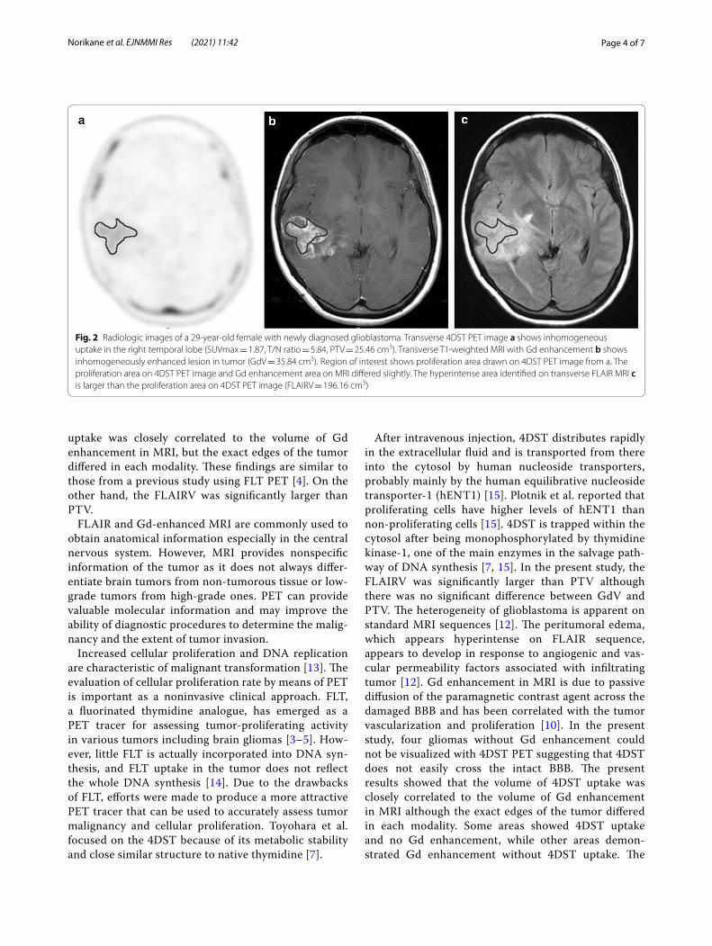
Fig. 2 Radiologic images of a 29-year-old female with newly diagnosed glioblastoma. Transverse 4DST PET image a shows inhomogeneous uptake in the right temporal lobe (SUVmax = 1.87, T/N ratio = 5.84, PTV = 25.46 cm3). Transverse T1-weighted MRI with Gd enhancement b shows inhomogeneously enhanced lesion in tumor (GdV = 35.84 cm3). Region of interest shows proliferation area drawn on 4DST PET image from a. The proliferation area on 4DST PET image and Gd enhancement area on MRI differed slightly. The hyperintense area identified on transverse FLAIR MRI c is larger than the proliferation area on 4DST PET image (FLAIRV = 196.16 cm3)

Page 5 of 7Norikane et al. EJNMMI Res (2021) 11:42
regional distribution of 4DST uptake with and with-out Gd enhancement in MRI suggests the presence of various tissue compartments in which 4DST uptake is due to the breakdown of the BBB, increased transport, increased proliferation or a variable combination of these components. 4DST PET may have a role not only in delineating neovascularization but also in evaluating the cellular proliferation and other aspects of tumo-rigenesis including invasion, progression and response
to chemoradiation. The present study included only patients with newly diagnosed gliomas. The role of 4DST PET may be more evident in patients with post-treatment and recurrent gliomas in which MRI abnor-malities were affected by radiation therapy. Further studies including histological analysis and patients with post-treatment and recurrent gliomas are needed to determine the relationship between 4DST uptake and Gd enhancement and FLAIR signal in the tumor.
Fig. 3 Plot of the volume of 4DST uptake and Gd enhancement in MRI (a), showing a significant correlation of the two parameters (Spearman correlation coefficient 0.941, P < 0.001). Plot of the volume of 4DST uptake and hyperintense FLAIR in MRI (b), showing a significant correlation of the two parameters (Spearman correlation coefficient 0.682, P < 0.001)

Page 6 of 7Norikane et al. EJNMMI Res (2021) 11:42
Although 4DST PET is feasible for brain glioma imag-ing, its mechanism of uptake and biological significance are not yet understood. The value of the current data is limited by the retrospective character of the study and the small number of patients. The most important limita-tion was the lack of pathological correlation to the imag-ing findings, especially regarding the area of difference between 4DST uptake and Gd enhancement and FLAIR signal. Nowosielski et al. compared MRI with FLT and O-(2-[18F]fluoroethyl)-L-tyrosine (FET) PET in high-grade gliomas [16]. They concluded that FET but not FLT PET was able to detect metabolic active tumor tissue beyond contrast enhancing tumor on MRI [16]. To date, no reports have compared 4DST PET with FET PET in glioma. Accurate tumor detection and delineation before surgical resection are of great importance [17]. Nikaki et al. reported that the non-specific binding of FLT may be related to the false positive results, and could impair proper tumor delineation and characterization in gliomas [17]. The dynamic 4DST PET was not performed in the present study. A previous study by Toyohara et al. was performed brain dynamic 4DST scans and reported that physiologic uptake was observed in salivary glands, nasal mucosa and bone marrow [18].
Recently, Ceccon et al. evaluated the response to adju-vant temozolomide chemotherapy using FET PET and Gd-enhanced MRI in patients with newly diagnosed glioma [19]. They showed that relative changes of meta-bolic tumor volume (MTV) and T/N ratio obtained from FET PET, not MRI, significantly longer progression-free survival and overall survival (OS) [19]. Schwarzenberg and colleagues also reported that relative 3,4-dihydroxy-6-[18F]fluoro-L-phenylalanine (FDOPA) MTV change relative to baseline following bevacizumab and irinotecan predicted a significantly prolonged OS [20]. An impor-tant indication is the possibility that the cell proliferation imaging such as 4DST could be used for early treatment response assessment. There are currently no reports for treatment response assessment using 4DST PET in patients with gliomas. Further studies of larger numbers of patients are needed to address the clinical utility of 4DST PET for early evaluation of treatment response.
ConclusionThese preliminary results in a small patient population indicate that tumor proliferation assessed by 4DST PET is closely associated with tumor-induced neovasculariza-tion determined by Gd-enhanced MRI in patients with newly diagnosed glioma. Further prospective studies, with greater number of patients, will demonstrate the usefulness of 4DST PET for tumor delineation, treatment response and prognosis, in patients with glioma.
AcknowledgementsNot applicable.
Authors’ contributionsAll authors contributed to the concept and design of the study. The study was designed by TN and YY. Material preparation, data collection and analysis were performed by TN, KM, YM, KT, TH and KM. The first draft of the manuscript was written by TN and reviewed by YY, JT and YN. All authors read and approved the final manuscript.
FundingNo funding was received.
Availability of data and materialAll results are provided in the manuscript.
Declarations
Ethics approval and consent to participateAll procedures performed in studies involving human participants were in accordance with the ethical standards of the institutional and/or national research committee and with the 1964 Declaration of Helsinki and its later amendments or comparable ethical standards. Informed consent was waived due to its retrospective nature.
Consent for publicationNot applicable.
Competing interestsThe authors declare that they have no competing interests.
Author details1 Department of Radiology, Faculty of Medicine, Kagawa University, 1750-1 Ikenobe, Miki-cho, Kita-gun, Kagawa 761-0793, Japan. 2 Department of Clinical Radiology, Kagawa University Hospital, Kita-gun, Kagawa, Japan. 3 Department of Neurological Surgery, Faculty of Medicine, Kagawa University, Kita-gun, Kagawa, Japan. 4 Research Team for Neuroimaging, Tokyo Metropolitan Insti-tute of Gerontology, Tokyo, Japan.
Received: 10 February 2021 Accepted: 23 April 2021
References 1. Costa A, Silvestrini R, Mochen C, Lequaglie C, Boracchi P, Faranda
A, et al. P53 expression, DNA ploidy and S-phase cell fraction in operable locally advanced non-small-cell lung cancer. Br J Cancer. 1996;73:914–9.
2. Rohren EM, Turkington TG, Coleman RE. Clinical applications of PET in oncology. Radiology. 2004;231:305–32.
3. Hatakeyama T, Kawai N, Nishiyama Y, Yamamoto Y, Sasakawa Y, Ichikawa T, et al. 11C-methionine (MET) and 18F-fluorothymidine (FLT) PET in patients with newly diagnosed glioma. Eur J Nucl Med Mol Imaging. 2008;35:2009–17.
4. Yamamoto Y, Wong TZ, Turkington TG, Hawk TC, Reardon DA, Coleman RE. 3’-deoxy-3’-[F-18]fluorothymidine positron emission tomography in patients with recurrent glioblastoma multiforme: comparison with Gd-DTPA enhanced magnetic resonance imaging. Mol Imaging Biol. 2006;8:340–7.
5. Yamamoto Y, Ono Y, Aga F, Kawai N, Kudomi N, Nishiyama Y. Correlation of 18F-FLT uptake with tumor grade and Ki-67 immunohistochemistry in patients with newly diagnosed and recurrent gliomas. J Nucl Med. 2012;53:1911–5.
6. Ogawa T, Kawai N, Miyake K, Shinomiya A, Yamamoto Y, Nishiyama Y, et al. Diagnostic value of PET/CT with 11C-methionine (MET) and 18F-fluoro-thymidine (FLT) in newly diagnosed glioma based on the 2016 WHO classification. EJNMMI Res. 2020;10:44.
Page 7 of 7Norikane et al. EJNMMI Res (2021) 11:42
7. Toyohara J, Kumata K, Fukushi K, Irie T, Suzuki K. Evaluation of [methyl-14C]4′-thiothymidine for in vivo DNA synthesis imaging. J Nucl Med. 2006;47:1717–22.
8. Tanaka K, Yamamoto Y, Maeda Y, Yamamoto H, Kudomi N, Kawai N, et al. Correlation of 4′-[methyl-11C]-thiothymidine uptake with Ki-67 immu-nohistochemistry and tumor grade in patients with newly diagnosed gliomas in comparison with 11C-methionine uptake. Ann Nucl Med. 2016;30:89–96.
9. Toyota Y, Miyake K, Kawai N, Hatakeyama T, Yamamoto Y, Toyohara J, et al. Comparison of 4′-[methyl-11C]thiothymidine (11C–4DST) and 3′-deoxy-3′-[18F]fluorothymidine (18F-FLT) PET/CT in human brain glioma imaging. EJNMMI Res. 2015;5:7.
10. Earnest F 4th, Kelly PJ, Scheithauer BW, Kall BA, Cascino TL, Ehman RL, et al. Cerebral astrocytomas: histopathologic correlation of MR and CT contrast enhancement with stereotactic biopsy. Radiology. 1988;166:823–7.
11. Chang PD, Malone HR, Bowden SG, Chow DS, Gill BJA, Ung TH, et al. A multiparametric model for mapping cellularity in glioblastoma using radiographically localized biopsies. AJNR Am J Neuroradiol. 2017;38:890–8.
12. Shukla G, Alexander GS, Bakas S, Nikam R, Talekar K, Palmer JD, et al. Advanced magnetic resonance imaging in glioblastoma: a review. Chin Clin Oncol. 2017;6:40.
13. la Fougère C, Suchorska B, Bartenstein P, Kreth FW, Tonn JC. Molecular imaging of gliomas with PET: opportunities and limitations. Neuro Oncol. 2011;13:806–19.
14. Rasey JS, Grierson JR, Wiens LW, Kolb PD, Schwartz JL. Validation of FLT uptake as a measure of thymidine kinase-1 activity in A549 carcinoma cells. J Nucl Med. 2002;43:1210–7.
15. Plotnik DA, Wu S, Linn GR, Yip FCT, Comandante NL, Krohn KA, Toyohara J, Schwartz JL. In vitro analysis of transport and metabolism of 4′-thiothy-midine in human tumor cells. Nucl Med Biol. 2015;42:470–4.
16. Nowosielski M, DiFranco MD, Putzer D, Seiz M, Recheis W, Jacobs AH, et al. An intra-individual comparison of MRI, [18F]-FET and [18F]-FLT PET in patients with high-grade gliomas. PLoS ONE. 2014;23:e95830.
17. Nikaki A, Angelidis G, Efthimiadou R, Tsougos I, Valotassiou V, Fountas K, et al. 18F-fluorothymidine PET imaging in gliomas: an update. Ann Nucl Med. 2017;31:495–505.
18. Toyohara J, Nariai T, Sakata M, Oda K, Ishii K, Kawabe T, et al. Whole-body distribution and brain tumor imaging with 11C–4DST: a pilot study. J Nucl Med. 2011;52:1322–8.
19. Ceccon G, Lohmann P, Werner JM, Tscherpel C, Dunkl V, Stoffels G, et al. Early treatment response assessment using 18F-FET PET compared to contrast-enhanced MRI in glioma patients following adjuvant temozolo-mide chemotherapy. J Nucl Med. 2020. https:// doi. org/ 10. 2967/ jnumed. 120. 254243.
20. Schwarzenberg J, Czernin J, Cloughesy TF, Ellingson BM, Pope WB, Grogan T, et al. Treatment response evaluation using 18F-FDOPA PET in patients with recurrent malignant glioma on bevacizumab therapy. Clin Cancer Res. 2014;20:3550–9.
Publisher’s NoteSpringer Nature remains neutral with regard to jurisdictional claims in pub-lished maps and institutional affiliations.


![Mechanical Engineering Research Journalconvection heat transfer of Al2O3 nanoparticle enhanced N-butyl-N-methyl pyrrolidinium bis{trifluoromethyl)sulfonyl} imide ([C4mpyrr][NTf2])](https://img.pdfslide.tips/doc/110x75/60180d6c8ee8432e99113cbb/mechanical-engineering-research-convection-heat-transfer-of-al2o3-nanoparticle-enhanced.jpg)