Embed Size (px)
Citation preview
Diplomarbeit
Correlation of microbiological findings in patients with
suspected clostridial myonecrosis
Martina Wernik
zur Erlangung des akademischen Grades
Doktorin der gesamten Heilkunde
(Dr. med. univ.)
an der
Medizinischen Universität Graz
ausgeführt am
Institut für Hygiene, Mikrobiologie und Umweltmedizin
unter der Anleitung von
Univ.- Prof. Dr. Andrea Grisold
Graz, 11.November.2014 Martina Wernik
i
Eidesstattliche Erklärung
Ich erkläre ehrenwörtlich, dass ich die vorliegende Arbeit selbstständig und ohne fremde
Hilfe verfasst habe, andere als die angegebenen Quellen nicht verwendet habe und die den
benutzten Quellen wörtlich oder inhaltlich entnommenen Stellen als solche kenntlich
gemacht habe.
Graz, am 11.November.2014 Martina Wernik eh.
ii
Acknowledgements
I would like to take this opportunity to thank Univ.-Prof. Dr. Grisold, who was in charge of
my diploma thesis, for her support and advice as well as helpful discussions during the
working process. I gratefully acknowledge Iris Fuchs and Anna Tamussino for editorial
assistance in the preparation of this work, and Dr. Fink-Neuböck for her assistance in
searching the patient database at the Medical University of Graz. I would also like to thank
Univ.-Prof. Dr. Smolle-Juettner for the opportunity of this thesis.
Moreover, with deep gratitude, I want to thank my family and friends, who went along my
side during my studies and were never tired of supporting.
iii
Zusammenfassung
Hintergrund. Infektionen durch Clostridien sind ein seltenes aber sehr komplexes
Krankheitsbild. Die Erkrankung ist aggressiv progredient und stellt sowohl an den
Patienten, der um sein Überleben kämpft, als auch an die behandelnden Ärzte in
Diagnosefindung und Therapie sehr hohe Ansprüche.
Methoden. Durchgeführt wurde eine retrospektive Studie, um die Methoden der
Diagnosefindung dieses aggressiven Krankheitsbildes näher zu untersuchen. Dabei wurden
zwei Methoden genauer betrachtet und die Ergebnisse miteinander verglichen: Zum einen
die Gramfärbung, welche im Labor zuallererst durchgeführt wird und schon einen Hinweis
auf das Erregerspektrum in der Probe liefern kann. Als nächstes wurden die jeweiligen
Ergebnisse der Erregerkulturen analysiert. Anschließend haben wir die Kulturergebnisse
mit den Ergebnissen der dazugehörigen Gramfärbung verglichen. Zusätzlich wurde die
Auslastung des 24h Telefon-Notdienstes, ein Service des Institutes für Hygiene,
Mikrobiologie und Umweltmedizin, der für Kliniker des Landeskrankenhauses Graz (LKH
Graz) zur Verfügung gestellt wird, in Hinblick auf dieses Krankheitsbild untersucht.
Resultate. In den Jahren 2009 bis 2013 wurden 132 Patienten (90 männliche and 42
weibliche), durchschnittliches Alter 57 Jahre, mit Verdacht auf Gasbrand oder
nekrotisierende Fasziitis am LKH Graz behandelt. 23 von 132 Patienten (17%) zeigten in
der Kultur einen Nachweis von Clostridien. 12 von 132 Patienten zeigten in der
Gramfärbung gram-positive Stäbchen. Zwei der 12 Gramfärbungen zeigten als
Endergebnis in der Kultur keine Clostridien, sondern Actinomyces turicensis, ebenfalls ein
gram-positives Stäbchen.
In den 5 Jahren des beobachteten Zeitraumes wurden insgesamt 61 Anrufe von
Mitarbeitern des Institutes für Hygiene, Mikrobiologie und Umweltmedizin gemacht,
bezüglich des Verdachtes auf Gasbrand. Von diesen 61 Anrufen wurden 45 außerhalb der
normalen Arbeitszeiten (8 Uhr - 16 Uhr) getätigt.
Schlussfolgerung. Die Studie hat gezeigt, dass, wenn in der Gramfärbung gram-positive
Stäbchen zu sehen sind, in 83% der Fälle auch tatsächlich Clostridien wachsen. Wenn
allerdings die Gramfärbung negativ war, kann aber nicht ausgeschlossen werden, dass
später in der Kultur Clostridien wachsen.
iv
Abstract
Background. A clostridial infection is a rare disease with a complex, mostly severe and
aggressive progressing course, with enormous demands not just for the patient and his or
her survival but also for clinicians in diagnosis and treatment.
Methods. We performed a retrospective analysis to determine correlation between two
microbiological methods, gram-staining and culture, with regard to clostridial
myonecrosis. In addition, we assessed capacity utilisation of a 24 hours emergency system
provided by the Institute of Hygiene, Microbiology and Environmental Medicine.
Results. From 2009 to 2013 a total of 132 patients (90 males and 42 females), average age
57 years, with suspected gas gangrene that were recorded were included in the analysis. 23
patients (17%) had laboratory evidence of an infection with a Clostridium species. 12 of
132 samples showed evidence of gram-positive rods in the gram-staining. Whereas in ten
cases growth of Clostridium spp. was observed in two cases the gram-positive rods turned
out to be Actinomyces turicensis. 61 calls regarding suspected gas gangrene were made
during the observed time period. 45 were made outside the routine working hours and
therefore carried out by the emergency service.
Conclusion. Our study indicates, regarding to samples with clinical suspicion of gas
gangrene, that the reliability of a gram-staining result with gram-positive rods is acceptable
good and in our case in 83% leads to the result of a cultured-proof of clostridia. In contrast,
a negative result for gram-positive rods in gram-stainings can show clostridial growth after
all.
v
Contents
ACKNOWLEDGEMENTS ...................................................................................... II
ZUSAMMENFASSUNG ........................................................................................ III
ABSTRACT ........................................................................................................... IV
CONTENTS ............................................................................................................ V
GLOSSARY AND ABBREVIATIONS .................................................................. VII
LIST OF FIGURES .............................................................................................. VIII
LIST OF TABLES ................................................................................................. IX
1 INTRODUCTION ............................................................................................ 10
1.1 History .................................................................................................................................... 10
1.2 Aetiology ................................................................................................................................ 10
1.2.1 Morphology 10
1.2.2 Pathogenicity 11
1.3 Epidemiology ......................................................................................................................... 13
1.3.1 Clostridial Infections 13
1.3.2 Occurrence 13
1.3.3 Incidence 13
1.4 Pathogenesis ......................................................................................................................... 14
1.4.1 Risk Factors 14
1.4.2 Traumatic vs. Nontraumatic or “Spontaneous” Gas Gangrene 14
1.5 Clinical Picture ...................................................................................................................... 15
1.5.1 Traumatic Gas Gangrene 15
1.5.2 Nontraumatic Gas Gangrene 16
1.5.3 Mortality 16
1.6 Laboratory and Microbiological Findings .......................................................................... 16
1.7 Clinical Diagnosis ................................................................................................................. 18
vi
1.8 Differential Diagnosis ........................................................................................................... 19
1.9 Therapy .................................................................................................................................. 23
1.10 Prognosis ............................................................................................................................. 26
1.11 Prevention ............................................................................................................................ 26
2 MATERIAL AND METHODS .......................................................................... 27
2.1 Study Setting ......................................................................................................................... 27
2.2 Patients .................................................................................................................................. 27
2.3 Inclusion Criteria ................................................................................................................... 28
2.4 Exclusion Criteria ................................................................................................................. 28
2.5 Data ........................................................................................................................................ 28
2.6 Statistical Analysis ............................................................................................................... 29
3 RESULTS ....................................................................................................... 30
3.1 Referral Diagnosis ................................................................................................................ 30
3.2 Annual distribution ............................................................................................................... 30
3.3 Age and Gender .................................................................................................................... 31
3.4 Distribution of specimens according to origin .................................................................. 33
3.5 Microbiological Results ........................................................................................................ 34
3.5.1 Results of Gram-staining 34
3.5.2 Comparison of gram-stainings with gram-positive rods and cultures with proof of
clostridia 39
3.5.3 Results regarding samples with clostridia 39
3.6 Analysis of emergency calls made by the microbiologists .............................................. 40
4 DISCUSSION ................................................................................................. 43
5 CONCLUSION ................................................................................................ 46
6 REFERENCES ............................................................................................... 47
vii
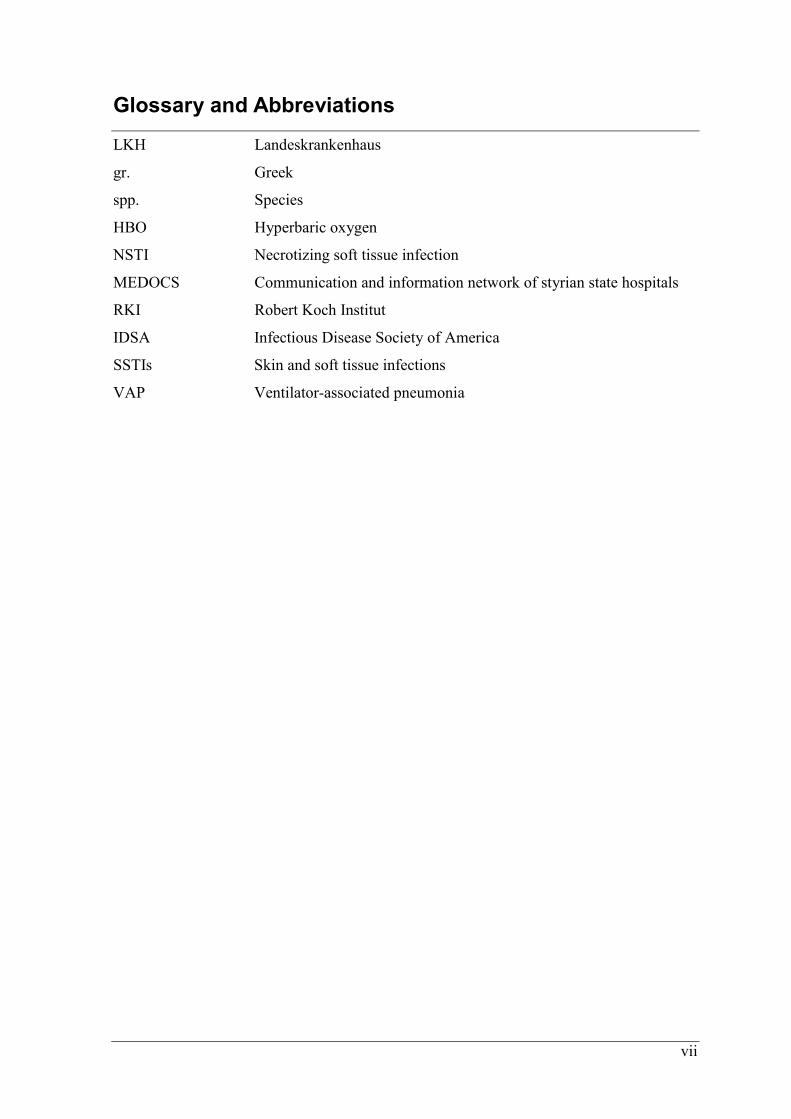
Glossary and Abbreviations
LKH Landeskrankenhaus
gr. Greek
spp. Species
HBO Hyperbaric oxygen
NSTI Necrotizing soft tissue infection
MEDOCS Communication and information network of styrian state hospitals
RKI Robert Koch Institut
IDSA Infectious Disease Society of America
SSTIs Skin and soft tissue infections
VAP Ventilator-associated pneumonia
viii
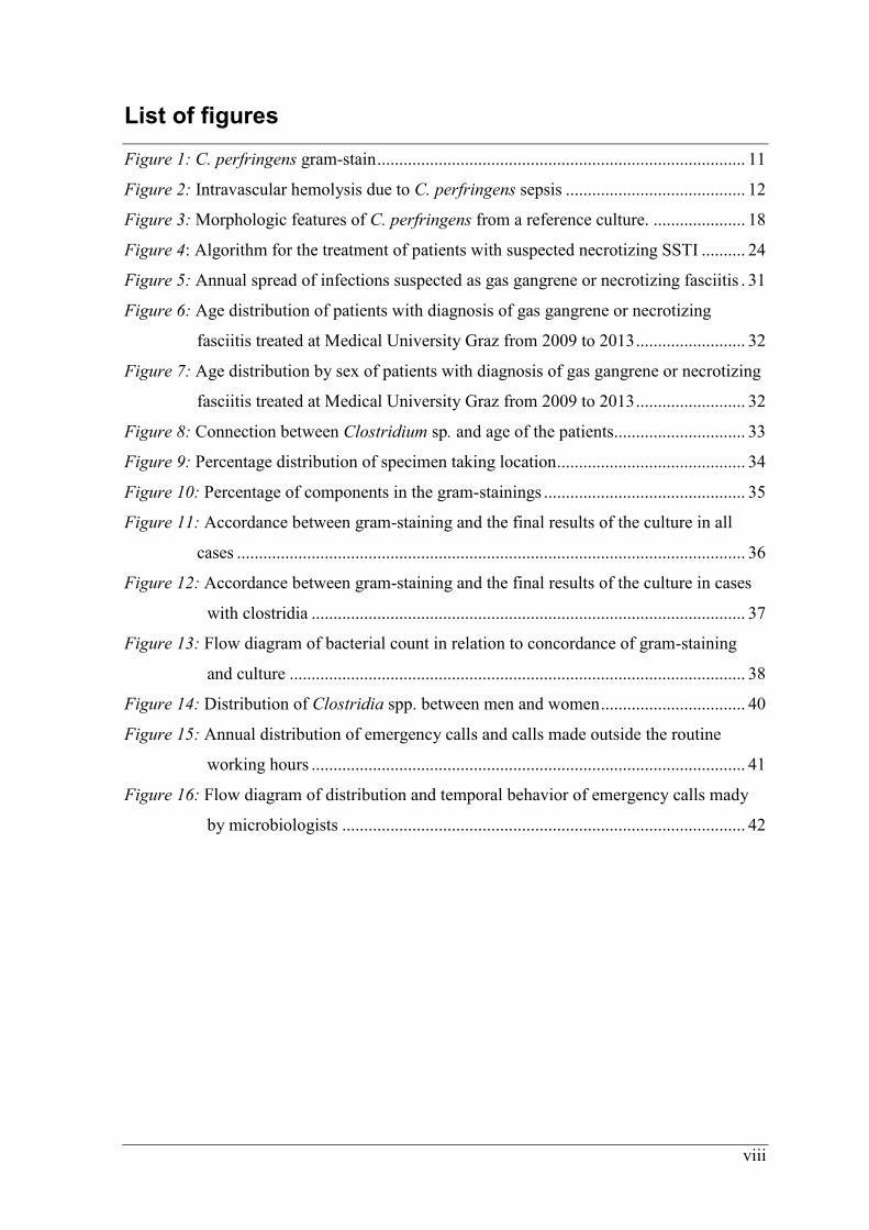
List of figures
Figure 1: C. perfringens gram-stain .................................................................................... 11
Figure 2: Intravascular hemolysis due to C. perfringens sepsis ......................................... 12
Figure 3: Morphologic features of C. perfringens from a reference culture. ..................... 18
Figure 4: Algorithm for the treatment of patients with suspected necrotizing SSTI .......... 24
Figure 5: Annual spread of infections suspected as gas gangrene or necrotizing fasciitis . 31
Figure 6: Age distribution of patients with diagnosis of gas gangrene or necrotizing
fasciitis treated at Medical University Graz from 2009 to 2013 ......................... 32
Figure 7: Age distribution by sex of patients with diagnosis of gas gangrene or necrotizing
fasciitis treated at Medical University Graz from 2009 to 2013 ......................... 32
Figure 8: Connection between Clostridium sp. and age of the patients.............................. 33
Figure 9: Percentage distribution of specimen taking location ........................................... 34
Figure 10: Percentage of components in the gram-stainings .............................................. 35
Figure 11: Accordance between gram-staining and the final results of the culture in all
cases .................................................................................................................... 36
Figure 12: Accordance between gram-staining and the final results of the culture in cases
with clostridia ................................................................................................... 37
Figure 13: Flow diagram of bacterial count in relation to concordance of gram-staining
and culture ........................................................................................................ 38
Figure 14: Distribution of Clostridia spp. between men and women ................................. 40
Figure 15: Annual distribution of emergency calls and calls made outside the routine
working hours ................................................................................................... 41
Figure 16: Flow diagram of distribution and temporal behavior of emergency calls mady
by microbiologists ............................................................................................ 42
ix
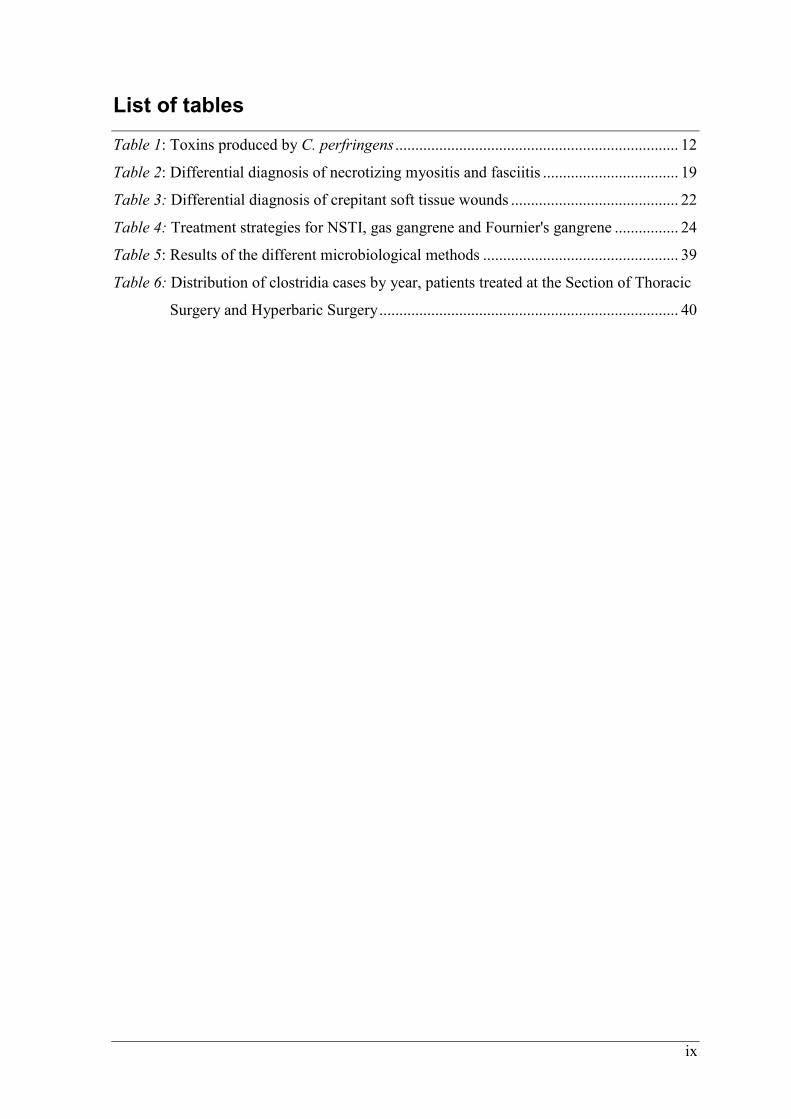
List of tables
Table 1: Toxins produced by C. perfringens ....................................................................... 12
Table 2: Differential diagnosis of necrotizing myositis and fasciitis .................................. 19
Table 3: Differential diagnosis of crepitant soft tissue wounds .......................................... 22
Table 4: Treatment strategies for NSTI, gas gangrene and Fournier's gangrene ................ 24
Table 5: Results of the different microbiological methods ................................................. 39
Table 6: Distribution of clostridia cases by year, patients treated at the Section of Thoracic
Surgery and Hyperbaric Surgery ........................................................................... 40
10
1 Introduction
1.1 History
Clostridium spp. include over 200 described species that have diversity in the meaning for
humans. Clostridia (gr: >> klostér<< rachis) occur ubiquitous in soil and marine sediments.
Therefore and because of their fulminant and often fatal course of disease, gas gangrene
has been highly feared, especially in times of war. Clostridium perfringens is beside other
Clostridium species and other species of bacteria the principal germ that causes the clinical
picture of necrotising fasciitis, vernacular better known as gas gangrene (clostridial
myonecrosis). Other species, than gas gangrene causing clostridia, that generated interest
due to their severity and often fatal nature are Clostridium botulinum and Clostridium
tetani, whose clinical features were already described by one of the earliest medical
writers, Hippocrates (1). C. perfringens (former name: Welch-Fraenkel-bacillus or
Clostridium welchii) has been first characterized by W.H. Welch and G.H.F. Nutall in
1892. Earlier Pirogoff, a Russian army doctor did the first description of the clinical
features during the Crimean War. The first microbiological characterization was conducted
by Veillon and Zuber in 1898. Heaped appearance was mainly seen and feared during wars
due to the number of traumatic injuries: in World War I approximately 100.000 soldiers
died of gas gangrene. Enteritis necroticans mainly caused by C.perfringens Typ C was first
described in Germany after World War II and later in the 1960’s in New Guinea (2).
1.2 Aetiology
1.2.1 Morphology
Clostridia are putrefactive, anaerobe, gram-positive rod-shaped bacteria that are spore
forming, and except for some species (C. perfringens) flagellate. Spores are relatively
resistant as far as environmental surrounding is concerned, especially to heat and
dehydration, whereby a survival beyond anaerobic conditions is possible. Among many
different species that have been isolated, there are only some that are regularly associated
with human diseases of any severity. Under human medical aspects there are four species
of clostridia that are of special interest: C. tetani (tetanus), C. botulinum (botulism), C.
perfringens (gas gangrene) and C. difficile (antibiotic associated colitis).
11
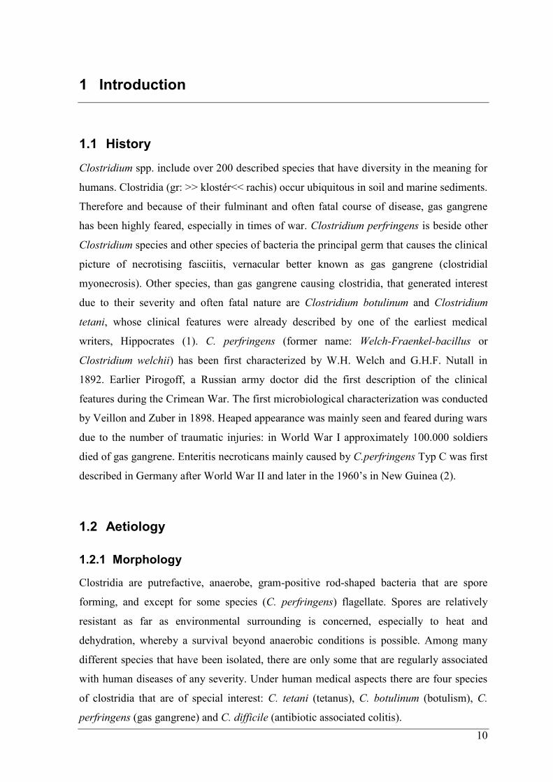
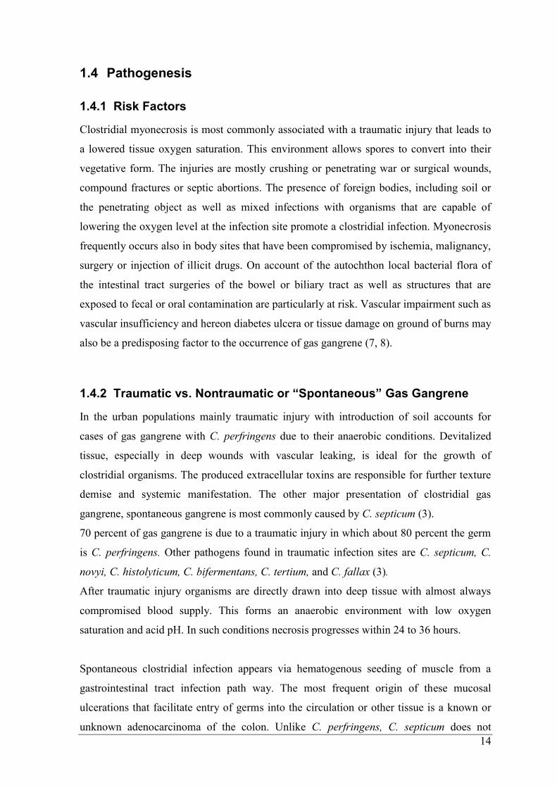
With a light optical microscope, one can usually see gram-positively stained, cloddy and
often pleomorphic rods (Fig.1). In working stocks and patient material the main difference
between C. perfringens and other Clostridium species of the gas gangrene group is that C.
perfringens is unmovable and spores are invisible.
Figure 1: C. perfringens gram-stain
Reproduced from: (3)
According to present knowledge gas gangrene is usually caused by C. perfringens, less
commonly clostridial myonecrosis is due to C. septicum, C. histolyticum, C. novyi, C.
sordellii, and C. bifermentans (1, 2, 4). These clostridial species are also called clostridia
of the gas gangrene group.
1.2.2 Pathogenicity
Pathogenicity and virulencefactors of gas gangrene agents are the exotoxins and
exoencymes (Tab. 1). They cause cell and tissue damage, muscle decomposition, oedema
and gas production in tissue as well as destruction of erythrocytes and leukocytes. Beside
the main exotoxin Phospholipase C (Lecithinase , alpha-Toxin), the vegetative forms of the
gas gangrene group clostridia also generate collagenases, hyaluronidases, haemolysines
and proteases. Typ A of C. perfringens is mainly responsible for gas gangrene and food
poisoning if it is producing enterotoxines. Typ C causes Enterocolitis necroticans by beta-
toxins (2).
12
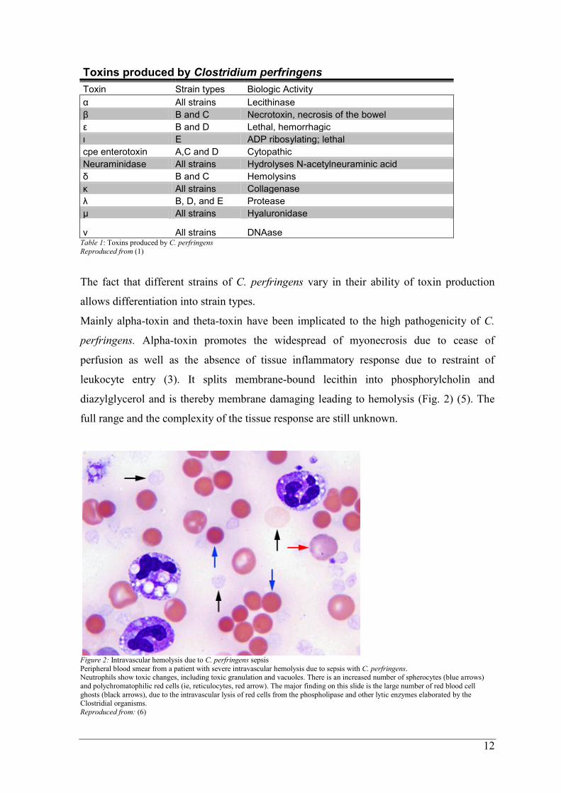
Toxins produced by Clostridium perfringens
Toxin Strain types Biologic Activity
α All strains Lecithinase
β B and C Necrotoxin, necrosis of the bowel
ε B and D Lethal, hemorrhagic
ι E ADP ribosylating; lethal
cpe enterotoxin A,C and D Cytopathic
Neuraminidase All strains Hydrolyses N-acetylneuraminic acid
δ B and C Hemolysins
κ All strains Collagenase
λ B, D, and E Protease
μ All strains Hyaluronidase
ν All strains DNAase Table 1: Toxins produced by C. perfringens
Reproduced from (1)
The fact that different strains of C. perfringens vary in their ability of toxin production
allows differentiation into strain types.
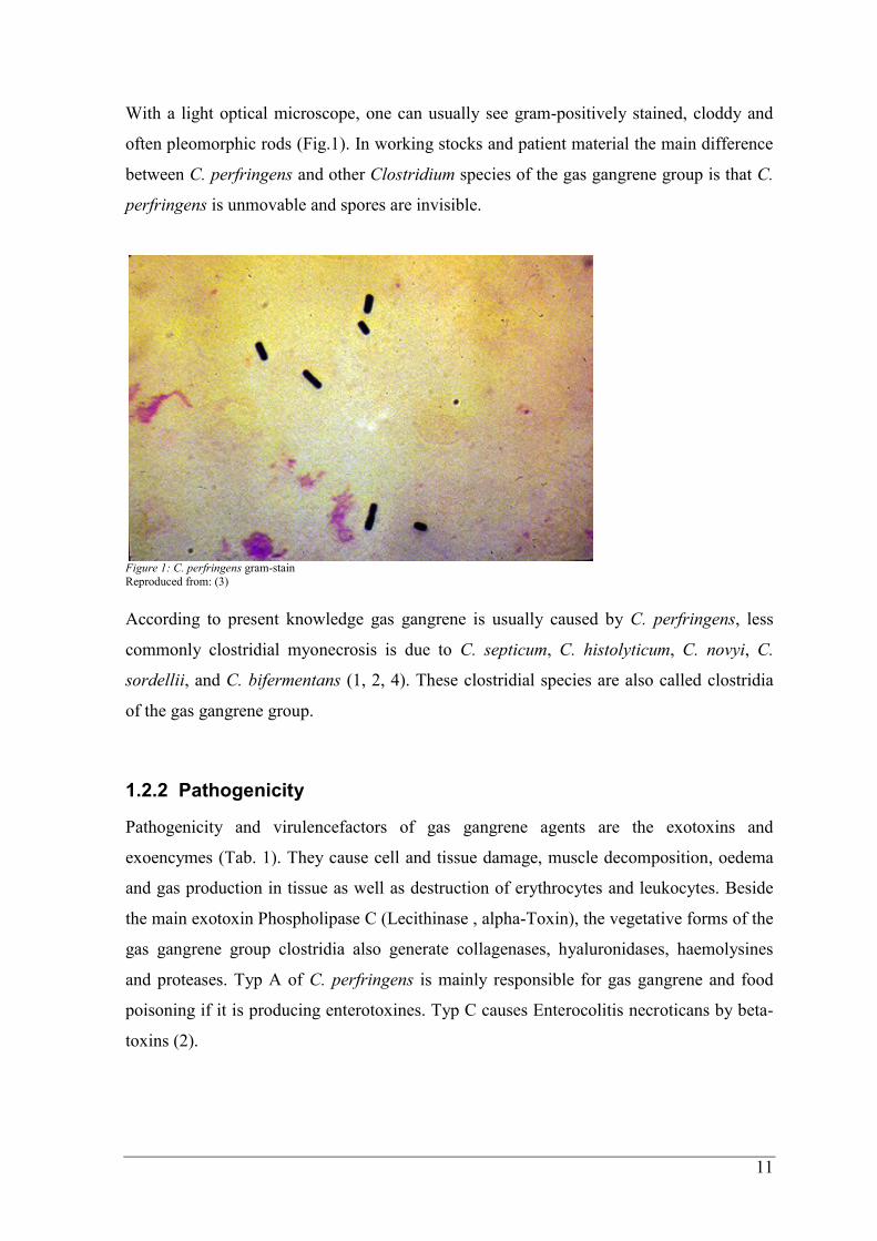
Mainly alpha-toxin and theta-toxin have been implicated to the high pathogenicity of C.
perfringens. Alpha-toxin promotes the widespread of myonecrosis due to cease of
perfusion as well as the absence of tissue inflammatory response due to restraint of
leukocyte entry (3). It splits membrane-bound lecithin into phosphorylcholin and
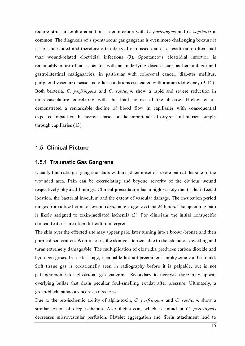
diazylglycerol and is thereby membrane damaging leading to hemolysis (Fig. 2) (5). The
full range and the complexity of the tissue response are still unknown.
Figure 2: Intravascular hemolysis due to C. perfringens sepsis
Peripheral blood smear from a patient with severe intravascular hemolysis due to sepsis with C. perfringens. Neutrophils show toxic changes, including toxic granulation and vacuoles. There is an increased number of spherocytes (blue arrows)
and polychromatophilic red cells (ie, reticulocytes, red arrow). The major finding on this slide is the large number of red blood cell
ghosts (black arrows), due to the intravascular lysis of red cells from the phospholipase and other lytic enzymes elaborated by the Clostridial organisms.
Reproduced from: (6)
13
1.3 Epidemiology
1.3.1 Clostridial Infections
In literature we find a differentiation between three major categories of clostridial soft
tissue infections: wound contamination, anaerobic cellulitis, and myonecrosis (gas
gangrene).
Wound contamination is due to spores or vegetative organisms found in soil. This
is a frequent type of contamination, but in the absence of damaged tissue and a
vessel impairment, it does not inevitably lead to infection.
Anaerobic cellulitis is the next step found in patients with clostridial contamination
in open wounds and devitalized tissue. The infection remains locally. Bacteremia
and invasion of healthy tissue is usually not seen. Prompt and adequate
management maintain a good outcome.
Clostridial gas gangrene is comparatively fulminant and life-threatening, due to the
extensive tissue damage and invasion of muscle tissue. This article is designed to
present the two types of clostridial myonecrosis: traumatic and spontaneous gas
gangrene.
1.3.2 Occurrence
Clostridia spores exist in the soil and marine sediments as well as they are a part of the
gastrointestinal microflora of 70 percent of humans and also animals (1). From the
remaining species only C. septicum occurs in two percent of humans. Thus it is not
considered a normal bowel inhabitant. Spores of C. perfringens were found numerously in
soils compared to other species that were rarely found (2).
Most gas gangrene causing clostridia need obligate anaerobic conditions to increase and
convert from the spore to the vegetative form. However, a few species such as C.
histolyticum, C. tertium, C. innocuum and some strains of C. perfringens are able to
develop in aerotolerant terms as well (1).
1.3.3 Incidence
There is no explicit data in literature available about the incidence of clostridial
myonecrosis in Austria. In 1997 122 cases have been reported in Germany (5). It is
assumable that the number of cases between these two countries is at a comparable count.
14
1.4 Pathogenesis
1.4.1 Risk Factors
Clostridial myonecrosis is most commonly associated with a traumatic injury that leads to
a lowered tissue oxygen saturation. This environment allows spores to convert into their
vegetative form. The injuries are mostly crushing or penetrating war or surgical wounds,
compound fractures or septic abortions. The presence of foreign bodies, including soil or
the penetrating object as well as mixed infections with organisms that are capable of
lowering the oxygen level at the infection site promote a clostridial infection. Myonecrosis
frequently occurs also in body sites that have been compromised by ischemia, malignancy,
surgery or injection of illicit drugs. On account of the autochthon local bacterial flora of
the intestinal tract surgeries of the bowel or biliary tract as well as structures that are
exposed to fecal or oral contamination are particularly at risk. Vascular impairment such as
vascular insufficiency and hereon diabetes ulcera or tissue damage on ground of burns may
also be a predisposing factor to the occurrence of gas gangrene (7, 8).
1.4.2 Traumatic vs. Nontraumatic or “Spontaneous” Gas Gangrene
In the urban populations mainly traumatic injury with introduction of soil accounts for
cases of gas gangrene with C. perfringens due to their anaerobic conditions. Devitalized
tissue, especially in deep wounds with vascular leaking, is ideal for the growth of
clostridial organisms. The produced extracellular toxins are responsible for further texture
demise and systemic manifestation. The other major presentation of clostridial gas
gangrene, spontaneous gangrene is most commonly caused by C. septicum (3).
70 percent of gas gangrene is due to a traumatic injury in which about 80 percent the germ
is C. perfringens. Other pathogens found in traumatic infection sites are C. septicum, C.
novyi, C. histolyticum, C. bifermentans, C. tertium, and C. fallax (3).
After traumatic injury organisms are directly drawn into deep tissue with almost always
compromised blood supply. This forms an anaerobic environment with low oxygen
saturation and acid pH. In such conditions necrosis progresses within 24 to 36 hours.
Spontaneous clostridial infection appears via hematogenous seeding of muscle from a
gastrointestinal tract infection path way. The most frequent origin of these mucosal
ulcerations that facilitate entry of germs into the circulation or other tissue is a known or
unknown adenocarcinoma of the colon. Unlike C. perfringens, C. septicum does not
15
require strict anaerobic conditions, a coinfection with C. perfringens and C. septicum is
common. The diagnosis of a spontaneous gas gangrene is even more challenging because it
is not entertained and therefore often delayed or missed and as a result more often fatal
than wound-related clostridial infections (3). Spontaneous clostridial infection is
remarkably more often associated with an underlying disease such as hematologic and
gastrointestinal malignancies, in particular with colorectal cancer, diabetes mellitus,
peripheral vascular disease and other conditions associated with immunodeficiency (9–12).
Both bacteria, C. perfringens and C. septicum show a rapid and severe reduction in
microvasculature correlating with the fatal course of the disease. Hickey et al.
demonstrated a remarkable decline of blood flow in capillaries with consequential
expected impact on the necrosis based on the importance of oxygen and nutrient supply
through capillaries (13).
1.5 Clinical Picture
1.5.1 Traumatic Gas Gangrene
Usually traumatic gas gangrene starts with a sudden onset of severe pain at the side of the
wounded area. Pain can be excruciating and beyond severity of the obvious wound
respectively physical findings. Clinical presentation has a high variety due to the infected
location, the bacterial inoculum and the extent of vascular damage. The incubation period
ranges from a few hours to several days, on average less than 24 hours. The upcoming pain
is likely assigned to toxin-mediated ischemia (3). For clinicians the initial nonspecific
clinical features are often difficult to interpret.
The skin over the effected site may appear pale, later turning into a brown-bronze and then
purple discoloration. Within hours, the skin gets tensens due to the edematous swelling and
turns extremely damageable. The multiplication of clostridia produces carbon dioxide and
hydrogen gases. In a later stage, a palpable but not preeminent emphyseme can be found.
Soft tissue gas is occasionally seen in radiography before it is palpable, but is not
pathognomonic for clostridial gas gangrene. Secondary to necrosis there may appear
overlying bullae that drain peculiar foul-smelling exudat after pressure. Ultimately, a
green-black cutaneous necrosis develops.
Due to the pro-ischemic ability of alpha-toxin, C. perfringens and C. septicum show a
similar extent of deep ischemia. Also theta-toxin, which is found in C. perfringens
decreases microvascular perfusion. Platelet aggregation and fibrin attachment lead to
16
obstruction of small vessels reducing blood supply unlike other soft tissue infections with
inflammation increasing blood flow. Studies show that vascular leukostasis and the lack of
neutrophils can be assessed as clinical characteristics of clostridial myonecrosis.
The patient soon looks severely ill and the course is usually rapidly deteriorating. Signs of
a systemic intoxication including tachycardia and low-grade fever develop fast. Later
anemia, jaundice and delirious changes often lead to profound shock and multiorgan
failure. Crucial for the outcome is the period between the first symptom and the beginning
of the treatment (14).
1.5.2 Nontraumatic Gas Gangrene
Occasionally, gas gangrene appears without an obvious external entry path. In this case C.
septicum is the primary contributor of Clostridium spp. found in samples. It is quite
aerotolerant and spreads by bacteremia and can grow in normal undamaged tissue. The
typical clinical findings of traumatic gas gangrene are more difficult to diagnose in
spontaneous myonecrosis, as there is a multifocal involvement observed with even more
fulminant progression than that of C. perfringens myonecrosis. Sometimes only heaviness
or numbness is named as symptom. Inflammatory cells are also rare as with C. perfringens
infections. There is very few knowledge in existing work on the impact to microcirculation
and the spectrum of its alpha-toxin. However, the virulence of infections with C. septicum
is imputable to its alpha-toxin.
1.5.3 Mortality
C. septicum, the main causative agent in spontaneous clostridial infections beside C.
perfringens, shows a higher rate of mortality in adults and children. The mortality rate in
adults with C. septicum myonecrosis is about 79 percent in comparison to 32 percent
mortality rate in adults with C. perfringens myonecrosis (15).
1.6 Laboratory and Microbiological Findings
Laboratory diagnosis is based on two steps: a) Gram-stain and b) microbiological culture
on agar plates. Gram-staining is a fast and well performable method to search for bacteria
in clinical samples. In case of suspected gas gangrene material for the gram-stain can be
extracted from ichor, or muscle biopsy. This provides a direct proof of the presumptive
17
diagnosis and counts as a microbiological emergency, while laboratories have an alert
system to inform the clinicians as soon as the gram-staining is ready.
In an infection with C. perfringens the gram-stain usually shows the typical boxcar-shaped
rods. Inflammation cells, especially leukocytes are often missing due to the fatal nature of
alpha-toxin. Therefore an absence of frank pus is also often seen. Few polymorphonuclear
cells and gram-positive rods in fluids or exudates be among earliest laboratory signs (7). In
the histological assessment sweeping myonecrosis with edematous tissue and thrombosis is
predominant beside a lack of perivascular leukocyte infiltration. Clinical samples usually
do not show any spores. If blood cultures are obtained, bacteremia is only found in about
15 percent of the patients and may be accompanied by intravascular hemolysis (3).
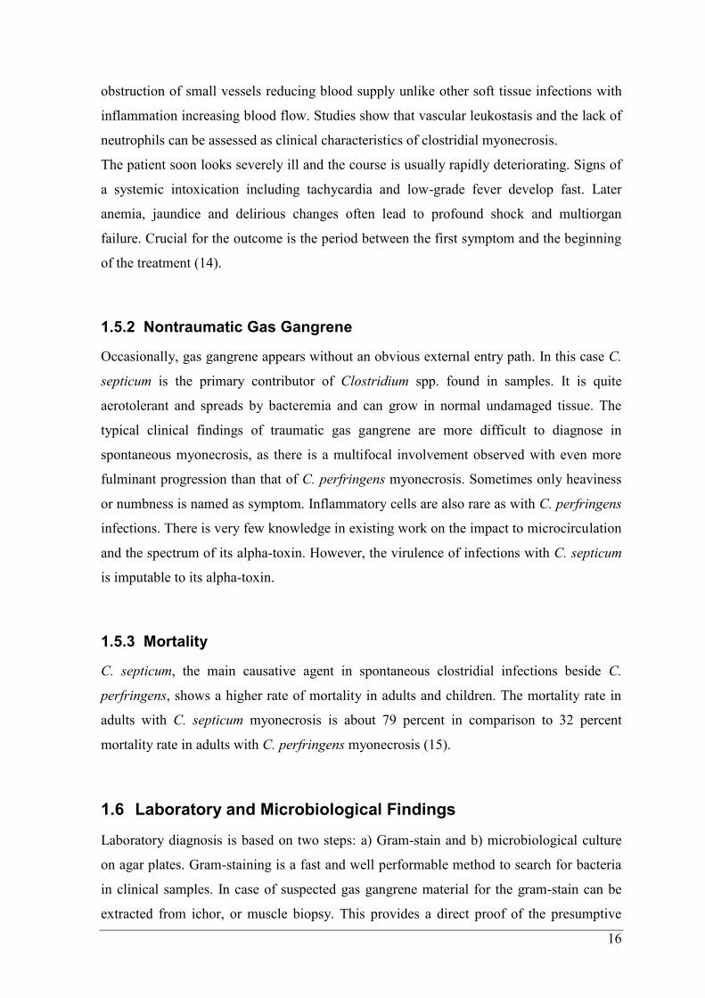
Microbiological cultures are performed on different nutrient agars, such as blood agar or
Schädler agar, with an incubation of the plates under anaerobic conditions at 37°C.
Incubation period is a minimum of 24 hours up to five days (Fig. 3) (16).
18
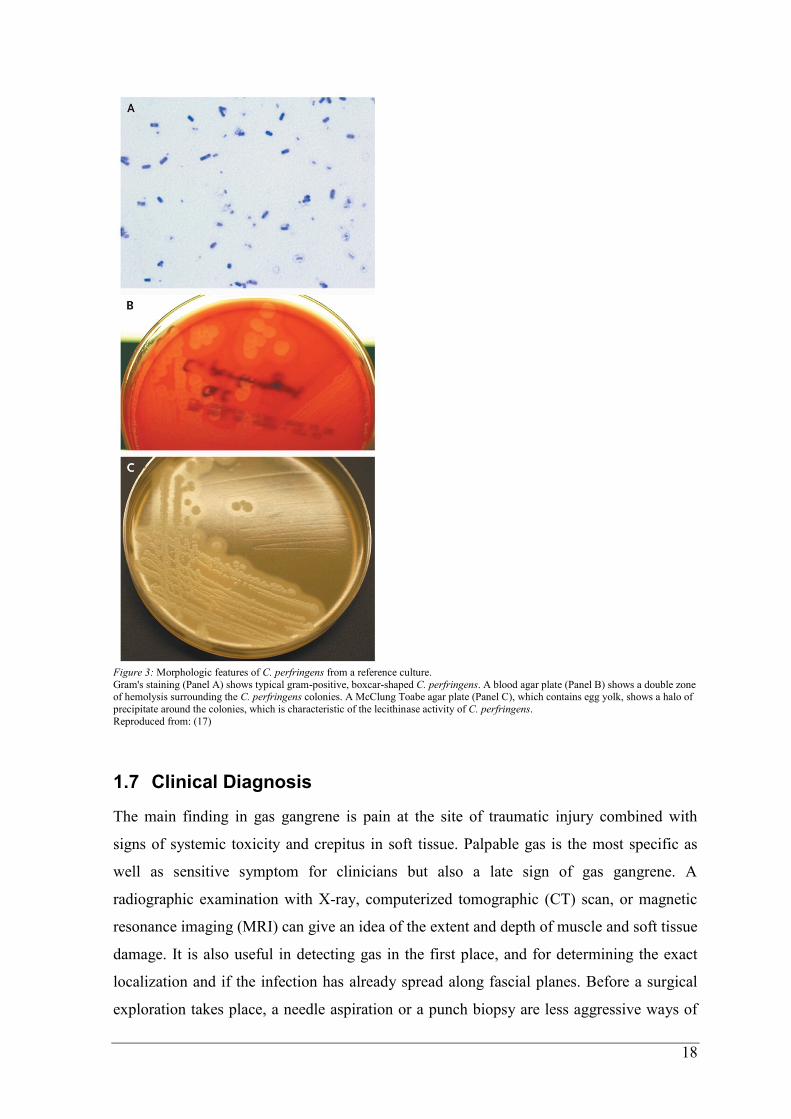
Figure 3: Morphologic features of C. perfringens from a reference culture.
Gram's staining (Panel A) shows typical gram-positive, boxcar-shaped C. perfringens. A blood agar plate (Panel B) shows a double zone of hemolysis surrounding the C. perfringens colonies. A McClung Toabe agar plate (Panel C), which contains egg yolk, shows a halo of
precipitate around the colonies, which is characteristic of the lecithinase activity of C. perfringens.
Reproduced from: (17)
1.7 Clinical Diagnosis
The main finding in gas gangrene is pain at the site of traumatic injury combined with
signs of systemic toxicity and crepitus in soft tissue. Palpable gas is the most specific as
well as sensitive symptom for clinicians but also a late sign of gas gangrene. A
radiographic examination with X-ray, computerized tomographic (CT) scan, or magnetic
resonance imaging (MRI) can give an idea of the extent and depth of muscle and soft tissue
damage. It is also useful in detecting gas in the first place, and for determining the exact
localization and if the infection has already spread along fascial planes. Before a surgical
exploration takes place, a needle aspiration or a punch biopsy are less aggressive ways of
19
getting samples for gram-stain and culture. If there is a reasonable suspicion for gas
gangrene a surgical exploration is unavoidable. As soon as possible patients have to be
transported to a Center for Hyperbaric oxygenation to receive additional therapy.
During surgical procedures a lack of bleeding or contraction after stimulation is a typical
sign for infected muscle tissue.
1.8 Differential Diagnosis
A number of clinical entities can mimic a similar clinical presentation as found in
clostridial myonecrosis (Tab. 3). Diseases under the umbrella term necrotizing soft tissue
infection (NSTI) contain different entities such as types of necrotizing cellulitis, myositis,
and fasciitis. The great diversity of described types of NSTI in the literature should be
remembered, due to their potential of entailing useful pointers in diagnosis and the time of
initiating measures if necessary.The major causatives leading to myonecrosis are Clostrium
spp., Group A Streptococci (Streptococcus pyogenes), or other ß-hemolytic Streptococci
(18). Presumably the biggest challenge lies in differentiating the entities of this clinical
presentation. Early signs include mainly subtle changes and seem to be entirely
indistinguishable (Tab. 2). Progressive changes and typical symptoms indicate an advanced
disease which is why an early diagnosis is the most crucial part of a successful
management.
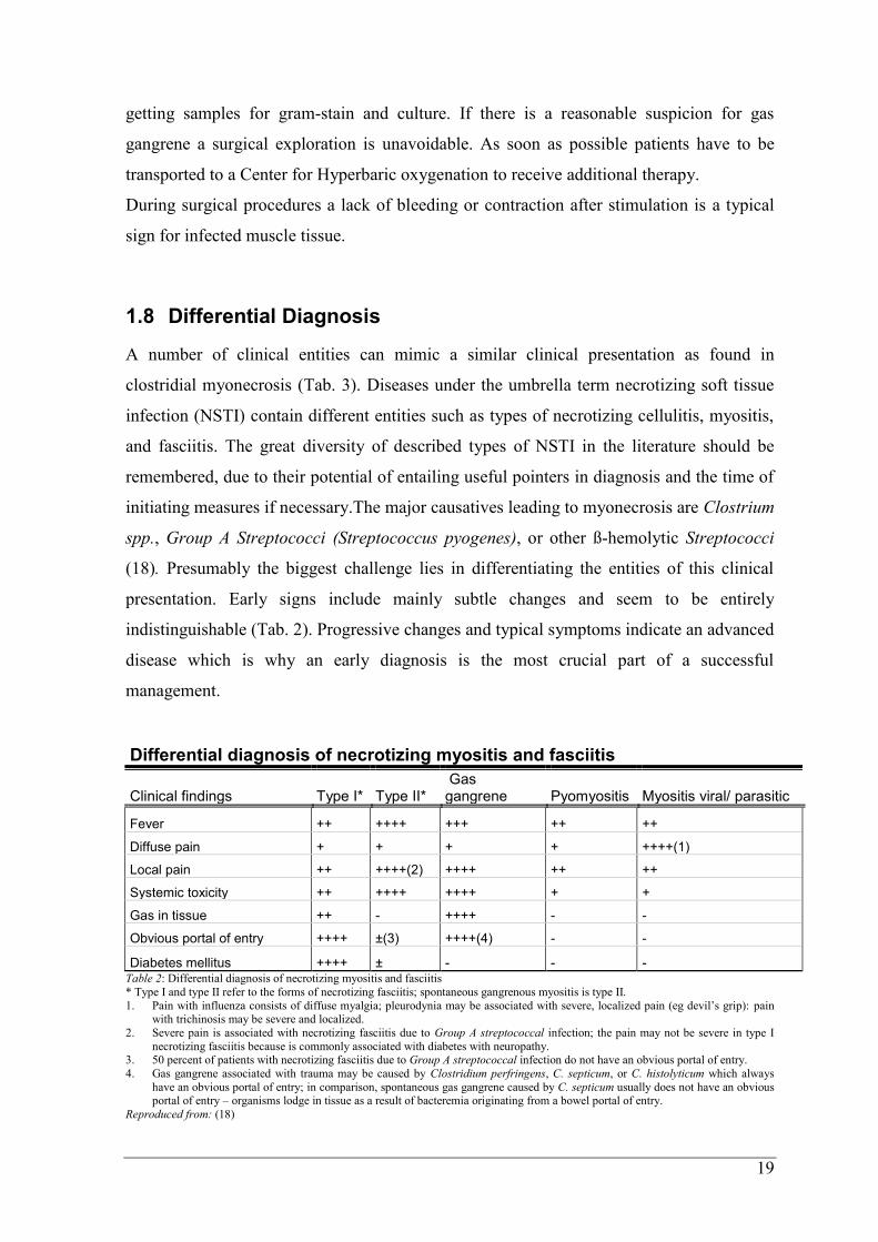
Differential diagnosis of necrotizing myositis and fasciitis
Clinical findings Type I* Type II* Gas gangrene Pyomyositis Myositis viral/ parasitic
Fever ++ ++++ +++ ++ ++
Diffuse pain + + + + ++++(1)
Local pain ++ ++++(2) ++++ ++ ++
Systemic toxicity ++ ++++ ++++ + +
Gas in tissue ++ - ++++ - -
Obvious portal of entry ++++ ±(3) ++++(4) - -
Diabetes mellitus ++++ ± - - - Table 2: Differential diagnosis of necrotizing myositis and fasciitis
* Type I and type II refer to the forms of necrotizing fasciitis; spontaneous gangrenous myositis is type II.
1. Pain with influenza consists of diffuse myalgia; pleurodynia may be associated with severe, localized pain (eg devil’s grip): pain with trichinosis may be severe and localized.
2. Severe pain is associated with necrotizing fasciitis due to Group A streptococcal infection; the pain may not be severe in type I
necrotizing fasciitis because is commonly associated with diabetes with neuropathy. 3. 50 percent of patients with necrotizing fasciitis due to Group A streptococcal infection do not have an obvious portal of entry.
4. Gas gangrene associated with trauma may be caused by Clostridium perfringens, C. septicum, or C. histolyticum which always
have an obvious portal of entry; in comparison, spontaneous gas gangrene caused by C. septicum usually does not have an obvious portal of entry – organisms lodge in tissue as a result of bacteremia originating from a bowel portal of entry.
Reproduced from: (18)
20
As in clostridial myonecrosis, diabetes counts as a predisposing or associated condition.
Also, there seems to be an assuming connection between diabetes and the occurence of
particular bacteria (19).
Group A streptococcal necrotizing myositis is also described as NSTI type II. The infection
is seen in young and healthy people, often to a recent trauma and leads to an aggressive
form of myositis, with intensive pain, fever, and swelling. Generally it is a monomicrobial
infection with Group A ß-hemolytic Streptococcus, occasionally a coinfection with other
species is possible, most commonly with Staphylococcus aureus (20). The skin over the
infected area may be uninvolved at first, or discolored with sometimes petechiae and
bullae. Type II NSTI is characterized by an aggressive course with vascular impairment
and signs of systemic toxicity. Unlike clostridial myonecrosis in streptococcal myonecrosis
bacteremia and toxemia are seen frequently and are attributed to the high mortality of this
entity. Also a typical finding is the compartment syndrome, due to the rapid spread of
infection and the increasing pressure (7).
Type I necrotizing soft tissue infection includes a poylmicrobial spectrum of causative
agents. A varying combination of anaerobes plus one or more facultative anaerobic
streptococci and Enterobacteriaceae can be isolated (18). The patient is comparatively
older with more than one medical condition and an overall state of poor health. Normally
there is no clear history of trauma. Diabetes, peripheral vascular disease, obesity, chronic
renal insufficiency, alcohol abuse, existing abscesses, blunt trauma, chicken pox, HIV or
AIDS, and i.v. drug abuse are predisposing conditions (20). The infected site is usually the
trunk or perineum, for example Fournier’s gangrene (infection of the male perineum). In
case of Fournier’s gangrene the spectrum varies from facultative organisms, E. coli.
Klebsiella sp., and Enterococci to a combination with anaerobes, such as Bacteroides sp.,
Fusobacterium sp., Clostridium sp. and Streptococci sp. (21). Due to the location of the
infection, the spectrum of bacilli can be further distinguished by the resident flora.
Fournier’s gangrene is often found in patients at the age between 50 and 60 years in both
sexes, with older men particularly affected. As with necrotizing fasciitis and gas gangrene
underlying diseases, like diabetes mellitus, are common (22). The disease starts apruptly
with intense pain and a similar picture as described above, and can have a progressive and
fatal course. Women can also be affected by infections of the vulvar or perineal
21
involvement, often in combination with predisposing diabetes and obesity (23). As in other
necrotizing diseases the treatment should consist of a prompt surgical evaluation and
debridement or drainage, together with an appropiate antimicrobial regime. To quote
Laucks: ‘Empiric broad-spectrum antibiotic therapy should be instituted, regardless of
Gram’s stain and culture results. The antibiotic regimen chosen must have a high degree
of effectiveness against staphylococcal and streptococcal bacteria, gram-negative
coliforms, pseudomonas, bacteroides and clostridia’(24).
A muscle infection with Staphylococcus aureus leads to pyomyositis (primary muscle
abscess) of the skeletal muscle. It is also characterized by fever, pain, edema, and
tenderness. Unanalogous most bacterial infections of the muscle, pyomyositis occurs
without a predisposing area of infection (7).
Viral infections, especially with Influenze A and B, can also lead to symptoms of myositis
(3).
22
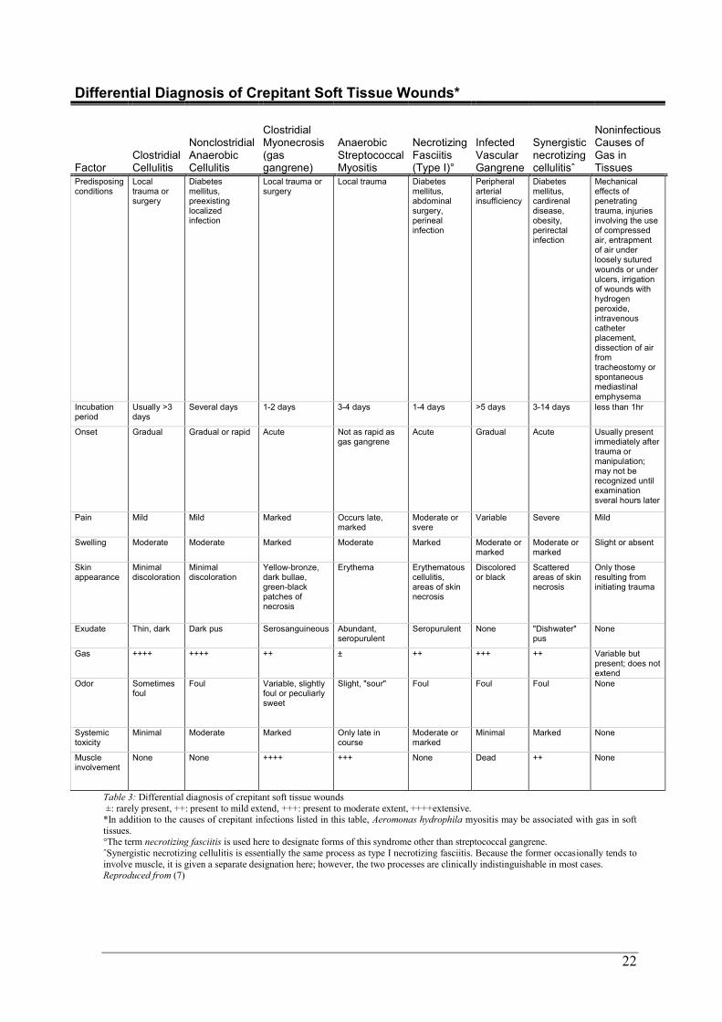
Table 3: Differential diagnosis of crepitant soft tissue wounds
±: rarely present, ++: present to mild extend, +++: present to moderate extent, ++++extensive. *In addition to the causes of crepitant infections listed in this table, Aeromonas hydrophila myositis may be associated with gas in soft
tissues. °The term necrotizing fasciitis is used here to designate forms of this syndrome other than streptococcal gangrene.
ˆSynergistic necrotizing cellulitis is essentially the same process as type I necrotizing fasciitis. Because the former occasionally tends to
involve muscle, it is given a separate designation here; however, the two processes are clinically indistinguishable in most cases. Reproduced from (7)
Differential Diagnosis of Crepitant Soft Tissue Wounds*
Factor Clostridial Cellulitis
Nonclostridial Anaerobic Cellulitis
Clostridial Myonecrosis (gas gangrene)
Anaerobic Streptococcal Myositis
Necrotizing Fasciitis (Type I)°
Infected Vascular Gangrene
Synergistic necrotizing cellulitisˆ
Noninfectious Causes of Gas in Tissues
Predisposing conditions
Local trauma or surgery
Diabetes mellitus, preexisting localized infection
Local trauma or surgery
Local trauma Diabetes mellitus, abdominal surgery, perineal infection
Peripheral arterial insufficiency
Diabetes mellitus, cardirenal disease, obesity, perirectal infection
Mechanical effects of penetrating trauma, injuries involving the use of compressed air, entrapment of air under loosely sutured wounds or under ulcers, irrigation of wounds with hydrogen peroxide, intravenous catheter placement, dissection of air from tracheostomy or spontaneous mediastinal emphysema
Incubation period
Usually >3 days
Several days 1-2 days 3-4 days 1-4 days >5 days 3-14 days less than 1hr
Onset Gradual Gradual or rapid Acute Not as rapid as gas gangrene
Acute Gradual Acute Usually present immediately after trauma or manipulation; may not be recognized until examination sveral hours later
Pain Mild Mild Marked Occurs late, marked
Moderate or svere
Variable Severe Mild
Swelling Moderate Moderate Marked Moderate Marked Moderate or marked
Moderate or marked
Slight or absent
Skin appearance
Minimal discoloration
Minimal discoloration
Yellow-bronze, dark bullae, green-black patches of necrosis
Erythema Erythematous cellulitis, areas of skin necrosis
Discolored or black
Scattered areas of skin necrosis
Only those resulting from initiating trauma
Exudate Thin, dark Dark pus Serosanguineous Abundant, seropurulent
Seropurulent None "Dishwater" pus
None
Gas ++++ ++++ ++ ± ++ +++ ++ Variable but present; does not extend
Odor Sometimes foul
Foul Variable, slightly foul or peculiarly sweet
Slight, "sour" Foul Foul Foul None
Systemic toxicity
Minimal Moderate Marked Only late in course
Moderate or marked
Minimal Marked None
Muscle involvement
None None ++++ +++ None Dead ++ None
23
1.9 Therapy
The mainstays of treatment are a combination of parenteral antibiotics and prompt surgical
intervention. The best outcome is achieved the earlier an aggressive and precise surgical
evaluation is performed (Fig. 4). Extensive debridement of necrotic tissue with amply
resection margins is as important as in time amputation of an extremity if necessary. The
overriding aim is the complete removal of necrotic tissue and to relieve and decompress
the swollen surrounding compartments to prevent further destruction. Pending results of
microbiological findings an empiric antibiotic therapy should be started. With regard to
possible differential diagnosis of myonecrotic infections the initial antibiotics treatment
should cover Clostridium spp., Group A Streptococci, facultative anaerobic streptococci,
and Enterobacteriaceae. Penicillin is the first choice for infections with C. perfringens
since most strains are susceptible (1). Other potential antibiotics that showed superior
activity in animal models are clindamycin, tetracycline, chloramphenicol, metronidazole
and a couple of cephalosporins (3). Experimental gas gangrene models comparing single
versus combinational use of antimicrobial agents indicated that clindamycin and
metronidazole used singly are more effective than penicillin. Since some strains of
clostridia may show resistances advisement of combinating penicillin and clindamycin
appears rational. The demands on optimal antimicrobial therapy are complex and struggle
with tissue penetration, ineffectiveness in high inocula, bacteria colony killing effects and
supposing toxin supression problems (25). The Infectious Disease Society of America
(IDSA) published this summer an update of their recommendations about the treatment of
skin and soft tissue infections (SSTIs), with a section about necrotizing fasciitis and
Fournier’s gangrene as well as clostridial gas gangrene and myonecrosis. Table 4 gives an
overview about the latest guidelines from the IDSA.
24
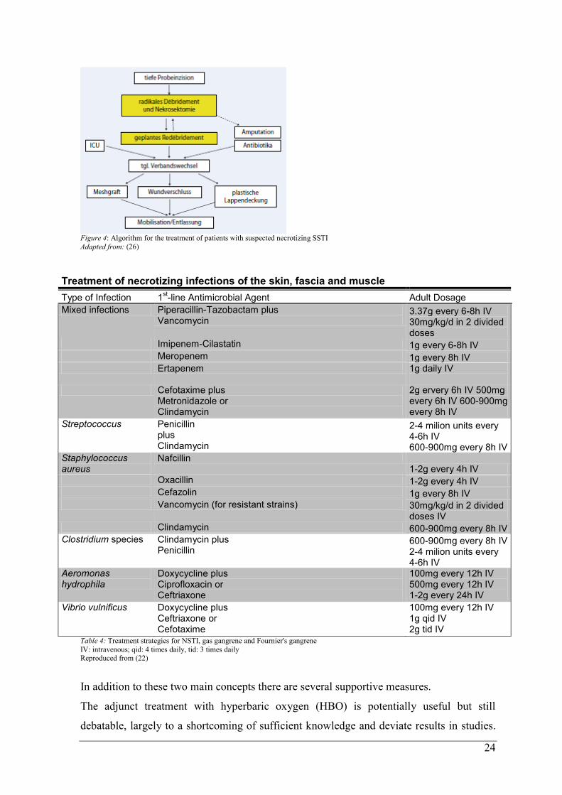
Figure 4: Algorithm for the treatment of patients with suspected necrotizing SSTI Adapted from: (26)
Treatment of necrotizing infections of the skin, fascia and muscle
Type of Infection 1st-line Antimicrobial Agent Adult Dosage
Mixed infections Piperacillin-Tazobactam plus Vancomycin
3.37g every 6-8h IV 30mg/kg/d in 2 divided doses
Imipenem-Cilastatin 1g every 6-8h IV Meropenem 1g every 8h IV Ertapenem 1g daily IV
Cefotaxime plus
Metronidazole or Clindamycin
2g ervery 6h IV 500mg every 6h IV 600-900mg every 8h IV
Streptococcus Penicillin plus Clindamycin
2-4 milion units every 4-6h IV 600-900mg every 8h IV
Staphylococcus aureus
Nafcillin 1-2g every 4h IV
Oxacillin 1-2g every 4h IV Cefazolin 1g every 8h IV Vancomycin (for resistant strains) 30mg/kg/d in 2 divided
doses IV Clindamycin 600-900mg every 8h IV Clostridium species Clindamycin plus
Penicillin 600-900mg every 8h IV 2-4 milion units every 4-6h IV
Aeromonas hydrophila
Doxycycline plus Ciprofloxacin or Ceftriaxone
100mg every 12h IV 500mg every 12h IV 1-2g every 24h IV
Vibrio vulnificus Doxycycline plus Ceftriaxone or Cefotaxime
100mg every 12h IV 1g qid IV 2g tid IV
Table 4: Treatment strategies for NSTI, gas gangrene and Fournier's gangrene
IV: intravenous; qid: 4 times daily, tid: 3 times daily Reproduced from (22)
In addition to these two main concepts there are several supportive measures.
The adjunct treatment with hyperbaric oxygen (HBO) is potentially useful but still
debatable, largely to a shortcoming of sufficient knowledge and deviate results in studies.
25
Studies in animals showed that HBO treatment alone does not improve the outcome. The
combination of HBO and antibiotics enhances the response rate of antibiotic-treated
bacteria to HBO and has an reductive effect on the bacteria population at the infected site.
As with surgical debridement, HBO is most beneficial if applicated as soon as possible.
The positive effects of HBO are attributed to the antimicrobial effect of high oxygen
concentrations, amelioration of tissue oxygenation and the bacteriostatic effect due to rise
of oxidation-reduction potential in and around the infected area and creation of an aerobe,
less favorable milieu for anaerobic growth. Murine models showed a direct in-vivo
inactivation of C. perfringens by HBO. Inhibition of alpha-toxin production was seen but
could not be confirmed and alters from trail to trail depending on used gas pressure and
exposure regime. The best outcome was observed with intense exposures to HBO in the
initial phase of the infection which leads to the supposition that a practical approach would
be the highest dose and frequence of HBO exposure a patient can tolerate safely (27).
There are no randomized, controlled trials in humans confirming that HBO solitary used or
in combination with antibiotics and surgery increases the survival. The major problems are
the small number of clostridial myonecrosis cases in normal population and no area-wide
availability of clinical centers with hyperbaric chambers. Additionally, the therapy regimes
used and the clinical techniques for exposure vary strongly. However, due to the positive
effects on infected tissue in several animal models there is rationale assumption for
profitable contribution of HBO in the treatment of gas gangrene. Besides, HBO is at large
safe, quite well tolerated and it’s only absolute contraindication is an untreated
pneumothorax.
Other adjuvant options but less often performed than HBO, include G-CSF (Granulocyte-
Colony Stimulation Factor) to impel hematopoietic growth and approaches direct against
toxin effect, such as vaccines (1). In one series, mice were protected against C.
perfringens, after active immunization with a subunit vaccine. The inoculant was a direct
derivative from the alpha-toxine (28). Another study confirmed these results and
demonstrated prime efficacy of immunization against the C-domain of C.perfringens
alpha-toxin. They also elucidated that alpha-toxin-induced activation of leukocytes,
platelets, and endothelial cells leads to occlusive intravascular aggregation, such that
hypoxic impairment extends and C. perfringens thrives under optimal anaerobic
conditions. Furthermore, symptoms of immunized mice showed to be more localized and
26
transient than in the control group (29). Antitoxin for a passive immunization or active
immunization in humans is not yet available.
1.10 Prognosis
Clostridial myonecrosis has an overall poor outcome. Though the affection of an limb has
a better prognosis than myonecrosis of the trunk or visceral organs. Patients with clostridial
bacteremia have a great probability of progression towards shock.
1.11 Prevention
A precise combined therapy approach with radical debridement of damaged tissue and
correction of blood circulation impairment is crucial and attributed to a better outcome.
27
2 Material and Methods
A clostridial infection is a disease with a complex, mostly severe and fatal course that
confronts clinicians with a difficult challenge in diagnosis and therapy. In Austria only the
Section of Thoracic Surgery and Hyperbaric Surgery of the Universitiy Hospital Graz
offers a hyperbaric facility with hyperbaric patient treatment chambers.
The aim of this study was to retrospectively analyse the correlations between different
findings of microbiological methods, as well as the reviewing of the epidemiology of
clostridial infections among the Graz conurbation in the last 5 years. Additional we took a
look in detail at the 24 hour emergency service for clinicians, which is provided by
employees of the Institute of Hygiene, Microbiology and Environmental Medicine. This
service mainly ensures an immediate alert and information exchange between clinicians
and microbiologists for the medical emergency of gas gangrene or necrotizing fasciitis.
Communication in both directions is facilitated in terms of clinicians call if they have a
sample with suspicion of gas gangrene or necrotizing fasciitis and microbiologists call
back after performing a gram-staining.
2.1 Study Setting
The period of time included in the analysis was from 2009 to 2013. Data was raised in a
cooperation between the Institute of Hygiene, Microbiology and Environmental Medicine
and the Section of Thoracic Surgery and Hyperbaric Surgery at the Medical University of
Graz.
2.2 Patients
Several concepts of terminology were recorded to determine and to limit a suitable patient
population. A search process with targeted search words was performed in the electronic
patients database (MEDOCS). In-patients from the 1st
of January 2009 to the 31st of
December 2013 were included.
28
2.3 Inclusion Criteria
- male and female of any age
- lab conducted by the Institute of Hygiene, Microbiology and Environmental
Medicine
- clinical suspicion of gas gangrene
search terms in MEDOCS:
- Wie “*gasbrand*“ Oder (Wie “*nekrotis*“ Und Wie “*fa[sz]ciit*“) Oder (Wie
“*fournier*“ Und Wie “*gangr*“)
2.4 Exclusion Criteria
- data that shows to be classified incorrect in accordance with the search terms (e.g.
sterile osteonecrosis)
- patients with no microbiological data at the Institute of Hygiene, Microbiology
and Environmental Medicine (samples were sent to an external microbiological
laboratory)
- infections with C. difficile
2.5 Data
All data regarding the diagnosis and classification of the infection were collected through
the electronic patient database (MEDOCS). Due to the availability of a hyperbaric chamber
in Graz, gas gangrene or any necrotizing disease is usually treated in cooperation with this
facility. To limit the patient data we contrasted the list of patients treated in the hyperbaric
chamber with those who matched the search terms.
The remaining information concerning age, sex, laboratory results and if an alert call was
made were gathered via the electronic and analog archieves of the Institute of Hygiene,
Microbiology and Environmental Medicine. Data from 2012 and later was electronically
available. Earlier data had to be collected manually for the most part.
In lab findings we differentiated any causing agent from Clostridium spp.. Furthermore all
emergency calls made from employees from the Institute of Hygiene, Microbiology and
Environmental Medicine to a clinician at the Medical University of Graz were listed and
sorted in order of outgoing time. The outgoing time is interesting from a economical point
of view since calls outside routine working hours are charged seperate.
29
2.6 Statistical Analysis
The study was performed retrospectively on medical records of the LKH Graz. The data
was accomplished via a computer-generated approach and completed through the
analogous archive. The analysis was undertaken with Microsoft Excel.
From the clinical side we counted the cases with proven infection with C. perfringens and
other Clostrium spp. itemized for each year and for the total period of the analysis.
Furthermore specific data of the patients (concerning age, gender, etc.) were investigated.
From the side of microbiologic laboratory we analyzed the frequency of emergency calls
outside of the routine working hours and the correlation between the results of the gram-
staining and the culture was also ascertained.
30
3 Results
3.1 Referral Diagnosis
The search was first based on specimens with the referral diagnosis gas gangrene or
necrotizing fasciitis. We found that differentiation between these two disease entities is
difficult, because a consistent description of the clinical presentation is not found and also
the nomenclature is used variously and ambiguously. The search terms were reconsidered
and extended to a list of diagnosis related to these entities. We identified a total of 157
patients that matched the terms at first search. Next we had to sort out patients with for
example sterile osteonecrosis and some other kinds of necrosis that did not fit to the
referral diagnosis. A further inconsistency we observed was that nomenclature did not
always meet the official ICD-10 nomenclature. Therefore, a ranking based on obtained
referral diagnosis, as we had hypothesized - on the one hand gas gangrene and on the other
hand necrotizing fasciitis - was unsatisfactory and unusable for evaluation.
3.2 Annual distribution
In total we abstracted 132 eligible patients treated from 2009-2013 at the Section of
Thoracic Surgery and Hyperbaric Surgery, University Hospital Graz that matched all the
inclusion criteria of the referral diagnosis of suspected gas gangrene or necrotizing fasciitis
and were subsequently analyzed in this study. 90 (68%) patients were male, 42 (32%) were
female. The review showed an average of 26.4 cases per year over the last 5 years with
suspected gas gangrene. Two peaks occured in 2011 and 2013 with each 32 cases. In 2009
the lowest number with only 20 cases was recorded, followed by 21 cases in 2012, and a
total of 27 cases in 2010 (Fig. 5).

31
Figure 5: Annual spread of infections suspected as gas gangrene or necrotizing fasciitis
3.3 Age and Gender
All age groups were included, the median age of all 132 patients was 57 years. The
youngest patient was an 11-year-old female, the oldest a 94-year-old female. The highest
number occurred between the ages of 40 - 49 years with 26 cases. The age groups older
than that deviated insignificantly between 21 cases between 50 - 59, 24 cases between 60 -
69 years, and 23 cases between the ages of 70 - 79 years. Older people were less affected.
Only 15 cases occurred with an age older than 80 years, and only one woman was older
than 90 years. The age groups 0 - 19 and 20 - 29 showed each only 3% of the cases. 15
patients (11%) were between 30 - 39 years, which created an abrupt increase in the case
number with rising age.
The distribution between men and women was in favour of men, 90 (68%) patients were
male, 42 (32%) were female. Between the median age of male patients, 57 years, and
female median age, 56 years, was no distinct difference. The highest peak for men was
between the ages of 40 - 49 years, for women over 80 years.
The following figures show the age distribution of the observed population and the age
distribution subdivided into males and females (Fig. 6, Fig. 7).
20
27
32
21
32
0
5
10
15
20
25
30
35
2009 2010 2011 2012 2013
No
. o
f cases
Year
Annual distribution of infections (n=132)
32
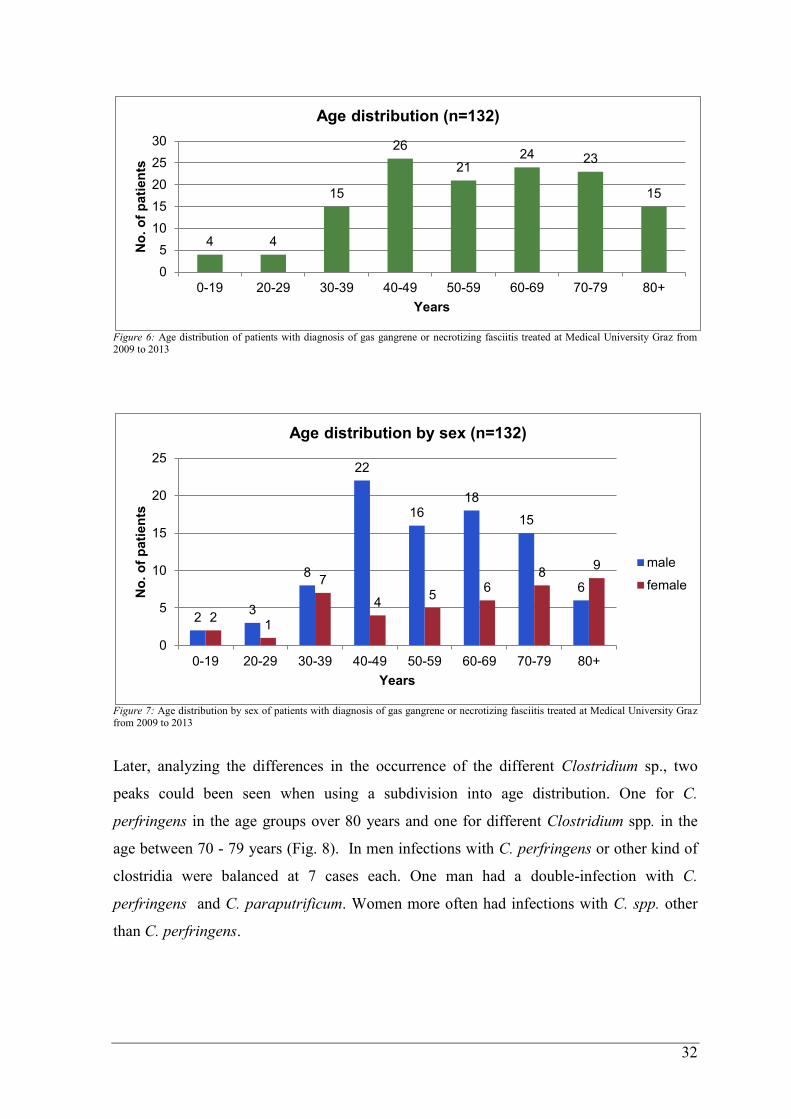
Figure 6: Age distribution of patients with diagnosis of gas gangrene or necrotizing fasciitis treated at Medical University Graz from 2009 to 2013
Figure 7: Age distribution by sex of patients with diagnosis of gas gangrene or necrotizing fasciitis treated at Medical University Graz from 2009 to 2013
Later, analyzing the differences in the occurrence of the different Clostridium sp., two
peaks could been seen when using a subdivision into age distribution. One for C.
perfringens in the age groups over 80 years and one for different Clostridium spp. in the
age between 70 - 79 years (Fig. 8). In men infections with C. perfringens or other kind of
clostridia were balanced at 7 cases each. One man had a double-infection with C.
perfringens and C. paraputrificum. Women more often had infections with C. spp. other
than C. perfringens.
4 4
15
26
21 24 23
15
0
5
10
15
20
25
30
0-19 20-29 30-39 40-49 50-59 60-69 70-79 80+
No
. o
f p
ati
en
ts
Years
Age distribution (n=132)
2 3
8
22
16 18
15
6
2 1
7
4 5
6 8
9
0
5
10
15
20
25
0-19 20-29 30-39 40-49 50-59 60-69 70-79 80+
No
. o
f p
ati
en
ts
Years
Age distribution by sex (n=132)
male
female
33
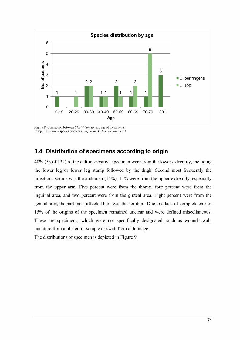
Figure 8: Connection between Clostridium sp. and age of the patients C.spp: Clostridium species (such as C. septicum, C. bifermentans, etc.)
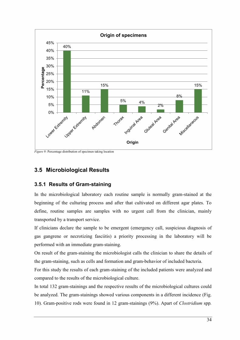
3.4 Distribution of specimens according to origin
40% (53 of 132) of the culture-positive specimen were from the lower extremity, including
the lower leg or lower leg stump followed by the thigh. Second most frequently the
infectious source was the abdomen (15%), 11% were from the upper extremity, especially
from the upper arm. Five percent were from the thorax, four percent were from the
inguinal area, and two percent were from the gluteal area. Eight percent were from the
genital area, the part most affected here was the scrotum. Due to a lack of complete entries
15% of the origins of the specimen remained unclear and were defined miscellaneous.
These are specimens, which were not specifically designated, such as wound swab,
puncture from a blister, or sample or swab from a drainage.
The distributions of specimen is depicted in Figure 9.
1
2
1
2
1 1
3
1
2
1 1
2
5
0
1
2
3
4
5
6
0-19 20-29 30-39 40-49 50-59 60-69 70-79 80+
No
. o
f p
ati
en
ts
Age
Species distribution by age
C. perfringens
C. spp
34
Figure 9: Percentage distribution of specimen taking location
3.5 Microbiological Results
3.5.1 Results of Gram-staining
In the microbiological laboratory each routine sample is normally gram-stained at the
beginning of the culturing process and after that cultivated on different agar plates. To
define, routine samples are samples with no urgent call from the clinician, mainly
transported by a transport service.
If clinicians declare the sample to be emergent (emergency call, suspicious diagnosis of
gas gangrene or necrotizing fasciitis) a priority processing in the laboratory will be
performed with an immediate gram-staining.
On result of the gram-staining the microbiologist calls the clinician to share the details of
the gram-staining, such as cells and formation and gram-behavior of included bacteria.
For this study the results of each gram-staining of the included patients were analyzed and
compared to the results of the microbiological culture.
In total 132 gram-stainings and the respective results of the microbiological cultures could
be analyzed. The gram-stainings showed various components in a different incidence (Fig.
10). Gram-positive rods were found in 12 gram-stainings (9%). Apart of Clostridium spp.
40%
11%
15%
5% 4% 2%
8%
15%
0%
5%
10%
15%
20%
25%
30%
35%
40%
45%P
erc
en
tag
e
Origin
Origin of specimens
35
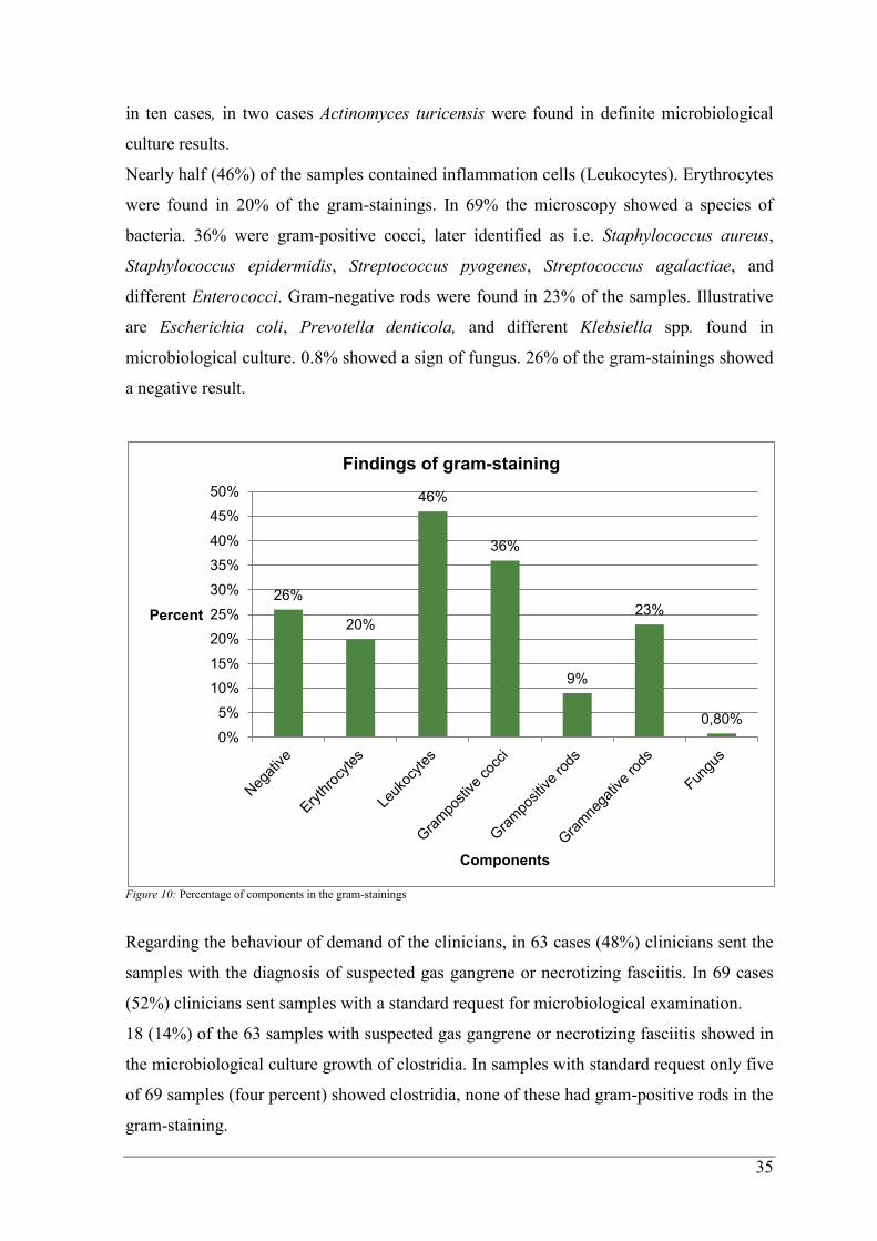
in ten cases, in two cases Actinomyces turicensis were found in definite microbiological
culture results.
Nearly half (46%) of the samples contained inflammation cells (Leukocytes). Erythrocytes
were found in 20% of the gram-stainings. In 69% the microscopy showed a species of
bacteria. 36% were gram-positive cocci, later identified as i.e. Staphylococcus aureus,
Staphylococcus epidermidis, Streptococcus pyogenes, Streptococcus agalactiae, and
different Enterococci. Gram-negative rods were found in 23% of the samples. Illustrative
are Escherichia coli, Prevotella denticola, and different Klebsiella spp. found in
microbiological culture. 0.8% showed a sign of fungus. 26% of the gram-stainings showed
a negative result.
Figure 10: Percentage of components in the gram-stainings
Regarding the behaviour of demand of the clinicians, in 63 cases (48%) clinicians sent the
samples with the diagnosis of suspected gas gangrene or necrotizing fasciitis. In 69 cases
(52%) clinicians sent samples with a standard request for microbiological examination.
18 (14%) of the 63 samples with suspected gas gangrene or necrotizing fasciitis showed in
the microbiological culture growth of clostridia. In samples with standard request only five
of 69 samples (four percent) showed clostridia, none of these had gram-positive rods in the
gram-staining.
26%
20%
46%
36%
9%
23%
0,80%
0%
5%
10%
15%
20%
25%
30%
35%
40%
45%
50%
Percent
Components
Findings of gram-staining
36
Regarding the total 23 samples with cultured clostridia, the result of the respective gram-
stainings showed gram-positive rods in 10 cases.
When comparing the negative gram-stainings and those with no evidence of gram-positive
rods, but other bacteria, in 13 out of 120 (11%) cultures, growth of a Clostridium spp. was
found.
To analyze the accordance between the results of the gram-staining and the results of the
cultures, we defined the accordance as complete accordance between the two methods,
partly accordance, for example if only one kind of bacterium was seen in the gram-staining
but different kinds were cultivated afterwards, and as no accordance, for those samples
with a negative gram-staining but bacterial growth in culture.
To note: gram-staining has an operational limitation, as it can only show bacteria with a
concentration of 105/ml or higher. Therefore it is possible that a negative result in the
gram-staining can lead to a growth of bacteria anyway.
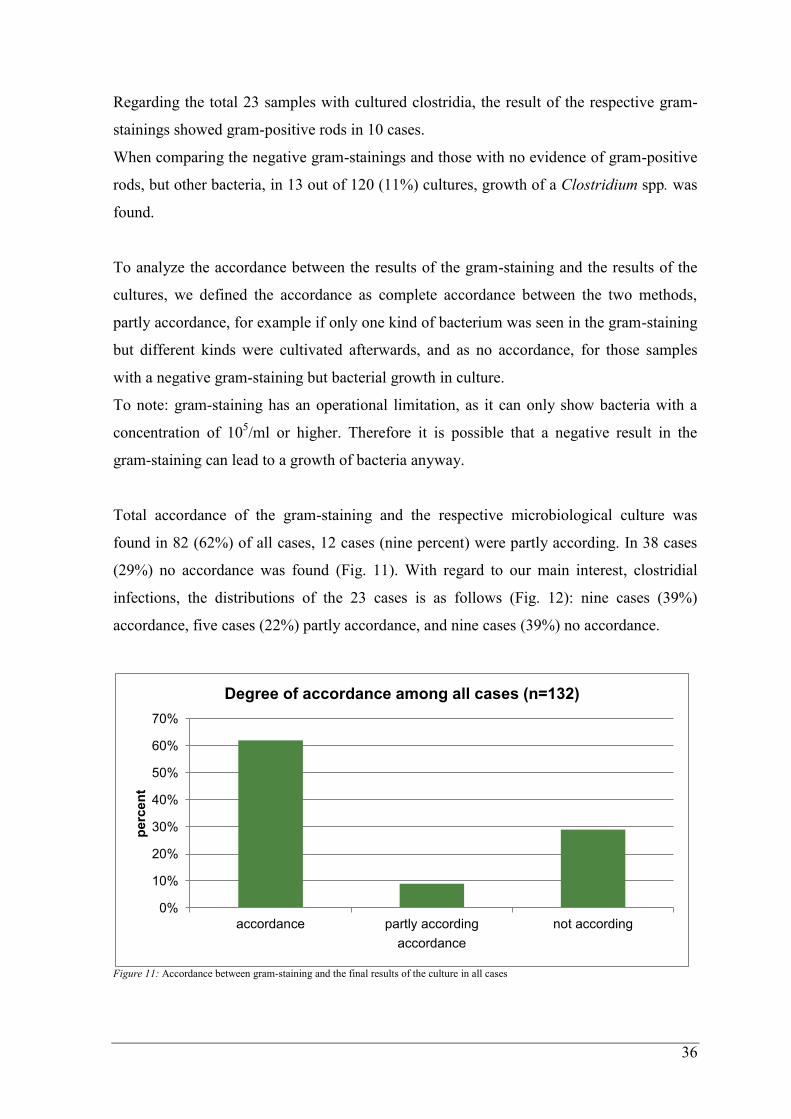
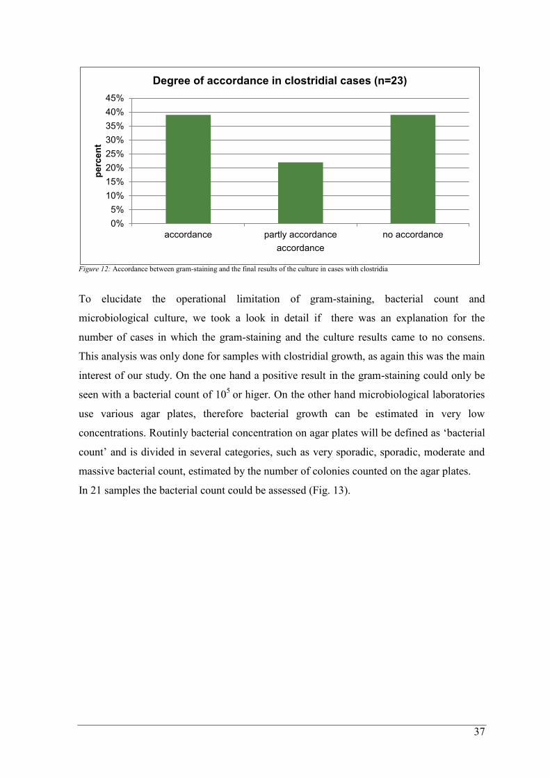
Total accordance of the gram-staining and the respective microbiological culture was
found in 82 (62%) of all cases, 12 cases (nine percent) were partly according. In 38 cases
(29%) no accordance was found (Fig. 11). With regard to our main interest, clostridial
infections, the distributions of the 23 cases is as follows (Fig. 12): nine cases (39%)
accordance, five cases (22%) partly accordance, and nine cases (39%) no accordance.
Figure 11: Accordance between gram-staining and the final results of the culture in all cases
0%
10%
20%
30%
40%
50%
60%
70%
accordance partly according not according
perc
en
t
accordance
Degree of accordance among all cases (n=132)
37
Figure 12: Accordance between gram-staining and the final results of the culture in cases with clostridia
To elucidate the operational limitation of gram-staining, bacterial count and
microbiological culture, we took a look in detail if there was an explanation for the
number of cases in which the gram-staining and the culture results came to no consens.
This analysis was only done for samples with clostridial growth, as again this was the main
interest of our study. On the one hand a positive result in the gram-staining could only be
seen with a bacterial count of 105
or higer. On the other hand microbiological laboratories
use various agar plates, therefore bacterial growth can be estimated in very low
concentrations. Routinly bacterial concentration on agar plates will be defined as ‘bacterial
count’ and is divided in several categories, such as very sporadic, sporadic, moderate and
massive bacterial count, estimated by the number of colonies counted on the agar plates.
In 21 samples the bacterial count could be assessed (Fig. 13).
0%
5%
10%
15%
20%
25%
30%
35%
40%
45%
accordance partly accordance no accordance
perc
en
t
accordance
Degree of accordance in clostridial cases (n=23)
38
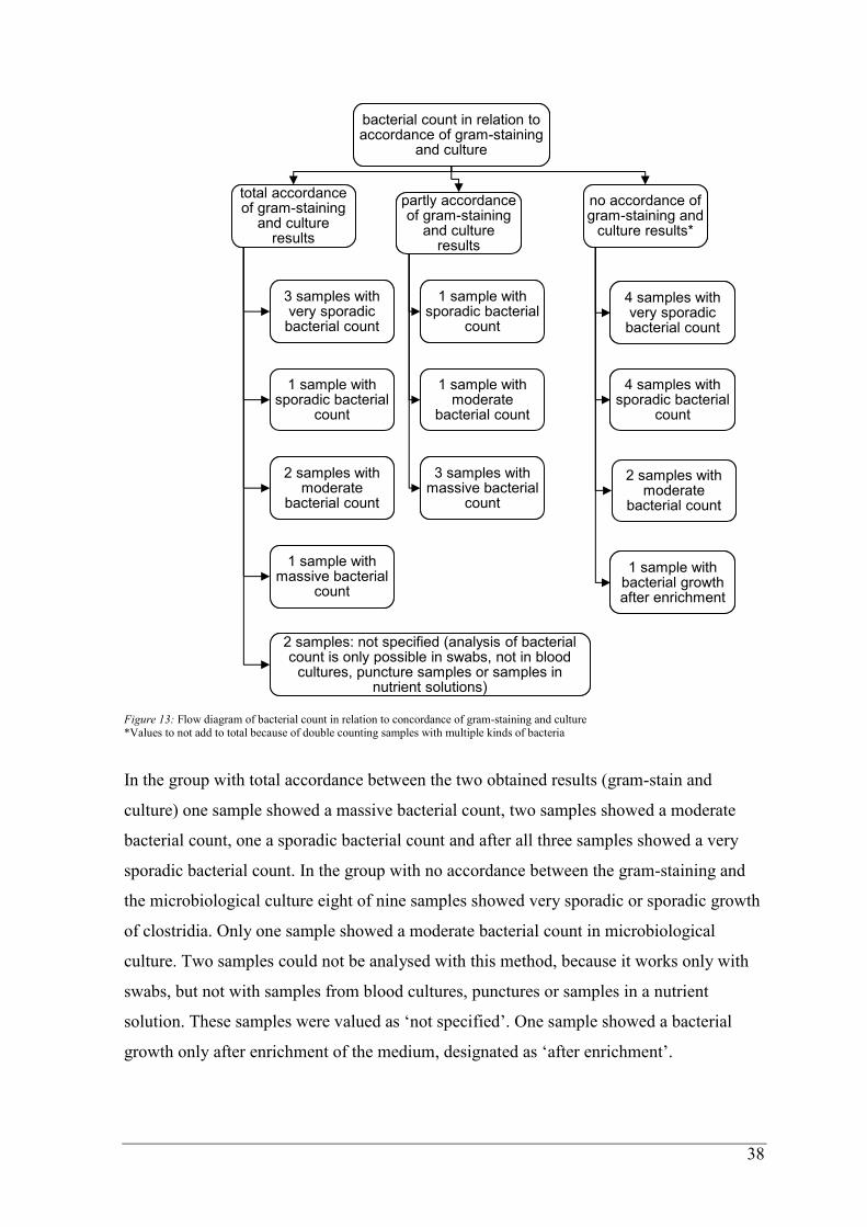
Figure 13: Flow diagram of bacterial count in relation to concordance of gram-staining and culture *Values to not add to total because of double counting samples with multiple kinds of bacteria
In the group with total accordance between the two obtained results (gram-stain and
culture) one sample showed a massive bacterial count, two samples showed a moderate
bacterial count, one a sporadic bacterial count and after all three samples showed a very
sporadic bacterial count. In the group with no accordance between the gram-staining and
the microbiological culture eight of nine samples showed very sporadic or sporadic growth
of clostridia. Only one sample showed a moderate bacterial count in microbiological
culture. Two samples could not be analysed with this method, because it works only with
swabs, but not with samples from blood cultures, punctures or samples in a nutrient
solution. These samples were valued as ‘not specified’. One sample showed a bacterial
growth only after enrichment of the medium, designated as ‘after enrichment’.
bacterial count in relation to accordance of gram-staining
and culture
bacterial count in relation to accordance of gram-staining
and culture
total accordance of gram-staining
and culture results
total accordance of gram-staining
and culture results
3 samples with very sporadic bacterial count
3 samples with very sporadic bacterial count
1 sample with sporadic bacterial
count
1 sample with sporadic bacterial
count
2 samples with moderate
bacterial count
2 samples with moderate
bacterial count
1 sample with massive bacterial
count
1 sample with massive bacterial
count
2 samples: not specified (analysis of bacterial count is only possible in swabs, not in blood
cultures, puncture samples or samples in nutrient solutions)
2 samples: not specified (analysis of bacterial count is only possible in swabs, not in blood
cultures, puncture samples or samples in nutrient solutions)
partly accordance of gram-staining
and culture results
partly accordance of gram-staining
and culture results
1 sample with sporadic bacterial
count
1 sample with sporadic bacterial
count
1 sample with moderate
bacterial count
1 sample with moderate
bacterial count
3 samples with massive bacterial
count
3 samples with massive bacterial
count
no accordance of gram-staining and
culture results*
no accordance of gram-staining and
culture results*
4 samples with very sporadic bacterial count
4 samples with very sporadic bacterial count
4 samples with sporadic bacterial
count
4 samples with sporadic bacterial
count
2 samples with moderate
bacterial count
2 samples with moderate
bacterial count
1 sample with bacterial growth after enrichment
1 sample with bacterial growth after enrichment
39
3.5.2 Comparison of gram-stainings with gram-positive rods and
cultures with proof of clostridia
In this section two different points of the study were analyzed:
On the one hand all gram-stainings of the included 132 samples with proof of gram-
positive rods were analyzed, and on the other hand gram-stainings with negative results or
no proof of gram-positive rods. This anaylsis showed that in the group with gram-positive
rods the results were according or at least partly according, while all disaccording results
were in the group with no clue for gram-positive rods in the gram-staining at the beginning
of microbiological laboratory.
10 of the 12 gram-stainings with gram-positive rods showed in the culture a growth of a
Clostridium spp.. The two other samples showed Actinomyces turicensis as gram-positive
rod as definite result.
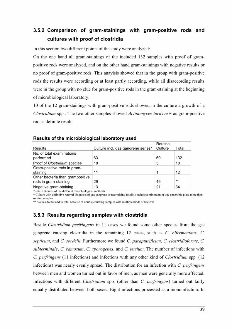
Results of the microbiological laboratory used
Results Culture incl. gas gangrene series* Routine Culture Total
No. of total examinations performed 63 69 132
Proof of Clostridium species 18 5 18
Gram-positive rods in gram-staining 11 1 12
Other bacteria than grampositive rods in gram-staining 29 49 **
Negative gram-staining 13 21 34 Table 5: Results of the different microbiological methods * Culture with definitive referral diagnosis of gas gangrene or necrotizing fasciitis include a minimum of one anaerobic plate more than
routine samples
** Values do not add to total because of double counting samples with multiple kinds of bacteria
3.5.3 Results regarding samples with clostridia
Beside Clostridium perfringens in 11 cases we found some other species from the gas
gangrene causing clostridia in the remaining 12 cases, such as C. bifermentans, C.
septicum, and C. sordelli. Furthermore we found C. paraputrificum, C. clostridioforme, C.
subterminale, C. ramosum, C. sporogenes, and C. tertium. The number of infections with
C. perfringens (11 infections) and infections with any other kind of Clostridium spp. (12
infections) was nearly evenly spread. The distribution for an infection with C. perfringens
between men and women turned out in favor of men, as men were generally more affected.
Infections with different Clostridium spp. (other than C. perfringens) turned out fairly
equally distributed between both sexes. Eight infections processed as a monoinfection. In
40
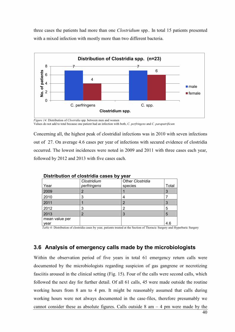
three cases the patients had more than one Clostridium spp.. In total 15 patients presented
with a mixed infection with mostly more than two different bacteria.
Figure 14: Distribution of Clostridia spp. between men and women Values do not add to total because one patient had an infection with both, C. perfringens and C. paraputrificum
Concerning all, the highest peak of clostridial infections was in 2010 with seven infections
out of 27. On average 4.6 cases per year of infections with secured evidence of clostridia
occurred. The lowest incidences were noted in 2009 and 2011 with three cases each year,
followed by 2012 and 2013 with five cases each.
Distribution of clostridia cases by year
Year Clostridium perfringens
Other Clostridia
species Total
2009 2 1 3
2010 3 4 7
2011 1 2 3
2012 3 2 5
2013 2 3 5
mean value per year 4.6
Table 6: Distribution of clostridia cases by year, patients treated at the Section of Thoracic Surgery and Hyperbaric Surgery
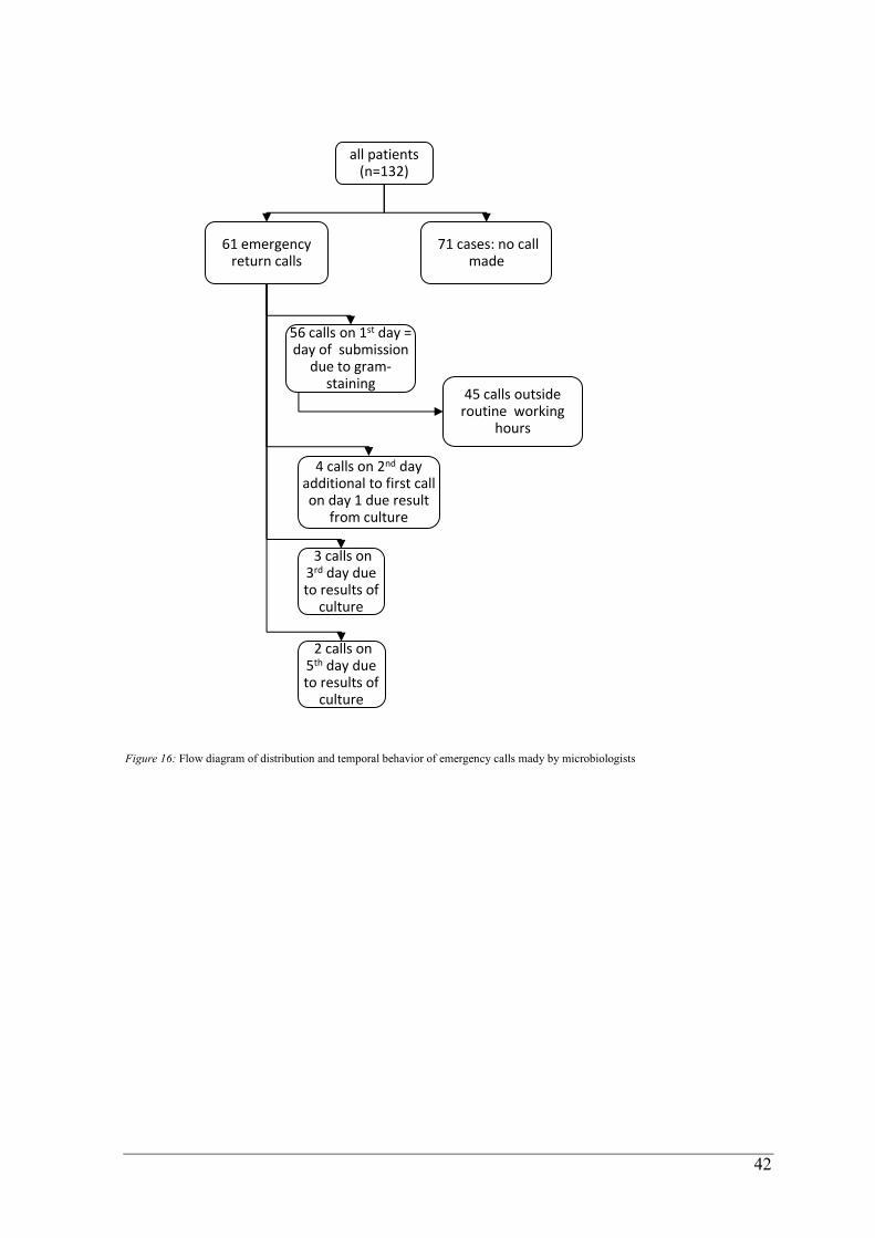
3.6 Analysis of emergency calls made by the microbiologists
Within the observation period of five years in total 61 emergency return calls were
documented by the microbiologists regarding suspicion of gas gangrene or necrotizing
fasciitis aroused in the clinical setting (Fig. 15). Four of the calls were second calls, which
followed the next day for further detail. Of all 61 calls, 45 were made outside the routine
working hours from 8 am to 4 pm. It might be reasonably assumed that calls during
working hours were not always documented in the case-files, therefore presumably we
cannot consider these as absolute figures. Calls outside 8 am – 4 pm were made by the
7 7
4
6
0
2
4
6
8
C. perfringens C. spp.
No
. o
f p
ati
en
ts
Clostridium spp.
Distribution of Clostridia spp. (n=23)
male
female
41
microbiological emergency service provided by the Institute of Hygiene, Microbiology and
Environmental Medicine.
Normally the calls take place on the day of entry of the samples after a gram-staining is
performed, in our study these were 56 calls (Fig. 16). Included were also calls which were
made on the second day or even later, due to new developments seen on the culture plates.
Our data showed three calls made on the 3rd
day and two on the 5th
day of incubation to
inform the clinicians about a definitive growth of C. perfringens that could be observed.
Four of these C. perfringens were foung in routine samples, only one C. perfringens came
with a request for gas gangrene/ necrotizing fasciitis.
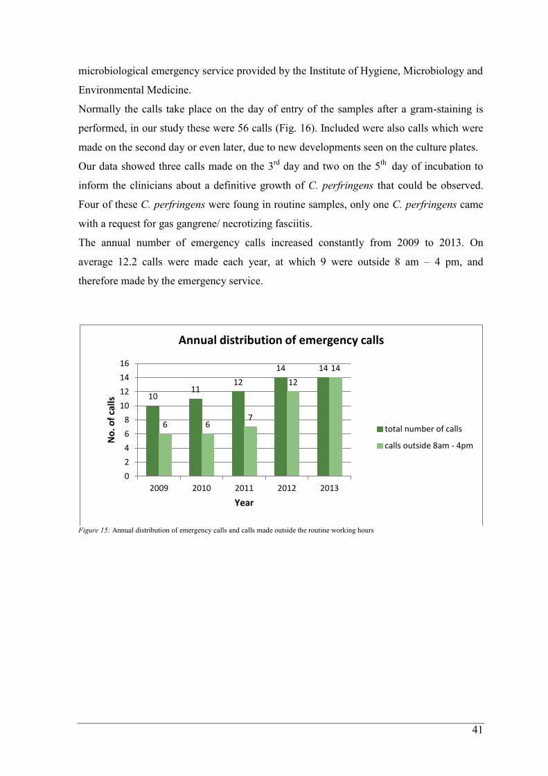
The annual number of emergency calls increased constantly from 2009 to 2013. On
average 12.2 calls were made each year, at which 9 were outside 8 am – 4 pm, and
therefore made by the emergency service.
Figure 15: Annual distribution of emergency calls and calls made outside the routine working hours
10 11
12
14 14
6 6 7
12
14
0
2
4
6
8
10
12
14
16
2009 2010 2011 2012 2013
No
. of
calls
Year
Annual distribution of emergency calls
total number of calls
calls outside 8am - 4pm
42
Figure 16: Flow diagram of distribution and temporal behavior of emergency calls mady by microbiologists
all patients (n=132)
61 emergency return calls
56 calls on 1st day = day of submission
due to gram- staining
45 calls outside routine working
hours
4 calls on 2nd day additional to first call on day 1 due result
from culture
3 calls on 3rd day due to results of
culture
2 calls on 5th day due to results of
culture
71 cases: no call made
43
4 Discussion
Clostridial myonecrosis due to Clostridium perfringens is a rare and uncommon disease in
civilian population. Optimization of fast and secure detection of infections with
Clostridium species has to be investigated because of the aggressive progression and the
high morbidity and mortality rate that are associated with this kind of disease. Gram
staining is a method, characterized by simple practicability, high availability and low costs.
Therefore, it would be an ideal supplement to facilitate diagnosis of clostridial
myonecrosis. Gram-staining is already used to support clinicians in planning and selecting
empiric clinical management for infections due to bacteria pending definite culture results.
For a disease with a fatal nature like clostridial infections the results of such methods are
crucial, and should be characterized by high reliability. Browsing literature about the use
of gram-staining compared with the use of culture regarding clostridial infections remained
unsatisfactory. However, there are studies on the importance of gram-staining regarding
e.g. bacterial meningitis and pneumonia. In a study of 10 patients with Klebsiella
pneumoniae meningitis Khan et al. were able to demonstrate, that ‘the examination of
Gram stain on admission can be negative or misleading; because of the small number of
organisms, patients being partially treated, and culture time of more than one day may be
required, thus delaying the final diagnosis of meningeal infection and representing a
prognostic factor of mortality’ (30). This statement meets the challenges that come with a
clostridial infection. Several other authors examinated the use of gram-staining in different
kinds of pneumonia. Mimoz et al. compared the results of two observers, performing
gram-staining on 10 fields and 50 fields, reporting that gram-staining of protected
pulmonary specimens is able to predict ventilator-associated pneumonia (VAP) and
additionally partly identified causing agents when they were growing at considerable
concentrations in culture (31). In a meta-analysis examination by O’Horo et al. an
admissible concordance between the negative result of gram-staining and probability of
occurrence of VAP was evaluated. Despite a positive gram-staining was not as specific for
VAP than previously assumed (32). Given all these unequal statements, these are subject to
considerable uncertainty about approaching a reliable method for bacterial infections with
the necessity of immediate action.
The aim of this retrospective study was to show how microbiological findings in relation to
a suspected clostridial myonecrosis complete each other and if special circumstances of
44
these form group, warrant special measures, in view of the 24h emergency service for
clinicians.
In a 5-year period, from 2009-2013, patient data was collected from the Institute of
Hygiene, Microbiology and Environmental Medicine and the Section of Thoracic Surgery
and Hyperbaric Surgery at the Medical University of Graz. The background of the
cooperation with the Section of Thoracic Surgery and Hyperbaric Surgery is based on the
fact, that for patients with this kind of disease a therapy session in a HBO facility is
essential part of the therapy.
The study was conducted at the Medical University Graz, where a total of 132 patients was
reviewed and analyzed. Our study was untertaken to see how reliable gram-staining is as a
first-line method (also performed by the emergency service of the microbiological
laboratory), compared to microbiological culture, for the detection of Clostridia spp. in the
context of clinical suspected gas gangrene. The population consists of patients with clinical
referral diagnosis of suspected gas gangrene or necrotizing fasciitis. 23 patients had
culture-proven infections with a Clostridium species. The current incidence of gas
gangrene is not well observed and literature about that is rare. Studies from the 1960-ties to
the 1980-ties reported an incidence about 0,1/100.000 population per year (10,33). In
Germany 100 - 140 cases of gas gangrene have been reported from the Robert Koch
Institute during 1992 and 1996. In 1997 data was published by the RKI with 122 cases of
gas gangrene, recorded in a register of notifiable infections/ contagious diseases (34). In
our study 90 (68%) patients were male, 42 (32%) female, giving the male-to-female ratio
of 2.2:1, men seem to be more often affected than women. Other studies have reported a
comparable distribution between men and woman (33,35).
We first tried to sort the patients by their referral diagnosis. This presented as mentioned
before as a big challenge that ultimately could not be solved. The classifications of NSTI
and gas gangrene vary considerably and do not meet ICD-10 classification in our case.
Even if the terminology is not uniform, the different definitions can give a clue to the
disease and about its course.
Based on origin of the specimens we classified groups by bodypart affected by the
infection. This showed that 40% of the infections were located in the lower extremity, most
frequently in the lower leg. Our study shares this findings with similar findings for gas
gangrene and necrotizing fasciitis in literature (36,37). It appeares that there might be a
conection between the higher incidence for pre-existing conditions and underlying diseases
in the lower extremity, such as diabetes, PAVK, foot ulcera and others mentioned above
45
(38). Subsequently we analysed the collected microbiological data. Therefore, we
evaluated first all patients and further on distinguished between patients who showed an
infection with clostridia and those who did not. All 132 patients had a gram-staining and a
culture result. First, we searched gram-stainings for samples with gram-positive rods. 12
out of 132 revealed a positive result for gram-positive rods. Afterwards microbiological
culture results were analyzed exclusively for clostridial growth. Clostridia were cultured in
23 out of 132 cases (17%). 11 of 23 patient samples showed C. perfringens, the remaining
12 samples belonged to different kinds of clostridia. We also noticed a broad spectrum of
coinfecting or causing bacteria other than clostridia species, such as Group A ß-hemolytic
Streptococci, and Staphylococcus aureus, responsible for the clinical appearance of gas
gangrene or necrotizing fasciitis. Just as Elliott reported in his analysis, an extremly wide
range of microbial pathogens could be found in patients with NSTI (39).
Gram-staining is an economical way of providing fast information which can point clinical
findings in a distinct direction. This method provides clinicians with useful clues directed
towars a fast diagnosis and supports their decision for empirical antibiotic treatment.
In this study in 12 gram-stainings gram-positive rods could be observed and were
confirmed as Clostridium sp. in ten and as Actinomyces turicensis in two cases. Regarding
the result of microbiological cultures, clostridia were found in 23 cases, wherease in these
cases gram-positive rods were seen in only ten cases (8%). In all investigated 132 patients
accordance of gram-stainings and the regarding microbiological cultures was found in 62%
(total accordance) and 9% (partly accordance). To analyse why the accordance was not
hundred percent we took a look at the bacterial count of the culture results. We hypothesize
that a lower bacterial count in the samples attributes to a negative result in the gram-
stainig. This may have affected the ability of detecting a match between gram-staining and
culture result. Our data showed that eight of nine samples with no accordance between
gram-staining and microbiological culture contained only very sporadic or sporadic
bacterial count.
At the Institute of Hygiene, Microbiology and Environmental Medicine a 24h emergency
call service is offered for patients with suspected gas gangrene or necrotizing fasciitis. This
allows a rapid exchange of information between clinicians and microbiologists. While
routine laboratory work is normally from 8 am to 4 pm, in case of patients with suspected
gas gangrene emergency return calls are provided 24 hours a day. Our analysis showed 61
46
emergency return calls in the evaluated time period. 45 of these calls were made outside
the routine working hours (8 am – 4 pm).
As mentionened above the study has some limitations that should be assessed. There are
limits that come with methods or a statistically approach as disscussed above. Apart from
the bacterial count necessary for a proper gram-staining, the evaluation of the documented
return calls is prone to error, since calls during routine working time might not be
documentent. This is leading to a false positive result in the number of emergency calls
outside the given time period.
5 Conclusion
Our study indicates, regarding to samples with clinical suspicion of gas gangrene, that the
reliability of a gram-staining result with gram-positive rods is acceptable good and in our
case in 83% lead to the result of a cultured-proof of clostridia. In contrast, a negative result
for gram-positive rods in gram-stainings can show clostridial growth after all. Also other
kinds of bacteria could be seen in the primarily gram-stain with an accordance (total or
partly) of more than 70% (gram-staining at the beginning and microbiological culture
result) This could be used as an argument to continue the emergency service provided by
the Instiute of Hygiene, Microbiology and Environmental Medicine. As well as the service
is not only for suspected gas gangrene but also provided for cases with suspected
meningitis.
47
6 References
1. Onderdonk AB, Garrett WS. Gas Gangrene and Other Clostridium-Associated Diseases.
In: Mandell GL, Bennett JE, Dolin R, editors. Mandell, Douglas, and Bennett’s Principals
and Practice of Infectious Diseases. 7th edition. Philadelphia: Churchill Livingston
Elsevier; p. 3103–9.
2. Schütt-Gerowitt H. Clostridien der Gasbrand-Gruppe. In: Darai G, Handermann M,
Sonntag H-G, Zöller L, editors. Lexikon der Infektionskrankheiten des Menschen. 4.
vollst. überarb. Auflage. Berlin Heidelberg: Springer; p. 180–3.
3. Stevens DL, Bryant A. Clostridial myonecrosis [Internet]. UpToDate. 2014 [updated
2014 Jan 30; cited 2014 Jun 25]. Available from:
http://www.uptodate.com/contents/clostridial-
myonecrosis?source=search_result&search=clostridial+myonecrosis&selectedTitle=1%7E
150
4. Crum-Cianflone NF. Bacterial, Fungal, Parasitic, and Viral Myositis. Clin Microbiol
Rev. 2008 Jul 1;21(3):473–94.
5. Rodloff AC. Obligat anaerob, sporenbildende Stäbchen (Clostridien). In: Suerbaum S,
Hahn H, Burchard G-D, Kaufmann SHE, Schulz TF, editors. Medizinische Mikrobiologie
und Infektiologie. 7. überarb. Auflage. Berlin Heidelberg: Springer; 2012. p. 324–7.
6. Schrier SL. Extrinsic nonimmune hemolytic anemia due to systemic disease [Internet].
UpToDate. 2014 [updated 2014 May 06; cited 2014 Oct 2]. Available from:
http://www.uptodate.com/contents/extrinsic-nonimmune-hemolytic-anemia-due-to-
systemic-
disease?source=search_result&search=extrinsic+nonimmune+hemolytic+anaemia&selecte
dTitle=1%7E150#
7. Pasternack MS, Swartz MN. Myositis and Myonecrosis. In: Mandell GL, Bennett JE,
Dolin R, editors. Mandell, Douglas, and Bennett’s Principals and Practice of Infectious
Diseases. 7th edition. Philadelphia: Churchill Livingston Elsevier; p. 1313–21.
8. Brook I. Microbiology and management of myositis. Int Orthop. 2004 Oct;28(5):257–
60.
9. Burke MP, Opeskin K. Nontraumatic clostridial myonecrosis. Am J Forensic Med
Pathol. 1999 Jun;20(2):158–62.
10. Hart GB, Lamb RC, Strauss MB. Gas gangrene. J Trauma. 1983 Nov;23(11):991–
1000.
11. Chew SSB, Lubowski DZ. Clostridium septicum and malignancy. ANZ J Surg. 2001
Nov;71(11):647–9.
12. Pelletier JPR, Plumbley JA, Rouse EA, Cina SJ. The Role of Clostridium septicum in
Paraneoplastic Sepsis. Arch Pathol Lab Med. 2000 Mar;124(3):353–6.
13. Hickey MJ, Kwan RYQ, Awad MM, Kennedy CL, Young LF, Hall P, et al. Molecular
48
and cellular basis of microvascular perfusion deficits induced by Clostridium perfringens
and Clostridium septicum. PLoS Pathog. 2008 Apr;4(4):e1000045.
14. Schumpelick V. Chirurgische Notfälle und Komplikationen. Kurzlehrbuch Chirurgie.
8. vollst. überarb. u. erw. Auflage. Stuttgart: Thieme; 2010. p. 213–4.
15. Smith-Slatas CL, Bourque M, Salazar JC. Clostridium septicum infections in children:
a case report and review of the literature. Pediatrics. 2006 Apr;117(4):e796–805.
16. Schaumann R, Rodloff A. Clostridium spp. In: Neumeister B, Geiss HK, Braun RW,
Kimming P, editors. Mikrobiologische Diagnostik. 2. vollst. überarb. Auflage. Stuttgart
New York: Georg Thieme; 2009. p. 539–50.
17. Soper DE, Lee SI, Kim JY, McDonald AG. Case 35-2011. N Engl J Med. 2011 Nov
16;365(20):1916–24.
18. Stevens DL, Baddour LM. Necrotizing soft tissue infections [Internet]. UpToDate.
2014 [updated 2014 Jan 21; cited 2014 Jun 25]. Available from:
http://www.uptodate.com/contents/necrotizing-soft-tissue-
infections?source=search_result&search=clostridium+perfringens+therapy&selectedTitle=
8~33
19. Brook I, Frazier EH. Clinical and microbiological features of necrotizing fasciitis. J
Clin Microbiol. 1995 Sep 1;33(9):2382–7.
20. Shiroff AM, Herlitz GN, Gracias VH. Necrotizing soft tissue infections. J Intensive
Care Med. 2014 Jun;29(3):138–44.
21. Eke N. Fournier’s gangrene: a review of 1726 cases. Br J Surg. 2000 Jun;87(6):718–
28.
22. Stevens DL, Bisno AL, Chambers HF, Dellinger EP, Goldstein EJC, Gorbach SL, et al.
Practice Guidelines for the Diagnosis and Management of Skin and Soft Tissue Infections:
2014 Update by the Infectious Diseases Society of America. Clin Infect Dis. 2014 Jul
15;59(2):e10–e52.
23. Addison WA, Livengood CHI, Hill GB, Sutton GP, Fortier KJ. Necrotizing Fasciitis of
Vulvar Origin in Diabetic Patients. : Obstetrics & Gynecology. Obstet Gynecol. 1984
Apr;63(4):473–9.
24. 2nd LS. Fournier’s gangrene. Surg Clin North Am. 1994 Dec;74(6):1339–52.
25. Stevens DL, Laine BM, Mitten JE. Comparison of single and combination
antimicrobial agents for prevention of experimental gas gangrene caused by Clostridium
perfringens. Antimicrob Agents Chemother. 1987 Feb;31(2):312–6.
26. Kujath PDP, Hoffmann M, Schlöricke E, Unger L, Bouchard R. Klinik und Therapie
nekrotisierender Haut- und Weichgewebsinfektionen. Chir. 2012 Nov 1;83(11):953–9.
27. Hill GB, Osterhout S. Experimental effects of hyperbaric oxygen on selected clostridial
species. II. In-vitro studies in mice. J Infect Dis. 1972 Jan;125(1):26–35.
49
28. Williamson ED, Titball RW. A genetically engineered vaccine against the alpha-toxin
of Clostridium perfringens protects mice against experimental gas gangrene. Vaccine. 1993
Sep;11(12):1253–8.
29. Stevens DL, Titball RW, Jepson M, Bayer CR, Hayes‐Schroer SM, Bryant AE.
Immunization with the C‐Domain of α‐Toxin Prevents Lethal Infection, Localizes Tissue
Injury, and Promotes Host Response to Challenge with Clostridium perfringens. J Infect
Dis. 2004 Aug 15;190(4):767–73.
30. Khan FY, Abukhattab M, Abukamar M, Anand D. Adult Klebsiella pneumoniae
meningitis in Qatar: clinical pattern of ten cases. Asian Pac J Trop Biomed. 2014 Jul
28;4(8):669–72.
31. Mimoz O, Karim A, Mazoit JX, Edouard A, Leprince S, Nordmann P. Gram staining
of protected pulmonary specimens in the early diagnosis of ventilator‐associated
pneumonia. Br J Anaesth. 2000 Nov 1;85(5):735–9.
32. O’Horo JC, Thompson D, Safdar N. Is the Gram Stain Useful in the Microbiologic
Diagnosis of VAP? A Meta-analysis. Clin Infect Dis. 2012 Aug 15;55(4):551–61.
33. Darke SG, King AM, Slack WK. Gas gangrene and related infection: classification,
clinical features and aetiology, management and mortality. A report of 88 cases. Br J Surg.
1977;(64):104–12.
34. Rasch G, Schöneberg I, Apitzsch L. Gasbrand - Einzellfallerfassung der Erkrankungs-
und Sterbefalle in den neuen Bundesländern und Berlin 1992 bis 1997.
Bundesgesungheitsblatt. 1998 May 1;41(5):203–7.
35. Bakleh M, Wold LE, Mandrekar JN, Harmsen WS, Dimashkieh HH, Baddour LM.
Correlation of Histopathologic Findings with Clinical Outcome in Necrotizing Fasciitis.
Clin Infect Dis. 2005 Feb 1;40(3):410–4.
36. Kalaivani V, Hiremath BV, V. A I. Necrotising Soft Tissue Infection-Risk Factors for
Mortality. J Clin Diagn Res JCDR. 2013 Aug;7(8):1662–5.
37. Lee C-Y, Kuo L-T, Peng K-T, Hsu W-H, Huang T-W, Chou Y-C. Prognostic factors
and monomicrobial necrotizing fasciitis: gram-positive versus gram-negative pathogens.
BMC Infect Dis. 2011;11(1):5.
38. Elliott DC, Kufera JA, Myers RA. Necrotizing soft tissue infections. Risk factors for
mortality and strategies for management. Ann Surg. 1996 Nov;224(5):672.
39. Elliott D, Kufera JA, Myers RA. The microbiology of necrotizing soft tissue infections.
Am J Surg. 2000 May;179(5):361–6.