Embed Size (px)
Citation preview
Cranial Nerve ExaminationDr Will RickettsClinical Teaching Fellow, Bart’s Health NHS TrustHonorary Lecturer, QMULthanks to Kate Breckenridge

BACKGROUND
CRANIAL NERVE EXAMINATION
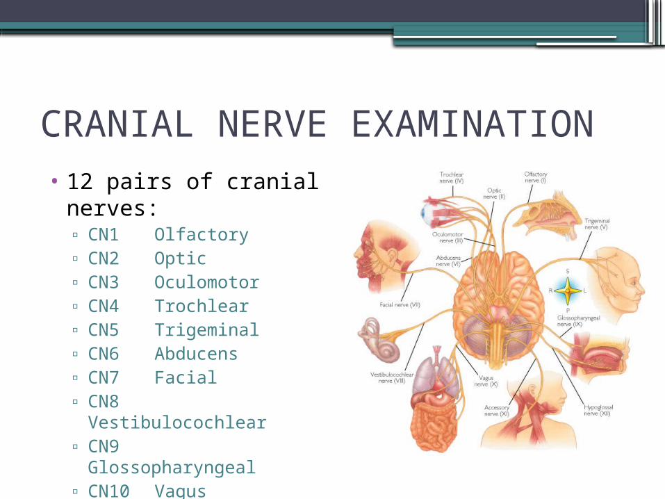
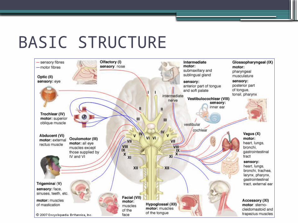
• 12 pairs of cranial nerves:▫ CN1 Olfactory▫ CN2 Optic▫ CN3 Oculomotor▫ CN4 Trochlear▫ CN5 Trigeminal▫ CN6 Abducens▫ CN7 Facial▫ CN8 Vestibulocochlear▫ CN9 Glossopharyngeal▫ CN10 Vagus▫ CN11 Accessory▫ CN12 Hypoglossal
CRANIAL NERVE EXAMINATION
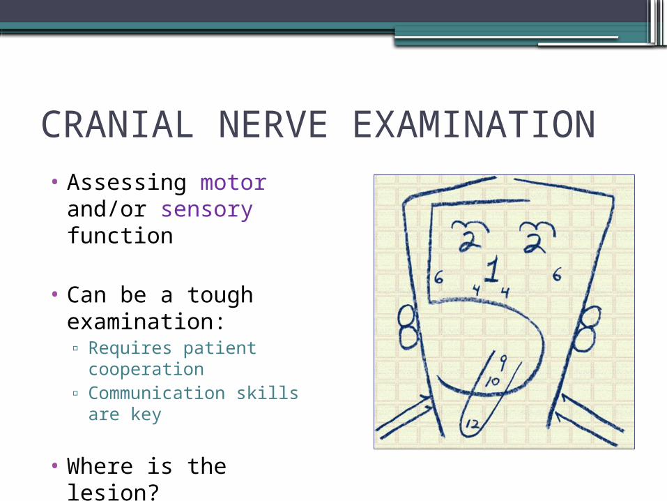
• Assessing motor and/or sensory function
• Can be a tough examination:▫ Requires patient
cooperation▫ Communication skills are
key
• Where is the lesion?

BASIC STRUCTURE
STARTING THE EXAMINATION
• WASH HANDS• INTRODUCE & CONSENT
▫“Hello, my name is **. I am a medical student. I would like to examine your eyes and the movement and feeling in your face today. Would that be OK?”
• POSITION▫Sitting (in bed/on couch/on chair)
• EXPOSE▫Head and neck
• RETREAT to end of bed to observe
INSPECTION – END OF THE BED
AROUND THE BED:•Sensory aids – Including Spectacles•Mobility aids•Special Diet•Catheter
THE PATIENT:•Well/Unwell?•Level of Consciousness•Obvious Neurological Signs?
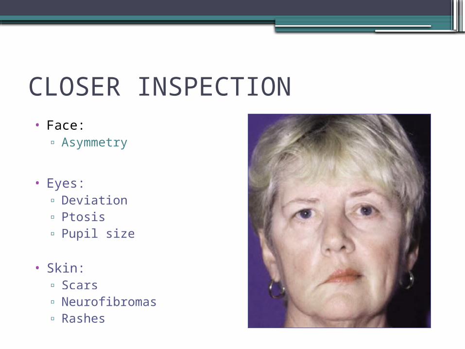
CLOSER INSPECTION• Face:
▫ Asymmetry
• Eyes:▫ Deviation▫ Ptosis▫ Pupil size
• Skin:▫ Scars▫ Neurofibromas▫ Rashes
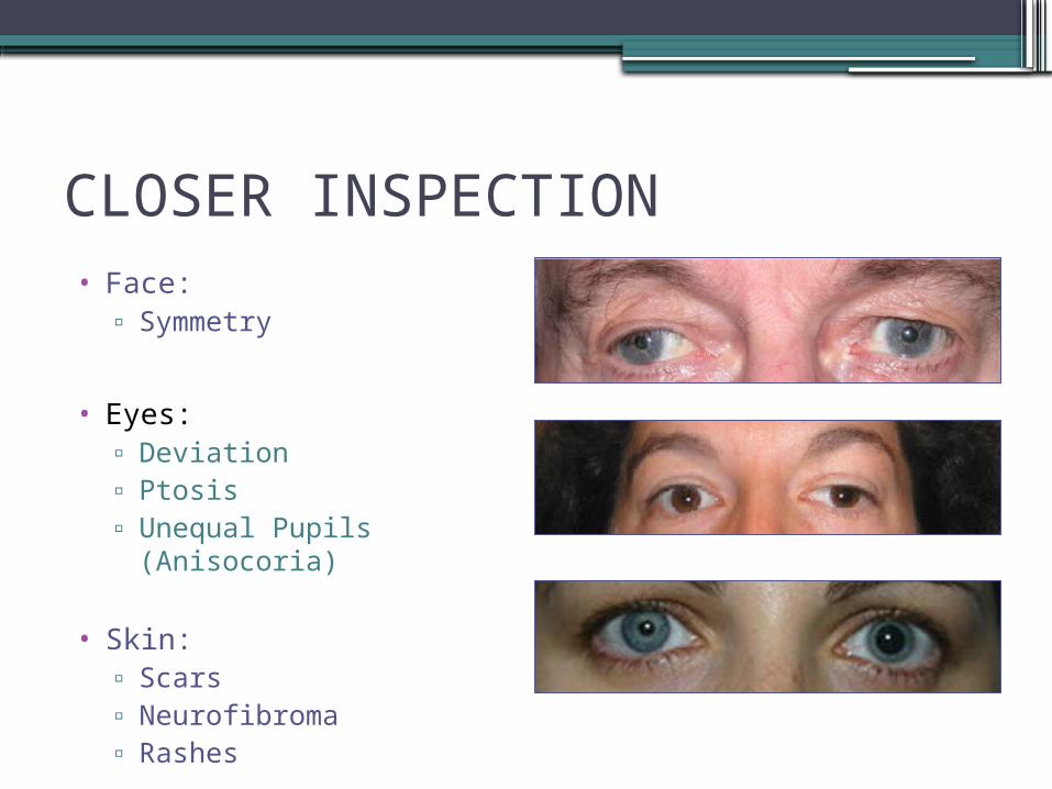
CLOSER INSPECTION• Face:
▫ Symmetry
• Eyes:▫ Deviation▫ Ptosis▫ Unequal Pupils
(Anisocoria)
• Skin:▫ Scars▫ Neurofibroma▫ Rashes
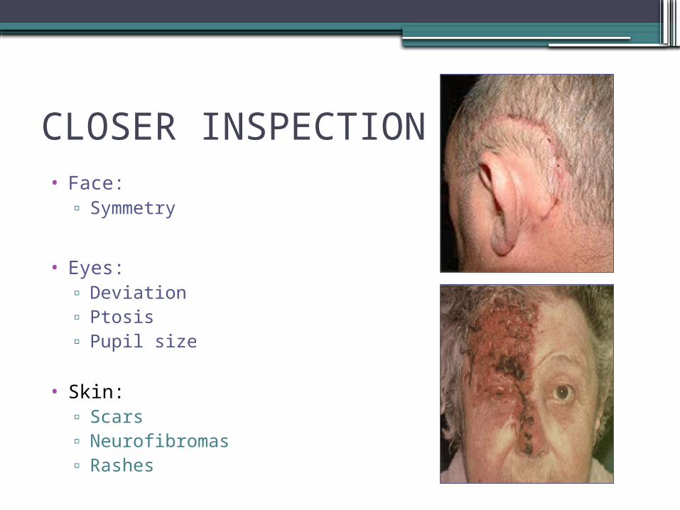
CLOSER INSPECTION• Face:
▫ Symmetry
• Eyes:▫ Deviation▫ Ptosis▫ Pupil size
• Skin:▫ Scars▫ Neurofibromas▫ Rashes
CRANIAL NERVE EXAMINATION
BASIC STRUCTURE
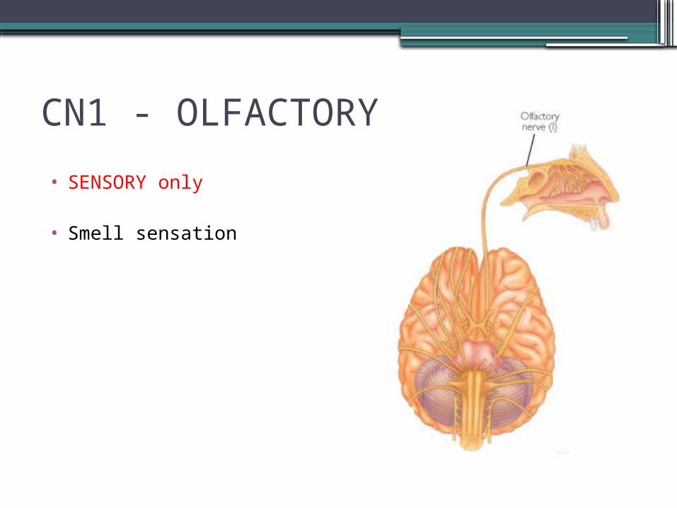
CN1 - OLFACTORY
• SENSORY only
• Smell sensation
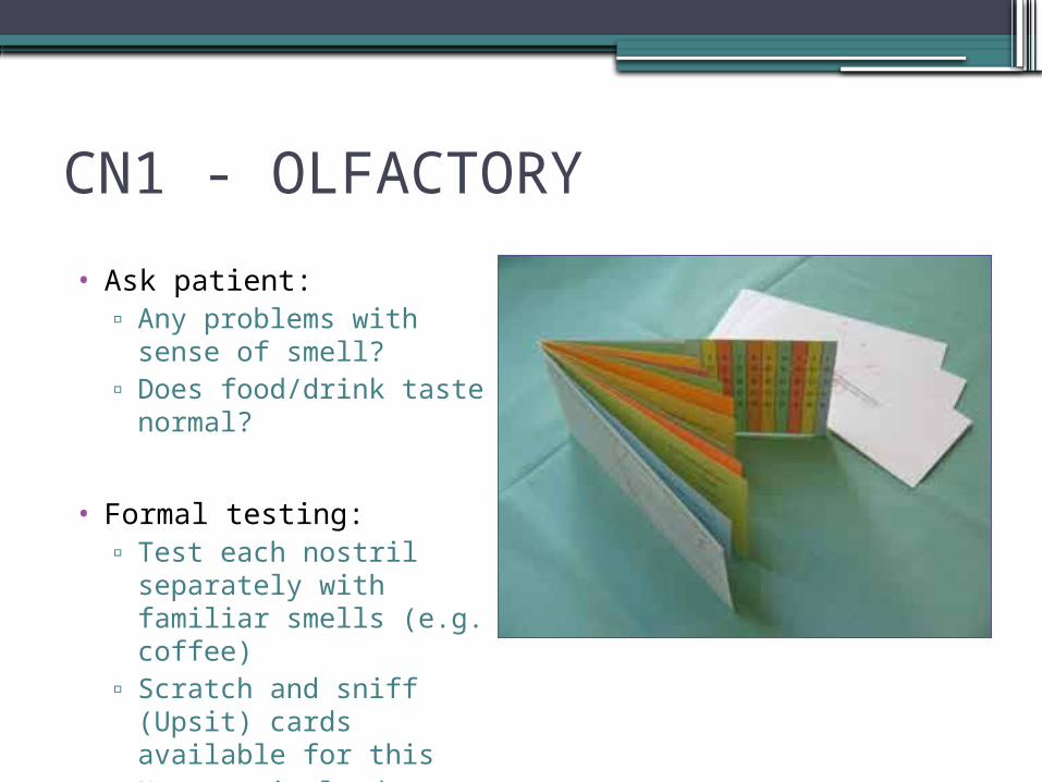
CN1 - OLFACTORY
• Ask patient:▫ Any problems with sense
of smell?▫ Does food/drink taste
normal?
• Formal testing:▫ Test each nostril
separately with familiar smells (e.g. coffee)
▫ Scratch and sniff (Upsit) cards available for this
▫ Not routinely done
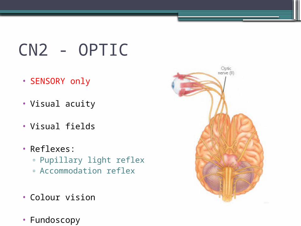
CN2 - OPTIC
• SENSORY only
• Visual acuity
• Visual fields
• Reflexes:▫ Pupillary light reflex▫ Accommodation reflex
• Colour vision
• Fundoscopy
CN2 - OPTIC
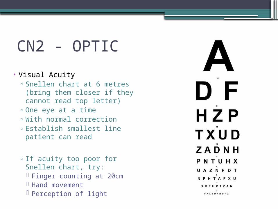
• Visual Acuity▫ Snellen chart at 6 metres
(bring them closer if they cannot read top letter)
▫ One eye at a time▫ With normal correction▫ Establish smallest line
patient can read
▫ If acuity too poor for Snellen chart, try: Finger counting at 20cm Hand movement Perception of light
CN2 - OPTIC
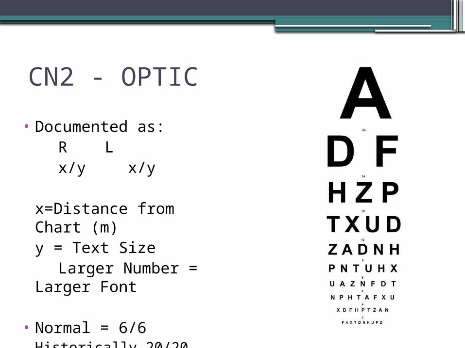
• Documented as:R Lx/y x/y
x=Distance from Chart (m)y = Text Size
Larger Number = Larger Font
• Normal = 6/6Historically 20/20
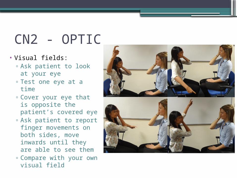
CN2 - OPTIC• Visual fields:
▫ Ask patient to look at your eye
▫ Test one eye at a time▫ Cover your eye that is
opposite the patient’s covered eye
▫ Ask patient to report finger movements on both sides, move inwards until they are able to see them
▫ Compare with your own visual field
CN2 - OPTIC
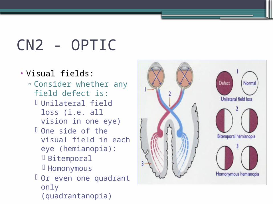
• Visual fields:▫ Consider whether any
field defect is: Unilateral field loss
(i.e. all vision in one eye)
One side of the visual field in each eye (hemianopia): Bitemporal Homonymous
Or even one quadrant only (quadrantanopia)
CN2 - OPTIC
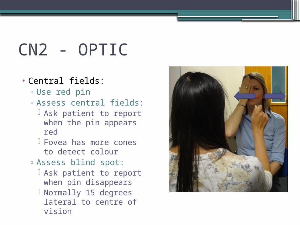
• Central fields:▫ Use red pin▫ Assess central fields:
Ask patient to report when the pin appears red
Fovea has more cones to detect colour
▫ Assess blind spot: Ask patient to report
when pin disappears Normally 15 degrees
lateral to centre of vision
CN2 - OPTIC
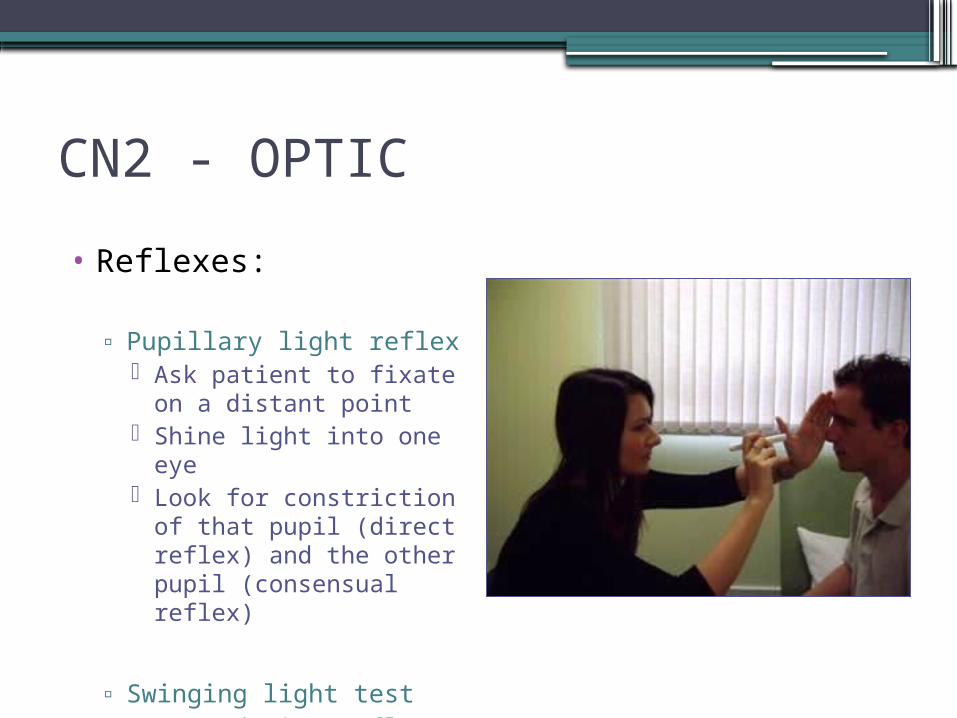
• Reflexes:
▫ Pupillary light reflex Ask patient to fixate on a
distant point Shine light into one eye Look for constriction of
that pupil (direct reflex) and the other pupil (consensual reflex)
▫ Swinging light test▫ Accommodation reflex
CN2 - OPTIC
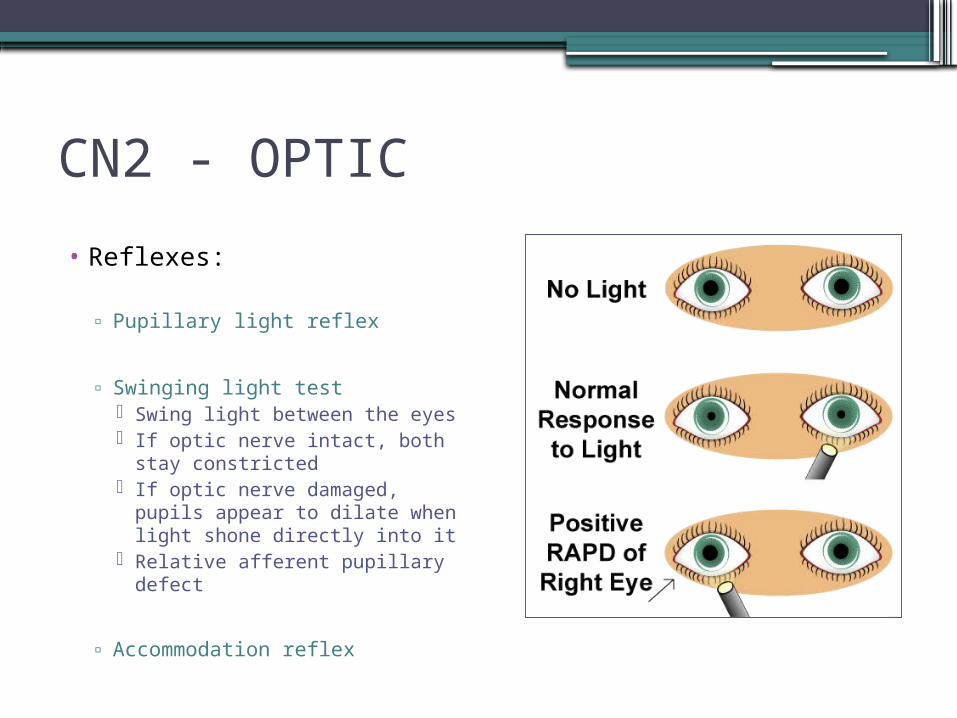
• Reflexes:
▫ Pupillary light reflex
▫ Swinging light test Swing light between the
eyes If optic nerve intact, both
stay constricted If optic nerve damaged,
pupils appear to dilate when light shone directly into it
Relative afferent pupillary defect
▫ Accommodation reflex
CN2 - OPTIC
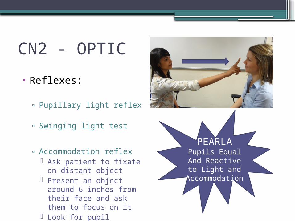
• Reflexes:
▫ Pupillary light reflex
▫ Swinging light test
▫ Accommodation reflex Ask patient to fixate on
distant object Present an object around
6 inches from their face and ask them to focus on it
Look for pupil constriction
PEARLAPupils Equal And Reactive to Light and
Accommodation
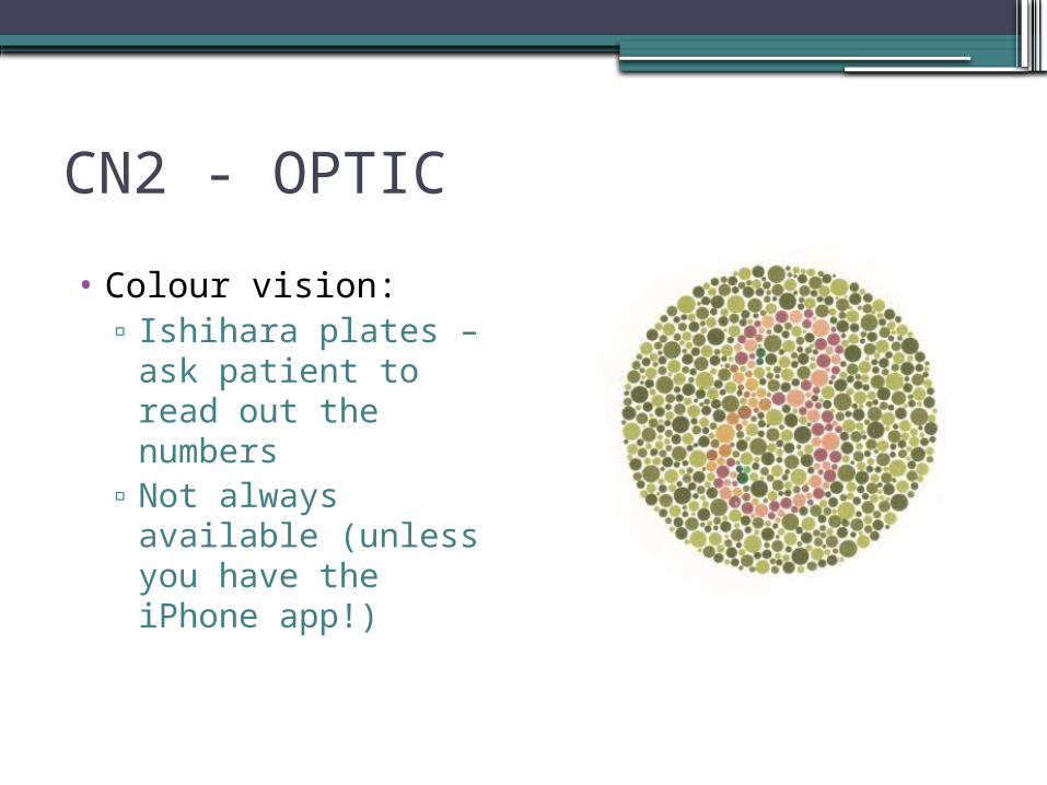
CN2 - OPTIC
• Colour vision:▫ Ishihara plates – ask
patient to read out the numbers
▫ Not always available (unless you have the iPhone app!)
CN2 - OPTIC
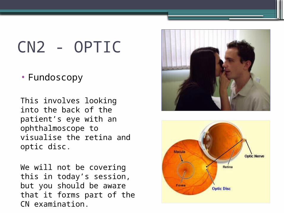
• Fundoscopy
This involves looking into the back of the patient’s eye with an ophthalmoscope to visualise the retina and optic disc.
We will not be covering this in today’s session, but you should be aware that it forms part of the CN examination.
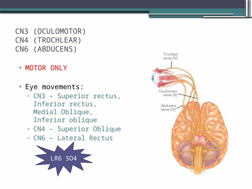
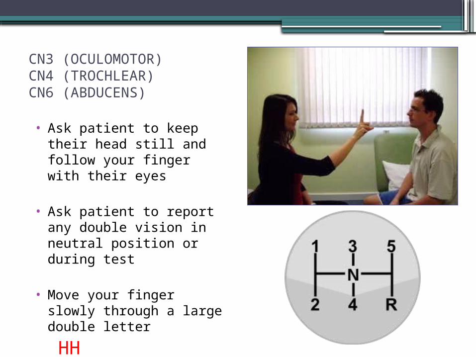
CN3 (OCULOMOTOR)CN4 (TROCHLEAR)CN6 (ABDUCENS)
• MOTOR ONLY
• Eye movements:▫ CN3 – Superior rectus,
Inferior rectus, Medial Oblique, Inferior oblique
▫ CN4 – Superior Oblique▫ CN6 – Lateral Rectus
LR6 SO4
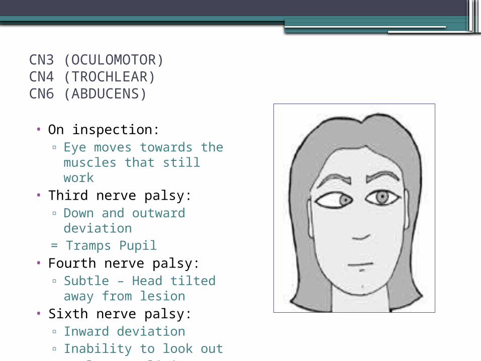
CN3 (OCULOMOTOR)CN4 (TROCHLEAR)CN6 (ABDUCENS)
• On inspection:▫ Eye moves towards the
muscles that still work• Third nerve palsy:
▫ Down and outward deviation
= Tramps Pupil• Fourth nerve palsy:
▫ Subtle – Head tilted away from lesion
• Sixth nerve palsy:▫ Inward deviation▫ Inability to look out▫ “False Localising Sign”
CN3 (OCULOMOTOR)CN4 (TROCHLEAR)CN6 (ABDUCENS)
• Ask patient to keep their head still and follow your finger with their eyes
• Ask patient to report any double vision in neutral position or during test
• Move your finger slowly through a large double letter
HH• Observe for full eye
movements
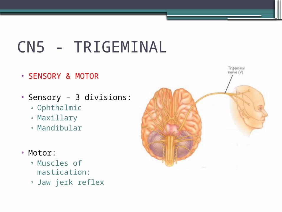
CN5 - TRIGEMINAL
• SENSORY & MOTOR
• Sensory – 3 divisions:▫ Ophthalmic▫ Maxillary▫ Mandibular
• Motor:▫ Muscles of mastication:▫ Jaw jerk reflex
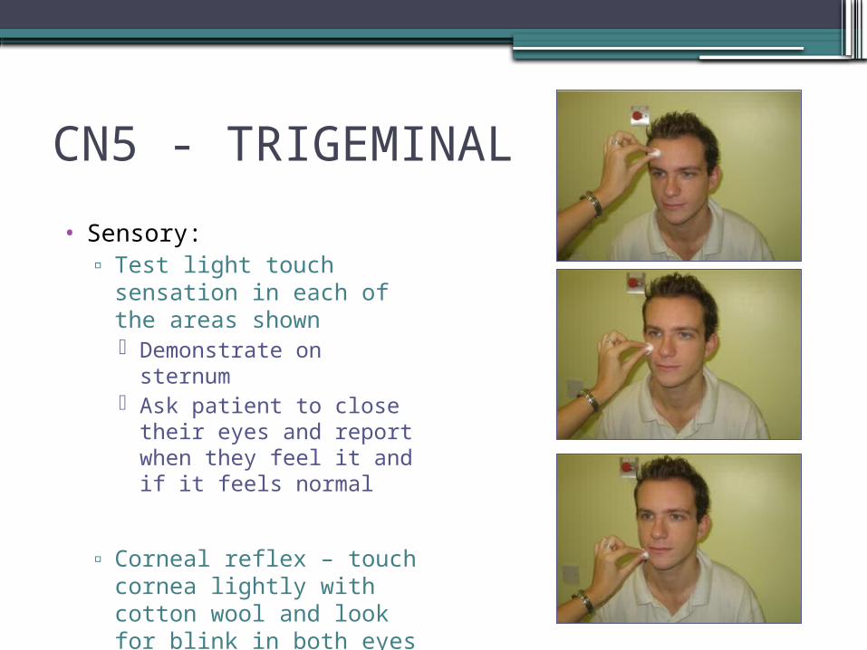
CN5 - TRIGEMINAL
• Sensory:▫ Test light touch sensation
in each of the areas shown Demonstrate on sternum Ask patient to close their
eyes and report when they feel it and if it feels normal
▫ Corneal reflex – touch cornea lightly with cotton wool and look for blink in both eyes Not done in exam setting
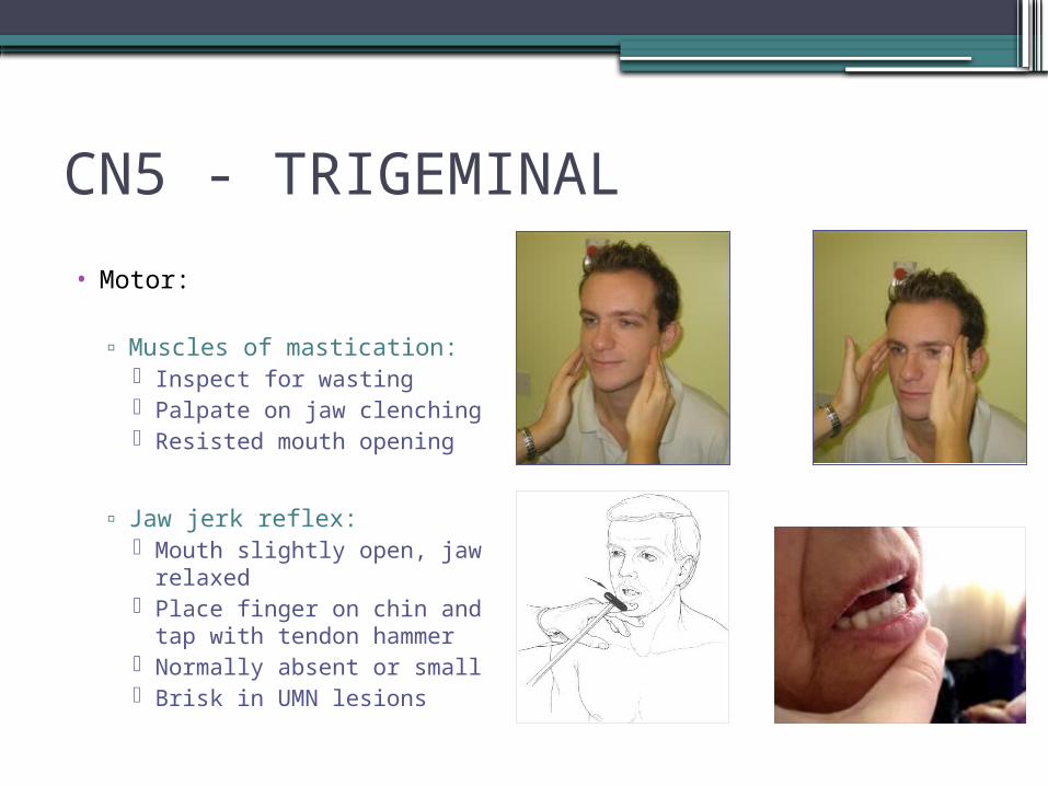
CN5 - TRIGEMINAL
• Motor:
▫ Muscles of mastication: Inspect for wasting Palpate on jaw clenching Resisted mouth opening
▫ Jaw jerk reflex: Mouth slightly open, jaw
relaxed Place finger on chin and
tap with tendon hammer Normally absent or small Brisk in UMN lesions
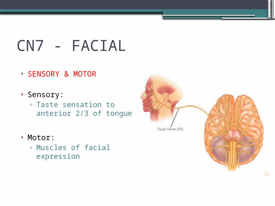
CN7 - FACIAL
• SENSORY & MOTOR
• Sensory:▫ Taste sensation to
anterior 2/3 of tongue
• Motor:▫ Muscles of facial
expression
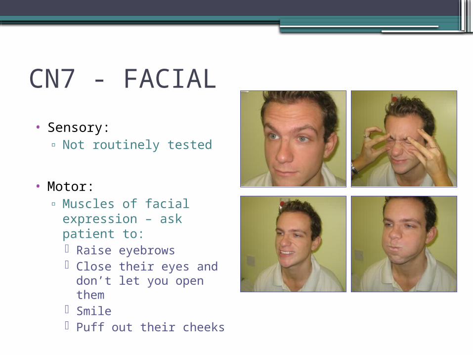
CN7 - FACIAL
• Sensory:▫ Not routinely tested
• Motor:▫ Muscles of facial
expression – ask patient to: Raise eyebrows Close their eyes and
don’t let you open them Smile Puff out their cheeks
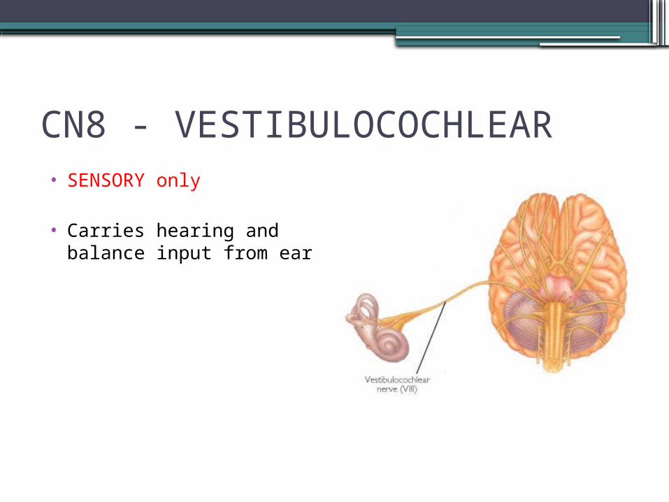
CN8 - VESTIBULOCOCHLEAR• SENSORY only
• Carries hearing and balance input from ear
CN8 - VESTIBULOCOCHLEAR
•Crudely test hearing:▫Whisper a number into each ear whilst
making a distracting sound in the other ear▫Ask patient to repeat the number
•If concerned, perform Weber’s and Rinne’s tests

CN8 - VESTIBULOCOCHLEAR• Weber’s test:
▫ Tuning fork in centre of forehead – in which ear does it sound louder?
▫ Normally equal in both ears.
▫ Conductive hearing loss: Lateralises to affected side
▫ Sensorineural hearing loss: Lateralises to non-affected
side How do you know which? Rinne’s Test
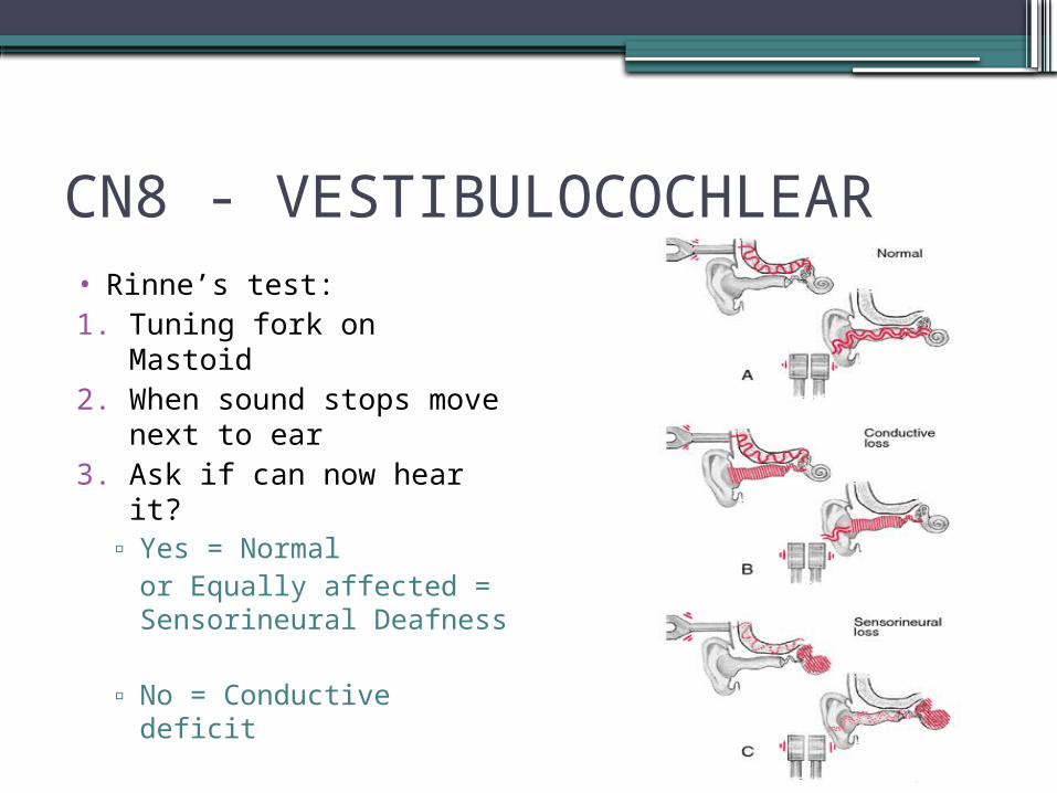
CN8 - VESTIBULOCOCHLEAR• Rinne’s test:1. Tuning fork on Mastoid 2. When sound stops move
next to ear3. Ask if can now hear it?
▫ Yes = Normal or Equally affected = Sensorineural Deafness
▫ No = Conductive deficit
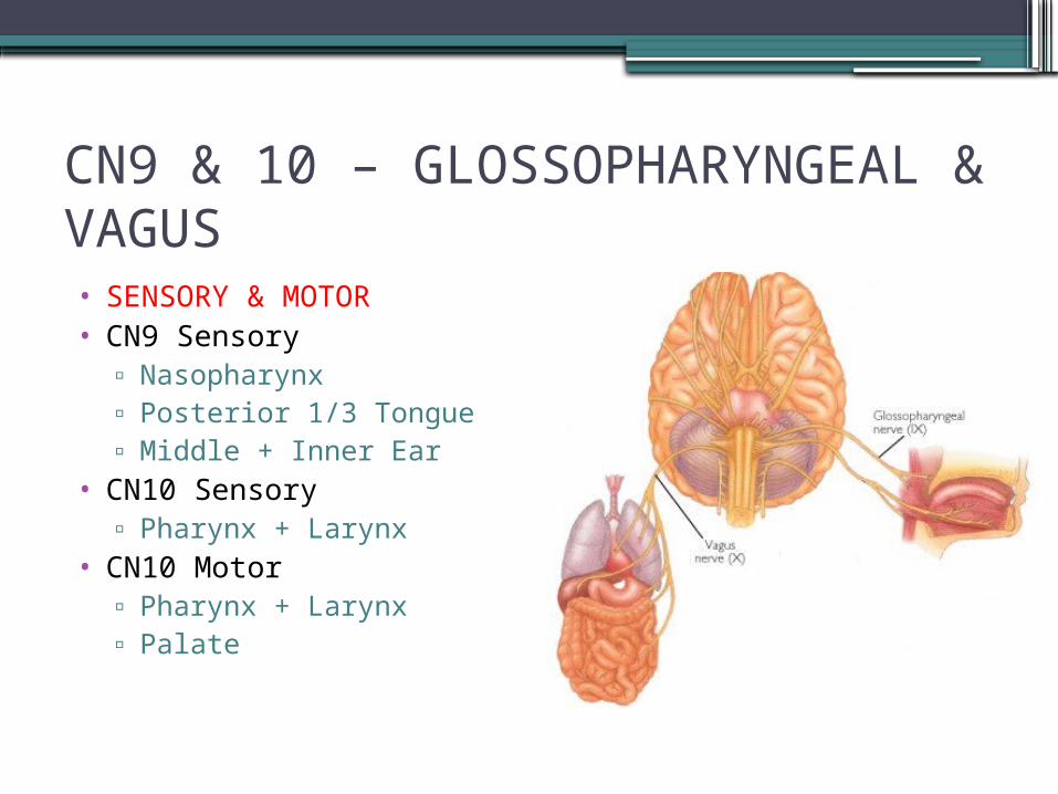
CN9 & 10 – GLOSSOPHARYNGEAL & VAGUS• SENSORY & MOTOR• CN9 Sensory
▫ Nasopharynx▫ Posterior 1/3 Tongue▫ Middle + Inner Ear
• CN10 Sensory▫ Pharynx + Larynx
• CN10 Motor▫ Pharynx + Larynx▫ Palate
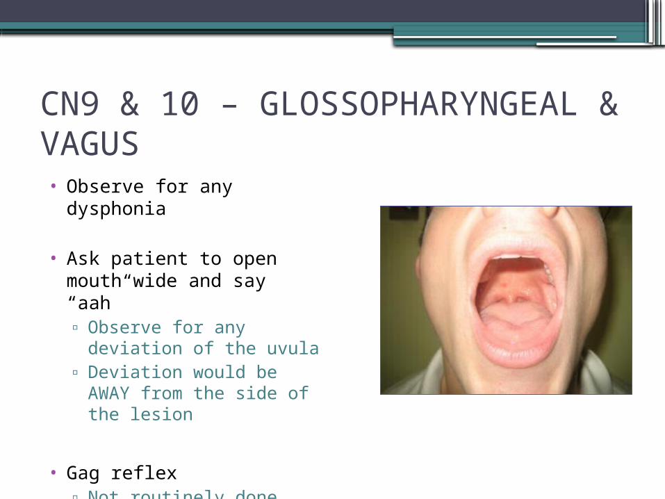
CN9 & 10 – GLOSSOPHARYNGEAL & VAGUS• Observe for any dysphonia
• Ask patient to open mouth wide and say “aah”▫ Observe for any deviation
of the uvula▫ Deviation would be AWAY
from the side of the lesion
• Gag reflex▫ Not routinely done
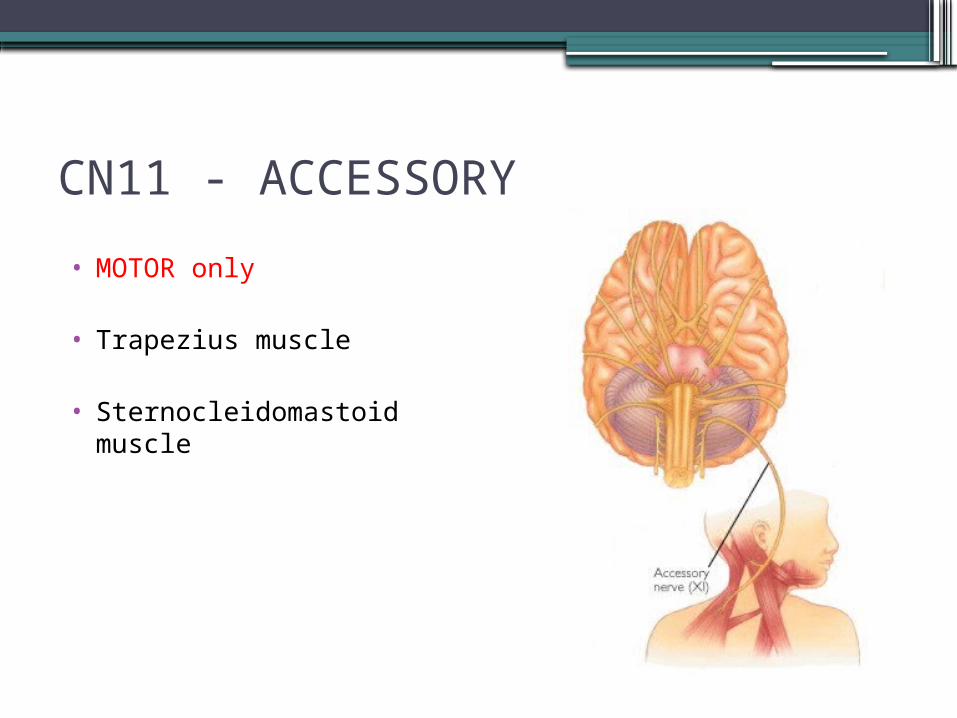
CN11 - ACCESSORY
• MOTOR only
• Trapezius muscle
• Sternocleidomastoid muscle
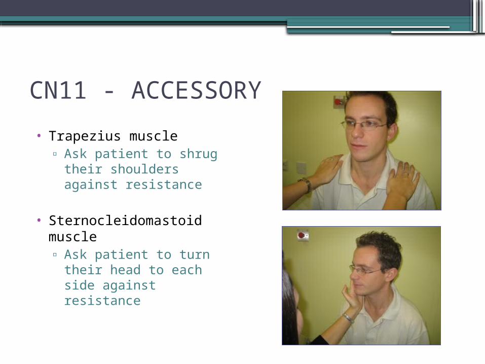
CN11 - ACCESSORY
• Trapezius muscle▫ Ask patient to shrug their
shoulders against resistance
• Sternocleidomastoid muscle▫ Ask patient to turn their
head to each side against resistance
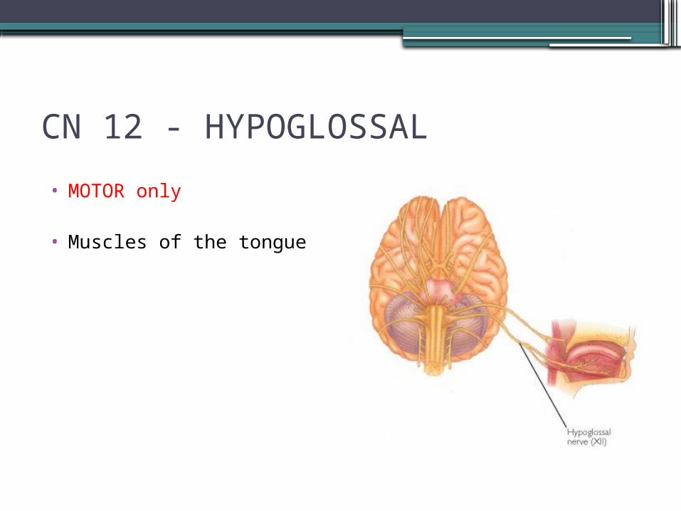
CN 12 - HYPOGLOSSAL
• MOTOR only
• Muscles of the tongue
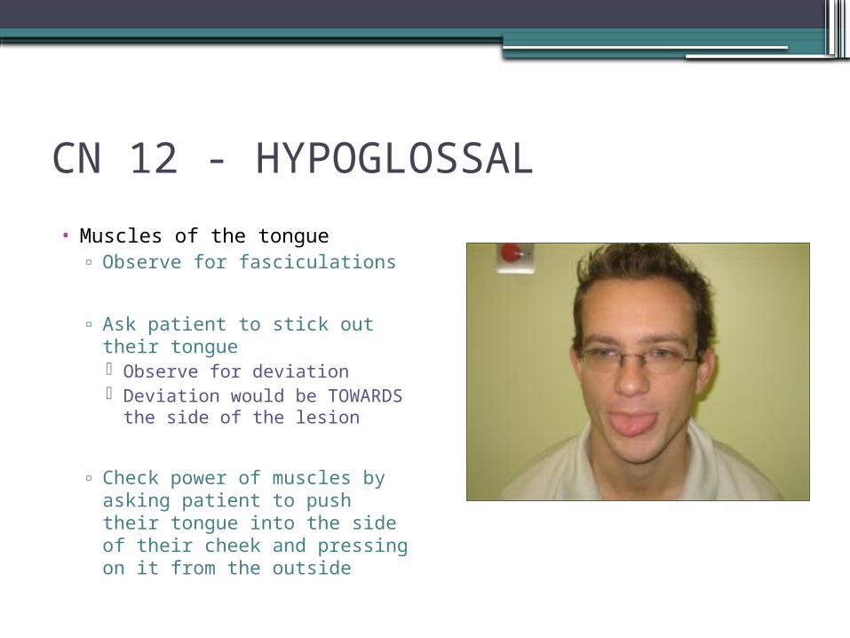
CN 12 - HYPOGLOSSAL
• Muscles of the tongue▫ Observe for fasciculations
▫ Ask patient to stick out their tongue Observe for deviation Deviation would be
TOWARDS the side of the lesion
▫ Check power of muscles by asking patient to push their tongue into the side of their cheek and pressing on it from the outside
COMPLETING THE EXAMINATION
•THANK PATIENT•ENSURE COMFORT•WASH HANDS
“To complete my examination I would like to perform the reflexes mentioned, plus a full peripheral nerve examination.”
TYING IT ALL TOGETHER
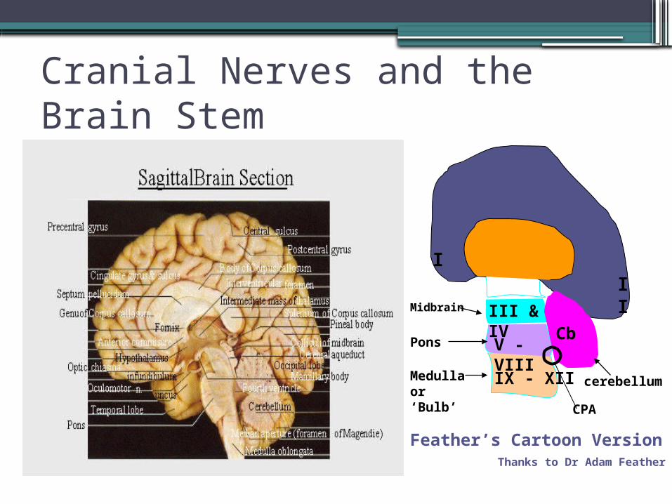
Cranial Nerves and the Brain Stem
I
II
III & IV
V - VIII
IX - XII
Cb
Feather’s Cartoon VersionThanks to Dr Adam Feather
Midbrain
Pons
Medulla or ‘Bulb’
cerebellum
CPA

![Oligomorphic permutation groups - QMUL Mathspjc/preprints/oligo.pdf · 2008-03-04 · groups. For further information about permutation groups, see [7, 14]. Note also that there are](https://img.pdfslide.tips/doc/110x75/5f9e488a7d777a0770675a71/oligomorphic-permutation-groups-qmul-pjcpreprintsoligopdf-2008-03-04-groups.jpg)