Embed Size (px)
Citation preview

Critical Flocculation Concentrations, BindingIsotherms, and Ligand Exchange Properties ofPeptide-Modified Gold Nanoparticles Studied byUV-Visible, Fluorescence, and Time-CorrelatedSingle Photon Counting Spectroscopies
Huan Xie,† Alexander G. Tkachenko,† Wilhelm R. Glomm,† Joseph A. Ryan,† Matthew K. Brennaman,‡John M. Papanikolas,‡ Stefan Franzen,*,† and Daniel L. Feldheim*,†
Department of Chemistry, North Carolina State University, Raleigh, North Carolina 27695, and Department of Chemistry,University of North Carolina, Chapel Hill, North Carolina 27599
Protocols for modifying gold nanoparticles with peptide-bovine serum albumin (BSA) conjugates are describedwithin. The resulting constructs were characterized usinga number of techniques including static fluorescencespectroscopy and time-correlated single photon countingspectroscopy (TCSPC) in order to quantify peptide-BSAbinding isotherms, exchange rates, critical flocculationconcentrations, and the composition of mixed peptide-BSA monolayers on gold nanoparticles. TCSPC has provento be a powerful technique for observing the microenvi-ronment of protein-gold nanoparticle conjugates becauseit can distinguish between surface-bound and solution-phase species without the need for separation steps. Fullcharacterization of the composition and stability of pep-tide-modified metal nanoparticles is an important step intheir use as intracellular delivery vectors and imagingagents.
This paper addresses fundamental issues involving the adsorp-tion of peptide-modified protein conjugates onto gold nanopar-ticles. Metal particle bioconjugates are important constructs forcellular imaging, therapeutic delivery, and biomolecule detection.1-4
For example, Schultz et al. used gold and silver particles asimmunohistochemical staining reagents to selectively label regionsof the Drosophila X chromosome.3 Because of the remarkablylarge scattering cross section of metal particles (10-10 cm2),individual nanoparticles can be imaged under white-light illumina-tion. Moreover, the plasmon resonance condition of metals canbe tuned across the visible spectrum and into the near-infrared
simply by changing particle size and shape.5-7 Thus, multicolorassays are possible with a single light source, without the needfor filters, and free from complications of fluorescence bleachingor blinking.
We have employed gold particle bioconjugates to study cellularuptake of peptides and antibodies.4 The constructs used in ourstudies are based on those described in the pioneering work ofFeldherr et al.2 In their work, cell-targeting peptides wereconjugated to bovine serum albumin (BSA). The peptide-BSAconjugates were subsequently adsorbed onto gold nanoparticles,microinjected into a HeLa cell line and imaged with transmissionelectron microscopy.
A major goal of modern therapeutic delivery and tissue imagingis to create a bioconjugate capable of seeking out specific cells invivo, traversing the cell and nuclear membranes, and releasing atherapeutic in the nucleus. Our work has sought to accomplishthese tasks by combining multiple membrane-translocating pep-tides and antibodies onto a single gold nanoparticle. Of importancein this work is understanding how to formulate BSA-goldbioconjugates that are stable in high ionic strength solutionscontaining exchangeable peptides and proteins and how toquantify the number of adsorbed biomolecules per gold particle.This paper addresses these fundamental issues using a combina-tion of classic and time-resolved fluorescence spectroscopy studies.
MATERIALS AND METHODSMaterials. Gold nanoparticles (20-nm diameter) were pur-
chased from Ted Pella. BSA was purchased from Roche (MW,67 000). 3-Maleimidobenzonic acid N-hydroxysuccinimide ester(MBS), dithiothreitol (DTT), rhodamine B isothiocyanate (RBITC),glutathione, lysine, fluorescamine, and 5,5′-dithio-bis(2-nitroben-zoic acid) (DTNB) were obtained from Sigma-Aldrich. Ruthenium-(II) tris(2,2′-bipyridine)monohexanethiol ([Ru(bipy)2bipy-C6H12-SH]2+) was synthesized as described previously.8-11
Sephadex G10-50 was purchased from Sigma. Centricon YM-30 and Microcon YM-100 were purchased from Millipore. The
* To whom correspondence should be addressed. E-mail:[email protected]; [email protected].
† North Carolina State University.‡ University of North Carolina.
(1) Cao, Y.; Jin, R.; Mirkin, C. A. J. Am. Chem. Soc. 2001, 123, 7961-7962.(2) Feldherr, C. M.; Lanford, R. E.; Akin, D. Proc. Natl. Acad. Sci. U.S.A. 1992,
89, 11002-11005.(3) Schultz, S.; Smith, D. R.; Mock, J. J.; Schultz, D. A. Proc. Natl. Acad. Sci.
U.S.A. 2000, 97, 996-1001.(4) Tkachenko, A. G.; Xie, H.; Coleman, D.; Glomm, W.; Ryan, J.; Anderson,
M. F.; Franzen, S.; Feldheim, D. L. J. Am. Chem. Soc. 2003, 125, 4700-4701.
(5) Haes, A.; Van Duyne, R. J. Am. Chem. Soc. 2002, 124, 10596-10604.(6) West, J. L.; Halas, N. J. Curr. Opin. Biotechnol. 2000, 11, 215-217.(7) Jin, R.; Cao, Y.; Mirkin, C. A.; Kelly, K.; Schatz, G.; Zheng, J. Science 2001,
294, 1901-1903.
Anal. Chem. 2003, 75, 5797-5805
10.1021/ac034578d CCC: $25.00 © 2003 American Chemical Society Analytical Chemistry, Vol. 75, No. 21, November 1, 2003 5797Published on Web 09/30/2003
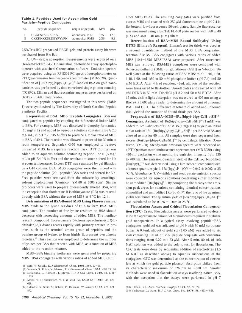
7.5% Tris-HCl prepacked PAGE gels and protein assay kit werepurchased from Bio-Rad.
All UV-visible absorption measurements were acquired on aHewlett-Packard 8453 Chemstation photodiode array spectropho-tometer with attached Chemstation software. Emission spectrawere acquired using an RF-5301 PC spectrofluorophotometer orPTI Quantamaster luminescence spectrometer (MD-5020). Quan-tification of [Ru(bipy)2bipy-C6H12-S]2+-labeled BSA on gold nano-particles was performed by time-correlated single photon counting(TCSPC). Ellman and fluorescamine analyses were performed onBioTek FL-600 plate reader.
The two peptide sequences investigated in this work (Table1) were synthesized by The University of North Carolina PeptideSynthesis Facility.
Preparation of BSA-MBS-Peptide Conjugates. BSA wasconjugated to peptides by coupling the bifunctional linker MBSto BSA. For example, MBS was dissolved in dimethylformamide(10 mg/mL) and added to aqueous solutions containing BSA (10mg/mL, in pH 7.2 PBS buffer) to produce a molar ratio of MBSto BSA of 40:1. The reaction was allowed to proceed for 30 min atroom temperature. Sephadex G-50 was employed to removeunreacted MBS. In a separate reaction flask, DTT (10 mg) wasadded to an aqueous solution containing peptide I or II (5 mg/mL in pH 7.4 PB buffer) and the resultant mixture stirred for 1 hat room temperature. Excess DTT was separated by gel filtrationon a G10 column. BSA-MBS conjugates were then mixed withthe peptide solution (20:1 peptide/BSA ratio) and stirred for 5 h.Free peptides were removed from the mixture by centrifugalsolvent displacement (Centricon YM-30 at 5000 rpm). Similarprotocols were used to prepare fluorescently labeled BSA, withthe exception that rhodamine B isothiocyanate (RB) was reacteddirectly with BSA without the use of MBS at 4 °C for 12 h.
Determination of BSA-Bound MBS Using Fluorescamine.MBS binds to the lysine residues of BSA to form BSA-MBSconjugates. The number of free lysine residues on BSA shoulddecrease with increasing amounts of added MBS. The nonfluo-rescent compound fluorescamine (4-phenylspiro[furan-2(3H)-1′-phthalan]-3,3′-dione) reacts rapidly with primary amines in pro-teins, such as the terminal amino group of peptides and theε-amino group of lysine, to form highly fluorescent pyrrolinonemoieties.12 This reaction was employed to determine the numberof lysines per BSA that reacted with MBS, as a function of MBSadded to the reaction mixture.
MBS-BSA binding isotherms were generated by preparingMBS-BSA conjugates with various ratios of added MBS (10:1-
135:1 MBS/BSA). The resulting conjugates were purified fromexcess MBS and reacted with 250 µM fluorescamine at pH 7.4 inflat-bottom low-fluorescence 96-well plates. Sample fluorescencewas measured using a BioTek FL-600 plate reader with 360 ( 40(EX) and 460 ( 40 nm (EM) filters.
Determination of BSA-MBS Bound Sulfhydryl UsingDTNB (Ellman’s Reagent). Ellman’s test for thiols was used asa second quantitative method of the MBS-BSA conjugationreaction.13 MBS-BSA conjugates with various ratios of addedMBS (10:1-135:1 MBS/BSA) were prepared. After unreactedMBS was removed, BSA-MBS complexes were combined withâ-mercaptoethanol (BME) or glutathione (GSH) in V-bottom 96-well plates at the following ratios of BSA/MBS/thiol: 1:10, 1:20,1:40, 1:60, and 1:80 in 50 mM phosphate buffer (pH 7.4) and 50mM EDTA. After 4 h of reaction, 45-µL aliquots of the reactionwere transferred to flat-bottom 96-well plates and reacted with 50µM DTNB in 50 mM Tris‚HCl pH 8.2 and 50 mM EDTA. After15 min, visible light absorption was measured at 405 nm using aBioTek FL-600 plate reader to determine the amount of unboundBME and GSH. The difference of total thiol added and unboundthiol yielded the number of bound thiols per BSA.
Preparation of BSA-MBS-[Ru(bipy)2bipy-C6H12-SH]2+
Conjugates. A solution of [Ru(bipy)2bipy-C6H12-SH]2+ (1 mM) wasadded to 1-mL aliquots of BSA/MBS (1 mg/mL) to yield an initialmolar ratio of 15:1 [Ru(bipy)2bipy-C6H12-SH]2+ per BSA-MBS andallowed to mix for 60 min. All samples were then separated fromexcess [Ru(bipy)2bipy-C6H12-SH]2+ by centrifugal filtration (Cen-tricon, YM- 30). Steady-state emission spectra were recorded ona PTI Quantamaster luminescence spectrometer (MD-5020) using450-nm excitation while monitoring emission intensity from 530to 700 nm. The emission quantum yield of the C6H12-SH-modified[Ru(bpy)3]2+ was determined using a luminescent compound witha known quantum yield; [Ru(bpy)3]2+ (QY ) 0.042 ( 0.002 at 25°C14). Absorbance (UV-visible) and steady-state emission spectrawere collected for aqueous solutions containing either modifiedor unmodified [Ru(bpy)3]2+. By comparing the steady-state emis-sion peak areas for solutions containing identical concentrationsof modified and unmodified [Ru(bpy)3]2+, the ratio of the quantumyields was found. The quantum yield of [Ru(bpy)2bpy-C6H12-SH]2+
was calculated to be 0.026 ( 0.003 at 25 °C.Flocculation Assays and Critical Flocculation Concentra-
tion (CFC) Tests. Flocculation assays were performed to deter-mine the approximate amount of biomolecules required to stabilizegold nanoparticles. In a typical assay involving peptide-BSAconjugates, gold sol was adjusted to pH 9 with 50 mM carbonatebuffer. A 0.7-mL aliquot of gold sol (1.05 nM) was added to sixvials containing 100 µL of BSA-peptide conjugate with concentra-tions ranging from 0.22 to 1.83 µM. After 5 min, 80 µL of 10%NaCl solution was added to the sols to test for flocculation. TheCFC tests were done by sequential addition of electrolytes (1.5M NaCl as described above) to aqueous suspensions of theconjugates. CFC was determined as the concentration of electro-lyte at which the gold particle plasmon absorption shifted fromits characteristic maximum of 526 nm to ∼600 nm. Similarmethods were used in flocculation assays involving native BSA,with the exception that the assays were performed in pH 7
(8) Sato, Y.; Uosaki, K. J. Electroanal. Chem. 1995, 384, 57-66.(9) Yamada, S.; Koide, Y.; Matsuo, T. J. Electroanal. Chem. 1997, 426, 23-26.
(10) Dellaciana, L.; Hamachi, I.; Meyer, T. J. J. Org. Chem. 1989, 54, 1731-1735.
(11) Maier, V. E.; Shafirovich, V. Y. B Acad. Sci. USSR Ch+ 1989, 38, 626-628.
(12) Udenfrie, S.; Stein, S.; Bohlen, P.; Dairman, W. Science 1972, 178, 871-872.
(13) Ellman, G. L. Arch. Biochem. Biophys. 1959, 82, 70-77.(14) Vanhouten, J.; Watts, R. J. J. Am. Chem. Soc. 1976, 98, 4853-4858.
Table 1. Peptides Used for Assembling GoldParticle-Peptide Conjugates
no. peptide sequence origin of peptide MW pKI
I CGGFSTSLRARKA adenoviral NLS 1353 12.3II CKKKKKKSEDEYPYVPN adenoviral RME 2084 9.3
5798 Analytical Chemistry, Vol. 75, No. 21, November 1, 2003
phosphate buffer and the gold sol was added to six vials containing100 µL of BSA with concentrations ranging from 1.85 to 5.85 µM.
Quantifying [Ru(bipy)2bipy-C6H12-S]2+-Labeled BSA onGold Nanoparticles by Time-Correlated Single Photon Count-ing. The surface coverage of [Ru(bipy)2bipy-C6H12-S]2+-labeledBSA on 20-nm gold colloids was determined using TCSPC.Transients were obtained for the pure [Ru(bipy)2bipy-C6H12-S]2+-labeled BSA solution (1 mg/mL; preparation described above)and for a solution with 20-nm gold colloids mixed with a ratio of500:1 [Ru(bipy)2bipy-C6H12-S]2+-labeled BSA/gold.
Time-Resolved Emission Methods. Time-resolved emissionspectra were acquired with an argon ion laser whose continuousoutput was used to pump a mode-locked Ti:sapphire oscillator.The Ti:sapphire laser output was frequency doubled to 423 nmusing a BBO crystal to produce ∼1-ps pulses with a pulse energyof ∼ 0.26 nJ/pulse. This pulse train was pulse picked and therepetition rate selected to be roughly 5 times the natural lifetimeof the sample. Luminescence lifetime data were collected usingthe time-correlated single photon counting technique publishedearlier.15 Samples were sparged with argon for ∼45 min prior touse.
The method for analysis of the fraction of luminescentmolecules on the surface of a nanoparticle and in solution hasbeen also published previously.15 Relaxation from the radiativeexcited states was modeled using the following exponentialdecays:
where N is the total number of phosphorescent components andAi and ki represent the amplitude is rate constant for eachcomponent, respectively. The final fit was approached using aniterative process where exponentials were added until no signifi-cant improvements in the correlation coefficient of the fit couldbe seen. The calculated populations associated with each observedlifetime were then compared to the initial added concentration ofemitter (free in solution) in order to determine the number ofemitters associated with the gold colloids. For example, if thereare two populations, there will be two observed lifetimes. Theradiative rate constant for unquenched Ru(bipy)3
2+ in solution is
where kobs ) 1/τobs, kphos ) 1/τphos, etc. The phosphorescencelifetime is τobs, and the nonradiative lifetime is τnon-rad. Quenchingcan occur by electron or energy transfer from [Ru(bipy)2bipy-C6H12-S]2+ to the gold nanoparticle with rate constant, ket to givean observed lifetime of
The phosphorescence quantum yield is
where ket ) 0 for the solution fraction. It turns out that the ratio
of the amplitudes, Ai/Aj in the fit correspond exactly to the ratioof the amounts of [Ru(bipy)2bipy-C6H12-S]2+ in phases i and j,respectively.16
Quantifying Fluorescently Labeled BSA or BSA-PeptideConjugates on Gold Nanoparticles by Fluorescence Spec-troscopy. BSA coverage on 20-nm-diameter gold particles wasdetermined with fluorescence spectroscopy. In a typical experi-mental procedure (e.g., to generate a binding isotherm plot), BSAwas labeled with rhodamine B isothiocyanate (RB-BSA) andpurified by centrifugal solvent displacement (Centricon YM-30 at5000 rpm). The concentration of RB-BSA following purificationwas determined with a protein assay (Bio-Rad DC protein assay),and standard curves of fluorescence versus RB-BSA concentra-tion were measured. RB-BSA solutions were prepared at con-centrations (nine solutions of 30 µL each) ranging from 0.39 to11.7 µM. Gold nanoparticles (450 µL of 1.05 µM solution) inaqueous phosphate solution buffered at pH 7.0 were added to eachRB-BSA solution. After stirring for 15 min, 20 µL of 2 mg/mLnative BSA (e.g., unlabeled BSA) was added to each solution.17,18
Native BSA was required in order to block open gold particlesurface sites and prevent flocculation. That is, the RB-BSAconcentrations corresponding to submonolayer coverage resultedin particles that were unstable toward flocculation. Experimentsin which the exchange of RB-BSA for native BSA was monitoredin time revealed this to be a slow process (hours). Thus, withinthe time scale of the RB-BSA adsorption isotherm experiments,bound RB-BSA was not displaced by the native BSA stabilizer.Following addition of RB-BSA to gold nanoparticle sols, wesought to measure the unbound fraction of RB-BSA. To removethe unbound fraction, the nanoparticle sols were centrifuged byMicrocon at 2.6 × 10³ rpm for 15 min. The sediment was washedthree times with 50 µL of 50 mM pH 7.0 phosphate buffer. Thefinal sediment was transferred to a new vial, centrifuged at 3.3 ×10³ rpm for 8 min, and resuspended in buffer. The aliquots ofsupernatant were diluted to 2 mL, and fluorescence of thesupernatant was measured and compared to the standard curveto determine the concentration of unbound RB-BSA in the initialmixture. The amount of RB-BSA bound to the gold particles wascalculated by subtracting free RB-BSA from the total amount ofRB-BSA added initially. The number of gold nanoparticles in thesol was determined by UV-visible spectroscopy. Any loss fromnonspecific adsorption of RB-BSA onto the Microcon was nottaken into account.
Adsorption isotherms for peptide-BSA conjugates were de-termined by conjugating peptide I or II to RB-BSA via an MBScross-linker. Gel electrophoresis was used to quantify the peptide/BSA ratio. Peptide-BSA-gold conjugates were assembled usingthe conditions reported for BSA-gold conjugates, with theexception that the gold sol was buffered with pH 9.0 carbonatebuffer.
Quantifying Mixed BSA Adlayers. To determine the molefraction of BSA-peptide I and BSA-peptide II conjugates boundto nanoparticles versus their mole fraction in a solution mixture,
(15) Brennaman, M. K.; Alstrum-Acevedo, J. H.; Fleming, C. N.; Jang, P.; Meyer,T. J.; Papanikolas, J. M. J. Am. Chem. Soc. 2002, 124, 15094-15098.
(16) Glomm, W. R.; Moses, S. J.; Brennaman, M. K.; Papanikolas, J. M.; Franzen,S. J. Phys. Chem., submitted.
(17) Deroe, C.; Courtoy, P. J.; Baudhuin, P. J. Histochem. Cytochem. 1987, 35,1191-1198.
(18) Horisberger, M.; Clerc, M. F. Histochemistry 1985, 82, 219-223.
Y(t) ) ∑i)1
N
Aie-kit (1)
1/τobs ) 1/τphos + 1/τnon-rad (2)
1/τobs ) 1/τphos + 1/τnon-rad + 1/τet (3)
φ ) kphos/(kphos + knon-rad + ket) (4)
Analytical Chemistry, Vol. 75, No. 21, November 1, 2003 5799
BSA-peptide II conjugates were labeled with rhodamine B, whileBSA-peptide I was kept unlabeled. Solution mixtures of BSA-peptide I and RB-BSA-peptide II conjugates were prepared withXpeptide II of 1, 0.8, 0.6, 0.4, and 0.2, keeping the total amount ofBSA constant. The amount of bound RB-BSA-peptide II pernanoparticle was determined using the procedures describedabove for single-component solutions. The amount of BSA-peptide I per nanoparticle was computed by subtracting thenumber of RB-BSA-peptide II per nanoparticle for Xpeptide II * 1from Xpeptide II ) 1.
BSA Exchange Studies. Three identical solutions of RB-BSA-gold complexes were prepared (1.05 nM). Glutathione (10µL of 65.0 mM) was added to each aliquot of the first solution,native BSA (10 µL of 74.6 µM) was added to each aliquot of thesecond solution, and phosphate buffer (10 µL of 50 mM) wasadded to each aliquot of the third solution as a control. All aliquotswere centrifuged and resuspended to separate bound and unboundBSA. The emission spectra of each supernatant were comparedover time (20-240 min) to determine the rate of exchange ofparticle-bound RB-BSA in the presence of glutathione and nativeBSA.
RESULTSBSA-MBS Conjugates. MBS is a heterobifunctional cross-
linker with an NHS ester on one end and a maleimide group onthe other. The NHS ester reacts with primary amines in BSA toform amide bonds, while the maleimide reacts with sulfhydrylgroups to form thiol linkages.19,20 The pH of the solution was keptin the range between 6.6 and 7.4 to promote attack of themaleimide by the thiol. At higher pH values, amines couldpotentially compete with the thiol for the maleimide binding sites,thus lowering the yield of thiol (RSH, where R ) peptide or [Ru-(bipy)3]2+) conjugation to the protein. Our first task in theconstruction of peptide-BSA-gold nanoparticle complexes wasthus to determine the yield of MBS conjugation to BSA undervarying reaction stoichiometry.
BSA contains 59 lysines out of which up to 35 were thoughtto reside on the surface and could potentially be used formaleimide modification (vide infra). The number of MBS ligandsbound to BSA was determined experimentally by a fluorescentassay using fluorescamine.12 Free fluorescamine does not fluo-resce, but when reacted with amines, the resulting product has astrong emission at 460 nm. Thus, the number of MBS ligandsconjugated to BSA can be quantified by determining the numberof unreacted lysines and subtracting this value from the numberof total reactive lysines on BSA (e.g., the number determined byperforming a fluorescamine assay on BSA not exposed to MBS).This assay requires calibration of fluorescamine emission for acompound similar in chemical form to that produced whenfluorescamine reacts with BSA. Two calibration standards wereexplored in this work: lysine monomer and (lysine)16. Lysinemonomers contain two potentially reactive amines; however, theyield of the reaction between fluorescamine and the N-terminalamine was unknown and could not be assumed as 100%. Tomeasure the contribution of the N-terminus to the fluorescencefrom the lysine-fluorescamine standard curve, a calibration plot
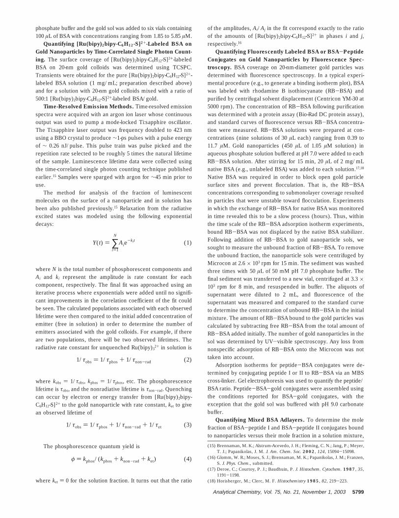
was generated using cysteine. Cysteine contains only the N-terminal amine, and as shown in Figure 1, the fluorescenceresulting from the cysteine reaction is much less than that fromthe ε-amine of lysine. The difference in the lysine and cysteinecalibration plots represents the fluorescence contribution from theε-amine of lysine. This curve was used as a standard fluorescencecurve for determining the number of lysines on BSA. To checkthe validity of this calibration standard, (lysine)16 was also usedto generate a standard curve. The fluorescence contribution fromthe N-terminus was assumed negligible compared to the other16 amines of the polymer. Indeed, the calibration plot for (lysine)16
was close to that generated by the lysine-cysteine differencemethod (Figure 1).
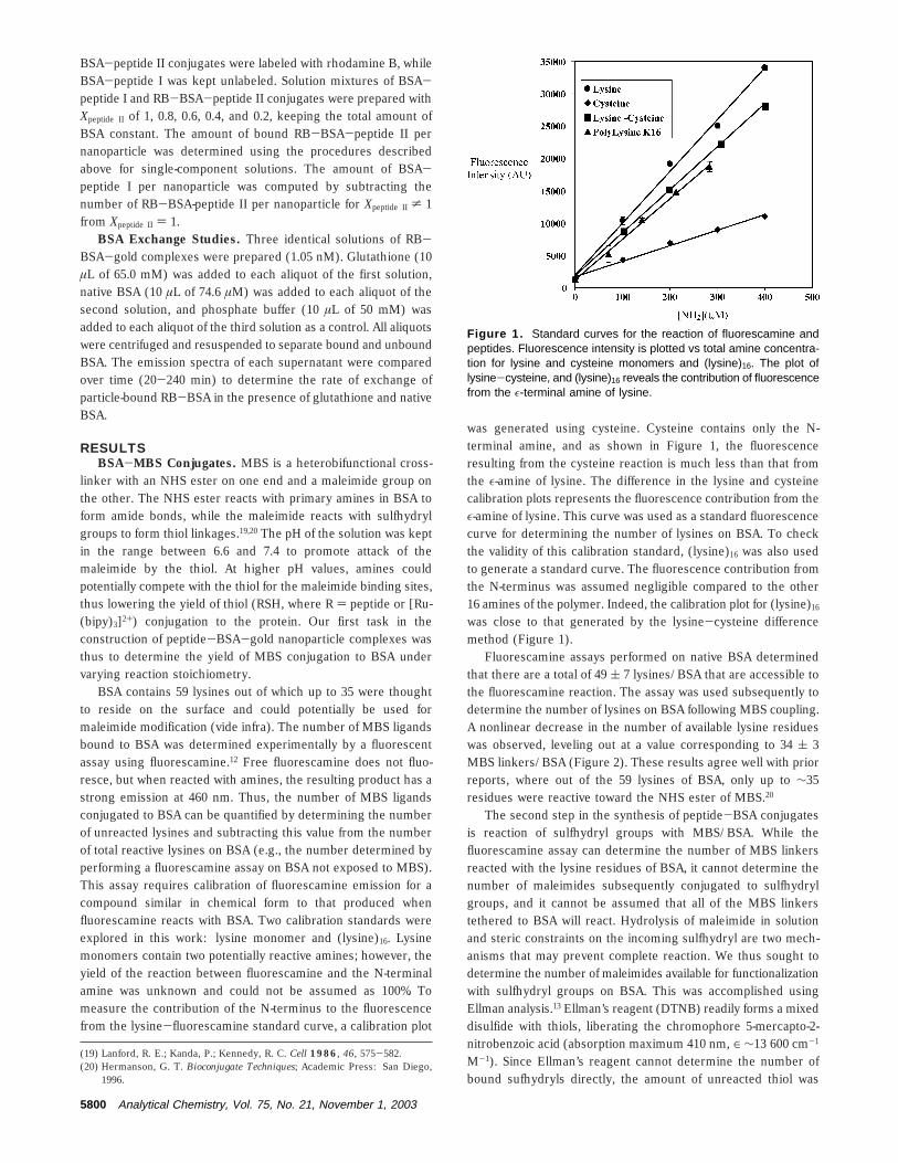
Fluorescamine assays performed on native BSA determinedthat there are a total of 49 ( 7 lysines/BSA that are accessible tothe fluorescamine reaction. The assay was used subsequently todetermine the number of lysines on BSA following MBS coupling.A nonlinear decrease in the number of available lysine residueswas observed, leveling out at a value corresponding to 34 ( 3MBS linkers/BSA (Figure 2). These results agree well with priorreports, where out of the 59 lysines of BSA, only up to ∼35residues were reactive toward the NHS ester of MBS.20
The second step in the synthesis of peptide-BSA conjugatesis reaction of sulfhydryl groups with MBS/BSA. While thefluorescamine assay can determine the number of MBS linkersreacted with the lysine residues of BSA, it cannot determine thenumber of maleimides subsequently conjugated to sulfhydrylgroups, and it cannot be assumed that all of the MBS linkerstethered to BSA will react. Hydrolysis of maleimide in solutionand steric constraints on the incoming sulfhydryl are two mech-anisms that may prevent complete reaction. We thus sought todetermine the number of maleimides available for functionalizationwith sulfhydryl groups on BSA. This was accomplished usingEllman analysis.13 Ellman’s reagent (DTNB) readily forms a mixeddisulfide with thiols, liberating the chromophore 5-mercapto-2-nitrobenzoic acid (absorption maximum 410 nm, ∈ ∼13 600 cm-1
M-1). Since Ellman’s reagent cannot determine the number ofbound sufhydryls directly, the amount of unreacted thiol was
(19) Lanford, R. E.; Kanda, P.; Kennedy, R. C. Cell 1986, 46, 575-582.(20) Hermanson, G. T. Bioconjugate Techniques; Academic Press: San Diego,
1996.
Figure 1. Standard curves for the reaction of fluorescamine andpeptides. Fluorescence intensity is plotted vs total amine concentra-tion for lysine and cysteine monomers and (lysine)16. The plot oflysine-cysteine, and (lysine)16 reveals the contribution of fluorescencefrom the ε-terminal amine of lysine.
5800 Analytical Chemistry, Vol. 75, No. 21, November 1, 2003
measured. GSH and BME were used as sources of thiol groups.Results for both thiols were essentially identical and showed 8.6( 1.2 thiol groups attached per BSA (Figure 2). This number waslower than expected since over 30 MBS ligands were attached toBSA.
Yield for Binding of [Ru(bipy)2bipy-C6H12-SH]2+ to BSA.The number of [Ru(bipy)2bipy-C6H12-SH]2+ labels attached to BSAwas determined by comparing the emission of the BSA-[Ru-(bipy)2bipy-C6H12-SH]2+ conjugate to that of a free [Ru(bipy)2bipy-C6H12-SH]2+ standard curve. After comparison of sample emissionsto the standard curve, it was determined that there were 8 ( 1[Ru(bipy)2bipy-C6H12-SH]2+ molecules/BSA under conditions where∼20 MBS molecules were conjugated to BSA. The yield forreaction of [Ru(bipy)2bipy-C6H12-SH]2+ with MBS was ∼0.5 underthe conditions studied here.
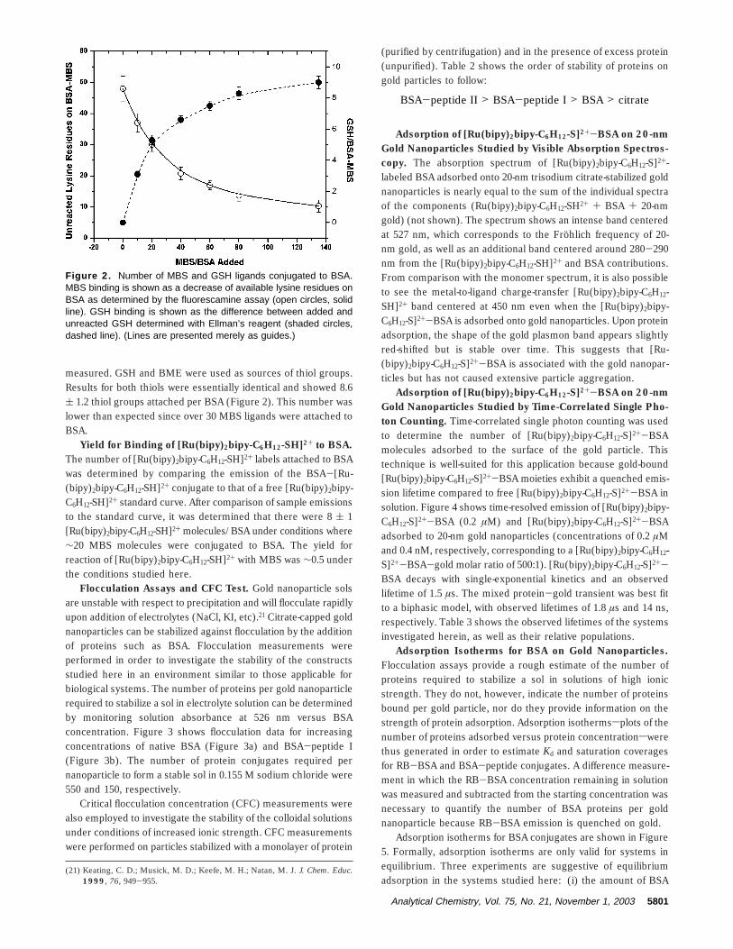
Flocculation Assays and CFC Test. Gold nanoparticle solsare unstable with respect to precipitation and will flocculate rapidlyupon addition of electrolytes (NaCl, KI, etc).21 Citrate-capped goldnanoparticles can be stabilized against flocculation by the additionof proteins such as BSA. Flocculation measurements wereperformed in order to investigate the stability of the constructsstudied here in an environment similar to those applicable forbiological systems. The number of proteins per gold nanoparticlerequired to stabilize a sol in electrolyte solution can be determinedby monitoring solution absorbance at 526 nm versus BSAconcentration. Figure 3 shows flocculation data for increasingconcentrations of native BSA (Figure 3a) and BSA-peptide I(Figure 3b). The number of protein conjugates required pernanoparticle to form a stable sol in 0.155 M sodium chloride were550 and 150, respectively.
Critical flocculation concentration (CFC) measurements werealso employed to investigate the stability of the colloidal solutionsunder conditions of increased ionic strength. CFC measurementswere performed on particles stabilized with a monolayer of protein
(purified by centrifugation) and in the presence of excess protein(unpurified). Table 2 shows the order of stability of proteins ongold particles to follow:
Adsorption of [Ru(bipy)2bipy-C6H12-S]2+-BSA on 20-nmGold Nanoparticles Studied by Visible Absorption Spectros-copy. The absorption spectrum of [Ru(bipy)2bipy-C6H12-S]2+-labeled BSA adsorbed onto 20-nm trisodium citrate-stabilized goldnanoparticles is nearly equal to the sum of the individual spectraof the components (Ru(bipy)2bipy-C6H12-SH2+ + BSA + 20-nmgold) (not shown). The spectrum shows an intense band centeredat 527 nm, which corresponds to the Frohlich frequency of 20-nm gold, as well as an additional band centered around 280-290nm from the [Ru(bipy)2bipy-C6H12-SH]2+ and BSA contributions.From comparison with the monomer spectrum, it is also possibleto see the metal-to-ligand charge-transfer [Ru(bipy)2bipy-C6H12-SH]2+ band centered at 450 nm even when the [Ru(bipy)2bipy-C6H12-S]2+-BSA is adsorbed onto gold nanoparticles. Upon proteinadsorption, the shape of the gold plasmon band appears slightlyred-shifted but is stable over time. This suggests that [Ru-(bipy)2bipy-C6H12-S]2+-BSA is associated with the gold nanopar-ticles but has not caused extensive particle aggregation.
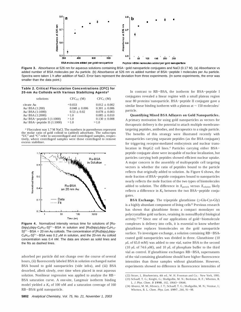
Adsorption of [Ru(bipy)2bipy-C6H12-S]2+-BSA on 20-nmGold Nanoparticles Studied by Time-Correlated Single Pho-ton Counting. Time-correlated single photon counting was usedto determine the number of [Ru(bipy)2bipy-C6H12-S]2+-BSAmolecules adsorbed to the surface of the gold particle. Thistechnique is well-suited for this application because gold-bound[Ru(bipy)2bipy-C6H12-S]2+-BSA moieties exhibit a quenched emis-sion lifetime compared to free [Ru(bipy)2bipy-C6H12-S]2+-BSA insolution. Figure 4 shows time-resolved emission of [Ru(bipy)2bipy-C6H12-S]2+-BSA (0.2 µM) and [Ru(bipy)2bipy-C6H12-S]2+-BSAadsorbed to 20-nm gold nanoparticles (concentrations of 0.2 µMand 0.4 nM, respectively, corresponding to a [Ru(bipy)2bipy-C6H12-S]2+-BSA-gold molar ratio of 500:1). [Ru(bipy)2bipy-C6H12-S]2+-BSA decays with single-exponential kinetics and an observedlifetime of 1.5 µs. The mixed protein-gold transient was best fitto a biphasic model, with observed lifetimes of 1.8 µs and 14 ns,respectively. Table 3 shows the observed lifetimes of the systemsinvestigated herein, as well as their relative populations.
Adsorption Isotherms for BSA on Gold Nanoparticles.Flocculation assays provide a rough estimate of the number ofproteins required to stabilize a sol in solutions of high ionicstrength. They do not, however, indicate the number of proteinsbound per gold particle, nor do they provide information on thestrength of protein adsorption. Adsorption isothermssplots of thenumber of proteins adsorbed versus protein concentrationswerethus generated in order to estimate Kd and saturation coveragesfor RB-BSA and BSA-peptide conjugates. A difference measure-ment in which the RB-BSA concentration remaining in solutionwas measured and subtracted from the starting concentration wasnecessary to quantify the number of BSA proteins per goldnanoparticle because RB-BSA emission is quenched on gold.
Adsorption isotherms for BSA conjugates are shown in Figure5. Formally, adsorption isotherms are only valid for systems inequilibrium. Three experiments are suggestive of equilibriumadsorption in the systems studied here: (i) the amount of BSA
(21) Keating, C. D.; Musick, M. D.; Keefe, M. H.; Natan, M. J. J. Chem. Educ.1999, 76, 949-955.
Figure 2. Number of MBS and GSH ligands conjugated to BSA.MBS binding is shown as a decrease of available lysine residues onBSA as determined by the fluorescamine assay (open circles, solidline). GSH binding is shown as the difference between added andunreacted GSH determined with Ellman’s reagent (shaded circles,dashed line). (Lines are presented merely as guides.)
BSA-peptide II > BSA-peptide I > BSA > citrate
Analytical Chemistry, Vol. 75, No. 21, November 1, 2003 5801
adsorbed per particle did not change over the course of severalhours, (ii) fluorescently labeled BSA in solution exchanged nativeBSA bound to gold nanoparticles (vide infra), and (iii) BSAdesorbed, albeit slowly, over time when placed in neat aqueoussolution. Nonlinear regression was applied to analyze the RB-BSA saturation curve. A one-site, Langmuir isotherm bindingmodel yielded a Kd of 100 nM and a saturation coverage of 160RB-BSA/gold nanoparticle.
In contrast to RB-BSA, the isotherm for BSA-peptide Iconjugates revealed a linear regime with a small plateau regionnear 80 proteins/nanoparticle. BSA-peptide II conjugate gave asimilar linear binding isotherm with a plateau at ∼ 150 molecules/particle.
Quantifying Mixed BSA Adlayers on Gold Nanoparticles.A primary motivation for using gold nanoparticles as vectors fortherapeutic delivery is the potential to attach multiple membrane-targeting peptides, antibodies, and therapeutics to a single particle.The benefits of this strategy were illustrated recently withnanoparticles carrying separate peptides (as the BSA conjugate)for triggering receptor-mediated endocytosis and nuclear trans-location in HepG2 cell lines.4 Particles carrying either BSA-peptide conjugate alone were incapable of nuclear localization, butparticles carrying both peptides showed efficient nuclear uptake.A major concern in the assembly of multi-peptide cell targetingvectors is whether the ratio of peptides bound to the particlereflects that originally added to solution. As Figure 6 shows, themole fraction of BSA-peptide conjugates bound to nanoparticlesnearly reflects the mole fraction of the two types of biomoleculesadded to solution. The difference in Xparticle versus Xsolution likelyreflects a difference in Kd between the two BSA-peptide conju-gates.
BSA Exchange. The tripeptide glutathione (γ-Glu-Cys-Gly)is a highly abundant component of living cells22 Previous researchhas shown that glutathione forms a compact monolayer onpolycrystalline gold surfaces, retaining its nonsulfhydryl biologicalactivity.23,24 Since one of our applications of gold-biomoleculecomplexes is delivery into cells, it is essential to know whetherglutathione replaces biomolecules on the gold nanoparticlesurface. To investigate exchange, a solution containing RB-BSA-coated gold nanoparticles was divided in three. Glutathione (10µL of 65.0 mM) was added to one vial, native BSA to the second(10 µL of 74.6 µM), and 10 µL of phosphate buffer to the thirdvial as control. If glutathione exchanges RB-BSA, supernatantsof the vial containing glutathione should have higher fluorescenceintensities than those samples without glutathione. However,experiments showed no difference in fluorescence intensities of
(22) Stryer, L. Biochemistry, 4th ed.; W. H. Freeman and Co.: New York, 1995.(23) Schaaff, T. G.; Knight, G.; Shafigullin, M. N.; Borkman, R. F.; Whetten, R.
L. J. Phys. Chem. B 1998, 102, 10643-10646.(24) Alvarez, M. M.; Khoury, J. T.; Schaaff, T. G.; Shafigullin, M. N.; Vezmar, I.;
Whetten, R. L. Chem. Phys. Lett. 1997, 266, 91-98.
Figure 3. Absorbance at 526 nm for aqueous solutions containing BSA-gold nanoparticle conjugates and NaCl (0.17 M). (a) Absorbance vsadded number of BSA molecules per Au particle. (b) Absorbance at 526 nm vs added number of BSA-peptide I molecules per Au particle.Spectra were taken 1 h after addition of NaCl. Error bars represent the deviation from three experiments. (In some experiments, the error wassmaller than the data point.)
Table 2. Critical Flocculation Concentrations (CFC) for20-nm Au Colloids with Various Stabilizing Agentsa
solutions CFCUC (M) CFCC (M)
citrate Au <0.033 0.012 ( 0.002Au/BSA (1:200) 0.048 ( 0.006 0.301 ( 0.006Au/BSA (1:1000) 0.53 ( 0.02 0.078 ( 0.003Au/BSA (1:2000) >1.0 0.085 ( 0.010Au/BSA-peptide I (1:1000) >1.0 0.130 ( 0.008Au/BSA-peptide II (1:1000) >1.0 >1.0
a Flocculent was 1.7 M NaCl. The numbers in parentheses representthe molar ratio of gold colloid to (added) adsorbate. The subscripts“UC” and “C” refer to uncentrifuged and centrifuged samples, respec-tively, where centrifuged samples were those centrifuged to removeexcess stabilizer.
Figure 4. Normalized intensity versus time for solutions of [Ru-(bipy)2bipy-C6H12-S]2+-BSA in solution and [Ru(bipy)2bipy-C6H12-S]2+-BSA + 20 nm Au colloids. The concentration of [Ru(bipy)2bipy-C6H12-S]2+-BSA was 0.2 µM in solution, and the 20-nm Au colloidconcentration was 0.4 nM. The data are shown as solid lines andthe fits as dashed lines.
5802 Analytical Chemistry, Vol. 75, No. 21, November 1, 2003
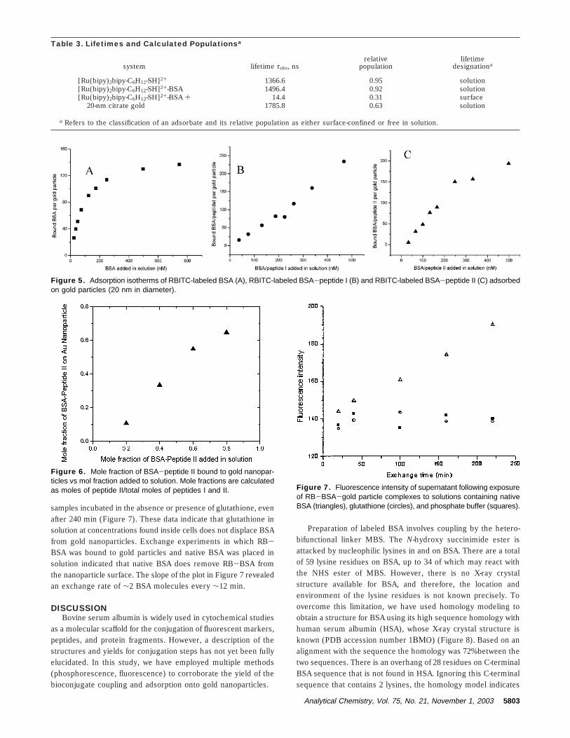
samples incubated in the absence or presence of glutathione, evenafter 240 min (Figure 7). These data indicate that glutathione insolution at concentrations found inside cells does not displace BSAfrom gold nanoparticles. Exchange experiments in which RB-BSA was bound to gold particles and native BSA was placed insolution indicated that native BSA does remove RB-BSA fromthe nanoparticle surface. The slope of the plot in Figure 7 revealedan exchange rate of ∼2 BSA molecules every ∼12 min.
DISCUSSIONBovine serum albumin is widely used in cytochemical studies
as a molecular scaffold for the conjugation of fluorescent markers,peptides, and protein fragments. However, a description of thestructures and yields for conjugation steps has not yet been fullyelucidated. In this study, we have employed multiple methods(phosphorescence, fluorescence) to corroborate the yield of thebioconjugate coupling and adsorption onto gold nanoparticles.
Preparation of labeled BSA involves coupling by the hetero-bifunctional linker MBS. The N-hydroxy succinimide ester isattacked by nucleophilic lysines in and on BSA. There are a totalof 59 lysine residues on BSA, up to 34 of which may react withthe NHS ester of MBS. However, there is no X-ray crystalstructure available for BSA, and therefore, the location andenvironment of the lysine residues is not known precisely. Toovercome this limitation, we have used homology modeling toobtain a structure for BSA using its high sequence homology withhuman serum albumin (HSA), whose X-ray crystal structure isknown (PDB accession number 1ΒΜO) (Figure 8). Based on analignment with the sequence the homology was 72% between thetwo sequences. There is an overhang of 28 residues on C-terminalBSA sequence that is not found in HSA. Ignoring this C-terminalsequence that contains 2 lysines, the homology model indicates
Table 3. Lifetimes and Calculated Populationsa
system lifetime τobs, nsrelative
populationlifetime
designationa
[Ru(bipy)2bipy-C6H12-SH]2+ 1366.6 0.95 solution[Ru(bipy)2bipy-C6H12-SH]2+-BSA 1496.4 0.92 solution[Ru(bipy)2bipy-C6H12-SH]2+-BSA + 14.4 0.31 surface
20-nm citrate gold 1785.8 0.63 solution
a Refers to the classification of an adsorbate and its relative population as either surface-confined or free in solution.
Figure 5. Adsorption isotherms of RBITC-labeled BSA (A), RBITC-labeled BSA-peptide I (B) and RBITC-labeled BSA-peptide II (C) adsorbedon gold particles (20 nm in diameter).
Figure 6. Mole fraction of BSA-peptide II bound to gold nanopar-ticles vs mol fraction added to solution. Mole fractions are calculatedas moles of peptide II/total moles of peptides I and II. Figure 7. Fluorescence intensity of supernatant following exposure
of RB-BSA-gold particle complexes to solutions containing nativeBSA (triangles), glutathione (circles), and phosphate buffer (squares).
Analytical Chemistry, Vol. 75, No. 21, November 1, 2003 5803
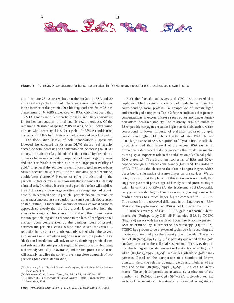
that there are 28 lysine residues on the surface of BSA and 30more that are partially buried. There were essentially no lysinesin the interior of the protein. Our binding isotherm for MBS hasa maximum of 34 MBS molecules per BSA, which suggests that∼6 MBS ligands are at least partially buried and likely unavailablefor further conjugation to thiol ligands (e.g., peptides). Of theremaining 28 surface-exposed MBS ligands, only 10 were foundto react with incoming thiols, for a yield of ∼35%. A combinationof sterics and MBS hydrolysis is a likely source of such low yields.
The flocculation assays of gold nanoparticle suspensionsfollowed the expected trends from DLVO theoryssol stabilitydecreased with increasing salt concentration. According to DLVOtheory, the stability of a gold colloid is determined by the balanceof forces between electrostatic repulsion of like-charged spheresand van der Waals attraction due to the large polarizability ofgold.25 In general, the addition of electrolytes to gold nanoparticlescauses flocculation as a result of the shielding of the repulsivedouble-layer charges.26 Proteins or polymers adsorbed to theparticle surface or free in solution will also influence the stabilityof metal sols. Proteins adsorbed to the particle surface will stabilizethe sol due simply to the large positive free energy input of proteindesorption required prior to particle-particle fusion. Proteins (orother macromolecules) in solution can cause particle flocculationor stabilization.27 Flocculation occurs whenever colloidal particlesapproach so closely that the free protein is excluded from theinterparticle region. This is an entropic effect; the protein leavesthe interparticle region in response to the loss of configurationalentropy upon compression by the particles. Loss of proteinbetween the particles leaves behind pure solvent molecules. Areduction in free energy is subsequently gained when the solventalso leaves the interparticle region to mix with the protein. This“depletion flocculation” will only occur by demixing protein chainsand solvent in the interparticle region. In good solvents, demixingis thermodynamically unfavorable, and the free protein in solutionwill actually stabilize the sol by preventing close approach of twoparticles (depletion stabilization).27
Both the flocculation assays and CFC tests showed thatpeptide-modified proteins stabilize gold sols better than thecorresponding native protein. The comparison of uncentrifugedand centrifuged samples in Table 2 further indicates that proteinconcentrations in excess of those required for monolayer forma-tion afford increased stability. The relatively large structures ofBSA-peptide conjugates result in higher steric stabilization, whichcorrespond to lower amounts of stabilizer required by goldparticles and higher CFC values than that of native BSA. The factthat a large excess of BSA is required to fully stabilize the colloidaldispersions and that removal of the excess BSA results indramatically decreased stability indicates that depletion mecha-nisms play an important role in the stabilization of colloidal gold-BSA systems.27 The adsorption isotherms of BSA and BSA-peptide conjugates differed considerably (Figure 5). The isothermof RB-BSA was the closest to the classic Langmuir type, whichdescribes the formation of a monolayer on the surface. We donote, however, that the plateau of this isotherm is not totally flat,suggesting a small percentage of loosely bound proteins mightexist. In contrast to RB-BSA, the isotherms of BSA-peptideconjugates revealed highly linear regimes, suggesting nonspecificbinding occurs to a much larger degree compared to RB-BSA.The reason for the observed difference in binding between RB-BSA and the peptide-modified BSA is not known at this time.
A surface coverage of 160 ( 8 BSA/gold nanoparticle deter-mined for [Ru(bipy)2bipy-C6H12-SH]2+-lableled BSA by TCSPC(Figure 4) agrees with the result of rhodamine B isothiocyanate-BSA determined by fluorescence spectroscopy (Figure 5A).TCSPC has proven to be a powerful technique for observing themicroenvironment of phosphorescent probe molecules. The emis-sion of [Ru(bipy)2bipy-C6H12-S]2+ is partially quenched on the goldsurfaces present in the colloidal suspensions. This is evident inthe shortening of the lifetime in the kinetic traces in Figure 4when [Ru(bipy)2bipy-C6H12-S]2+ molecules adsorb to gold nano-particles. Based on the comparison to a standard of knownquantum yield, the relative quantum yields and lifetimes of thefree and bound [Ru(bipy)2bipy-C6H12-S]2+-BSA can be deter-mined. These yields permit an accurate determination of thenumber of [Ru(bipy)2bipy-C6H12-S]2+-BSA molecules on thesurface of a nanoparticle. Interestingly, earlier radiolabeling studies
(25) Adamson, A. W. Physical Chemistry of Surfaces, 5th ed.; John Wiley & Sons:New York, 1990.
(26) Niemeyer, C. M. Angew. Chem., Int. Ed. 2001, 40, 4128-4158.(27) Hunter, R. J. Foundations of Colloid Science; Oxford University Press Inc.:
New York, 1991.
Figure 8. (A) 1BMO X-ray structure for human serum albumin. (B) Homology model for BSA. Lysines are shown in pink.
5804 Analytical Chemistry, Vol. 75, No. 21, November 1, 2003

done on 15-nm citrate-capped gold nanoparticles17,28 concluded that39 native BSA molecules are adsorbed at monolayer coverage.The corresponding monolayer coverage for 20-nm gold nanopar-ticles, taking only the relative surface areas into consideration,would be ∼70 BSA/gold. The discrepancy with the numberobserved above (160 ( 8) could be due to electrostatic differencesthat arise from the different labels on BSA, differences incrystallinity between the gold particles (i.e., surface area may notscale precisely with particle radius), or different solvent conditions.Another important difference between radiolabeling studies andTCSPC is that TCSPC is sensitive to the distance between thephosphorescent tag and the gold nanoparticle surface: only BSAbound to the nanoparticle surface is measured in TCSPC (e.g.,monolayer adsorption). In this context, the 160 RB-BSA observedper gold nanoparticle signifies a dense mode of binding in whicheach BSA occupies ∼7.5 nm2 of the nanoparticle surface. Giventhat BSA is an oblong protein 10 nm long × 3 nm wide, the datasuggest that BSA adsorbs onto gold nanoparticles end-on.
Irrespective of the detailed orientation of BSA binding to goldnanoparticles, the ease of the current methods compared toradiolabeling provides a ready experimental protocol to compareadsorption isotherms under a variety of solvent conditions, forpeptide tags and bioconjugates and for various nanoparticlecoatings. The combination of fluorescence quenching and lifetimemeasurements provides a simple one-pot assay for distinguishingbetween the bound and unbound fractions of BSA adsorbed onmetal nanoparticles. Moreover, TCSPC should in principle beeasily extended to most any fluorescent adsorbate.
CONCLUSIONSA primary goal of our research is to increase the stability of
biomolecule-nanoparticle complexes in biologically relevantmedia (e.g., solutions of high ionic strength such as cell growthmedia and blood plasma). The studies presented here demonstratethat BSA can be an effective linker for peptide conjugation to goldnanoparticles. Particle coagulation or exchange of surface-boundBSA in the presence of electrolytes and thiol molecules found inbiofluids is sufficiently slow that a nanoparticle-BSA complex canbe used as a research tool in cellular imaging studies. In addition,we have demonstrated that two peptides with different cell-targeting properties partition onto nanoparticles with an averagemole fraction that is not dramatically different from their molefraction in solution. The observation of such partitioning isimportant to the development of multifunctional nanoparticles forstudies in cell biology and medicine. Finally, the characterizationof metal particle bioconjugates will be aided by the application ofnew tools that can monitor adsorption kinetics and ligandexchange mechanisms in real time. The work presented heresuggests that time-correlated single photon counting spectroscopyis a useful tool for studying such processes.
ACKNOWLEDGMENTThe authors thank the David and Lucile Packard Foundation,
The Arnold and Mabel Beckman Foundation, NSF DMR (9900073),NIH, and NSF-MDB (9874895) for support of this work.
Received for review May 29, 2003. Accepted August 21,2003.
AC034578D
(28) Baudhuin, P.; Van der Smissen, P.; Beauvois, S.; Courtoy, P. J. ColloidalGold: Principles, Methods, and Applications; Academic Press: New York,1989.
Analytical Chemistry, Vol. 75, No. 21, November 1, 2003 5805