Embed Size (px)
Citation preview
8004 Journal of the American Chemical Society / 100.25 December 6 , 1978
(8) J. L. Corbin and D. E. Work, J. Org. Chem., 41, 489 (1976). (9) P. Kroneck and J. T. Spence. Biochemistry, 12, 5020 (1973).
(10) W. E. Newton and J. W. McDonald, J. Less Common Metals, 54, 51
(1 1) N. Pariyadath, W. E. Newton, and E. I. Stiefel, J. Am. Chem. Soc., 98,5388
(12) K. Yamanouchi and J. Enemark, Inorg. Chem., 17, 1981 (1978). (13) A. F. lsbell and D. T. Sawyer, Inorg. Chem., 10, 2449 (1971). (14) R. D. Taylor, J. P. Street, M. Minelli, and J. T. Spence. inorg. Chem., 17,
(15) I. W. Boyd, I. G. Dance, K. S. Murray, and A. G. Wedd, Aust. J. Chem., 31,
(1 977).
(1976).
3207 (1978).
279 (1978).
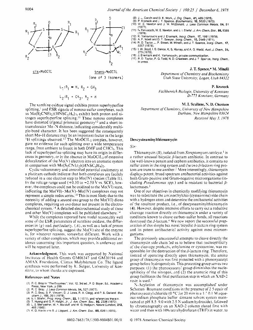
c i s - M o O C 1 L
L , : R 1 H,
W - M O O C l L (one o f 3 isomers)
R2 = CH3
L : R 2 1 = CH3, R 2 = H
The xanthine oxidase signal exhibits proton superhyperfine splitting,' and ESR signals of nonoxo sulfur complexes, such as Mo(S2CNEt2)( HNSC6H4)2, exhibit both proton and ni- trogen superhyperfine splitting.' l These nonoxo complexes have distorted trigonal prismatic geometry1* and a short in- tramolecular Mo-N distance, indicating considerably multi- ple-bond character. It has been suggested the consequently short Mo-H distance may be an important factor in the large I H splittings observed.12 The MoOClL2 complex, however, gave no evidence for such splitting over a wide temperature range, from ambient to frozen in both D M F and CHC13. This lack of superhyperfine splitting may have its origin in differ- ences in geometry, or in the absence in MoOClLz of extensive delocalization of the Mo(V) electron into an aromatic system in comparison with Mo(S2CNEt*)(HNSCsHd)2.
Cyclic voltammetry and controlled potential coulometry at a platinum cathode indicate that both complexes are facilely reduced in a one electron step to Mo(IV) species (Table 11). In the voltage range used (+0.50 to -2.50 V vs. SCE) , how- ever, the complexes could not be oxidized to the Mo(V1) state, indicating the Mo(V1)-Mo(V)-Mo( IV) complexes may not represent a simple redox series.14 This is most likely due to the necessity of adding a second oxo group to the Mo(V1) dioxo complexes, requiring an oxo donor not present in the electro- chemical system.I4 A detailed electrochemical study of these and other Mo(V) complexes will be published elsewhere.lj
While the complexes reported here model reasonably well some of the ESR parameters of xanthine oxidase, the differ- ences in (g) and, particularly, ( A ), as well as a lack of proton superhyperfine splitting, suggest the Mo(V) site of the enzyme is, for whatever reasons, somewhat different. Work with a variety of other complexes, which may provide additional ev- idence concerning this important question, is underway and will be reported later.
Acknowledgment. This work was supported by National Institutes of Health Grants GM08347 and GM20194 and AMAX Foundation, Climax Molybdenum Co. The ligand syntheses were performed by K . Sulger, University of Kon- stanz, to whom thanks are expressed.
References and Notes (1) R. C. Bray in "The Enzymes", Vol. 12, 3rd ed.. P. D. Boyer, Ed., Academic
(2) R. C. Bray, J. Less Common Metals, 54, 527 (1977). (3) R. C. Bray, S. P. Vincent, D. J. Lowe. R. A. Clegg. and P. B. Garland, Bio-
(4) E. I. Stiefel, frog. Inorg. Chern., 22, 1 (1977), and references therein. (5) T. Huang and G. P. Haight, Jr., J. Am. Chem. Soc., 92, 2336 (1970). (6) L. S. Meriwether, W. F. Marziuff, and W. G. Hodgson, Nature(London), 212,
(7) K. D. Kariin and S. J. Lippard. J. Am. Chem. SOC., 98, 6951 (1976)
Press, New York, p 299.
chernJ., 155, 201 (1976).
465 (1966).
0002-7863/78/l500-8004$01 .OO/O
(16) J. Enemark and K Yamanouchi, private communication. (17) R. D. Taylor, P. G. Todd, N. D. Chasteen. and J. T. Spence, Inorg. Chem.,
in press.
J. T. Spence,* M. Minelli Department of Chemistry and Biochemistry
Utah State Unicersity, Logan, Utah 84322
P. Kroneck Fachbereich Biologie, Uniuersity of Konstanz
0 - 7 7 5 Konstanz, Germany
M. I. Scullane, N. D. Chasteen Department of Chemistry, Unicersity of New Hampshire
Durham, New Hampshire 03824 Receiced May 3, 1978
Descysteaminylthienarnycin
Sir: Thienamycin ( l ) , isolated from Streptomyces cattleya, is
a rather unusual bicyclic p-lactam antibiotic. In contrast to the well-known penam and cephem antibiotics, it contains no sulfur atom in the ring system and the two P-lactani ring pro- tons are trans to one another.' More interestingly, thienamycin displays potent, broad spectrum antibacterial activities against both Gram-positive and Gram-negative microorganisms (in- cluding Pseudomonas spp . ) and is resistant to bacterial 0- l a c t a m a ~ e s . ~
One of our objectives in chemically modifying thienamycin was to substitute the aminoethylthio (cysteamine) side chain with a hydrogen atom and determine the antibacterial activities of the resultant product, i.e., of descysteaminylthienamycin (4). However, despite intensive efforts to carry out a reductive cleavage reaction directly on thienamycin under a variety of conditions known to cleave carbon-sulfur bonds, all reactions destroyed the /3-lactam.4 We now report the successful prep- aration of this simple but novel bicyclic P-lactam ring system and its potent antibacterial activity against most microor- ganisms.
The previously unsuccessful attempts to cleave directly the thienamycin side chain led us to believe that nucleophilicity of the cleavage products, ethylamine or cysteamine, was re- sponsible for the destruction of the p-lactam ring. Therefore, instead of operating directly upon thienamycin, the amino group of thienamycin was first protected with a phenoxyacetyl group before hydrogenolysis. This protecting group serves two purposes: ( I ) the phenoxyacetyl group diminishes the nucle- ophilicity of the nitrogen, and ( 2 ) the aromatic ring of this group facilitates the final purification step in which an XAD-2 resin is used.'
N-Acylation of thienamycin was accomplished' under Schotten-Raumann conditions in the presence of 2.5 cquiv of phenoxyacetyl chloride (0 "C for 20 min in a 1 : 1 0.1 M aque- ous sodium phosphate buffer-dioxane solvent system main- tained at pH 8.5-9.0 with 2.5 N sodium hydroxide), followed by chromatography on an XAD-2 column eluted first w i t h water and then with 10% tetrahydrofuran (THF) in water, to
0 1978 American Chemical Society

Communications to the Editor 8005
Table I. Inhibitory Zone Diameters (Millimeters) vs. Penicillin-Sensitive and Resistant Bacterial Strains disc content, S. aureus E. coli Enterobacter clocae Pseudomonas aeruginosa
compd gg (nmol MB2985 MB2314 MB2482 MB2964 MB2647 MB2646 MB3835 MB3350
carbenicillin 50 (1 18) 34 18 24 0 22 16 21 0
4 25.6 (117) 40 38 31 33 31 30 23 20 thienamycin 25 (92) 41 40 28 29 26 26 26 24
18.8 Hz 8.2 H t
2 . 9 H 2.5 Hz
4 R = h a @ R ' = H
*_ j R = C H ~ 0, R ' = H
6 . i( = CH2 0, R ' = Ac
give N-phenoxyacetylthienamycin sodium salt (2, 70%): UV (HzO) 302 nm ( t 7800); N M R (DzO, 60 MHz) , 1.25 (d, 3, J = 6 Hz, CH3), 2.65-3.80 (m, 7), 4.10 (m, 2, H j and Hg), 4.53 (s, 2, CHzO), 7.12 ppm (m, 5, aromatic protons). Treatment of 2 with p-bromobenzylbromide in hexarnethylphosphoramide ( H M P A ) for 30 min a t room temperature followed by T L C purification (silica gel GF, ethyl acetate eluant, RfO.20) gave the expected p-bromobenzyl ester 3 (80%): mass spectrum 574 ( M f ) , 530 ( M - 44), and 488 ( M - 86); IR (CHC13) 1776 cm-l (0-lactarn); UV (EtOH) 320 nm ( 6 8000); N M R (CDC13,60 MHz) 1.28 (d, 3, CH3), 2.76-3.72 (m, 7),4.15 (m, 2, H j and Hg), 4.46 (s, 2, CH20) , 5.12 (q, 2,p-bromobenzyl CH2), 6.70-7.40 ppm (m, 9, aromatic protons). The reductive cleavage of 2 was conducted in aqueous solution (pH 7.0) at room temperature under 1 atm of hydrogen in the presence of five times its weight of palladium oxide. The progress of the reaction was monitored by reverse-phase high-pressure liquid chromatography6 and electrophoresis,' until 90% of the sodium N-phenoxyacetylt hienamycin was consumed (2.5 h). After filtration, the aqueous solution (pH 9.5) was adjusted to pH 7.0 with 2.5 N aqueous phosphoric acid and chromatographed on an XAD-2 column, eluted wi th water, to give the desired product, sodium descysteaminylthienamycin (4, 40% yield). Subsequent elution of the column with 20% THF-H20 gave the expected cleavage product, N-ethylphenoxyacetamide (50% yield); mass spectrum 179 (M+); N M R (CDCl3, 60 MHz), 1.20 (t , 3, CH3) , 3.40 (quintet, 2, CH2), 4.50 (s, 2, C H 2 0 ) , 6.10 (NH) . 7.10 (m, 5, aromatic protons).
The structural assignment of sodium descysteaminyl- thienamycin is based on the spectral data and chemical deri- vatization: UV (H2O) 265 nm ( 6 6500); IR (Nujol mull) 1750 cm-I (0-lactam); N M R (D20 , 300 MHz) 1.27 (d, 3 , J = 6.0 Hz, CH3), 2.83 (octet, I , J4p-3 = 2.5, J40-5 = 8.2, J40-n = 18.8 Hz, H40), 2.95 (octet, 1,J4n-3 = 2.9,J4,-5 = l0 .0 ,and
H6). 4.26 (m, H5 and Hg), 6.29 ppm (q, 1, J 3 - d O = 2.9, J3-4p = 2.5 Hz, H3). The 300-MHz N M R of 4, i n which the AB pattern of H4n and H40 is completely resolved, makes the structure assignment unequivocal (Figure
Attempts to obtain a field desorption mass spectrum of 4 were unsuccessful. However, esterification of 4 with benzyl bromide in HMPA yielded descysteaminylthienamycin benzyl ester ( 5 , 75%) which showed a molecular ion in the El mass spectrum at m/e 287, and the expected fragments a t m/e 243
J4a-,j = 18.8 Hz, H4<?), 3.42 (q, 1, J 6 - 5 = 3.1,56-9 = 6.2 Hz,
Figure 1. 300-VH7 N M R bpectrum of Hj, , m d H4,j of sodruni descys- teaminqlthrenamycin
( M - 44) and 202 ( M - 85); IR (CHC13) 1781 (p-lactam), 1726 cm-' (ester); UV (EtOH) 276 nm ( t 7800); N M R (CDC13, 100 MHz), 1.33 (d, 3, J = 6.1 Hz, CH3), 2.84 (m, 2, H4), 3.18 (q, I , J 6 - 5 = 2.9,Jh-9 = 6.8 Hz, H6),4.20 (m, 2, Hs and Hg), 5.18 (d, l , J = 12 Hz, benzyl CH-) , 5.32 (d, 1, J = 12.0 Hz, benzyl CH2), 6.46 (t, I , = 3.0 Hz), 7.34 ppm (m, 5, aromatic protons). Treatment of 5 with acetic anhydride- pyridine at room temperature for 3 h provided O-acetyldes- cysteaminylthienamycin benzyl ester ( 6 ) i n 68% yield: IR (CHC13) 1780 (0-lactam), 1740 cm-' (ester); N M R (CDC13, 60 MHz), 1.38 (d, 3, CH3), 2.02 (s, 3, OAc), 2.82 (m, 2, Hd), 3.30 (q, 1 , H6), 4.20 (d of t, I , H j ) , 5.19 (m, I , Hg), 5.28 (s, 2, benzyl CHz), 6.49 (q, I , H3), 7.40 ppm (m, 5, aromatic pro- tons).
Sodium descysteaminylthienamycin possesses in vitro ac- tivity comparable with that of the parent thienamycin against both Gram-positive species and the Enterobacteriaceae Gram-negative microorganisms. Although the average activity of 4 against thienamycin-sensitive pseudomonas strains is one third that of thienamycin itself, descysteaminylthienamycin is nevertheless still superior to carbenicillin and active against Gram-negative strains resistant to carbenicillin (Table I ) . Descysteaminylthienamycin represents the simplest bicyclic p-lactam antibiotic to date which still possesses such extraor- dinary potency.
Acknowledgments. We thank Dr. Byron H . Arison and Mr. H . Flynn for providing 300- and 100-MHz ' H N M R spectra, Mr. Jack Smith for obtaining mass spectra, and Ms. Jean S. Kahan for providing antibacterial assays.
References and Notes ( 1 ) J. S. Kahan, F. M. Kahan, E. 0. Stapley, R. T. Goegelman, and S. Hernandez,
U.S. Patent 3 950 357 (1976). (2) The structure has been established by chemical, spectroscopic, and X-ray
crystallographic means, and confirmed by total synthesis: (a) D. B. R. Johnston, S. M. Schmitt, F . A. Bouffard, and 6. G. Christensen, J. Am. Chem. Soc., 100, 313 (1978); (b) G. Albers-Schonberg, B. A. Arison, E. A . Kaczka. F. M. Kahan, J. S. Kahan, B. Lago, W. M. Maiese, R. E. Rhodes, and J. L. Smith, Sixteenth Interscience Conference on Antimicrobial Agents and Chemotherapy, Chicago, Ill., Oct 1976, Abstract 229; (c) G. Albers-
8006 Journal of the American Chemical Society 1 100:25 / December 6 , 1978
Schonberg, B. Arison, 0. D. Hensens, J. Hirshfield, K. Hoogsteen, E. A. Kaczka, R. E. Rhodes, J. S. Kahan, F. M. Kahan, R. W. Ratcliffe, E. Walton, L. J. Ruswinkle, R. B. Morin, and B. G. Christensen, J. Am. Chem. SOC., 100, 6491 (1976).
(3) H. Kropp, J. S . Kahan. F. M. Kahan, J. Sundelof, G. Darland, and J. Birnbaum, ref 2b, Abstract 228.
(4) Personal communication from Dr. E. Walton. (5) The XAD-2 resin (obtained from the Rohm & Haas Co.) is a polystyrene resin
which has a strong affinity for compounds containing aromatic moieties when the chromatographic separation is carried out using water as solvent.
(6) The reverse-phase HPLC analysis of the reaction mixture was performed using a Waters Associates high pressure liquid chromatograph equipped with a 2.5-mm i.d. X 61 cm Bondapak C1&orosil column. Using 10% THF-HpO as solvent at 1 .O-mL/min flow rate, sodium N-phenoxyacetyi- thienamycin and sodium descysteaminylthienamycin showed retention times of 9 and 3 min, respectively.
(7) High voltage electrophoresis was also used to analyze the reaction mixture. In a typical electrophoretic separation (0.05 M, pH 7.0, sodium phosphate buffer at 2 kv for 20 min on Whatman chromatography paper, followed by bioautographic visualization), sodium Kphenoxyacetylthienamycin and sodium descysteaminylthienamycin showed mobilities of 5 and 8 cm, re- spectively, toward the anode.
(8) The H4 protons of 4 appeared as two doublets in the 60-MHz NMR spectrum, and as two triplets in the 100-MHz spectrum. Since the outer peaks of the AB pattern could not be observed in the 60- and 100-MHz spectra, the as- signment of the chemical shifts of the H l n and H4g protons remain uncertain: K . M. Silverstein and G. C. Bassler, "Spectrometric Identification of Organic Compounds", Wiley, New York, 1967, p 130.
D. H. Shih,* J . Hannah, B. G . Christensen Merck Sharp & Dohme Research Laboratories
Rahway, New Jersey 07065 Receiced September 8, I978
Total Synthesis of Thienamycin Analogues. 1. Synthesis of the Thienamycin Nucleus and dl-Descysteaminylthienamycin
Sir: Thienamycin (16), a novel p-lactam antibiotic isolated from
Streptomyces cattleya, is an unusually potent antibiotic.' It has three structural features not found in the classical P-lactam antibiotics, the penicillins and cephalosporins:* (1) an cy-
hydroxyethyl side chain instead of a &amido side chain at C-6, (2) an unusual cysteamine side chain at position 2, and (3) a highly strained nucleus consisting of an unsaturated five- membered ring fused to a P-lactam in which a methylene re- places the sulfur a t position 1, found in conventional p-lactam antibiotics.
Introduction of the hydroxyethyl side chain a t the 6(7) po- sition of a penicillin ( c e p h a l ~ s p o r i n ) ~ or of a cysteamine side chain a t position 3 of a cephalosporin4 did not increase the activity of these nuclei, leading us to believe that the nucleus of thienamycin, 1 -carba-2-penem-3-carboxylic acid, may have high antibiotic activity and may be a major contributor to the unusual antibiotic activity of thienamycin.
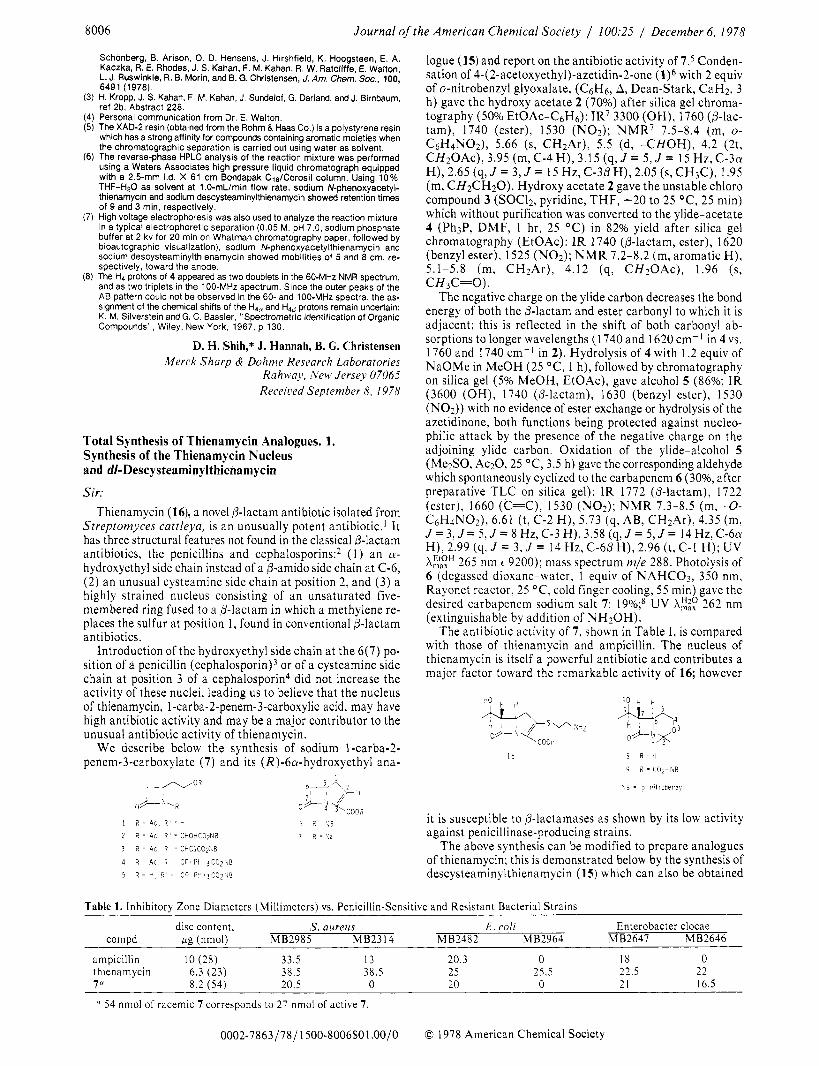
We describe below the synthesis of sodium l-carba-2- penem-3-carboxylate (7) and its (R)-6a-hydroxyethyl ana-
l 6 - 5 4
r - f - + O R O + h k R #
1 2 = 4c. R ' = b 0 2 = '.6
2 R = Ac R ' = CHOt iCO?Na 7 R = ' l a
3 i Ac. R ' = CHClCOZ ' IB
4 R = A c , i 1 8 = C P t P h i 3 C 0 2 1 u 6
5 R = b Q ' = C P I P Q ~ ~ C O ~ ~ V B
logue (15) and report on the antibiotic activity of 7.5 Conden- sation of 4-(2-acetoxyethyl)-azetidin-2-one (1)6 with 2 equiv of o-nitrobenzyl glyoxalate, (C&, A, Dean-Stark, CaH2, 3 h) gave the hydroxy acetate 2 (70%) after silica gel chroma- tography (50% EtOAc-C,&): IR' 3300 (OH), 1760 @-lac- tam), 1740 (ester), 1530 (NO2); NMR' 7.5-8.4 (m, o-
C H ~ O A C ) , 3.95 ( m , C - 4 H ) , 3.15 (q, J = 5, J = 15 H z , C - 3 a
(m, CH2CH20). Hydroxy acetate 2 gave the unstable chloro compound 3 (SOC12, pyridine, T H F , -20 to 25 OC, 25 min) which without purification was converted to the ylide-acetate 4 (Ph3P, DMF, 1 hr, 25 "C) in 82% yield after silica gel chromatography (EtOAc): IR 1740 (@-lactam, ester), 1620 (benzyl ester), 1525 (N02) ; N M R 7.2-8.2 (m, aromatic H) , 5.1-5.8 (m, CH2Ar), 4.12 (q, C H ~ O A C ) , 1.96 (s, CH3C=O).
The negative charge on the ylide carbon decreases the bond energy of both the 6-lactam and ester carbonyl to which it is adjacent; this is reflected in the shift of both carbonyl ab- sorptions to longer wavelengths (1740 and 1620 cm-' in 4 vs. 1760 and 1740 cm-I in 2). Hydrolysis of 4 with 1.2 equiv of NaOMe in MeOH (25 OC, 1 h), followed by chromatography on silica gel (5% MeOH, EtOAc), gave alcohol 5 (86%; IR (3600 (OH) , 1740 (p-lactam), 1630 (benzyl ester), 1530 (N02)) with no evidence of ester exchange or hydrolysis of the azetidinone, both functions being protected against nucleo- philic attack by the presence of the negative charge on the adjoining ylide carbon. Oxidation of the ylide-alcohol 5 (Me2S0, Ac20,25 OC, 3.5 h) gave the corresponding aldehyde which spontaneously cyclized to the carbapenem 6 (30%, after preparative T L C on silica gel): I R 1772 (6-lactam), 1722 (ester), 1660 (C=C), 1530 (NO*); N M R 7.3-8.5 (m, -0- C G H ~ N O ~ ) , 6.61 (t, C-2 H) , 5.73 (q, AB, CH*Ar), 4.35 (m,
C6H4NO2), 5.66 (s, CH2Ar), 5.5 (d, -CHOH), 4.2 (2t,
H) , 2.65 (9, J = 3, J = 15 Hz, C-30 H ) , 2.05 (s, CH3C), 1.95
J =3, J = 5, J = 8 Hz, C-3 H), 3.58 (q, J = 5, J = 14 Hz, C-6a H), 2.99 (q, J = 3, J = 14 Hz, C-66 H), 2.96 (t, C - l H); U V A::" 265 nm 6 9200); mass spectrum m/e 288. Photolysis of 6 (degassed dioxane-water, 1 equiv of NAHC03, 350 nm, Rayonet reactor, 25 OC, cold finger cooling, 55 min) gave the desired carbapenem sodium salt 7: 19%;8 UV A:$) 262 nm (extinguishable by addition of "*OH).
The antibiotic activity of 7, shown in Table I, is compared with those of thienamycin and ampicillin. The nucleus of thienamycin is itself a powerful antibiotic and contributes a major factor toward the remarkable activity of 16; however
it is susceptible to 6-lactamases as shown by its low activity against penicillinase-producing strains.
The above synthesis can be modified to prepare analogues of thienamycin; this is demonstrated below by the synthesis of descysteaminylthienamycin (15) which can also be obtained
Table 1 . lnhibitorv Zone Diameters (Millimeters) vs. Penicillin-Sensitive and Resistant Bacterial Strains
disc content, S. aureus E . coli Enterobacter clocae compd ue (nmol) MB2985 MB2314 MB2482 MB2964 MB2647 MB2646
ampicillin I O (28) 33.5 13 20.3 0 18 0 thienamycin 6.3 (23) 38.5 38.5 25 25.5 22.5 22 70 8.2 (54) 20.5 0 20 0 21 16.5
54 nmol of racemic 7 corresponds to 27 nmol of active 7 .
0002-7863/78/ 1500-8006$01 .OO/O 0 1978 American Chemical Society


