Embed Size (px)
Citation preview
UNIVERSITE DE GENEVE FACULTE DE MEDECINE
Section de médecine Clinique
Département de Gynécologie et
d’Obstétrique
Service d’obstétrique
______________________________________________________________________________
_
Thèse préparée sous la direction du Dr Antoine Weil, PD
DETECTION ECHOGRAPHIQUE DES COLLECTIONS LIQUIDIENNES APRES CESARIENNE ET
HYSTERECTOMIE ET MORBIDITE POSTOPERATOIRE ASSOCIEE
Thèse
Présentée à la Faculté de Médecine
de l’Université de Genève
pour obtenir le grade de Docteur en médecine
par
Eric Antonelli
de
Genève (GE)
Thèse no 10412
2
Table des matières
Introduction française 3
Résumé de l’étude 9
Introduction 11
Methods 12
Results 15
Discussion 18
Legends for illustration 21
Bibliography 22
Tables 24
4
Détection échographique des collections liquidiennes après césarienne et
hystérectomie et morbidité postopératoire associée.
Introduction :
La césarienne et l’hystérectomie sont les interventions chirurgicales les plus
fréquemment pratiquées en gynécologie-obstétrique et ont chacune un impact et
une signification particulière sur la vie des femmes.
D’un point de vue purement médical, la césarienne et l’hystérectomie, quelle que
soit sa voie d’abord, sont classiquement associées à certaines complications. A
court terme, la morbidité majeure concerne principalement le risque opératoire
(plaies des structures avoisinantes) et l’hémorragie. La fréquence des complications
dites mineures est élevée et touche 30-50% des femmes après césariennes, et peut
atteindre 85% lorsque les patientes sont interrogées à domicile : endométrite,
infection urinaire, anémie, asthénie, douleurs abdominales et fièvre inexpliquée.
A long terme, la réalisation d’une césarienne expose à un risque de césarienne
d’environ 50% lors de l’accouchement suivant, et ceci contribue probablement à
une diminution du nombre total de grossesses désirées par les femmes au total. On
observe en effet une diminution de la fécondité ultérieure après une césarienne
chez les femmes multipares. Par ailleurs, la cicatrice utérine expose à un risque de
rupture utérine en cas de tentative d’accouchement par voie basse. Les taux
rapportés dans la littérature varient de 0,3 à 2,3% pour les ruptures vraies; ces taux
sont doublés pour les déhiscences de cicatrices. Ce risque augmente en fonction
du nombre de cicatrices utérines. La rupture utérine augmente également la
morbidité néonatale et peut être parfois fatale pour l’enfant (environ 1% des cas de
5 rupture utérine). L’antécédent de césarienne semble également être associé à une
nette augmentation du risque ultérieur d’anomalie de l’insertion placentaire (praevia)
et des complications hémorragiques qui lui sont liées. Notons aussi l’augmentation
du risque de placenta accreta, menaçant la vie maternelle et nécessitant la
réalisation d’une hystérectomie en urgence.
De nombreuses études observationnelles ont étudié la morbidité à long terme après
réalisation d’une hystérectomie et démontré la relation que pouvait avoir cette
intervention sur la qualité de vie générale et la fonction sexuelle. Ces études ont
aussi clairement montré l’augmentation du risque d’incontinence urinaire et de
développement d’une insuffisance ovarienne précoce.
Parmi les complications mineures précoces liées à la césarienne et à
l’hystérectomie, l’état fébrile postopératoire en est un des plus fréquent. Cette
morbidité fébrile, définie comme une température de plus de 37.5° à deux reprises,
mesurée dès le premier jour postopératoire, est classiquement associée à des
infections urinaires, des surinfections ou abcès de plaies ou à des complications
thromboemboliques. En dehors de ces diagnostics, l’état fébrile postopératoire
pourrait être associé à la présence d’un état inflammatoire transitoire, à une nécrose
tissulaire ou à la présence de collections liquidiennes postopératoires. Ces
collections se situent soit dans le pelvis, soit dans la paroi abdominale.
En cas de suspicion de collection liquidienne postopératoire, l’échographie
transabdominale ou endovaginale peut être utilisée comme moyen d’investigation et
de diagnostic. Lorsque ces collections sont infectées et volumineuses, l’examen
échographique permet dans certains cas de réaliser un drainage.
6 La revue de la littérature retrouve très peu d’études prospectives ayant été réalisées
afin d’évaluer l’association entre des collections liquidiennes détectées par
échographie et un état fébrile postopératoire.
Concernant l’évaluation échographique de collections liquidiennes après
césariennes et la morbidité postopératoire, seules deux études ont étudié cette
association. La première, de Faustin et al., incluant cent femmes après césarienne
retrouve 29% de collections postopératoires, sans que leur localisation soit
indiquée. Parmi celles-ci, plus de 75% développent un état fébrile postopératoire,
alors que parmi les patientes qui ne présentent pas de collections, 54% développent
une morbidité fébrile. De plus l’administration d’antibiotique n’était pas
systématique, créant un certain biais entre les groupes. Les résultats de cette étude
suggèrent que seuls de volumineux hématomes, dont la taille serait égale ou plus
grande à 3,5 cm, seraient en relation avec une morbidité postopératoire, en
particulier un état fébrile. Mais seules huit femmes présentaient cette condition. La
deuxième étude, plus récente, de Gemer et al., qui inclus également une centaine
de patientes, confirme que l’état fébrile postopératoire est fréquent puisque 25% des
femmes présentent cette complication. Toutefois, le taux de collections liquidiennes
détectées par échographie est moins important que dans l’étude de Faustin,
puisque seules 15% des patientes présentent une collection. Les auteurs
suggérèrent que seules les collections sous-aponévrotiques sont associées à un
état fébrile postopératoire, mais cette conclusion n’est basée que sur cinq femmes
présentant cette complication.
Ces deux études, confirme que l’état fébrile postopératoire est une complication
fréquente de la césarienne sans toutefois clairement démontrer l’association entre
7 cette morbidité fébrile et la présence de collection (antibiothérapie non systématique
et localisation de la collection non précisée dans l’étude de Faustin et très faible
échantillon de patientes présentant une association dans l’étude de Gemer et al.).
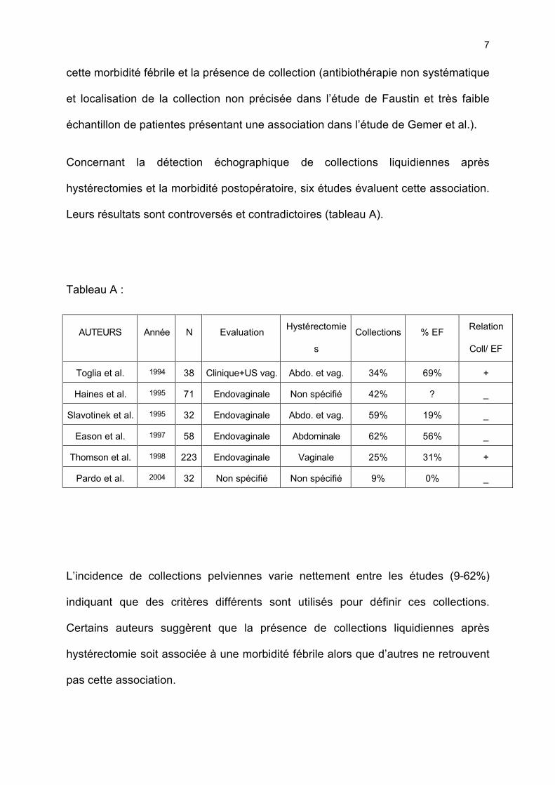
Concernant la détection échographique de collections liquidiennes après
hystérectomies et la morbidité postopératoire, six études évaluent cette association.
Leurs résultats sont controversés et contradictoires (tableau A).
Tableau A :
AUTEURS Année N Evaluation Hystérectomie
s
Collections % EF Relation
Coll/ EF
Toglia et al. 1994 38 Clinique+US vag. Abdo. et vag. 34% 69% +
Haines et al. 1995 71 Endovaginale Non spécifié 42% ? _
Slavotinek et al. 1995 32 Endovaginale Abdo. et vag. 59% 19% _
Eason et al. 1997 58 Endovaginale Abdominale 62% 56% _
Thomson et al. 1998 223 Endovaginale Vaginale 25% 31% +
Pardo et al. 2004 32 Non spécifié Non spécifié 9% 0% _
L’incidence de collections pelviennes varie nettement entre les études (9-62%)
indiquant que des critères différents sont utilisés pour définir ces collections.
Certains auteurs suggèrent que la présence de collections liquidiennes après
hystérectomie soit associée à une morbidité fébrile alors que d’autres ne retrouvent
pas cette association.
8 Deux études retrouvent une association significative entre la présence d’une
collection postopératoire et la morbidité fébrile. L’étude de Toglia et al. vise à
déterminer l’incidence des collections pelviennes chez 38 patientes après
hystérectomies abdominales et vaginales détectées par toucher vaginal et
échographie endovaginale et à corréler ces examens au développement d’une
morbidité postopératoire. Les auteurs évaluent la présence d’une collection
uniquement par abord échographique vaginal, alors que certaines patientes ont eu
une hystérectomie abdominale avec possibilité de collections pariétales. Ce biais
peut également être mentionné pour deux études (Slavotinek et al. et Eason et al.)
qui ne retrouvent pas d’association entre le développement de collections et une
morbidité fébrile mais qui utilisent la même approche méthodologique (échographie
vaginale dans le cadre d’hystérectomie abdominale). La seule publication dont la
méthodologie semble correcte est le travail de Thompson et al. qui a évalué les
suites opératoires de 223 hystérectomies vaginales. Le but de cette étude est de
déterminer si le développement d’un hématome de la tranche vaginale permet de
prédire l’apparition de complications postopératoires et en particulier d’un état
fébrile. Les résultats de cette étude démontrent que la présence d’un hématome de
la cicatrice vaginale détecté par échographie est associée de façon significative à
un état fébrile postopératoire, à la nécessité de transfusions sanguines et à
l’augmentation de la durée du séjour hospitalier. Toutefois, seules les
hystérectomies vaginales sont inclues dans cette étude.
Dans la plupart des études, les facteurs de risque liés au développement de ces
collections tels que le poids de la femme, le type d’opération (césarienne
programmée ou d’urgence, voie d’abord de l’hystérectomie), les pertes sanguines

9 en cours d’intervention ou l’utilisation des drains ou d’antibiotiques sont rarement
évalués ou discutés.
En conclusion, la revue de la littérature montre qu’il n’existe pas de preuve claire
quant à l’existence d’une association entre la mise en évidence d’une collection par
échographie et le développement d’une morbidité postopératoire, raison pour
laquelle cette étude est réalisée.
10
Résumé de l’étude :
Cette étude prospective à été réalisée conjointement dans trois maternités de
Suisse romande (Genève, Neuchâtel, La Chaux-de-Fonds) entre janvier 1999 et
décembre 2000. Le protocole d’étude a été approuvé par le comité d’éthique de ces
institutions et un consentement éclairé a été obtenu de chaque patiente.
Objectifs : Le but de cette étude est d’évaluer la signification clinique de collections
liquidiennes détectées par échographie après césarienne et hystérectomie, et
d’identifier des facteurs de risque liés au développement de ces collections.
Méthode : Nous avons réalisé une étude prospective incluant 280 femmes, dont
145 ont eu une césarienne et 135 une hystérectomie abdominale ou vaginale. Une
échographie était pratiquée systématiquement au quatrième jour postopératoire
afin de déterminer la présence d’une collection liquidienne, soit au niveau de la
paroi abdominale, soit dans le pelvis. Les échographistes étaient tenus à l’écart de
l’évolution clinique de patientes et n’étaient impliqués dans aucune décision
clinique. Le diagnostic de collection était corrélé aux données cliniques et à la
morbidité postopératoire.
Résultats : Une collection liquidienne a été retrouvée chez 69 femmes (48%) dans
le groupe césarienne et chez 59 femmes (44%) dans le groupe hystérectomie. Nous
n’avons pas identifié de facteurs de risque liés au développement de collections
postopératoires, que ce soit après césarienne ou hystérectomie. Le risque de
développer un état fébrile après ces interventions, n’est pas associé à la présence,
la taille ou la localisation d’une collection.
Conclusion : Les collections liquidiennes détectées par échographie après
césarienne et hystérectomie sont fréquentes. Au vu de l’absence d’association entre

11
ces collections et une morbidité postopératoire, en particulier un état fébrile, il ne
semble pas utile de les rechercher systématiquement par échographie en cas de
fièvre postopératoire.
12
Introduction
Caesarean section and hysterectomy are the most frequent surgical operations in
obstetrics and gynaecology. The occurrence of postoperative febrile morbidity is
often observed.1-3 In addition to specific conditions such as urinary tract infection,
wound abscess or thrombophlebitis, postoperative fluid collections may be
associated with febrile morbidity. Endovaginal or transabdominal sonography may
be used to detect and, eventually, drain fluid collections.
Few prospective studies have been conducted to evaluate the association between
postoperative fluid collections detected by ultrasonography and febrile morbidity. A
study of women after caesarean section suggested that large haematomas are
associated with postoperative morbidity, including fever.4 Gemer et al. suggested
that subfascial haematoma are associated with febrile morbidity, but this conclusion
is based on only 5 women presenting with this condition.5 Results of studies
conducted after hysterectomy are controversial. The incidence of pelvic fluid
collections vary greatly between studies, which may indicate that various definitions
(and criteria) were used.6-9 Some authors suggested that the presence of
ultrasonographically-diagnosed pelvic fluid collections is associated with febrile
morbidity8,9, while others were unable to demonstrate such a relationship.6,7,10 Risk
factors for developing postoperative fluid collections were rarely assessed or
discussed.
Our objective was to evaluate the clinical significance of ultrasonographically-
diagnosed fluid collections after caesarean section and hysterectomy, and to identify
risk factors associated with their formation.
13
Methods
A prospective study was conducted between January 2000 and December 2001 in
the obstetric and gynecology departments of a Swiss university tertiary care hospital
and two public teaching hospitals. The study protocol was approved by the
institutional ethics committees and written informed consent was obtained from all
women before inclusion in the study.
One hundred and forty five women who had a caesarean section and 135 who had
abdominal or vaginal total hysterectomy participated in the study. Women were
included consecutively, except when the participating sonographers were
unavailable. Exclusion criteria included any preoperative infection, administration of
anticoagulants other than prophylaxis for thrombophlebitis (subcutaneous
nadroparin 2850 IU/day), and symptomatic postoperative urinary tract infection.
Specific exclusion criteria in the caesarean section group were rupture of the
membranes for more than 36 hours and, in the hysterectomy group, neoplasia,
ascitis or endometriosis.
For both groups, indication for operation, surgical technique, duration and amount of
blood loss were recorded. Collected data included age, preoperative weight, parity
(for women who had a caesarean section), and number of years after menopause
for women who had an hysterectomy. Type of caesarean section (i.e., elective or
emergency) and of hysterectomy (i.e., abdominal or vaginal) were also recorded.
Postoperative morbidity included fever, blood loss and subcutaneous serous flow.
Body temperature was measured every 8 hours. Febrile morbidity was defined as a
temperature of at least 37.5°C on any two hospital days after surgery, excluding the
first 24 hours. All women who underwent caesarean section received prophylactic
antibiotics. Antibiotic prophylaxis was not administered systematically during
14
hysterectomy. Significant blood loss was defined as a difference of at least 20 g/L
between the preoperative and the day 3 postoperative haemoglobin level.
Subcutaneous serous flow was considered present if additional wound care was
needed.
All women underwent a sonographic examination on day 4 postoperatively to
assess the presence of parietal or pelvic fluid collection. Sonographic evaluation
was performed by one of 3 investigators (EA, PD or MM) who were blinded to the
women clinical course before examination and were not involved in any clinical
decision making. Health care providers were unaware of the results of the
ultrasonography. Examination was performed with a real-time ultrasound scanner
(Toshiba vssA-270a, Nasu, Japan) using either a 3.5 MHz or a 5MHz abdominal
probe, or with a 6.5 MHz vaginal probe. The caesarean section group had only
abdominal ultrasonography. Women in the vaginal hysterectomy group had only
vaginal ultrasonography and the abdominal hysterectomy group had both vaginal
and abdominal ultrasound scanning. In the latter group, vaginal examination was
carried out first with the woman in semi-recumbent position and with an empty
bladder. Transabdominal examination was then conducted in the recumbent
position. The three main diameters of any detected echo-free areas were measured
(the radius was obtained by dividing this measurement by two). Volume of fluid
collection was calculated using the formula for an ellipse (4/3π x r1 x r2 x r3).6 The
vaginal vault, the Douglas pouch, the bladder flap area, and the abdominal wall were
systematically examined. Characteristics of the fluid collection were recorded. A
parietal wall collection was defined as any subcutaneous or subfascial echo-free
area. Pelvic collections were diagnosed when the volume of the echo-free area was
superior to 20 mL. This cut-off value was chosen because it represents the average
volume of fluid found in women undergoing laparoscopic tubal sterilisation.11
15
Measurement of fluid collection by vaginal ultrasonography was considered as a
valid method when compared with direct laparoscopic measurement.12,13
Statistical significance was assessed with the χ2 test or the Fisher exact test in the
case of proportions and with the Student t test in the case of continuous variables. A
P value of less than 0.05 was considered as indicating statistical significance. Data
analysis was performed with EpiInfo software (CDC, Atlanta, GA).
16
Results
Two hundred and ninety-six women were eligible for the study. Sixteen were
subsequently excluded for postoperative urinary tract infection (9 after caesarean
section, and 7 after hysterectomy). Of the remaining 280 participants, 145
underwent a caesarean section and 135 underwent an hysterectomy. Most of these
operations were performed by residents. In the caesarean section group, the mean
age was 29.6 years and the mean weight was 79.5 kg. In the hysterectomy group,
the mean age was 51.8 years and the mean weight was 67.6 kg.
Following caesarean section, we detected a fluid collection in 69 women (48%), 58
located in the abdominal wall and 11 in the pelvis. None of the women had
collections in both sites. The median volume of abdominal wall fluid collections was
25 mL (range, 3-387 mL), and the median volume of pelvic fluid collections was 59
mL (range, 41-274 mL). Most of the latter were described as fixed, hypoechogenic
and heterogeneous, suggesting a diagnosis of pelvic haematoma. In two cases, free
peritoneal fluid was observed. We have not identified risk factors for the
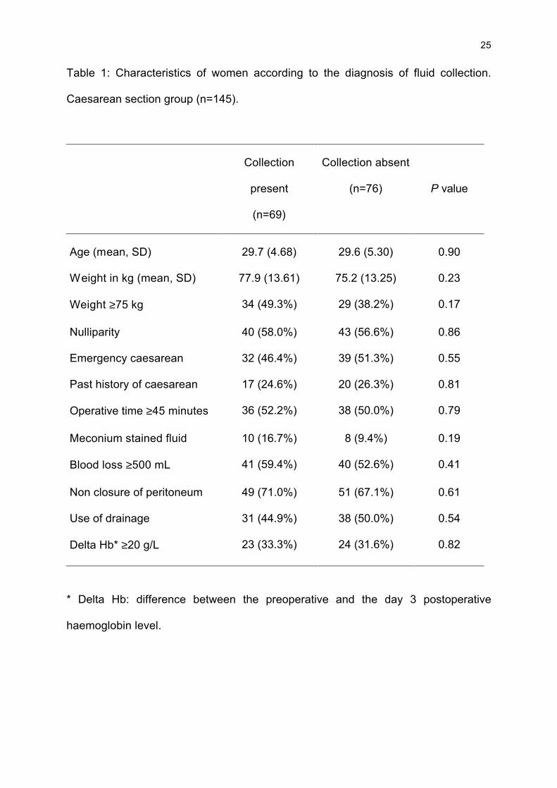
development of fluid collections after (Table 1). Obstetric data, surgical technique
(closure of the peritoneum), and operative drainage were not statistically associated
with the presence of a fluid collection. There was also no association between the
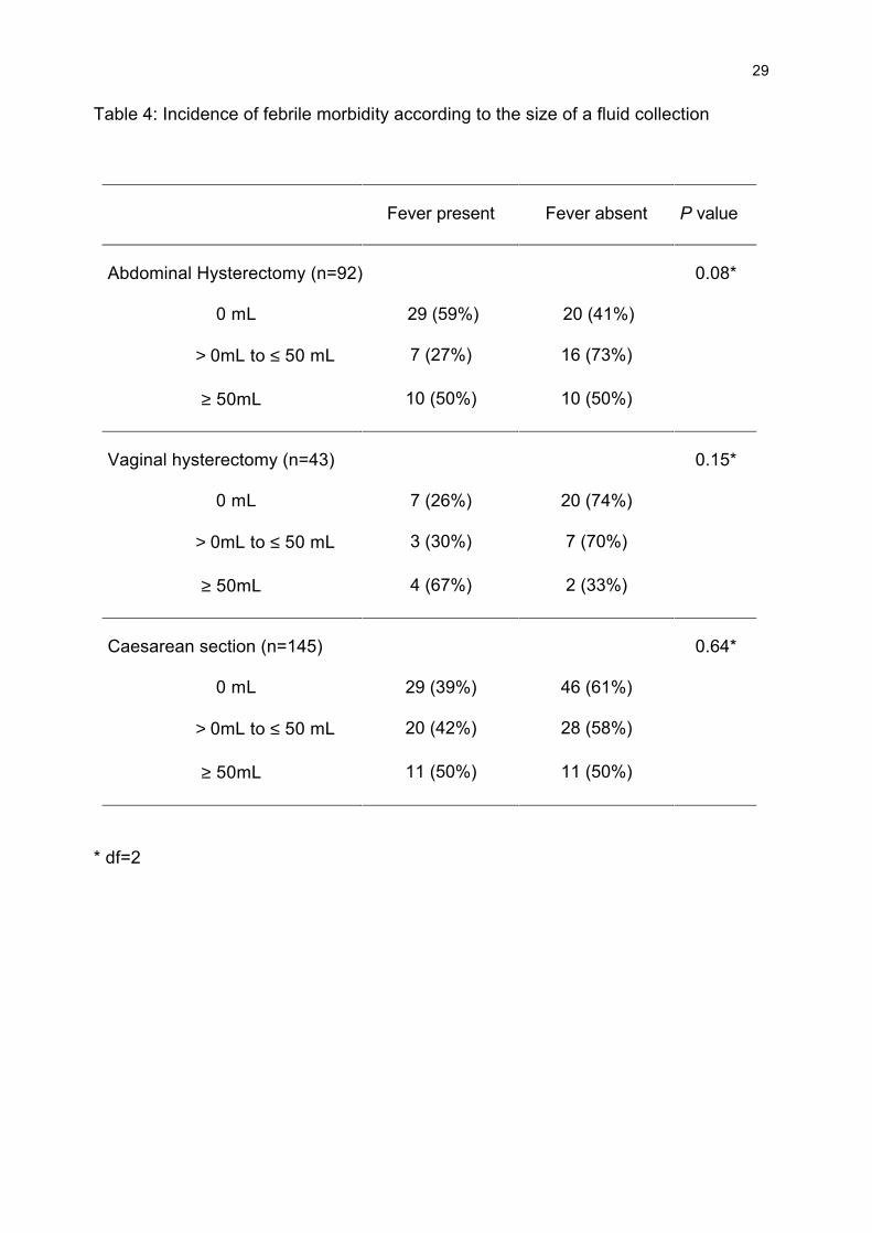
size of the collection and febrile morbidity following caesarean section , even when
the fluid collection was greater than 50 mL (Table 4).
Following hysterectomy, a fluid collection was diagnosed in 59 women (44%).
Nineteen women had fluid detected in the abdominal wall only, 33 in the pelvis only
and 7 women had both pelvic and abdominal wall collections. The median volume of
the abdominal wall fluid collections was 21 mL (range, 3-227 mL) and of the pelvic
17
fluid collections was 44 mL (range, 20-554 mL). The two largest collections (297
and 554 mL) were diagnosed as free peritoneal fluid by sonography (Figure 1). All
other collections were fixed, and hypoechogenic, suggesting the diagnosis of
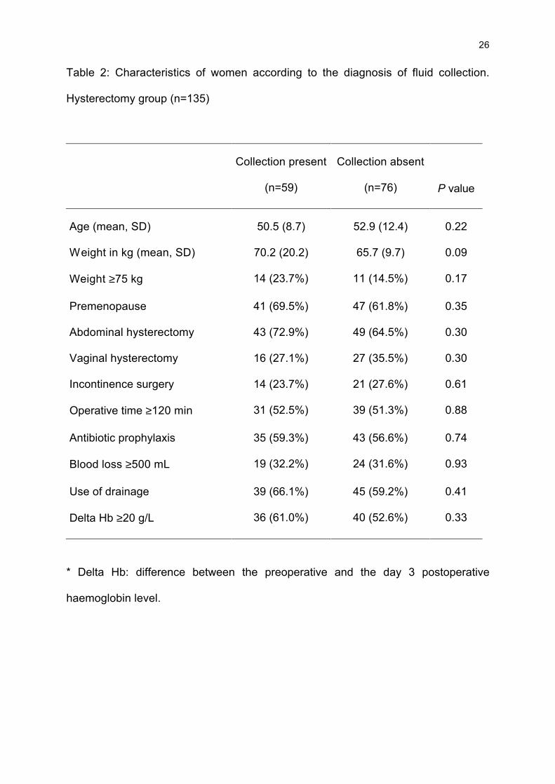
haematoma. W e have not identified risk factors for developing fluid collections after
hysterectomy (Table 2). Menopausal status, type of hysterectomy, associated
incontinence surgery and postoperative drainage were not statistically associated
with the presence of a fluid collection.
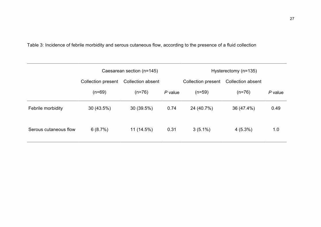
The risk of developing febrile morbidity was not related to the presence of a fluid
collection in any of the groups (Table 3). Febrile morbidity was diagnosed in 60
women following caesarean section. Thirty women (44%) diagnosed with a
collection developed fever, compared with 30 (40%) without fluid collection. Women
who had abdominal wall collection were no more likely to develop fever than those
who had a pelvic collection (39% and 42%, respectively). Following hysterectomy,
24 women with a fluid collection (41%) had febrile morbidity, compared to 36 (47%)
without a fluid collection. There was also no association between the size of the
collection and febrile morbidity following hysterectomy, even when the fluid
collection was greater than 50 mL (Table 4).
Twenty-four women (17 after caesarean section and seven after hysterectomy)
developed serous cutaneous flow during hospitalisation and required ambulatory
care. There was no association between the ultrasonographically-detected fluid
collections and serous cutaneous flow (Table 3). Duration of stay in hospital was not
increased because of this complication.
Seven women developed severe complications. Two women who underwent
caesarean section and one who had abdominal hysterectomy had wound
complications which required surgical drainage, antibiotics and prolonged hospital

18
stay. All three had a large subcutaneous haematoma, clinically evident. Following
caesarean section, two women had persistent fever and wound abscess not
requiring surgical drainage. Two women who underwent hysterectomy developed a
fistula. One had a vesico-vaginal fistula after vaginal hysterectomy, but no fluid
collection was diagnosed by ultrasonography; the second had ileo-vaginal fistula 10
days following surgery and underwent surgical repair.
19
Discussion
Our results show that fluid collections are frequently detected by ultrasonography
both after caesarean section and after hysterectomy. However, their significance
remains unclear, given the absence of an association between presence, location
and size of a fluid collection, and postoperative fever or serous cutaneous flow.
The definition of postoperative febrile morbidity is controversial.8 We chose a
relatively low cut-off (at least 37.5°C twice, excluding the first postoperative day) to
increase the sensitivity of the detection of fever.
Ultrasonographic examinations were performed on day 4 postoperatively to
minimise discomfort and to detect the presence of fluid collections not resorbed
soon after surgery. To minimise the risk of bacterial contamination with vaginal
probes, only transabdominal ultrasonography was performed after caesarean
section. The disadvantage of this approach is related to the difficulty to detect small
pelvic collections, thus leading to a possible underestimation of total fluid volume
detected. Intraoperative blood loss was assessed by recording both estimated
intraoperative blood loss and differences in haemoglobin levels measured before
and after surgery.
Few data are available on fluid collections following caesarean section. In a study
including 100 women who underwent caesarean section, fluid collection was
detected in 29.4 The association between fluid collection and postoperative
morbidity was found to be statistically significant in eight women who presented a
large haematoma (largest diameter >3.5 cm). Antibiotics were administered not only
to these eight women, but also as a prophylaxis when membranes were ruptured for
more than 6 hours. In our study, we have not found an association between large
collection and febrile morbidity. More recently, a report including 111 women
20
showed fluid collection in 14%.5 Five women with subfascial haematoma had fever.
Unfortunately, antibiotic prophylaxis, surgical techniques and risk factors for the
development of fluid collection were not reported.
Association between morbidity and fluid collections after hysterectomy is a
controversial issue. Recent studies showed that the incidence of fluid collection
varies from 25% to 62%.6-10 Different criteria and methods used to assess fluid
collection might account for these variations. Toglia et al. performed transvaginal
sonography in 38 women after hysterectomy, of whom 34% developed pelvic fluid
collections.9 They reported an association between fluid collection and febrile
morbidity. The majority of women (32/38) had undergone abdominal hysterectomy,
but possible parietal wall collection was not investigated. Recently, an association
between fluid collection and postoperative morbidity following vaginal hysterectomy
was reported.8
In contrast, three studies reported an absence of association between fluid
collection detected by sonography and postoperative morbidity following
hysterectomy. In a study including 58 women who had abdominal hysterectomy,
62% had pelvic fluid collection detected by endovaginal sonography.6 Slavotinek et
al. performed endovaginal sonography in 32 women to determine the incidence of
pelvic collection after abdominal and vaginal hysterectomy.7 However, they failed to
show a significant association between the presence of fluid collection and
postoperative pyrexia or other clinical parameters. Haines et al. performed
endovaginal sonography in 66 women after abdominal or vaginal hysterectomy and
reported an incidence of 42% of vault haematoma, which was not associated with
postoperative morbidity.10
21
The incidence of postoperative fever with no obvious cause is common. Our study
did not demonstrate any association between postoperative morbidity and fluid
collection detected by ultrasonography, even when a cut-off greater than 20 mL was
used to define a pelvic collection. Therefore, a systematic search for postoperative
collection by sonography in case of transient fever might not be justified. A more
useful approach may be to perform ultrasonography only in women presenting
prolonged or recurring fever, after common causes of postoperative pyrexia are ruled
out. Similarly, the presence of a fluid collection was not a good predictor of serous
subcutaneous flow or other postoperative morbidity. As almost half of all women
had an ultrasonographically-detected fluid collection following caesarean section or
hysterectomy without increased morbidity, such a finding in asymptomatic women
may be ignored. Only rare conditions require surgical drainage, such as abscess
refractory to antibiotic treatment. Our results do not justify a systematic search for
postoperative collection by sonography.
22
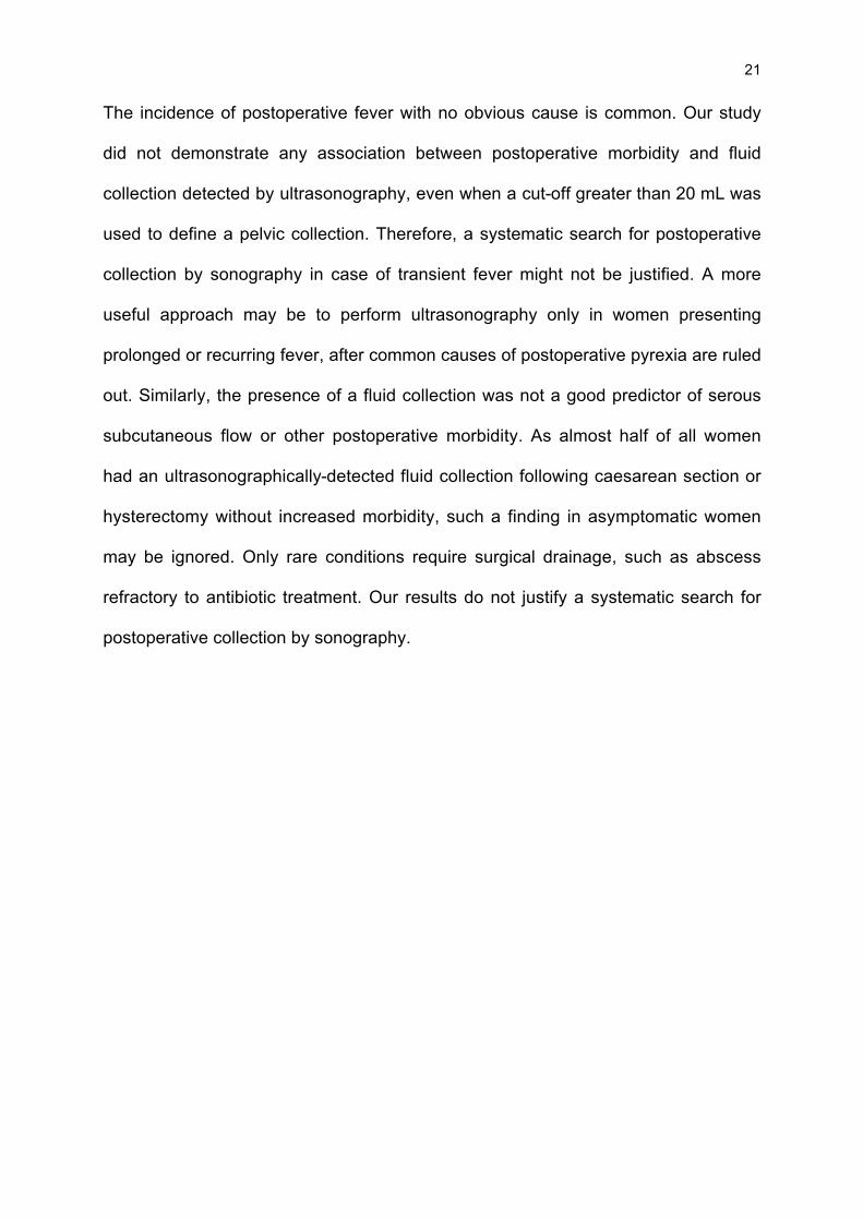
Legends for illustration
Figure 1:
Panel A: Sagittal view of the pelvis showing a large hypoechogenic fluid collection
Panel B: Transverse view of the same fluid collection
23
References
1. van Ham MA, van Dongen PW, Mulder J. Maternal consequences of
caesarean section. A retrospective study of intra-operative and postoperative
maternal complications of caesarean section during a 10-year period. Eur J
Obstet Gynecol Reprod Biol 1997;74:1-6.
2. Hill DJ. Complications of hysterectomy. Baillieres Clin Obstet Gynaecol
1997;11:181-97.
3. Harris WJ. Early complications of abdominal and vaginal hysterectomy.
Obstet Gynecol Surv 1995;50:795-805.
4. Faustin D, Minkoff H, Schaffer R, Crombleholme W, Schwarz R. Relationship
of ultrasound findings after cesarean section to operative morbidity. Obstet
Gynecol 1985;66:195-8.
5. Gemer O, Shenhav S, Segal S, Harari D, Segal O, Zohav E. Sonographically
diagnosed pelvic hematomas and postcesarean febrile morbidity. Int J
Gynaecol Obstet 1999;65:7-9.
6. Eason E, Aldis A, Seymour RJ. Pelvic fluid collections by sonography and
febrile morbidity after abdominal hysterectomy. Obstet Gynecol 1997;90:58-
62.
7. Slavotinek J, Berman L, Burch D, Keefe B. The incidence and significance of
acute post-hysterectomy pelvic collections. Clin Radiol 1995;50:322-6.
8. Thomson AJ, Sproston AR, Farquharson RG. Ultrasound detection of vault
haematoma following vaginal hysterectomy. Br J Obstet Gynaecol
1998;105:211-5.
9. Toglia MR, Pearlman MD. Pelvic fluid collections following hysterectomy and
their relation to febrile morbidity. Obstet Gynecol 1994;83:766-70.
24
10. Haines CJ, Shan YO, Hung TW, Chung TK, Leung DH. Sonographic
assessment of the vaginal vault following hysterectomy. Acta Obstet Gynecol
Scand 1995;74:220-3.
11. Koninckx PR, Renaer M, Brosens IA. Origin of peritoneal fluid in women: an
ovarian exudation product. Br J Obstet Gynaecol 1980;87:177-83.
12. Khalife S, Falcone T, Hemmings R, Cohen D. Diagnostic accuracy of
transvaginal ultrasound in detecting free pelvic fluid. J Reprod Med
1998;43:795-8.
13. Nichols JE, Steinkampf MP. Detection of free peritoneal fluid by transvaginal
sonography. J Clin Ultrasound 1993;21:171-4.
25
Table 1: Characteristics of women according to the diagnosis of fluid collection.
Caesarean section group (n=145).
Collection
present
(n=69)
Collection absent
(n=76)
P value
Age (mean, SD) 29.7 (4.68) 29.6 (5.30) 0.90
Weight in kg (mean, SD) 77.9 (13.61) 75.2 (13.25) 0.23
Weight ≥75 kg 34 (49.3%) 29 (38.2%) 0.17
Nulliparity 40 (58.0%) 43 (56.6%) 0.86
Emergency caesarean 32 (46.4%) 39 (51.3%) 0.55
Past history of caesarean 17 (24.6%) 20 (26.3%) 0.81
Operative time ≥45 minutes 36 (52.2%) 38 (50.0%) 0.79
Meconium stained fluid 10 (16.7%) 8 (9.4%) 0.19
Blood loss ≥500 mL 41 (59.4%) 40 (52.6%) 0.41
Non closure of peritoneum 49 (71.0%) 51 (67.1%) 0.61
Use of drainage 31 (44.9%) 38 (50.0%) 0.54
Delta Hb* ≥20 g/L 23 (33.3%) 24 (31.6%) 0.82
* Delta Hb: difference between the preoperative and the day 3 postoperative
haemoglobin level.
26
Table 2: Characteristics of women according to the diagnosis of fluid collection.
Hysterectomy group (n=135)
Collection present
(n=59)
Collection absent
(n=76)
P value
Age (mean, SD) 50.5 (8.7) 52.9 (12.4) 0.22
Weight in kg (mean, SD) 70.2 (20.2) 65.7 (9.7) 0.09
Weight ≥75 kg 14 (23.7%) 11 (14.5%) 0.17
Premenopause 41 (69.5%) 47 (61.8%) 0.35
Abdominal hysterectomy 43 (72.9%) 49 (64.5%) 0.30
Vaginal hysterectomy 16 (27.1%) 27 (35.5%) 0.30
Incontinence surgery 14 (23.7%) 21 (27.6%) 0.61
Operative time ≥120 min 31 (52.5%) 39 (51.3%) 0.88
Antibiotic prophylaxis 35 (59.3%) 43 (56.6%) 0.74
Blood loss ≥500 mL 19 (32.2%) 24 (31.6%) 0.93
Use of drainage 39 (66.1%) 45 (59.2%) 0.41
Delta Hb ≥20 g/L 36 (61.0%) 40 (52.6%) 0.33
* Delta Hb: difference between the preoperative and the day 3 postoperative
haemoglobin level.
27
Table 3: Incidence of febrile morbidity and serous cutaneous flow, according to the presence of a fluid collection
Caesarean section (n=145) Hysterectomy (n=135)
Collection present
(n=69)
Collection absent
(n=76)
P value
Collection present
(n=59)
Collection absent
(n=76)
P value
Febrile morbidity 30 (43.5%) 30 (39.5%) 0.74 24 (40.7%) 36 (47.4%) 0.49
Serous cutaneous flow 6 (8.7%) 11 (14.5%) 0.31 3 (5.1%) 4 (5.3%) 1.0

28
29
Table 4: Incidence of febrile morbidity according to the size of a fluid collection
Fever present Fever absent P value
Abdominal Hysterectomy (n=92) 0.08*
0 mL 29 (59%) 20 (41%)
> 0mL to ≤ 50 mL 7 (27%) 16 (73%)
≥ 50mL 10 (50%) 10 (50%)
Vaginal hysterectomy (n=43) 0.15*
0 mL 7 (26%) 20 (74%)
> 0mL to ≤ 50 mL 3 (30%) 7 (70%)
≥ 50mL 4 (67%) 2 (33%)
Caesarean section (n=145) 0.64*
0 mL 29 (39%) 46 (61%)
> 0mL to ≤ 50 mL 20 (42%) 28 (58%)
≥ 50mL 11 (50%) 11 (50%)
* df=2