Embed Size (px)
Citation preview

Progress in Cardiovascular Diseases 52 (2010) 336–346www.onlinepcd.com
Diagnosis and Management of Cardiac SarcoidosisSimon W. Dubreya, Rodney H. Falkb,⁎
aHillingdon Hospital Uxbridge, United KingdombHarvard Vanguard Medical Associates, Harvard Medical School, Boston, MA
Abstract Cardiac sarcoidosis is an underdiagnosed disease that may be present in as many as 25% of
Statement of Conf⁎ Address reprin
Vanguard Medical AsE-mail address: rf
0033-0620/$ – see frodoi:10.1016/j.pcad.20
patients with systemic sarcoidosis. Although most commonly recognized in patients with othermanifestations of sarcoidosis, it may occur in isolation and its presence is often notappreciated. Cardiac sarcoidosis may present as asymptomatic left ventricular dysfunction,congestive heart failure, atrioventricular block, atrial or ventricular arrhythmia and suddendeath. Although untested in clinical trials, early use of high-dose steroid therapy may halt orreverse cardiac damage. This article reviews the clinical manifestations, diagnosis andtreatment of sarcoidosis, with an emphasis on new imaging techniques and therapies. (ProgCardiovasc Dis 2010;52:336-346)
© 2010 Elsevier Inc. All rights reserved.Keywords: Cardiac MRI; Cardiac PET scan; Cardiac sarcoidosis; Cardiomyopathy; Endomyocardial biopsy; Sarcoidosis
Sarcoidosis is a multisystem disease characterizedhistologically by the formation of granulomas in manytissues. Most patients present with pulmonary involvementof whom approximately half have asymptomatic pulmonaryinvolvement, characterized by hilar lymphadenopathy onchest x-ray.1 The remainder have parenchymal disease andpresent most commonly with cough or dyspnea on exertion.In addition to the lungs, virtually any other organ systemmay be involved by sarcoidosis. Skin involvement,characterized by erythema nodosum or granulomas inscars and tattoos, seems to be the second commonestmanifestation.2,3 After this, in decreasing order of frequen-cy, are hepatic and gastrointestinal involvement, ocularinvolvement, and neurological sarcoidosis.2 A recent reviewof sarcoidosis placed cardiac involvement at 2%, one of theleast common manifestations.2 However, cardiac sarcoido-sis may be an asymptomatic accompaniment to pulmonarydisease or may be the presenting feature of sarcoidosis.Among patients dying with sarcoidosis in whom an autopsy
lict of Interest: see page 345.t requests to Rodney H. Falk, MD, Harvardsociates, 133, Brookline Ave, Boston, MA [email protected] (R.H. Falk).
nt matter © 2010 Elsevier Inc. All rights reserved.09.11.010
is performed, the figure has been estimated to be between20% and 25%,4-7 and sophisticated imaging studies showthat a significantly higher proportion of patients have cardiacabnormalities than suspected from symptoms and examina-tion alone.1 Thus, it is likely that cardiac sarcoidosis iscommoner than clinically recognized, although it remains arelatively uncommon disease.
In the United States, sarcoidosis is commonest in theAfrican-American population. In a population-based studydrawn from patients enrolled in a Health MaintenanceOrganization in Detroit, the age-adjusted incidence ofsarcoidosis for the age group 20 to 69 years was 39.1 per100,000 African-American females, 29.8 for African-American males, 12.1 for white females, and 9.6 in whitemales.8 African-American females in the age group of 30to 39 years had the highest annual incidence at 107 per100,000, representing an approximately 3-fold higher age-adjusted annual incidence compared with whites. Outsidethe United States, there are several populations that have ahigh incidence of sarcoidosis, particularly in Scandinaviaand Ireland. Sarcoidosis also seems to be relativelycommon in Japan, and a large body of literature on thediagnosis and management of cardiac sarcoidosis comesfrom this country. Although considerable data exist on the
336
ig 1. Lymph node from a patient withonnecrotizing granulomas typical of sarcain. Original magnification, ×40. Co
Brigham and Women's Hospital, Boston
Abbreviations and Acronyms
ACE = angiotensin-convert-ing enzyme
AV = atrioventricular
CT = computed tomography
FDG = fluoro-2-deoxy-D-glu-cose
MRI = magnetic resonanceimaging
PET = positron emissiontomography
337S.W. Dubrey, R.H. Falk / Progress in Cardiovascular Diseases 52 (2010) 336–346
epidemiology and clini-cal manifestations of sar-coidosis, controlled trialsof therapy are few forpulmonary sarcoidosisand are nonexistent incardiac sarcoidosis.
Etiology
Theetiologyof sarcoid-osis remains unknown, al-though infectious agents
and environmental exposures (eg, insecticides and agri-cultural employment) have been proposed.2,9,10 Reports ofcommunity outbreaks and clustering of cases amongworking colleagues, including nurses, firefighters, USNavy personnel, and neighbors are described, and a highprevalence of sarcoidosis, or sarcoid-like pulmonarydisease, has been described in firefighters who were atthe scene of the 2001 World Trade Center terroristattack.11 The likelihood is that there is a geneticpredisposition as evidenced by such clustering and thatan as yet unknown stimulus triggers an exaggeratedimmune response. Sarcoidosis occurs more commonly inmonozygotic than dizygotic twins, and familial clusters ofthe disease have been described.12-14
Pathology
The granulomas that characterize sarcoidosis arenoncaseating and consist of aggregates of epithelioidhistiocytes with minimal inflammation and large multinu-cleated giant cells (Fig 1).15-18 A similar histologic
sarcoidosis showing numerousoidosis. Hematoxylin and eosinurtesy of Dr Robert Padera,
Fnst
, Mass.
appearance can be seen in tuberculosis (in which caseationis usually present) and in a variety of other conditionsincluding lymphoproliferative disorders and foreignbodies. Thus, it is important to interpret the histologicfindings in conjunction with the clinical picture. Theseverity of the disease is not proportional to the number ofgranulomas, because advanced cases develop a fibroticreaction that may cause permanent tissue damage.Whether fibrosis is a reaction to the granuloma itself oris a nonspecific reaction is unclear.
Among 84 patients with sarcoidosis who had anautopsy performed at Johns Hopkins Hospital during an88-year period, 23 had myocardial sarcoidosis based onthe histologic findings of myocardial granulomas, but only4 had grossly visible myocardial abnormalities.6 Allchambers were involved, although the ventricles werefavored. In a more recent publication, autopsy findings in25 patients dying of cardiac sarcoidosis were comparedwith 16 patients with sarcoidosis in whom sudden deathwas attributed to other causes.7 Sarcoidosis had beendiagnosed premortem in only 16 of 46 patients. A dilatedleft ventricle was present in 25% of both those dying ofcardiac sarcoidosis in those with other causes of suddendeath. Gross involvement of the myocardium andpericardium with sarcoid was more frequent in sarcoid-related death than in incidental death. Sudden deathattributed to sarcoidosis was associated with visiblemyocardial scarring, and no cases of death attributable tocardiac sarcoidosis occurred in patients in whom autopsyexamination of the heart showed only microscopicfeatures of the disease. Of note, 10 of 25 deaths attributedto sarcoid heart disease had no evidence of extracardiacdisease. Both of these autopsy series, although suggestingthat sarcoidosis clinically isolated to the heart is common,suffer from selection bias because a patient withunexpected death is more likely to be subject to anautopsy. Nevertheless, the findings underscore the factthat isolated cardiac sarcoidosis does occur and that itsinitial manifestation may be sudden death.
Clinical presentation of cardiac sarcoidosis
As noted above, asymptomatic heart involvement inpatients with pulmonary sarcoidosis may be relativelyfrequent. Cardiac involvement may be present in theabsence of any abnormality on standard cardiac testing,may manifest as an abnormal electrocardiogram alone, ormay be recognized as an asymptomatic abnormality onechocardiography or cardiac magnetic resonance imaging(MRI). In a patient with established extracardiac sarcoid-osis, the presence of congestive heart failure or the findingof impaired systolic function on echocardiography strong-ly raises the suspicion of cardiac involvement, unlessanother obvious cause, such as coronary artery disease,
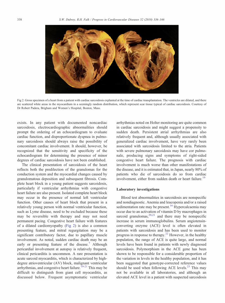
Fig 2. Gross specimen of a heart from a patient with cardiac sarcoidosis explanted at the time of cardiac transplantation. The ventricles are dilated, and thereare scattered white areas in the myocardium in a seemingly random distribution, which represent scar tissue typical of cardiac sarcoidosis. Courtesy ofDr Robert Padera, Brigham and Women's Hospital, Boston, Mass.
338 S.W. Dubrey, R.H. Falk / Progress in Cardiovascular Diseases 52 (2010) 336–346
exists. In any patient with documented noncardiacsarcoidosis, electrocardiographic abnormalities shouldprompt the ordering of an echocardiogram to evaluatecardiac function, and disproportionate dyspnea in pulmo-nary sarcoidosis should always raise the possibility ofconcomitant cardiac involvement. It should, however, berecognized that the sensitivity and specificity of theechocardiogram for determining the presence of minordegrees of cardiac sarcoidosis have not been established.
The clinical presentation of sarcoidosis of the heartreflects both the predilection of the granulomas for theconduction system and the myocardial changes caused bygranulomatous deposition and subsequent fibrosis. Com-plete heart block in a young patient suggests sarcoidosis,particularly if ventricular arrhythmias with congestiveheart failure are also present. Isolated complete heart blockmay occur in the presence of normal left ventricularfunction. Other causes of heart block that present in arelatively young person with normal ventricular function,such as Lyme disease, need to be excluded because thesemay be reversible with therapy and may not needpermanent pacing. Congestive heart failure with featuresof a dilated cardiomyopathy (Fig 2) is also a commonpresenting feature, and mitral regurgitation may be asignificant contributory factor, due to papillary muscleinvolvement. As noted, sudden cardiac death may be anearly or presenting feature of the disease.7 Althoughpericardial involvement at autopsy is relatively frequent,clinical pericarditis is uncommon. A rare presentation isacute sarcoid myocarditis, which is characterized by high-degree atrioventricular (AV) block, malignant ventriculararrhythmias, and congestive heart failure.19-21 This may bedifficult to distinguish from giant cell myocarditis, asdiscussed below. Frequent asymptomatic ventricular
arrhythmias noted on Holter monitoring are quite commonin cardiac sarcoidosis and might suggest a propensity tosudden death. Persistent atrial arrhythmias are alsorelatively frequent and, although usually associated withgeneralized cardiac involvement, have very rarely beenassociated with sarcoidosis limited to the atria. Patientswith severe pulmonary sarcoidosis may have cor pulmo-nale, producing signs and symptoms of right-sidedcongestive heart failure. The prognosis with cardiacinvolvement is much worse than other manifestations ofthe disease, and it is estimated that, in Japan, nearly 80% ofpatients who die of sarcoidosis do so from cardiacinvolvement, either from sudden death or heart failure.19
Laboratory investigations
Blood test abnormalities in sarcoidosis are nonspecificand nondiagnostic. Anemia and leucopenia and/or a raisedsedimentation rate may be present.16 Hypercalcaemia mayoccur due to an activation of vitamin D by macrophages insarcoid granulomas,20,21 and there may be nonspecificincrease in serum immunoglobulins. Serum angiotensin-converting enzyme (ACE) level is often elevated inpatients with sarcoidosis and has been used to monitorprogress in response to therapy.22 However, in the healthypopulation, the range of ACE is quite large, and normallevels have been found in patients with newly diagnosedsarcoidosis. Polymorphism in the ACE gene has beenshown to be responsible for a considerable proportion ofthe variation in levels in the healthy population, and it hasbeen suggested that genotype-corrected reference valuesshould be used when following ACE levels.23 This maynot be available in all laboratories, and although anelevated ACE level in a patient with suspected sarcoidosis
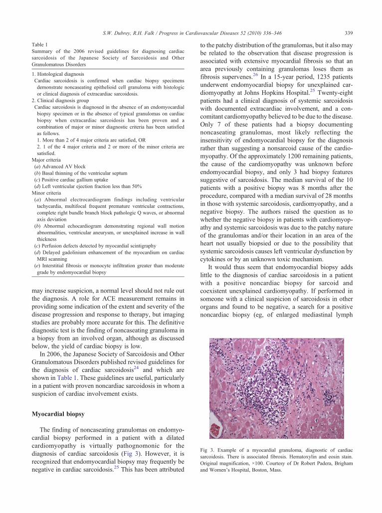
Table 1Summary of the 2006 revised guidelines for diagnosing cardiacsarcoidosis of the Japanese Society of Sarcoidosis and OtherGranulomatous Disorders
1. Histological diagnosisCardiac sarcoidosis is confirmed when cardiac biopsy specimensdemonstrate noncaseating epithelioid cell granuloma with histologicor clinical diagnosis of extracardiac sarcoidosis.
2. Clinical diagnosis groupCardiac sarcoidosis is diagnosed in the absence of an endomyocardialbiopsy specimen or in the absence of typical granulomas on cardiacbiopsy when extracardiac sarcoidosis has been proven and acombination of major or minor diagnostic criteria has been satisfiedas follows.1. More than 2 of 4 major criteria are satisfied, OR2. 1 of the 4 major criteria and 2 or more of the minor criteria aresatisfied.
Major criteria(a) Advanced AV block(b) Basal thinning of the ventricular septum(c) Positive cardiac gallium uptake(d) Left ventricular ejection fraction less than 50%Minor criteria(a) Abnormal electrocardiogram findings including ventriculartachycardia, multifocal frequent premature ventricular contractions,complete right bundle branch block pathologic Q waves, or abnormalaxis deviation(b) Abnormal echocardiogram demonstrating regional wall motionabnormalities, ventricular aneurysm, or unexplained increase in wallthickness(c) Perfusion defects detected by myocardial scintigraphy(d) Delayed gadolinium enhancement of the myocardium on cardiacMRI scanning(e) Interstitial fibrosis or monocyte infiltration greater than moderategrade by endomyocardial biopsy
339S.W. Dubrey, R.H. Falk / Progress in Cardiovascular Diseases 52 (2010) 336–346
may increase suspicion, a normal level should not rule outthe diagnosis. A role for ACE measurement remains inproviding some indication of the extent and severity of thedisease progression and response to therapy, but imagingstudies are probably more accurate for this. The definitivediagnostic test is the finding of noncaseating granuloma ina biopsy from an involved organ, although as discussedbelow, the yield of cardiac biopsy is low.
In 2006, the Japanese Society of Sarcoidosis and OtherGranulomatous Disorders published revised guidelines forthe diagnosis of cardiac sarcoidosis24 and which areshown in Table 1. These guidelines are useful, particularlyin a patient with proven noncardiac sarcoidosis in whom asuspicion of cardiac involvement exists.
ig 3. Example of a myocardial granuloma, diagnostic of cardiacrcoidosis. There is associated fibrosis. Hematoxylin and eosin stain.riginal magnification, ×100. Courtesy of Dr Robert Padera, Brigham
and Women's Hospital, Boston, Mass.
Myocardial biopsy
The finding of noncaseating granulomas on endomyo-cardial biopsy performed in a patient with a dilatedcardiomyopathy is virtually pathognomonic for thediagnosis of cardiac sarcoidosis (Fig 3). However, it isrecognized that endomyocardial biopsy may frequently benegative in cardiac sarcoidosis.25 This has been attributed
to the patchy distribution of the granulomas, but it also maybe related to the observation that disease progression isassociated with extensive myocardial fibrosis so that anarea previously containing granulomas loses them asfibrosis supervenes.26 In a 15-year period, 1235 patientsunderwent endomyocardial biopsy for unexplained car-diomyopathy at Johns Hopkins Hospital.25 Twenty-eightpatients had a clinical diagnosis of systemic sarcoidosiswith documented extracardiac involvement, and a con-comitant cardiomyopathy believed to be due to the disease.Only 7 of these patients had a biopsy documentingnoncaseating granulomas, most likely reflecting theinsensitivity of endomyocardial biopsy for the diagnosisrather than suggesting a nonsarcoid cause of the cardio-myopathy. Of the approximately 1200 remaining patients,the cause of the cardiomyopathy was unknown beforeendomyocardial biopsy, and only 3 had biopsy featuressuggestive of sarcoidosis. The median survival of the 10patients with a positive biopsy was 8 months after theprocedure, compared with a median survival of 28 monthsin those with systemic sarcoidosis, cardiomyopathy, and anegative biopsy. The authors raised the question as towhether the negative biopsy in patients with cardiomyop-athy and systemic sarcoidosis was due to the patchy natureof the granulomas and/or their location in an area of theheart not usually biopsied or due to the possibility thatsystemic sarcoidosis causes left ventricular dysfunction bycytokines or by an unknown toxic mechanism.
It would thus seem that endomyocardial biopsy addslittle to the diagnosis of cardiac sarcoidosis in a patientwith a positive noncardiac biopsy for sarcoid andcoexistent unexplained cardiomyopathy. If performed insomeone with a clinical suspicion of sarcoidosis in otherorgans and found to be negative, a search for a positivenoncardiac biopsy (eg, of enlarged mediastinal lymph
FsaO

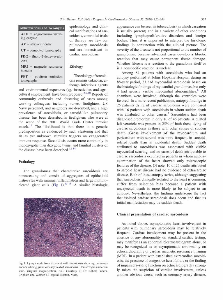
Fig 5. Parasternal long-axis view of a the echocardiogram from a 38-year-old woman with peripartum heart failure, originally attributed toperipartum cardiomyopathy. Her left ventricular systolic function wasmildly impaired but did not improve postpartum. The basal septum(arrow) shows marked thinning and increased echogenicity, highlysuggestive of sarcoidosis. She had a history of pulmonary sarcoidosis andwas found to have marked hilar adenopathy and evidence of activedisease. Abbreviations: Ao, aorta; LA, left atrium; LV, left ventricle.
340 S.W. Dubrey, R.H. Falk / Progress in Cardiovascular Diseases 52 (2010) 336–346
nodes) should be pursued. Most patients will have bilateralhilar lymphadenopathy, often with right paratrachealadenopathy, and mediastinoscopy can be performed tobiopsy paratracheal lymph nodes.1 Up to 75% of patientswill have asymptomatic liver granulomas, providing anadditional biopsy site. Approximately 1 in 4 will haveocular involvement in the form of an anterior uveitis,which can be diagnosed on slit lamp examination.
The histologic differential diagnosis of sarcoid granu-lomas includes giant cell myocarditis. Occasionally,cardiac sarcoidosis may present acutely with fulminantcongestive heart failure, ventricular arrhythmias, and AVblock, and it may be difficult to distinguish this from giantcell myocarditis or myocarditis of another origin. In acomparison of 42 patients with cardiac sarcoidosis to 73patients with idiopathic giant cell myocarditis, 31% ofpatients with sarcoidosis were of African origin comparedwith only 4% with giant cell myocarditis.17 Syncope andAV block were much commoner in sarcoidosis, whereasthere was a slight increase in the prevalence of left-sidedheart failure in the giant cell myocarditis group. In amultivariate analysis, patients with giant cell myocarditismore commonly presented with heart failure of shortduration, whereas more than 9 weeks of symptoms andpresentation with heart block was more commonly seen incardiac sarcoidosis. Histologically, myocyte damage,eosinophils, and foci of lymphocytic myocarditis favoredgiant cell myocarditis, whereas granulomas and fibrosiswere more frequent in cardiac sarcoidosis. Intriguingly,only one third of the patients ultimately diagnosed ascardiac sarcoidosis had evidence of extracardiac disease.
Fig 4. Chest x-ray from a 49-year-old man who presented with congestiveheart failure and global left ventricular hypokinesis, an estimated ejectionfraction of 25% to 35%, and mild to moderate mitral regurgitationCoronary angiography was normal, and endomyocardial biopsy showednonspecific changes only. He was readmitted with worsening hearfailure, and after diuresis, the x-ray above was obtained. It shows markedcardiomegaly with bilateral hilar fullness. The hilar fullness, originallyattributed to heart failure, had not resolved with diuresis, and a chest CTscan showed widespread hilar adenopathy. Mediastinoscopic lymph nodebiopsy showed noncaseating granulomas in all biopsied lymph nodesand his heart failure improved after high-dose steroid therapy.
.
t
,
Although this may represent selection bias (patientswithout organ involvement elsewhere are probably morelikely to have a cardiac biopsy), cardiac involvement as thepresenting feature of sarcoidosis has been describedfrequently in other series.
Imaging and cardiac sarcoidosis
Different imaging techniques have been used both for thediagnosis and follow-up of patients with suspected cardiacsarcoidosis. These include echocardiography, myocardialperfusion with thallium or Tc-99 nuclear scintigraphy,gallium-67 scintigraphy, positron emission tomography(PET) with fluoro-2-deoxy-D-glucose (18FDG), and MRIand cardiac computed tomography. With increasing sophis-tication of these techniques, it is becoming possible todiagnose cardiac sarcoidosis noninvasively.
Chest x-ray
There is nothing specific about the cardiac contour incardiac sarcoidosis on the chest x-ray. Nonspecificcardiomegaly is often present, reflecting the dilatedventricle. If cardiac sarcoidosis coexists with pulmonarysarcoid, hilar lymphadenopathy and/or pulmonary paren-chymal changes may give a clue as to the diagnosis (Fig 4).However, hilar fullness on a chest x-ray may be a feature ofcongestive heart failure or may represent pulmonary arterydilation secondary to the cardiac disease. If hilar adeno-pathy is suspected on a chest x-ray, computed tomography(CT) imaging of the chest should be performed to look forhilar adenopathy and pulmonary parenchymal disease.High-resolution CT is particularly sensitive for detection ofpulmonary involvement,19,20 whereas standard contrast-

341S.W. Dubrey, R.H. Falk / Progress in Cardiovascular Diseases 52 (2010) 336–346
enhanced CT may be better for delineation of mediastinaland hilar lymphadenopathy.
Echocardiography
Generally, echocardiography is nondiagnostic in termsof etiology in a patient in whom a new cardiomyopathyhas been found, but it may be very helpful in suggestingcardiac sarcoidosis if abnormal in a patient with biopsy-proven systemic sarcoidosis at another site. The usualechocardiographic appearance of cardiac sarcoidosis isthat of a dilated cardiomyopathy. The ventricle may beglobally hypokinetic or the patchy nature of sarcoidinfiltration of the heart may result in regional wall motionabnormalities. Mild wall thickening may be presentrelated to edema or infiltration. More commonly, areasof wall thinning are seen, most commonly in theventricular septum and probably associated with scarring.A typical but uncommon finding is the thinning of thebasal anterior septum, the appearance of which in a youngpatient with a dilated cardiomyopathy is highly sugges-tive of sarcoidosis27 (Fig 5). On rare occasions, anappearance similar to hypertrophic cardiomyopathy hasbeen described.28,29 Diastolic dysfunction is a commonbut nonspecific finding.30
Radionuclide scintigraphy
201Thallium scintigraphy myocardial perfusion studieshave been used for a number of years in evaluating patientswith suspected cardiac sarcoidosis. If sarcoidosis is presentin the heart, there are typically segmental areas ofdecreased uptake in the ventricular myocardium thatdisappear or decrease in size during stress or afterintravenous administration of dipyridamole.31,32 However,this reverse distribution is not specific for cardiacsarcoidosis because it may also occur in other cardiomy-opathies. Gallium-67 scintigraphy has also been used todemonstrate both cardiac and extracardiac disease,1 and thedetection of clinically silent extrathoracic uptake mayprovide sites for biopsy. It has also been used for follow-upof active disease (both cardiac and extracardiac) withtreatment. Studies in which both 201thallium and gallium-67 scintigraphy were performed in patients with suspectedsarcoidosis showed that areas of reduced uptake were morecommon using 201thallium than with gallium-67.33 Inpatients in which both scans were positive, there was moresevere cardiac involvement, as indicated by a reducedejection fraction. 99-mTc sestamibi has been described incombination with gallium-67 scintigraphy as increasingthe sensitivity for the diagnosis of cardiac sarcoidosis.34
Cardiac MRI
With more widespread availability of cardiac MRI andincreasing availability of cardiac PET scanning, thallium (or
sestamibi) and gallium imaging has become less frequentlyused, because MRI and PET seem to be more sensitive andspecific for the diagnosis of both pulmonary and extra-pulmonary sarcoidosis. Due to its high spatial and soft tissueresolution, MRI is now increasingly being shown to be atechnique of choice for evaluation and diagnosis of cardiacsarcoidosis.35 Cardiac MRI can demonstrate both scar andmyocardial edema.Acutemyocardial inflammation presentsas focal areas of thickening and increased signal intensity onT2-weighted images and early gadolinium-enhancedimages. Myocardial edema, which is present in the activestage of cardiac sarcoidosis, can rapidly resolve with steroidtreatment, resulting in rapid decrease in thickened areas andresolution of increased T2 hyperintensity.36 In cardiacsarcoidosis, delayed gadolinium enhancement is foundpredominantly in the midmyocardium and epicardial areasbut rarely in the endocardium.37 A predilection forabnormalities in basal and lateral segments of the leftventricle has been described,38,39 and the papillary musclesare frequently involved.37 In addition to the delayedgadolinium enhancement, cardiac MRI can preciselydetermine regional wall motion abnormalities and areas ofwall thickening but may represent myocardial edema orinfiltration. Abnormalities of delayed gadolinium enhance-ment or T2 imaging for edema have been used to targetendomyocardial biopsy in an attempt to increase thesensitivity of this technique.
Cardiac MRI is useful not only in patients with acardiomyopathy of unexplained origin but also in determin-ing the presence or absence of cardiac sarcoidosis in patientswith documented pulmonary disease. In a series of 58patients with histologically proven pulmonary sarcoidosiswho underwent both thallium myocardial scintigraphy andcardiac MRI, late gadolinium enhancement was found in 19patients, most commonly in the basal and lateral leftventricular segments.38 Ten patients were found to have areduction in left ventricular ejection fraction, for a total of 22patients with abnormalities. Only 12 patients had anabnormality on thallium scintigraphy. In a similar series ofpatients with documented pulmonary sarcoidosis and nopreviously diagnosed cardiac sarcoidosis, one third ofpatients had abnormalities on cardiac MRI and 80% hadmetabolic abnormalities on PET scanning.40 All of thesepatients had 1 or more cardiac abnormalities on theelectrocardiogram or on a Holter monitoring, and thus, therelatively high percentage potential disease represents arelatively selected population. All these studies indicate thatcardiac abnormalities are relatively common in patients withnoncardiac sarcoidosis, a finding that is concordant with theprevalence of cardiac sarcoid involvement at autopsy.
18FDG PET scanning
18F-fluoro-2-deoxy-D-glucose PET scanning is widelyused in evaluation of tumors, vasculitis, and inflammatory
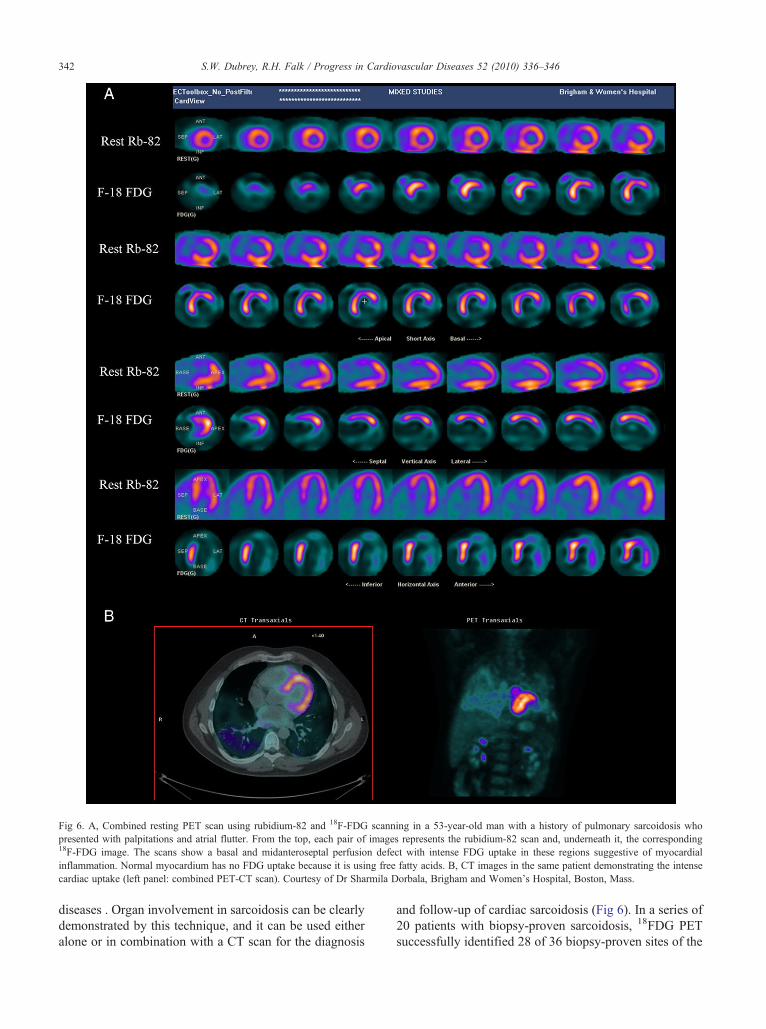
Fig 6. A, Combined resting PET scan using rubidium-82 and 18F-FDG scanning in a 53-year-old man with a history of pulmonary sarcoidosis whopresented with palpitations and atrial flutter. From the top, each pair of images represents the rubidium-82 scan and, underneath it, the corresponding18F-FDG image. The scans show a basal and midanteroseptal perfusion defect with intense FDG uptake in these regions suggestive of myocardialinflammation. Normal myocardium has no FDG uptake because it is using free fatty acids. B, CT images in the same patient demonstrating the intensecardiac uptake (left panel: combined PET-CT scan). Courtesy of Dr Sharmila Dorbala, Brigham and Women's Hospital, Boston, Mass.
342 S.W. Dubrey, R.H. Falk / Progress in Cardiovascular Diseases 52 (2010) 336–346
diseases . Organ involvement in sarcoidosis can be clearlydemonstrated by this technique, and it can be used eitheralone or in combination with a CT scan for the diagnosis
and follow-up of cardiac sarcoidosis (Fig 6). In a series of20 patients with biopsy-proven sarcoidosis, 18FDG PETsuccessfully identified 28 of 36 biopsy-proven sites of the

343S.W. Dubrey, R.H. Falk / Progress in Cardiovascular Diseases 52 (2010) 336–346
disease.41 In a Japanese series of 21 patients withsarcoidosis and suspected cardiac involvement, cardiacMRI, 18F-FDG PET, and sestamibi scintigraphy wereperformed.42 Based on the Japanese guidelines fordiagnosing cardiac sarcoidosis (Table 1; which includesan abnormal myocardial sestamibi scan as one of thefeatures), 8 patients were considered to have cardiacsarcoidosis before the scans. Fifteen patients had abnormalPET scans, and 11 had abnormal cardiac MRI scanscharacterized by delayed gadolinium enhancement in 9 (1of whom also had features of myocardial edema) andabnormal T2 images suggesting myocardial edema in 2. Itis likely that abnormalities on PET scanning or MRI inthese patients represent cardiac sarcoidosis even in theabsence of clinical cardiac abnormalities. Interestingly, 7patients exhibited abnormalities on PET scanning aloneand only 1 on MRI alone. Although it is conceivable thatthis indicates that PET scanning is more sensitive thanMRI, it may be that the PET abnormalities werenonspecific or that they represent active sarcoid inflam-mation without myocardial edema or scarring, whichmightnot be apparent on cardiac MRI. Further work needs to bedone on the significance of the discrepant findings todetermine whether or not cardiac MRI and PET scans willbe complementary to one another for the investigation andfollow-up of cardiac sarcoidosis.
Management of cardiac sarcoidosis
The management of cardiac sarcoidosis is similar tothat of other forms of dilated cardiomyopathy, namely,diuretics, ACE inhibitors, or angiotensin-receptor blockersand β- blocking agents. Angiotensin-converting enzymeinhibitors and angiotensin-receptor blockers have antifi-brotic properties and have the theoretical potential tomodify the progressive fibrosis that accompanies manycases of cardiac sarcoidosis. Although there are nocontrolled randomized trials addressing the arrhythmiamanagement in this disease, the apparently high incidenceof sudden cardiac death has led many clinicians to adopt arelatively aggressive approach in terms of prophylacticdefibrillators. In addition to the management of congestiveheart failure and cardiac arrhythmias, attention in cardiacsarcoidosis should be direct toward therapies aimed atdecreasing the inflammatory process. Corticosteroids arethe mainstay of such therapy.43 Arrhythmia therapy andthe value of steroids and other anti-inflammatory therapiesare addressed in the following sections.
Cardiac arrhythmia
The first manifestation of cardiac sarcoidosis may behigh-degree AV block, sustained atrial arrhythmias, ormalignant ventricular arrhythmia (including sudden
death). The substrate for arrhythmias may be activegranuloma formation in the myocardium or myocardialfibrosis. The widespread involvement of the heart by thedisease may result in the coexistence of these arrhythmiasin a single individual, and the onset of an atrial arrhythmiamay portend a ventricular arrhythmia. An intriguingpresentation of cardiac sarcoidosis is as a mimic ofarrhythmogenic right ventricular dysplasia. Twenty-threepatients referred for suspected arrhythmogenic rightventricular dysplasia underwent endomyocardial biopsyand 3 were found to have noncaseating granulomasconsistent with sarcoid.44 Of note, a reduced leftventricular ejection fraction was present in all 3 of thesepatients but was only found in 2 of 17 patients withsuspected arrhythmogenic right ventricular dysplasia.
Presentation with sustained ventricular tachycardia orventricular fibrillation is a clear indication for implantationof a defibrillator. Catheter ablation of the site of origin ofventricular arrhythmias has been used to decrease thefrequency of defibrillator shocks. However, the wide-spread scarring and progressive nature of cardiac sarcoid-osis are generally considered unfavorable for such therapy.Koplan and coworkers26 reported their experience withattempted ablation therapy for refractory ventriculartachycardia in 8 patients with sarcoidosis. Althoughsmall, this case series underscores some of the difficultiesin the diagnosis and treatment of this disorder. Endomyo-cardial biopsy had been performed in 7 patients and wasnegative for sarcoid granulomas in 3. In 1 patient, whosubsequently underwent cardiac transplantation, theexplanted heart showed extensive fibrosis without gran-ulomas, whereas an endomyocardial biopsy performedjust 4 months before had shown granulomatous infiltra-tion. Two of the patients had a previous diagnosis ofarrhythmogenic right ventricular dysplasia and presentedwith ventricular arrhythmias before a diagnosis ofsarcoidosis was been made. Multiple monomorphicventricular tachycardias were induced and ablation,although they abolished individual ventricular tachycar-dias, was rarely completely successful in an individualpatient. Five patients subsequently underwent cardiactransplantation, the indication being intractable arrhythmiain 4 of them.
The decision in whom to implant a defibrillator inpatients with cardiac sarcoidosis not presenting withventricular arrhythmia is complex, and there are nocontrolled data to guide the clinician. Prystowsky hascogently argued that the progressive nature of the diseaseis such that a defibrillator (all of which also have pacingcapabilities) is the appropriate choice in a patient withcardiac sarcoidosis presenting with high-degree AVblock.45 He also favors an aggressive approach in apatient with documented extracardiac sarcoidosis andevidence of cardiac involvement on an abnormal
344 S.W. Dubrey, R.H. Falk / Progress in Cardiovascular Diseases 52 (2010) 336–346
electrocardiogram or imaging study, based on theunpredictable and relatively high risk of sudden death insuch patients. In contrast, Soejima and Yada24 havesuggested an algorithmic approach to sarcoidosis andarrhythmia, with pacemaker implantation alone forpatients presenting with AV block in the absence ofsevere left ventricular dysfunction or heart failure,defibrillator implantation alone for those with spontaneousventricular tachycardia/fibrillation and a narrow QRS and,a defibrillator plus cardiac resynchronization therapy forspontaneous ventricular arrhythmias associated withimpaired ventricular function and a wide QRS. Thisapproach is similar to that generally taken in idiopathicdilated cardiomyopathy with a moderately or severelyreduced ejection fraction, a condition in which the risk ofsudden death is also elevated. It is possible that PETimaging may serve as a “tie-breaker” in deciding in whichpatients with cardiac sarcoidosis and normal or mildlyreduced ejection fraction to implant a defibrillator.Significant myocardial PET uptake, if assumed to indicateactive disease, may identify a group at higher risk ofsudden death either because of the activity of the disease orgreater likelihood that it will progress. On the other hand, apatient with a mildly reduced ejection fraction andsystemic sarcoidosis in whom there is no cardiac PETabnormality could be considered to have quiescent diseaseand may have a lower risk of sudden death. At present,there are no data to support this approach, but in theory, itremains attractive.
Immunosuppressive therapy
The treatment of pulmonary sarcoidosis (other thanasymptomatic hilar adenopathy in which treatment is notrequired) and many forms of extrapulmonary sarcoidosisis with high-dose corticosteroids,43 even though defini-tive data on efficacy are lacking.46 Several uncontrolledseries of patients with cardiac sarcoidosis have beenpublished, which suggest that steroid use may bevaluable in this condition. However, there are norandomized controlled data. Because many patientswith cardiac sarcoidosis have significant congestiveheart failure, the usual complications of high-dose steroidtherapy such as weight gain, hyperglycemia, osteoporo-sis, and increased susceptibility to infection additionallyinclude the possibility of exacerbation of congestive heartfailure. Consequently, if steroids are to be considered, itis vital that the diagnosis is as certain as possible and thatthe patient is followed very carefully. Most series of theeffects of steroids in cardiac sarcoidosis suggesteffectiveness of this therapy, although the optimal doseand duration of therapy are unclear. In a retrospectiveseries from Japan of 48 patients with cardiac sarcoidosis,high-dose steroid therapy seemed to be ineffective inpatients with a pretreatment left ventricular ejection
fraction of less than 30%, whereas there was animprovement in ejection fraction and a decrease in leftventricular end-diastolic volume in patients whosepretreatment ejection fraction was between 30% and55%.47 The authors suggested that severe left ventriculardysfunction in cardiac sarcoidosis may represent end-stage fibrosis, which is refractory to corticosteroidtherapy. Corticosteroid therapy was used in most of the41 patients described in a retrospective study fromFrance.48 Improvement in abnormal echocardiographicparameters was described in 78%, and 9 of 17 patientspresenting with congestive heart failure had completeresolution of symptoms.
The dose of corticosteroids that is effective in cardiacsarcoidosis is unclear. The French group recommended aninitial dose of 1 mg per kilogram per day of prednisone,maintained for 6 to 8 weeks followed by tapering of thedose with careful assessment of cardiac function.48 In aretrospective study of cardiac sarcoidosis in 30 Japanesepatients who were prescribed 40 mg or more of prednisonedaily (range, 40-60 mg) and in 45 who were prescribedless than 30 mg/d (range, 10-30 mg), there was noapparent survival benefit of high-dose prednisone over thelower dose.49 Based on these observations, the authorsrecommended prescribing an initial dose of 60 mg ofprednisone every other day tapering, over a few months, to10 mg every other day. Whether a high or intermediatedose is used, it is clear that the eventual dose should be thelowest possible dose effective in suppressing diseaseactivity. Steroid therapy may eventually be discontinued,but it should be recognized that cessation of steroidtherapy has been associated with a relapse rates ofapproximately 25%.
Additional immunosuppressive therapy has been triedin sarcoidosis including cyclophosphamide, methotrexate,and cyclosporine, but there are no data as to their efficacyin cardiac sarcoidosis. Recently, infliximab, an agent usedfor the treatment of rheumatoid arthritis that acts byopposing tumor necrosis factor-α has been proposed as atreatment for sarcoidosis. In a small study of refractorypulmonary sarcoidosis, changes imaged by 18F-FDG PETscanning correlated with improvement in pulmonaryfunction50 and case reports have described improvementin cardiac sarcoidosis after infliximab treatment.51-53
Infliximab poses an infectious risk because there is alreadya cellular defect due to the sarcoidosis and is not withoutserious adverse effects, and a case of fatal disseminatedcryptococcosis in a patient with sarcoidosis treated withinfliximab has been described.54
Heart transplantation for sarcoid heart disease
Sarcoidosis can result in severe congestive heart failurenecessitating consideration of cardiac transplantation.

345S.W. Dubrey, R.H. Falk / Progress in Cardiovascular Diseases 52 (2010) 336–346
Partly because of the uncommon nature of the diseaseand partly because of coexisting disease in other organs,the number of patients undergoing cardiac transplanta-tion in whom cardiac sarcoidosis was documentedpretransplant is quite small. Review of the UnitedNetwork for Organ Sharing database between 1987 and2005 revealed that 65 patients with cardiac sarcoidosisunderwent cardiac transplantation.55 This represents lessthan 0.2% of all transplants. Surprisingly, the 1-year posttransplant survival for sarcoidosis patients was signifi-cantly better than the other contemporaneous patients(87.7% versus 84.5%), and survival benefit persisted outto 5 years.
The difficulty in diagnosing cardiac sarcoidosis has ledto several cases in which cardiac transplantation wasperformed without realizing the etiology of congestiveheart failure. Three of four patients with cardiacsarcoidosis transplanted in a Danish center had thediagnosis made at the time all examination of theexplanted heart.56 There were 2 long-term survivors, 1patient died of primary graft failure of the other fromcytomegalovirus. Recurrence of the disease has beenreported in a transplanted heart,57 but this seems to be arare phenomenon. Thus, it seems that, in carefully selectedcases, the outcome from cardiac transplantation in cardiacsarcoidosis is good.
Summary
Cardiac sarcoidosis, although an uncommon disease,should be considered in all cases of unexplainedcardiomyopathy, particularly in young patients. A nega-tive endomyocardial biopsy should not be taken asevidence of the absence of cardiac sarcoidosis, particularlyif suspicion is high. Advances in cardiac imaging seem tobe improving the diagnosis and, when highly suggestive,should prompt a search for a positive tissue biopsy in avisualized abnormal area of the myocardium or from anoncardiac site. Steroid therapy, although untested inrandomized clinical trial, seem to be beneficial particularlywhen used relatively early in the course of the disease.Attention should be given to the risk of arrhythmia in thisdisease, with a strong consideration for prophylacticimplantation of a cardiac defibrillator, particularly in thepresence of ventricular arrhythmias or a reduced ejectionfraction. Careful follow-up is mandatory when using high-dose steroids, and in the patient with severe heart failureand minimal extracardiac sarcoid, consideration can begiven cardiac transplantation.
Statement of Conflict of Interest
All authors declare that there are no conflicts of interest.
References
1. Akbar JJ, Meyer CA, Shipley RT, et al: Cardiopulmonary imaging insarcoidosis. Clin Chest Med 2008;29:429-443.
2. Baughman RP, Teirstein AS, Judson MA, et al: Clinical character-istics of patients in a case control study of sarcoidosis. Am J RespirCrit Care Med 2001;15:1885-1889.
3. Lodha S, Sanchez M, Prystowsky S: Sarcoidosis of the skin: a reviewfor the pulmonologist. Chest 2009;136:583-596.
4. Brown ML, Reeder G, Unni KK, et al: Intraoperative diagnosis ofisolated cardiac sarcoid. Heart Lung Circ 2007;16:315-317.
5. Roberts WC, McAllister Jr HA, Ferrans VJ: Sarcoidosis of the heart.A clinicopathologic study of 35 necropsy patients (group 1) andreview of 78 previously described necropsy patients (group 2). Am JMed 1977;63:86-108.
6. Silverman KJ, Hutchins GM, Bulkley BH: Cardiac sarcoid: aclinicopathologic study of 84 unselected patients with systemicsarcoidosis. Circulation 1978;58:1204-1211.
7. Tavora F, Cresswell N, Li L, et al: Comparison of necropsy findingsin patients with sarcoidosis dying suddenly from cardiac sarcoidosisversus dying suddenly from other causes. Am J Cardiol 2009;104:571-577.
8. Rybicki BA, Major M, Popovich Jr J, et al: Racial differences insarcoidosis incidence: a 5-year study in a health maintenanceorganization. Am J Epidemiol 1997;145:234-241.
9. Barnard J, Rose C, Newman L, et al: Job and industry classificationsassociated with sarcoidosis in A Case-Control Etiologic Study ofSarcoidosis (ACCESS). J Occup Environ Med 2005;47:226-234.
10. Newman LS, Rose CS, Bresnitz EA, et al: A case control etiologicstudy of sarcoidosis: environmental and occupational risk factors.Am J Respir Crit Care Med 2004;170:1324-1330.
11. Izbicki G, Chavko R, Banauch GI, et al: World Trade Center“sarcoid-like” granulomatous pulmonary disease in New York CityFire Department rescue workers. Chest 2007;131:1414-1423.
12. Kucera GP, Rybicki BA, Kirkey KL, et al: Occupational risk factorsfor sarcoidosis in African-American siblings. Chest 2003;123:1527-1535.
13. Rybicki BA, Maliarik MJ, Major M, et al: Epidemiology,demographics, and genetics of sarcoidosis. Semin Respir Infect1998;13:166-173.
14. Sverrild A, Backer V, Kyvik KO, et al: Heredity in sarcoidosis: aregistry-based twin study. Thorax 2008;63:894-896.
15. Butany J, Bahl NE, Morales K, et al: The intricacies of cardiacsarcoidosis: a case report involving the coronary arteries and a reviewof the literature. Cardiovasc Pathol 2006;15:222-227.
16. Dempsey OJ, Paterson EW, Kerr KM, et al: Sarcoidosis. BMJ 2009;b3206:339.
17. Okura Y, Dec GW, Hare JM, et al: A clinical and histopathologiccomparison of cardiac sarcoidosis and idiopathic giant cellmyocarditis. J Am Coll Cardiol 2003;41:322-329.
18. Sekiguchi M, Numao Y, Imai M, et al: Clinical and histopathologicalprofile of sarcoidosis of the heart and acute idiopathic myocarditis.Concepts through a study employing endomyocardial biopsy. I.Sarcoidosis. Jpn Circ J 1980;44:249-263.
19. Iwai K, Sekiguti M, Hosoda Y, et al: Racial difference in cardiacsarcoidosis incidence observed at autopsy. Sarcoidosis 1994;11:26-31.
20. Fine RM: The mechanism of hypercalcemia in sarcoidosis. Int JDermatol 1987;26:22-23.
21. Gardner DG: Hypercalcemia and sarcoidosis—another piece of thepuzzle falls into place. Am J Med 2001;110:736-737.
22. Ainslie GM, Benatar SR: Serum angiotensin converting enzyme insarcoidosis: sensitivity and specificity in diagnosis: correlations withdisease activity, duration, extra-thoracic involvement, radiographictype and therapy. Q J Med 1985;55:253-270.
346 S.W. Dubrey, R.H. Falk / Progress in Cardiovascular Diseases 52 (2010) 336–346
23. Biller H, Zissel G, Ruprecht B, et al: Genotype-corrected referencevalues for serum angiotensin-converting enzyme. Eur Respir J 2006;28:1085-1090.
24. Soejima K, Yada H: The work-up and management of patients withapparent or subclinical cardiac sarcoidosis: with emphasis on theassociated heart rhythm abnormalities. J Cardiovasc Electrophysiol2009;20:578-583.
25. Ardehali H, Howard DL, Hariri A, et al: A positive endomyocardialbiopsy result for sarcoid is associated with poor prognosis in patientswith initially unexplained cardiomyopathy. Am Heart J 2005;150:459-463.
26. Koplan BA, Soejima K, Baughman K, et al: Refractory ventriculartachycardia secondary to cardiac sarcoid: electrophysiologic char-acteristics, mapping, and ablation. Heart Rhythm 2006;3:924-929.
27. Uemura A, Morimoto S, Kato Y, et al: Relationship between basalthinning of the interventricular septum and atrioventricular block inpatients with cardiac sarcoidosis. Sarcoidosis Vasc Diffuse Lung Dis2005;22:63-65.
28. Matsumori A, Hara M, Nagai S, et al: Hypertrophic cardiomyopathyas a manifestation of cardiac sarcoidosis. Jpn Circ J 2000;64:679-683.
29. Yazaki Y, Isobe M, Hayasaka M, et al: Cardiac sarcoidosismimicking hypertrophic cardiomyopathy: clinical utility of radio-nuclide imaging for differential diagnosis. Jpn Circ J 1998;62:465-468.
30. Smedema JP: Tissue Doppler imaging in cardiac sarcoidosis. Eur JEchocardiogr 2008;9:579-580.
31. Okayama K, Kurata C, Tawarahara K, et al: Diagnostic andprognostic value of myocardial scintigraphy with thallium-201 andgallium-67 in cardiac sarcoidosis. Chest 1995;107:330-334.
32. Tawarahara K, Kurata C, Okayama K, et al: Thallium-201 andgallium 67 single photon emission computed tomographic imagingin cardiac sarcoidosis. Am Heart J 1992;124:1383-1384.
33. Hirose Y, Ishida Y, Hayashida K, et al: Myocardial involvement inpatients with sarcoidosis. An analysis of 75 patients. Clin Nucl Med1994;19:522-526.
34. Nakazawa A, Ikeda K, Ito Y, et al: Usefulness of dual 67Ga and99mTc-sestamibi single-photon-emission CT scanning in thediagnosis of cardiac sarcoidosis. Chest 2004;126:1372-1376.
35. Tadamura E, Yamamuro M, Kubo S, et al: Effectiveness of delayedenhanced MRI for identification of cardiac sarcoidosis: comparisonwith radionuclide imaging. AJR Am J Roentgenol 2005;185:110-115.
36. Radulescu B, Imperiale A, Germain P, et al: Severe ventriculararrhythmias in a patient with cardiac sarcoidosis: insights from MRIand PET imaging and importance of early corticosteroid therapy. EurHeart J 2009 (in press).
37. Doughan AR, Williams BR: Cardiac sarcoidosis. Heart 2006;92:282-288.
38. Smedema JP, Snoep G, van Kroonenburgh MP, et al: Evaluation ofthe accuracy of gadolinium-enhanced cardiovascular magneticresonance in the diagnosis of cardiac sarcoidosis. J Am Coll Cardiol2005;45:1683-1690.
39. Smedema JP, Truter R, de Klerk PA, et al: Cardiac sarcoidosisevaluated with gadolinium-enhanced magnetic resonance and
contrast-enhanced 64-slice computed tomography. Int J Cardiol2006;112:261-263.
40. Mehta D, Lubitz SA, Frankel Z, et al: Cardiac involvement inpatients with sarcoidosis: diagnostic and prognostic value ofoutpatient testing. Chest 2008;133:1426-1435.
41. Braun JJ, Kessler R, Constantinesco A, et al: 18F-FDG PET/CT insarcoidosis management: review and report of 20 cases. Eur J NuclMed Mol Imaging 2008;35:1537-1543.
42. Ohira H, Tsujino I, Ishimaru S, et al: Myocardial imaging with 18F-fluoro-2-deoxyglucose positron emission tomography and magneticresonance imaging in sarcoidosis. Eur J Nucl Med Mol Imaging2008;35:933-941.
43. Sugisaki K, Yamaguchi T, Nagai S, et al: Clinical characteristics of195 Japanese sarcoidosis patients treated with oral corticosteroids.Sarcoidosis Vasc Diffuse Lung Dis 2003;20:222-226.
44. Vasaiwala SC, Finn C, Delpriore J, et al: Prospective study of cardiacsarcoid mimicking arrhythmogenic right ventricular dysplasia.J Cardiovasc Electrophysiol 2009;20:473-476.
45. Kim JS, Judson MA, Donnino R, et al: Cardiac sarcoidosis. AmHeart J 2009;157:9-21.
46. Paramothayan NS, Jones PW: Corticosteroids for pulmonarysarcoidosis. Cochrane Database Syst Rev 2000:CD001114.
47. Chiu CZ, Nakatani S, Zhang G, et al: Prevention of left ventricularremodeling by long-term corticosteroid therapy in patients withcardiac sarcoidosis. Am J Cardiol 2005;95:143-146.
48. Chapelon-Abric C, de Zuttere D, Duhaut P, et al: Cardiacsarcoidosis: a retrospective study of 41 cases. Medicine 2004;83:315-334.
49. Yazaki Y, Isobe M, Hiroe M, et al: Prognostic determinants of long-term survival in Japanese patients with cardiac sarcoidosis treatedwith prednisone. Am J Cardiol 2001;88:1006-1010.
50. Keijsers RG, Verzijlbergen JF, van Diepen DM, et al: 18F-FDG PETin sarcoidosis: an observational study in 12 patients treated withinfliximab. Sarcoidosis Vasc Diffuse Lung Dis 2008;25:143-149.
51. Barnabe C, McMeekin J, Howarth A, et al: Successful treatment ofcardiac sarcoidosiswith infliximab. J Rheumatol 2008;35:1686-1687.
52. Judson MA, Baughman RP, Costabel U, et al: Efficacy of infliximabin extrapulmonary sarcoidosis: results from a randomised trial. EurRespir J 2008;31:1189-1196.
53. Uthman I, Touma Z, Khoury M: Cardiac sarcoidosis respondingto monotherapy with infliximab. Clin Rheumatol 2007;26:2001-2003.
54. Arnaud L, Sene D, Costedoat-Chalumeau N, et al: Disseminatedcryptococcal infection and anti-tumor necrosis factor-alpha treatmentfor refractory sarcoidosis: an expected association? J Rheumatol2009;36:462-463.
55. Zaidi AR, Zaidi A, Vaitkus PT: Outcome of heart transplantation inpatients with sarcoid cardiomyopathy. J Heart Lung Transplant2007;26:714-717.
56. Milman N, Andersen CB, Mortensen SA, et al: Cardiac sarcoidosisand heart transplantation: a report of four consecutive patients.Sarcoidosis Vasc Diffuse Lung Dis 2008;25:51-59.
57. Yager JE, Hernandez AF, Steenbergen C, et al: Recurrence of cardiacsarcoidosis in a heart transplant recipient. J Heart Lung Transplant2005;24:1988-1990.